EP1415998A2 - Conjugués de fragments d'anticorps et des polymères et des anticorps monoclonaux humanisés contre l'IL-8 - Google Patents
Conjugués de fragments d'anticorps et des polymères et des anticorps monoclonaux humanisés contre l'IL-8 Download PDFInfo
- Publication number
- EP1415998A2 EP1415998A2 EP03019832A EP03019832A EP1415998A2 EP 1415998 A2 EP1415998 A2 EP 1415998A2 EP 03019832 A EP03019832 A EP 03019832A EP 03019832 A EP03019832 A EP 03019832A EP 1415998 A2 EP1415998 A2 EP 1415998A2
- Authority
- EP
- European Patent Office
- Prior art keywords
- amino acid
- antibody fragment
- conjugate
- acid sequence
- peg
- Prior art date
- Legal status (The legal status is an assumption and is not a legal conclusion. Google has not performed a legal analysis and makes no representation as to the accuracy of the status listed.)
- Withdrawn
Links
Images



















































Classifications
-
- C—CHEMISTRY; METALLURGY
- C07—ORGANIC CHEMISTRY
- C07K—PEPTIDES
- C07K16/00—Immunoglobulins [IG], e.g. monoclonal or polyclonal antibodies
- C07K16/18—Immunoglobulins [IG], e.g. monoclonal or polyclonal antibodies against material from animals or humans
- C07K16/24—Immunoglobulins [IG], e.g. monoclonal or polyclonal antibodies against material from animals or humans against cytokines, lymphokines or interferons
- C07K16/244—Interleukins [IL]
-
- A—HUMAN NECESSITIES
- A61—MEDICAL OR VETERINARY SCIENCE; HYGIENE
- A61K—PREPARATIONS FOR MEDICAL, DENTAL OR TOILETRY PURPOSES
- A61K47/00—Medicinal preparations characterised by the non-active ingredients used, e.g. carriers or inert additives; Targeting or modifying agents chemically bound to the active ingredient
- A61K47/50—Medicinal preparations characterised by the non-active ingredients used, e.g. carriers or inert additives; Targeting or modifying agents chemically bound to the active ingredient the non-active ingredient being chemically bound to the active ingredient, e.g. polymer-drug conjugates
- A61K47/51—Medicinal preparations characterised by the non-active ingredients used, e.g. carriers or inert additives; Targeting or modifying agents chemically bound to the active ingredient the non-active ingredient being chemically bound to the active ingredient, e.g. polymer-drug conjugates the non-active ingredient being a modifying agent
- A61K47/56—Medicinal preparations characterised by the non-active ingredients used, e.g. carriers or inert additives; Targeting or modifying agents chemically bound to the active ingredient the non-active ingredient being chemically bound to the active ingredient, e.g. polymer-drug conjugates the non-active ingredient being a modifying agent the modifying agent being an organic macromolecular compound, e.g. an oligomeric, polymeric or dendrimeric molecule
- A61K47/59—Medicinal preparations characterised by the non-active ingredients used, e.g. carriers or inert additives; Targeting or modifying agents chemically bound to the active ingredient the non-active ingredient being chemically bound to the active ingredient, e.g. polymer-drug conjugates the non-active ingredient being a modifying agent the modifying agent being an organic macromolecular compound, e.g. an oligomeric, polymeric or dendrimeric molecule obtained otherwise than by reactions only involving carbon-to-carbon unsaturated bonds, e.g. polyureas or polyurethanes
- A61K47/60—Medicinal preparations characterised by the non-active ingredients used, e.g. carriers or inert additives; Targeting or modifying agents chemically bound to the active ingredient the non-active ingredient being chemically bound to the active ingredient, e.g. polymer-drug conjugates the non-active ingredient being a modifying agent the modifying agent being an organic macromolecular compound, e.g. an oligomeric, polymeric or dendrimeric molecule obtained otherwise than by reactions only involving carbon-to-carbon unsaturated bonds, e.g. polyureas or polyurethanes the organic macromolecular compound being a polyoxyalkylene oligomer, polymer or dendrimer, e.g. PEG, PPG, PEO or polyglycerol
-
- A—HUMAN NECESSITIES
- A61—MEDICAL OR VETERINARY SCIENCE; HYGIENE
- A61K—PREPARATIONS FOR MEDICAL, DENTAL OR TOILETRY PURPOSES
- A61K47/00—Medicinal preparations characterised by the non-active ingredients used, e.g. carriers or inert additives; Targeting or modifying agents chemically bound to the active ingredient
- A61K47/50—Medicinal preparations characterised by the non-active ingredients used, e.g. carriers or inert additives; Targeting or modifying agents chemically bound to the active ingredient the non-active ingredient being chemically bound to the active ingredient, e.g. polymer-drug conjugates
- A61K47/51—Medicinal preparations characterised by the non-active ingredients used, e.g. carriers or inert additives; Targeting or modifying agents chemically bound to the active ingredient the non-active ingredient being chemically bound to the active ingredient, e.g. polymer-drug conjugates the non-active ingredient being a modifying agent
- A61K47/68—Medicinal preparations characterised by the non-active ingredients used, e.g. carriers or inert additives; Targeting or modifying agents chemically bound to the active ingredient the non-active ingredient being chemically bound to the active ingredient, e.g. polymer-drug conjugates the non-active ingredient being a modifying agent the modifying agent being an antibody, an immunoglobulin or a fragment thereof, e.g. an Fc-fragment
- A61K47/6835—Medicinal preparations characterised by the non-active ingredients used, e.g. carriers or inert additives; Targeting or modifying agents chemically bound to the active ingredient the non-active ingredient being chemically bound to the active ingredient, e.g. polymer-drug conjugates the non-active ingredient being a modifying agent the modifying agent being an antibody, an immunoglobulin or a fragment thereof, e.g. an Fc-fragment the modifying agent being an antibody or an immunoglobulin bearing at least one antigen-binding site
- A61K47/6845—Medicinal preparations characterised by the non-active ingredients used, e.g. carriers or inert additives; Targeting or modifying agents chemically bound to the active ingredient the non-active ingredient being chemically bound to the active ingredient, e.g. polymer-drug conjugates the non-active ingredient being a modifying agent the modifying agent being an antibody, an immunoglobulin or a fragment thereof, e.g. an Fc-fragment the modifying agent being an antibody or an immunoglobulin bearing at least one antigen-binding site the antibody targeting a cytokine, e.g. growth factors, VEGF, TNF, a lymphokine or an interferon
-
- A—HUMAN NECESSITIES
- A61—MEDICAL OR VETERINARY SCIENCE; HYGIENE
- A61K—PREPARATIONS FOR MEDICAL, DENTAL OR TOILETRY PURPOSES
- A61K47/00—Medicinal preparations characterised by the non-active ingredients used, e.g. carriers or inert additives; Targeting or modifying agents chemically bound to the active ingredient
- A61K47/50—Medicinal preparations characterised by the non-active ingredients used, e.g. carriers or inert additives; Targeting or modifying agents chemically bound to the active ingredient the non-active ingredient being chemically bound to the active ingredient, e.g. polymer-drug conjugates
- A61K47/51—Medicinal preparations characterised by the non-active ingredients used, e.g. carriers or inert additives; Targeting or modifying agents chemically bound to the active ingredient the non-active ingredient being chemically bound to the active ingredient, e.g. polymer-drug conjugates the non-active ingredient being a modifying agent
- A61K47/68—Medicinal preparations characterised by the non-active ingredients used, e.g. carriers or inert additives; Targeting or modifying agents chemically bound to the active ingredient the non-active ingredient being chemically bound to the active ingredient, e.g. polymer-drug conjugates the non-active ingredient being a modifying agent the modifying agent being an antibody, an immunoglobulin or a fragment thereof, e.g. an Fc-fragment
- A61K47/6835—Medicinal preparations characterised by the non-active ingredients used, e.g. carriers or inert additives; Targeting or modifying agents chemically bound to the active ingredient the non-active ingredient being chemically bound to the active ingredient, e.g. polymer-drug conjugates the non-active ingredient being a modifying agent the modifying agent being an antibody, an immunoglobulin or a fragment thereof, e.g. an Fc-fragment the modifying agent being an antibody or an immunoglobulin bearing at least one antigen-binding site
- A61K47/6883—Polymer-drug antibody conjugates, e.g. mitomycin-dextran-Ab; DNA-polylysine-antibody complex or conjugate used for therapy
-
- A—HUMAN NECESSITIES
- A61—MEDICAL OR VETERINARY SCIENCE; HYGIENE
- A61P—SPECIFIC THERAPEUTIC ACTIVITY OF CHEMICAL COMPOUNDS OR MEDICINAL PREPARATIONS
- A61P29/00—Non-central analgesic, antipyretic or antiinflammatory agents, e.g. antirheumatic agents; Non-steroidal antiinflammatory drugs [NSAID]
-
- A—HUMAN NECESSITIES
- A61—MEDICAL OR VETERINARY SCIENCE; HYGIENE
- A61P—SPECIFIC THERAPEUTIC ACTIVITY OF CHEMICAL COMPOUNDS OR MEDICINAL PREPARATIONS
- A61P37/00—Drugs for immunological or allergic disorders
-
- A—HUMAN NECESSITIES
- A61—MEDICAL OR VETERINARY SCIENCE; HYGIENE
- A61K—PREPARATIONS FOR MEDICAL, DENTAL OR TOILETRY PURPOSES
- A61K39/00—Medicinal preparations containing antigens or antibodies
- A61K2039/505—Medicinal preparations containing antigens or antibodies comprising antibodies
-
- A—HUMAN NECESSITIES
- A61—MEDICAL OR VETERINARY SCIENCE; HYGIENE
- A61K—PREPARATIONS FOR MEDICAL, DENTAL OR TOILETRY PURPOSES
- A61K38/00—Medicinal preparations containing peptides
-
- C—CHEMISTRY; METALLURGY
- C07—ORGANIC CHEMISTRY
- C07K—PEPTIDES
- C07K2317/00—Immunoglobulins specific features
- C07K2317/20—Immunoglobulins specific features characterized by taxonomic origin
- C07K2317/24—Immunoglobulins specific features characterized by taxonomic origin containing regions, domains or residues from different species, e.g. chimeric, humanized or veneered
-
- C—CHEMISTRY; METALLURGY
- C07—ORGANIC CHEMISTRY
- C07K—PEPTIDES
- C07K2317/00—Immunoglobulins specific features
- C07K2317/50—Immunoglobulins specific features characterized by immunoglobulin fragments
- C07K2317/54—F(ab')2
-
- C—CHEMISTRY; METALLURGY
- C07—ORGANIC CHEMISTRY
- C07K—PEPTIDES
- C07K2317/00—Immunoglobulins specific features
- C07K2317/50—Immunoglobulins specific features characterized by immunoglobulin fragments
- C07K2317/55—Fab or Fab'
-
- C—CHEMISTRY; METALLURGY
- C07—ORGANIC CHEMISTRY
- C07K—PEPTIDES
- C07K2317/00—Immunoglobulins specific features
- C07K2317/70—Immunoglobulins specific features characterized by effect upon binding to a cell or to an antigen
- C07K2317/76—Antagonist effect on antigen, e.g. neutralization or inhibition of binding
-
- C—CHEMISTRY; METALLURGY
- C07—ORGANIC CHEMISTRY
- C07K—PEPTIDES
- C07K2317/00—Immunoglobulins specific features
- C07K2317/90—Immunoglobulins specific features characterized by (pharmaco)kinetic aspects or by stability of the immunoglobulin
- C07K2317/92—Affinity (KD), association rate (Ka), dissociation rate (Kd) or EC50 value
-
- C—CHEMISTRY; METALLURGY
- C07—ORGANIC CHEMISTRY
- C07K—PEPTIDES
- C07K2319/00—Fusion polypeptide
Definitions
- This application relates to the field of antibody fragments derivatized with polymers, and in particular to the use of such derivatization to increase the circulation half-lives of antibody fragment-polymer conjugates.
- This application also relates to humanized anti-interleukin-8 (IL-8) antibodies and to high affinity variants of such antibodies.
- IL-8 humanized anti-interleukin-8
- PEGylation Modification of proteins with polyethylene glycol
- PEGylation has not been widely exploited for clinical applications.
- antibody fragments PEGylation has not been shown to extend serum half-life to useful levels. Delgado et al., Br. J.
- Zapata et al., FASEB J., 9: A1479 (1995) reported that low molecular weight (5 or 10 kD) PEG attached to a sulfhydryl group in the hinge region of a Fab' fragment reduced clearance compared to the parental Fab' molecule.
- interleukin-8 is neutrophil chemotactic peptide secreted by a variety of cells in response to inflammatory mediators (for a review see Hebert et al. Cancer Investigation 11(6):743 (1993)). IL-8 can play an important role in the pathogenesis of inflammatory disorders, such as adult respiratory distress syndrome (ARDS), septic shock, and multiple organ failure. Immune therapy for such inflammatory disorders can include treatment of an affected patient with anti-IL-8 antibodies.
- Sticherling et al. J. Immunol. 143:1628 (1989) disclose the production and characterization of four monoclonal antibodies against IL-8.
- WO 92/04372, published March 19, 1992 discloses polyclonal antibodies which react with the receptor-interacting site of IL-8 and peptide analogs of IL-8, along with the use of such antibodies to prevent an inflammatory response in patients.
- St. John et al. (Chest 103:932 (1993)) review immune therapy for ARDS, septic shock, and multiple organ failure, including the potential therapeutic use of anti-IL-8 antibodies. Sekido et al.
- WO 95/23865 International Application No. PCT/US95/02589 published September 8, 1995 demonstrates that anti-IL-8 monoclonal antibodies can be used therapeutically in the treatment of other inflammatory disorders, such as bacterial pneumonias and inflammatory bowel disease.
- One aspect of the invention is a conjugate consisting essentially of one or more antibody fragments covalently attached to one or more polymer molecules, wherein the apparent size of the conjugate is at least about 500 kD.
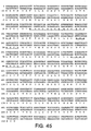
- Another aspect of the invention is an anti-IL-8 monoclonal antibody or antibody fragment comprising the complementarity determining regions of the 6G4.2.SLV11N35E light chain polypeptide amino acid sequence of Fig. 45 (SEQ ID NO:62).
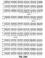
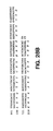
- Fig. 29 depicts an amino acid sequence alignment of murine 6G425 light chain variable domain (SEQ ID NO: 45), humanized 6G425 F(ab)-1 light chain variable domain (SEQ ID NO: 46), and human light chain ⁇ I consensus framework (SEQ ID NO: 47) amino acid sequences, and an amino acid sequence alignment of murine 6G425 heavy chain variable domain (SEQ ID NO: 48), humanized 6G425 F(ab)-1 heavy chain variable domain (SEQ ID NO: 49), and human IgGI subgroup III heavy chain variable domain (SEQ ID NO: 50) amino acid sequences, used in the humanization of 6G425.
- Light chain CDRs are labeled L1, L2, L3; heavy chain CDRs are labeled H1, H2, and H3.
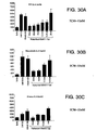
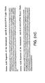
- Panel B presents inhibition data for F(ab)-9 samples at concentrations of 6.25 nM, 12.5 nM, 25 nM, and 50 nM, for an isotype control antibody (denoted “4D5") sample at a concentration of 100 nM, and for a no antibody control sample, in the presence of 4 nM human monomeric IL-8 (denoted as “BD59” and as “monomeric IL-8").
- panels B, C and D each presents data for human wild type IL-8 control (denoted “HuIL-8”) samples at a concentration of 2 nM in the respective assay
- panels A, B, C, and D each presents data for a no IL-8 buffer control (denoted "Buffer”) sample in the respective assay.
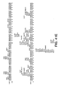
- Fig. 35 depicts the amino acid sequences of the humanized anti-IL-8 6G4.2.5V11N35A light chain in an N-terminal fusion with the STII leader peptide (SEQ ID NO: 56), the humanized anti-IL-8 6G4.2.5V11N35A heavy chain in an N-terminal fusion with the STII leader peptide (SEQ ID NO: 52), and the GCN4 leucine zipper peptide (SEQ ID NO: 57).
- the Ala residue (substituted for the wild type Asn residue) at amino acid position 35 in the 6G4.2.5V11N35A light chain appears in bold case.
- a putative pepsin cleavage site in the GCN4 leucine zipper sequence is underlined.
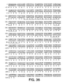
- Fig. 36 depicts the DNA sequence (SEQ ID NO: 58) and the amino acid sequence (SEQ ID NO: 56) of the humanized anti-IL-8 6G4.2.5V11N35A light chain in an N-terminal fusion with the STII leader peptide.
- Complementarity determining regions L1, L2, and L3 are underlined
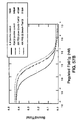
- Fig. 38 is a Scatchard plot depicting the inhibition of 125 I-IL-8 binding to human neutrophils exhibited by 6G4.2.SV11N35A Fab (denoted Fab), 6G4.2.5V11N35A F(ab') 2 (denoted F(ab') 2 ), and human wild type IL-8 control (denoted IL-8).
- Fig. 39 is a graph depicting a comparison of the wild type human IL-8 mediated neutrophil chemotaxis inhibition activities of the 6G4.2.5V11N35A F(ab') 2 and 6G4.2.5V11N35A Fab.
- Inhibition data are presented for 6G4.2.5V11N35A Fab samples (denoted "N35A Fab") and 6G4.2.5V11N35A F(ab') 2 samples (denoted N35A F(ab') 2 ) at concentrations of 0.3, 1, 3, 10, 30, and 100 nM, for an isotype control antibody (denoted as "4D5") sample at a concentration of 100 nM, and for a no antibody control sample, in the presence of 2 nM human wild type IL-8.
- inhibition data are presented for no IL-8 buffer control samples (denoted "Buffer").
- Fig. 40 is a graph depicting the ability of 6G4.2.SV11N35A F(ab') 2 to inhibit human monomeric IL-8, rhesus IL-8, and rabbit IL-8 mediated neutrophil chemotaxis.
- Human monomeric IL-8 mediated neutrophil chemotaxis data are presented for 6G4.2.5V11N35A F(ab') 2 samples at concentrations of 0.3, 1, 3, and 10 nM, for an isotype control antibody (denoted as "4D5") sample at a concentration of 100 nM, and for a no antibody control sample (denoted as "BD59”), in the presence of human monomeric IL-8 (denoted as "BD59”) at a concentration of 0.5 nM.
- Rhesus IL-8 mediated neutrophil chemotaxis data are presented for 6G4.2.5V11N35A F(ab') 2 samples at concentrations of 0.3, 1, 3, and 10 nM, and for a no antibody control sample, in the presence of rhesus IL-8 at a concentration of 2 nM.
- Rabbit IL-8 mediated neutrophil chemotaxis data are presented for 6G4.2.5V11N35A F(ab') 2 samples at concentrations of 0.3, 1, 3, and 10 nM, and for a no antibody control sample, in the presence of rabbit IL-8 at a concentration of 2 nM.
- inhibition data are presented for a no IL-8 buffer control sample (denoted as "Buffer") and for a 2 nM human wild type IL-8 (denoted as "HuIL-8").
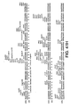
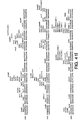
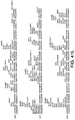
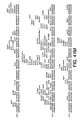
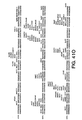
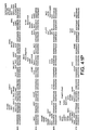
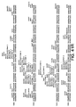
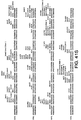
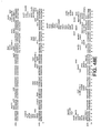
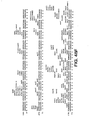
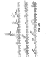
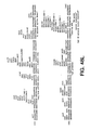
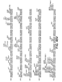
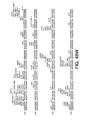
- Figs. 41A-41Q depict the nucleic acid sequence (SEQ ID NO: 61) of the p6G4VHN35A.F(ab') 2 vector.
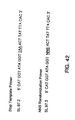
- Fig. 42 depicts the nucleic acid sequences of the stop template primer (SEQ ID NO: 63) and the NNS randomization primer (SEQ ID NO: 64) used for random mutagenesis of amino acid position 35 in variable light chain CDR-L of humanized antibody 6G4V11.
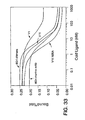
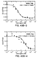
- Fig. 43B contains graphs of displacement curves depicting the inhibition of 125 I-IL-8 binding to neutrophils exhibited by the 6G4V11N35A, 6G4V11N3SD, 6G4V11N35E and 6G4V11N3SG Fab's.
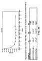
- Fig. 44 contains a graph depicting the typical kinetics of an anti-IL-8 antibody fragment (6G4V11N35A F(ab') 2 ) binding to IL-8.
- Fig. 44 also contains a table of data providing the equilibrium constant for 6G4V11N35A Fab binding to IL-8 (rate constants were not determined "ND"), and the equilibrium and rate constants for 6G4V11N35A F(ab') 2 and 6G4V11N35E Fab binding to IL-8.
- Fig. 45 depicts the DNA sequence (SEQ ID NO: 65) and amino acid sequence (SEQ ID NO: 62) of the 6G4V11N35E light chain in an N-terminal fusion with the STII leader peptide. Complementarity determining regions L1, L2 and L3 are underlined.
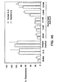
- Fig. 46 is a graph depicting the ability of 6G4V11N35E Fab to inhibit human IL-8 (dark columns) and rabbit IL-8 (light columns) mediated neutrophil chemotaxis. Data are presented for 6G4V11N35E Fab samples at concentrations of 0.4, 1.2, 3.7, 11 and 33 nM, and for an isotype control antibody (4D5) sample at a concentration of 100 nM, in the presence of 2 nM human IL-8 or 2 nM rabbit IL-8. In addition, inhibition data are presented for a no IL-8 buffer control sample (denoted "Buffer”) and for human and rabbit IL-8 control samples (denoted "IL-8").
- Fig. 47 depicts the DNA sequence of the sense (SEQ ID NO: 66) and anti-sense (SEQ ID NO: 67) strands of a PvuII-XhoI synthetic nucleotide encoding amino acids Leu4 to Phe29 of the 6G4V11N35A heavy chain.
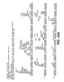
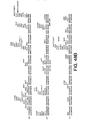
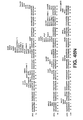
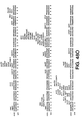
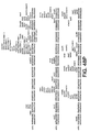
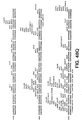
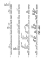
- Figs. 48A-48T depict the DNA sequence (SEQ ID NO: 68) of plasmid p6G4V11N35A.choSD9.
- Fig. 49 contains graphs of displacement curves depicting the inhibition of 125 I-IL-8 binding to neutrophils exhibited by the full length IgG1 forms of variants 6G4V11N35A and 6G4V11N35E.
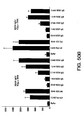
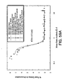
- Figs. 50A-50B are graphs depicting the ability of full length 6G4V11N35A IgGI and 6G4V11N35E IgGI to inhibit human IL-8 (Fig. 50A) and rabbit IL-8 (Fig. 50B) mediated neutrophil chemotaxis.
- Fig. 51 contains a graph depicting the typical kinetics of a full length anti-IL8 antibody (6G4V11N3SA IgG1) binding to IL-8.
- Fig. 51 also contains a table of data providing the equilibrium and rate constants for full length murine 6G4.2.5 IgG2a, 6G4V11N35A IgG1 and 6G4V11N35E IgGI binding to IL-8.
- Fig. 52 contains graphs of displacement curves depicting the results of an unlabeled IL-8/ 125 I-IL-8 competition radioimmunoassay performed with full length 6G4V11N35A IgG1 and 6G4V11N35E IgG1
- Fig. 53 depicts the DNA sequence (SEQ ID NO: 69) and amino acid sequence (SEQ ID NO: 70) of the 6G4V11N35A Fab' heavy chain (6G4V11N35A Fab heavy chain modified to contain a cysteine residue in the hinge region).
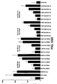
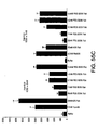
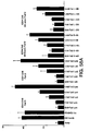
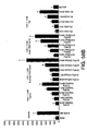
- Figs. 58A-58B are graphs depicting the ability of PEG-succinimide modified 6G4V11N35A F(ab') 2 molecules to inhibit human IL-8 mediated neutrophil chemotaxis.
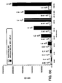
- Fig. 60 is a graph depicting the theoretical molecular weight (dotted bars) and effective size (solid bars) of PEG-maleimide modified 6G4V11N35A Fab' molecules as determined by SEC-HPLC.
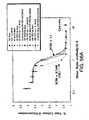
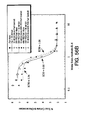
- Fig. 62 contains size exclusion chromatograms (SEC-HPLC) depicting the retention times and effective (hydrodynamic) sizes of various PEG-succinimide modified 6G4V11N35A F(ab') 2 molecules.
- Fig. 63 is a graph depicting the theoretical molecular weight (open columns), effective size determined by SEC-HPLC (solid columns), and the actual molecular weight determined by SEC-light scattering (shaded columns) for various PEG-succinimide modified 6G4V11N35A F(ab') 2 molecules.
- Fig. 64 is an SDS-PAGE gel depicting the electrophoretic mobility of various PEG-succinimide modified 6G4V11N35A F(ab') 2 molecules.
- lane I contains unmodified F(ab') 2
- lane 2 contains F(ab') 2 coupled to two 40 kD branched PEG-succinimide molecules (denoted "Br(2)-40kD(N)-F(ab')2")
- lane 3 contains F(ab') 2 coupled to one 40 kD branched PEG-succinimide molecule (denoted "Br(1)-40kD-(N)-Fab'2")
- lane 4 contains a mixture of F(ab') 2 coupled to four 20 kD linear PEG-succinimide molecules and F(ab') 2 coupled to five 20 kD linear PEG-succinimide molecules (denoted "L(4+5)-20kD-(N)-Fab'2”)
- Fig. 65 contains graphs comparing the serum concentration vs. time profiles of various PEG-maleimide modified 6G4V11N35A Fab' molecules (upper graph) and various PEG-succinimide modified 6G4V11N35A F(ab') 2 molecules (lower graph) in rabbits.
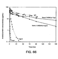
- Fig. 66 contains graphs comparing the serum concentration vs. time profiles of 6G4V11N35A Fab' coupled to one 40 kD branched PEG-maleimide molecule (denoted as "bran.(1)40K(s)Fab”'), 6G4V11N35A F(ab') 2 coupled to one 40 kD branched PEG-succinimide molecule (denoted as "bran.(1)40K(N)Fab'2”), unmodified 6G4V11N35A F(ab') 2 (denoted as "Fab'2"), unmodified 6G4V11N35A Fab' (denoted as "Fab”'), and a full length IgG1 (denoted as "IgG”) equivalent of the human-murine chimeric anti-rabbit IL-8 Fab described in Example F below.
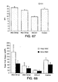
- Fig. 67 is a graph depicting the effect of 6G4V11N35A Fab' coupled to one 40 kD branched PEG-maleimide molecule (denoted as "PEG 40 Kd”) and murine anti-rabbit IL-8 monoclonal antibody 6G4.2.5 (full length IgG2a) (denoted as "6G4.2.5") on gross weight of entire lung in an ARDS rabbit model.
- Fig. 68 is a graph depicting the effect of 6G4V11N35A Fab' coupled to one branched 40 kD PEG-maleimide molecule (denoted as "PEG 40 Kd”) and murine anti-rabbit IL-8 monoclonal antibody 6G4.2.5 (full length IgG2a) (denoted as "6G4.2.5") on BAL total leukocyte (light columns) and polymorphonuclear cell (dark columns) counts in an ARDS rabbit model. Untreated (no therapeutics) control animal data is denoted as "Control”.
- Fig. 69 is a graph depicting the effect of 6G4V11N35A Fab' coupled to one branched 40 kD PEG-maleimide molecule (denoted as "PEG 40 Kd”) and murine anti-rabbit IL-8 monoclonal antibody 6G4.2.5 (full length IgG2a) (denoted as "6G4.2.5") on PaO2/FiO2 ratio at 24 hours-post treatment (light columns) and 48 hours post-treatment (dark columns) in an ARDS rabbit model. Untreated (no therapeutics) control animal data is denoted as "Control”.
- PCR Polymerase chain reaction
- sequence information from the ends of the region of interest or beyond needs to be available, such that oligonucleotide primers can be designed; these primers will be identical or similar in sequence to opposite strands of the template to be amplified.
- the 5' terminal nucleotides of the two primers can coincide with the ends of the amplified material.
- PCR can be used to amplify specific RNA sequences, specific DNA sequences from total genomic DNA, and cDNA transcribed from total cellular RNA, bacteriophage or plasmid sequences, etc. See generally Mullis et al., Cold Spring Harbor Symp. Quant. Biol. 51:263 (1987); Erlich, ed., PCR Technology (Stockton Press, NY, 1989).
- PCR is considered to be one, but not the only, example of a nucleic acid polymerase reaction method for amplifying a nucleic acid test sample comprising the use of a known nucleic acid as a primer and a nucleic acid polymerase to amplify or generate a specific piece of nucleic acid.
- the CDRs in each chain are held together in close proximity by the FR regions and, with the CDRs from the other chain, contribute to the formation of the antigen-binding site of antibodies (see Kabat et al., Sequences of Proteins of Immunological Interest, Fifth Edition, National Institute of Health, Bethesda, MD (1991)).
- the constant domains are not involved directly in binding an antibody to an antigen, but exhibit various effector functions, such as participation of the antibody in antibody-dependent cellular toxicity.
- Fv is the minimum antibody fragment which contains a complete antigen-recognition and - binding site.
- this region consists of a dimer of one heavy- and one light-chain variable domain in tight, non-covalent association.
- scFv single-chain Fv species
- one heavy- and one light-chain variable domain can be covalently linked by a flexible peptide linker such that the light and heavy chains can associate in a "dimeric" structure analogous to that in a two-chain Fv species. It is in this configuration that the three CDRs of each variable domain interact to define an antigen-binding site on the surface of the VH-VL dimer.
- the Fab fragment also contains the constant domain of the light chain and the first constant domain (CH1) of the heavy chain.
- Fab' fragments differ from Fab fragments by the addition of a few residues at the carboxy terminus of the heavy chain CH1 domain including one or more cysteines from the antibody hinge region.
- Fab'-SH is the designation herein for Fab' in which the cysteine residue(s) of the constant domains bear a free thiol group.
- F(ab') 2 antibody fragments originally were produced as pairs of Fab' fragments which have hinge cysteines between them. Other chemical couplings of antibody fragments are also known.
- the "light chains" of antibodies (immunoglobulins) from any vertebrate species can be assigned to one of two clearly distinct types, called kappa (k) and lambda (1), based on the amino acid sequences of their constant domains.
- antibody is used in the broadest sense and specifically covers single monoclonal antibodies (including agonist and antagonist antibodies) and antibody compositions with polyepitopic specificity.
- antibody fragments include Fab, Fab', Fab'-SH, F(ab') 2 , and Fv fragments; diabodies; any antibody fragment that is a polypeptide having a primary structure consisting of one uninterrupted sequence of contiguous amino acid residues (referred to herein as a "single-chain antibody fragment” or “single chain polypeptide"), including without limitation (1)single-chain Fv (scFv) molecules (2)single chain polypeptides containing only one light chain variable domain, or a fragment thereof that contains the three CDRs of the light chain variable domain, without an associated heavy chain moiety and (3)single chain polypeptides containing only one heavy chain variable region, or a fragment thereof containing the three CDRs of the heavy chain variable region, without an associated light chain moiety; and multispecific or multivalent structures formed from antibody fragments.
- single-chain antibody fragment or “single chain polypeptide”
- scFv single chain polypeptide
- the heavy chain(s) can contain any constant domain sequence (e.g. CH 1 in the IgG isotype) found in a non-Fc region of an intact antibody, and/or can contain any hinge region sequence found in an intact antibody, and/or can contain a leucine zipper sequence fused to or situated in the hinge region sequence or the constant domain sequence of the heavy chain(s).
- Suitable leucine zipper sequences include the jun and fos leucine zippers taught by Kostelney et al., J. Immunol., 148 : 1547-1553 (1992) and the GCN4 leucine zipper described in the Examples below.
- conjugate encompasses the aforementioned heterogeneous molecule in a precipitate, sediment, bioerodible matrix or other solid capable of releasing the heterogeneous molecule into aqueous solution upon hydration of the solid.
- mAb monoclonal antibody
- monoclonal antibody refers to an antibody obtained from a population of substantially homogeneous antibodies, i.e., the individual antibodies comprising the population are identical except for possible naturally occurring mutations that may be present in minor amounts.
- Monoclonal antibodies are highly specific, being directed against a single antigenic site.
- polyclonal antibody preparations which typically include different antibodies directed against different determinants (epitopes)
- each mAb is directed against a single determinant on the antigen.
- the monoclonal antibodies are advantageous in that they can be synthesized by hybridoma culture, uncontaminated by other immunoglobulins.
- the modifier "monoclonal” indicates the character of the antibody as being obtained from a substantially homogeneous population of antibodies, and is not to be construed as requiring production of the antibody by any particular method.
- the monoclonal antibodies to be used in accordance with the present invention may be made by the hybridoma method first described by Kohler et al., Nature, 256:495 (1975), or may be made by recombinant DNA methods (see, e.g., U.S. Patent No. 4,816,567 to Cabilly et al.).
- the monoclonal antibodies herein specifically include "chimeric" antibodies (immunoglobulins) in which a portion of the heavy and/or light chain is identical with or homologous to corresponding sequences in antibodies derived from a particular species or belonging to a particular antibody class or subclass, while the remainder of the chain(s) is identical with or homologous to corresponding sequences in antibodies derived from another species or belonging to another antibody class or subclass, as well as fragments of such antibodies, so long as they exhibit the desired biological activity (Cabilly et al., supra; Morrison et al., Proc. Natl. Acad. Sci. U.S.A. 81:6851 (1984)).
- chimeric antibodies immunoglobulins in which a portion of the heavy and/or light chain is identical with or homologous to corresponding sequences in antibodies derived from a particular species or belonging to a particular antibody class or subclass, while the remainder of the chain(s) is identical with or homologous to corresponding sequences in antibodies
- Treatment refers to both therapeutic treatment and prophylactic or preventative measures. Those in need of treatment include those already with the disorder as well as those prone to have the disorder or those in which the disorder is to be prevented.
- mammal for purposes of treatment refers to any animal classified as a mammal, including humans, domestic and farm animals, and zoo, sports, or pet animals, such as dogs, horses, cats, cows, etc.
- the mammal herein is human.
- inflammatory disorders refers to pathological states resulting in inflammation, typically caused by neutrophil chemotaxis.
- disorders include inflammatory skin diseases including psoriasis; responses associated with inflammatory bowel disease (such as Crohn's disease and ulcerative colitis); ischemic reperfusion; adult respiratory distress syndrome; dermatitis; meningitis; encephalitis; uveitis; autoimmune diseases such as rheumatoid arthritis, Sjorgen's syndrome, vasculitis; diseases involving leukocyte diapedesis; central nervous system (CNS) inflammatory disorder, multiple organ injury syndrome secondary to septicaemia or trauma; alcoholic hepatitis, bacterial pneumonia, antigen-antibody complex mediated diseases; inflammations of the lung, including pleurisy, alveolitis, vasculitis, pneumonia, chronic bronchitis, bronchiectasis, and cystic fibrosis; etc.
- the preferred indications are bacterial pneumonia and inflammatory
- hydrodynamic size refers to the size of a molecule as determined by comparison to a standard curve produced with globular protein molecular weight standards in a size exclusion chromatography system, wherein the standard curve is created by mapping the actual molecular weight of each standard against its elution time observed in the size exclusion chromatography system.
- the apparent size of a test molecule is derived by using the molecule's elution time to extrapolate a putative molecular weight from the standard curve.
- the molecular weight standards used to create the standard curve are selected such that the apparent size of the test molecule falls within the linear portion of the standard curve.
- the invention arises from the surprising and unexpected discovery that antibody fragment-polymer conjugates having an effective or apparent size significantly greater than the antibody fragment-polymer conjugates described in the art confers an increase in serum half-life, an increase in mean residence time in circulation (MRT), and/or a decrease in serum clearance rate over underivatized antibody fragment which far exceed the modest changes in such biological property or properties obtained with the art-known antibody fragment-polymer conjugates.
- MRT mean residence time in circulation
- the present inventors have determined for the first time that increasing the effective size of an antibody fragment to at least about 500,000 D, or increasing the effective size of an antibody fragment by at least about 8 fold over the effective size of the parental antibody fragment, or derivatizing an antibody fragment with a polymer of at least about 20,000 D in molecular weight, yields a molecule with a commercially useful pharmacokinetic profile.
- the greatly extended serum half-life, extended MRT, and/or reduced serum clearance rate of the conjugates of the invention makes such conjugates viable alternatives to intact antibodies used for therapeutic treatment of many disease indications.
- Antibody fragments provide significant advantages over intact antibodies, notably the fact that recombinant antibody fragments can be made in bacterial cell expression systems. Bacterial cell expression systems provide several advantages over mammalian cell expression systems, including reduced time and cost at both the research and development and manufacturing stages of a product.
- the present invention also arises from the humanization of the 6G4.2.5 murine anti-rabbit IL-8 monoclonal antibody ("6G4.2.5") described in WO 95/23865 (PCT/US95/02589 published September 8, 1995), the entire disclosure of which is specifically incorporated herein by reference.
- the hybridoma producing antibody 6G4.2.5 was deposited on September 28, 1994 with the American Type Culture Collection and assigned ATCC Accession No.
- the invention provides an antibody fragment covalently attached to a polymer to form a conjugate having an effective or apparent size of at least about 500,000 Daltons (D). In another aspect, the invention provides an antibody fragment covalently attached to a polymer to form a conjugate having an apparent size that is at least about 8 fold greater than the apparent size of the parental antibody fragment. In yet another aspect, the invention provides an antibody fragment covalently attached to a polymer of at least about 20,000 D in molecular weight (MW).
- MW molecular weight
- the unexpectedly and surprisingly large increase in antibody fragment serum half-life, increase in MRT, and/or decrease in serum clearance rate can be achieved by using any type of polymer or number of polymer molecules which will provide the conjugate with an effective size of at least about 500,000 D, or by using any type of polymer or number of polymer molecules which will provide the conjugate with an effective size that is at least about 8 fold greater than the effective size of the parental antibody fragment, or by using any type or number of polymers wherein each polymer molecule is at least about 20,000 D in MW.
- the invention is not dependent on the use of any particular polymer or molar ratio of polymer to antibody fragment in the conjugate.
- the beneficial aspects of the invention extend to antibody fragments without regard to antigen specificity.
- the antigen specificity of a given antibody will not substantially impair the extraordinary improvement in serum half-life, MRT, and/or serum clearance rate for antibody fragments thereof that can be obtained by derivatizing the antibody fragments as taught herein.
- the conjugate has an effective size of at least about 500,000 D, or at least about 800,000 D, or at least about 900,000 D, or at least about 1,000,000 D, or at least about 1,200,000 D, or at least about 1,400,000 D, or at least about 1,500,000 D, or at least about 1,800,000 D, or at least about 2,000,000 D, or at least about 2,500,000 D.
- the conjugate has an effective size of at or about 500,000 D to at or about 10,000,000 D, or an effective size of at or about 500,000 D to at or about 8,000,000 D, or an effective size of at or about 500,000 D to at or about 5,000,000 D, or an effective size of at or about 500,000 D to at or about 4,000,000 D, or an effective size of at or about 500,000 D to at or about 3,000,000 D, or an effective size of at or about 500,000 D to at or about 2,500,000 D, or an effective size of at or about 500,000 D to at or about 2,000,000 D, or an effective size of at or about 500,000 D to at or about 1,800,000 D, or an effective size of at or about 500,000 D to at or about 1,600,000 D, or an effective size of at or about 500,000 D to at or about 1,500,000 D, or an effective size of at or about 500,000 D to at or about 1,000,000 D.
- the conjugate has an effective size of at or about 800,000 D to at or about 10,000,000 D, or an effective size of at or about 800,000 D to at or about 8,000,000 D, or an effective size of at or about 800,000 D to at or about 5,000,000 D, or an effective size of at or about 800,000 D to at or about 4,000,000 D, or an effective size of at or about 800,000 D to at or about 3,000,000 D, or an effective size of at or about 800,000 D to at or about 2,500,000 D, or an effective size of at or about 800,000 D to at or about 2,000,000 D, or an effective size of at or about 800,000 D to at or about 1,800,000 D, or an effective size of at or about 800,000 D to at or about 1,600,000 D, or an effective size of at or about 800,000 D to at or about 1,500,000 D, or an effective size of at or about 800,000 D to at or about 1,000,000 D.
- the conjugate has an effective size of at or about 1,000,000 D to at or about 10,000,000 D, or an effective size of at or about 1,000,000 D to at or about 8,000,000 D, or an effective size of at or about 1,000,000 D to at or about 5,000,000 D, or an effective size of at or about 1,000,000 D to at or about 4,000,000 D, or an effective size of at or about 1,000,000 D to at or about 3,000,000 D, or an effective size of at or about 1,000,000 D to at or about 2,500,000 D, or an effective size of at or about 1,000,000 D to at or about 2,000,000 D, or an effective size of at or about 1,000,000 D to at or about 1,800,000 D, or an effective size of at or about 1,000,000 D to at or about 1,600,000 D, or an effective size of at or about 1,000,000 D to at or about 1,500,000 D.
- the conjugate has an effective size that is at least about 8 fold greater, or at least about 10 fold greater, or at least about 12 fold greater, or at least about 15 fold greater, or at least about 18 fold greater, or at least about 20 fold greater, or at least about 25 fold greater, or at least about 28 fold greater, or at least about 30 fold greater, or at least about 40 fold greater, than the effective size of the parental antibody fragment.
- the conjugate has an effective size that is about 8 fold to about 100 fold greater, or is about 8 fold to about 80 fold greater, or is about 8 fold to about 50 fold greater, or is about 8 fold to about 40 fold greater, or is about 8 fold to about 30 fold greater, or is about 8 fold to about 28 fold greater, or is about 8 fold to about 25 fold greater, or is about 8 fold to about 20 fold greater, or is about 8 fold to about 18 fold greater, or is about 8 fold to about 15 fold greater, than the effective size of the parental antibody fragment.
- the conjugate has an effective size that is about 12 fold to about 100 fold greater, or is about 12 fold to about 80 fold greater, or is about 12 fold to about 50 fold greater, or is about 12 fold to about 40 fold greater, or is about 12 fold to about 30 fold greater, or is about 12 fold to about 28 fold greater, or is about 12 fold to about 25 fold greater, or is about 12 fold to about 20 fold greater, or is about 12 fold to about 18 fold greater, or is about 12 fold to about 15 fold greater, than the effective size of the parental antibody fragment.
- the conjugate has an effective size that is about 15 fold to about 100 fold greater, or is about 15 fold to about 80 fold greater, or is about 15 fold to about 50 fold greater, or is about 15 fold to about 40 fold greater, or is about 15 fold to about 30 fold greater, or is about 15 fold to about 28 fold greater, or is about 15 fold to about 25 fold greater, or is about 15 fold to about 20 fold greater, or is about 15 fold to about 18 fold greater, than the effective size of the parental antibody fragment.
- the conjugate has an effective size that is about 18 fold to about 100 fold greater, or is about 18 fold to about 80 fold greater, or is about 18 fold to about 50 fold greater, or is about 18 fold to about 40 fold greater, or is about 18 fold to about 30 fold greater, or is about 18 fold to about 28 fold greater, or is about 18 fold to about 25 fold greater, or is about 18 fold to about 20 fold greater, than the effective size of the parental antibody fragment.
- the conjugate has an effective size that is about 20 fold to about 100 fold greater, or is about 20 fold to about 80 fold greater, or is about 20 fold to about 50 fold greater, or is about 20 fold to about 40 fold greater, or is about 20 fold to about 30 fold greater, or is about 20 fold to about 28 fold greater, or is about 20 fold to about 25 fold greater, than the effective size of the parental antibody fragment.
- the conjugate has an effective size that is about 25 fold to about 100 fold greater, or is about 25 fold to about 80 fold greater, or is about 25 fold to about 50 fold greater, or is about 25 fold to about 40 fold greater, or is about 25 fold to about 30 fold greater, or is about 25 fold to about 28 fold greater, than the effective size of the parental antibody fragment.
- the conjugate has an effective size that is about 30 fold to about 100 fold greater, or is about 30 fold to about 80 fold greater, or is about 30 fold to about 50 fold greater, or is about 30 fold to about 40 fold greater, than the effective size of the parental antibody fragment.
- the conjugate has an effective size that is about 40 fold to about 100 fold greater, or is about 40 fold to about 80 fold greater, or is about 40 fold to about 50 fold greater, than the effective size of the parental antibody fragment.
- the conjugate is an antibody fragment covalently attached to at least one polymer having an actual MW of at least about 20,000 D.
- the conjugate is an antibody fragment covalently attached to at least one polymer having an actual MW of at least about 30,000 D.
- the conjugate is an antibody fragment covalently attached to at least one polymer having an actual MW of at least about 40,000 D.
- the conjugate is an antibody fragment covalently attached to at least one polymer having an actual MW that is at or about 20,000 D to at or about 300,000 D, or is at or about 30,000 D to at or about 300,000 D, or is at or about 40,000 D to at or about 300,000 D.
- the conjugate is an antibody fragment covalently attached to at least one polymer having an actual MW that is at or about 20,000 D to at or about 100,000 D, or is at or about 30,000 D to at or about 100,000 D, or is at or about 40,000 D to at or about 100,000 D.
- the conjugate is an antibody fragment covalently attached to at least one polymer having an actual MW that is at or about 20,000 D to at or about 70,000 D, or is at or about 30,000 D to at or about 70,000 D, or is at or about 40,000 D to at or about 70,000 D.
- the conjugate is an antibody fragment covalently attached to at least one polymer having an actual MW that is at or about 20,000 D to at or about 50,000 D, or is at or about 30,000 D to at or about 50,000 D, or is at or about 40,000 D to at or about 50,000 D.
- the conjugate is an antibody fragment covalently attached to at least one polymer having an actual MW that is at or about 20,000 D to at or about 40,000 D, or is at or about 30,000 D to at or about 40,000 D.
- the conjugates of the invention can be made using any suitable technique now known or hereafter developed for derivatizing antibody fragments with polymers. It will be appreciated that the invention is not limited to conjugates utilizing any particular type of linkage between an antibody fragment and a polymer.
- the conjugates of the invention include species wherein a polymer is covalently attached to a non-specific site or non-specific sites on the parental antibody fragment, i.e. polymer attachment is not targeted to a particular region or a particular amino acid residue in the parental antibody fragment.
- the coupling chemistry can, for example, utilize the free epsilon amino groups of lysine residues in the parental antibody as attachment sites for the polymer, wherein such lysine residue amino groups are randomly derivatized with polymer.
- the conjugates of the invention include species wherein a polymer is covalently attached to a specific site or specific sites on the parental antibody fragment, i.e. polymer attachment is targeted to a particular region or a particular amino acid residue or residues in the parental antibody fragment.
- the coupling chemistry can, for example, utilize the free sulfhydryl group of a cysteine residue not in a disulfide bridge in the parental antibody fragment.
- one or more cysteine residue(s) is (are) engineered into a selected site or sites in the parental antibody fragment for the purpose of providing a specific attachment site or sites for polymer.
- the polymer can be activated with any functional group that is capable of reacting specifically with the free sulfhydryl or thiol group(s) on the parental antibody, such as maleimide, sulfhydryl, thiol, triflate, tesylate, aziridine, exirane, and 5-pyridyl functional groups.
- the polymer can be coupled to the parental antibody fragment using any protocol suitable for the chemistry of the coupling system selected, such as the protocols and systems described in Section (II)(1)(b) or in Section (T) of the Examples below.
- polymer attachment is targeted to the hinge region of the parental antibody fragment.
- the location of the hinge region varies according to the isotype of the parental antibody.
- the hinge region of IgG, IgD and IgA isotype heavy chains is contained in a proline rich peptide sequence extending between the C H 1 and C H 2 domains.
- a cysteine residue or residues is (are) engineered into the hinge region of the parental antibody fragment in order to couple polymer specifically to a selected location in the hinge region.
- the invention encompasses a conjugate having any molar ratio of polymer to antibody fragment that endows the conjugate with an apparent size in the desired range as taught herein.
- the apparent size of the conjugate will depend in part upon the size and shape of the polymer used, the size and shape of the antibody fragment used, the number of polymer molecules attached to the antibody fragment, and the location of such attachment site(s) on the antibody fragment. These parameters can easily be identified and maximized to obtain the a conjugate with the desired apparent size for any type of antibody fragment, polymer and linkage system.
- the invention encompasses a conjugate with a polymer to antibody fragment molar ratio of no more than about 10:1, or no more than about 5:1, or no more than about 4:1, or no more than about 3:1, or no more than about 2:1, or no more than 1:1.
- the invention encompasses a conjugate wherein the antibody fragment is attached to about 10 or fewer polymer molecules, each polymer molecule having a molecular weight of at least about 20,000 D, or at least about 30,000 D, or at least about 40,000 D.
- the conjugate contains an antibody fragment attached to about 5 or fewer polymer molecules, each polymer molecule having a molecular weight of at least about 20,000 D, or at least about 30,000 D, or at least about 40,000 D.
- the conjugate contains an antibody fragment attached to about 4 or fewer polymer molecules, each polymer molecule having a molecular weight of at least about 20,000 D, or at least about 30,000 D, or at least about 40,000 D.
- the conjugate contains an antibody fragment attached to about 3 or fewer polymer molecules, each polymer molecule having a molecular weight of at least about 20,000 D, or at least about 30,000 D, or at least about 40,000 D. In an additional embodiment the conjugate contains an antibody fragment attached to about 2 or fewer polymer molecules, each polymer molecule having a molecular weight of at least about 20,000 D, or at least about 30,000 D, or at least about 40,000 D. Also provided herein is a conjugate containing an antibody fragment attached to a single polymer molecule having a molecular weight of at least about 20,000 D, or at least about 30,000 D, or at least about 40,000 D.
- the invention encompasses a conjugate wherein every polymer molecule in the conjugate has a molecular weight that is at or about 20,000 D to at or about 300,000 D, or is at or about 30,000 D to at or about 300,000 D, or is at or about 40,000 D to at or about 300,000 D, and wherein the conjugate contains no more than about 10 polymer molecules, or no more than about 5 polymer molecules, or no more than about 4 polymer molecules, or no more than about 3 polymer molecules, or no more than about 2 polymer molecules, or no more than 1 polymer molecule.
- the invention encompasses a conjugate wherein every polymer molecule in the conjugate has a molecular weight that is at or about 20,000 D to at or about 100,000 D, or is at or about 30,000 D to at or about 100,000 D, or is at or about 40,000 D to at or about 100,000 D, and wherein the conjugate contains no more than about 10 polymer molecules, or no more than about 5 polymer molecules, or no more than about 4 polymer molecules, or no more than about 3 polymer molecules, or no more than about 2 polymer molecules, or no more than 1 polymer molecule.
- the invention encompasses a conjugate wherein every polymer molecule in the conjugate has a molecular weight that is at or about 20,000 D to at or about 70,000 D, or is at or about 30,000 D to at or about 70,000 D, or is at or about 40,000 D to at or about 70,000 D, and wherein the conjugate contains no more than about 10 polymer molecules, or no more than about 5 polymer molecules, or no more than about 4 polymer molecules, or no more than about 3 polymer molecules, or no more than about 2 polymer molecules, or no more than 1 polymer molecule.
- the invention encompasses a conjugate wherein every polymer molecule in the conjugate has a molecular weight that is at or about 20,000 D to at or about 50,000 D, or is at or about 30,000 D to at or about 50,000 D, or is at or about 40,000 D to at or about 50,000 D, and wherein the conjugate contains no more than about 10 polymer molecules, or no more than about 5 polymer molecules, or no more than about 4 polymer molecules, or no more than about 3 polymer molecules, or no more than about 2 polymer molecules, or no more than 1 polymer molecule.
- the invention encompasses a conjugate wherein every polymer molecule in the conjugate has a molecular weight that is at or about 20,000 D to at or about 40,000 D, or is at or about 30,000 D to at or about 40,000 D, and wherein the conjugate contains no more than about 10 polymer molecules, or no more than about 5 polymer molecules, or no more than about 4 polymer molecules, or no more than about 3 polymer molecules, or no more than about 2 polymer molecules, or no more than 1 polymer molecule.
- the conjugate contains an antibody fragment selected from the group consisting of Fab, Fab', and Fab'-SH, wherein every polymer molecule in the conjugate is attached to the hinge region of the antibody fragment.
- the conjugate contains a F(ab') 2 antibody fragment attached to no more than about 2 polymer molecules, wherein every polymer molecule is attached to a cysteine residue in the light or heavy chain of the antibody fragment that would ordinarily form the disulfide bridge linking the light and heavy chains, wherein the disulfide bridge is avoided by substituting another amino acid, such as serine, for the corresponding cysteine residue in the opposite chain.
- the conjugate contains an antibody fragment selected from the group consisting of Fab, Fab', and Fab'-SH, wherein the antibody fragment is attached to no more than I polymer molecule and the polymer is coupled to a cysteine residue in the light or heavy chain of the antibody fragment that would ordinarily form the disulfide bridge linking the light and heavy chains, wherein the disulfide bridge is avoided by substituting another amino acid, such as serine, for the corresponding cysteine residue in the opposite chain.
- the conjugate contains an antibody fragment selected from the group consisting of Fab, Fab', and Fab'-SH, every polymer molecule in the conjugate is at or about 20,000 D to at or about 300,000 D in molecular weight, or is at or about 30,000 D to at or about 300,000 D in molecular weight, or is at or about 40,000 D to at or about 300,000 D in molecular weight, every polymer molecule in the conjugate is attached to the hinge region of the antibody fragment, and the conjugate contains no more than about 10 polymer molecules, or no more than about 5 polymer molecules, or no more than about 4 polymer molecules, or no more than about 3 polymer molecules, or no more than about 2 polymer molecules, or no more than 1 polymer molecule.
- the conjugate contains an antibody fragment selected from the group consisting of Fab, Fab', and Fab'-SH, every polymer molecule in the conjugate is at or about 20,000 D to at or about 50,000 D in molecular weight, or is at or about 30,000 D to at or about 50,000 D in molecular weight, or is at or about 40,000 D to at or about 50,000 D in molecular weight, every polymer molecule in the conjugate is attached to the hinge region of the antibody fragment, and the conjugate contains no more than about 10 polymer molecules. or no more than about 5 polymer molecules, or no more than about 4 polymer molecules, or no more than about 3 polymer molecules, or no more than about 2 polymer molecules, or no more than 1 polymer molecule.
- the conjugate contains an antibody fragment selected from the group consisting of Fab, Fab', and Fab'-SH, every polymer molecule in the conjugate is at or about 20,000 D to at or about 40,000 D in molecular weight, or is at or about 30,000 D to at or about 40,000 D in molecular weight, every polymer molecule in the conjugate is attached to the hinge region of the antibody fragment, and the conjugate contains no more than about 10 polymer molecules, or no more than about 5 polymer molecules, or no more than about 4 polymer molecules, or no more than about 3 polymer molecules, or no more than about 2 polymer molecules, or no more than 1 polymer molecule.
- the conjugate contains a F(ab') 2 antibody fragment attached to no more than about 2 polymer molecules, wherein every polymer molecule in the conjugate is at least about 20,000 D in molecular weight, or at least about 30,000 D in molecular weight, or at least about 40,000 D in molecular weight, and wherein every polymer molecule in the conjugate is attached to a cysteine residue in the light or heavy chain of the antibody fragment that would ordinarily form the disulfide bridge linking the light and heavy chains, wherein the disulfide bridge is avoided by substituting another amino acid, such as serine, for the corresponding cysteine residue in the opposite chain.
- every polymer molecule in the conjugate is at least about 20,000 D in molecular weight, or at least about 30,000 D in molecular weight, or at least about 40,000 D in molecular weight
- every polymer molecule in the conjugate is attached to a cysteine residue in the light or heavy chain of the antibody fragment that would ordinarily form the disulfide bridge linking the
- the conjugate contains a F(ab') 2 antibody fragment attached to no more than about 2 polymer molecules, wherein every polymer molecule in the conjugate is at or about 20,000 D to at or about 300,000 D in molecular weight, or is at or about 30,000 D to at or about 300,000 D in molecular weight, or is at or about 40,000 D to at or about 300,000 D in molecular weight, and wherein every polymer molecule in the conjugate is attached to a cysteine residue in the light or heavy chain of the antibody fragment that would ordinarily form the disulfide bridge linking the light and heavy chains, wherein the disulfide bridge is avoided by substituting another amino acid, such as serine, for the corresponding cysteine residue in the opposite chain.
- every polymer molecule in the conjugate is at or about 20,000 D to at or about 300,000 D in molecular weight, or is at or about 30,000 D to at or about 300,000 D in molecular weight, or is at or about 40,000 D to at or about 300
- the conjugate contains a F(ab') 2 antibody fragment attached to no more than about 2 polymer molecules, wherein every polymer molecule in the conjugate is at or about 20,000 D to at or about 100,000 D in molecular weight, or is at or about 30,000 D to at or about 100,000 D in molecular weight, or is at or about 40,000 D to at or about 100,000 D in molecular weight, and wherein every polymer molecule in the conjugate is attached to a cysteine residue in the light or heavy chain of the antibody fragment that would ordinarily form the disulfide bridge linking the light and heavy chains, wherein the disulfide bridge is avoided by substituting another amino acid, such as serine, for the corresponding cysteine residue in the opposite chain.
- every polymer molecule in the conjugate is at or about 20,000 D to at or about 100,000 D in molecular weight, or is at or about 30,000 D to at or about 100,000 D in molecular weight, or is at or about 40,000 D to at or about 100,000 D in molecular weight
- the conjugate contains a F(ab') 2 antibody fragment attached to no more than about 2 polymer molecules, wherein every polymer molecule in the conjugate is at or about 20,000 D to at or about 70,000 D in molecular weight, or is at or about 30,000 D to at or about 70,000 D in molecular weight, or is at or about 40,000 D to at or about 70,000 D in molecular weight, and wherein every polymer molecule in the conjugate is attached to a cysteine residue in the light or heavy chain of the antibody fragment that would ordinarily form the disulfide bridge linking the light and heavy chains, wherein the disulfide bridge is avoided by substituting another amino acid, such as serine, for the corresponding cysteine residue in the opposite chain.
- every polymer molecule in the conjugate is at or about 20,000 D to at or about 70,000 D in molecular weight, or is at or about 30,000 D to at or about 70,000 D in molecular weight, or is at or about 40,000 D to at or about 70,000 D in mole
- the conjugate contains a F(ab') 2 antibody fragment attached to no more than about 2 polymer molecules, wherein every polymer molecule in the conjugate is at or about 20,000 D to at or about 40,000 D in molecular weight, or is at or about 30,000 D to at or about 40,000 D in molecular weight, and wherein every polymer molecule in the conjugate is attached to a cysteine residue in the light or heavy chain of the antibody fragment that would ordinarily form the disulfide bridge linking the light and heavy chains, wherein the disulfide bridge is avoided by substituting another amino acid, such as serine, for the corresponding cysteine residue in the opposite chain.
- every polymer molecule in the conjugate is at or about 20,000 D to at or about 40,000 D in molecular weight, or is at or about 30,000 D to at or about 40,000 D in molecular weight
- every polymer molecule in the conjugate is attached to a cysteine residue in the light or heavy chain of the antibody fragment that would ordinarily form the disulf
- the conjugate contains an antibody fragment selected from the group consisting of Fab, Fab', and Fab'-SH, wherein the antibody fragment is attached to no more than I polymer molecule, wherein the polymer molecule is at least about 20,000 D in molecular weight, or at least about 30,000 D in molecular weight, or at least about 40,000 D in molecular weight, wherein the polymer molecule is attached to a cysteine residue in the light or heavy chain of the antibody fragment that would ordinarily form the disulfide bridge linking the light and heavy chains, wherein the disulfide bridge is avoided by substituting another amino acid, such as serine, for the corresponding cysteine residue in the opposite chain.
- an antibody fragment selected from the group consisting of Fab, Fab', and Fab'-SH, wherein the antibody fragment is attached to no more than I polymer molecule, wherein the polymer molecule is at least about 20,000 D in molecular weight, or at least about 30,000 D in molecular weight, or at least about 40,000 D
- the conjugate contains an antibody fragment selected from the group consisting of Fab, Fab', and Fab'-SH, wherein the antibody fragment is attached to no more than 1 polymer molecule, wherein the polymer molecule is at or about 20,000 D to at or about 300,000 D in molecular weight, or is at or about 30,000 D to at or about 300,000 D in molecular weight, or is at or about 40,000 D to at or about 300,000 D in molecular weight, wherein the polymer molecule is attached to a cysteine residue in the light or heavy chain of the antibody fragment that would ordinarily form the disulfide bridge linking the light and heavy chains, wherein the disulfide bridge is avoided by substituting another amino acid, such as serine, for the corresponding cysteine residue in the opposite chain.
- the polymer molecule is at or about 20,000 D to at or about 300,000 D in molecular weight, or is at or about 30,000 D to at or about 300,000 D in molecular weight, or is at or about 40,000
- the conjugate contains an antibody fragment selected from the group consisting of Fab, Fab', and Fab'-SH, wherein the antibody fragment is attached to no more than 1 polymer molecule, wherein the polymer molecule is at or about 20,000 D to at or about 100,000 D in molecular weight, or is at or about 30,000 D to at or about 100,000 D in molecular weight, or is at or about 40,000 D to at or about 100,000 D in molecular weight, wherein the polymer molecule is attached to a cysteine residue in the light or heavy chain of the antibody fragment that would ordinarily form the disulfide bridge linking the light and heavy chains, wherein the disulfide bridge is avoided by substituting another amino acid, such as serine, for the corresponding cysteine residue in the opposite chain.
- an antibody fragment selected from the group consisting of Fab, Fab', and Fab'-SH, wherein the antibody fragment is attached to no more than 1 polymer molecule, wherein the polymer molecule is at or about 20,000 D to at or about 100,000 D
- the conjugate contains an antibody fragment selected from the group consisting of Fab, Fab', and Fab'-SH, wherein the antibody fragment is attached to no more than 1 polymer molecule, wherein the polymer molecule is at or about 20,000 D to at or about 70,000 D in molecular weight, or is at or about 30,000 D to at or about 70,000 D in molecular weight, or is at or about 40,000 D to at or about 70,000 D in molecular weight, wherein the polymer molecule is attached to a cysteine residue in the light or heavy chain of the antibody fragment that would ordinarily form the disulfide bridge linking the light and heavy chains, wherein the disulfide bridge is avoided by substituting another amino acid, such as serine, for the corresponding cysteine residue in the opposite chain.
- an antibody fragment selected from the group consisting of Fab, Fab', and Fab'-SH, wherein the antibody fragment is attached to no more than 1 polymer molecule, wherein the polymer molecule is at or about 20,000 D to at or
- the conjugate contains an antibody fragment selected from the group consisting of Fab, Fab', and Fab'-SH, wherein the antibody fragment is attached to no more than 1 polymer molecule, wherein the polymer molecule is at or about 20,000 D to at or about 50,000 D in molecular weight, or is at or about 30,000 D to at or about 50,000 D in molecular weight, or is at or about 40,000 D to at or about 50,000 D in molecular weight, wherein the polymer molecule is attached to a cysteine residue in the light or heavy chain of the antibody fragment that would ordinarily form the disulfide bridge linking the light and heavy chains, wherein the disulfide bridge is avoided by substituting another amino acid, such as serine, for the corresponding cysteine residue in the opposite chain.
- an antibody fragment selected from the group consisting of Fab, Fab', and Fab'-SH, wherein the antibody fragment is attached to no more than 1 polymer molecule, wherein the polymer molecule is at or about 20,000 D to at or
- the conjugate contains an antibody fragment selected from the group consisting of Fab, Fab', and Fab'-SH, wherein the antibody fragment is attached to no more than 1 polymer molecule, wherein the polymer molecule is at or about 20,000 D to at or about 40,000 D in molecular weight, or is at or about 30,000 D to at or about 40,000 D in molecular weight, wherein the polymer molecule is attached to a cysteine residue in the light or heavy chain of the antibody fragment that would ordinarily form the disulfide bridge linking the light and heavy chains, wherein the disulfide bridge is avoided by substituting another amino acid, such as serine, for the corresponding cysteine residue in the opposite chain.
- an antibody fragment selected from the group consisting of Fab, Fab', and Fab'-SH, wherein the antibody fragment is attached to no more than 1 polymer molecule, wherein the polymer molecule is at or about 20,000 D to at or about 40,000 D in molecular weight, or is at or about 30,000 D to at or
- the conjugate contains an antibody fragment selected from the group consisting of Fab, Fab', and Fab'-SH, wherein the antibody fragment is attached to no more than 1 polymer molecule, wherein the polymer molecule is at least about 20,000 D in molecular weight, or at least about 30,000 D in molecular weight, or at least about 40,000 D in molecular weight, and wherein the polymer molecule is attached to the hinge region of the antibody fragment.
- the conjugate contains an antibody fragment selected from the group consisting of Fab, Fab', and Fab'-SH, wherein the antibody fragment is attached to no more than 1 polymer molecule, wherein the polymer molecule is at or about 20,000 D to at or about 300,000 D in molecular weight, or is at or about 30,000 D to at or about 300,000 D in molecular weight, or is at or about 40,000 D to at or about 300,000 D in molecular weight, and wherein the polymer molecule is attached to the hinge region of the antibody fragment.
- the conjugate contains an antibody fragment selected from the group consisting of Fab, Fab', and Fab'-SH, wherein the antibody fragment is attached to no more than 1 polymer molecule, wherein the polymer molecule is at or about 20,000 D to at or about 50,000 D in molecular weight, or is at or about 30,000 D to at or about 50,000 D in molecular weight, or is at or about 40,000 D to at or about 50,000 D in molecular weight, and wherein the polymer molecule is attached to the hinge region of the antibody fragment.
- the conjugate contains an antibody fragment selected from the group consisting of Fab, Fab', and Fab'-SH, wherein the antibody fragment is attached to no more than 1 polymer molecule, wherein the polymer molecule is at or about 20,000 D to at or about 40,000 D in molecular weight, or is at or about 30,000 D to at or about 40,000 D in molecular weight, and wherein the polymer molecule is attached to the hinge region of the antibody fragment.
- polyethylene glycol (PEG) polymers are preferred for use herein.
- the conjugate is an antibody fragment covalently attached to at least one PEG having an actual MW that is at or about 20,000 D to at or about 300,000 D, or is at or about 30,000 D to at or about 300,000 D, or is at or about 40,000 D to at or about 300,000 D.
- the conjugate is an antibody fragment covalently attached to at least one PEG having an actual MW that is at or about 20,000 D to at or about 100,000 D, or is at or about 30,000 D to at or about 100,000 D, or is at or about 40,000 D to at or about 100,000 D.
- the conjugate is an antibody fragment covalently attached to at least one PEG having an actual MW that is at or about 20,000 D to at or about 50,000 D, or is at or about 30,000 D to at or about 50,000 D, or is at or about 40,000 D to at or about 50,000 D.
- the conjugate is an antibody fragment covalently attached to at least one PEG having an actual MW that is at or about 20,000 D to at or about 40,000 D, or is at or about 30,000 D to at or about 40,000 D.
- the invention encompasses a conjugate with a PEG to antibody fragment molar ratio of no more than about 10:1, or no more than about 5:1, or no more than about 4:1, or no more than about 3:1, or no more than about 2:1, or no more than 1:1.
- the invention encompasses a conjugate wherein the antibody fragment is attached to about 10 or fewer PEG molecules, each PEG molecule having a molecular weight of at least about 20,000 D, or at least about 30,000 D, or at least about 40,000 D.
- the conjugate contains an antibody fragment attached to about 5 or fewer PEG molecules, each PEG molecule having a molecular weight of at least about 20,000 D, or at least about 30,000 D, or at least about 40,000 D.
- the conjugate contains an antibody fragment attached to about 4 or fewer PEG molecules, each PEG molecule having a molecular weight of at least about 20,000 D, or at least about 30,000 D, or at least about 40,000 D.
- the conjugate contains an antibody fragment attached to about 3 or fewer PEG molecules, each PEG molecule having a molecular weight of at least about 20,000 D, or at least about 30,000 D, or at least about 40,000 D. In an additional embodiment, the conjugate contains an antibody fragment attached to about 2 or fewer PEG molecules, each PEG molecule having a molecular weight of at least about 20,000 D, or at least about 30,000 D, or at least about 40,000 D. Also provided herein is a conjugate containing an antibody fragment attached to a single PEG molecule having a molecular weight of at least about 20,000 D, or at least about 30,000 D, or at least about 40,000 D.
- the invention encompasses a conjugate wherein the antibody fragment is derivatized with PEG, wherein every PEG molecule in the conjugate is at or about 20,000 D to at or about 300,000 D in molecular weight, or is at or about 30,000 D to at or about 300,000 D in molecular weight, or is at or about 40,000 D to at or about 300,000 D in molecular weight, and wherein the conjugate contains no more than about 10 PEG molecules, or no more than about 5 PEG molecules, or no more than about 4 PEG molecules, or no more than about 3 PEG molecules, or no more than about 2 PEG molecules, or no more than 1 PEG molecule.
- the invention encompasses a conjugate wherein the antibody fragment is derivatized with PEG, wherein every PEG molecule in the conjugate is at or about 20,000 D to at or about 100,000 D in molecular weight, or is at or about 30,000 D to at or about 100,000 D in molecular weight, or is at or about 40,000 D to at or about 100,000 D in molecular weight, and wherein the conjugate contains no more than about 10 PEG molecules, or no more than about 5 PEG molecules, or no more than about 4 PEG molecules, or no more than about 3 PEG molecules, or no more than about 2 PEG molecules, or no more than 1 PEG molecule.
- the invention encompasses a conjugate wherein the antibody fragment is derivatized with PEG, wherein every PEG molecule in the conjugate is at or about 20,000 D to at or about 70,000 D in molecular weight, or is at or about 30,000 D to at or about 70,000 D in molecular weight, or is at or about 40,000 D to at or about 70,000 D in molecular weight, and wherein the conjugate contains no more than about 10 PEG molecules, or no more than about 5 PEG molecules, or no more than about 4 PEG molecules, or no more than about 3 PEG molecules, or no more than about 2 PEG molecules, or no more than 1 PEG molecule.
- the invention encompasses a conjugate wherein the antibody fragment is derivatized with PEG, wherein every PEG molecule in the conjugate is at or about 20,000 D to at or about 50,000 D in molecular weight, or is at or about 30,000 D to at or about 50,000 D in molecular weight, or is at or about 40,000 D to at or about 50,000 D in molecular weight, and wherein the conjugate contains no more than about 10 PEG molecules, or no more than about 5 PEG molecules, or no more than about 4 PEG molecules, or no more than about 3 PEG molecules, or no more than about 2 PEG molecules, or no more than 1 PEG molecule.
- the invention encompasses a conjugate containing an antibody fragment selected from the group consisting of Fab, Fab', Fab'-SH and F(ab') 2 , wherein the antibody fragment is attached to about 10 or fewer PEG molecules, each PEG molecule having a molecular weight of at least about 20,000 D, or at least about 30,000 D, or at least about 40,000 D.
- the foregoing conjugate contains an antibody fragment attached to about 5 or fewer PEG molecules, each PEG molecule having a molecular weight of at least about 20,000 D, or at least about 30,000 D, or at least about 40,000 D.
- the foregoing conjugate contains an antibody fragment attached to about 4 or fewer PEG molecules, each PEG molecule having a molecular weight of at least about 20,000 D, or at least about 30,000 D, or at least about 40,000 D. In a further embodiment, the foregoing conjugate contains an antibody fragment attached to about 3 or fewer PEG molecules, each PEG molecule having a molecular weight of at least about 20,000 D, or at least about 30,000 D, or at least about 40,000 D. In an additional embodiment, the foregoing conjugate contains an antibody fragment attached to about 2 or fewer PEG molecules, each PEG molecule having a molecular weight of at least about 20,000 D, or at least about 30,000 D, or at least about 40,000 D. Also provided herein is the foregoing conjugate that contains an antibody fragment attached to a single PEG molecule having a molecular weight of at least about 20,000 D, or at least about 30,000 D, or at least about 40,000 D.
- the invention encompasses a conjugate containing an antibody fragment selected from the group consisting of Fab, Fab', Fab'-SH and F(ab') 2 , wherein the antibody fragment is derivatized with PEG, wherein every PEG molecule in the conjugate is at or about 20,000 D to at or about 300,000 D in molecular weight, or is at or about 30,000 D to at or about 300,000 D in molecular weight, or is at or about 40,000 D to at or about 300,000 D in molecular weight, and wherein the conjugate contains no more than about 10 PEG molecules, or no more than about 5 PEG molecules, or no more than about 4 PEG molecules, or no more than about 3 PEG molecules, or no more than about 2 PEG molecules, or no more than 1 PEG molecule.
- the invention encompasses a conjugate containing an antibody fragment selected from the group consisting of Fab, Fab', Fab'-SH and F(ab') 2 wherein the antibody fragment is derivatized with PEG, wherein every PEG molecule in the conjugate is at or about 20,000 D to at or about 100,000 D in molecular weight, or is at or about 30,000 D to at or about 100,000 D in molecular weight, or is at or about 40,000 D to at or about 100,000 D in molecular weight, and wherein the conjugate contains no more than about 10 PEG molecules, or no more than about 5 PEG molecules, or no more than about 4 PEG molecules, or no more than about 3 PEG molecules, or no more than about 2 PEG molecules, or no more than 1 PEG molecule.
- the invention encompasses a conjugate containing an antibody fragment selected from the group consisting of Fab, Fab', Fab'-SH and F(ab') 2 , wherein the antibody fragment is derivatized with PEG, wherein every PEG molecule in the conjugate is at or about 20,000 D to at or about 70,000 D in molecular weight, or is at or about 30,000 D to at or about 70,000 D in molecular weight, or is at or about 40,000 D to at or about 70,000 D in molecular weight, and wherein the conjugate contains no more than about 10 PEG molecules, or no more than about 5 PEG molecules, or no more than about 4 PEG molecules, or no more than about 3 PEG molecules, or no more than about 2 PEG molecules, or no more than 1 PEG molecule.
- the invention encompasses a conjugate containing an antibody fragment selected from the group consisting of Fab, Fab', Fab'-SH and F(ab') 2 , wherein the antibody fragment is derivatized with PEG, wherein every PEG molecule in the conjugate is at or about 20,000 D to at or about 50,000 D in molecular weight, or is at or about 30,000 D to at or about 50,000 D in molecular weight, or is at or about 40,000 D to at or about 50,000 D in molecular weight, and wherein the conjugate contains no more than about 10 PEG molecules, or no more than about 5 PEG molecules, or no more than about 4 PEG molecules, or no more than about 3 PEG molecules, or no more than about 2 PEG molecules, or no more than 1 PEG molecule.
- the invention encompasses a conjugate containing an antibody fragment selected from the group consisting of Fab, Fab', Fab'-SH and F(ab') 2 , wherein the antibody fragment is derivatized with PEG, wherein every PEG molecule in the conjugate is at or about 20,000 D to at or about 40,000 D in molecular weight, or is at or about 30,000 D to at or about 40,000 D in molecular weight, and wherein the conjugate contains no more than about 10 PEG molecules, or no more than about 5 PEG molecules, or no more than about 4 PEG molecules, or no more than about 3 PEG molecules, or no more than about 2 PEG molecules, or no more than 1 PEG molecule.
- the conjugate contains an antibody fragment selected from the group consisting of Fab, Fab', and Fab'-SH, wherein the antibody fragment is derivatized with PEG having a molecular weight that is at or about 20,000 D to about 300,000 D, or is at or about 30,000 D to at or about 300,000 D, or is at or about 40,000 D to at or about 300,000 D, and wherein every PEG molecule in the conjugate is attached to the hinge region of the antibody fragment.
- the conjugate contains an antibody fragment selected from the group consisting of Fab, Fab', and Fab'-SH, wherein the antibody fragment is derivatized with PEG having a molecular weight that is at or about 20,000 D to about 100,000 D, or is at or about 30,000 D to at or about 100,000 D, or is at or about 40,000 D to at or about 100,000 D, and wherein every PEG molecule in the conjugate is attached to the hinge region of the antibody fragment.
- the conjugate contains an antibody fragment selected from the group consisting of Fab, Fab', and Fab'-SH, wherein the antibody fragment is derivatized with PEG having a molecular weight that is at or about 20,000 D to about 70,000 D, or is at or about 30,000 D to at or about 70,000 D, or is at or about 40,000 D to at or about 70,000 D, and wherein every PEG molecule in the conjugate is attached to the hinge region of the antibody fragment.
- the conjugate contains an antibody fragment selected from the group consisting of Fab, Fab', and Fab'-SH, wherein the antibody fragment is derivatized with PEG having a molecular weight that is at or about 20,000 D to about 50,000 D, or is at or about 30,000 D to at or about 50,000 D, or is at or about 40,000 D to at or about 50,000 D, and wherein every PEG molecule in the conjugate is attached to the hinge region of the antibody fragment.
- the conjugate contains an antibody fragment selected from the group consisting of Fab, Fab', and Fab'-SH, wherein the antibody fragment is derivatized with PEG, wherein every PEG molecule in the conjugate is at least about 20,000D in molecular weight, or at least about 30,000D in molecular weight, or at least about 40,000D in molecular weight, wherein every PEG molecule in the conjugate molecule is attached to the hinge region of the antibody fragment, and wherein the conjugate contains no more than about 10 PEG molecules, or no more than about 5 PEG molecules, or no more than about 4 PEG molecules, or no more than about 3 PEG molecules, or no more than about 2 PEG molecules, or no more than 1 PEG molecule.
- the conjugate contains an antibody fragment selected from the group consisting of Fab, Fab', and Fab'-SH, wherein the antibody fragment is derivatized with PEG, wherein every PEG molecule in the conjugate is at or about 20,000 D to at or about 300,000 D in molecular weight, or is at or about 30,000 D to at or about 300,000 D in molecular weight, or is at or about 40,000 D to at or about 300,000 D in molecular weight, wherein every PEG molecule in the conjugate molecule is attached to the hinge region of the antibody fragment, and wherein the conjugate contains no more than about 10 PEG molecules, or no more than about 5 PEG molecules, or no more than about 4 PEG molecules, or no more than about 3 PEG molecules, or no more than about 2 PEG molecules, or no more than 1 PEG molecule.
- the conjugate contains an antibody fragment selected from the group consisting of Fab, Fab', and Fab'-SH, wherein the antibody fragment is derivatized with PEG, wherein every PEG molecule in the conjugate is at or about 20,000 D to at or about 100,000 D in molecular weight, or is at or about 30,000 D to at or about 100,000 D in molecular weight, or is at or about 40,000 D to at or about 100,000 D in molecular weight, wherein every PEG molecule in the conjugate molecule is attached to the hinge region of the antibody fragment, and wherein the conjugate contains no more than about 10 PEG molecules, or no more than about 5 PEG molecules, or no more than about 4 PEG molecules, or no more than about 3 PEG molecules, or no more than about 2 PEG molecules, or no more than 1 PEG molecule.
- the conjugate contains an antibody fragment selected from the group consisting of Fab, Fab', and Fab'-SH, wherein the antibody fragment is derivatized with PEG, wherein every PEG molecule in the conjugate is at or about 20,000 D to at or about 70,000 D in molecular weight, or is at or about 30,000 D to at or about 70,000 D in molecular weight, or is at or about 40,000 D to at or about 70,000 D in molecular weight, wherein every PEG molecule in the conjugate molecule is attached to the hinge region of the antibody fragment, and wherein the conjugate contains no more than about 10 PEG molecules, or no more than about 5 PEG molecules, or no more than about 4 PEG molecules, or no more than about 3 PEG molecules, or no more than about 2 PEG molecules, or no more than 1 PEG molecule.
- the conjugate contains an antibody fragment selected from the group consisting of Fab, Fab', and Fab'-SH, wherein the antibody fragment is derivatized with PEG, wherein every PEG molecule in the conjugate is at or about 20,000 D to at or about 50,000 D in molecular weight, or is at or about 30,000 D to at or about 50,000 D in molecular weight, or is at or about 40,000 D to at or about 50,000 D in molecular weight, wherein every PEG molecule in the conjugate molecule is attached to the hinge region of the antibody fragment, and wherein the conjugate contains no more than about 10 PEG molecules, or no more than about 5 PEG molecules, or no more than about 4 PEG molecules, or no more than about 3 PEG molecules, or no more than about 2 PEG molecules, or no more than 1 PEG molecule.
- the conjugate contains an antibody fragment selected from the group consisting of Fab, Fab', and Fab'-SH, wherein the antibody fragment is derivatized with PEG, wherein every PEG molecule in the conjugate is at or about 20,000 D to at or about 40,000 D in molecular weight, or is at or about 30,000 D to at or about 40,000 D in molecular weight, wherein every PEG molecule in the conjugate molecule is attached to the hinge region of the antibody fragment, and wherein the conjugate contains no more than about 10 PEG molecules, or no more than about 5 PEG molecules, or no more than about 4 PEG molecules, or no more than about 3 PEG molecules, or no more than about 2 PEG molecules, or no more than 1 PEG molecule.
- the conjugate contains a F(ab') 2 antibody fragment derivatized with PEG, wherein every PEG molecule in the conjugate is at least about 20,000D in molecular weight, or at least about 30,000D in molecular weight, or at least about 40,000D in molecular weight, wherein the antibody fragment is attached to no more than about 2 PEG molecules, and wherein every PEG molecule is attached to a cysteine residue in the light or heavy chain of the antibody fragment that would ordinarily form the disulfide bridge linking the light and heavy chains, wherein the disulfide bridge is avoided by substituting another amino acid, such as serine, for the corresponding cysteine residue in the opposite chain.
- every PEG molecule in the conjugate is at least about 20,000D in molecular weight, or at least about 30,000D in molecular weight, or at least about 40,000D in molecular weight
- the antibody fragment is attached to no more than about 2 PEG molecules
- every PEG molecule is attached to a cysteine residue in the
- the conjugate contains a F(ab') 2 antibody fragment derivatized with PEG, wherein every PEG molecule in the conjugate is at or about 20,000 D to at or about 300,000 D in molecular weight, or is at or about 30,000 D to at or about 300,000 D in molecular weight, or is at or about 40,000 D to at or about 300,000 D in molecular weight, wherein the antibody fragment is attached to no more than about 2 PEG molecules, and wherein every PEG molecule is attached to a cysteine residue in the light or heavy chain of the antibody fragment that would ordinarily form the disulfide bridge linking the light and heavy chains, wherein the disulfide bridge is avoided by substituting another amino acid, such as serine, for the corresponding cysteine residue in the opposite chain.
- every PEG molecule in the conjugate is at or about 20,000 D to at or about 300,000 D in molecular weight, or is at or about 30,000 D to at or about 300,000 D in molecular weight, or is at
- the conjugate contains a F(ab') 2 antibody fragment derivatized with PEG, wherein every PEG molecule in the conjugate is at or about 20,000 D to at or about 70,000 D in molecular weight, or is at or about 30,000 D to at or about 70,000 D in molecular weight, or is at or about 40,000 D to at or about 70,000 D in molecular weight, wherein the antibody fragment is attached to no more than about 2 PEG molecules, and wherein every PEG molecule is attached to a cysteine residue in the light or heavy chain of the antibody fragment that would ordinarily form the disulfide bridge linking the light and heavy chains, wherein the disulfide bridge is avoided by substituting another amino acid, such as serine, for the corresponding cysteine residue in the opposite chain.
- every PEG molecule in the conjugate is at or about 20,000 D to at or about 70,000 D in molecular weight, or is at or about 30,000 D to at or about 70,000 D in molecular weight, or is at or about 40,000 D
- the conjugate contains a F(ab') 2 antibody fragment derivatized with PEG, wherein every PEG molecule in the conjugate is at or about 20,000 D to at or about 50,000 D in molecular weight, or is at or about 30,000 D to at or about 50,000 D in molecular weight, or is at or about 40,000 D to at or about 50,000 D in molecular weight, wherein the antibody fragment is attached to no more than about 2 PEG molecules, and wherein every PEG molecule is attached to a cysteine residue in the light or heavy chain of the antibody fragment that would ordinarily form the disulfide bridge linking the light and heavy chains, wherein the disulfide bridge is avoided by substituting another amino acid, such as serine, for the corresponding cysteine residue in the opposite chain.
- every PEG molecule in the conjugate is at or about 20,000 D to at or about 50,000 D in molecular weight, or is at or about 30,000 D to at or about 50,000 D in molecular weight, or is at or about 40,000 D
- the conjugate contains a F(ab') 2 antibody fragment derivatized with PEG, wherein every PEG molecule in the conjugate is at or about 20,000 D to at or about 40,000 D in molecular weight, or is at or about 30,000 D to at or about 40,000 D in molecular weight, wherein the antibody fragment is attached to no more than about 2 PEG molecules, and wherein every PEG molecule is attached to a cysteine residue in the light or heavy chain of the antibody fragment that would ordinarily form the disulfide bridge linking the light and heavy chains, wherein the disulfide bridge is avoided by substituting another amino acid, such as serine, for the corresponding cysteine residue in the opposite chain.
- every PEG molecule in the conjugate is at or about 20,000 D to at or about 40,000 D in molecular weight, or is at or about 30,000 D to at or about 40,000 D in molecular weight
- the antibody fragment is attached to no more than about 2 PEG molecules
- every PEG molecule is attached to a cyste
- the conjugate contains an antibody fragment selected from the group consisting of Fab, Fab', and Fab'-SH, wherein the antibody fragment is derivatized with PEG, wherein every PEG molecule in the conjugate is at least about 20,000 D in molecular weight, or at least about 30,000 in molecular weight, or at least about 40,000 D in molecular weight, wherein the antibody fragment is attached to no more than I PEG molecule, and wherein the PEG molecule is attached to a cysteine residue in the light or heavy chain of the antibody fragment that would ordinarily form the disulfide bridge linking the light and heavy chains, wherein the disulfide bridge is avoided by substituting another amino acid, such as serine, for the corresponding cysteine residue in the opposite chain.
- PEG amino acid
- the conjugate contains an antibody fragment selected from the group consisting of Fab, Fab', and Fab'-SH, wherein the antibody fragment is derivatized with PEG, wherein every PEG molecule in the conjugate is at or about 20,000 D to at or about 300,000 D in molecular weight, or is at or about 30,000 D to at or about 300,000 D in molecular weight, or is at or about 40,000 D to at or about 300,000 D in molecular weight, wherein the antibody fragment is attached to no more than 1 PEG molecule, and wherein the PEG molecule is attached to a cysteine residue in the light or heavy chain of the antibody fragment that would ordinarily form the disulfide bridge linking the light and heavy chains, wherein the disulfide bridge is avoided by substituting another amino acid, such as serine, for the corresponding cysteine residue in the opposite chain.
- PEG an antibody fragment selected from the group consisting of Fab, Fab', and Fab'-SH
- the conjugate contains an antibody fragment selected from the group consisting of Fab, Fab', and Fab'-SH, wherein the antibody fragment is derivatized with PEG, wherein every PEG molecule in the conjugate is at or about 20,000 D to at or about 100,000 D in molecular weight, or is at or about 30,000 D to at or about 100,000 D in molecular weight, or is at or about 40,000 D to at or about 100,000 D in molecular weight, wherein the antibody fragment is attached to no more than 1 PEG molecule, and wherein the PEG molecule is attached to a cysteine residue in the light or heavy chain of the antibody fragment that would ordinarily form the disulfide bridge linking the light and heavy chains, wherein the disulfide bridge is avoided by substituting another amino acid, such as serine, for the corresponding cysteine residue in the opposite chain.
- PEG an antibody fragment selected from the group consisting of Fab, Fab', and Fab'-SH
- the conjugate contains an antibody fragment selected from the group consisting of Fab, Fab', and Fab'-SH, wherein the antibody fragment is derivatized with PEG, wherein every PEG molecule in the conjugate is at or about 20,000 D to at or about 70,000 D in molecular weight, or is at or about 30,000 D to at or about 70,000 D in molecular weight, or is at or about 40,000 D to at or about 70,000 D in molecular weight, wherein the antibody fragment is attached to no more than 1 PEG molecule, and wherein the PEG molecule is attached to a cysteine residue in the light or heavy chain of the antibody fragment that would ordinarily form the disulfide bridge linking the light and heavy chains, wherein the disulfide bridge is avoided by substituting another amino acid, such as serine, for the corresponding cysteine residue in the opposite chain.
- PEG an antibody fragment selected from the group consisting of Fab, Fab', and Fab'-SH
- the conjugate contains an antibody fragment selected from the group consisting of Fab, Fab', and Fab'-SH, wherein the antibody fragment is derivatized with PEG, wherein every PEG molecule in the conjugate is at or about 20,000 D to at or about 50,000 D in molecular weight, or is at or about 30,000 D to at or about 50,000 D in molecular weight, or is at or about 40,000 D to at or about 50,000 D in molecular weight, wherein the antibody fragment is attached to no more than 1 PEG molecule, and wherein the PEG molecule is attached to a cysteine residue in the light or heavy chain of the antibody fragment that would ordinarily form the disulfide bridge linking the light and heavy chains, wherein the disulfide bridge is avoided by substituting another amino acid, such as serine, for the corresponding cysteine residue in the opposite chain.
- PEG an antibody fragment selected from the group consisting of Fab, Fab', and Fab'-SH
- the conjugate contains an antibody fragment selected from the group consisting of Fab, Fab', and Fab'-SH, wherein the antibody fragment is attached to no more than 1 PEG molecule, and wherein the PEG molecule is branched and at least about 40,000 D in molecular weight.
- the inventors discovered that the foregoing conjugate exhibits a serum half-life, MRT and serum clearance rate approaching that of full length antibody as shown in Example X below.
- the conjugate contains an antibody fragment selected from the group consisting of Fab, Fab', and Fab'-SH, wherein the antibody fragment is attached to no more than 1 PEG molecule, and wherein the PEG molecule is branched and has a molecular weight that is at or about 40,000 D to at or about 300,000 D.
- the conjugate contains an antibody fragment selected from the group consisting of Fab, Fab', and Fab'-SH, wherein the antibody fragment is attached to no more than 1 PEG molecule, and wherein the PEG molecule is branched and has a molecular weight that is at or about 40,000 D to at or about 100,000 D.
- the invention provides a conjugate containing an antibody fragment selected from the group consisting of Fab, Fab', and Fab'-SH, wherein the antibody fragment is attached to no more than I PEG molecule, wherein the PEG molecule is branched and at least 40,0000 in molecular weight, and the PEG molecule is attached to the hinge region of the antibody fragment.
- the invention provides a conjugate containing an antibody fragment selected from the group consisting of Fab, Fab', and Fab'-SH, wherein the antibody fragment is attached to no more than 1 PEG molecule, wherein the PEG molecule is branched and has a molecular weight that is at or about 40,000 D to at or about 300,000 D, and the PEG molecule is attached to the hinge region of the antibody fragment.
- the invention provides a conjugate containing an antibody fragment selected from the group consisting of Fab, Fab', and Fab'-SH, wherein the antibody fragment is attached to no more than 1 PEG molecule, wherein the PEG molecule is branched and has a molecular weight that is at or about 40,000 D to at or about 50,000 D, and the PEG molecule is attached to the hinge region of the antibody fragment.
- the invention provides any of the above-described conjugates wherein the conjugate contains no more than one antibody fragment. Additionally provided herein is any of the above-described conjugates wherein the conjugate contains one or more antibody fragment(s) covalently linked to one or more polymer molecule(s), such as conjugates containing two or more antibody fragments covalently linked together by polymer molecule(s). In one embodiment, a polymer molecule is used to link together two antibody fragments to form a dumbbell-shaped structure. Also encompassed herein are conjugates formed by more than two antibody fragments joined by polymer molecule(s) to form a rosette or other shapes.
- the invention encompasses any of the above-described conjugates utilizing an antibody fragment comprising an antigen recognition site that binds to rabbit IL-8 and/or human IL-8.
- the invention encompasses any of the above-described conjugates utilizing an antibody fragment comprising 6G4.2.5LV/L1N35A or 6G4.2.5LV/L1N35E as defined below.
- the invention encompasses any of the above-described conjugates utilizing an antibody fragment comprising 6G4.5.2.5HV11 as defined below.
- any of the above described conjugates utilizing an antibody fragment comprising hu6G4.2.5LV/L1N35A or hu6G4.2.5LV/L1N35E and further comprising hu6G4.2.5HV as defined below. Additionally encompassed herein are any of the above-described conjugates utilizing an antibody fragment comprising 6G4.2.5LV11N35A or 6G4.2.5LV11N35E as defined below. Further provided herein are any of the above-described conjugates utilizing an antibody fragment comprising 6G4.2.5LV11N35A or 6G4.2.5LV11N35E and further comprising 6G4.2.5HV11 as defined below.
- Antibody fragments can be produced by any method known in the art. Generally, an antibody fragment is derived from a parental intact antibody.
- the parental antibody can be generated by raising polyclonal sera against the desired antigen by multiple subcutaneous (sc) or intraperitoneal (ip) injections of antigen and an adjuvant, such as monophosphoryl lipid A (MPL)/trehalose dicrynomycolate (TDM) (Ribi Immunochem. Research, Inc., Hamilton, MT), at multiple sites. Two weeks later the animals are boosted. 7 to 14 days later animals are bled and the serum is assayed for anti-antigen titer. Animals are boosted until titer plateaus.
- MPL monophosphoryl lipid A
- TDM trehalose dicrynomycolate
- Sera are harvested from animals, and polyclonal antibodies are isolated from sera by conventional immunoglobulin purification procedures, such as protein A-Sepharose chromatography, hydroxylapatite chromatography, gel filtration, dialysis, or antigen affinity chromatography.
- the desired antibody fragments can be generated from purified polyclonal antibody preparations by conventional enzymatic methods, e.g. F(ab') 2 fragments are produced by pepsin cleavage of intact antibody, and Fab fragments are produced by briefly digesting intact antibody with papain.
- antibody fragments are derived from monoclonal antibodies generated against the desired antigen.
- Monoclonal antibodies may be made using the hybridoma method first described by Kohler et al., Nature, 256:495 (1975), or may be made by recombinant DNA methods (U.S. Patent No. 4,816,567).
- Culture medium in which hybridoma cells are growing is assayed for production of monoclonal antibodies directed against the antigen.
- the binding specificity of monoclonal antibodies produced by hybridoma cells is determined by immunoprecipitation or by an in vitro binding assay, such as radioimmunoassay (RIA) or enzyme-linked immunoabsorbent assay (ELISA).
- RIA radioimmunoassay
- ELISA enzyme-linked immunoabsorbent assay
- the binding affinity of the monoclonal antibody can, for example, be determined by the Scatchard analysis of Munson et al., Anal. Biochem., 107:220 (1980).
- the clones may be subcloned by limiting dilution procedures and grown by standard methods (Goding, Monoclonal Antibodies: Principles and Practice, pp.59-103 (Academic Press, 1986)). Suitable culture media for this purpose include, for example, D-MEM or RPMI-1640 medium.
- the hybridoma cells may be grown in vivo as ascites tumors in an animal.
- the monoclonal antibodies secreted by the subclones are suitably separated from the culture medium, ascites fluid, or serum by conventional immunoglobulin purification procedures such as, for example, protein A-Sepharose, hydroxylapatite chromatography, gel electrophoresis, dialysis, or affinity chromatography.
- the antibody fragment is derived from a humanized antibody.
- Methods for humanizing non-human antibodies are well known in the art.
- a humanized antibody has one or more amino acid residues introduced into it from a source which is non-human. These non-human amino acid residues are often referred to as "import" residues, which are typically taken from an "import” variable domain.
- variable domain sequences obtained from any non-human animal phage display library-derived Fv clone or from any non-human animal hybridoma-derived antibody clone provided as described herein can serve as the "import" variable domain used in the construction of the humanized antibodies of the invention.
- Humanization can be essentially performed following the method of Winter and co-workers (Jones et al. , Nature, 321 : 522 (1986); Riechmann et al., Nature, 332 : 323 (1988); Verhoeyen et al., Science, 239 : 1534 (1988)), by substituting non-human animal, e.g. rodent, CDRs or CDR sequences for the corresponding sequences of a human antibody.
- non-human animal e.g. rodent, CDRs or CDR sequences
- such "humanized” antibodies are chimeric antibodies (Cabilly et al., supra), wherein substantially less than an intact human variable domain has been substituted by the corresponding sequence from a non-human species.
- humanized antibodies are typically human antibodies in which some CDR residues and possibly some FR residues are substituted by residues from analogous sites in non-human animal, e.g. rodent, antibodies.
- variable domains both light and heavy
- the choice of human variable domains, both light and heavy, to be used in making the humanized antibodies is very important to reduce antigenicity.
- the sequence of the variable domain of a non-human animal, e.g. rodent, antibody is screened against the entire library of known human variable-domain sequences.
- the human sequence which is closest to that of the non-human animal is then accepted as the human framework (FR) for the humanized antibody (Sims et al., J Immunol., 151 : 2296 (1993); Chothia and Lesk, J. Mol. Biol., 196 : 901 (1987)).
- Another method uses a particular framework derived from the consensus sequence of all human antibodies of a particular subgroup light or heavy chains.
- humanized antibodies are prepared by a process of analysis of the parental sequences and various conceptual humanized products using three-dimensional models of the parental and humanized sequences. Three-dimensional immunoglobulin models are commonly available and are familiar to those skilled in the art. Computer programs are available which illustrate and display probable three-dimensional conformational structures of selected candidate immunoglobulin sequences.
- antibody fragments for use herein can be derived from human monoclonal antibodies.
- Human monoclonal antibodies against the antigen of interest can be made by the hybridoma method.
- Human myeloma and mouse-human heteromyeloma cell lines for the production of human monoclonal antibodies have been described, for example, by Kozbor J. Immunol., 133 : 3001 (1984); Brodeur et at., Monoclonal Antibody Production Techniques and Applications, pp. 51-63 (Marcel Dekker, Inc., New York, 1987); and Boerner et al., J Immunol., 147 : 86 (1991).
- transgenic animals e.g. mice
- transgenic animals e.g. mice
- JH antibody heavy-chain joining region
- transfer of the human germ-line immunoglobulin gene array in such germ-line mutant mice will result in the production of human antibodies upon antigen challenge.
- Jakobovits et al. Proc. Natl. Acad. Sci USA, 90 : 2551 (1993); Jakobovits et al., Nature, 362 : 255 (1993); Bruggermann et al. , Year in Immunol., 7 : 33 (1993).
- phage display technology can be used to produce human antibodies and antibody fragments in vitro, from immunoglobulin variable (V) domain gene repertoires from unimmunized donors.
- V domain genes are cloned in-frame into either a major or minor coat protein gene of a filamentous bacteriophage, such as M13 or fd, and displayed as functional, antibody fragments on the surface of the phage particle. Because the filamentous particle contains a single-stranded DNA copy of the phage genome, selections based on the functional properties of the antibody also result in selection of the gene encoding the antibody exhibiting those properties.
- the phage mimics some of the properties of the B-cell.
- Phage display can be performed in a variety of formats; for their review see, e.g., Johnson et al., Current Opinion in Structural Biology 3:564 (1993).
- V-gene segments can be used for phage display. Clackson et al., Nature 352:624 (1991) isolated a diverse array of anti-oxazolone antibodies from a small random combinatorial library of V genes derived from the spleens of immunized mice.
- a repertoire of V genes from unimmunized human donors can be constructed and antibodies to a diverse array of antigens (including self-antigens) can be isolated essentially following the techniques describes by Marks et al., J Mol. Biol. 222:581 (1991), or Griffith et al., EMBO J. 12:725 (1993).
- Gene shuffling can also be used to derive human antibodies from non-human, e.g. rodent, antibodies, where the human antibody has similar affinities and specificities to the starting non-human antibody.
- this method which is also called “epitope imprinting"
- either the heavy or light chain variable region of a non-human antibody fragment obtained by phage display techniques as described above is replaced with a repertoire of human V domain genes, creating a population of non-human chain/human chain scFv or Fab chimeras.
- bispecific and heteroconjugate antibody fragments having specificities for at least two different antigens can be prepared as full length antibodies or as antibody fragments (e.g. F(ab') 2 bispecific antibody fragments). Antibody fragments having more than two valencies (e.g. trivalent or higher valency antibody fragments) are also contemplated for use herein. Bispecific antibodies, heteroconjugate antibodies, and multi-valent antibodies can be prepared as described in Section (II)(3)(C) below.
- DNA encoding the monoclonal antibody or antibody fragment of interest can be isolated from its hybridoma or phage display clone of origin, and then manipulated to create humanized and/or affinity matured constructs.
- known techniques can be employed to introduce an amino acid residue or residues into any desired location on the polypeptide backbone of the antibody fragment, e.g. a cysteine residue placed in the hinge region of the heavy chain, thereby providing a site for specific attachment of polymer molecule(s).
- the native cysteine residue in either the light or heavy chain of the antibody fragment that would ordinarily form the disulfide bridge linking the light and heavy chains is substituted with another amino acid, such as serine, in order to leave the partner cysteine residue in the opposite chain with a free suflhydryl for specific attachment of polymer molecule.
- the clone can be used for recombinant production of the antibody fragment as described in Section (II)(4) below.
- the antibody or antibody fragment product can be recovered from host cell culture and purified as described in Section (II)(4)(F) below.
- preferred recombinant production systems include bacterial expression and product recovery procedures utilizing the low pH osmotic shock method described in the "Alternative Fab'-SH Purification" section of Example T below. If a full length antibody is produced, the desired antibody fragment can be obtained therefrom by subjecting the intact antibody to enzymatic digestion according to known methods, e.g. as described in Section (II)(4)(G) below.
- the antibody fragment-polymer conjugates of the invention can be made by derivatizing the desired antibody fragment with an inert polymer. It will be appreciated that any inert polymer which provides the conjugate with the desired apparent size or which has the selected actual MW as taught herein is suitable for use in constructing the antibody fragment-polymer conjugates of the invention.
- polyalkylene ethers such as polyethylene glycol (PEG); polyoxyalkylenes such as polyoxyethylene, polyoxypropylene, and block copolymers of polyoxyethylene and polyoxypropylene (Pluronics); polymethacrylates; carbomers; branched or unbranched polysaccharides which comprise the saccharide monomers D-mannose, D- and L-galactose, fucose, fructose, D-xylose, L-arabinose, D-glucuronic acid, sialic acid, D-galacturonic acid, D-mannuronic acid (e.g.
- polymannuronic acid or alginic acid
- D-glucosamine D-galactosamine
- D-glucose and neuraminic acid including homopolysaccharides and heteropolysaccharides such as lactose, amylopectin, starch, hydroxyethyl starch, amylose, dextrane sulfate, dextran, dextrins, glycogen, or the polysaccharide subunit of acid mucopolysaccharides, e.g. hyaluronic acid; polymers of sugar alcohols such as polysorbitol and polymannitol; heparin or heparon.
- the polymer prior to cross-linking need not be, but preferably is, water soluble, but the final conjugate must be water soluble.
- the conjugate exhibits a water solubility of at least about 0.01 mg/ml, and more preferably at least about 0.1 mg/ml, and still more preferably at least about 1 mg/ml.
- the polymer should not be highly immunogenic in the conjugate form, nor should it possess viscosity that is incompatible with intravenous infusion or injection if the conjugate is intended to be administered by such routes.
- the polymer contains only a single group which is reactive. This helps to avoid cross-linking of protein molecules. However, it is within the scope herein to maximize reaction conditions to reduce cross-linking, or to purify the reaction products through gel filtration or ion exchange chromatography to recover substantially homogenous derivatives. In other embodiments, the polymer contains two or more reactive groups for the purpose of linking multiple antibody fragments to the polymer backbone. Again, gel filtration or ion exchange chromatography can be used to recover the desired derivative in substantially homogeneous form.
- the molecular weight of the polymer can range up to about 500,000 D, and preferably is at least about 20,000 D, or at least about 30,000 D, or at least about 40,000 D.
- the molecular weight chosen can depend upon the effective size of the conjugate to be achieved, the nature (e.g. structure, such as linear or branched) of the polymer, and the degree of derivatization, i.e. the number of polymer molecules per antibody fragment, and the polymer attachment site or sites on the antibody fragment.
- the polymer can be covalently linked to the antibody fragment through a multifunctional crosslinking agent which reacts with the polymer and one or more amino acid residues of the antibody fragment to be linked.
- a multifunctional crosslinking agent which reacts with the polymer and one or more amino acid residues of the antibody fragment to be linked.
- directly crosslink the polymer by reacting a derivatized polymer with the antibody fragment, or vice versa.
- the covalent crosslinking site on the antibody fragment includes the N-terminal amino group and epsilon amino groups found on lysine residues, as well as other amino, imino, carboxyl, sulfhydryl, hydroxyl or other hydrophilic groups.
- the polymer may be covalently bonded directly to the antibody fragment without the use of a multifunctional (ordinarily bifunctional) crosslinking agent.
- Covalent binding to amino groups is accomplished by known chemistries based upon cyanuric chloride, carbonyl diimidazole, aldehyde reactive groups (PEG alkoxide plus diethyl acetal of bromoacetaldehyde; PEG plus DMSO and acetic anhydride, or PEG chloride plus the phenoxide of 4-hydroxybenzaldehyde, activated succinimidyl esters, activated dithiocarbonate PEG, 2,4,5-trichlorophenylcloroformate or P-nitrophenylcloroformate activated PEG.)
- Carboxyl groups are derivatized by coupling PEG-amine using carbodiimide.
- Sulfhydryl groups are derivatized by coupling to maleimido-substituted PEG (e.g. alkoxy-PEG amine plus sulfosuccinimidyl 4-(N-maleimidomethyl)cyclohexane-1-carboxylate) as described in WO 97/10847 published March 27, 1997, or PEG-maleimide commercially available from Shearwater Polymers, Inc., Huntsville, AL).
- PEG e.g. alkoxy-PEG amine plus sulfosuccinimidyl 4-(N-maleimidomethyl)cyclohexane-1-carboxylate
- PEG-maleimide commercially available from Shearwater Polymers, Inc., Huntsville, AL.
- free amino groups on the antibody fragment e.g.
- epsilon amino groups on lysine residues can be thiolated with 2-imino-thiolane (Traut's reagent) and then coupled to maleimide-containing derivatives of PEG as described in Pedley et al., Br. J. Cancer, 70 : 1126-1130 (1994).
- Water soluble in reference to the starting polymer means that the polymer or its reactive intermediate used for conjugation is sufficiently water soluble to participate in a derivatization reaction.
- the degree of substitution with such a polymer will vary depending upon the number of reactive sites on the antibody fragment, the molecular weight, hydrophilicity and other characteristics of the polymer, and the particular antibody fragment derivatization sites chosen.
- the conjugate contains from 1 to about 10 polymer molecules, but greater numbers of polymer molecules attached to the antibody fragments of the invention are also contemplated.
- the desired amount of derivatization is easily achieved by using an experimental matrix in which the time, temperature and other reaction conditions are varied to change the degree of substitution, after which the level of polymer substitution of the conjugates is determined by size exclusion chromatography or other means known in the art.
- the polymer e.g PEG
- PEG polymer
- Cyanuronic chloride chemistry leads to many side reactions, including protein cross-linking. In addition, it may be particularly likely to lead to inactivation of proteins containing sulfhydryl groups.
- Carbonyl diimidazole chemistry (Beauchamp et al., Anal Biochem. 131, 25-33 [1983]) requires high pH (>8.5), which can inactivate proteins.
- PEG polymers to modify the antibody fragments of the invention are available from Shearwater Polymers, Inc. (Huntsville, AL).
- PEG derivatives include, but are not limited to, amino-PEG, PEG amino acid esters, PEG-hydrazide, PEG-thiol, PEG-succinate, carboxymethylated PEG, PEG-propionic acid, PEG amino acids, PEG succinimidyl succinate, PEG succinimidyl propionate, succinimidyl ester of carboxymethylated PEG, succinimidyl carbonate of PEG, succinimidyl esters of amino acid PEGs, PEG-oxycarbonylimidazole, PEG-nitrophenyl carbonate, PEG tresylate, PEG-glycidyl ether, PEG-aldehyde, PEG vinylsulfone, PEG-maleimide, PEG-orthopyridyldisulfide, heterofunctional PEGs, PEG vinyl
- the reaction conditions for coupling these PEG derivatives will vary depending on the protein, the desired degree of PEGylation, and the PEG derivative utilized. Some factors involved in the choice of PEG derivatives include: the desired point of attachment (such as lysine or cysteine R-groups), hydrolytic stability and reactivity of the derivatives, stability, toxicity and antigenicity of the linkage, suitability for analysis, etc. Specific instructions for the use of any particular derivative are available from the manufacturer.
- the conjugates of this invention are separated from the unreacted starting materials by gel filtration or ion exchange HPLC. Heterologous species of the conjugates are purified from one another in the same fashion.
- the conjugates may also be purified by ion-exchange chromatography.
- the chemistry of many of the electrophilically activated PEG's results in a reduction of amino group charge of the PEGylated product.
- high resolution ion exchange chromatography can be used to separate the free and conjugated proteins, and to resolve species with different levels of PEGylation.
- the resolution of different species e.g. containing one or two PEG residues
- species with difference levels of PEGylation are resolved according to the methods described in WO 96/34015 (International Application No. PCT/US96/05550 published October 31, 1996).
- the conjugate is generated by utilizing the derivatization and purification methods described in Section (T) of the Examples below.
- the invention provides any of the above-described conjugates formed by its component parts, i.e. one or more antibody fragment(s) covalently attached to one or more polymer molecule(s), without any extraneous matter in the covalent molecular structure of the conjugate.
- any of the above-described conjugates can be modified to contain one or more component(s) in addition to the antibody fragment component(s) and polymer component(s) that form the conjugate, wherein the modification does not alter the essential functional property of the conjugate, namely, the substantially improved serum half-life, MRT and/or serum clearance rate as compared to that of the parental antibody fragment from which the conjugate is derived.
- the invention provides any of the above-described conjugates modified to incorporate one or more nonproteinaceous functional group(s).
- any conjugate of the invention is modified by derivatizing the antibody fragment component with any of the above-described non-proteinaceous labels, wherein the label is directly or indirectly (through a coupling agent) attached to the antibody fragment, and wherein such derivatization of the antibody fragment does not contribute or introduce any polymer moiety into the molecular structure of the conjugate.
- coupling agents such as dialdehydes, carbodiimides, dimaleimides, bis-imidates, bis-diazotized benzidine, and the like can be used to tag the antibody fragment with the above-described fluorescent or chemiluminescent labels.
- a particularly preferred chelating agent is l-isothiocycmatobenzyl-3-methyldiothelene triaminepent acetic acid ("MX-DTPA").
- MX-DTPA l-isothiocycmatobenzyl-3-methyldiothelene triaminepent acetic acid
- direct labeling and “direct labeling approach” both mean that a radionuclide is covalently attached directly to the antibody fragment moiety (typically via an amino acid residue) or to the polymer moiety of the conjugate.
- Preferred radionuclides for use in direct labeling of conjugate are provided in Srivagtava and Mease, supra.
- the conjugate is directly labeled with 131 I covalently attached to tyrosine residues.
- the antibody fragment component of the conjugate is directly or indirectly labeled with any of the above-described radiolabels, wherein such labeling of the antibody fragment does not contribute or introduce any polymer moiety into the molecular structure
- the conjugate of the invention is useful for treating the disease indications that are treated with the parent intact antibody.
- a conjugate derived from an anti-IL-8 antibody or fragment is useful in the treatment of inflammatory disorders as described in Section (II)(5)(B) below.
- Therapeutic formulations of the conjugate of the invention can be prepared by utilizing the same procedures described for the formulation of the anti-IL-8 antibodies and fragments of the invention in Section (II)(5)(B) below.
- the conjugate of the invention can be administered in place of the parent antibody for a given disease indication by modifying the formulation, dosage, administration protocol, and other aspects of a therapeutic regimen as required by the different pharmacodynamic characteristics of the conjugate and as dictated by common medical knowledge and practice.
- the conjugate of the invention also finds application as a reagent in an animal model system for in vivo study of the biological functions of the antigen recognized by the conjugate.
- the conjugate would enable the practitioner to inactivate or detect the cognate antigen in circulation or in tissue for a far greater period of time than would be possible with art-known constructs while removing any Fc interaction (which could attend the use of an intact antibody) from the system.
- the increased half-life of the conjugate of the invention can be applied advantageously to the induction of tolerance for the underivatized antibody fragment in a test animal by employing the Wie et al., Int. Archs. Allergy Appl. Immunol., 64: 84-99 (1981) method for allergen tolerization, which would permit the practitioner to repeatedly challenge the tolerized animal with the underivatized parental antibody fragment without generating an immune response against the parental fragment.
- the invention provides an antibody fragment or full length antibody comprising a heavy chain comprising the amino acid sequence of amino acids 1-230 (herein referred to as "6G4.2.5HV11") of the humanized anti-IL-8 6G4.2.5v11 heavy chain polypeptide amino acid sequence of Figs. 37A-37B (SEQ ID NO: 60).
- the invention encompasses a single chain antibody fragment comprising the 6G4.2.5HV11, with or without any additional amino acid sequence.
- the invention provides a single chain antibody fragment comprising the 6G4.2.5HV11 without any associated light chain amino acid sequence, i.e. a single chain species that makes up one half of a Fab fragment.
- the invention provides a single chain antibody fragment wherein the 6G4.2.5HV11 and the 6G4.2.5LV11 are contained in a single chain polypeptide species.
- the single chain antibody fragment comprises the 6G4.2.5HV11 joined to the 6G4.2.5LV11 by means of a flexible peptide linker sequence, wherein the heavy chain and light chain domains can associate in a "dimeric" structure analogous to that formed in a two-chain Fab species.
- the single chain antibody fragment is a species comprising the 6G4.2.5HV11 joined to the 6G4.2.5LV11 by a linker that is too short to permit intramolecular pairing of complementary domains, i.e. a single chain polypeptide monomer that forms a diabody upon dimerization with another monomer.
- the invention also provides an antibody or antibody fragment comprising a heavy chain containing the 6G4.2.5HV11 and optionally further comprising a light chain containing the 6G4.2.5LV11, wherein the heavy chain, and optionally the light chain, is (are) fused to an additional moiety, such as additional immunoglobulin constant domain sequence.
- Constant domain sequence can be added to the heavy chain and/or light chain sequence(s) to form species with full or partial length heavy and/or light chain(s).
- constant regions of any isotype can be used for this purpose, including IgG, IgM, IgA, IgD, and IgE constant regions, and that such constant regions can be obtained from any human or animal species.
- the constant domain sequence is human in origin. Suitable human constant domain sequences can be obtained from Kabat et al. (supra).
- the antibody or antibody fragment comprises the 6G4.2.5HV11 in a heavy chain that is fused to or contains a leucine zipper sequence.
- the leucine zipper can increase the affinity and/or production efficiency of the antibody or antibody fragment of interest.
- Suitable leucine zipper sequences include the jun and fos leucine zippers taught by Kostelney et al., J. Immunol., 148: 1547-1553 (1992) and the GCN4 leucine zipper described in the Examples below.
- the antibody or antibody fragment comprises the 6G4.2.5HV1 1 fused at its C-terminus to the GCN4 leucine zipper to yield the amino acid sequence of amino acids 1-275 (herein referred to as "6G4.2.5HV11GCN4") of the heavy chain polypeptide amino acid sequence of Figs. 37A-37B (SEQ ID NO: 60).
- the invention provides humanized anti-IL-8 monoclonal antibodies and antibody fragments comprising the complementarity determining regions (referred to herein as the "CDRs of 6G4.2.5LV”) L1, L2, and L3 of the 6G4.2.5 light chain variable domain amino acid sequence of Fig. 24, wherein L1 corresponds to amino acids 24-39 of the amino acid sequence of Fig. 24, L2 corresponds to amino acids 55-61 of the amino acid sequence of Fig. 24 (SEQ ID NO: 35), and L3 corresponds to amino acids 94-102 of the amino acid sequence of Fig. 24 (SEQ ID NO: 35).
- CDRs of 6G4.2.5LV complementarity determining regions
- the invention provides a variant 6G4.2.5 humanized antibody or antibody fragment comprising a humanized light chain variable domain comprising a variant (hereinafter referred to a "6G4.2.5LV CDRs variant") of the complementarity determining regions L1, L2, and L3 of the 6G4.2.5 variable light chain domain amino acid sequence of Fig. 24 (SEQ ID NO: 35).
- the invention provides a variant 6G4.2.5 humanized antibody or antibody fragment comprising a 6G4.2.5LV CDRs variant (herein referred to as "6G4.2.5LV/L1N35X35") wherein L1 corresponds to amino acids 24-39 of the amino acid sequence of Fig.
- the invention provides a variant 6G4.2.5 humanized antibody or antibody fragment comprising a 6G4.2.5LV CDRs variant (herein referred to as "6G4.2.5LV/L1N35A”) wherein L1 corresponds to amino acids 24-39 of the amino acid sequence of Fig.
- the invention provides a variant 6G4.2.5 humanized antibody or antibody fragment comprising a 6G4.2.SLV CDRs variant (herein referred to as "6G4.2.5LV/L1N35E") wherein L1 corresponds to amino acids 24-39 of the amino acid sequence of Fig.
- L2 corresponds to amino acids 55-61 of the amino acid sequence of Fig. 24 (SEQ ID NO: 35)
- L3 corresponds to amino acids 94-102 of the amino acid sequence of Fig. 24 (SEQ ID NO: 35).
- the invention provides a variant 6G4.2.5 humanized antibody or antibody fragment comprising a 6G4.2.5LV CDRs variant (herein referred to as "6G4.2.5LV/L1826X 26 ") wherein L1 corresponds to amino acids 24-39 of the amino acid sequence of Fig. 24 (SEQ ID NO: 35) with the proviso that any amino acid other than Ser (denoted as "X 26 ") is substituted for Ser at amino acid position 26, L2 corresponds to amino acids 55-61 of the amino acid sequence of Fig. 24 (SEQ ID NO: 35), and L3 corresponds to amino acids 94-102 of the amino acid sequence of Fig. 24 (SEQ ID NO: 35).
- L1 corresponds to amino acids 24-39 of the amino acid sequence of Fig. 24 (SEQ ID NO: 35) with the proviso that any amino acid other than Ser (denoted as "X 26 ") is substituted for Ser at amino acid position 26
- L2 corresponds to amino acids 55-61 of the amino acid sequence of Fig. 24 (S
- the invention provides a variant 6G4.2.5 humanized antibody or antibody fragment comprising a 6G4.2.5LV CDRs variant (herein referred to as "6G4.2.5LV/L1S26A") wherein L1 corresponds to amino acids 24-39 of the amino acid sequence of Fig. 24 (SEQ ID NO: 35) with the proviso that Ala is substituted for Ser at amino acid position 26, L2 corresponds to amino acids 55-61 of the amino acid sequence of Fig. 24 (SEQ ID NO: 35), and L3 corresponds to amino acids 94-102 of the amino acid sequence of Fig. 24 (SEQ ID NO: 35).
- L1 corresponds to amino acids 24-39 of the amino acid sequence of Fig. 24 (SEQ ID NO: 35) with the proviso that Ala is substituted for Ser at amino acid position 26
- L2 corresponds to amino acids 55-61 of the amino acid sequence of Fig. 24 (SEQ ID NO: 35)
- L3 corresponds to amino acids 94-102 of the amino acid sequence of Fig. 24
- the invention provides a variant 6G4.2.5 humanized antibody or antibody fragment comprising a 6G4.2.5LV CDRs variant (herein referred to as "6G4.2.5LV/L3H98X 98 ") wherein L1 corresponds to amino acids 24-39 of the amino acid sequence of Fig. 24 (SEQ ID NO: 35), L2 corresponds to amino acids 55-61 of the amino acid sequence of Fig. 24 (SEQ ID NO: 35), and L3 corresponds to amino acids 94-102 of the amino acid sequence of Fig. 24 (SEQ ID NO: 35) with the proviso that any amino acid other than His (denoted as "X 98 ") is substituted for His at amino acid position 98.
- L1 corresponds to amino acids 24-39 of the amino acid sequence of Fig. 24 (SEQ ID NO: 35)
- L2 corresponds to amino acids 55-61 of the amino acid sequence of Fig. 24 (SEQ ID NO: 35)
- L3 corresponds to amino acids 94-102 of the amino acid sequence of Fig
- the invention provides a variant 6G4.2.5 humanized antibody or antibody fragment comprising a 6G4.2.5LV CDRs variant (herein referred to as "6G4.2.5LV/L3H98A") wherein L1 corresponds to amino acids 24-39 of the amino acid sequence of Fig. 24 (SEQ ID NO: 35), L2 corresponds to amino acids 55-61 of the amino acid sequence of Fig. 24 (SEQ ID NO: 35), and L3 corresponds to amino acids 94-102 of the amino acid sequence of Fig. 24 (SEQ ID NO: 35) with the proviso that Ala is substituted for His at amino acid position 98.
- L1 corresponds to amino acids 24-39 of the amino acid sequence of Fig. 24 (SEQ ID NO: 35)
- L2 corresponds to amino acids 55-61 of the amino acid sequence of Fig. 24 (SEQ ID NO: 35)
- L3 corresponds to amino acids 94-102 of the amino acid sequence of Fig. 24 (SEQ ID NO: 35) with the proviso that Ala
- the invention provides a variant 6G4.2.5 humanized antibody or antibody fragment comprising a 6G4.2.5LV CDRs variant (herein referred to as "6G4.2.5LV/L1S26X 26 ,N35X 35 ") wherein L1 corresponds to amino acids 24-39 of the amino acid sequence of Fig. 24 (SEQ ID NO: 35) with the proviso that any amino acid other than Ser (denoted as "X 26 ”) is substituted for Ser at amino acid position 26 and any amino acid other than Asn (denoted as "X 35 ”) is substituted for Asn at amino acid position 35, L2 corresponds to amino acids 55-61 of the amino acid sequence of Fig.
- the invention provides a variant 6G4.2.5 humanized antibody or antibody fragment comprising a 6G4.2.5LV CDRs variant (herein referred to as "6G4.2.5LV/L1N35X 35 /L3H98X 98 ") wherein L1 corresponds to amino acids 24-39 of the amino acid sequence of Fig. 24 (SEQ ID NO 35) with the proviso that any amino acid other than Asn (denoted as "'X 35 ") is substituted for Asn at amino acid position 35, L2 corresponds to amino acids 55-61 of the amino acid sequence of Fig. 24 (SEQ ID NO 35), and L3 corresponds to amino acids 94-102 of the amino acid sequence of Fig.
- L1 corresponds to amino acids 24-39 of the amino acid sequence of Fig. 24 (SEQ ID NO 35) with the proviso that any amino acid other than Asn (denoted as "'X 35 ") is substituted for Asn at amino acid position 35
- L2 corresponds to amino acids 55-61 of the amino acid sequence of
- the invention provides a variant 6G4.2.5 humanized antibody or antibody fragment comprising a 6G4.2.5LV CDRs variant (herein referred to as "6G4.2.5LV/L1N35A/L3H98A”) wherein L1 corresponds to amino acids 24-39 of the amino acid sequence of Fig. 24 (SEQ ID NO 35) with the proviso that Ala is substituted for Asn at amino acid position 35, L2 corresponds to amino acids 55-61 of the amino acid sequence of Fig. 24 (SEQ ID NO 35), and L3 corresponds to amino acids 94-102 of the amino acid sequence of Fig. 24 (SEQ ID NO 35) with the proviso that Ala is substituted for His at amino acid position 98.
- L1 corresponds to amino acids 24-39 of the amino acid sequence of Fig. 24 (SEQ ID NO 35) with the proviso that Ala is substituted for Asn at amino acid position 35
- L2 corresponds to amino acids 55-61 of the amino acid sequence of Fig. 24 (SEQ ID NO 35
- the invention provides a variant 6G4.2.5 humanized antibody or antibody fragment comprising a 6G4.2.SLV CDRs variant (herein referred to as "6G4.2.5LV/L1S 26 X26/L3H98X 98 ") wherein L1 corresponds to amino acids 24-39 of the amino acid sequence of Fig. 24 (SEQ ID NO 35) with the proviso that any amino acid other than Ser (denoted as "X 26 ") is substituted for Ser at amino acid position 26, L2 corresponds to amino acids 55-61 of the amino acid sequence of Fig. 24 (SEQ ID NO 35), and L3 corresponds to amino acids 94-102 of the amino acid sequence of Fig.
- 6G4.2.5 humanized antibody or antibody fragment comprising a 6G4.2.SLV CDRs variant (herein referred to as "6G4.2.5LV/L1S 26 X26/L3H98X 98 ”) wherein L1 corresponds to amino acids 24-39 of the amino acid sequence of Fig. 24 (SEQ ID NO 35)
- the invention provides a variant 6G4.2.5 humanized antibody or antibody fragment comprising a 6G4.2.5LV CDRs variant (herein referred to as "6G4.2.SLV/L1S26A/L3H98A”) wherein L1 corresponds to amino acids 24-39 of the amino acid sequence of Fig. 24 (SEQ ID NO 35) with the proviso that Ala is substituted for Ser at amino acid position 26, L2 corresponds to amino acids 55-61 of the amino acid sequence of Fig. 24 SEQ ID NO 35), and L3 corresponds to amino acids 94-102 of the amino acid sequence of Fig. 24 (SEQ ID NO: 35) with the proviso that Ala is substituted for His at amino acid position 98.
- 6G4.2.SLV/L1S26A/L3H98A 6G4.2.SLV/L1S26A/L3H98A
- the invention provides a variant 6G4.2.5 humanized antibody or antibody fragment comprising a 6G4.2.5LV CDRs variant (here referred to as "6G4.2.5LV/L1S26X 26 N35X 35 /L3H98X 98 ") wherein L1 corresponds to amino acids 24-39 of the amino acid sequence of Fig. 24 (SEQ ID NO: 35) with the proviso that any amino acid other than Ser (denoted as "X 26 ") is substituted for Ser at amino acid position 26 and any amino acid other than Asn (denoted as "X 35 ”) is substituted for Asn at amino acid position 35, L2 corresponds to amino acids 55-61 of the amino acid sequence of Fig.
- the invention provides a variant 6G4.2.5 humanized antibody or antibody fragment comprising a 6G4.2.SLV CDRs variant (here referred to as "6G4.2.5LV/L1S26A,N35A/L3H98A”) wherein L1 corresponds to amino acids 24-39 of the amino acid sequence of Fig.
- L2 corresponds to amino acids 55-61 of the amino acid sequence of Fig. 24 (SEQ ID NO: 35)
- L3 corresponds to amino acids 94-102 of the amino acid sequence of Fig. 24 (SEQ ID NO: 35) with the proviso that Ala is substituted for His at amino acid position 98.
- the humanized light chain variable domains of the invention can be constructed by using any of the techniques for antibody humanization known in the art. Humanization can be essentially performed following the method of Winter and co-workers (Jones et al., Nature 321:522 (1986); Riechmann et al., Nature 332:323 (1988); Verhoeyen et al., Science 239:1534 (1988)), by substituting the CDRs of 6G4.2.SLV or the CDRs of a 6G4.2.5LV CDRs variant for the corresponding sequences of a human antibody light chain variable domain.
- Such "humanized" derivatives containing the CDRs of 6G4.2.SLV or the CDRs of a 6G4.2.5VL CDRs variant are chimeric (Cabilly et al., supra).
- the humanized light chain variable domain comprising the CDRs of 6G4.2.5LV or the CDRs of a 6G4.2.5LV CDRs variant can also contain some FR residues that are substituted by residues from analogous sites in the murine 6G4.2.5 antibody light chain variable domain ("6G4.2.5LV").
- the complete amino acid sequence of 6G4.2.5LV is set out as amino acids 1-114 of the amino acid sequence of Fig. 24 (SEQ ID NO: 35).
- the invention further provides a humanized antibody or antibody fragment comprising a humanized light chain variable domain comprising the CDRs of 6G4.2.5LV or the CDRs of a 6G4.2.5LV CDRs variant as described above, and further comprising a humanized heavy chain variable domain comprising the complementarity determining regions (CDRs) H1, H2, and H3 of the 6G4.2.5 (murine monoclonal antibody) variable heavy chain domain amino acid sequence of Fig. 25 (SEQ ID NO: 37), wherein H1 correspond to amino acids 26-35 of the amino acid sequence of Fig. 25 (SEQ ID NO: 37), wherein H2 corresponds to amino acids 50-66 of the amino acid sequence of Fig.
- CDRs complementarity determining regions
- H3 corresponds to amino acids 99-111 of the amino acid sequence of Fig. 25 (SEQ ID NO: 37).
- the above-described H1, H2, and H3 CDRs of the 6G4.2.5 heavy chain variable domain (“6G4.2.5HV”) are collectively referred to as the "CDRs of 6G4.2.5HV”.
- the invention provides a humanized antibody or antibody fragment comprising a humanized light chain variable domain comprising the CDRs of 6G4.2.5LV or the CDRs of a 6G4.2.5LV CDRs variant as described above, and further comprising a humanized heavy chain variable domain comprising a variant (herein referred to as a "6G4.2.5HV CDRs variant") of the H1, H2, and H3 CDRs of the 6G4.2.5 (murine monoclonal antibody) variable heavy chain domain amino acid sequence of Fig. 25 (SEQ ID NO: 37).
- H1 correspond to amino acids 26-35 of the amino acid sequence of Fig. 25 (SEQ ID NO: 37) with the proviso that Ala is substituted for Ser at amino acid position 31, H2 corresponds to amino acids 50-66 of the amino acid sequence of Fig. 25 (SEQ ID NO: 37), and H3 corresponds to amino acids 99-111 of the amino acid sequence of Fig. 25 (SEQ ID NO: 37).
- H1 corresponds to amino acids 26-35 of the amino acid sequence of Fig. 25 (SEQ ID NO: 37)
- H2 corresponds to amino acids 50-66 of the amino acid sequence of Fig. 25 (SEQ ID NO: 37) with the proviso that any amino acid other than Ser (denoted as "Z 54 ") is substituted for Ser at amino acid position 54
- H3 corresponds to amino acids 99-111 of the amino acid sequence of Fig. 25 (SEQ ID NO: 37).
- H1 corresponds to amino acids 26-35 of the amino acid sequence of Fig. 25 (SEQ ID NO: 37)
- H2 corresponds to amino acids 50-66 of the amino acid sequence of Fig. 25 (SEQ ID NO: 37) with the proviso that Ala is substituted for Ser at amino acid position 54
- H3 corresponds to amino acids 99-111 of the amino acid sequence of Fig. 25 (SEQ ID NO: 37).
- 6G4.2.5HV CDRs variant referred to herein as "6G4.2.5HV/H3D100E"
- H1 correspond to amino acids 26-35 of the amino acid sequence of Fig. 25 (SEQ ID NO: 37)
- H2 corresponds to amino acids 50-66 of the amino acid sequence of Fig. 25 (SEQ ID NO: 37)
- H3 corresponds to amino acids 99-111 of the amino acid sequence of Fig. 25 (SEQ ID NO: 37) with the proviso that Glu is substituted for Asp at amino acid position 100.
- 6G4.2.5HV CDRs variant (referred to herein as "6G4.2.5HV/H3R102K"), wherein H1 correspond to amino acids 26-35 of the amino acid sequence of Fig. 25 (SEQ ID NO: 37), wherein H2 corresponds to amino acids 50-66 of the amino acid sequence of Fig. 25 (SEQ ID NO: 37), and wherein H3 corresponds to amino acids 99-111 of the amino acid sequence of Fig. 25 (SEQ ID NO: 37) with the proviso that Lys is substituted for Arg at amino acid position 102.
- H1 correspond to amino acids 26-35 of the amino acid sequence of Fig. 25 (SEQ ID NO: 37)
- H2 corresponds to amino acids 50-66 of the amino acid sequence of Fig. 25 (SEQ ID NO: 37)
- H3 corresponds to amino acids 99-111 of the amino acid sequence of Fig. 25 (SEQ ID NO: 37) with the proviso that Lys is substituted for Arg at amino acid position 102.
- 6G4.2.5HV CDRs variant (referred to herein as "6G4.2.5HV/H3R102K,D106E"), wherein H correspond to amino acids 26-35 of the amino acid sequence of Fig. 25 (SEQ ID NO: 37), wherein H2 corresponds to amino acids 50-66 of the amino acid sequence of Fig. 25 (SEQ ID NO: 37), and wherein H3 corresponds to amino acids 99-111 of the amino acid sequence of Fig. 25 (SEQ ID NO: 37) with the proviso that Lys is substituted for Arg at amino acid position 102 and Glu is substituted for Asp at amino acid position 106.
- 6G4.2.5HV CDRs variant (referred to herein as "6G4.2.5HV/H3D100E,R102K,D106E")
- H1 correspond to amino acids 26-35 of the amino acid sequence of Fig. 25 (SEQ ID NO: 37)
- H2 corresponds to amino acids 50-66 of the amino acid sequence of Fig. 25 (SEQ ID NO: 37)
- H3 corresponds to amino acids 99-111 of the amino acid sequence of Fig. 25 (SEQ ID NO: 37) with the proviso that Glu is substituted for Asp at amino acid position 100, Lys is substituted for Arg at amino acid position 102, and Glu is substituted for Asp at amino acid position 106.
- H1 correspond to amino acids 26-35 of the amino acid sequence of Fig. 25 (SEQ ID NO: 37) with the proviso that any amino acid other than Ser (denoted as "Z 31 ") is substituted for Ser at amino acid position
- H2 corresponds to amino acids 50-66 of the amino acid sequence of Fig. 25 (SEQ ID NO: 37) with the proviso that any amino acid other than Ser (denoted as "Z 54 ") is substituted for Ser at amino acid position 54
- H3 corresponds to amino acids 99-111 of the amino acid sequence of Fig.
- H1 correspond to amino acids 26-35 of the amino acid sequence of Fig. 25 (SEQ ID NO: 37) with the proviso that Ala is substituted for Ser at amino acid position 31
- H2 corresponds to amino acids 50-66 of the amino acid sequence of Fig. 25 (SEQ ID NO: 37) with the proviso that Ala is substituted for Ser at amino acid position 54
- H3 corresponds to amino acids 99-111 of the amino acid sequence of Fig. 25 (SEQ ID NO: 37).
- H1 correspond to amino acids 26-35 of the amino acid sequence of Fig. 25 (SEQ ID NO: 37) with the proviso that any amino acid other than Ser (denoted as “Z 31 ") is substituted for Ser at amino acid position
- H2 corresponds to amino acids 50-66 of the amino acid sequence of Fig. 25 (SEQ ID NO: 37)
- H3 corresponds to amino acids 99-111 of the amino acid sequence of Fig. 25 (SEQ ID NO: 37) with the proviso that Glu is substituted for Asp at amino acid position 100.
- H1 correspond to amino acids 26-35 of the amino acid sequence of Fig. 25 (SEQ ID NO: 37) with the proviso that Ala is substituted for Ser at amino acid position 31
- H2 corresponds to amino acids 50-66 of the amino acid sequence of Fig. 25 (SEQ ID NO: 37)
- H3 corresponds to amino acids 99-111 of the amino acid sequence of Fig. 25 (SEQ ID NO: 37) with the proviso that Glu is substituted for Asp at amino acid position 100.
- H1 correspond to amino acids 26-35 of the amino acid sequence of Fig. 25 (SEQ ID NO: 37) with the proviso that any amino acid other than Ser (denoted as “Z 31 ") is substituted for Ser at amino acid position 31
- H2 corresponds to amino acids 50-66 of the amino acid sequence of Fig. 25 (SEQ ID NO: 37)
- H3 corresponds to amino acids 99-111 of the amino acid sequence of Fig. 25 (SEQ ID NO: 37) with the proviso that Lys is substituted for Arg at amino acid position 102.
- H1 correspond to amino acids 26-35 of the amino acid sequence of Fig. 25 (SEQ ID NO: 37) with the proviso that Ala is substituted for Ser at amino acid position 31
- H2 corresponds to amino acids 50-66 of the amino acid sequence of Fig. 25 (SEQ ID NO: 37)
- H3 corresponds to amino acids 99-111 of the amino acid sequence of Fig. 25 (SEQ ID NO: 37) with the proviso that Lys is substituted for Arg at amino acid position 102.
- H1 correspond to amino acids 26-35 of the amino acid sequence of Fig. 25 (SEQ ID NO: 37) with the proviso that any amino acid other than Ser (denoted as “Z 31 ”) is substituted for Ser at amino acid position
- H2 corresponds to amino acids 50-66 of the amino acid sequence of Fig. 25 (SEQ ID NO: 37)
- H3 corresponds to amino acids 99-111 of the amino acid sequence of Fig. 25 (SEQ ID NO: 37) with the proviso that Glu is substituted for Asp at amino acid position 106.
- H1 correspond to amino acids 26-35 of the amino acid sequence of Fig. 25 (SEQ ID NO: 37) with the proviso that any amino acid other than Ser (denoted as “Z 31 ”) is substituted for Ser at amino acid position
- H2 corresponds to amino acids 50-66 of the amino acid sequence of Fig. 25 (SEQ ID NO: 37)
- H3 corresponds to amino acids 99-111 of the amino acid sequence of Fig.
- H1 correspond to amino acids 26-35 of the amino acid sequence of Fig. 25 (SEQ ID NO: 37) with the proviso that Ala is substituted for Ser at amino acid position 31
- H2 corresponds to amino acids 50-66 of the amino acid sequence of Fig. 25 (SEQ ID NO: 37)
- H3 corresponds to amino acids 99-111 of the amino acid sequence of Fig. 25 (SEQ ID NO: 37) with the proviso that Glu is substituted for Asp at amino acid position 100 and Lys is substituted for Arg at amino acid position 102.
- H1 correspond to amino acids 26-35 of the amino acid sequence of Fig. 25 (SEQ ID NO: 37) with the proviso that any amino acid other than Ser (denoted as "Z 31 is substituted for Ser at amino acid position 31, H2 corresponds to amino acids 50-66 of the amino acid sequence of Fig. 25 (SEQ ID NO: 37), and H3 corresponds to amino acids 99-111 of the amino acid sequence of Fig.
- H1 correspond to amino acids 26-35 of the amino acid sequence of Fig. 25 (SEQ ID NO: 37) with the proviso that Ala is substituted for Ser at amino acid position 31
- H2 corresponds to amino acids 50-66 of the amino acid sequence of Fig. 25 (SEQ ID NO: 37)
- H3 corresponds to amino acids 99-111 of the amino acid sequence of Fig. 25 (SEQ ID NO: 37) with the proviso that Lys is substituted for Arg at amino acid position 102 and Glu is substituted for Asp at amino acid position 106.
- H1 correspond to amino acids 26-35 of the amino acid sequence of Fig. 25 (SEQ ID NO: 37) with the proviso that any amino acid other than Ser (denoted as '"Z 31 ") is substituted for Ser at amino acid position 31, H2 corresponds to amino acids 50-66 of the amino acid sequence of Fig. 25 (SEQ ID NO: 37), and H3 corresponds to amino acids 99-111 of the amino acid sequence of Fig.
- H1 correspond to amino acids 26-35 of the amino acid sequence of Fig. 25 (SEQ ID NO: 37) with the proviso that Ala is substituted for Ser at amino acid position 31
- H2 corresponds to amino acids 50-66 of the amino acid sequence of Fig. 25 (SEQ ID NO: 37)
- H3 corresponds to amino acids 99-111 of the amino acid sequence of Fig. 25 (SEQ ID NO: 37) with the proviso that Glu is substituted for Asp at amino acid position 100 and Glu is substituted for Asp at amino acid position 106.
- H1 correspond to amino acids 26-35 of the amino acid sequence of Fig. 25 (SEQ ID NO: 37) with the proviso that any amino acid other than Ser (denoted as “Z 31 ") is substituted for Ser at amino acid position
- H2 corresponds to amino acids 50-66 of the amino acid sequence of Fig. 25 (SEQ ID NO: 37)
- H3 corresponds to amino acids 99-111 of the amino acid sequence of Fig.
- H1 correspond to amino acids 26-35 of the amino acid sequence of Fig. 25 (SEQ ID NO: 37) with the proviso that Ala is substituted for Ser at amino acid position 31, H2 corresponds to amino acids 50-66 of the amino acid sequence of Fig.
- H3 corresponds to amino acids 99-111 of the amino acid sequence of Fig. 25 (SEQ ID NO: 37) with the proviso that Glu is substituted for Asp at amino acid position 100, Lys is substituted for Arg at amino acid position 102 and Glu is substituted for Asp at amino acid position 106.
- H1 corresponds to amino acids 26-35 of the amino acid sequence of Fig. 25 (SEQ ID NO: 37)
- H2 corresponds to amino acids 50-66 of the amino acid sequence of Fig. 25 (SEQ ID NO: 37) with the proviso that any amino acid other than Ser (denoted as “Z 54 ”) is substituted for Ser at amino acid position 54
- H3 corresponds to amino acids 99-111 of the amino acid sequence of Fig. 25 (SEQ ID NO: 37) with the proviso that Glu is substituted for Asp at amino acid position 100.
- H1 corresponds to amino acids 26-35 of the amino acid sequence of Fig. 25 (SEQ ID NO: 37)
- H2 corresponds to amino acids 50-66 of the amino acid sequence of Fig. 25 (SEQ ID NO: 37) with the proviso that Ala is substituted for Ser at amino acid position 54
- H3 corresponds to amino acids 99-111 of the amino acid sequence of Fig. 25 (SEQ ID NO: 37) with the proviso that Glu is substituted for Asp at amino acid position 100.
- H1 corresponds to amino acids 26-35 of the amino acid sequence of Fig. 25 (SEQ ID NO: 37)
- H2 corresponds to amino acids 50-66 of the amino acid sequence of Fig. 25 (SEQ ID NO: 37) with the proviso that any amino acid other than Ser (denoted as “Z 54 ”) is substituted for Ser at amino acid position 54
- H3 corresponds to amino acids 99-111 of the amino acid sequence of Fig. 25 (SEQ ID NO: 37) with the proviso that Lys is substituted for Arg at amino acid position 102.
- H1 corresponds to amino acids 26-35 of the amino acid sequence of Fig. 25 (SEQ ID NO: 37)
- H2 corresponds to amino acids 50-66 of the amino acid sequence of Fig. 25 (SEQ ID NO: 37) with the proviso that Ala is substituted for Ser at amino acid position 54
- H3 corresponds to amino acids 99-111 of the amino acid sequence of Fig. 25 (SEQ ID NO: 37) with the proviso that Lys is substituted for Arg at amino acid position 102.
- H1 corresponds to amino acids 26-35 of the amino acid sequence of Fig. 25 (SEQ ID NO: 37)
- H2 corresponds to amino acids 50-66 of the amino acid sequence of Fig. 25 (SEQ ID NO: 37) with the proviso that any amino acid other than Ser (denoted as “Z 54 ”) is substituted for Ser at amino acid position 54
- H3 corresponds to amino acids 99-111 of the amino acid sequence of Fig. 25 (SEQ ID NO: 37) with the proviso that Glu is substituted for Asp at amino acid position 106.
- H1 corresponds to amino acids 26-35 of the amino acid sequence of Fig. 25 (SEQ ID NO: 37)
- H2 corresponds to amino acids 50-66 of the amino acid sequence of Fig. 25 (SEQ ID NO: 37) with the proviso that Ala is substituted for Ser at amino acid position 54
- H3 corresponds to amino acids 99-111 of the amino acid sequence of Fig. 25 (SEQ ID NO: 37) with the proviso that Glu is substituted for Asp at amino acid position 106.
- H1 corresponds to amino acids 26-35 of the amino acid sequence of Fig. 25 (SEQ ID NO: 37)
- H2 corresponds to amino acids 50-66 of the amino acid sequence of Fig. 25 (SEQ ID NO: 37) with the proviso that Ala is substituted for Ser at amino acid position 54
- H3 corresponds to amino acids 99-111 of the amino acid sequence of Fig. 25 (SEQ ID NO: 37) with the proviso that Glu is substituted for Asp at amino acid position 100 and Lys is substituted for Arg at amino acid position 102.
- H1 corresponds to amino acids 26-35 of the amino acid sequence of Fig. 25 (SEQ ID NO: 37)
- H2 corresponds to amino acids 50-66 of the amino acid sequence of Fig. 25 (SEQ ID NO: 37) with the proviso that any amino acid other than Ser (denoted as “Z 54 ”) is substituted for Ser at amino acid position 54
- H3 corresponds to amino acids 99-111 of the amino acid sequence of Fig.
- H1 corresponds to amino acids 26-35 of the amino acid sequence of Fig. 25 (SEQ ID NO: 37)
- H2 corresponds to amino acids 50-66 of the amino acid sequence of Fig. 25 (SEQ ID NO: 37) with the proviso that any amino acid other than Ser (denoted as “Z 54 ”) is substituted for Ser at amino acid position 54
- H3 corresponds to amino acids 99-111 of the amino acid sequence of Fig.
- H1 corresponds to amino acids 26-35 of the amino acid sequence of Fig. 25 (SEQ ID NO: 37)
- H2 corresponds to amino acids 50-66 of the amino acid sequence of Fig. 25 (SEQ ID NO: 37) with the proviso that Ala is substituted for Ser at amino acid position 54
- H3 corresponds to amino acids 99-111 of the amino acid sequence of Fig. 25 (SEQ ID NO: 37) with the proviso that Glu is substituted for Asp at amino acid position 100 and Glu is substituted for Asp at amino acid position 106.
- H1 corresponds to amino acids 26-35 of the amino acid sequence of Fig. 25 (SEQ ID NO: 37)
- H2 corresponds to amino acids 50-66 of the amino acid sequence of Fig.
- H1 correspond to amino acids 26-35 of the amino acid sequence of Fig. 25 (SEQ ID NO: 37) with the proviso that any amino acid other than Ser (denoted as "Z 31 ") is substituted for Ser at amino acid position 31, H2 corresponds to amino acids 50-66 of the amino acid sequence of Fig.
- HI corresponds to amino acids 26-35 of the amino acid sequence of Fig.
- H2 corresponds to amino acids 50-66 of the amino acid sequence of Fig. 25 (SEQ ID NO: 37) with the proviso that Ala is substituted for Ser at amino acid position 54
- H3 corresponds to amino acids 99-111 of the amino acid sequence of Fig. 25 (SEQ ID NO: 37) with the proviso that Glu is substituted for Asp at amino acid position 100.
- H1 correspond to amino acids 26-35 of the amino acid sequence of Fig. 25 (SEQ ID NO: 37) with the proviso that any amino acid other than Ser (denoted as "Z 31 ”) is substituted for Ser at amino acid position 31, H2 corresponds to amino acids 50-66 of the amino acid sequence of Fig.
- H3 corresponds to amino acids 99-111 of the amino acid sequence of Fig. 25 (SEQ ID NO: 37) with the proviso that Glu is substituted for Asp at amino acid position 106.
- H1 correspond to amino acids 26-35 of the amino acid sequence of Fig.
- H2 corresponds to amino acids 50-66 of the amino acid sequence of Fig. 25 (SEQ ID NO: 37) with the proviso that Ala is substituted for Ser at amino acid position 54
- H3 corresponds to amino acids 99-111 of the amino acid sequence of Fig. 25 (SEQ ID NO: 37) with the proviso that Glu is substituted for Asp at amino acid position 106.
- H1 correspond to amino acids 26-35 of the amino acid sequence of Fig. 25 (SEQ ID NO: 37) with the proviso that any amino acid other than Ser (denoted as "Z 31 ”) is substituted for Ser at amino acid position 31, H2 corresponds to amino acids 50-66 of the amino acid sequence of Fig.
- H2 corresponds to amino acids 50-66 of the amino acid sequence of Fig. 25 (SEQ ID NO: 37) with the proviso that Ala is substituted for Ser at amino acid position 54
- H3 corresponds to amino acids 99-111 of the amino acid sequence of Fig. 25 (SEQ ID NO: 37) with the proviso that Glu is substituted for Asp at amino acid position 100 and Lys is substituted for Arg at amino acid position 102.
- H1 correspond to amino acids 26-35 of the amino acid sequence of Fig. 25 (SEQ ID NO: 37) with the proviso that any amino acid other than Ser (denoted as "Z 31 ”) is substituted for Ser at amino acid position 31, H2 corresponds to amino acids 50-66 of the amino acid sequence of Fig.
- H3 corresponds to amino acids 99-111 of the amino acid sequence of Fig. 25 (SEQ ID NO: 37) with the proviso that Lys is substituted for Arg at amino acid position 102 and Glu is substituted for Asp at amino acid position 106.
- H1 correspond to amino acids 26-35 of the amino acid sequence of Fig.
- H1 correspond to amino acids 26-35 of the amino acid sequence of Fig. 25 (SEQ ID NO: 37) with the proviso that any amino acid other than Ser (denoted as "Z 31 ") is substituted for Ser at amino acid position
- H2 corresponds to amino acids 50-66 of the amino acid sequence of Fig.
- H3 corresponds to amino acids 99-111 of the amino acid sequence of Fig. 25 (SEQ ID NO: 37) with the proviso that Glu is substituted for Asp at amino acid position 100 and Glu is substituted for Asp at amino acid position 106.
- H1 correspond to amino acids 26-35 of the amino acid sequence of Fig.
- H2 corresponds to amino acids 50-66 of the amino acid sequence of Fig. 25 (SEQ ID NO: 37) with the proviso that Ala is substituted for Ser at amino acid position 54
- H3 corresponds to amino acids 99-111 of the amino acid sequence of Fig. 25 (SEQ ID NO: 37) with the proviso that Glu is substituted for Asp at amino acid position 100 and Glu is substituted for Asp at amino acid position 106.
- H1 correspond to amino acids 26-35 of the amino acid sequence of Fig. 25 (SEQ ID NO: 37) with the proviso that any amino acid other than Ser (denoted as “2 31 ”) is substituted for Ser at amino acid position 31, H2 corresponds to amino acids 50-66 of the amino acid sequence of Fig.
- H3 corresponds to amino acids 99-111 of the amino acid sequence of Fig. 25 (SEQ ID NO: 37) with the proviso that Glu is substituted for Asp at amino acid position 100, Lys is substituted for Arg at amino acid position 102 and Glu is substituted for Asp at amino acid position 106.
- variable domains both light and heavy
- the choice of human variable domains, both light and heavy, to be used in making the humanized antibodies and antibody fragments is very important to reduce antigenicity.
- the sequence of the variable domain of a rodent antibody is screened against the entire library of known human variable-domain sequences.
- the human sequence which is closest to that of the rodent is then accepted as the human framework (FR) for the humanized antibody (Sims et al., J. Immunol. 151: 2296 (1993); Chothia and Lesk, J. Mol. Biol. 196:901 (1987))
- Another method uses a particular framework derived from the consensus sequence of all human antibodies of a particular subgroup of light or heavy chains.
- the humanized antibodies and antibody fragments of the invention are prepared by a process of analysis of the parental sequences and various conceptual humanized products using three-dimensional models of the parental and humanized sequences.
- Three-dimensional immunoglobulin models are commonly available and are familiar to those skilled in the art.
- Computer programs are available which illustrate and display probable three-dimensional conformational structures of selected candidate immunoglobulin sequences. Inspection of these displays permits analysis of the likely role of the residues in the functioning of the candidate immunoglobulin sequence, i.e., the analysis of residues that influence the ability of the candidate immunoglobulin to bind its antigen.
- FR residues can be selected and combined from the consensus and parental sequences so that the desired antibody characteristic, such as increased affinity for the target antigen(s), is achieved.
- the invention provides a humanized antibody or antibody fragment that comprises a light chain variable domain comprising the hu6G4.2.5LV/vL1-3X. In another embodiment, the invention provides a humanized antibody or antibody fragment that comprises a light chain variable domain comprising the hu6G4.2.5LV/vL1-3A. In yet another embodiment, the invention provides a humanized antibody or antibody fragment that comprises a light chain variable domain comprising the hu6G4.2.5LV/L1N35X 35 . In still another embodiment, the invention provides a humanized antibody or antibody fragment that comprises a light chain variable domain comprising the hu6G4.2.5LV/L1N35A. In a further embodiment, the invention provides a humanized antibody or antibody fragment that comprises a light chain variable domain comprising the hu6G4.2.5LV/L1N35E.
- the invention additionally provides a humanized antibody or antibody fragment that comprises a light chain variable domain comprising the hu6G4.2.5LV/vL1-3X and further comprises a heavy chain variable domain comprising the hu6G4.2.5HV or hu6G4.2.5HV/vH1-3Z.
- the invention provides a humanized antibody or antibody fragment that comprises a light chain variable domain comprising the hu6G4.2.5LV/vL1-3 and further comprises a heavy chain variable domain comprising the hu6G4.2.5HV or hu6G4.2.5HV/vH1-3Z.
- the invention provides a humanized antibody or antibody fragment that comprises a light chain variable domain comprising the hu6G4.2.5LV/vL1-3A, and further comprises a heavy chain variable domain comprising the hu6G4.2.5HV/vH1-3A.
- the invention provides a humanized antibody or antibody fragment that comprises a light chain variable domain comprising the hu6G4.2.5LV/L1N35X 35 , and further comprises a heavy chain variable domain comprising the hu6G4.2.5HV or hu6G4.2.5HV/vH1-3Z.
- the invention provides a humanized antibody or antibody fragment that comprises a light chain variable domain comprising the hu6G4.2.5LV/N35X 35 , and further comprises a heavy chain variable domain comprising the hu6G4.2.5HV/vH1-3A.
- the antibody or antibody fragment comprises a light chain variable domain comprising the hu6G4.2.5LV/L1N35X 35 and further comprises a humanized heavy chain comprising the amino acid sequence of 6G4.2.5HV11.
- the invention provides a humanized antibody or antibody fragment that comprises a light chain variable domain comprising the hu6G4.2.5LV/L1N35A, and further comprises a heavy chain variable domain comprising the hu6G4.2.5HV or hu6G4.2.5HV/vH1-3Z.
- the invention provides a humanized antibody or antibody fragment that comprises a light chain variable domain comprising the hu6G4.2.5LV/N35A, and further comprises a heavy chain variable domain comprising the hu6G4.2.5HV/vH1-3A.
- the humanized antibody or antibody fragment comprises a light chain variable domain comprising the hu6G4.2.5LV/L1N35A, and further comprises a heavy chain variable domain comprising the hu6G4.2.5HV.
- the humanized antibody or antibody fragment comprises a light chain variable domain comprising the hu6G4.2.5LV/L1N35E, and further comprises a heavy chain variable domain comprising the hu6G4.2.5HV.
- the antibody or antibody fragment comprises a light chain variable domain comprising the hu6G4.2.5LV/L1N35A and further comprises a humanized heavy chain comprising the amino acid sequence of 6G4.2.5HV1 1.
- the antibody or antibody fragment comprises a light chain variable domain comprising the hu6G4.2.5LV/L1N35E and further comprises a humanized heavy chain comprising the amino acid sequence of 6G4.2.5HV 11.
- the invention encompasses a single chain antibody fragment comprising the hu6G4.2.5LV/vL1-3X, with or without any additional amino acid sequence.
- the invention provides a single chain antibody fragment comprising the hu6G4.2.5LV/vLI-3X without any associated heavy chain variable domain amino acid sequence, i.e. a single chain species that makes up one half of an Fv fragment.
- the invention provides a single chain antibody fragment comprising the hu6G4.2.5LV/vL1-3A without any associated heavy chain variable domain amino acid sequence.
- the invention provides a single chain antibody fragment comprising the hu6G4.2.5LV/L1N35X 35 without any associated heavy chain variable domain amino acid sequence.
- the invention provides a single chain antibody fragment comprising the hu6G4.2.5LV/L1N35A without any associated heavy chain variable domain amino acid sequence. In another preferred embodiment, the invention provides a single chain antibody fragment comprising the hu6G4.2.5LV/L1N35E without any associated heavy chain variable domain amino acid sequence.
- the invention provides a single chain antibody fragment wherein the hu6G4.2.5LV/vL1-3X and the hu6G4.2.5HV or hu6G4.2.5HV/vH1-3Z are contained in a single chain polypeptide species.
- the single chain antibody fragment is a scFv species comprising the hu6G4.2.5LV/vL1-3X joined to the hu6G4.2.5HV or hu6G4.2.5HV/vH1-3 by means of a flexible peptide linker sequence, wherein the heavy chain and light chain variable domains can associate in a "dimeric" structure analogous to that formed in a two-chain Fv species.
- the single chain antibody fragment is a species comprising the hu6G4.2.5LV/vL1-3X joined to the hu6G4.2.5HV or hu6G4.2.5HV/vH1-3Z by a linker that is too short to permit intramolecular pairing of the two variable domains, i.e. a single chain polypeptide monomer that forms a diabody upon dimerization with another monomer.
- the invention provides a single chain antibody fragment wherein the hu6G4.2.5LV/vL1-3A and the hu6G4.2.5HV or hu6G4.2.5HV/vH1-3Z are contained in a single chain polypeptide species.
- the single chain antibody fragment is a scFv species comprising the hu6G4.2.5LV/vL1-3 joined to the hu6G4.2.5HV or hu6G4.2.5HV/vH1-3Z by means of a flexible peptide linker sequence, wherein the heavy chain and light chain variable domains can associate in a "dimeric" structure analogous to that formed in a two-chain Fv species.
- the invention provides a single chain antibody fragment wherein the hu6G4.2.5LV/vL1-3A and the hu6G4.2.5HV/vH1-3A are contained in a single chain polypeptide species.
- the single chain antibody fragment is a scFv species comprising the hu6G4.2.5LV/vL1-3A joined to the hu6G4.2.5HV/vH1-3A by means of a flexible peptide linker sequence, wherein the heavy chain and light chain variable domains can associate in a "dimeric" structure analogous to that formed in a two-chain Fv species.
- the invention provides a single chain antibody fragment wherein the hu6G4.2.5LV/L1N35X 35 and the hu6G4.2.5HV or hu6G4.2.5HV/vH1-3Z are contained in a single chain polypeptide species.
- the single chain antibody fragment is a scFv species comprising the hu6G4.2.5LV/L1N35X 35 joined to the hu6G4.2.5HV or hu6G4.2.5HV/vH1-3Z by means of a flexible peptide linker sequence, wherein the heavy chain and light chain variable domains can associate in a "dimeric" structure analogous to that formed in a two-chain Fv species.
- the single chain antibody fragment is a species comprising the hu6G4.2.5LV/L1N35X 35 joined to the hu6G4.2.5HV or hu6G4.2.5HV/vH1-3Z by a linker that is too short to permit intramolecular pairing of the two variable domains, i.e. a single chain polypeptide monomer that forms a diabody upon dimerization with another monomer.
- the invention provides a single chain antibody fragment wherein the hu6G4.2.5LV/L1N35X 35 and the hu6G4.2.5HV/vH1-3A are contained in a single chain polypeptide species.
- the single chain antibody fragment is a scFv species comprising the hu6G4.2.5LV/L1N35X 35 joined to the hu6G4.2.5HV/vH1-3A by means of a flexible peptide linker sequence, wherein the heavy chain and light chain variable domains can associate in a "dimeric" structure analogous to that formed in a two-chain Fv species.
- the invention provides a single chain antibody fragment wherein the hu6G4.2.5LV/L1N35A and the hu6G4.2.5HV or hu6G4.2.5HV/vH1-3Z are contained in a single chain polypeptide species.
- the single chain antibody fragment is a scFv species comprising the hu6G4.2.5LV/L1N35A joined to the hu6G4.2.5HV or hu6G4.2.5HV/vH1-3Z by means of a flexible peptide linker sequence, wherein the heavy chain and light chain variable domains can associate in a "dimeric" structure analogous to that formed in a two-chain Fv species.
- the single chain antibody fragment is a species comprising the hu6G4.2.5LV/L1N35A joined to the hu6G4.2.5HV or hu6G4.2.5HV/vH1-3Z by a linker that is too short to permit intramolecular pairing of the two variable domains, i.e. a single chain polypeptide monomer that forms a diabody upon dimerization with another monomer.
- the invention provides an antibody fragment comprising a plurality of polypeptide chains, wherein one polypeptide chain comprises the hu6G4.2.5LV/vL1-3X and a second polypeptide chain comprises the hu6G4.25HV or hu6G4.2.5HV/vH1-3Z and the two polypeptide chains are covalently linked by one or more interchain disulfide bonds.
- the invention provides an antibody fragment comprising a plurality of polypeptide chains, wherein one polypeptide chain comprises the hu6G4.2.5LV/vL1-3X and a second polypeptide chain comprises the hu6G4.2.5HV/vH1-3A and the two polypeptide chains are covalently linked by one or more interchain disulfide bonds.
- the invention provides an antibody fragment comprising a plurality of polypeptide chains, wherein one polypeptide chain comprises the hu6G4.2.5LV/vL1-3X and a second polypeptide chain comprises the amino acid sequence of 6G4.2.5HV11 and the two polypeptide chains are covalently linked by one or more interchain disulfide bonds.
- the invention provides an antibody fragment comprising a plurality of polypeptide chains, wherein one polypeptide chain comprises the hu6G4.2.5LV/vL1-3A and a second polypeptide chain comprises the hu6G4.2.5HV or hu6G4.2.5HV/vH1-3Z and the two polypeptide chains are covalently linked by one or more interchain disulfide bonds.
- the invention provides an antibody fragment comprising a plurality of polypeptide chains, wherein one polypeptide chain comprises the hu6G4.2.5LV/vL1-3A and a second polypeptide chain comprises the hu6G4.2.5HV/vH1-3A and the two polypeptide chains are covalently linked by one or more interchain disulfide bonds.
- the invention provides an antibody fragment comprising a plurality of polypeptide chains, wherein one polypeptide chain comprises the hu6G4.2.5LV/vL1-3A and a second polypeptide chain comprises the amino acid sequence of 6G4.2.5HV11 and the two polypeptide chains are covalently linked by one or more interchain disulfide bonds.
- the invention also encompasses an antibody fragment comprising a plurality of polypeptide chains, wherein one polypeptide chain comprises the hu6G4.2.5LV/L1N35X 35 and a second polypeptide chain comprises the hu6G4.2.5HV or hu6G4.2.5HV/vH1-3Z and the two polypeptide chains are covalently linked by one or more interchain disulfide bonds.
- the invention provides an antibody fragment comprising a plurality of polypeptide chains, wherein one polypeptide chain comprises the hu6G4.2.5LV/L1N35X 35 and a second polypeptide chain comprises the hu6G4.2.5HV/vH1-3A and the two polypeptide chains are covalently linked by one or more interchain disulfide bonds.
- the invention provides an antibody fragment comprising a plurality of polypeptide chains, wherein one polypeptide chain comprises the hu6G4.2.5LV/L1N35X 35 and a second polypeptide chain comprises the amino acid sequence of 6G4.2.5HV11 and the two polypeptide chains are covalently linked by one or more interchain disulfide bonds.
- the invention provides an antibody fragment comprising a plurality of polypeptide chains, wherein one polypeptide chain comprises the hu6G4.2.5LV/L1N35A and a second polypeptide chain comprises the hu6G4.2.5HV/vH1-3A and the two polypeptide chains are covalently linked by one or more interchain disulfide bonds.
- the invention provides an antibody fragment comprising a plurality of polypeptide chains, wherein one polypeptide chain comprises the hu6G4.2.5LV/L1N35A and a second polypeptide chain comprises the amino acid sequence of 6G4.2.5HV11 and the two polypeptide chains are covalently linked by one or more interchain disulfide bonds.
- the invention provides an antibody fragment comprising a plurality of polypeptide chains, wherein one polypeptide chain comprises the hu6G4.2.5LV/L1N35E and a second polypeptide chain comprises the amino acid sequence of 6G4.2.5HV1 I and the two polypeptide chains are covalently linked by one or more interchain disulfide bonds.
- any of the foregoing two-chain antibody fragments are selected from the group consisting of Fab, Fab', Fab'-SH, Fv, and F(ab')2.
- the antibody fragment is selected from the group consisting of Fab, Fab', Fab'-SH, Fv, and F(ab') 2 , wherein the antibody fragment comprises one polypeptide chain comprising the hu6G4.2.5LV/L1N35X 35 and a second polypeptide chain comprising the hu6G4.2.5HV.
- the antibody fragment is selected from the group consisting of Fab, Fab', Fab'-SH, Fv, and F(ab') 2 , wherein the antibody fragment comprises one polypeptide chain comprising the hu6G4.2.5LV/L1N35A and a second polypeptide chain comprising the hu6G4.2.5HV.
- the antibody fragment is selected from the group consisting of Fab, Fab', Fab'-SH, Fv, and F(ab') 2 , wherein the antibody fragment comprises one polypeptide chain comprising the hu6G4.2.5LV/L1N35E and a second polypeptide chain comprising the hu6G4.2.5HV.
- the antibody fragment is a F(ab') 2 that comprises one polypeptide chain comprising the hu6G4.2.5LV/L1N35A and a second polypeptide chain comprising the amino acid sequence of 6G4.2.5HV11.
- the antibody fragment is a F(ab') 2 that comprises one polypeptide chain comprising the hu6G4.2.5LV/L1N35E and a second polypeptide chain comprising the amino acid sequence of 6G4.2.5HV11.
- the invention also provides an antibody or antibody fragment comprising a light chain variable domain containing the hu6G4.2.5LV/vL1-3X and optionally further comprising a heavy chain variable domain containing the hu6G4.2.5HV or hu6G4.2.5HV/vH1-3Z, wherein the light chain variable domain, and optionally the heavy chain variable domain, is (are) fused to an additional moiety, such as a immunoglobulin constant domain.
- Constant domain sequence can be added to the heavy chain and/or light chain sequence(s) to form species with full or partial length heavy and/or light chain(s).
- constant regions of any isotype can be used for this purpose, including IgG, IgM, IgA, IgD, and IgE constant regions, and that such constant regions can be obtained from any human or animal species.
- the constant domain sequence is human in origin. Suitable human constant domain sequences can be obtained from Kabat et al.
- the invention additionally provides an antibody or antibody fragment comprising a light chain variable domain containing the hu6G4.2.5LV/vL1-3X and optionally further comprising a heavy chain variable domain containing the hu6G4.2.5HV/vH1-3A, wherein the light chain variable domain, and optionally the heavy chain variable domain, is (are) fused to an additional moiety, such as a immunoglobulin constant domain.
- Constant domain sequence can be added to the heavy chain and/or light chain sequence(s) to form species with full or partial length heavy and/or light chain(s).
- constant regions of any isotype can be used for this purpose, including IgG, IgM, IgA, IgD, and IgE constant regions, and that such constant regions can be obtained from any human or animal species.
- the constant domain sequence is human in origin. Suitable human constant domain sequences can be obtained from Kabat et al.
- the invention further provides an antibody or antibody fragment comprising a light chain variable domain containing the hu6G4.2.5LV/L1N35X 35 and optionally further comprising a heavy chain variable domain containing the hu6G4.2.5HV or hu6G4.2.5HV/vH1-3Z, wherein the light chain variable domain, and optionally the heavy chain variable domain, is (are) fused to an additional moiety, such as a immunoglobulin constant domain.
- Constant domain sequence can be added to the heavy chain and/or light chain sequence(s) to form species with full or partial length heavy and/or light chain(s).
- constant regions of any isotype can be used for this purpose, including IgG, IgM, IgA, IgD, and IgE constant regions, and that such constant regions can be obtained from any human or animal species.
- the constant domain sequence is human in origin. Suitable human constant domain sequences can be obtained from Kabat et al.
- the invention additionally provides an antibody or antibody fragment comprising a light chain variable domain containing the hu6G4.2.5LV/L1N35X 35 and optionally further comprising a heavy chain variable domain containing the hu6G4.2.5HV/vH1-3A, wherein the light chain variable domain, and optionally the heavy chain variable domain, is (are) fused to an additional moiety, such as a immunoglobulin constant domain.
- Constant domain sequence can be added to the heavy chain and/or light chain sequence(s) to form species with full or partial length heavy and/or light chain(s).
- constant regions of any isotype can be used for this purpose, including IgG, IgM, IgA, IgD, and IgE constant regions, and that such constant regions can be obtained from any human or animal species.
- the constant domain sequence is human in origin. Suitable human constant domain sequences can be obtained from Kabat et al.
- the invention also encompasses an antibody or antibody fragment comprising a light chain variable domain containing the hu6G4.2.5LV/L1N35A and optionally further comprising a heavy chain variable domain containing the hu6G4.2.5HV or hu6G4.2.5MV/vH1-3Z, wherein the light chain variable domain, and optionally the heavy chain variable domain, is (are) fused to an additional moiety, such as a immunoglobulin constant domain.
- Constant domain sequence can be added to the heavy chain and/or light chain sequence(s) to form species with full or partial length heavy and/or light chain(s).
- constant regions of any isotype can be used for this purpose, including IgG, IgM, IgA, IgD, and IgE constant regions, and that such constant regions can be obtained from any human or animal species.
- the constant domain sequence is human in origin. Suitable human constant domain sequences can be obtained from Kabat et al.
- the invention additionally provides an antibody or antibody fragment comprising a light chain variable domain containing the hu6G4.2.5LV/L1N35A and optionally further comprising a heavy chain variable domain containing the hu6G4.2.5HV/vH1-3A, wherein the light chain variable domain, and optionally the heavy chain variable domain, is (are) fused to an additional moiety, such as a immunoglobulin constant domain.
- Constant domain sequence can be added to the heavy chain and/or light chain sequence(s) to form species with full or partial length heavy and/or light chain(s).
- constant regions of any isotype can be used for this purpose, including IgG, IgM, IgA, IgD, and IgE constant regions, and that such constant regions can be obtained from any human or animal species.
- the constant domain sequence is human in origin. Suitable human constant domain sequences can be obtained from Kabat et al.
- the invention additionally encompasses an antibody or antibody fragment comprising a light chain variable domain containing the hu6G4.2.5LV/L1N35A and optionally further comprising a heavy chain containing the amino acid sequence of 6G4.2.5HV11, wherein the light chain variable domain, and optionally the heavy chain, is (are) fused to an additional moiety, such as immunoglobulin constant domain sequences.
- Constant domain sequence can be added to the heavy chain and/or light chain sequence(s) to form species with full or partial length heavy and/or light chain(s).
- constant regions of any isotype can be used for this purpose, including IgG, IgM, IgA, IgD, and IgE constant regions, and that such constant regions can be obtained from any human or animal species.
- the constant domain sequence is human in origin. Suitable human constant domain sequences can be obtained from Kabat et al.
- the invention further encompasses an antibody or antibody fragment comprising a light chain variable domain containing the hu6G4.2.5LV/L1N35E and optionally further comprising a heavy chain containing the amino acid sequence of 6G4.2.5HV11, wherein the light chain variable domain, and optionally the heavy chain, is (are) fused to an additional moiety, such as immunoglobulin constant domain sequences.
- Constant domain sequence can be added to the heavy chain and/or light chain sequence(s) to form species with full or partial length heavy and/or light chain(s).
- constant regions of any isotype can be used for this purpose, including IgG, IgM, IgA, IgD, and IgE constant regions, and that such constant regions can be obtained from any human or animal species.
- the constant domain sequence is human in origin. Suitable human constant domain sequences can be obtained from Kabat et al.
- the antibody or antibody fragment comprises a light chain variable domain containing the hu6G4.2.5LV/vL1-3X, and further comprises the hu6G4.2.5HV or hu6G4.2.5HV/vH1-3Z in a heavy chain that is fused to or contains a leucine zipper sequence.
- the leucine zipper can increase the affinity or production efficiency of the antibody or antibody fragment of interest.
- Suitable ieucine zipper sequences include the jun and tos leucine zippers taught by Kostelney et al., J. Immunol., 148: 1547-1553 (1992) and the GCN4 leucine zipper described in the Examples below.
- the invention provides an antibody or antibody fragment comprising a light chain comprising the amino acid sequence of amino acids 1-219 of the variant humanized anti-IL-8 6G4.2.5v11 light chain polypeptide amino acid sequence of Fig. 31B (SEQ ID NO: 51) with the proviso that any amino acid other than Asn (denoted as “X 35 ”) is substituted for Asn at amino acid position 35 (herein referred to as "6G4.2.5LV11N35X 35 ").
- the invention provides an antibody or antibody fragment comprising a light chain comprising the amino acid sequence of amino acids 1-219 of the variant humanized anti-IL-8 6G4.2.5v11 light chain polypeptide amino acid sequence of Fig. 31B (SEQ ID NO: 51) with the proviso that any amino acid other than Ser (denoted as "X 26 ”) is substituted for Ser at amino acid position 26 (herein referred to as "6G4.2.5LV11S26X 26 ").
- the invention provides an antibody or antibody fragment comprising a light chain comprising the amino acid sequence of amino acids 1-219 of the variant humanized anti-IL-8 6G4.2.5v11 light chain polypeptide amino acid sequence of Fig. 31B (SEQ ID NO: 51) with the proviso that any amino acid other than His (denoted as "X 98 ”) is substituted for His at amino acid position 98 (herein referred to as "6G4.2.5LV11H98X 98 ").
- the invention provides an antibody or antibody fragment comprising a light chain comprising the amino acid sequence of amino acids 1-219 of the variant humanized anti-IL-8 6G4.2.5v1 1 light chain polypeptide amino acid sequence of Fig. 31B (SEQ ID NO: 51) with the proviso that any amino acid other than Ser (denoted as "X 26 ”) is substituted for Ser at amino acid position 26 and any amino acid other than Asn (denoted as "X 35 ”) is substituted for Asn at amino acid position 35 (herein referred to as "6G4.2.5LV11526X 26 /N35X 35 ").
- the invention provides an antibody or antibody fragment comprising a light chain comprising the amino acid sequence of amino acids 1-219 of the variant humanized anti-IL-8 6G4.2.Sv1 light chain polypeptide amino acid sequence of Fig. 31B (SEQ ID NO: 51) with the proviso that any amino acid other than Asn (denoted as "X 35 ”) is substituted for Asn at amino acid position 35 and any amino acid other than His (denoted as "X 98 ”) is substituted for His at amino acid position 98 (herein referred to as "6G4.2.5LV11N35X 35 /H98X 98 ").
- the invention provides an antibody or antibody fragment comprising a light chain comprising the amino acid sequence of amino acids 1-219 of the variant humanized anti-IL-8 6G4.2.5v11 light chain polypeptide amino acid sequence of Fig. 31B (SEQ ID NO: 51) with the proviso that any amino acid other than Ser (denoted as "X 26 ”) is substituted for Ser at amino acid position 26 and any amino acid other than His (denoted as "X 98 ”) is substituted for His at amino acid position 98 (herein referred to as "6G4.2.5LV11S26X 26 /H95X 98 ").
- the invention also encompasses an antibody or antibody fragment comprising a light chain comprising the amino acid sequence of amino acids 1-219 of the variant humanized anti-IL-8 6G4.2.5v11 light chain polypeptide amino acid sequence of Fig. 31B (SEQ ID NO: 51) with the proviso that any amino acid other than Ser (denoted as "X 26 ") is substituted for Ser at amino acid position 26, any amino acid other than Asn (denoted as "X 35 ”) is substituted for Asn at amino acid position 35 and any amino acid other than His (denoted as "X 98 ”) is substituted for His at amino acid position 98 (herein referred to as "6G4.2.5LV11S26X 26 /N35X 35 /H98X 98 ").
- the invention provides an antibody or antibody fragment comprising a light chain comprising the amino acid sequence of amino acids 1-219 of the variant humanized anti-IL-8 6G4.2.5v11 light chain polypeptide amino acid sequence (SEQ ID NO: 56) of Fig. 36 (herein referred to as "6G4.2.5LV11N35A").
- an antibody or antibody fragment comprising a light chain comprising the amino acid sequence of amino acids 1-219 of the variant humanized anti-IL-8 6G4.2.5v11 light chain polypeptide amino acid sequence (SEQ ID NO: 62) of Fig. 45 (herein referred to as "6G4.2.5LV11N35E").
- the invention provides an antibody or antibody fragment comprising a light chain comprising the amino acid sequence of amino acids 1-219 of the variant humanized anti-IL-8 6G4.2.5v11 light chain polypeptide amino acid sequence of Fig. 31B (SEQ ID NO: 51) with the proviso that Ala is substituted for Ser at amino acid position 26 (herein referred to as "6G4.2.5LV11S26A").
- the invention provides an antibody or antibody fragment comprising a light chain comprising the amino acid sequence of amino acids 1-219 of the variant humanized anti-IL-8 6G4.2.5v1 1 light chain polypeptide amino acid sequence of Fig. 31B (SEQ ID NO: 51) with the proviso that Ala is substituted for Ser at amino acid position 26 and Ala is substituted for Asn at amino acid position 35 (herein referred to as "6G4.2.5LV11S26A/N35A").
- the invention provides an antibody or antibody fragment comprising a light chain comprising the amino acid sequence of amino acids 1-219 of the variant humanized anti-IL-8 6G4.2.5vl11 light chain polypeptide amino acid sequence of Fig. 31B (SEQ ID NO: 51) with the proviso that Ala is substituted for Ser at amino acid position 26 and Ala is substituted for His at amino acid position 98 (herein referred to as "6G4.2.5LV 11S26A/H98A").
- the invention also encompasses an antibody or antibody fragment comprising a light chain comprising the amino acid sequence of amino acids 1-219 of the variant humanized anti-IL-8 6G4.2.5v1 light chain polypeptide amino acid sequence of Fig. 31B (SEQ ID NO: 51) with the proviso that Ala is substituted for Asn at amino acid position 35 and Ala is substituted for His at amino acid position 98 (herein referred to as "6G4.2.5LV11N35A/H98A").
- the invention further encompasses an antibody or antibody fragment comprising a light chain comprising the amino acid sequence of amino acids 1-219 of the variant humanized anti-IL-8 6G4.2.5v11 light chain polypeptide amino acid sequence of Fig. 31B (SEQ ID NO: 51) with the proviso that Ala is substituted for Ser at amino acid position 26, Ala is substituted for Asn at amino acid position 35, and Ala is substituted for His at amino acid position 98 (herein referred to as "6G4.2.5LV 11 S26AM35A/H98A").
- the invention provides a single chain antibody fragment comprising a variant light chain selected from the group consisting of 6G4.2.5LV11N35X 35 , 6G4.2.5LV11S26X 26 , 6G4.2.5LV11H98X 98 , 6G4.2.5LV11S26X 26 /N35X 35 , 6G4.2.5LV11N35X 35 /H98X 98 , 6G4.2.5LV11S26X 26 /H98X 98 , and 6G4.2.5LV11S26X 26 /N35X 35 /H98X 98 , with or without any additional amino acid sequence.
- the group consisting of 6G4.2.5LV11N35X 35 , 6G4.2.5LV11S26X 26 , 6G4.2.5LV11H98X 98 , 6G4.2.5LV11S26X 26 /N35X 35 , 6G4.2.5LV11N35X 35 /H98X 98 , 6G4.2.5LV11S26X 26 /H98X 98 , and 6G4.2.5LV11S26X 26 /N35X 35 /H98X 98 is collectively referred to herein as the "group of 6G4.2.5LV11X variants", and that individual members of this group are generically referred to herein as a "6G4.2.5LV11X variant.”
- the invention provides a single chain antibody fragment comprising a 6G4.2.5LV11X variant without any associated heavy chain amino acid sequence, i.e. a single chain species that makes up one half of a Fab fragment.
- the invention provides a 6G4.2.5LV11N35
- the invention encompasses a single chain antibody fragment comprising a variant light chain selected from the group consisting of 6G4.2.5LV11N35A, 6G4.2.5LV11S26A, 6G4.2.5LV11H98A, 6G4.2.5LV11S26A/ N35A, 6G4.2.5LV11N35A/ H98A, 6G4.2.5LV11S26A/H98A, and 6G4.2.5LV11S26A/ N35A/H98A, with or without any additional amino acid sequence.
- the group consisting of 6G4.2.5LV11N35A, 6G4.2.5LV11S26A, 6G4.2.5LV11H98A, 6G4.2.5LV11S26A/ N35A, 6G4.2.5LV11N35A/ H98A, 6G4.2.5LV11S26A/H98A, and 6G4.2.5LV11S26A/ N35A/H98A is collectively referred to herein as the "group of 6G4.2.5LV11A variants", and that individual members of this group are generically referred to herein as a "6G4.2.5LV11A variant.”
- the invention provides a single chain antibody fragment comprising a 6G4.2.5LV11A variant without any associated heavy chain amino acid sequence, i.e. a single chain species that makes up one half of a Fab fragment.
- the invention provides the 6G4.2.5LV11N35A without any associated heavy chain amino acid sequence.
- an antibody or antibody fragment comprising a light chain comprising a 6G4.2.5LV11X variant, and further comprising a heavy chain comprising the 6G4.2.5HV11.
- the invention provides an antibody or antibody fragment comprising a 6G4.2.5LV11N35X 35 variant and further comprising the 6G4.2.SHV11.
- the invention provides an antibody or antibody fragment comprising the 6G4.2.5LV11N35A and further comprising the 6G4.2.5HV11.
- the invention provides an antibody or antibody fragment comprising the 6G4.2.5LV11N35E and further comprising the 6G4.2.5HV11.
- the invention provides a single chain antibody fragment wherein a 6G4.2.5LV11X variant and the 6G4.2.5HV11 are contained in a single chain polypeptide species.
- the single chain antibody fragment comprises a 6G4.2.5LV11X variant joined to the 6G4.2.5HV11 by means of a flexible peptide linker sequence, wherein the heavy chain and light chain domains can associate in a "dimeric" structure analogous to that formed in a two-chain Fab species.
- the single chain antibody fragment is a species comprising a 6G4.2.SLV 11X variant joined to the 6G4.2.5HV11 by a linker that is too short to permit intramolecular pairing of complementary domains, i.e. a single chain polypeptide monomer that forms a diabody upon dimerization with another monomer.
- the invention provides a single chain antibody fragment wherein a 6G4.2.5LV11N35X 35 variant and the 6G4.2.5HV11 are contained in a single chain polypeptide species-In a preferred embodiment, the single chain antibody fragment comprises a 6G4.2.5LV11N35X 35 variant joined to the 6G4.2.5HV11 by means of a flexible peptide linker sequence, wherein the heavy chain and light chain domains can associate in a "dimeric" structure analogous to that formed in a two-chain Fab species.
- the invention provides a single chain antibody fragment wherein the 6G4.2.5LV11N35A and the 6G4.2.5HV11 are contained in a single chain polypeptide species.
- the single chain antibody fragment comprises the 6G4.2.5LV11N35A joined to the 6G4.2.5HV11 by means of a flexible peptide linker sequence, wherein the heavy chain and light chain domains can associate in a "dimeric" structure analogous to that formed in a two-chain Fab species.
- the single chain antibody fragment is a species comprising the 6G4.2.5LV11N35A joined to the 6G4.2.5HV11 by a linker that is too short to permit intramolecular pairing of complementary domains, i.e. a single chain polypeptide monomer that forms a diabody upon dimerization with another monomer.
- the invention provides an antibody fragment comprising a plurality of polypeptide chains, wherein one polypeptide chain comprises a 6G4.2.5LV11X variant and a second polypeptide chain comprises the 6G4.2.5HV11 and the two polypeptide chains are covalently linked by one or more interchain disulfide bonds.
- the invention provides an antibody fragment comprising a plurality of polypeptide chains, wherein one polypeptide chain comprises a 6G4.2.5LV11N35X 35 variant and a second polypeptide chain comprises the 6G4.2.5HV11 and the two polypeptide chains are covalently linked by one or more interchain disulfide bonds.
- any of the foregoing two-chain antibody fragments is selected from the group consisting of Fab, Fab', Fab'-SH, and F(ab') 2 .
- the two-chain antibody fragment is a F(ab') 2 wherein one polypeptide chain comprises the 6G4.2.5LV11N35A and the second polypeptide chain comprises the 6G4.2.5HV11.
- the antibody fragment is a Fab, Fab', Fab'-SH, or F(ab') 2 wherein one polypeptide chain comprises the 6G4.2.5LV11N35E and the second polypeptide chain comprises the 6G4.2.5HV11.
- the antibody fragment is the 6G4V11N35A F(ab') 2 GCN4 leucine zipper species described in the Examples below.
- the antibody fragment is the 6G4V11N35E F(ab') 2 GCN4 leucine zipper species described in the Examples below.
- the antibody fragment is the 6G4V11N35E Fab described in the Examples below.
- the invention also provides an antibody or antibody fragment comprising a light chain containing a 6G4.2.5LV11X variant and optionally further comprising a heavy chain containing the 6G4.2.5HV11, wherein the light chain, and optionally the heavy chain, is (are) fused to an additional moiety, such as additional immunoglobulin constant domain sequence.
- Constant domain sequence can be added to the heavy chain and/or light chain sequence(s) to form species with full or partial length heavy and/or light chain(s). It will be appreciated that constant regions of any isotype can be used for this purpose, including IgG, IgM, IgA, IgD, and IgE constant regions, and that such constant regions can be obtained from any human or animal species.
- the constant domain sequence is human in origin. Suitable human constant domain sequences can be obtained from Kabat et al.
- the invention further provides an antibody or antibody fragment comprising a light chain containing the 6G4.2.5LV11N35A and optionally further comprising a heavy chain containing the 6G4.2.5HV11, wherein the light chain, and optionally the heavy chain, is (are) fused to an additional moiety, such as additional immunoglobulin constant domain sequence.
- Constant domain sequence can be added to the heavy chain and/or light chain sequence(s) to form species with full or partial length heavy and/or light chain(s). It will be appreciated that constant regions of any isotype can be used for this purpose, including IgG, IgM, IgA, IgD, and IgE constant regions, and that such constant regions can be obtained from any human or animal species.
- the constant domain sequence is human in origin. Suitable human constant domain sequences can be obtained from Kabat et al.
- the invention further provides an antibody or antibody fragment comprising a light chain containing the 6G4.2.5LV11N35E and optionally further comprising a heavy chain containing the 6G4.2.5HV11, wherein the light chain, and optionally the heavy chain, is (are) fused to an additional moiety, such as additional immunoglobulin constant domain sequence.
- Constant domain sequence can be added to the heavy chain and/or light chain sequence(s) to form species with full or partial length heavy and/or light chain(s). It will be appreciated that constant regions of any isotype can be used for this purpose, including IgG, IgM, IgA, IgD, and IgE constant regions, and that such constant regions can be obtained from any human or animal species.
- the constant domain sequence is human in origin. Suitable human constant domain sequences can be obtained from Kabat et al.
- the antibody or antibody fragment comprises a light chain containing a 6G4.2.5LV11X variant, and further comprises the 6G4.2.5HV11 in a heavy chain that is fused to or contains a leucine zipper sequence.
- the leucine zipper can increase the affinity or production efficiency of the antibody or antibody fragment of interest. Suitable leucine zipper sequences include the jun and fos leucine zippers taught by Kostelney et al., J. Immunol., 148: 1547-1553 (1992) and the GCN4 leucine zipper described in the Examples below.
- the antibody or antibody fragment comprises a light chain containing the 6G4.2.5LV11N35A, and further comprises a heavy chain containing the 6G4.2.5HV11 fused to the GCN4 leucine zipper.
- the antibody or antibody fragment comprises a light chain containing the 6G4.2.5LV11N35E, and further comprises a heavy chain containing the 6G4.2.5HV11 fused to the GCN4 leucine zipper.
- the invention provides humanized antibodies and antibody fragments comprising the CDRs of a 6G4.2.5HV CDR variant.
- the use of a 6G4.2.5HV CDRs variant in the humanized antibodies and antibody fragments of the invention confer the advantages of higher affinity for human IL-8 and/or improved recombinant manufacturing economy.
- a heavy chain variable domain comprising the CDRs of a 6G4.2.5HV CDRs variant can be humanized in conjunction with a light chain comprising the CDRs of 6G4.2.5LV or the CDRs of a 6G4.2.5LV CDRs variant, essentially as described in Section (II)(2)(A) above.
- the invention provides a humanized antibody or antibody fragment comprising a 6G4.2.5HV CDRs variant selected from the group consisting of 6G4.2.5HV/H1S31Z 31 , 6G4.2.5HV/H2S54Z 54 , and 6G4.2.5HV/H1S31Z 31 /H2S54Z 54 .
- the invention further encompasses a single chain humanized antibody fragment comprising the hu6G4.2.5HV/vH1-3Z, with or without any additional amino acid sequence.
- the invention provides a single chain antibody fragment comprising the hu6G4.2.5HV/vH1-3Z without any associated heavy chain variable domain amino acid sequence, i.e. a single chain species that makes up one half of an Fv fragment.
- the invention provides a single chain humanized antibody fragment wherein the hu6G4.2.5HV/vH1-3Z and the hu6G4.2.5LV or hu6G4.2.5LV/vL1-3X are contained in a single chain polypeptide species.
- the single chain antibody fragment is a scFv species comprising the hu6G4.2.5HV/vH1-3Z joined to the hu6G4.2.5LV or hu6G4.2.5LV/vL1-3X by means of a flexible peptide linker sequence, wherein the heavy chain and light chain variable domains can associate in a "dimeric" structure analogous to that formed in a two-chain Fv species.
- constant regions of any isotype can be used for this purpose, including IgG, IgM, IgA, IgD, and IgE constant regions, and that such constant regions can be obtained from any human or animal species.
- the constant domain sequence is human in origin. Suitable human constant domain sequences can be obtained from Kabat et al.
- the humanized antibody or antibody fragment comprises the hu6G4.2.5HV/vH1-3Z in a heavy chain that is fused to or contains a leucine zipper sequence.
- the leucine zipper can increase the affinity or production efficiency of the antibody or antibody fragment of interest Suitable leucine zipper sequences include the jun and fos leucine zippers taught by Kostelney et al., J. Immunol. , 148: 1547-1553 (1992) and the GCN4 leucine zipper described in the Examples below.
- the invention provides a humanized antibody or antibody fragment comprising a heavy chain comprising the amino acid sequence of amino acids 1-230 of the 6G4.2.5HV11 polypeptide amino acid sequence of Figs. 37A-37B (SEQ ID NO: 60) with the proviso that Ala is substituted for Ser at amino acid position 31 (hereinafter referred to as "6G4.2.5HV11S31A").
- the invention provides a humanized antibody or antibody fragment comprising a heavy chain comprising the amino acid sequence of amino acids 1-230 of the 6G4.2.5HV11 polypeptide amino acid sequence of Figs. 37A-37B (SEQ ID NO: 60) with the proviso that Ala is substituted for Ser at amino acid position 54 (hereinafter referred to as "6G4.2.5HV11S54A").
- the invention provides a humanized antibody or antibody fragment comprising a heavy chain comprising the amino acid sequence of amino acids 1-230 of the 6G4.2.5HV11 polypeptide amino acid sequence of Figs. 37A-37B (SEQ ID NO: 60) with the proviso that Ala is substituted for Ser at amino acid position 31 and Ala is substituted for Ser at amino acid position 54 (hereinafter referred to as "6G4.2.5HV11S31A/S54A”)
- the invention encompasses a single chain humanized antibody fragment comprising a variant heavy chain selected from the group consisting of 6G4.2.5HV11S31A, 6G4.2.5HV11S54A, and 6G4.2.5HV11S31A/ S54A, with or without any additional amino acid sequence.
- the group consisting of 6G4.2.5HV11S31A, 6G4.2.5HV11S54A, and 6G4.2.5HV11S31A/ S54A is collectively referred to herein as the "group of 6G4.2.5HV11A variants", and that individual members of this group are generically referred to herein as a "6G4.2.5HV11A variant.”
- the invention provides a single chain humanized antibody fragment comprising a 6G4.2.5HV11A variant without any associated light chain amino acid sequence, i.e. a single chain species that makes up one half of a Fab fragment.
- a humanized antibody or antibody fragment comprising a heavy chain comprising a 6G4.2.5HV11A variant, and further comprising a light chain comprising a 6G4.2.5LV11A variant or a 6G4.2.5LV11X variant.
- the humanized antibody or antibody fragment comprises any combination of light and heavy chains listed in Tables 1 and 2 above.
- the invention provides a humanized antibody or antibody fragment comprising a 6G4.2.5HV11A variant and further comprising the 6G4.2.5LV11N35X 35 .
- the invention provides a humanized antibody or antibody fragment comprising a 6G4.2.5HV11A variant and further comprising the 6G4.2.5LV11N35A.
- the invention provides a single chain humanized antibody fragment wherein a 6G4.2.5HV11A variant and the 6G4.2.5LV11 are contained in a single chain polypeptide species.
- the invention provides a single chain humanized antibody fragment wherein any pair of light and heavy chains listed in Tables 1-2 above is contained in a single chain polypeptide species.
- the invention provides a single chain humanized antibody fragment wherein a 6G4.2.5HV11A variant and a 6G4.2.5LV11X variant are contained in a single chain polypeptide species.
- the single chain humanized antibody fragment comprises a 6G4.2.5HV11A variant joined to a 6G4.2.5LV11X variant, 6G4.2.5LV11N35X 35 variant, 6G4.2.5LV11N35A variant, or 6G4.2.5LV11 by means of a flexible peptide linker sequence, wherein the heavy chain and light chain domains can associate in a "dimeric" structure analogous to that formed in a two-chain Fab species.
- the single chain humanized antibody fragment comprises any pair of light and heavy chains listed in Table 1 above joined by means of a flexible peptide linker sequence, wherein the heavy chain and light chain domains can associate in a "dimeric"structure analogous to that formed in a two-chain Fab species.
- the single chain humanized antibody fragment comprises any pair of light and heavy chains listed in Tables 1-2 above joined by a linker that is too short to permit intramolecular pairing of complementary domains, i.e. a single chain polypeptide monomer that forms a diabody upon dimerization with another monomer.
- the invention provides a humanized antibody fragment comprising a plurality of polypeptide chains, wherein one polypeptide chain comprises a 6G4.2.5HV11A variant and a second polypeptide chain comprises a 6G4.2.5LV11X variant, 6G4.2.5LV11N35X 35 variant, 6G4.2.5LV11N35A variant, or 6G4.2.5LV11, and the two polypeptide chains are covalently linked by one or more interchain disulfide bonds.
- the foregoing two-chain antibody fragment is selected from the group consisting of Fab, Fab', Fab'-SH, and F(ab') 2 .
- the invention provides a two-chain humanized antibody fragment comprising any pair of heavy and light chains listed in Tables 1-2 above, wherein each chain is contained on a separate molecule.
- the two-chain antibody fragment comprising any pair of heavy and light chains listed in Tables 1-2 above is selected from the group consisting of Fab, Fab', Fab'-SH, and F(ab') 2 .
- the two-chain humanized antibody fragment is a F(ab') 2 comprising any pair of heavy and light chains listed in Tables 1-2 above.
- the two-chain humanized antibody fragment is a F(ab') 2 wherein one polypeptide chain comprises a 6G4.2.5HV11A variant and the second polypeptide chain comprises the 6G4.2.5LV11N35A.
- the invention also provides a humanized antibody or antibody fragment comprising a heavy chain containing a 6G4.2.5HV11A variant and optionally further comprising a light chain containing a 6G4.2.5LV11X variant, 6G4.2.5LV11N35X 35 variant, 6G4.2.5LV11N35A, or 6G4.2.SHV11, wherein the heavy chain, and optionally the light chain, is (are) fused to an additional moiety, such as additional immunoglobulin constant domain sequence. Constant domain sequence can be added to the heavy chain and/or light chain sequence(s) to form species with full or partial length heavy and/or light chain(s).
- constant regions of any isotype can be used for this purpose, including IgG, IgM, IgA, IgD, and IgE constant regions, and that such constant regions can be obtained from any human or animal species.
- the constant domain sequence is human in origin. Suitable human constant domain sequences can be obtained from Kabat et al. (supra).
- the humanized antibody or antibody fragment comprises a 6G4.2.5HV11A variant in a heavy chain that is fused to or contains a leucine zipper sequence.
- the leucine zipper can increase the affinity or production efficiency of the antibody or antibody fragment of interest.
- Suitable leucine zipper sequences include the jun and fos leucine zippers taught by Kostelney et al. , J. Immunol., 148: 1547-1553 (1992) and the GCN4 leucine zipper described in the Examples below.
- bispecific antibodies are known in the art. Traditionally, the recombinant production of bispecific antibodies is based on the co-expression of two immunoglobulin heavy chain-light chain pairs, where the two heavy chains have different specificities (Milstein and Cuello, Nature 305:537 (1983)). Because of the random assortment of immunoglobulin heavy and light chains, these hybridomas (quadromas) produce a potential mixture of 10 different antibody molecules, of which only one has the correct bispecific structure. The purification of the correct molecule, which is usually done by affinity chromatography steps, is rather cumbersome, and the product yields are low. Similar procedures are disclosed in WO 93/08829 published 13 May 1993, and in Traunecker et al., EMBO J. 10:3655 (1991).
- antibody-variable domains with the desired binding specificities are fused to immunoglobulin constant-domain sequences.
- the fusion preferably is with an immunoglobulin heavy-chain constant domain, comprising at least part of the hinge, CH2, and CH3 regions. It is preferred to have the first heavy-chain constant region (CH1), containing the site necessary for light-chain binding, present in at least one of the fusions.
- DNAs encoding the immunoglobulin heavy chain fusions and, if desired, the immunoglobulin light chain are inserted into separate expression vectors, and are co-transfected into a suitable host organism.
- the bispecific antibodies are composed of a hybrid immunoglobulin heavy chain with a first binding specificity in one arm, and a hybrid immunoglobulin heavy chain-light chain pair (providing a second binding specificity) in the other arm.
- This asymmetric structure facilitates the separation of the desired bispecific compound from unwanted immunoglobulin chain combinations, as the presence of an immunoglobulin light chain in only one half of the bispecific molecule provides for a facile way of separation.
- bispecific antibodies see, for example, Suresh et al., Methods in Enzymology 121:210(1986).
- the interface between a pair of antibody molecules can be engineered to maximize the percentage of heterodimers which are recovered from recombinant cell culture.
- the preferred interface comprises at least a part of the C H 3 domain of an antibody constant domain.
- one or more small amino acid side chains from the interface of the first antibody molecule are replaced with larger side chains (e.g. tyrosine or tryptophan).
- Compensatory "cavities" of identical or similar size to the large side chain(s) are created on the interface of the second antibody molecule by replacing large amino acid side chains with smaller ones (e.g. alanine or threonine). This provides a mechanism for increasing the yield of the heterodimer over other unwanted end-products such as homodimers.
- Bispecific antibodies include cross-linked or "heteroconjugate" antibodies.
- one of the antibodies in the heteroconjugate can be coupled to avidin, the other to biotin.
- Such antibodies have, for example, been proposed to target immune system cells to unwanted cells (US Patent No. 4,676,980), and for treatment of HIV infection (WO 91/00360, WO 92/00373, and EP 03089).
- Heteroconjugate antibodies may be made using any convenient cross-linking methods. Suitable cross-linking agents are well known in the art, and are disclosed in US Patent No. 4,676,980, along with a number of cross-linking techniques.
- bispecific antibodies can be prepared using chemical linkage.
- Brennan et al., Science, 229 : 81 (1985) describe a procedure wherein intact antibodies are proteolytically cleaved to generate F(ab') 2 fragments. These fragments are reduced in the presence of the dithiol complexing agent sodium arsenite to stabilize vicinal dithiols and prevent intermolecular disulfide formation.
- the Fab' fragments generated are then converted to thionitrobenzoate (TNB) derivatives.
- One of the Fab'-TNB derivatives is then reconverted to the Fab'-thiol by reduction with mercaptoethylamine and is mixed with an equimolar amount of the other Fab'-TNB derivative to form the bispecific antibody.
- the bispecific antibodies produced can be used as agents for the selective immobilization of enzymes.
- bispecific antibodies have been produced using leucine zippers.
- the leucine zipper peptides from the Fos and Jun proteins were linked to the Fab' portions of two different antibodies by gene fusion.
- the antibody homodimers were reduced at the hinge region to form monomers and then re-oxidized to form the antibody heterodimers. This method can also be utilized for the production of antibody homodimers.
- the fragments comprise a heavy-chain variable domain (VH) connected to a light-chain variable domain (VL) by a linker which is too short to allow pairing between the two domains on the same chain. Accordingly, the VH and VL domains of one fragment are forced to pair with the complementary VL and VH domains of another fragment, thereby forming two antigen-binding sites.
- VH and VL domains of one fragment are forced to pair with the complementary VL and VH domains of another fragment, thereby forming two antigen-binding sites.
- sFv single-chain Fv
- Antibodies with more than two valencies are contemplated.
- trispecific antibodies can be prepared. Tutt et al. J. Immunol. 147: 60 (1991).
- the antibodies and antibody fragments of the invention can be produced using any convenient antibody manufacturing process known in the art.
- the antibody or antibody fragment is made using recombinant expression systems.
- a multiple polypeptide chain antibody or antibody fragment species can be made in a single host cell expression system wherein the host cell produces each chain of the antibody of antibody fragment and assembles the polypeptide chains into a multimeric structure to form the antibody or antibody fragment in vivo, followed by recovery of the antibody or antibody fragment from the host cell.
- suitable recombinant expression systems for the production of complete antibody or antibody fragment are described in Lucas et al., Nucleic Acids Res., 24: 1774-1779 (1996).
- the separate polypeptide chains of the desired antibody or antibody fragment can be made in separate expression host cells, separately recovered from the respective host cells, and then mixed in vitro under conditions permitting the formation of the multi-subunit antibody or antibody fragment of interest.
- U.S. Pat. No. 4,816,567 to Cabilly et al. and Carter et al., Bio/Technology. 10 : 163-167 (1992) provide methods for recombinant production of antibody heavy and light chains in separate expression hosts followed by assembly of antibody from separate heavy and light chains in vitro.
- production of the various subunits can be effected by expression of multiple polypeptide-encoding nucleic acid sequences carried on a single vector or by expression of polypeptide-encoding nucleic acid sequences carried on multiple vectors contained in the host cell.
- DNA encoding the desired antibody or antibody fragment can be prepared by a variety of methods known in the art. These methods include, but are not limited to, chemical synthesis by any of the methods described in Engels et al., Agnew. Chem. Int. Ed. Engl., 28: 716-734 (1989), the entire disclosure of which is incorporated herein by reference, such as the triester, phosphite, phosphoramidite and H-phosphonate methods.
- a variation on the above procedures contemplates the use of gene fusions, wherein the gene(s) encoding the antibody or antibody fragment is associated, in the vector, with a gene encoding another protein or a fragment of another protein.
- the "other" protein is often a protein or peptide which can be secreted by the cell, making it possible to isolate and purify the desired protein from the culture medium and eliminating the necessity of destroying the host cells which arises when the desired protein remains inside the cell.
- the fusion protein can be expressed intracellularly. It is advantageous to use fusion proteins that are highly expressed.
- Fusion proteins can be cleaved using chemicals, such as cyanogen bromide, which cleaves at a methionine, or hydroxylamine, which cleaves between an Asn and Gly.
- chemicals such as cyanogen bromide, which cleaves at a methionine, or hydroxylamine, which cleaves between an Asn and Gly.
- the nucleotide base pairs encoding these amino acids may be inserted just prior to the 5' end of the antibody or antibody fragment gene(s).
- proteolytic cleavage of fusion proteins which has been recently reviewed (Carter, P. (1990) in Protein Purification: From Molecular Mechanisms to Large-Scale Processes, Ladisch, M. R., Willson, R. C., Painton, C. C., and Builder, S. E., eds., American Chemical Society Symposium Series No. 427, Ch 13, 181-193).
- a peptide linker that is amenable to cleavage by the protease used is inserted between the "other" protein (e.g., the Z domain of protein A) and the protein of interest, such as humanized anti-IL-8 antibody or antibody fragment.
- the nucleotide base pairs encoding the linker are inserted between the genes or gene fragments coding for the other proteins.
- Proteolytic cleavage of the partially purified fusion protein containing the correct linker can then be carried out on either the native fusion protein, or the reduced or denatured fusion protein.
- site specific mutagenesis (Kunkel et al., Methods Enzymol. 204:125-139 (1991); Carter, P., et al., Nucl. Acids. Res. 13 :4331 (1986); Zoller, M. J. et al., Nucl. Acids Res. 10 :6487 (1982)), cassette mutagenesis (Wells, J. A., et al., Gene 34 :315 (1985)), restriction selection mutagenesis (Wells, J. A., et al., Philos. Trans, R. Soc.
- the DNA encoding the antibody or antibody fragment is inserted into a replicable vector for further cloning(amplification of the DNA) or for expression
- a replicable vector for further cloning(amplification of the DNA) or for expression
- Many vectors are available, and selection of the appropriate vector will depend on (1) whether it is to be used for DNA amplification or for DNA expression, (2) the size of the DNA to be inserted into the vector, and (3) the host cell to be transformed with the vector.
- Each vector contains various components depending on its function (amplification of DNA or expression of DNA) and the host cell for which it is compatible.
- the vector components generally include, but are not limited to, one or more of the following: a signal sequence, an origin of replication, one or more marker genes, an enhancer element, a promoter, and a transcription termination sequence.
- a signal sequence may be a component of the vector, or it may be a part of the antibody or antibody fragment DNA that is inserted into the vector.
- a heterologous signal sequence selected and fused to the antibody or antibody fragment DNA such that the signal sequence in the corresponding fusion protein is recognized, transported and processed (i.e., cleaved by a signal peptidase) in the host cell's protein secretion system.
- the signal sequence is selected, for example, from the group of the alkaline phosphatase, penicillinase, Ipp, or heat-stable enterotoxin II leaders.
- the STII signal sequence is used as described in the Examples below.
- Both expression and cloning vectors contain a nucleic acid sequence that enables the vector to replicate in one or more selected host cells.
- this sequence is one that enables the vector to replicate independently of the host chromosomal DNA, and includes origins of replication or autonomously replicating sequences.
- origins of replication or autonomously replicating sequences are well known for a variety of bacteria, yeast, and viruses.
- the origin of replication from the plasmid pBR322 is suitable for most Gram-negative bacteria, the 2 ⁇ plasmid origin is suitable for yeast, and various viral origins (SV40, polyoma, adenovirus, VSV or BPV) are useful for cloning vectors in mammalian cells.
- the origin of replication component is not needed for mammalian expression vectors (the SV40 origin may typically be used only because it contains the early promoter).
- Most expression vectors are "shuttle" vectors, i.e. they are capable of replication in at least one class of organisms but can be transfected into another organism for expression.
- a vector is cloned in E. coli and then the same vector is transfected into yeast or mammalian cells for expression even though it is not capable of replicating independently of the host cell chromosome.
- DNA may also be amplified by insertion into the host genome. This is readily accomplished using Bacillus species as hosts, for example, by including in the vector a DNA sequence that is homologous to a sequence found in Bacillus genomic DNA. Transfection of Bacillus with this vector results in homologous recombination with the genome and insertion of the antibody or antibody fragment DNA.
- Selection genes should contain a selection gene, also termed a selectable marker. This gene encodes a protein necessary for the survival or growth of transformed host cells grown in a selective culture medium. Host cells not transformed with the vector containing the selection gene will not survive in the culture medium.
- Typical selection genes encode proteins that (a) confer resistance to antibiotics or other toxins, e.g. ampicillin, neomycin, methotrexate, or tetracycline, (b) complement auxotrophic deficiencies, or (c) supply critical nutrients not available from complex media, e.g. the gene encoding D-alanine racemase for Bacilli.
- a selection scheme utilizes a drug to arrest growth of a host cell. Those cells that are successfully transformed with a heterologous gene express a protein conferring drug resistance and thus survive the selection regimen. Examples of such dominant selection use the drugs neomycin (Southern et al., J. Molec. Appl. Genet., 1: 327 (1982)), mycophenolic acid (Mulligan et al., Science, 209 : 1422 (1980)) or hygromycin (Sugden et al., Mol. Cell. Biol., 5: 410-413 (1985)).
- the three examples given above employ bacterial genes under eukaryotic control to convey resistance to the appropriate drug (G418 or neomycin (geneticin), xgpt (mycophenolic acid), and hygromycin, respectively.)
- suitable selectable markers for mammalian cells are those that enable the identification of cells competent to take up the antibody or antibody fragment nucleic acid, such as dihydrofolate reductase (DHFR) or thymidine kinase.
- DHFR dihydrofolate reductase
- the mammalian cell transformants are placed under selection pressure which only the transformants are uniquely adapted to survive by virtue of having taken up the marker. Selection pressure is imposed by culturing the transformants under conditions in which the concentration of selection agent in the medium is successively changed, thereby leading to amplification of both the selection gene and the DNA that encodes the antibody or antibody fragment.
- Amplification is the process by which genes in greater demand for the production of a protein critical for growth are reiterated in tandem within the chromosomes of successive generations of recombinant cells. Increased quantities of the antibody or antibody fragment are synthesized from the amplified DNA.
- cells transformed with the DHFR selection gene are first identified by culturing all of the transformants in a culture medium that contains methotrexate (Mtx), a competitive antagonist of DHFR.
- Mtx methotrexate
- An appropriate host cell when wild-type DHFR is employed is the Chinese hamster ovary (CHO) cell line deficient in DHFR activity, prepared and propagated as described by Urlaub and Chasin, Proc. Natl. Acad. Sci. USA, 77 : 4216 (1980).
- the transformed cells are then exposed to increased levels of methotrexate. This leads to the synthesis of multiple copies of the DHFR gene, and, concomitantly, multiple copies of other DNA comprising the expression vectors, such as the DNA encoding the antibody or antibody fragment.
- host cells transformed or co-transformed with DNA sequences encoding the antibody or antibody fragment, wild-type DHFR protein, and another selectable marker such as aminoglycoside 3' phosphotransferase (APH) can be selected by cell growth in medium containing a selection agent for the selectable marker such as an aminoglycosidic antibiotic, e.g., kanamycin, neomycin, or G418. See U.S. Pat. No. 4,965,199.
- APH aminoglycoside 3' phosphotransferase
- a suitable selection gene for use in yeast is the trp 1 gene present in the yeast plasmid YRp7. Stinchcomb et al., Nature, 282: 39 (1979); Kingsman et al., Gene, 7 : 141 (1979); or Tschemper et al., Gene, 10 : 157 (1980).
- the trp 1 gene provides a selection marker for a mutant strain of yeast lacking the ability to grow in tryptophan. for example, ATCC No. 44076 or PEP4-1. Jones, Genetics, 85 : 12 (1977). The presence of the trp 1 lesion in the yeast host cell genome then provides an effective environment for detecting transformation by growth in the absence of tryptophan.
- Leu2 -deficient yeast strains (ATCC 20,622 or 38,626) are complemented by known plasmids bearing the Leu2 gene.
- Expression vectors usually contain a promoter that is recognized by the host organism and is operably linked to the antibody or antibody fragment nucleic acid. Promoters are untranslated sequences located upstream (5') to the start codon of a structural gene (generally within about 100 to 1000 bp) that control the transcription and translation of a particular nucleic acid sequence, such as the antibody or antibody fragment encoding sequence, to which they are operably linked. Such promoters typically fall into two classes, inducible and constitutive. Inducible promoters are promoters that initiate increased levels of transcription from DNA under their control in response to some change in culture conditions, e.g. the presence or absence of a nutrient or a change in temperature. At this time a large number of promoters recognized by a variety of potential host cells are well known.
- Promoters suitable for use with prokaryotic hosts include the ⁇ -lactamase and lactose promoter systems (Chang et al., Nature, 275: 615 (1978); and Goeddel et al., Nature, 281: 544 (1979)), alkaline phosphatase, a tryptophan (trp) promoter system (Goeddel, Nucleic Acids Res. , 8: 4057 (1980) and EP 36,776) and hybrid promoters such as the tac promoter (deBoer et al., Proc. Natl. Acad. Sci. USA, 80 : 21-25 (1983)).
- trp tryptophan
- hybrid promoters such as the tac promoter (deBoer et al., Proc. Natl. Acad. Sci. USA, 80 : 21-25 (1983)).
- other known bacterial promoters are suitable.
- Suitable promoting sequences for use with yeast hosts include the promoters for 3-phosphoglycerate kinase (Hitzeman et al., J. Biol. Chem., 255: 2073 (1980)) or other glycolytic enzymes (Hess et al., J. Adv. Enzyme Reg. , 7 : 149 (1968); and Holland, Biochemistry.
- enolase such as enolase, glyceraldehyde-3-phosphate dehydrogenase, hexokinase, pyruvate decarboxylase, phospho-fructokinase, glucose-6-phosphate isomerase, 3-phosphoglycerate mutase, pyruvate kinase, triosephosphate isomerase, phosphoglucose isomerase, and glucokinase.
- enolase such as enolase, glyceraldehyde-3-phosphate dehydrogenase, hexokinase, pyruvate decarboxylase, phospho-fructokinase, glucose-6-phosphate isomerase, 3-phosphoglycerate mutase, pyruvate kinase, triosephosphate isomerase, phosphoglucose isomerase, and glucokinase.
- yeast promoters which are inducible promoters having the additional advantage of transcription controlled by growth conditions, are the promoter regions for alcohol dehydrogenase 2, isocytochrome C, acid phosphatase, degradative enzymes associated with nitrogen metabolism, metallothionein, glyceraldehyde-3-phosphate dehydrogenase, and enzymes responsible for maltose and galactose utilization.
- Suitable vectors and promoters for use in yeast expression are further described in Hitzeman et al., EP 73,657A.
- Yeast enhancers also are advantageously used with yeast promoters.
- Promoter sequences are known for eukaryotes. Virtually all eukaryotic genes have an AT-rich region located approximately 25 to 30 bases upstream from the site where transcription is initiated. Another sequence found 70 to 80 bases upstream from the start of transcription of many genes is a CXCAAT region where X may be any nucleotide. At the 3' end of most eukaryotic genes is an AATAAA sequence that may be the signal for addition of the poly A tail to the 3' end of the coding sequence. All of these sequences are suitably inserted into mammalian expression vectors.
- Vector driven transcription of antibody or antibody fragment encoding DNA in mammalian host cells can be controlled by promoters obtained from the genomes of viruses such as polyoma virus, fowlpox virus (UK 2,211,504 published 5 July 1989), adenovirus (such as Adenovirus 2), bovine papilloma virus, avian sarcoma virus, cytomegalovirus, a retrovirus, hepatitis-B virus and most preferably Simian Virus 40 (SV40), from heterologous mammalian promoters, e.g. the actin promoter or an immunoglobulin promoter, and from heat-shock promoters, provided such promoters are compatible with the host cell systems.
- viruses such as polyoma virus, fowlpox virus (UK 2,211,504 published 5 July 1989), adenovirus (such as Adenovirus 2), bovine papilloma virus, avian sarcoma virus, cytomegalovirus,
- the early and late promoters of the SV40 virus are conveniently obtained as an SV40 restriction fragment that also contains the SV40 viral origin of replication. Fiers et al., Nature, 273: 113 (1978); Mulligan and Berg, Science, 209: 1422-1427 (1980); Pavlakis et al., Proc. Natl. Acad. Sci. USA, 78 : 7398-7402 (1981).
- the immediate early promoter of the human cytomegalovirus is conveniently obtained as a Hin dIII E restriction fragment. Greenaway et al., Gene, 18 :355-360 (1982).
- a system for expressing DNA in mammalian hosts using the bovine papilloma virus as a vector is disclosed in U.S.
- Examples include the SV40 enhancer on the late side of the replication origin (bp 100-270), the cytomegalovirus early promoter enhancer, the polyoma enhancer on the late side of the replication origin, and adenovirus enhancers. See also Yaniv, Nature, 297: 17-18 (1982) on enhancing elements for activation of eukaryotic promoters.
- the enhancer may be spliced into the vector at a position 5' or 3' to the antibody or antibody fragment DNA, but is preferably located at a site 5' from the promoter.
- Expression vectors used in eukaryotic host cells can also contain sequences necessary for the termination of transcription and for stabilizing the mRNA. Such sequences are commonly available from the 5' and, occasionally 3' untranslated regions of eukaryotic or viral DNAs or cDNAs. These regions contain nucleotide segments transcribed as polyadenylated fragments in the untranslated portion of the mRNA encoding the antibody or antibody fragment. The 3' untranslated regions also include transcription termination sites.
- Suitable vectors containing one or more of the above listed components and the desired coding and control sequences are constructed by standard ligation techniques. Isolated plasmids or DNA fragments are cleaved, tailored, and religated in the form desired to generate the plasmids required.
- transient expression involves the use of an expression vector that is able to replicate efficiently in a host cell, such that the host cell accumulates many copies of the expression vector and, in turn, synthesizes high levels of a desired polypeptide encoded by the expression vector.
- a particularly useful plasmid for mammalian cell culture expression of the IgE peptide antagonist is pRK5 (EP pub. no. 307,247) or pSVI6B (PCT pub. no. WO 91/08291 published 13 June 1991).
- Suitable host cells for cloning or expressing the vectors herein are the prokaryote, yeast, or higher eukaryote cells described above.
- Suitable prokaryotes include eubacteria, such as Gram-negative or Gram-positive organisms, for example, E. coli , Bacilli such as B. subtilis, Pseudomonas species such as P. aeruginosa, Salmonella typhimurium, or Serratia marcescens.
- E. coli cloning host is E. coli 294 (ATCC 31,446), although other strains such as E. coli B, E. coli 1776 (ATCC 31,537), and E. coli W3110 (ATCC 27,325) are suitable.
- the host cell should secrete minimal amounts of proteolytic enzymes.
- the E. coli strain 49D6 is used as the expression host as describeb in the Examples below. Review articles describing the recombinant production of antibodies in bacterial host cells include Skerra et al., Curr. Opinion in Immunol., 5 : 256 (1993) and Pluckthun, Immunol. Revs., 130: 151 (1992).
- Host cells derived from multicellular organisms can also be used in the recombinant production of antibody or antibody fragment. Such host cells are capable of complex processing and glycosylation activities. In principle, any higher eukaryotic cell culture is workable, whether from vertebrate or invertebrate culture. Examples of invertebrate cells include plant and insect cells. Numerous baculoviral strains and variants and corresponding permissive insect host cells from hosts such as Spodoptera frugiperda (caterpillar), Aedes aegypti (mosquito), Aedes albopictus (mosquito), Drosophila melanogaster (fruitfly), and Bombyx mori host cells have been identified.
- Plant cell cultures of cotton, corn, potato, soybean, petunia, tomato, and tobacco can be utilized as hosts.
- plant cells are transfected by incubation with certain strains of the bacterium Agrobacterium tumefaciens, which has been previously manipulated to contain the antibody or antibody fragment DNA.
- A. tumefaciens the DNA encoding antibody or antibody fragment is transferred to the plant cell host such that it is transfected, and will, under appropriate conditions, express the antibody or antibody fragment DNA.
- regulatory and signal sequences compatible with plant cells are available, such as the nopaline synthase promoter and polyadenylation signal sequences. Depicker et al., J. Mol. Appl.
- Vertebrate cell culture is preferred for the recombinant production of full length antibodies.
- tissue culture The propagation of vertebrate cells in culture (tissue culture) has become a routine procedure in recent years (Tissue Culture, Academic Press, Kruse and Patterson, editors (1973)).
- useful mammalian host cell lines are monkey kidney CV1 line transformed by SV40 (COS-7, ATCC CRL 1651); human embryonic kidney line (293 or 293 cells subcloned for growth in suspension culture, Graham et al., J. Gen Virol., 36 : 59 (1977)): baby hamster kidney cells (BHK. ATCC CCL 10): Chinese hamster ovary cells/-DHFR (CHO, Urlaub and Chasin, Proc. Natl. Acad.
- mice sertoli cells TM4, Mather, Biol. Reprod., 23: 243-251 (1980)); monkey kidney cells (CV1 ATCC CCL 70); African green monkey kidney cells (VERO-76, ATCC CRL-1587); human cervical carcinoma cells (HELA, ATCC CCL 2); canine kidney cells (MDCK, ATCC CCL 34); buffalo rat liver cells (BRL 3A, ATCC CRL 1442); human lung cells (W138, ATCC CCL 75); human liver cells (Hep G2, HB 8065); mouse mammary tumor (MMT 060562, ATCC CCL51); TRI cells (Mather et al., Annals N.Y. Acad.
- MRC 5 cells are human embryonic kidney 293 and Chinese hamster ovary cells.
- Myeloma cells that do not otherwise produce immunoglobulin protein are also useful host cells for the recombinant production of full length antibodies.
- Host cells are transfected and preferably transformed with the above-described expression or cloning vectors of this invention and cultured in conventional nutrient media modified as appropriate for inducing promoters, selecting transformants, or amplifying the genes encoding the desired sequences.
- Transformation means introducing DNA into an organism so that the DNA is replicable, either as an extrachromosomal element or by chromosomal integrant. Depending on the host cell used, transformation is done using standard techniques appropriate to such cells.
- the calcium treatment employing calcium chloride, as described in section 1.82 of Sambrook et al., supra, is generally used for prokaryotes or other cells that contain substantial cell-wall barriers.
- Infection with Agrobacterium tumefaciens is used for transformation of certain plant cells, as described by Shaw et al., Gene, 23: 315 (1983) and WO 89/05859 published 29 June 1989.
- Prokaryotic cells used to produce the antibody or antibody fragment are cultured in suitable media as described generally in Sambrook et al., supra.
- the mammalian host cells used to produce the antibody or antibody fragment can be cultured in a variety of media.
- Commercially available media such as Ham's F10 (Sigma), Minimal Essential Medium ((MEM), Sigma), RPMI-1640 (Sigma), and Dulbecco's Modified Eagle's Medium ((DMEM), Sigma) are suitable for culturing the host cells.
- Pat. Re. 30,985; or U.S. 5,122,469, the disclosures of all of which are incorporated herein by reference, may be used as culture media for the host cells. Any of these media may be supplemented as necessary with hormones and/or other growth factors (such as insulin, transferrin, or epidermal growth factor), salts (such as sodium chloride, calcium, magnesium, and phosphate), buffers (such as HEPES), nucleosides (such as adenosine and thymidine), antibiotics (such as Gentamycin TM drug), trace elements (defined as inorganic compounds usually present at final concentrations in the micromolar range), and glucose or an equivalent energy source. Any other necessary supplements may also be included at appropriate concentrations that would be known to those skilled in the art.
- the culture conditions, such as temperature, pH, and the like, are those previously used with the host cell selected for expression, and will be apparent to the ordinarily skilled artisan.
- the host cells referred to in this disclosure encompass cells in in vitro culture as well as cells that are within a host animal.
- Gene amplification and/or expression may be measured in a sample directly, for example, by conventional Southern blotting, northern blotting to quantitate the transcription of mRNA (Thomas, Proc. Natl. Acad. Sci. USA, 77: 5201-5205 (1980)), dot blotting (DNA analysis), or in situ hybridization using an appropriately labeled probe based on the sequences provided herein.
- Various labels may be employed, most commonly radioisotopes, particularly 32 P.
- other techniques may also be employed, such as using biotin-modified nucleotides for introduction into a polynucleotide.
- the biotin then serves as the site for binding to avidin or antibodies, which may be labeled with a wide variety of labels, such as radionuclides, fluorescers, enzymes, or the like.
- antibodies may be employed that can recognize specific duplexes, including DNA duplexes, RNA duplexes, and DNA-RNA hybrid duplexes or DNA-protein duplexes.
- the antibodies in turn may be labeled and the assay may be carried out where the duplex is bound to a surface, so that upon the formation of duplex on the surface, the presence of antibody bound to the duplex can be detected.
- Gene expression may be measured by immunological methods, such as immunohistochemical staining of tissue sections and assay of cell culture or body fluids, to quantitate directly the expression of gene product.
- immunohistochemical staining techniques a cell sample is prepared, typically by dehydration and fixation, followed by reaction with labeled antibodies specific for the gene product, where the labels are usually visually detectable, such as enzymatic labels, fluorescent labels, luminescent labels, and the like.
- a particularly sensitive staining technique suitable for use in the present invention is described by Hsu et al., Am. J. Clin. Path., 75: 734-738 (1980).
- the antibody or antibody fragment is recovered from the culture medium.
- the antibody can be produced intracellularly, or produced in the periplasmic space of a bacterial host cell. If the antibody is produced intracellularly, as a first step, the host cells are lysed, and the resulting particulate debris is removed, for example, by centrifugation or ultrafiltration. Carter et al., Bio / Technology 10:163-167 (1992) describe a procedure for isolating antibodies which are secreted to the periplasmic space of E. coli.
- cell paste is thawed in the presence of sodium acetate (pH 3.5), EDTA, and phenylmethylsulfonylfluoride (PMSF) over about 30 min.
- PMSF phenylmethylsulfonylfluoride
- Cell debris can be removed by centrifugation.
- supernatants from such expression systems are generally first concentrated using a commercially available protein concentration filter, for example, an Amicon or Millipore Pellicon ultrafiltration unit.
- a protease inhibitor such as PMSF may be included in any of the foregoing steps to inhibit proteolysis and antibiotics may be included to prevent the growth of adventitious contaminants.
- the antibody composition prepared from the cells can be purified using, for example, hydroxylapatite chromatography, gel electrophoresis, dialysis, and affinity chromatography, with affinity chromatography being the preferred purification technique.
- affinity chromatography is the preferred purification technique.
- the suitability of protein A as an affinity ligand depends on the species and isotype of any immunoglobulin Fc domain that is present in the antibody.
- Protein A can be used to purify antibodies that are based on human ⁇ 1, ⁇ 2, or ⁇ 4 heavy chains (Lindmark et al., J. Immunol. Meth. 62:1-13 (1983)).
- Protein G is recommended for all mouse isotypes and for human ⁇ 3 (Guss et al., EMBO J. 5:15671575 (1986)).
- the matrix to which the affinity ligand is attached is most often agarose, but other matrices are available.
- Mechanically stable matrices such as controlled pore glass or poly(styrenedivinyl)benzene allow for faster flow rates and shorter processing times than can be achieved with agarose.
- the antibody comprises a C H 3 domain
- the Bakerbond ABX TM resin J. T. Baker, Phillipsburg, NJ is useful for purification.
- the mixture comprising the antibody of interest and contaminants may be subjected to low pH hydrophobic interaction chromatography using an elution buffer at a pH between about 2.5-4.5, preferably performed at low salt concentrations (e.g. from about 0-0.25M salt).
- the antibodies or antibody fragments of the invention typically will be labeled with a detectable moiety.
- the detectable moiety can be any one which is capable of producing, either directly or indirectly, a detectable signal.
- the detectable moiety can be a radioisotope, such as 3 H, 14 C, 32 P, 35 S, or 125 I; a fluorescent or chemiluminescent compound, such as fluorescein isothiocyanate, rhodamine, or luciferin; radioactive isotopic labels, such as, e.g., 125 I, 32 P, 14 C, or 3 H; or an enzyme, such as alkaline phosphatase, beta-galactosidase, or horseradish peroxidase.
- a radioisotope such as 3 H, 14 C, 32 P, 35 S, or 125 I
- a fluorescent or chemiluminescent compound such as fluorescein isothiocyanate, rhodamine, or luciferin
- radioactive isotopic labels such as, e.g., 125 I, 32 P, 14 C, or 3 H
- an enzyme such as alkaline phosphatase, beta-galactosi
- any method known in the art for separately conjugating the antibody or antibody fragment to the detectable moiety can be employed, including those methods described by Hunter et al., Nature 144:945 (1962); David et al., Biochemistry 13:1014 (1974); Pain et al., J. Immunol. Meth. 40:219 (1981); and Nygren, J. Histochem. and Cytochem . 30:407 (1982).
- the antibodies and antibody fragments of the present invention can be employed in any known assay method, such as competitive binding assays, direct and indirect sandwich assays, and immunoprecipitation assays.
- assay method such as competitive binding assays, direct and indirect sandwich assays, and immunoprecipitation assays.
- Zola Monoclonal Antibodies: A Manual of Techniques, pp. 147-158 (CRC Press, Inc., 1987).
- a labeled standard which can be a IL-8 or an immunologically reactive portion thereof
- IL-8 analyte
- the amount of IL-8 in the test sample is inversely proportional to the amount of standard that becomes bound to the antibodies.
- the antibodies or antibody fragments generally are insolubilized before or after the competition, so that the standard and analyte that are bound to the antibodies can conveniently be separated from the standard and analyte which remain unbound.
- Sandwich assays involve the use of two antibodies, each capable of binding to a different antigenic portion, or epitope, of the protein (IL-8) to be detected.
- the test sample analyte is bound by a first antibody which is immobilized on a solid support, and thereafter a second antibody binds to the analyte, thus forming an insoluble three-part complex (U.S. Patent No. 4,376,110).
- the second antibody can itself be labeled with a detectable moiety (direct sandwich assays) or can be measured using an anti-immunoglobulin antibody that is labeled with a detectable moiety (indirect sandwich assay).
- sandwich assay is an ELISA assay, in which case the detectable moiety is an enzyme (e.g., horseradish peroxidase).
- IL-8 antibodies and antibody fragments also are useful for the affinity purification of IL-8 from recombinant cell culture or natural sources.
- these antibodies can be fixed to a solid support by techniques well known in the art so as to purify IL-8 from a source such as culture supernatant or tissue.
- the humanized anti-IL-8 antibodies and antibody fragments of the invention are useful in the treatment of inflammatory disorders, such as adult respiratory distress syndrome (ARDS), hypovolemic shock, ulcerative colitis, and rheumatoid arthritis.
- Therapeutic formulations of the humanized anti-IL-8 antibodies and antibody fragments are prepared for storage by mixing the antibody or antibody fragment having the desired degree of purity with optional physiologically acceptable carriers, excipients, or stabilizers (Remington's Pharmaceutical Sciences, supra), in the form of lyophilized cake or aqueous solutions.
- Acceptable carriers, excipients or stabilizers are nontoxic to recipients at the dosages and concentrations employed, and include buffers such as phosphate, citrate, and other organic acids; antioxidants including ascorbic acid; low molecular weight (less than about 10 residues) polypeptides; proteins, such as serum albumin, gelatin, or immunoglobulins; hydrophilic polymers such as polyvinylpyrrolidone; amino acids such as glycine, glutamine, asparagine, arginine or lysine; monosaccharides, disaccharides, and other carbohydrates including glucose, mannose, or dextrins; chelating agents such as EDTA; sugar alcohols such as mannitol or sorbitol; salt-forming counterions such as sodium; and/or nonionic surfactants such as Tween, Pluronics or polyethylene glycol (PEG).
- buffers such as phosphate, citrate, and other organic acids
- antioxidants including ascorbic acid
- Therapeutic humanized anti-IL-8 mAb or antibody fragment compositions generally are placed into a container having a sterile access port, for example, an intravenous solution bag or vial having a stopper pierceable by a hypodermic injection needle.
- humanized anti-IL-8 mAb or antibody fragment administration is in accord with known methods, e.g., inhalation, injection or infusion by intravenous, intraperitoneal, intracerebral, intramuscular, intraocular, intraarterial, or intralesional routes, by enema or suppository, or by sustained release systems as noted below.
- the antibody is given systemically or at a site of inflammation.
- sustained-release preparations include semipermeable polymer matrices in the form of shaped articles, e.g. films, or microcapsules.
- Sustained release matrices include polyesters, hydrogels, polylactides (U.S. 3,773,919, EP 58,481), copolymers of L-glutamic acid and gamma ethyl-L-glutamate (Sidman et al., Biopolymers 22:547 (1983)), poly (2-hydroxyethyl-methacrylate) (Langer et al., J. Biomed. Mater. Res. 15:167 (1981) and Langer, Chem. Tech.
- Sustained-release humanized anti-IL-8 antibody or antibody fragment compositions also include liposomally entrapped antibody or antibody fragment.
- Liposomes containing an antibody or antibody fragment are prepared by methods known per se: DE 3,218,121; Epstein et al., Proc. Natl. Acad. Sci. U.S.A. 82:3688 (1985); Hwang et al., Proc. Natl. Acad. Sci. U.S.A.
- the liposomes are of the small (about 200-800 Angstroms) unilamelar type in which the lipid content is greater than about 30 mole percent cholesterol, the selected proportion being adjusted for the most efficacious antibody or antibody fragment therapy.
- the antibody composition will be formulated, dosed, and. administered in a fashion consistent with good medical practice.
- Factors for consideration in this context include the particular disorder being treated, the particular mammal being treated, the clinical condition of the individual patient, the cause of the disorder, the site of delivery of the antibody, the particular type of antibody, the method of administration, the scheduling of administration, and other factors known to medical practitioners.
- the "therapeutically effective amount" of antibody to be administered will be governed by such considerations, and is the minimum amount necessary to prevent, ameliorate, or treat the inflammatory disorder, including treating acute or chronic respiratory diseases and reducing inflammatory responses. Such amount is preferably below the amount that is toxic to the host or renders the host significantly more susceptible to infections.
- the antibody or antibody fragment need not be, but is optionally formulated with one or more agents currently used to prevent or treat the inflammatory disorder in question.
- agents currently used to prevent or treat the inflammatory disorder in question For example, in rheumatoid arthritis, the antibody can be given in conjunction with a glucocorticosteroid.
- the effective amount of such other agents depends on the amount of antibody or antibody fragment present in the formulation, the type of disorder or treatment, and other factors discussed above. These are generally used in the same dosages and with administration routes as used hereinbefore or about from 1 to 99% of the heretofore employed dosages.
- IL-8 recombinant human IL-8
- set-IL-8 human IL-8
- MPL/TDM PeproTech, Inc., Rocky Hill, NJ
- IL-8 is intended to mean (ser-IL-8) 72 unless otherwise specified.
- a final boost of 10 ⁇ g of IL-8 was given 3 days before the fusion.
- Spleen cells or popliteal lymph node cells were fused with mouse myeloma P3X63Ag8U.1 (ATCC CRL1597), a non-secreting clone of the myeloma P3X63Ag8, using 35% polyethylene glycol as described before.
- culture supernatant was screened for the presence of monoclonal antibodies to IL-8 by ELISA.
- the ELISA was performed as follows. Nunc 96-well immunoplates (Flow Lab, McLean, VA) were coated with 50 ⁇ l/well of 2 ⁇ g/ml IL-8 in phosphate-buffered saline (PBS) overnight at 4°C. The remaining steps were carried out at room temperature. Nonspecific binding sites were blocked with 0.5% bovine serum albumin (BSA) for 1 hour (hr).
- BSA bovine serum albumin
- Immunodot blot analysis was performed to assess antibody reactivity to IL-8 immobilized on nitrocellulose paper. All seven antibodies recognized IL-8 immobilized on paper, whereas a control mouse IgG antibody did not.
- Immune complexes were precipitated by the addition of 0.5 ml of 6% polyethylene glycol (M.W. 8000) kept at 4°C. After centrifugation at 2,000 x g for 20 min at 4°C, the supernatant was removed by aspiration and the radioactivity remaining in the pellet was counted in a gamma counter. Percent specific binding was calculated as (precipitated cpm - background cpm)/ (total cpm - background cpm).
- Monoclonal antibodies 4.1.3, 5.2.3, 4.8, 5.12.14 and 12.3.9 captured 125 I-IL-8 very efficiently, while antibodies 9.2.4 and 8.9.1 were not able to capture soluble 125 I-IL-8 in the RIP even though they could bind to IL-8 coated onto ELISA plates (Table I).
- the dissociation constants of these monoclonal antibodies were determined using a competitive binding RIP assay. Briefly, competitive inhibition of the binding each antibody to 125 I-IL-8 (20,000-40,000 cpm per assay ) by various amounts of unlabeled IL-8 was determined by the RIP described above.
- the dissociation constant (affinity) of each mAb was determined by using Scatchard plot analysis (Munson, et al., Anal. Biochem. 107:220 (1980)) as provided in the VersaTerm-PRO computer program (Synergy Software, Reading, PA). The K d 's of these monoclonal antibodies (with the exception of 9.2.4. and 8.9.1) were in the range from 2 x 10 -8 to 3 x 10 -10 M.
- the receptor binding assay was done as follows. 50 ⁇ l of 125 I-IL-8 (5 ng/ml) was incubated with 50 ⁇ l of unlabeled IL-8 (100 ⁇ g/ml) or monoclonal antibodies in PBS containing 0.1% BSA for 30 min at room temperature. The mixture was then incubated with 100 ⁇ l of neutrophils (10 7 cells/ml) for 15 min at 37°C. The 125 I-IL-8 bound was separated from the unbound material by loading mixtures onto 0.4 ml of PBS containing 20% sucrose and 0.1% BSA and by centrifugation at 300 x g for 15 min. The supernatant was removed by aspiration and the radioactivity associated with the pellet was counted in a gamma counter.
- Monoclonal antibodies 4.1.3, 5.2.3, 4.8, 5.12.14, and 12.3.9 inhibited greater than 85% of the binding of IL-8 to human neutrophils at a 1:25 molar ratio of IL-8 to mAb.
- monoclonal antibodies 9.2.4 and 8.9.1 appeared to enhance the binding of IL-8 to its receptors on human neutrophils. Since a control mouse IgG also enhanced the binding of IL-8 on neutrophils, the enhancement of IL-8 binding to its receptors by mAb 9.2.4 and 8.9.1 appears to be nonspecific.
- monoclonal antibodies, 4.1.3, 5.1.3, 4.8, 5.12.14, and 12.3.9 are potential neutralizing monoclonal antibodies while monoclonal antibodies 8.9.1 and 9.2.4 are non-neutralizing monoclonal antibodies.
- the ability of the anti-IL-8 antibodies to block neutrophil chemotaxis induced by IL-8 was tested as follows. Neutrophil chemotaxis induced by IL-8 was determined using a Boyden chamber method (Larsen, et al. Science 243:1464 (1989)). One hundred ⁇ l of human neutrophils (10 6 cells/ml) resuspended in RPMI containing 0.1% BSA were placed in the upper chamber and 29 ⁇ l of the IL-8 (20 nM) with or without monoclonal antibodies were placed in the lower chamber. Cells were incubated for 1 hr at 37°C.
- Neutrophils migrated into the lower chamber were stained with Wright's Giemsa stain and counted under the microscope (100x magnification). Approximately 10 different fields per experimental group were examined. Neutralizing monoclonal antibodies 5.12.14 and 4.1.3 blocked almost 70% of the neutrophil chemotactic activity of IL-8 at 1:10 ratio of IL-8 to mAb.
- the isoelectric focusing (IEF) pattern of each mAb was determined by applying purified antibodies on an IEF polyacrylamide gel (pH 3-9, Pharmacia) using the Fast gel system (Pharmacia, Piscataway, NJ).
- the IEF gel was pretreated with pharmalyte containing 1% Triton X100 (Sigma, St. Louis, MO) for 10 min before loading the samples.
- the IEF pattern was visualized by silver staining according to the instructions from the manufacturer. All of the monoclonal antibodies had different IEF patterns, confirming that they originated from different clones.
- the pI values for the antibodies are listed in Table 3.
- IL-8 has greater than 30% sequence homology with certain other members of the platelet factor 4 (PF4) family of inflammatory cytokines such as ⁇ -TG (Van Damme et al., Eur. J. Biochem. 181:337(1989); Tanaka et al., FEB 236(2):467 (1988)) and PF4 (Deuel et al, Proc. Natl. Acad. Sci. U.S.A.
- PF4 platelet factor 4
- mAb 5.12.14 was further studied to determine whether it could block the IL-8 mediated release of elastase by neutrophils. Briefly, human neutrophils were resuspended in Hanks balanced salt solution (Gibco, Grand Island, NY) containing 1.0% BSA, Fraction V (Sigma, St. Louis, MO), 2 mg/ml alpha-D-glucose (Sigma), 4.2 mM sodium bicarbonate (Sigma) and 0.01 M HEPES, pH 7.1 (JRH Bioscience, Lenexa, KS). A stock of cytochalasin B (Sigma) was prepared (5 mg/ml in dimethylsulfoxide (Sigma) and stored at 2-8°C.
- Cytochalasin B was added to the neutrophil preparation to produce a final concentration of 5 ⁇ g/ml, and incubated for 15 min at 37°C.
- Human IL-8 was incubated with mAb 5.12.14 (20 ⁇ l), or a negative control antibody, in 1 ml polypropylene tubes (DBM Scientific, San Fernando, CA) for 30 min at 37°C.
- the final assay concentrations of IL-8 were 50 and 500 nM.
- the monoclonal antibodies were diluted to produce the following ratios (IL-8:Mab): 1:50, 1:10, 1:2, 1:1, and 1:0.25. Cytochalasin B-treated neutrophils were added (100 ⁇ l/tube) and incubated for 2 hours at 25°C.
- the tubes were centrifuged (210 X g, 2-8°C) for 10 min, and supernatants were transferred to 96 well tissue culture plates (30 ⁇ l/well).
- Elastase substrate stock 10 mM methoxysuccinyl-alanyl-alanyl-propyl-valyl-p-nitroanilide (Calbiochem, La Jolla, CA) in DMSO was prepared and stored at 2-8°C.
- Elastase substrate solution (1.2 mM substrate, 1.2 M NaCI (Mallinckrodt, Paris, Kentucky), 0.12 M HEPES pH 7.2 in distilled water) was added (170 ⁇ l/well) to the supernatants and incubated for 0.5 to 2 hours at 37°C (until control O.D. of 1.0 was reached). Absorbance was measured at 405 nm (SLT 340 ATTC plate reader, SLT Lab Instruments, Austria).
- the hybridoma producing antibody 5.12.14 was deposited on February 15, 1993 with the American Type Culture Collection, 12301 Parklawn Drive, Rockville, MD, U.S.A. (ATCC) and assigned ATTC Accession No. HB 11553.
- Antibodies against rabbit IL-8 were generated in essentially the same process as anti-human IL-8 antibodies using rabbit IL-8 as immunogen (kindly provided by C. Broaddus; see also Yoshimura et al. J. Immunol. 146:3483 (1991)). The antibody was characterized as described above for binding to other cytokines coated onto ELISA plates; no measurable binding was found to MGSA, fMLP, C5a, b-TG, TNF, PF4, or IL-1.
- the hybridoma producing antibody 6G4.2.5 was deposited on September 28, 1994, with the American Type Culture Collection, 12301 Parklawn Drive, Rockville, MD, U.S.A. (ATCC) and assigned ATTC Accession No. HB 11722.
- Recombinant human-murine chimeric Fabs for 5.12.14 and 6G4.2.5 were constructed as described below.
- a chimeric 6G.4.25 Fab is compared with a chimeric 5.12.14 Fab in detail below.
- the ability of the two chimeric Fabs, 5.12.14-Fab and 6G4.2.5-Fab, to efficiently bind IL-8 and prevent IL-8 from binding to IL-8 receptors on human neutrophils was determined by performing a competition binding assay which allows the calculation of the IC 50 - concentration required to achieve 50% inhibition of IL-8 binding.
- Human neutrophils (5 X 10 5 ) were incubated for 1 hour at 4°C with 0.5nM 125 I-IL-8 in the presence of various concentrations (0 to 300 nM) of 5.12.14-Fab, 6G4.2.5-Fab, an isotype control (4D5-Fab) or unlabeled IL-8. After the incubation, the unbound 1 25 I-IL-8 was removed by centrifugation through a solution of 20% sucrose and 0.1% bovine serum albumin in phosphate buffered saline and the amount of 125 I-IL-8 bound to the cells was determined by counting the cell pellets in a gamma counter.
- Figure 2 demonstrates the inhibition of 125 I-IL-8 binding to neutrophils by unlabeled IL-8.
- Figure 3 demonstrates that a negative isotype matched Fab does not inhibit the binding of 125 I-IL-8 to human neutrophils.
- Both the anti-IL-8 Fabs, 5.12.14 Fab ( Figure 4) and 6G.4.25 Fab ( Figure 5) were able to inhibit the binding of 125 I-IL-8 to human neutrophils with an average IC 50 of 1.6 nM and 7.5 nM, respectively.
- Chemotaxis experiments were carried out in a Neuro Probe (Cabin John, MD) 96-well chamber. model MBB96. Experimental samples (buffer only control, IL-8 alone or IL-8 + Fabs) were loaded in a Polyfiltronics 96-well View plate (Neuro Probe Inc.) placed in the lower chamber. 100 ⁇ l of the calcein AM-labeled neutrophils were added to the upper chambers and allowed to migrate through a 5 micrometer porosity PVP free polycarbonate framed filter (Neuro Probe Inc.) toward the bottom chamber sample. The chemotaxis apparatus was then incubated for 40 to 60 minutes at 37°C with 5% CO 2 .
- the relative number of neutrophils migrating through the filter was determined by measuring fluorescence intensity of the filter and the fluorescence intensity of the contents of the lower chamber and adding the two values together. Fluorescence intensity was measured with a CytoFluor 2300 fluorescent plate reader (Millipore Corp. Bedford, MA) configured to read a Corning 96-well plate using the 485-20 nm excitation filter and a 530-25 emission filter, with the sensitivity set at 3.
- Figure 6 demonstrates the inhibition of human IL-8 mediated neutrophil chemotaxis by chimeric 6G4.2.5 and 5.12.14 Fabs.
- Figure 7 demonstrates the relative abilities of chimeric 6G4.2.5 and 5.12.14 Fabs to inhibit rabbit IL-8 mediated neutrophil chemotaxis.
- IL-8 (10 ⁇ l) was mixed with anti-IL-8 Fab, an isotype control Fab, or buffer (20 ⁇ l) in 1 ml polypropylene tubes and incubated in a 37°C water bath for 30 min.
- IL-8 was used at final concentrations ranging from 0.01 to 1000 nM in dose response studies ( Figure 8) and at a final concentration of 100 nM in the experiments addressing the effects of the Fabs on elastase release ( Figures 9 and 10).
- Fab concentrations ranged from approximately 20 nM to 300 nM, resulting in Fab:IL-8 molar ratios of 0.2:1 to 3:1.
- Cytochalasin B (Sigma) was added to the neutrophil suspension at a concentration of 5 ⁇ /ml (using a 5 mg/ml stock solution made up in DMSO), and the cells were incubated for 15 min in a 37°C water bath. Cytochalasin B-treated neutrophils (100 ⁇ l) were then added to the IL-8/Fab mixtures. After a 3 hr incubation at room temperature, the neutrophils were pelleted by centrifugation (200 x g for 5 min), and aliquots of the cell-free supernatants were transferred to 96 well plates (30 ⁇ l/well).
- the elastase substrate methoxysuccinyl-alanyl-alanyl-prolyl-valyl-p-nitroanilide (Calbiochem, La Jolla, CA), was prepared as a 10 mM stock solution in DMSO and stored at 4°C. Elastase substrate working solution was prepared just prior to use (1.2 mM elastase substrate, 1.2 M Nacl, 0.12 M HEPES, pH 7.2), and 170 ⁇ l was added to each sample-containing well. The plates were placed in a 37°C tissue culture incubator for 30 min or until an optical density reading for me positive controls reached at least 1.0. Absorbance was measured at 405 nm using an SLT 340 plate reader (SLT Lab Instruments, Austria).
- the N-terminal sequence of the first eight amino acids of either the light or heavy chains of 5.12.14 was used to generate a putative murine DNA sequence corresponding to this region. (A total of 29 amino acids was sequenced from the N-terminus of both the light chain and heavy chain variable regions using the Edman degradation protein sequencing technique.) This information was used to design the forward amplification primers which were made degenerate in the third position for some codons to increase the chances of primer hybridization to the natural murine DNA codons and also included the unique restriction site, MluI, for both the light chain variable region forward primer and the heavy chain variable region forward primer to facilitate ligation to the 3' end of the STII element in the cloning vector.
- MluI unique restriction site
- the reverse amplification primers were designed to anneal with the murine DNA sequence corresponding to a portion of the constant region of the light or heavy chains near the variable/constant junction.
- the light chain variable region reverse primer contained a unique BstBI restriction site and the heavy chain variable region reverse primer contained a unique Apal restriction site for ligation to the 5' end of either the human IgG1 constant light or IgG1 constant heavy regions in the vectors, pB13.1 (light chain) and pB14 (heavy chain).
- the polymerase chain reaction using these primer sets yielded DNA fragments of approximately 400 bp.
- the cDNA encoding the 5.12.14 light chain variable region was cloned into the vector pB13.1, to form pA51214VL and the 5.12.14 heavy chain variable region was cloned into the vector, pB14, to form pA51214VH.
- the cDNA inserts were characterized by DNA sequencing and are presented in the DNA sequence (SEQ ID NO: 16) and amino acid sequence (SEQ ID NO: 17) of Figure 16 (murine light chain variable region) and in the DNA sequence (SEQ ID NO: 18) and amino acid (SEQ ID NO: 19) of Figure 17 (murine heavy chain variable region).
- FIG. 18 shows the amplification primers used to make these modifications.
- the forward primer, VL.front (SEQ ID NO: 20) was designed to match the last five amino acids of the STII signal sequence, including the MIu cloning site, and the first 4 amino acids of the 5.12.14 murine light chain variable sequence.
- the sequence was altered from the original cDNA in the third position of the first two codons D (T to C) and 12 (C to T) to create a unique EcoRV cloning site which was used for later constructions.
- VL.rear The reverse primer, VL.rear (SEQ ID NO: 21), was designed to match the first three amino acids of the human IgGl constant light sequence and the last seven amino acids of the 5.12.14 light chain variable sequence which included a unique BstBI cloning site.
- the nucleotide sequence encoding several amino acids were altered: L106 (TTG to CTT), K107 (AAA to CGA) resulting in a conservative amino acid substitution to arginine, and R108 (CGG to AGA).
- the PCR product encoding the modified 5.12.14 light chain variable sequence was then subcloned into pB13.1 in a two-part ligation.
- the MluI-BstBI digested 5.12.14 PCR product encoding the light chain variable region was ligated into MluI-BstBI digested vector to form the plasmid, pA51214VL'.
- the modified cDNA was characterized by DNA sequencing. The coding sequence for the 5.12.14 light chain is shown in Figure 19.
- the DNA sequence between the end of the heavy chain variable region and the unique cloning site, Apal, in the human IgG1 heavy chain constant domain of pA51214VH was reconstructed to change the amino acids in this area from murine to human. This was done by the polymerase chain reaction. Amplification of the murine 5.12.14 heavy chain variable sequence was accomplished using the primers shown in Figure 18.
- the forward PCR primer (SEQ ID NO: 22) was designed to match nucleotides 867-887 in pA51214VH upstream of the STII signal sequence and the putative cDNA sequence encoding the heavy chain variable region and included the unique cloning site Spel.
- the reverse PCR primer (SEQ ID NO: 23) was designed to match the last four amino acids of the 5.12.14 heavy chain variable sequence and the first six amino acids corresponding to the human IgG1 heavy constant sequence which also included the unique cloning site, Apal.
- the PCR product encoding the modified 5.12.14 heavy chain variable sequence was then subcloned to the expression plasmid, pMHM24.2.28 in a two-part ligation.
- the vector was digested with Spel-Apal and the Spel-Apal digested 5.12.14 PCR product encoding the heavy chain variable region was ligated into it to form the plasmid, pA51214VH'.
- the modified cDNA was characterized by DNA sequencing.
- the coding sequence for the 5.12.14 heavy chain is shown in the DNA sequence (SEQ ID NO: 26) and amino acid sequence (SEQ ID NO: 27) of Figures 20A-20B.
- the first expression plasmid, pantiIL-8.1, encoding the chimeric Fab of 5.12.14 was made by digesting pA51214VH' with EcoRV and Bpu1 102I to replace the EcoRV-Bpu1102I fragment with a EcoRV-Bpu1 102I fragment encoding the murine 5.12.14 light chain variable region of pA51214VL'.
- the resultant plasmid thus contained the murine-human variable/constant regions of both the light and heavy chains of 5.12.14.
- the second expression plasmid, pantiIL-8.2 was constructed using the plasmid, pmy187, as the vector. Plasmid pantiIL-8.2 was made by digesting pmy187 with MluI and SphI and the MluI (partial)-SphI fragment encoding the murine 5.12.14 murine-human chimeric Fab of pantiIL-8.1 was ligated into it. The resultant plasmid thus contained the murine-human variable/constant regions of both the light and heavy chains of 5.12.14.
- the plasmid pantiIL-8.2 was deposited on February 10, 1995 with the American Type Culture Collection, 12301 Parklawn Drive, Rockville, MD, U.S.A. (ATCC) and assigned ATTC Accession No. ATCC 97056.
- Three primers (SEQ ID NOS: SEQ ID NOS: 1-6) were designed for each the light and heavy chains to increase the chances of primer hybridization and efficiency of first strand cDNA synthesis (Figure 21).
- Amplification of the first strand cDNA to double-stranded (ds) DNA was accomplished using two sets of synthetic DNA oligonucleotide primers: one forward primer (SEQ ID NOS: 28-30) and one reverse primer (SEQ ID NO: 31) for the light chain variable region amplification ( Figure 22) and one forward primer (SEQ ID NOS: 32-33) and one reverse primer (SEQ ID NOS: 11,15,14 and 13) for the heavy chain variable region amplification ( Figure 23).
- the reverse amplification primers were designed to anneal with the murine DNA sequence corresponding to a portion of the constant region of the light or heavy chains near the variable/constant junction.
- the light chain variable region reverse primer contained a unique Munl restriction site and the heavy chain variable region reverse primer contained a unique Apal restriction site for ligation to the 5' end of either the human IgG 1 constant light or IgG I constant heavy regions in the vector, pchimFab.
- the polymerase chain reaction using these primer sets yielded DNA fragments of approximately 400 bp and were cloned individually into the vector, pchimFab, to form p6G425V1, and p6G425VH.
- the cDNA inserts were characterized by DNA sequencing and are presented in the DNA sequence (SEQ ID NO: 34) and amino acid sequence (SEQ ID NO: 35) of Figure 24 (murine light chain variable region) and the DNA sequence (SEQ ID NO: 36) and amino acid sequence (SEQ ID NO: 37) of Figure 25 (murine heavy chain variable region).
- the construction was performed as a three-part ligation because the plasmid, p6G425VH, contained an additional BstEII site within the vector sequence.
- a 5309 bp fragment of p6G425VH digested with MluI-ApaI was ligated to a 388 bp fragment carrying the 6G4.2.5 heavy chain variable region and a 27 bp synthetic DNA fragment encoding the first six amino acids of the human IgG1 constant region to form the plasmid, p6G425VH'.
- the insertion of the synthetic piece of DNA was confirmed by DNA sequencing.
- the coding sequence is shown in the DNA sequence (SEQ ID NO: 43) and amino acid sequence (SEQ ID NO: 44) of Figures 28A-28B.
- the expression plasmid, p6G425chim2, encoding the chimeric Fab of 6G4.2.5 was made by digesting p6G425chimVL' with Mlul and Apal to remove the STII-murine HPC4 heavy chain variable region and replacing it with the MluI-Apal fragment encoding the STII-murine 6G4.2.5 heavy chain variable region of p6G425chimVH'.
- the resultant plasmid thus contained the murine-human variable/constant regions of both the light and heavy chains of 6G4.2.5.
- the plasmid p6G425chim2 was deposited on February 10, 1995 with the American Type Culture Collection, 12301 Parklawn Drive, Rockville, MD, U.S.A. (ATCC) and assigned ATTC Accession No. 97055.
- the first humanized variant, F(ab)-1 was made by grafting synthetic DNA oligonucleotide primers encoding the murine CDRs of the heavy and light chains onto a phagemid vector, pEMX1 (Werther et al., J. Immunol. 157:4986-4995 (1996)), which contains a human 6-subgroup I light chain and a human IsG1 subgroup III heavy chain (Fig. 29).
- Amino acids comprising the framework of the antibody that were potentially important for maintaining the conformations necessary for high affinity binding to IL-8 by the complementarity-determining regions (CDR) were identified by comparing molecular models of the murine and humanized 6G4.2.5 (F(ab)-1) variable domains using methods described by Carter et al., PNAS 89:4285 (1992) and Eigenbrot, et. al., J. Mol. Biol. 229:969 (1993). Additional humanized framework variants (F(ab) 2-9) were constructed from the information obtained from these models and are presented in Table 2 below. In these variants, the site-directed mutagenesis methods of Kunkel, Proc. Natl. Acad.
- each F(ab) variant was utilized to exchange specific human framework residues with their corresponding 6G4.2.5 murine counterparts. Subsequently, the entire coding sequence of each variant was confirmed by DNA sequencing. Expression and purification of each F(ab) variant was performed as previously described by Werther et. al., supra, with the exception that hen egg white lysozyme was omitted from the purification protocol. The variant antibodies were analyzed by SDS-PAGE, electrospray mass spectroscopy and amino acid analysis. - Humanized 6G425 Variants IC50+----- + Variant Version Template Changes Purpose Mean S.D.
- the first humanized variant, F(ab)-1 was an unaltered CDR swap in which all the murine CDR amino acids defined by both x-ray crystallography and sequence hypervariability were transferred to the human framework.
- F(ab) The purified F(ab) was tested for its ability to inhibit 125 I-IL-8 binding to human neutrophils according to the methods described in Section (B)(1) above, a 5.5 fold reduction in binding affinity was evident as shown in Table 4 above.
- Subsequent versions of F(ab)-1 were engineered to fashion the 3-dimensional structure of the CDR loops into a more favorable conformation for binding IL-8.
- the relative affinities of the F(ab) variants determined from competition binding experiments using human neutrophils as described in Section (B)(1) above are presented in Table 4 above.
- F(ab)-2-3 A slight decrease in IL-8 binding ( ⁇ 2 fold) was observed for F(ab)-2-3 while only slight increases in IL-8 binding were noted for F(ab)3-5.
- Variant F(ab)-6 had the highest increase in affinity for IL-8 (approximately 2 fold), exhibiting an IL-8 binding affinity of 34.6nM compared to the F(ab)-1 IL-8 binding affinity of 63nM.
- the substitutions of murine Leu for Ile at H69 and murine Ala for Leu at H78 are predicted to influence the packing of CDRs H1 and H2. Further framework substitutions using the F(ab)-6 variant as template were made to bring the binding affinity closer to that of the chimeric F(ab).
- F(ab)-9 was chosen over F(ab)-8 for further affinity maturation studies.
- the amino acid sequence for variant F(ab)-8 is provided in Fig. 31c.
- An expression plasmid, pPh6G4.V11, encoding a fusion protein (heavy chain of the humanized 6G4.2.5 version 11 antibody and the M13 phage gene-III coat protein) and the light chain of the humanized 6G4.2.5 version 11 antibody was assembled to produce a monovalent display of the anti-IL-8 antibody on phage particles.
- the construct was made by digesting the plasmid, pFPHX, with EcoRV and Apal to remove the existing irrelevant antibody coding sequence and replacing it with a 1305bp EcoRV-Apal fragment from the plasmid, p6G4.V11, encoding the humanized 6G4.2.5 version 11 anti-IL-8 antibody.
- the translated sequence of the humanized 6G4.2.5 version 11 heavy chain (SEQ ID NO: 52), peptide linker and gene III coat protein (SEQ ID NO: 53) is shown in Fig. 31A.
- the pFPHX plasmid is a derivative of phGHam-3 which contains an in-frame amber codon (TAG) between the human growth hormone and gene-III DNA coding sequences.
- TAG in-frame amber codon
- the codon (TAG) is read as Glutamate producing a growth hormone (hGH)-gene III fusion protein.
- hGH growth hormone
- the codon (TAG) is read as a stop preventing translational read-through into the gene-III sequence and thus allowing the production of soluble hGH.
- the pGHam-3 plasmid is described in Methods: A Companion to Methods in Enzymology , 3 :205 (1991).
- the final product, pPh6G4.V11 was used as the template for the alanine scanning mutagenesis of the CDRs and for the construction of randomized CDR libraries of the humanized 6G4.V11 antibody.
- the solvent exposed amino acid residues in the CDRs of the humanized anti-IL-8 6G4.2.5 version 11 antibody (h6G4V11) were identified by analysis of a 3-D computer-generated model of the anti-IL-8 antibody.
- each of the solvent exposed amino acids was individually changed to alanine, creating a panel of mutant antibodies wherein each mutant contained an alanine substitution at a single solvent exposed residue.
- the alanine scanning mutagenesis was performed as described by Leong et. al., J. Biol. Chem., 269 : 19343 (1994)).
- the IC50's (relative affinities) of h6G4V11 wt and mutated antibodies were established using a Competition Phage ELISA Assay described by Cunningham el. al., (EMBO J. 13:2508 (1994)) and Lee et. al., (Science 270:1657 (1995)).
- the assay measures the ability of each antibody to bind IL-8 coated onto a 96-well plate in the presence of Various concentrations of free IL 8(0.2 to 1uM in solution.
- the first step of the assay requires that the concentrations of the phage carrying the wild type and mutated antibodies be normalized, allowing a comparison of the relative affinities of each antibody.
- the normalization was accomplished by titering the phage on the IL-8 coated plates and establishing their EC 50 .
- Sulfhydryl coated 96-well binding plates (Coming-Costar; Wilmington, MA) were incubated with a 0.1mg/ml solution of K64C IL-8 (Lysine 64 is substituted with Cysteine to allow the formation of a disulfide bond between the free thiol group of K64C IL-8 and the sulfhydryl coated plate, which results in the positioning of the IL-8 receptor binding domains towards the solution interface) in phosphate buffered saline (PBS) pH 6.5 containing 1mM EDTA for 1 hour at 25EC followed by three washes with PBS and a final incubation with a solution of PBS containing 1.75mg/ml of L-cysteine-HCl and 0.1M NaHCO 3 to block any free reactive sulfhydryl groups on the plate.
- PBS
- the plates were washed once more and stored covered at 4EC with 200ul of PBS/well.
- Phage displaying either the reference antibody, h6G4V11, or the mutant h6G4V1 antibodies were grown and harvested by PEG precipitation.
- the phage were resuspended in 500ul 10mM Tris-HCI pH 7.5, 1mM EDTA and 100mM NaCI and held at 4EC for no longer than 3 hours.
- An aliquot of each phage was diluted 4-fold in PBS containing 0.05% Tween-20 (BioRad, Richmond, Ca.) and 0.5% BSA RIA grade (Sigma, St.
- PBB Louis, Mo.
- IL-8 coated plates blocked for at least 2 hours at 25EC with 50mg/ml skim milk powder in 25mM Carbonate Buffer pH 9.6.
- the phage were next serially diluted in 3 fold steps down the plate from well A through H.
- the plates were incubated for 1 hour at 25EC followed by nine quick washes with PBS containing 0.05% Tween-20 (PBST).
- PBST PBS containing 0.05% Tween-20
- the plates were then incubated with a 1:3200 dilution of rabbit anti-phage antibody and a 1:1600 dilution of secondary goat-anti-rabbit Fc HRP-conjugated antibody for 15 minutes at 25EC followed by nine quick washes with PBST.
- the plates were developed with 80ul/well of 1mg/ml OPD (Sigma, St. Louis, Mo) in Citrate Phosphate buffer pH 5.0 containing 0.015% H 2 O 2 for 4 minutes at 25EC and the reaction stopped with the addition of 40ul of 4.5M H 2 SO 4 .
- the plates were analyzed at wavelength 8 492 in a SLT model 340ATTC plate reader (SLT Lab Instruments).
- the individual EC 50 s were determined by analyzing the data using the program Kaleidagraph (Synergy Software, Reading, Pa.) and a 4-parameter fit equation.
- the phage held at 4EC were then immediately diluted in PBB to achieve a final concentration corresponding to their respective EC 50 or target OD 492 for the competition segment of the experiment, and dispensed into a 96 well plate containing 4-fold serial dilutions of soluble IL-8 ranging from 1uM in well A and ending with 0.2uM in well H.
- 100ul of the phage/IL-8 mixture was transferred to an IL-8 coated 96-well plate and executed as described above. Each sample was done in triplicate - 3 columns/sample.
- amino acids, Y597, Y599 and D602 are of particular interest because it was determined from the computer generated model of the anti-IL-8 antibody that these residues are solvent exposed and that these residues might participate in hydrogen bonding or charge interactions with IL-8 or other amino acids of the antibody that influence either binding to IL-8 or the conformation of the CDR-H3 loop structure. (See the model depicted in Fig. 32). Unexpected increases in binding affinity (1.8 > 2.7 fold) were noted for S528 and S531 of CDR-H 1 and S553 of CDR-H2.
- Soluble 6G4V11N35A Fab antibody was made by transforming an amber non-suppressor strain of E. coli, 34B8, with pPh6G4.V11 and growing the culture in low phosphate medium for 24 hours.
- the periplasmic fraction was collected and passed over a Hi-Trap Protein-G column (Pharmacia, Piscataway, NJ.) followed by a desalting and concentration step.
- the protein was analyzed by SDS-PAGE, mass spectrometry and amino acid analysis. The protein had the correct size and amino acid composition (Fig. 35).
- the 6G4V11N35A Fab was tested for its ability to inhibit 125 I-IL-8 binding to human neutrophils and to inhibit IL-8 mediated neutrophil chemotaxis as described in Section (B)(1) and (B)(2) above.
- hybridoma-derived intact murine antibody (6G4 murine mAB), recombinant 6G4 murine-human chimera Fab, recombinant humanized Fab versions 1 and 11, and 6G4V11N35A Fab were found to inhibit 125 I-IL-8 binding to human neutrophils with an average IC 50 of 5nM, 8nM, 40nM, 10nM and 3nM, respectively.
- the 6G4V11N35A Fab had at least a 2-fold higher affinity than the 6G4.2.5 chimera Fab and a 3-fold higher affinity than 6G4V11. As shown in Fig. 34, the 6G4V11N35A Fab was found to inhibit IL-8 mediated neutrophil chemotaxis induced by both wild type and monomeric human IL-8, and by two different animal species of IL-8, namely, rabbit and rhesus. The irrelevant isotype control Fab (4D5) did not inhibit neutrophil migration. The average IC 50 values were 3nM (wt IL-8), 1 nM (monomeric IL-8), 5nM (Rabbit IL-8), and 10nM (Rhesus IL-8).
- F(ab') 2 version of the humanized anti-IL-8 6G4V11N35A Fab was accomplished by constructing a fusion protein with the yeast GCN4 leucine zipper.
- the expression plasmid p6G4V11N35A.F(ab') 2 was made by digesting the plasmid p6G425chim2.fab2 with the restriction enzymes bsal and apal to remove the DNA sequence encoding the 6G4.2.5 murine-human chimeric Fab and replacing it with a 2620bp bsal-apal fragment from pPh6G4.V11N35A.
- the plasmid p6G425chim2.fab2 is a derivative of pS1130 which encodes a fusion protein (the GCN4 leucine zipper fused to the heavy chain of anti-CD18) and the light chain of anti-CD18 antibody.
- the expression plasmid p6G4V11N35A.F(ab') 2 was deposited on February 20, 1996 with the American Type Culture Collection, 12301 Parklawn Drive, Rockville, MD, U.S.A. (ATCC) and assigned ATCC Accession No. 97890.
- a pepsin cleavage site in the hinge region of the antibody facilitates the removal of the leucine zipper leaving the two immunoglobin monomers joined by the cysteines that generate the interchain disulfide bonds.
- the DNA and protein sequence of the h6G4V11N35A.F(ab') 2 are depicted in Figs. 35-37.
- An expression host cell was obtained by transforming E. coli strain 49D6 with p6G4V11N35A.F(ab') 2 essentially as described in Section (II)(3)(C) above.
- the transformed host E. coli 49D6 (p6G4V11N35A.F(ab') 2 ) was deposited on February 20, 1997 at the ATCC and assigned ATCC Accession No. 98332.
- Transformed host cells were grown in culture, and the 6G4V11N35A F(ab') 2 product was harvested from the host cell periplasmic space essentially as described in Section (II)(3)(F) above.
- the 6G4V11N35A Fab and F(ab') 2 were tested for their ability to inhibit 125 I-IL-8 binding to neutrophils according to the procedures described in Section (B)(1) above.
- the displacement curves from a representative binding experiment performed in duplicate is depicted in Fig. 38. Scatchard analysis of this data shows that 6G4V11N35A F(ab') 2 inhibited 125 I-IL-8 binding to human neutrophils with an average IC 50 of 0.7 nM (+/- 0.2). This is at least a 7 fold increase in affinity compared to the hybridoma-derived intact murine antibody (average IC 50 of 5 nM) and at least a 2.8 fold increase in affinity over the Fab version (average IC 50 of 2 nM).
- the 6G4V11N35A F(ab') 2 was also tested for its ability to inhibit IL-8 mediated neutrophil chemotaxis according to the procedures described in Section (B)(2) above.
- the results of a representative chemotaxis experiment performed in quadruplicate are depicted in Fig. 39.
- the 6G4V11N35A F(ab') 2 inhibited human IL-8 mediated neutrophil chemotaxis.
- the 6G4V11N35A F(ab') 2 exhibited an average IC 50 value of 1.5nM versus 2.7nM for the 6G4V11N35A Fab, which represents an approximately 2 fold improvement in the antibody's ability to neutralize the effects of IL-8.
- the irrelevant isotype control Fab (4D5) did not inhibit neutrophil migration. Furthermore, the 6G4V11N35A F(ab') 2 antibody retained its ability to inhibit IL-8 mediated neutrophil chemotaxis by monomeric IL-8 and by two different animal species of IL-8, namely rabbit and rhesus, in neutrophil chemotaxis experiments conducted as described above. An individual experiment is shown in Fig. 40. The average IC 50 values were 1nM (monomeric IL-8), 4nM (Rabbit IL-8), and 2.0nM (Rhesus IL-8).
- a stop template pPh6G4.V11-stop
- pPH6G4V 11 described in Section (H) above
- AAC codon for N35
- TAA stop codon
- coli CJ236 transformed with the stop template was used to generate an antibody-phage library following the method described by Lowman (Methods in Molecular Biology, 87 Chapter 25: 1-15 (1997).
- the variants generated from this library were predicted to produce a collection of antibodies containing one of the 20 known amino acids at position N35 in CDR-L1.
- This codon usage should allow for the expression of any of the 20 amino acids - including the amber stop codon (TAG).
- the collection of antibody-phage variants was transfected into E. coli strain XL-I blue (Stratagene, San Diego, CA) by electroporation and grown at 37°C overnight to amplify the library. Selection of tight binding humanized 6G4V11 Fab's were accomplished by panning the library on IL-8 coated 96-well plates as described in Section (I) above.
- blocking solution 25mN Carbonate buffer containing 50mg/ml skim milk
- the phage were titered and propagated as described in Section (I) above.
- the stringency of the washes were successively increased with each round of panning depending upon the percent recovery of phage at the end of a sort.
- the total number of phage recovered was progressively reduced after each sort suggesting that non- or weak- binders were being selected against.
- the recovery of the negative control (the antibody-phage stop variant) was constant throughout the panning (approximately 0.0001 to 0.00001 percent).
- Glycine may suggest that a much wider range of conformations might be allowed for the loop of CDR-L1 which may be attributed to the reduction in steric hindrance of bond angle ( ⁇ - ⁇ ) pairing as a result of the single hydrogen atom as the side chain.
- Glutamic Acid at position 35 might restrict the flexibility of the loop by imposing less freedom of rotation imposed by the more rigid and bulky charged polar side chain.
- Soluble Fab's of the affinity matured variants were made as described in Section (J) above for evaluating their ability to block IL-8 binding.
- variants N35A, N35D, N35E and N35G were found to inhibit 125 I-IL-8 binding to human neutrophils with an approximate IC 50 of 0.2nM, 0.9nM, 0.1nM and 3.0nM, respectively.
- All of the affinity matured variants showed an improvement in binding IL-8 ranging from 3 - 100 fold compared to the humanized 6G4V11 mAb.
- the procedure for preparing the antigen-coated particles was modified as follows: 1 ml of activated agarose beads (Reacti-Gel 6X; Pierce, Rockford, IL ) were coated with antigen in 50mM Carbonate buffer pH 9.6 containing 20ug/ml of human IL-8 and incubated with gentle agitation on a rocker overnight at 25°C. The IL-8 coated beads were then washed twice with 1M Tris-HCI pH 7.5 to inactivate any unreactive groups on the beads and blocked with Superblock (Pierce, Rockford, IL) for 1 hour at 25C to reduce non-specific binding.
- the beads were resuspended in assay buffer (0.1% bovine serum albumin in PBS) to a final volume of 30 ml. A 550ul aliquot of the IL-8 coated bead suspension was used each time to pack a fresh 4mm high column in the KinEXA observation cell. The amount of unbound antibody from the antibody-antigen mixtures captured by the IL-8-coated beads in both the equilibrium and kinetic experiments was quantified using a fluorescently labeled secondary antibody.
- Rates of association (ka) and dissociation (kd) were determined by incubating together a constant amount of antibody and antigen, and measuring the amount of uncomplexed anti-IL-8 bound to the IL-8 coated beads over time.
- the concentration of antibody used in the kinetic experiments was identical to that used in the equilibrium experiment described above.
- the amount of human IL-8 used was the concentration derived from the binding curves of the equilibrium experiment that resulted in 70% inhibition of anti-IL-8 binding to the IL-8 coated beads. Measurements were made every 15 minutes to collect approximately nine data points.
- Figure 44 shows the equilibrium constants (Kd) for the affinity matured variants 6G4V11N35E and 6G4V11N35A Fab's were approximately 54pM and 114pM, respectively.
- the improvement in affinity of 6G4V11N35E Fab for IL-8 can be attributed to a 2-fold faster rate of association (K on ) of 4.7x10 6 for 6G4V11N35E Fab versus 2.0x10 6 for 6G4V11N35A F(ab') 2 .
- the Kd of the 6G4V11N35A F(ab')2 and 6G4V11N35A Fab are similar.
- the dissociation rates (K off ) were not significantly different.
- the affinity matured Fab variant, 6G4V11N35E was tested for its ability to inhibit IL-8 mediated neutrophil chemotaxis as described in Section (B)(2) above.
- the reuseable 96-well chemotaxis chamber described in Section (B)(2) was replaced with endotoxin-free disposable chemotaxis chambers containing 5-micron PVP-free polycarbonate filters (ChemoTx101-5, Neuro Probe, Inc. Cabin John, MD).
- variant N35E effectively blocks IL-8 mediated neutrophil chemotaxis induced by a 2nM stimulus of either rabbit or human IL-8.
- the full length IgG 1 version of the humanized anti-IL8 variant 6G4V11N35A was made using a dicistronic DHFR-Intron expression vector (Lucas et al., Nucleic Acids Res., 24 : 1774 1779 (1996)) which contained the full length recombinant murine-human chimera of the 6G4.2.5 anti-IL8 mAb.
- the expression plasmid encoding the humanized variant 6G4V11N35A was assembled as follows.
- pSL-3 First an intermediate plasmid (pSL-3) was made to shuttle the sequence encoding the variable heavy chain of humanized anti-IL-8 variant 6G4V11N35A to pRK56G4chim.2Vh - which contains the variable heavy region of the chimeric 6G4.5 anti-IL8 antibody.
- the vector pRK56G4chim.Vh was digested with PvuII and Apal to remove the heavy chain variable region of the chimeric antibody and religated with an 80bp Pvuii - XhoI synthetic oligonucleotide (encoding Leu4 to Phe29 of 6G4V11N35A) (Fig.
- a mammalian expression vector for the humanized 6G4V11N35E was made by swapping the light chain variable region of 6G4V11N35A with 6G4V11N35E as follows: a 7,566bp EcoRV - BIpI fragment (void of the 554bp fragment encoding the light chain variable region of 6G4V11N35A) from p6G4V11N35A.choSD.9 was ligated to a 554bp EcoRV - BIpI fragment (encoding the light chain variable region of 6G4V11N35E) from pPH6G4V11N35E.7. The mutation at position N35 of the light chain of p6G4V11N35E.choSD.10 was confirmed by DNA sequencing.
- CHO DP-12 cells were transfected with the above-described dicistronic vectors (p6G4V11N35A.choSD.9 and p6G4V11N35E.choSD.10, respectively) designed to coexpress both heavy and light chains (Lucas et al., Nucleic Acid Res. 24:1774-79 (1996)). Plasmids were introduced into CHO DP12 cells via lipofection and selected for growth in GHT-free medium (Chisholm, V. High efficiency gene transfer in mammalian cells. In: Glover, DM, Hames, BD.
- the humanized full length IgG variants of 6G4.2.5 were tested for their ability to inhibit 125 I-IL-8 binding and to neutralize activation of human neutrophils; the procedures are described in Sections (B)(1) and (B)(2) above.
- the two anti-IL-8 variants showed equivalent neutralizing capabilities with respect to inhibiting IL-8 mediated human neutrophil chemotaxis ( Figures 50A-50B).
- the IC 50 's of 6G4V11N35A IgG1 and 6G4V11N35E IgG1 for human IL-8 were 4.0nM and 6.0nM, respectively, and for rabbit IL-8 were 4.0nM and 2.0nM, respectively.
- the irrelevant isotype control Fab (4D5) did not inhibit neutrophil migration.
- the amount of 125 I-IL-8 specifically bound to the anti-IL-8 antibodies was determined by counting the protein-A pellets in a gamma counter. The approximate Kd values were similar to those determined by KinEXA. The average Kd for 6G4V11N35A IgGI and 6G4V11N35E IgGI were 54pM (18 -90pM) and 19pM (5-34pM), respectively ( Figure 52).
- a Fab' expression vector for 6G4V11N35A was constructed by digesting p6G4V1 1N35A.F(ab') 2 with the restriction enzymes Apal and Ndel to remove the 2805 bp fragment encoding the human *IgG 1 constant domain fused with the yeast GCN4 leucine zipper and replacing it with the 2683bp ApaI-NdeI fragment from the plasmid pCDNA.18 described in Eigenbrot et al., Proteins: Struct. Funct. Genet., 18: 49-62 (1994).
- the pCDNA.18 ApaI-NdeI fragment carries the coding sequence for the human constant IgG 1 heavy domain, including the free cysteine in the hinge region that was used to attach the PEG molecule.
- the 3758bp ApaI-NdeI fragment encodes the light chain and heavy variable domain of 6G4V11N35A) isolated from p6G4V11N35A.F(ab') 2 was ligated to the 2683bp ApaI-NdeI fragment of pCDNA.18 to create p6G4V11N35A.PEG-1.
- the integrity of the entire coding sequence was confirmed by DNA sequencing.
- the nucleotide and translated amino acid sequences of heavy chain constant domain with the cysteine in the hinge are presented in Figure 53.
- a Fab' expression plasmid for 6G4V11N35E was made similarly by digesting pPH6G4V11N35E (from Section (O) above) with the restriction enzymes ApaI and NdeI to isolate the 3758bp Apal-Ndel DNA fragment carrying the intact light chain and heavy variable domain of 6G4V11N35E and ligating it to the 2683 bp ApaI-NdeI DNA fragment from p6G4V11N35A.PEG-1 to create p6G4V11N35E.PEG-3. The integrity of the entire coding sequence was confirmed by DNA sequencing.
- Anti-IL-8 6G4V11N35A Fab' variant was modified with 20 kD linear methoxy-PEG-maleimide, 30 kD linear methoxy-PEG-maleimide, 40 kD linear methoxy-PEG-maleimide, or 40 kD branched methoxy-PEG-maleimide as described below. All PEG's used were obtained commercially from Shearwater Polymers, Inc.
- a Fab'-SH antibody fragment of the affinity matured antibody 6G4V11N35A was expressed in E. coli grown to high cell density in the fermentor as described by Carter et al., Bio / Technology 10 , 163-167 (1992).
- Preparation of Fab'-SH fragments was accomplished by protecting the Fab'-SH fragments with 4',4'-dithiodipyridine (PDS), partially purifying the protected Fab'-PDS fragments, deprotect the Fab'-PDS with dithiothreitol (DTT) and finally isolate the free Fab'-SH by using gel permeation chromatography.
- PDS 4',4'-dithiodipyridine
- DTT dithiothreitol
- Fermentation paste samples were dissolved in 3 volumes of 20mM MES, 5mM EDTA, pH 6.0 containing 10.7mg of 4',4'-dithiodipyridine per gram fermentation paste, resulting in a suspension with a pH close to 6.0
- the suspension was passed through a homogenizer followed by addition of 5% PEI (w/v), pH 6 to the homogenate to a final concentration of 0.25%.
- the mixture was then centrifuged to remove solids and the clear supernatant was conditioned to a conductivity of less than 3mS by the addition of cold water.
- the conditioned supernatant was loaded onto an ABX (Baker) column equilibrated in 20 mM MES, pH 6.0.
- the column was washed with the equilibration buffer followed by elution of the Fab'-SH with a 15 column volume linear gradient from 20 mM MES, pH 6.0 to 20 mM MES, 350 mM sodium chloride.
- the column was monitored by absorbance at 280nm, and the eluate was collected in fractions.
- the pH of the ABX pool was adjusted to 4.0 by the addition of dilute HCI.
- the pH adjusted solution was then deprotected by adding DTT to a final concentration of 0.2mM.
- the solution was incubated for about 30 minutes and then applied to a gel filtration Sephadex G25 column, equilibrated with 15mM sodium phosphate, 25mM MES, pH 4.0. After elution, the pH of the pool was raised to pH 5.5 and immediately flash frozen at -70°C for storage or derivatized with PEG-MAL as described below.
- Fab'-SH fragments can be purified using the following procedure. 100 g fermentation paste is thawed in the presence of 200 ml 50 mM acetic acid, pH 2.8, 2 mM EDTA, I mM PMSF. After mixing vigorously for 30 min at room temperature, the extract is incubated with 100 mg hen egg white lysozyme. DEAE fast flow resin (approximately 100 mL) is equilibrated with 10 mM MES, pH 5.5, 1 mM EDTA on a sintered glass funnel. The osmotic shock extract containing the Fab'-SH fragment is then filtered through the resin.
- a protein G Sepharose column is equilibrated with 10 mM MES, pH 5.5, 1 mM EDTA and then loaded with the DEAE flow-through sample.
- the column is washed followed by three 4 column volume washes with 10 mM MES, pH 5.5, 1 mM EDTA.
- the Fab'-SH antibody fragment containing a free thiol is eluted from the column with 100 mM acetic acid, pH 2.8, 1 mM EDTA. After elution, the pH of the pool is raised to pH 5.5 and immediately flash frozen at -70°C for storage or derivatized with PEG-MAL as described below.
- the free thiol content of the Fab'-SH preparation obtained as described above was determined by reaction with 5,5'-dithiobis(2-nitrobenzoic acid) (DTNB) analysis according to the method of Creighton in Protein Structure: A Practical Approach, Creighton, T.E., ed, IRL Press (Oxford, UK: 1990), pp. 155-167.
- 5 molar equivalents of PEG-MAL were added and the pH was immediately adjusted to pH 6.5 with 10% NaOH.
- the Fab'-S-PEG was purified using a 2.5 x 20 cm cation exchange column (Poros 50-HS).
- the column was equilibrated with a buffer containing 20 mM MES, pH 5.5.
- the coupling reaction containing the PEGylated antibody fragment was diluted with deionized water to a conductivity of approximately 2.0 mS.
- the conditioned coupling reaction was then loaded onto the equilibrated Poros 50 HS column. Unreacted PEG-MAL was washed from the column with 2 column volumes of 20 mM MES, pH 5.5.
- the Fab'-S-PEG was eluted from the column using a linear gradient from 0 to 400 mM NaCI, in 20 mM MES pH 5.5, over 15 column volumes.
- a Bakerbond ABX column can be used to purify the Fab'-S-PEG molecule.
- the column is equilibrated with 20 mM MES, pH 6.0 (Buffer A).
- the coupling reaction is diluted with deionized water until the conductivity equaled that of the Buffer A (approximately 2.0 mS) and loaded onto the column.
- Unreacted PEG-MAL is washed from the column with 2 column volumes of 20 mM MES, pH 6.0.
- the Fab'-S-PEG is eluted from the column using a linear gradient from 0 to 100 mM (NH 4 ) 2 SO 4 , in 20 mM MES pH 6.0, over 15 column volumes.
- the hydrodynamic or effective size of each molecule was determined using a Pharmacia Superose-6 HR 10/30 column (10x300mm).
- the mobile phase was 200 mM NaCI, 50 mM sodium phosphate pH 6.0.
- Flow rate was at 0.5 ml/min and the column was kept at ambient temperature.
- Absorbance at 280 nm was monitored where PEG contributed little signal.
- Biorad MW standards containing cyanocobalamin, myoglobin, ovalbumin, IgG, Thyroglobulin monomer and dimer were used to generate a standard curve from which the effective size of the pegylated species was estimated.
- the effective size of each modified species was characterized using size exclusion chromatography. The results are shown in Fig. 60 below.
- the theoretical molecular weight of the anti-IL8 Fab fragments modified with PEG 5kD, 10kD, 20kD, 30kD, 40kD (linear), 40kD (branched) or 100,000kD is shown along with the apparent molecular weight of the PEGylated fragments obtained by HPLC size exclusion chromatography.
- the apparent molecular weight increases dramatically by increasing the size of the PEG attached to the fragments.
- the upper panel shows the size of the anti-IL-8 Fab fragments modified with PEG of molecular weight 5kD (linear), 10kD (linear), 20kD (linear), 30kD (linear), 40kD (linear), 40kD (branched) or 100kD (linear) under reduced conditions.
- the unmodified Fab is shown in lane 2 from right to left. Both the heavy and light chains of the Fab had a molecular weight of approximately 30kD as determined by PAGE.
- Each PEGylated fragment sample produced two bands: (1) a first band (attributed to the light chain) exhibiting a molecular weight of 30kD; and (2) a second band (attiubbed to the heavy chain to which the PEG is attached specifically at the hinge SH) exhibiting increasing molecular weights of 40, 45, 70, 110, 125, 150 and 300kD. This result suggested that PEGylation was specifically restricted to the heavy chain of the Fab's whereas the light chain remained unmodified.
- the lower panel is non-reduced PAGE showing the size of the anti-IL-8 Fab fragments modified with PEG of molecular weight 5kD (linear), 20kD (linear), 30kD (linear), 40kD (linear), 40kD (branched), or 100kD (linear).
- the PEGylated fragments exhibited molecular weights of approximately 70kD, 115kD, 120kD, 140kD,200kD and 300kD.
- the SDS PAGE gels confirm that all Fab'-S-PEG molecules were purified to homogeneity and that the molecules differed only with respect to the size of the PEG molecule attached to them.
- a methoxy-succinimidyl derivative of an NHS-PEG was used because of the significantly longer half-life in solution (17 minutes at 25°C at pH 8.0) compared to the NHS esters of PEGs (which have 5-7 minute half life under the above conditions).
- a PEG that is less prone to hydrolysis a greater extent of modification is achieved with less PEG.
- Branched PEGs were used to induce a large increase in effective size of the antibody fragments.
- PEG reagents were purchased from Shearwater Polymers and stored at -70°C in a desiccator branched N-hydroxysuccinamide-PEG (PEG2-NHS-40KDa) has a 20 kDa PEG on each of the two branches, methoxy-succinimidyl-propionic acid-PEG (M-SPA-20000) is a linear PEG molecule with 20 kDa PEG. Protein was recombinantly produced in E. coli and purified as a (Fab)' 2 as described in Sections (K) and (O) above.
- SEC-HPLC The hydrodynamic or effective size of each molecule was determined using a Pharmacia Superose-6 HR 10/30 column (10x300mm) .
- the mobile phase was 200 mM NaCI, 50 mM sodium phosphate pH 6.0.
- Flow rate was at 0.5 ml/min and the column was kept at ambient temperature.
- Absorbance at 280 nm was monitored where PEG contributed little signal.
- Biorad MW standards containing cyanocobalamin, myoglobin, ovalbumin, IgG, Thyroglobulin monomer and dimer were used to generate a standard curve from which the effective size of the pegylated species was estimated.
- R 0 is the Rayleigh ratio
- K is an optical constant relating to the refractive index of the solvent, the wavelength of the incident light, and dn/dc, the differential refractive index between the solvent and the solute with respect to the change in solute concentration, c.
- the system was calibrated with toluene (R 0 of 1.406x10 -5 at 632.8 nm); a dn/dc of 0.18, and an extinction coefficient of 1.2 was used. The system had a mass accuracy of ⁇ 5%.
- the above reaction generated a mixture of 50% singly-labeled anti-IL8.
- the other 50% unreacted anti-IL8 was recycled through the pegylation/purification steps.
- the pooled pegylated product was dialyzed against a pH 5.5 buffer for in vitro assays and animal PK studies. Endotoxin levels were measured before administration to animals or for the cell based assays. Levels were below 0.5 eu/ml The fractions were also run on SDS-PAGE to confirm homogeneity. Concentration of the final product was assessed by absorbance at 280 nm using an extinction coefficient of 1.34, as well as by amino acid analysis.
- the extent of modification was evaluated by SDS-PAGE using a 4-12% Tris-Glycine (Novex) gel and stained with colloidal Coomasie blue (Novex).
- Novex Tris-Glycine
- Novex colloidal Coomasie blue
- the 5 mL PEG-protein mixture was injected on the ion exchange column for removal of any unreacted PEG.
- the above reaction generated a mixture of unreacted (37%), singly-labelled (45%), doubly and triply-labeled (18%) species. These were the optimal conditions for obtaining the greatest recovery of the protein with only 1 PEG per antibody rather than the higher molecular weight adducts.
- the unmodified anti-IL8 was recycled.
- the pegylated products were separated and fractionated in falcon tubes and then dialyzed against a pH 5.5 buffer for assays and animal PK studies. Endotoxin levels were below 0.5 eu/ml. The fractions were also run on SDS-PAGE to confirm homogeneity. The concentration of the final product was assessed by absorbance at 280 nm using an extinction coefficient of 1.34, as well as by amino acid analysis.
- branched(2)-40KDa-(N)(Fab')2 This molecule was most efficiently made by adding three times in 15 minute intervals a 3:1 molar ratio of PEG to the already modified branched(1)-40KDa-(N)-(Fab')2. The molecule was purified on IEX as 50% branched(2)-40KDa-(N)-(Fab')2. The unmodified molecule was recycled until ⁇ 20 mg protein was isolated for animal PK studies. The product was characterized by SEC-light scattering and SDS-PAGE.
- FIG. 62 shows the SEC profile of the pegylated F(ab') 2 species with UV detection at 280 nm.
- the hydrodynamic size of each molecule was estimated by reference to the standard MW calibrators.
- the increase in the effective size of (Fab') 2 was about 7-fold by adding one linear 20 kDa PEG molecule and about 11-fold by adding one branched ("Br(1)") 40 kDa PEG molecule, and somewhat more with addition of two branched ("Br(2)”) PEG molecules.
- Anti-IL-8 6G4V11N35A Fab' variants modified with 5-40kD linear PEG molecules and a 40kD branched PEG molecule were tested for their ability to inhibit both IL-8 binding and activation of human neutrophils; the procedures were described in Sections (B)(1), (B)(2) and (B)(3) above.
- the binding curves and IC 50 's for PEG-maleimide modified 6G4V11N35A Fab' molecules are presented in Figures 54A-54C.
- the IC 50 's of the 20kD linear PEG (3,4,5) F(ab') 2 and 40kD branch PEG (2) F(ab') 2 variants were 437pM and 510pM, respectively, compared to 349pM of the F(ab') 2 control ( Figure 57B).
- FIGS 59A and 59B Shown in Figures 59A and 59B are the results of the ⁇ -glucuronidase release assay which is a measure of degranulation by IL-8 stimulated human neutrophils.
- the single 20kD linear PEG-modified F(ab') 2 and the single 40kD branched PEG-modified F(ab') 2 variants were able to inhibit release of ⁇ -glucuronidase as well as the F(ab') 2 control ( Figure 59A).
- the 40kD branch PEG (2) F(ab') 2 inhibited this response within 2-fold of the F(ab') 2 control ( Figure 59B).
- the 20kD linear PEG (3,4,5) molecule was not tested.
- the F(ab') 2 pegylated anti-IL-8 antibodies were biologically active and effectively prevented IL-8 binding to human neutrophils and the signaling events leading to cellular activation.
- the objective of this study was to evaluate the effect of pegylation on the pharmacokinetics and safety of six pegylated humanized anti-IL-8 constructs (pegylated 6G4V11N35A.Fab' and pegylated 6G4V11N35A.F(ab') 2 obtained as described in Sections (T) and (U) above) relative to the non-pegylated fragments in normal rabbits.
- Eight groups of two/three male rabbits received equivalent protein amounts of pegylated 6G4V11N35A.Fab' or pegylated 6G4V11N35A.F(ab') 2 constructs (2 mg/kg) via a single intravenous (IV) bolus dose of one anti-IL8 construct.
- Serum samples were collected according to the schedule shown in Table 8 below and analyzed for anti-IL8 protein concentrations and antibody formation against nati-IL8 constructs by ELISA.
- Group No. Dose level/ Route Material Blood Collection 1 2 mg/kg (protein conc.) IV bolus Fab' control Fab' control 0,5,30 min; 1,2,3,4,6,8,10, 14,20,24,360 hr 2 linear(1)20K(s)Fab' 3 linear(1)40K(s)Fab' linear(1)40K(s)Fab' 0,5,30 min; 1,2,4,6,8,10,12, 24,28,32,48,72,96,168,216, 4 branched(1)40K(N)F(ab') 2 264,336,360 hr 264,336,360 hr 5 F(ab') 2 control 0,5,30 min; 1,2,4,6,8,10,12, 24,28,32,48,52,56,336 hr 6 branched(2)40K(s)Fab' 0,5,30 min; 1,2,4,6,8,
- 6G4V11N35A protein Fab' or F(ab') 2
- Amino acid composition analysis and absorbance at 280 nm using extinction coefficients of 1.26 for 6G4V11N35A Fab' constructs and 1.34 for 6G4V11N35A F(ab') 2 constructs were performed to determine the protein concentration.
- Whole blood samples were collected via an ear artery cannulation (ear opposing dosing ear) at the above time points.
- Fig. 65 The mean concentration-time profiles of the eight anti-IL8 constructs in normal rabbits are depicted in Fig. 65, and the pharmacokinetic parameters for the eight constructs are summarized in Table 9 below. Significant antibodies to the anti-IL-8 constructs were present at Day 13/14 in all dose groups except Group I (Fab' control).
- the initial volume of distribution approximated the plasma volume for both the Fab' and F(ab') 2 .
- Pegylation decreased serum CL of anti-IL8 fragments and extended both the terminal half-life and MRT as shown in Table 10 below.
- Angiocath and intra-venous catheter 24G, 1 in. angiocath were be placed in the ear central artery and posterior marginal ear vein for blood samplings (arterial blood gases and CBC) and anti-IL-8 and fluid administration, respectively.
- the anaesthetized animals were transferred in a supine position to an operating tray; the abdominal area was shaved and prepared for surgery.
- the superior mesenteric artery SMA was isolated and a microvascular arterial clip applied at the aortic origin.
- 9 mm wound clip Autoclip, Baxter
- a tracheotomy was performed under anesthesia. Normal physiologic saline was diluted 1:3 with water and adjusted to pH 1.5 (adjusted by using IN HCL); 3 ml/kg body weight was then instilled intra-tracheally. Rectal temperature was maintained at 37 +/- 1 degree C using a homeothermic heat therapy pad (K-Mod II, Baxter). Fluid supplements (LRS) at a rate of 5 ml/kg/hour IV were given. Blood gases were monitored every hour. The rabbits were returned to the cage after 6 hr of continuous monitoring.
- K-Mod II, Baxter homeothermic heat therapy pad
- PaO2/FiO2 ratios were measured at 24hr and 48hr in room air and 100% oxygen.
- the gross lung weight, total leukocyte and polymorphonuclear cell counts in BAL, and PaO2/FiO2 data obtained are depicted in Figs. 67, 68 and 69, respectively.
- Treatment with 40 kD branched PEG-6G4V11N35A Fab' exhibited no effect on the biological parameters measured in the model as compared to the "Control" group.
- the data do not contradict the pharmacokinetic analysis or the in vitro activity analysis for the 40 kD branched PEG-6G4V11N35A Fab' presented in Sections (V) and (X) above.
- these data do not contradict the ability of the 40 kD branched PEG-6G4V11N35A Fab' to reach and act on disease effector targets in circulation or other tissues.
Landscapes
- Health & Medical Sciences (AREA)
- Chemical & Material Sciences (AREA)
- Life Sciences & Earth Sciences (AREA)
- Medicinal Chemistry (AREA)
- General Health & Medical Sciences (AREA)
- Engineering & Computer Science (AREA)
- Bioinformatics & Cheminformatics (AREA)
- Public Health (AREA)
- Pharmacology & Pharmacy (AREA)
- Animal Behavior & Ethology (AREA)
- Veterinary Medicine (AREA)
- Immunology (AREA)
- Organic Chemistry (AREA)
- Epidemiology (AREA)
- Molecular Biology (AREA)
- Proteomics, Peptides & Aminoacids (AREA)
- General Chemical & Material Sciences (AREA)
- Chemical Kinetics & Catalysis (AREA)
- Nuclear Medicine, Radiotherapy & Molecular Imaging (AREA)
- Biotechnology (AREA)
- Biochemistry (AREA)
- Biophysics (AREA)
- Genetics & Genomics (AREA)
- Rheumatology (AREA)
- Pain & Pain Management (AREA)
- Peptides Or Proteins (AREA)
- Medicines Containing Antibodies Or Antigens For Use As Internal Diagnostic Agents (AREA)
- Preparation Of Compounds By Using Micro-Organisms (AREA)
- Micro-Organisms Or Cultivation Processes Thereof (AREA)
- Medicines That Contain Protein Lipid Enzymes And Other Medicines (AREA)
Applications Claiming Priority (5)
| Application Number | Priority Date | Filing Date | Title |
|---|---|---|---|
| US804444 | 1997-02-21 | ||
| US08/804,444 US6117980A (en) | 1997-02-21 | 1997-02-21 | Humanized anti-IL-8 monoclonal antibodies |
| US1211698A | 1998-01-22 | 1998-01-22 | |
| US12116 | 1998-01-22 | ||
| EP98911392A EP0968291B1 (fr) | 1997-02-21 | 1998-02-20 | Conjugues de polymeres et de fragments d'anticorps |
Related Parent Applications (1)
| Application Number | Title | Priority Date | Filing Date |
|---|---|---|---|
| EP98911392A Division EP0968291B1 (fr) | 1997-02-21 | 1998-02-20 | Conjugues de polymeres et de fragments d'anticorps |
Publications (2)
| Publication Number | Publication Date |
|---|---|
| EP1415998A2 true EP1415998A2 (fr) | 2004-05-06 |
| EP1415998A3 EP1415998A3 (fr) | 2005-11-23 |
Family
ID=26683183
Family Applications (2)
| Application Number | Title | Priority Date | Filing Date |
|---|---|---|---|
| EP03019832A Withdrawn EP1415998A3 (fr) | 1997-02-21 | 1998-02-20 | Conjugués de fragments d'anticorps et des polymères et des anticorps monoclonaux humanisés contre l'IL-8 |
| EP98911392A Expired - Lifetime EP0968291B1 (fr) | 1997-02-21 | 1998-02-20 | Conjugues de polymeres et de fragments d'anticorps |
Family Applications After (1)
| Application Number | Title | Priority Date | Filing Date |
|---|---|---|---|
| EP98911392A Expired - Lifetime EP0968291B1 (fr) | 1997-02-21 | 1998-02-20 | Conjugues de polymeres et de fragments d'anticorps |
Country Status (15)
| Country | Link |
|---|---|
| US (1) | US6870033B1 (fr) |
| EP (2) | EP1415998A3 (fr) |
| JP (1) | JP4108760B2 (fr) |
| CN (1) | CN1297480A (fr) |
| AT (1) | ATE258599T1 (fr) |
| AU (1) | AU746819B2 (fr) |
| BR (2) | BRPI9807464B1 (fr) |
| CA (2) | CA2615918C (fr) |
| DE (1) | DE69821338T2 (fr) |
| DK (1) | DK0968291T3 (fr) |
| ES (1) | ES2215295T3 (fr) |
| IL (2) | IL131406A0 (fr) |
| NZ (2) | NZ336839A (fr) |
| PT (1) | PT968291E (fr) |
| WO (1) | WO1998037200A2 (fr) |
Cited By (2)
| Publication number | Priority date | Publication date | Assignee | Title |
|---|---|---|---|---|
| EP2829608A1 (fr) | 2013-07-23 | 2015-01-28 | Universität Bielefeld | Procédé de production de protéine recombinante dans des cellules de mammifères |
| CN111615518A (zh) * | 2017-11-01 | 2020-09-01 | 南特生物科学公司 | 阻断emt途径并克服癌干细胞的il8 |
Families Citing this family (49)
| Publication number | Priority date | Publication date | Assignee | Title |
|---|---|---|---|---|
| US7122636B1 (en) | 1997-02-21 | 2006-10-17 | Genentech, Inc. | Antibody fragment-polymer conjugates and uses of same |
| NZ336839A (en) | 1997-02-21 | 2001-08-31 | Genentech Inc | Antibody fragment-polymer conjugates and humanized anti-IL-8 monoclonal antibodies |
| AU7266698A (en) | 1997-04-30 | 1998-11-24 | Enzon, Inc. | Polyalkylene oxide-modified single chain polypeptides |
| US7153943B2 (en) | 1997-07-14 | 2006-12-26 | Bolder Biotechnology, Inc. | Derivatives of growth hormone and related proteins, and methods of use thereof |
| CN1269805A (zh) | 1997-07-14 | 2000-10-11 | 博尔德生物技术公司 | 生长激素和相关蛋白的衍生物 |
| US7495087B2 (en) | 1997-07-14 | 2009-02-24 | Bolder Biotechnology, Inc. | Cysteine muteins in the C-D loop of human interleukin-11 |
| US20080076706A1 (en) | 1997-07-14 | 2008-03-27 | Bolder Biotechnology, Inc. | Derivatives of Growth Hormone and Related Proteins, and Methods of Use Thereof |
| US6458355B1 (en) | 1998-01-22 | 2002-10-01 | Genentech, Inc. | Methods of treating inflammatory disease with anti-IL-8 antibody fragment-polymer conjugates |
| US7005504B2 (en) | 1998-01-22 | 2006-02-28 | Genentech, Inc. | Antibody fragment-peg conjugates |
| US6468532B1 (en) | 1998-01-22 | 2002-10-22 | Genentech, Inc. | Methods of treating inflammatory diseases with anti-IL-8 antibody fragment-polymer conjugates |
| JP2002534119A (ja) * | 1999-01-14 | 2002-10-15 | ボルダー バイオテクノロジー, インコーポレイテッド | 自由システイン残基を有するタンパク質の生産方法 |
| US8288126B2 (en) | 1999-01-14 | 2012-10-16 | Bolder Biotechnology, Inc. | Methods for making proteins containing free cysteine residues |
| DK1283214T3 (da) | 2000-04-21 | 2007-06-11 | Fuso Pharmaceutical Ind | Hidtil ukendte kollektiner |
| US7521047B2 (en) | 2000-05-12 | 2009-04-21 | Gpc Biotech Ag | Human polypeptides causing or leading to the killing of cells including lymphoid tumor cells |
| ITMI20010347A1 (it) | 2001-02-21 | 2002-08-21 | Grisotech S A | Complessi di immunoglobuline e polisaccaridi per assorbimento orale etrans-mucosale |
| GB0315457D0 (en) * | 2003-07-01 | 2003-08-06 | Celltech R&D Ltd | Biological products |
| EP1874820A4 (fr) * | 2005-04-20 | 2010-09-22 | Amgen Fremont Inc | Anticorps monoclonaux entièrement humains à affinité élevée d'interleukine-8 et épitopes destinés à ces anticorps |
| US8529897B2 (en) | 2007-08-16 | 2013-09-10 | Carnegie Mellon University | Inflammation-regulating compositions and methods |
| US8629246B2 (en) | 2007-09-26 | 2014-01-14 | Ucb Pharma S.A. | Dual specificity antibody fusions |
| CN101348526B (zh) * | 2008-08-29 | 2011-08-17 | 浙江大学 | 一种抗白细胞介素-8抗体 |
| US8030026B2 (en) * | 2009-02-24 | 2011-10-04 | Abbott Laboratories | Antibodies to troponin I and methods of use thereof |
| ES2667258T3 (es) | 2009-09-10 | 2018-05-10 | Ucb Biopharma Sprl | Anticuerpos multivalentes |
| GB201005063D0 (en) | 2010-03-25 | 2010-05-12 | Ucb Pharma Sa | Biological products |
| GB0920127D0 (en) * | 2009-11-17 | 2009-12-30 | Ucb Pharma Sa | Antibodies |
| GB0920324D0 (en) * | 2009-11-19 | 2010-01-06 | Ucb Pharma Sa | Antibodies |
| US8309299B2 (en) * | 2010-05-19 | 2012-11-13 | Hoffmann-La Roche Inc. | Combination therapy and method for assessing resistance to treatment |
| US9023791B2 (en) * | 2010-11-19 | 2015-05-05 | Novartis Ag | Fibroblast growth factor 21 mutations |
| US9803004B2 (en) | 2011-11-11 | 2017-10-31 | Ucb Biopharma Sprl | Albumin binding antibodies and binding fragments thereof |
| WO2014089177A2 (fr) | 2012-12-04 | 2014-06-12 | Massachusetts Institute Of Technology | Composés, conjugués et compositions d'épipolythiodicétopipérazines et de polythiodicétopipérazines |
| CN105378099B (zh) | 2013-03-14 | 2021-05-11 | 雅培制药有限公司 | Hcv核心脂质结合结构域单克隆抗体 |
| EP2970947A4 (fr) | 2013-03-14 | 2016-10-12 | Abbott Lab | Antigènes recombinants ns3 de vhc et leurs mutants pour la détection d'anticorps améliorée |
| JP6505076B2 (ja) | 2013-03-14 | 2019-04-24 | アボット・ラボラトリーズAbbott Laboratories | Hcv抗原−抗体組み合わせアッセイおよびこれに使用するための方法および組成物 |
| PE20160674A1 (es) * | 2013-08-28 | 2016-07-21 | Stemcentrx Inc | Metodos de conjugacion de anticuerpos especificos de sitio y composiciones |
| US9840553B2 (en) | 2014-06-28 | 2017-12-12 | Kodiak Sciences Inc. | Dual PDGF/VEGF antagonists |
| EP3789039A1 (fr) * | 2014-12-22 | 2021-03-10 | The Rockefeller University | Anticorps agonistes anti-mertk et leurs utilisations |
| US20180271998A1 (en) | 2015-12-04 | 2018-09-27 | Merrimack Pharmaceuticals, Inc. | Disulfide-stabilized fabs |
| KR20250057128A (ko) | 2015-12-30 | 2025-04-28 | 코디악 사이언시스 인코포레이티드 | 항체 및 이의 접합체 |
| US10918627B2 (en) | 2016-05-11 | 2021-02-16 | Massachusetts Institute Of Technology | Convergent and enantioselective total synthesis of Communesin analogs |
| US11932650B2 (en) | 2017-05-11 | 2024-03-19 | Massachusetts Institute Of Technology | Potent agelastatin derivatives as modulators for cancer invasion and metastasis |
| US10640508B2 (en) | 2017-10-13 | 2020-05-05 | Massachusetts Institute Of Technology | Diazene directed modular synthesis of compounds with quaternary carbon centers |
| MX2020006171A (es) | 2018-01-12 | 2020-09-03 | Bristol Myers Squibb Co | Terapia de combinacion con anticuerpos anti interleucina-8 (il-8) y anticuerpos anti receptor de muerte programada (pd-1) para tratar cancer. |
| US12071476B2 (en) | 2018-03-02 | 2024-08-27 | Kodiak Sciences Inc. | IL-6 antibodies and fusion constructs and conjugates thereof |
| US11535634B2 (en) | 2019-06-05 | 2022-12-27 | Massachusetts Institute Of Technology | Compounds, conjugates, and compositions of epipolythiodiketopiperazines and polythiodiketopiperazines and uses thereof |
| AU2020364071A1 (en) | 2019-10-10 | 2022-05-26 | Kodiak Sciences Inc. | Methods of treating an eye disorder |
| GB201919061D0 (en) | 2019-12-20 | 2020-02-05 | Ucb Biopharma Sprl | Multi-specific antibody |
| KR20230117588A (ko) | 2020-12-07 | 2023-08-08 | 유씨비 바이오파마 에스알엘 | 다중특이적 항체 및 항체 조합 |
| US12030888B2 (en) | 2021-02-24 | 2024-07-09 | Massachusetts Institute Of Technology | Himastatin derivatives, and processes of preparation thereof, and uses thereof |
| WO2023192478A1 (fr) | 2022-04-01 | 2023-10-05 | Bristol-Myers Squibb Company | Polythérapie avec des anticorps anti-il-8 et des anticorps anti-pd-1 pour le traitement du cancer |
| CN116003616B (zh) * | 2023-02-07 | 2026-03-24 | 杭州旭科生物技术有限公司 | 一种抗甲苯噻嗪单克隆抗体及其应用 |
Family Cites Families (33)
| Publication number | Priority date | Publication date | Assignee | Title |
|---|---|---|---|---|
| US4179337A (en) | 1973-07-20 | 1979-12-18 | Davis Frank F | Non-immunogenic polypeptides |
| US4002531A (en) | 1976-01-22 | 1977-01-11 | Pierce Chemical Company | Modifying enzymes with polyethylene glycol and product produced thereby |
| US4732863A (en) | 1984-12-31 | 1988-03-22 | University Of New Mexico | PEG-modified antibody with reduced affinity for cell surface Fc receptors |
| JP2761881B2 (ja) | 1988-03-10 | 1998-06-04 | チッソ株式会社 | 抗体を固定化したアフイニテイクロマトグラフイ用担体 |
| US5652338A (en) | 1988-03-16 | 1997-07-29 | The United States Of America As Represented By The Department Of Health And Human Services | Neutrophil chemotactic factor |
| US5166322A (en) | 1989-04-21 | 1992-11-24 | Genetics Institute | Cysteine added variants of interleukin-3 and chemical modifications thereof |
| US5527528A (en) | 1989-10-20 | 1996-06-18 | Sequus Pharmaceuticals, Inc. | Solid-tumor treatment method |
| US5620689A (en) | 1989-10-20 | 1997-04-15 | Sequus Pharmaceuuticals, Inc. | Liposomes for treatment of B-cell and T-cell disorders |
| US5766897A (en) | 1990-06-21 | 1998-06-16 | Incyte Pharmaceuticals, Inc. | Cysteine-pegylated proteins |
| IE913192A1 (en) | 1990-09-12 | 1992-02-25 | Scripps Research Inst | Active site of interleukin-8: polypeptide analogs and¹antibodies |
| US5595732A (en) | 1991-03-25 | 1997-01-21 | Hoffmann-La Roche Inc. | Polyethylene-protein conjugates |
| US5169627A (en) | 1991-10-28 | 1992-12-08 | Mount Sinai School Of Medicine Of The City University Of New York | Oral pharmaceutical composition containing a polyethylene glycol-immunoglobulin G conjugate for reconstitution of secretory immunity and method of reconstituting secretory immunity |
| CA2101361A1 (fr) | 1992-08-27 | 1994-02-28 | Robert A. Snow | Substances proteiniques a action biologique liees a un oxyde de polyalkylene a faible teneur en diols |
| EP0679095A1 (fr) | 1992-11-25 | 1995-11-02 | Amgen Boulder Inc. | Facteur de croissance insulinoide modifie |
| US6326353B1 (en) | 1993-03-23 | 2001-12-04 | Sequus Pharmaceuticals, Inc. | Enhanced circulation effector composition and method |
| US5643575A (en) | 1993-10-27 | 1997-07-01 | Enzon, Inc. | Non-antigenic branched polymer conjugates |
| EP0730660A4 (fr) | 1993-10-29 | 1998-02-25 | Incyte Pharma Inc | Proteines chimeres contenant des variantes de la protease nexine-1 |
| US5443953A (en) | 1993-12-08 | 1995-08-22 | Immunomedics, Inc. | Preparation and use of immunoconjugates |
| US5702946A (en) | 1994-03-03 | 1997-12-30 | Genentech, Inc. | Anti-IL-8 monoclonal antibodies for treatment of inflammatory disorders |
| US5874080A (en) * | 1994-03-03 | 1999-02-23 | Genentech, Inc. | Anti-IL-8 monoclonal antibodies for treatment of asthma |
| CA2183550A1 (fr) | 1994-03-04 | 1995-09-08 | Craig M. Lewis | Maturation d'anticorps in vitro par affinite utilisant la mutagenese par balayage a l'alanine |
| WO1995032003A1 (fr) | 1994-05-24 | 1995-11-30 | Amgen Boulder Inc. | Facteurs de croissance proches de l'insuline modifies |
| CA2194907A1 (fr) | 1994-07-13 | 1996-02-01 | Kouji Matsushima | Anticorps humain reconstitue contre l'interleukine-8 humaine |
| US5670132A (en) | 1994-09-20 | 1997-09-23 | Immunomedics, Inc. | Modified radioantibody fragments for reduced renal uptake |
| AU4771696A (en) | 1995-01-23 | 1996-08-14 | Regents Of The University Of California, The | Methods for the treatment of acid aspiration-induced acute lung injury |
| US5695760A (en) | 1995-04-24 | 1997-12-09 | Boehringer Inglehiem Pharmaceuticals, Inc. | Modified anti-ICAM-1 antibodies and their use in the treatment of inflammation |
| US6359114B1 (en) | 1995-06-07 | 2002-03-19 | Aphton Corp. | System for method for the modification and purification of proteins |
| KR19990063648A (ko) | 1995-09-21 | 1999-07-26 | 프레드한 아룬디프 에스 | 글루타민산 데카복실라제에 대한 항체와 폴리(에틸렌 글리콜)공액물을 섬세포에 제공하는 방법 |
| US5679532A (en) | 1995-12-13 | 1997-10-21 | University Technology Corporation | Serum ferritin as a predictor of the acute respiratory distress syndrome |
| WO1997040215A1 (fr) | 1996-04-23 | 1997-10-30 | Chugai Seiyaku Kabushiki Kaisha | Medicament contre l'infarctus du myocarde contenant comme principe actif un anticorps anti-il-8 |
| GB9625640D0 (en) | 1996-12-10 | 1997-01-29 | Celltech Therapeutics Ltd | Biological products |
| NZ336839A (en) | 1997-02-21 | 2001-08-31 | Genentech Inc | Antibody fragment-polymer conjugates and humanized anti-IL-8 monoclonal antibodies |
| WO1999037779A1 (fr) | 1998-01-22 | 1999-07-29 | Genentech, Inc. | Conjugues fragments d'anticorps-polymeres et anticorps monoclonaux humanises anti-il-8, et leurs utilisations |
-
1998
- 1998-02-20 NZ NZ336839A patent/NZ336839A/en unknown
- 1998-02-20 PT PT98911392T patent/PT968291E/pt unknown
- 1998-02-20 ES ES98911392T patent/ES2215295T3/es not_active Expired - Lifetime
- 1998-02-20 JP JP53690598A patent/JP4108760B2/ja not_active Expired - Lifetime
- 1998-02-20 BR BRPI9807464-4A patent/BRPI9807464B1/pt unknown
- 1998-02-20 EP EP03019832A patent/EP1415998A3/fr not_active Withdrawn
- 1998-02-20 IL IL13140698A patent/IL131406A0/xx active IP Right Grant
- 1998-02-20 CA CA2615918A patent/CA2615918C/fr not_active Expired - Lifetime
- 1998-02-20 WO PCT/US1998/003337 patent/WO1998037200A2/fr not_active Ceased
- 1998-02-20 DK DK98911392T patent/DK0968291T3/da active
- 1998-02-20 CN CN98802735A patent/CN1297480A/zh active Pending
- 1998-02-20 CA CA002281794A patent/CA2281794A1/fr not_active Abandoned
- 1998-02-20 NZ NZ511956A patent/NZ511956A/xx not_active IP Right Cessation
- 1998-02-20 AT AT98911392T patent/ATE258599T1/de active
- 1998-02-20 AU AU65357/98A patent/AU746819B2/en not_active Expired
- 1998-02-20 EP EP98911392A patent/EP0968291B1/fr not_active Expired - Lifetime
- 1998-02-20 US US09/355,014 patent/US6870033B1/en not_active Expired - Fee Related
- 1998-02-20 BR BR9807464-4A patent/BR9807464A/pt not_active IP Right Cessation
- 1998-02-20 DE DE69821338T patent/DE69821338T2/de not_active Expired - Lifetime
-
1999
- 1999-08-15 IL IL131406A patent/IL131406A/en not_active IP Right Cessation
Cited By (3)
| Publication number | Priority date | Publication date | Assignee | Title |
|---|---|---|---|---|
| EP2829608A1 (fr) | 2013-07-23 | 2015-01-28 | Universität Bielefeld | Procédé de production de protéine recombinante dans des cellules de mammifères |
| CN111615518A (zh) * | 2017-11-01 | 2020-09-01 | 南特生物科学公司 | 阻断emt途径并克服癌干细胞的il8 |
| US11773158B2 (en) | 2017-11-01 | 2023-10-03 | Nantbio, Inc. | Method of treatment of a tumor with an IL-8 antibody |
Also Published As
| Publication number | Publication date |
|---|---|
| CN1297480A (zh) | 2001-05-30 |
| AU6535798A (en) | 1998-09-09 |
| BR9807464A (pt) | 2000-05-09 |
| EP0968291A2 (fr) | 2000-01-05 |
| PT968291E (pt) | 2004-06-30 |
| EP1415998A3 (fr) | 2005-11-23 |
| EP0968291B1 (fr) | 2004-01-28 |
| DE69821338D1 (de) | 2004-03-04 |
| DK0968291T3 (da) | 2004-06-07 |
| US6870033B1 (en) | 2005-03-22 |
| NZ511956A (en) | 2002-11-26 |
| ATE258599T1 (de) | 2004-02-15 |
| AU746819B2 (en) | 2002-05-02 |
| CA2615918C (fr) | 2013-11-26 |
| JP4108760B2 (ja) | 2008-06-25 |
| JP2001512489A (ja) | 2001-08-21 |
| CA2281794A1 (fr) | 1998-08-27 |
| IL131406A0 (en) | 2001-01-28 |
| BRPI9807464B1 (pt) | 2017-11-21 |
| DE69821338T2 (de) | 2004-10-21 |
| IL131406A (en) | 2007-03-08 |
| WO1998037200A2 (fr) | 1998-08-27 |
| CA2615918A1 (fr) | 1998-08-27 |
| NZ336839A (en) | 2001-08-31 |
| WO1998037200A3 (fr) | 1999-01-28 |
| ES2215295T3 (es) | 2004-10-01 |
Similar Documents
| Publication | Publication Date | Title |
|---|---|---|
| EP0968291B1 (fr) | Conjugues de polymeres et de fragments d'anticorps | |
| US7005504B2 (en) | Antibody fragment-peg conjugates | |
| US8652468B2 (en) | Methods of binding TNF-α using anti-TNF-α antibody fragment-polymer conjugates | |
| US6133426A (en) | Humanized anti-IL-8 monoclonal antibodies | |
| WO1999037779A1 (fr) | Conjugues fragments d'anticorps-polymeres et anticorps monoclonaux humanises anti-il-8, et leurs utilisations | |
| US6025158A (en) | Nucleic acids encoding humanized anti-IL-8 monoclonal antibodies | |
| US6458355B1 (en) | Methods of treating inflammatory disease with anti-IL-8 antibody fragment-polymer conjugates | |
| US6468532B1 (en) | Methods of treating inflammatory diseases with anti-IL-8 antibody fragment-polymer conjugates | |
| RU2499001C2 (ru) | Антитела к nkg2a и их применения | |
| KR101605908B1 (ko) | Cd154에 특이적으로 결합하는, 항체, 항체 유도체 및 항체 단편을 포함하는 결합 단백질, 및 그의 용도 | |
| TWI616456B (zh) | 人類化之抗因子d抗體及其用途 | |
| US6117980A (en) | Humanized anti-IL-8 monoclonal antibodies | |
| KR20180077271A (ko) | 항-cd3-폴레이트 컨쥬게이트 및 이의 용도 | |
| KR20230017207A (ko) | 항-cd103 항체 | |
| US20240366777A1 (en) | Anti-folate receptor conjugate combination therapy with bevacizumab | |
| AU2002300457B2 (en) | Antibody Fragment-Polymer Conjugates and Humanized Anti-IL-8 Monoclonal Antibodies | |
| MXPA99007584A (en) | Antibody fragment-polymer conjugates and humanized anti-il-8 monoclonal antibodies | |
| EP4602070A1 (fr) | Anticorps anti-vegf |
Legal Events
| Date | Code | Title | Description |
|---|---|---|---|
| PUAI | Public reference made under article 153(3) epc to a published international application that has entered the european phase |
Free format text: ORIGINAL CODE: 0009012 |
|
| 17P | Request for examination filed |
Effective date: 20030918 |
|
| AC | Divisional application: reference to earlier application |
Ref document number: 0968291 Country of ref document: EP Kind code of ref document: P |
|
| AK | Designated contracting states |
Kind code of ref document: A2 Designated state(s): AT BE CH DE DK ES FI FR GB GR IE IT LI LU MC NL PT SE |
|
| AX | Request for extension of the european patent |
Extension state: AL LT LV MK RO SI |
|
| RIN1 | Information on inventor provided before grant (corrected) |
Inventor name: ZAPATA, GERARDO A. Inventor name: SHAHROKH, ZAHRA Inventor name: PRESTA, LEONARD R. Inventor name: LEONG, STEVEN R. Inventor name: KOUMENIS, IPHIGENIA Inventor name: HSEI, VANESSA |
|
| PUAL | Search report despatched |
Free format text: ORIGINAL CODE: 0009013 |
|
| AK | Designated contracting states |
Kind code of ref document: A3 Designated state(s): AT BE CH DE DK ES FI FR GB GR IE IT LI LU MC NL PT SE |
|
| AX | Request for extension of the european patent |
Extension state: AL LT LV MK RO SI |
|
| RIC1 | Information provided on ipc code assigned before grant |
Ipc: 7A 61P 37/00 B Ipc: 7A 61K 39/395 B Ipc: 7C 12N 15/63 B Ipc: 7C 12N 5/10 B Ipc: 7C 12N 15/85 B Ipc: 7C 07K 16/24 B Ipc: 7A 61K 47/48 B Ipc: 7C 07K 19/00 B Ipc: 7C 12N 15/13 A |
|
| AKX | Designation fees paid |
Designated state(s): AT BE CH DE DK ES FI FR GB GR IE IT LI LU MC NL PT SE |
|
| AXX | Extension fees paid |
Extension state: SI Payment date: 20030918 Extension state: RO Payment date: 20030918 Extension state: MK Payment date: 20030918 Extension state: LV Payment date: 20030918 Extension state: LT Payment date: 20030918 Extension state: AL Payment date: 20030918 |
|
| STAA | Information on the status of an ep patent application or granted ep patent |
Free format text: STATUS: THE APPLICATION IS DEEMED TO BE WITHDRAWN |
|
| 18D | Application deemed to be withdrawn |
Effective date: 20060524 |