EP1420800B1 - Modulation de l'activite du recepteur (at 4) d'aminopeptidase (irap)/d'angiotensine iv regulee par l'insuline - Google Patents
Modulation de l'activite du recepteur (at 4) d'aminopeptidase (irap)/d'angiotensine iv regulee par l'insuline Download PDFInfo
- Publication number
- EP1420800B1 EP1420800B1 EP02747109A EP02747109A EP1420800B1 EP 1420800 B1 EP1420800 B1 EP 1420800B1 EP 02747109 A EP02747109 A EP 02747109A EP 02747109 A EP02747109 A EP 02747109A EP 1420800 B1 EP1420800 B1 EP 1420800B1
- Authority
- EP
- European Patent Office
- Prior art keywords
- irap
- receptor
- ang
- cells
- seq
- Prior art date
- Legal status (The legal status is an assumption and is not a legal conclusion. Google has not performed a legal analysis and makes no representation as to the accuracy of the status listed.)
- Expired - Lifetime
Links
- 230000000694 effects Effects 0.000 title claims abstract description 128
- 102000004400 Aminopeptidases Human genes 0.000 title claims abstract description 21
- 108090000915 Aminopeptidases Proteins 0.000 title claims abstract description 21
- NOESYZHRGYRDHS-UHFFFAOYSA-N insulin Chemical compound N1C(=O)C(NC(=O)C(CCC(N)=O)NC(=O)C(CCC(O)=O)NC(=O)C(C(C)C)NC(=O)C(NC(=O)CN)C(C)CC)CSSCC(C(NC(CO)C(=O)NC(CC(C)C)C(=O)NC(CC=2C=CC(O)=CC=2)C(=O)NC(CCC(N)=O)C(=O)NC(CC(C)C)C(=O)NC(CCC(O)=O)C(=O)NC(CC(N)=O)C(=O)NC(CC=2C=CC(O)=CC=2)C(=O)NC(CSSCC(NC(=O)C(C(C)C)NC(=O)C(CC(C)C)NC(=O)C(CC=2C=CC(O)=CC=2)NC(=O)C(CC(C)C)NC(=O)C(C)NC(=O)C(CCC(O)=O)NC(=O)C(C(C)C)NC(=O)C(CC(C)C)NC(=O)C(CC=2NC=NC=2)NC(=O)C(CO)NC(=O)CNC2=O)C(=O)NCC(=O)NC(CCC(O)=O)C(=O)NC(CCCNC(N)=N)C(=O)NCC(=O)NC(CC=3C=CC=CC=3)C(=O)NC(CC=3C=CC=CC=3)C(=O)NC(CC=3C=CC(O)=CC=3)C(=O)NC(C(C)O)C(=O)N3C(CCC3)C(=O)NC(CCCCN)C(=O)NC(C)C(O)=O)C(=O)NC(CC(N)=O)C(O)=O)=O)NC(=O)C(C(C)CC)NC(=O)C(CO)NC(=O)C(C(C)O)NC(=O)C1CSSCC2NC(=O)C(CC(C)C)NC(=O)C(NC(=O)C(CCC(N)=O)NC(=O)C(CC(N)=O)NC(=O)C(NC(=O)C(N)CC=1C=CC=CC=1)C(C)C)CC1=CN=CN1 NOESYZHRGYRDHS-UHFFFAOYSA-N 0.000 title abstract description 94
- 108090001061 Insulin Proteins 0.000 title abstract description 47
- 102000004877 Insulin Human genes 0.000 title abstract description 47
- 229940125396 insulin Drugs 0.000 title abstract description 47
- 230000001105 regulatory effect Effects 0.000 title abstract description 18
- 108020003175 receptors Proteins 0.000 title description 257
- 102000005962 receptors Human genes 0.000 title description 255
- QSBGWDDCOJYQGY-KOQODJNWSA-N Angiotensin IV Chemical compound C([C@@H](C(=O)N[C@@H]([C@@H](C)CC)C(=O)N[C@@H](CC=1NC=NC=1)C(=O)N1[C@@H](CCC1)C(=O)N[C@@H](CC=1C=CC=CC=1)C(O)=O)NC(=O)[C@@H](N)C(C)C)C1=CC=C(O)C=C1 QSBGWDDCOJYQGY-KOQODJNWSA-N 0.000 title description 6
- 102400000349 Angiotensin-4 Human genes 0.000 title description 5
- 101800000737 Angiotensin-4 Proteins 0.000 title description 5
- 101100510671 Rattus norvegicus Lnpep gene Proteins 0.000 title 1
- 238000000034 method Methods 0.000 claims abstract description 32
- 230000015654 memory Effects 0.000 claims abstract description 28
- 230000013016 learning Effects 0.000 claims abstract description 21
- 150000001875 compounds Chemical class 0.000 claims description 46
- 230000002255 enzymatic effect Effects 0.000 claims description 12
- 238000000338 in vitro Methods 0.000 claims description 10
- 238000012216 screening Methods 0.000 claims description 3
- 210000004027 cell Anatomy 0.000 abstract description 137
- 108090000765 processed proteins & peptides Proteins 0.000 abstract description 115
- 102000004196 processed proteins & peptides Human genes 0.000 abstract description 60
- 108010075470 AT4 receptor Proteins 0.000 abstract description 22
- 230000014509 gene expression Effects 0.000 abstract description 18
- 230000033115 angiogenesis Effects 0.000 abstract description 16
- 238000011161 development Methods 0.000 abstract description 16
- 230000018109 developmental process Effects 0.000 abstract description 16
- 230000004190 glucose uptake Effects 0.000 abstract description 13
- 208000026139 Memory disease Diseases 0.000 abstract description 9
- 230000004936 stimulating effect Effects 0.000 abstract description 6
- 230000004663 cell proliferation Effects 0.000 abstract description 5
- 230000017423 tissue regeneration Effects 0.000 abstract description 5
- 230000002708 enhancing effect Effects 0.000 abstract description 4
- 230000006870 function Effects 0.000 abstract description 4
- 230000002503 metabolic effect Effects 0.000 abstract description 4
- 230000019491 signal transduction Effects 0.000 abstract description 4
- 238000011069 regeneration method Methods 0.000 abstract description 3
- 230000006907 apoptotic process Effects 0.000 abstract description 2
- 230000008467 tissue growth Effects 0.000 abstract description 2
- 108090000623 proteins and genes Proteins 0.000 description 80
- 102000004169 proteins and genes Human genes 0.000 description 73
- 235000018102 proteins Nutrition 0.000 description 70
- 230000027455 binding Effects 0.000 description 58
- 241001465754 Metazoa Species 0.000 description 53
- 241000700159 Rattus Species 0.000 description 45
- 210000004556 brain Anatomy 0.000 description 43
- 210000004379 membrane Anatomy 0.000 description 42
- 239000012528 membrane Substances 0.000 description 42
- 238000011282 treatment Methods 0.000 description 41
- 239000003795 chemical substances by application Substances 0.000 description 36
- 102000058061 Glucose Transporter Type 4 Human genes 0.000 description 34
- 108091006300 SLC2A4 Proteins 0.000 description 34
- 239000000758 substrate Substances 0.000 description 34
- 108091034117 Oligonucleotide Proteins 0.000 description 33
- 239000003446 ligand Substances 0.000 description 30
- 210000001519 tissue Anatomy 0.000 description 29
- 238000012360 testing method Methods 0.000 description 28
- FAPWRFPIFSIZLT-UHFFFAOYSA-M Sodium chloride Chemical compound [Na+].[Cl-] FAPWRFPIFSIZLT-UHFFFAOYSA-M 0.000 description 27
- 108020004999 messenger RNA Proteins 0.000 description 27
- 241000283690 Bos taurus Species 0.000 description 26
- 239000000203 mixture Substances 0.000 description 25
- 108020004414 DNA Proteins 0.000 description 24
- 238000009826 distribution Methods 0.000 description 24
- 230000001965 increasing effect Effects 0.000 description 24
- 210000002569 neuron Anatomy 0.000 description 24
- 239000002299 complementary DNA Substances 0.000 description 22
- 238000004885 tandem mass spectrometry Methods 0.000 description 21
- 229930000680 A04AD01 - Scopolamine Natural products 0.000 description 20
- -1 Ang IV Proteins 0.000 description 20
- STECJAGHUSJQJN-GAUPFVANSA-N Hyoscine Natural products C1([C@H](CO)C(=O)OC2C[C@@H]3N([C@H](C2)[C@@H]2[C@H]3O2)C)=CC=CC=C1 STECJAGHUSJQJN-GAUPFVANSA-N 0.000 description 20
- STECJAGHUSJQJN-UHFFFAOYSA-N N-Methyl-scopolamin Natural products C1C(C2C3O2)N(C)C3CC1OC(=O)C(CO)C1=CC=CC=C1 STECJAGHUSJQJN-UHFFFAOYSA-N 0.000 description 20
- 229960002646 scopolamine Drugs 0.000 description 20
- STECJAGHUSJQJN-FWXGHANASA-N scopolamine Chemical compound C1([C@@H](CO)C(=O)O[C@H]2C[C@@H]3N([C@H](C2)[C@@H]2[C@H]3O2)C)=CC=CC=C1 STECJAGHUSJQJN-FWXGHANASA-N 0.000 description 20
- 208000037265 diseases, disorders, signs and symptoms Diseases 0.000 description 19
- 239000000074 antisense oligonucleotide Substances 0.000 description 18
- 238000012230 antisense oligonucleotides Methods 0.000 description 18
- 108090000723 Insulin-Like Growth Factor I Proteins 0.000 description 17
- 241000699670 Mus sp. Species 0.000 description 17
- JLCPHMBAVCMARE-UHFFFAOYSA-N [3-[[3-[[3-[[3-[[3-[[3-[[3-[[3-[[3-[[3-[[3-[[5-(2-amino-6-oxo-1H-purin-9-yl)-3-[[3-[[3-[[3-[[3-[[3-[[5-(2-amino-6-oxo-1H-purin-9-yl)-3-[[5-(2-amino-6-oxo-1H-purin-9-yl)-3-hydroxyoxolan-2-yl]methoxy-hydroxyphosphoryl]oxyoxolan-2-yl]methoxy-hydroxyphosphoryl]oxy-5-(5-methyl-2,4-dioxopyrimidin-1-yl)oxolan-2-yl]methoxy-hydroxyphosphoryl]oxy-5-(6-aminopurin-9-yl)oxolan-2-yl]methoxy-hydroxyphosphoryl]oxy-5-(6-aminopurin-9-yl)oxolan-2-yl]methoxy-hydroxyphosphoryl]oxy-5-(6-aminopurin-9-yl)oxolan-2-yl]methoxy-hydroxyphosphoryl]oxy-5-(6-aminopurin-9-yl)oxolan-2-yl]methoxy-hydroxyphosphoryl]oxyoxolan-2-yl]methoxy-hydroxyphosphoryl]oxy-5-(5-methyl-2,4-dioxopyrimidin-1-yl)oxolan-2-yl]methoxy-hydroxyphosphoryl]oxy-5-(4-amino-2-oxopyrimidin-1-yl)oxolan-2-yl]methoxy-hydroxyphosphoryl]oxy-5-(5-methyl-2,4-dioxopyrimidin-1-yl)oxolan-2-yl]methoxy-hydroxyphosphoryl]oxy-5-(5-methyl-2,4-dioxopyrimidin-1-yl)oxolan-2-yl]methoxy-hydroxyphosphoryl]oxy-5-(6-aminopurin-9-yl)oxolan-2-yl]methoxy-hydroxyphosphoryl]oxy-5-(6-aminopurin-9-yl)oxolan-2-yl]methoxy-hydroxyphosphoryl]oxy-5-(4-amino-2-oxopyrimidin-1-yl)oxolan-2-yl]methoxy-hydroxyphosphoryl]oxy-5-(4-amino-2-oxopyrimidin-1-yl)oxolan-2-yl]methoxy-hydroxyphosphoryl]oxy-5-(4-amino-2-oxopyrimidin-1-yl)oxolan-2-yl]methoxy-hydroxyphosphoryl]oxy-5-(6-aminopurin-9-yl)oxolan-2-yl]methoxy-hydroxyphosphoryl]oxy-5-(4-amino-2-oxopyrimidin-1-yl)oxolan-2-yl]methyl [5-(6-aminopurin-9-yl)-2-(hydroxymethyl)oxolan-3-yl] hydrogen phosphate Polymers Cc1cn(C2CC(OP(O)(=O)OCC3OC(CC3OP(O)(=O)OCC3OC(CC3O)n3cnc4c3nc(N)[nH]c4=O)n3cnc4c3nc(N)[nH]c4=O)C(COP(O)(=O)OC3CC(OC3COP(O)(=O)OC3CC(OC3COP(O)(=O)OC3CC(OC3COP(O)(=O)OC3CC(OC3COP(O)(=O)OC3CC(OC3COP(O)(=O)OC3CC(OC3COP(O)(=O)OC3CC(OC3COP(O)(=O)OC3CC(OC3COP(O)(=O)OC3CC(OC3COP(O)(=O)OC3CC(OC3COP(O)(=O)OC3CC(OC3COP(O)(=O)OC3CC(OC3COP(O)(=O)OC3CC(OC3COP(O)(=O)OC3CC(OC3COP(O)(=O)OC3CC(OC3COP(O)(=O)OC3CC(OC3COP(O)(=O)OC3CC(OC3CO)n3cnc4c(N)ncnc34)n3ccc(N)nc3=O)n3cnc4c(N)ncnc34)n3ccc(N)nc3=O)n3ccc(N)nc3=O)n3ccc(N)nc3=O)n3cnc4c(N)ncnc34)n3cnc4c(N)ncnc34)n3cc(C)c(=O)[nH]c3=O)n3cc(C)c(=O)[nH]c3=O)n3ccc(N)nc3=O)n3cc(C)c(=O)[nH]c3=O)n3cnc4c3nc(N)[nH]c4=O)n3cnc4c(N)ncnc34)n3cnc4c(N)ncnc34)n3cnc4c(N)ncnc34)n3cnc4c(N)ncnc34)O2)c(=O)[nH]c1=O JLCPHMBAVCMARE-UHFFFAOYSA-N 0.000 description 17
- 238000003556 assay Methods 0.000 description 17
- 239000000872 buffer Substances 0.000 description 17
- 230000015556 catabolic process Effects 0.000 description 17
- 239000011780 sodium chloride Substances 0.000 description 17
- 241000282414 Homo sapiens Species 0.000 description 16
- DTQVDTLACAAQTR-UHFFFAOYSA-N Trifluoroacetic acid Chemical compound OC(=O)C(F)(F)F DTQVDTLACAAQTR-UHFFFAOYSA-N 0.000 description 16
- 238000006731 degradation reaction Methods 0.000 description 16
- 208000035475 disorder Diseases 0.000 description 16
- 238000002474 experimental method Methods 0.000 description 16
- 210000001320 hippocampus Anatomy 0.000 description 16
- 239000000523 sample Substances 0.000 description 16
- 238000001228 spectrum Methods 0.000 description 16
- WQZGKKKJIJFFOK-GASJEMHNSA-N Glucose Natural products OC[C@H]1OC(O)[C@H](O)[C@@H](O)[C@@H]1O WQZGKKKJIJFFOK-GASJEMHNSA-N 0.000 description 15
- LOKCTEFSRHRXRJ-UHFFFAOYSA-I dipotassium trisodium dihydrogen phosphate hydrogen phosphate dichloride Chemical compound P(=O)(O)(O)[O-].[K+].P(=O)(O)([O-])[O-].[Na+].[Na+].[Cl-].[K+].[Cl-].[Na+] LOKCTEFSRHRXRJ-UHFFFAOYSA-I 0.000 description 15
- 239000003112 inhibitor Substances 0.000 description 15
- 239000002953 phosphate buffered saline Substances 0.000 description 15
- 238000002415 sodium dodecyl sulfate polyacrylamide gel electrophoresis Methods 0.000 description 15
- 102000043136 MAP kinase family Human genes 0.000 description 14
- 108091054455 MAP kinase family Proteins 0.000 description 14
- 241000699666 Mus <mouse, genus> Species 0.000 description 14
- 210000001175 cerebrospinal fluid Anatomy 0.000 description 14
- 239000000499 gel Substances 0.000 description 14
- 239000008103 glucose Substances 0.000 description 14
- 210000002966 serum Anatomy 0.000 description 14
- 101001137975 Homo sapiens Leucyl-cystinyl aminopeptidase Proteins 0.000 description 13
- 230000000692 anti-sense effect Effects 0.000 description 13
- 238000009472 formulation Methods 0.000 description 13
- WEVYAHXRMPXWCK-UHFFFAOYSA-N Acetonitrile Chemical compound CC#N WEVYAHXRMPXWCK-UHFFFAOYSA-N 0.000 description 12
- 102000004190 Enzymes Human genes 0.000 description 12
- 108090000790 Enzymes Proteins 0.000 description 12
- 102400000050 Oxytocin Human genes 0.000 description 12
- 101800000989 Oxytocin Proteins 0.000 description 12
- XNOPRXBHLZRZKH-UHFFFAOYSA-N Oxytocin Natural products N1C(=O)C(N)CSSCC(C(=O)N2C(CCC2)C(=O)NC(CC(C)C)C(=O)NCC(N)=O)NC(=O)C(CC(N)=O)NC(=O)C(CCC(N)=O)NC(=O)C(C(C)CC)NC(=O)C1CC1=CC=C(O)C=C1 XNOPRXBHLZRZKH-UHFFFAOYSA-N 0.000 description 12
- 230000001919 adrenal effect Effects 0.000 description 12
- 210000003169 central nervous system Anatomy 0.000 description 12
- 229940088598 enzyme Drugs 0.000 description 12
- 230000005764 inhibitory process Effects 0.000 description 12
- XNOPRXBHLZRZKH-DSZYJQQASA-N oxytocin Chemical compound C([C@H]1C(=O)N[C@H](C(N[C@@H](CCC(N)=O)C(=O)N[C@@H](CC(N)=O)C(=O)N[C@@H](CSSC[C@H](N)C(=O)N1)C(=O)N1[C@@H](CCC1)C(=O)N[C@@H](CC(C)C)C(=O)NCC(N)=O)=O)[C@@H](C)CC)C1=CC=C(O)C=C1 XNOPRXBHLZRZKH-DSZYJQQASA-N 0.000 description 12
- 229960001723 oxytocin Drugs 0.000 description 12
- 230000026731 phosphorylation Effects 0.000 description 12
- 238000006366 phosphorylation reaction Methods 0.000 description 12
- 230000000638 stimulation Effects 0.000 description 12
- 108020000948 Antisense Oligonucleotides Proteins 0.000 description 11
- KBZOIRJILGZLEJ-LGYYRGKSSA-N argipressin Chemical compound C([C@H]1C(=O)N[C@@H](CCC(N)=O)C(=O)N[C@@H](CC(N)=O)C(=O)N[C@@H](CSSC[C@@H](C(N[C@@H](CC=2C=CC(O)=CC=2)C(=O)N1)=O)N)C(=O)N1[C@@H](CCC1)C(=O)N[C@@H](CCCN=C(N)N)C(=O)NCC(N)=O)C1=CC=CC=C1 KBZOIRJILGZLEJ-LGYYRGKSSA-N 0.000 description 11
- NJPPVKZQTLUDBO-UHFFFAOYSA-N novaluron Chemical compound C1=C(Cl)C(OC(F)(F)C(OC(F)(F)F)F)=CC=C1NC(=O)NC(=O)C1=C(F)C=CC=C1F NJPPVKZQTLUDBO-UHFFFAOYSA-N 0.000 description 11
- 239000013612 plasmid Substances 0.000 description 11
- 238000000746 purification Methods 0.000 description 11
- XLYOFNOQVPJJNP-UHFFFAOYSA-N water Chemical compound O XLYOFNOQVPJJNP-UHFFFAOYSA-N 0.000 description 11
- YFGBQHOOROIVKG-BHDDXSALSA-N (2R)-2-[[(2R)-2-[[2-[[2-[[(2S)-2-amino-3-(4-hydroxyphenyl)propanoyl]amino]acetyl]amino]acetyl]amino]-3-phenylpropanoyl]amino]-4-methylsulfanylbutanoic acid Chemical compound C([C@H](C(=O)N[C@H](CCSC)C(O)=O)NC(=O)CNC(=O)CNC(=O)[C@@H](N)CC=1C=CC(O)=CC=1)C1=CC=CC=C1 YFGBQHOOROIVKG-BHDDXSALSA-N 0.000 description 10
- KCXVZYZYPLLWCC-UHFFFAOYSA-N EDTA Chemical compound OC(=O)CN(CC(O)=O)CCN(CC(O)=O)CC(O)=O KCXVZYZYPLLWCC-UHFFFAOYSA-N 0.000 description 10
- 102000004218 Insulin-Like Growth Factor I Human genes 0.000 description 10
- 102400000988 Met-enkephalin Human genes 0.000 description 10
- 108010042237 Methionine Enkephalin Proteins 0.000 description 10
- 241000283973 Oryctolagus cuniculus Species 0.000 description 10
- 241001494479 Pecora Species 0.000 description 10
- OIPILFWXSMYKGL-UHFFFAOYSA-N acetylcholine Chemical compound CC(=O)OCC[N+](C)(C)C OIPILFWXSMYKGL-UHFFFAOYSA-N 0.000 description 10
- 229960004373 acetylcholine Drugs 0.000 description 10
- 230000004913 activation Effects 0.000 description 10
- 125000003275 alpha amino acid group Chemical group 0.000 description 10
- 238000006243 chemical reaction Methods 0.000 description 10
- 230000000971 hippocampal effect Effects 0.000 description 10
- YBYRMVIVWMBXKQ-UHFFFAOYSA-N phenylmethanesulfonyl fluoride Chemical compound FS(=O)(=O)CC1=CC=CC=C1 YBYRMVIVWMBXKQ-UHFFFAOYSA-N 0.000 description 10
- 230000006886 spatial memory Effects 0.000 description 10
- 208000024827 Alzheimer disease Diseases 0.000 description 9
- JWHURRLUBVMKOT-HNNXBMFYSA-N L-leucine 2-naphthylamide Chemical compound C1=CC=CC2=CC(NC(=O)[C@@H](N)CC(C)C)=CC=C21 JWHURRLUBVMKOT-HNNXBMFYSA-N 0.000 description 9
- 210000001715 carotid artery Anatomy 0.000 description 9
- 210000000170 cell membrane Anatomy 0.000 description 9
- 230000006378 damage Effects 0.000 description 9
- 108700013197 divalinal-angiotensin IV Proteins 0.000 description 9
- 210000002889 endothelial cell Anatomy 0.000 description 9
- 238000001802 infusion Methods 0.000 description 9
- 239000002773 nucleotide Substances 0.000 description 9
- 125000003729 nucleotide group Chemical group 0.000 description 9
- 239000004094 surface-active agent Substances 0.000 description 9
- 102400000059 Arg-vasopressin Human genes 0.000 description 8
- 101800001144 Arg-vasopressin Proteins 0.000 description 8
- 108091000069 Cystinyl Aminopeptidase Proteins 0.000 description 8
- 101800001814 Neurotensin Proteins 0.000 description 8
- 102400001103 Neurotensin Human genes 0.000 description 8
- 210000001789 adipocyte Anatomy 0.000 description 8
- 230000003109 amnesic effect Effects 0.000 description 8
- 229960001231 choline Drugs 0.000 description 8
- 230000007423 decrease Effects 0.000 description 8
- 238000011534 incubation Methods 0.000 description 8
- PCJGZPGTCUMMOT-ISULXFBGSA-N neurotensin Chemical compound C([C@@H](C(=O)N[C@@H]([C@@H](C)CC)C(=O)N[C@@H](CC(C)C)C(O)=O)NC(=O)[C@H]1N(CCC1)C(=O)[C@H](CCCN=C(N)N)NC(=O)[C@H](CCCN=C(N)N)NC(=O)[C@H]1N(CCC1)C(=O)[C@H](CCCCN)NC(=O)[C@H](CC(N)=O)NC(=O)[C@H](CCC(O)=O)NC(=O)[C@H](CC=1C=CC(O)=CC=1)NC(=O)[C@H](CC(C)C)NC(=O)[C@H]1NC(=O)CC1)C1=CC=C(O)C=C1 PCJGZPGTCUMMOT-ISULXFBGSA-N 0.000 description 8
- 238000004007 reversed phase HPLC Methods 0.000 description 8
- 230000005945 translocation Effects 0.000 description 8
- QKNYBSVHEMOAJP-UHFFFAOYSA-N 2-amino-2-(hydroxymethyl)propane-1,3-diol;hydron;chloride Chemical compound Cl.OCC(N)(CO)CO QKNYBSVHEMOAJP-UHFFFAOYSA-N 0.000 description 7
- 208000000044 Amnesia Diseases 0.000 description 7
- VGGGPCQERPFHOB-MCIONIFRSA-N Bestatin Chemical compound CC(C)C[C@H](C(O)=O)NC(=O)[C@@H](O)[C@H](N)CC1=CC=CC=C1 VGGGPCQERPFHOB-MCIONIFRSA-N 0.000 description 7
- VGGGPCQERPFHOB-UHFFFAOYSA-N Bestatin Natural products CC(C)CC(C(O)=O)NC(=O)C(O)C(N)CC1=CC=CC=C1 VGGGPCQERPFHOB-UHFFFAOYSA-N 0.000 description 7
- 101001076407 Homo sapiens Interleukin-1 receptor antagonist protein Proteins 0.000 description 7
- 206010028980 Neoplasm Diseases 0.000 description 7
- 102000013275 Somatomedins Human genes 0.000 description 7
- 238000004458 analytical method Methods 0.000 description 7
- 210000004204 blood vessel Anatomy 0.000 description 7
- 230000000875 corresponding effect Effects 0.000 description 7
- 239000003814 drug Substances 0.000 description 7
- 230000012010 growth Effects 0.000 description 7
- 102000046824 human IL1RN Human genes 0.000 description 7
- 230000007062 hydrolysis Effects 0.000 description 7
- 238000006460 hydrolysis reaction Methods 0.000 description 7
- 150000002500 ions Chemical class 0.000 description 7
- 210000003140 lateral ventricle Anatomy 0.000 description 7
- 230000004048 modification Effects 0.000 description 7
- 238000012986 modification Methods 0.000 description 7
- 239000012071 phase Substances 0.000 description 7
- 239000002243 precursor Substances 0.000 description 7
- 230000008569 process Effects 0.000 description 7
- 239000002904 solvent Substances 0.000 description 7
- 229950009811 ubenimex Drugs 0.000 description 7
- 108091032973 (ribonucleotides)n+m Proteins 0.000 description 6
- 238000010600 3H thymidine incorporation assay Methods 0.000 description 6
- QTBSBXVTEAMEQO-UHFFFAOYSA-N Acetic acid Chemical compound CC(O)=O QTBSBXVTEAMEQO-UHFFFAOYSA-N 0.000 description 6
- 208000031091 Amnestic disease Diseases 0.000 description 6
- 102000015427 Angiotensins Human genes 0.000 description 6
- 108010064733 Angiotensins Proteins 0.000 description 6
- 230000006820 DNA synthesis Effects 0.000 description 6
- 206010012289 Dementia Diseases 0.000 description 6
- 102100037852 Insulin-like growth factor I Human genes 0.000 description 6
- 238000012347 Morris Water Maze Methods 0.000 description 6
- 108090000189 Neuropeptides Proteins 0.000 description 6
- HEMHJVSKTPXQMS-UHFFFAOYSA-M Sodium hydroxide Chemical compound [OH-].[Na+] HEMHJVSKTPXQMS-UHFFFAOYSA-M 0.000 description 6
- 229920004890 Triton X-100 Polymers 0.000 description 6
- 208000027418 Wounds and injury Diseases 0.000 description 6
- 230000009471 action Effects 0.000 description 6
- 230000006986 amnesia Effects 0.000 description 6
- 230000003542 behavioural effect Effects 0.000 description 6
- 230000015572 biosynthetic process Effects 0.000 description 6
- 238000005119 centrifugation Methods 0.000 description 6
- 230000001684 chronic effect Effects 0.000 description 6
- 238000003776 cleavage reaction Methods 0.000 description 6
- 238000004128 high performance liquid chromatography Methods 0.000 description 6
- 238000009396 hybridization Methods 0.000 description 6
- 208000014674 injury Diseases 0.000 description 6
- 230000003993 interaction Effects 0.000 description 6
- 230000003834 intracellular effect Effects 0.000 description 6
- QWTDNUCVQCZILF-UHFFFAOYSA-N isopentane Chemical compound CCC(C)C QWTDNUCVQCZILF-UHFFFAOYSA-N 0.000 description 6
- 150000002632 lipids Chemical class 0.000 description 6
- 239000002502 liposome Substances 0.000 description 6
- 239000003550 marker Substances 0.000 description 6
- 239000000463 material Substances 0.000 description 6
- 230000007246 mechanism Effects 0.000 description 6
- 230000014511 neuron projection development Effects 0.000 description 6
- 239000000816 peptidomimetic Substances 0.000 description 6
- 150000004713 phosphodiesters Chemical group 0.000 description 6
- 239000002287 radioligand Substances 0.000 description 6
- 230000002829 reductive effect Effects 0.000 description 6
- 230000004044 response Effects 0.000 description 6
- 230000007017 scission Effects 0.000 description 6
- 210000002027 skeletal muscle Anatomy 0.000 description 6
- 239000011537 solubilization buffer Substances 0.000 description 6
- 239000006228 supernatant Substances 0.000 description 6
- 210000003568 synaptosome Anatomy 0.000 description 6
- 230000031836 visual learning Effects 0.000 description 6
- YPFNACALNKVZNK-MFNIMNRCSA-N (2s)-2-[(2-aminoacetyl)amino]-n-[(2s)-1-[[(2s)-1-[[(2s)-1-[[(2s,3r)-1-[[2-[[(2s)-1-[[(2s)-1-[[(2s)-1-amino-4-methylsulfanyl-1-oxobutan-2-yl]amino]-1-oxo-3-phenylpropan-2-yl]amino]-3-(1h-imidazol-5-yl)-1-oxopropan-2-yl]amino]-2-oxoethyl]amino]-3-hydroxy-1- Chemical compound C([C@@H](C(=O)N[C@@H](CCSC)C(N)=O)NC(=O)[C@H](CC=1NC=NC=1)NC(=O)CNC(=O)[C@@H](NC(=O)[C@H](C)NC(=O)[C@H](CC=1C2=CC=CC=C2NC=1)NC(=O)[C@H](CC(C)C)NC(=O)[C@H](CC(N)=O)NC(=O)CN)[C@@H](C)O)C1=CC=CC=C1 YPFNACALNKVZNK-MFNIMNRCSA-N 0.000 description 5
- NBQSSMDPPLHNDC-GSOGOTFGSA-N (2s)-2-[[(2s)-2-[[(2s)-5-amino-2-[[(2s,3r)-2-[[(2s)-2-[[(2s)-1-[(2s)-2-[[(2s)-2-[[(2s)-2-[[(2s)-2-amino-4-methylpentanoyl]amino]-3-methylbutanoyl]amino]-3-methylbutanoyl]amino]-3-(4-hydroxyphenyl)propanoyl]pyrrolidine-2-carbonyl]amino]-3-(1h-indol-3-yl)pr Chemical compound C([C@H](NC(=O)[C@H](C(C)C)NC(=O)[C@H](C(C)C)NC(=O)[C@@H](N)CC(C)C)C(=O)N1[C@@H](CCC1)C(=O)N[C@@H](CC=1C2=CC=CC=C2NC=1)C(=O)N[C@@H]([C@@H](C)O)C(=O)N[C@@H](CCC(N)=O)C(=O)N[C@@H](CCCN=C(N)N)C(=O)N[C@@H](CC=1C=CC=CC=1)C(O)=O)C1=CC=C(O)C=C1 NBQSSMDPPLHNDC-GSOGOTFGSA-N 0.000 description 5
- UMCMPZBLKLEWAF-BCTGSCMUSA-N 3-[(3-cholamidopropyl)dimethylammonio]propane-1-sulfonate Chemical compound C([C@H]1C[C@H]2O)[C@H](O)CC[C@]1(C)[C@@H]1[C@@H]2[C@@H]2CC[C@H]([C@@H](CCC(=O)NCCC[N+](C)(C)CCCS([O-])(=O)=O)C)[C@@]2(C)[C@@H](O)C1 UMCMPZBLKLEWAF-BCTGSCMUSA-N 0.000 description 5
- WOVKYSAHUYNSMH-RRKCRQDMSA-N 5-bromodeoxyuridine Chemical compound C1[C@H](O)[C@@H](CO)O[C@H]1N1C(=O)NC(=O)C(Br)=C1 WOVKYSAHUYNSMH-RRKCRQDMSA-N 0.000 description 5
- QMMRCKSBBNJCMR-KMZPNFOHSA-N Angiotensin III Chemical compound C([C@@H](C(=O)N[C@@H]([C@@H](C)CC)C(=O)N[C@@H](CC=1NC=NC=1)C(=O)N1[C@@H](CCC1)C(=O)N[C@@H](CC=1C=CC=CC=1)C(O)=O)NC(=O)[C@@H](NC(=O)[C@@H](N)CCCN=C(N)N)C(C)C)C1=CC=C(O)C=C1 QMMRCKSBBNJCMR-KMZPNFOHSA-N 0.000 description 5
- 101800000738 Angiotensin-3 Proteins 0.000 description 5
- 102400000348 Angiotensin-3 Human genes 0.000 description 5
- 108091003079 Bovine Serum Albumin Proteins 0.000 description 5
- 101800005151 Cholecystokinin-8 Proteins 0.000 description 5
- 102400000888 Cholecystokinin-8 Human genes 0.000 description 5
- 102400000242 Dynorphin A(1-17) Human genes 0.000 description 5
- 108010065372 Dynorphins Proteins 0.000 description 5
- 108010037362 Extracellular Matrix Proteins Proteins 0.000 description 5
- 102000010834 Extracellular Matrix Proteins Human genes 0.000 description 5
- 101000599951 Homo sapiens Insulin-like growth factor I Proteins 0.000 description 5
- 108090001117 Insulin-Like Growth Factor II Proteins 0.000 description 5
- 102000048143 Insulin-Like Growth Factor II Human genes 0.000 description 5
- FYSKZKQBTVLYEQ-FSLKYBNLSA-N Kallidin Chemical compound NCCCC[C@H](N)C(=O)N[C@@H](CCCN=C(N)N)C(=O)N1CCC[C@H]1C(=O)N1[C@H](C(=O)NCC(=O)N[C@@H](CC=2C=CC=CC=2)C(=O)N[C@@H](CO)C(=O)N2[C@@H](CCC2)C(=O)N[C@@H](CC=2C=CC=CC=2)C(=O)N[C@@H](CCCN=C(N)N)C(O)=O)CCC1 FYSKZKQBTVLYEQ-FSLKYBNLSA-N 0.000 description 5
- 108010003195 Kallidin Proteins 0.000 description 5
- 101800005093 LVV-hemorphin-7 Proteins 0.000 description 5
- 229940121948 Muscarinic receptor antagonist Drugs 0.000 description 5
- HEAUFJZALFKPBA-YRVBCFNBSA-N Neurokinin A Chemical compound C([C@@H](C(=O)N[C@H](C(=O)NCC(=O)N[C@@H](CC(C)C)C(=O)N[C@@H](CCSC)C(N)=O)C(C)C)NC(=O)[C@H](CO)NC(=O)[C@H](CC(O)=O)NC(=O)[C@@H](NC(=O)[C@H](CCCCN)NC(=O)[C@@H](N)CC=1NC=NC=1)C(C)O)C1=CC=CC=C1 HEAUFJZALFKPBA-YRVBCFNBSA-N 0.000 description 5
- 102400000097 Neurokinin A Human genes 0.000 description 5
- 101800000399 Neurokinin A Proteins 0.000 description 5
- 102100038819 Neuromedin-B Human genes 0.000 description 5
- 101800001639 Neuromedin-B Proteins 0.000 description 5
- 102000035195 Peptidases Human genes 0.000 description 5
- 108091005804 Peptidases Proteins 0.000 description 5
- BQCADISMDOOEFD-UHFFFAOYSA-N Silver Chemical compound [Ag] BQCADISMDOOEFD-UHFFFAOYSA-N 0.000 description 5
- 108010056088 Somatostatin Proteins 0.000 description 5
- 102000005157 Somatostatin Human genes 0.000 description 5
- 239000007983 Tris buffer Substances 0.000 description 5
- 239000013504 Triton X-100 Substances 0.000 description 5
- 102000004142 Trypsin Human genes 0.000 description 5
- 108090000631 Trypsin Proteins 0.000 description 5
- 238000007792 addition Methods 0.000 description 5
- 125000000539 amino acid group Chemical group 0.000 description 5
- 230000033228 biological regulation Effects 0.000 description 5
- 230000001413 cellular effect Effects 0.000 description 5
- 210000001638 cerebellum Anatomy 0.000 description 5
- 238000004587 chromatography analysis Methods 0.000 description 5
- 238000001360 collision-induced dissociation Methods 0.000 description 5
- 230000003750 conditioning effect Effects 0.000 description 5
- 229940079593 drug Drugs 0.000 description 5
- JMNJYGMAUMANNW-FIXZTSJVSA-N dynorphin a Chemical compound C([C@@H](C(=O)N[C@@H](CC(C)C)C(=O)N[C@@H](CCCNC(N)=N)C(=O)N[C@@H](CCCNC(N)=N)C(=O)N[C@@H]([C@@H](C)CC)C(=O)N[C@@H](CCCNC(N)=N)C(=O)N1[C@@H](CCC1)C(=O)N[C@@H](CCCCN)C(=O)N[C@@H](CC(C)C)C(=O)N[C@@H](CCCCN)C(=O)N[C@@H](CC=1C2=CC=CC=C2NC=1)C(=O)N[C@@H](CC(O)=O)C(=O)N[C@@H](CC(N)=O)C(=O)N[C@@H](CCC(N)=O)C(O)=O)NC(=O)CNC(=O)CNC(=O)[C@@H](N)CC=1C=CC(O)=CC=1)C1=CC=CC=C1 JMNJYGMAUMANNW-FIXZTSJVSA-N 0.000 description 5
- 238000000132 electrospray ionisation Methods 0.000 description 5
- 230000000763 evoking effect Effects 0.000 description 5
- 210000002744 extracellular matrix Anatomy 0.000 description 5
- 230000006377 glucose transport Effects 0.000 description 5
- 238000007901 in situ hybridization Methods 0.000 description 5
- 238000001727 in vivo Methods 0.000 description 5
- 238000000185 intracerebroventricular administration Methods 0.000 description 5
- 230000014759 maintenance of location Effects 0.000 description 5
- 108010082117 matrigel Proteins 0.000 description 5
- 239000003149 muscarinic antagonist Substances 0.000 description 5
- 239000002245 particle Substances 0.000 description 5
- 235000019833 protease Nutrition 0.000 description 5
- 230000002285 radioactive effect Effects 0.000 description 5
- 230000019464 regulation of glucose import Effects 0.000 description 5
- 230000002441 reversible effect Effects 0.000 description 5
- 229910052709 silver Inorganic materials 0.000 description 5
- 239000004332 silver Substances 0.000 description 5
- IZTQOLKUZKXIRV-YRVFCXMDSA-N sincalide Chemical compound C([C@@H](C(=O)N[C@@H](CCSC)C(=O)NCC(=O)N[C@@H](CC=1C2=CC=CC=C2NC=1)C(=O)N[C@@H](CCSC)C(=O)N[C@@H](CC(O)=O)C(=O)N[C@@H](CC=1C=CC=CC=1)C(N)=O)NC(=O)[C@@H](N)CC(O)=O)C1=CC=C(OS(O)(=O)=O)C=C1 IZTQOLKUZKXIRV-YRVFCXMDSA-N 0.000 description 5
- 229960002959 sincalide Drugs 0.000 description 5
- 239000000243 solution Substances 0.000 description 5
- NHXLMOGPVYXJNR-ATOGVRKGSA-N somatostatin Chemical compound C([C@H]1C(=O)N[C@H](C(N[C@@H](CO)C(=O)N[C@@H](CSSC[C@@H](C(=O)N[C@@H](CCCCN)C(=O)N[C@@H](CC(N)=O)C(=O)N[C@@H](CC=2C=CC=CC=2)C(=O)N[C@@H](CC=2C=CC=CC=2)C(=O)N[C@@H](CC=2C3=CC=CC=C3NC=2)C(=O)N[C@@H](CCCCN)C(=O)N[C@H](C(=O)N1)[C@@H](C)O)NC(=O)CNC(=O)[C@H](C)N)C(O)=O)=O)[C@H](O)C)C1=CC=CC=C1 NHXLMOGPVYXJNR-ATOGVRKGSA-N 0.000 description 5
- 229960000553 somatostatin Drugs 0.000 description 5
- 238000003786 synthesis reaction Methods 0.000 description 5
- 239000012588 trypsin Substances 0.000 description 5
- 239000013598 vector Substances 0.000 description 5
- WOVKYSAHUYNSMH-UHFFFAOYSA-N BROMODEOXYURIDINE Natural products C1C(O)C(CO)OC1N1C(=O)NC(=O)C(Br)=C1 WOVKYSAHUYNSMH-UHFFFAOYSA-N 0.000 description 4
- 108010084976 Cholesterol Side-Chain Cleavage Enzyme Proteins 0.000 description 4
- 102000005636 Cyclic AMP Response Element-Binding Protein Human genes 0.000 description 4
- 108010045171 Cyclic AMP Response Element-Binding Protein Proteins 0.000 description 4
- 229920002271 DEAE-Sepharose Polymers 0.000 description 4
- 102000053602 DNA Human genes 0.000 description 4
- 239000006144 Dulbecco’s modified Eagle's medium Substances 0.000 description 4
- 108040008097 MAP kinase activity proteins Proteins 0.000 description 4
- 102000019149 MAP kinase activity proteins Human genes 0.000 description 4
- 108010052285 Membrane Proteins Proteins 0.000 description 4
- 241001529936 Murinae Species 0.000 description 4
- 101001055320 Myxine glutinosa Insulin-like growth factor Proteins 0.000 description 4
- 208000034827 Neointima Diseases 0.000 description 4
- 238000010847 SEQUEST Methods 0.000 description 4
- AYFVYJQAPQTCCC-UHFFFAOYSA-N Threonine Natural products CC(O)C(N)C(O)=O AYFVYJQAPQTCCC-UHFFFAOYSA-N 0.000 description 4
- 239000004473 Threonine Substances 0.000 description 4
- IQFYYKKMVGJFEH-XLPZGREQSA-N Thymidine Chemical compound O=C1NC(=O)C(C)=CN1[C@@H]1O[C@H](CO)[C@@H](O)C1 IQFYYKKMVGJFEH-XLPZGREQSA-N 0.000 description 4
- 108010046516 Wheat Germ Agglutinins Proteins 0.000 description 4
- 235000001014 amino acid Nutrition 0.000 description 4
- 238000005349 anion exchange Methods 0.000 description 4
- 239000003963 antioxidant agent Substances 0.000 description 4
- 235000006708 antioxidants Nutrition 0.000 description 4
- 230000004071 biological effect Effects 0.000 description 4
- 239000003153 chemical reaction reagent Substances 0.000 description 4
- 210000002932 cholinergic neuron Anatomy 0.000 description 4
- 230000006735 deficit Effects 0.000 description 4
- 230000001419 dependent effect Effects 0.000 description 4
- 231100000673 dose–response relationship Toxicity 0.000 description 4
- 239000000796 flavoring agent Substances 0.000 description 4
- 239000012634 fragment Substances 0.000 description 4
- 239000003102 growth factor Substances 0.000 description 4
- 230000002401 inhibitory effect Effects 0.000 description 4
- 238000005040 ion trap Methods 0.000 description 4
- 230000003902 lesion Effects 0.000 description 4
- 238000005259 measurement Methods 0.000 description 4
- 230000001404 mediated effect Effects 0.000 description 4
- 239000002609 medium Substances 0.000 description 4
- 230000006883 memory enhancing effect Effects 0.000 description 4
- 230000001537 neural effect Effects 0.000 description 4
- 102000039446 nucleic acids Human genes 0.000 description 4
- 108020004707 nucleic acids Proteins 0.000 description 4
- 150000007523 nucleic acids Chemical class 0.000 description 4
- 230000037361 pathway Effects 0.000 description 4
- 230000002093 peripheral effect Effects 0.000 description 4
- 239000008194 pharmaceutical composition Substances 0.000 description 4
- 239000000047 product Substances 0.000 description 4
- 230000011664 signaling Effects 0.000 description 4
- 238000013222 sprague-dawley male rat Methods 0.000 description 4
- 239000000126 substance Substances 0.000 description 4
- 238000006467 substitution reaction Methods 0.000 description 4
- 238000001356 surgical procedure Methods 0.000 description 4
- 230000002889 sympathetic effect Effects 0.000 description 4
- 238000013519 translation Methods 0.000 description 4
- LENZDBCJOHFCAS-UHFFFAOYSA-N tris Chemical compound OCC(N)(CO)CO LENZDBCJOHFCAS-UHFFFAOYSA-N 0.000 description 4
- 230000000007 visual effect Effects 0.000 description 4
- 238000001262 western blot Methods 0.000 description 4
- 230000029663 wound healing Effects 0.000 description 4
- WZUVPPKBWHMQCE-XJKSGUPXSA-N (+)-haematoxylin Chemical compound C12=CC(O)=C(O)C=C2C[C@]2(O)[C@H]1C1=CC=C(O)C(O)=C1OC2 WZUVPPKBWHMQCE-XJKSGUPXSA-N 0.000 description 3
- PORPENFLTBBHSG-MGBGTMOVSA-N 1,2-dihexadecanoyl-sn-glycerol-3-phosphate Chemical group CCCCCCCCCCCCCCCC(=O)OC[C@H](COP(O)(O)=O)OC(=O)CCCCCCCCCCCCCCC PORPENFLTBBHSG-MGBGTMOVSA-N 0.000 description 3
- JKMHFZQWWAIEOD-UHFFFAOYSA-N 2-[4-(2-hydroxyethyl)piperazin-1-yl]ethanesulfonic acid Chemical compound OCC[NH+]1CCN(CCS([O-])(=O)=O)CC1 JKMHFZQWWAIEOD-UHFFFAOYSA-N 0.000 description 3
- 201000001320 Atherosclerosis Diseases 0.000 description 3
- 201000006474 Brain Ischemia Diseases 0.000 description 3
- 206010007559 Cardiac failure congestive Diseases 0.000 description 3
- 206010007572 Cardiac hypertrophy Diseases 0.000 description 3
- 208000006029 Cardiomegaly Diseases 0.000 description 3
- 206010008120 Cerebral ischaemia Diseases 0.000 description 3
- 102000018711 Facilitative Glucose Transport Proteins Human genes 0.000 description 3
- 108091052347 Glucose transporter family Proteins 0.000 description 3
- PEDCQBHIVMGVHV-UHFFFAOYSA-N Glycerine Chemical compound OCC(O)CO PEDCQBHIVMGVHV-UHFFFAOYSA-N 0.000 description 3
- 102000003886 Glycoproteins Human genes 0.000 description 3
- 108090000288 Glycoproteins Proteins 0.000 description 3
- 239000007995 HEPES buffer Substances 0.000 description 3
- WZUVPPKBWHMQCE-UHFFFAOYSA-N Haematoxylin Natural products C12=CC(O)=C(O)C=C2CC2(O)C1C1=CC=C(O)C(O)=C1OC2 WZUVPPKBWHMQCE-UHFFFAOYSA-N 0.000 description 3
- 206010019280 Heart failures Diseases 0.000 description 3
- 206010020772 Hypertension Diseases 0.000 description 3
- KDXKERNSBIXSRK-YFKPBYRVSA-N L-lysine Chemical compound NCCCC[C@H](N)C(O)=O KDXKERNSBIXSRK-YFKPBYRVSA-N 0.000 description 3
- 102400001228 LVV-hemorphin-7 Human genes 0.000 description 3
- 102100020872 Leucyl-cystinyl aminopeptidase Human genes 0.000 description 3
- KDXKERNSBIXSRK-UHFFFAOYSA-N Lysine Natural products NCCCCC(N)C(O)=O KDXKERNSBIXSRK-UHFFFAOYSA-N 0.000 description 3
- 239000004472 Lysine Substances 0.000 description 3
- OKKJLVBELUTLKV-UHFFFAOYSA-N Methanol Chemical compound OC OKKJLVBELUTLKV-UHFFFAOYSA-N 0.000 description 3
- 102000004232 Mitogen-Activated Protein Kinase Kinases Human genes 0.000 description 3
- 108090000744 Mitogen-Activated Protein Kinase Kinases Proteins 0.000 description 3
- 125000000729 N-terminal amino-acid group Chemical group 0.000 description 3
- 101710163270 Nuclease Proteins 0.000 description 3
- ZLMJMSJWJFRBEC-UHFFFAOYSA-N Potassium Chemical compound [K] ZLMJMSJWJFRBEC-UHFFFAOYSA-N 0.000 description 3
- 102000004535 Tankyrases Human genes 0.000 description 3
- 108010017601 Tankyrases Proteins 0.000 description 3
- 239000002253 acid Substances 0.000 description 3
- 230000002378 acidificating effect Effects 0.000 description 3
- 230000001154 acute effect Effects 0.000 description 3
- 238000001042 affinity chromatography Methods 0.000 description 3
- 239000000556 agonist Substances 0.000 description 3
- 230000004075 alteration Effects 0.000 description 3
- 150000001413 amino acids Chemical class 0.000 description 3
- 238000013459 approach Methods 0.000 description 3
- 238000000376 autoradiography Methods 0.000 description 3
- 230000006399 behavior Effects 0.000 description 3
- 210000004369 blood Anatomy 0.000 description 3
- 239000008280 blood Substances 0.000 description 3
- 229940098773 bovine serum albumin Drugs 0.000 description 3
- 201000011510 cancer Diseases 0.000 description 3
- 150000001718 carbodiimides Chemical class 0.000 description 3
- 125000003178 carboxy group Chemical group [H]OC(*)=O 0.000 description 3
- 210000000748 cardiovascular system Anatomy 0.000 description 3
- 230000003197 catalytic effect Effects 0.000 description 3
- 210000003710 cerebral cortex Anatomy 0.000 description 3
- 206010008118 cerebral infarction Diseases 0.000 description 3
- 230000001713 cholinergic effect Effects 0.000 description 3
- 230000002860 competitive effect Effects 0.000 description 3
- 238000004132 cross linking Methods 0.000 description 3
- 210000005220 cytoplasmic tail Anatomy 0.000 description 3
- 238000012217 deletion Methods 0.000 description 3
- 230000037430 deletion Effects 0.000 description 3
- AFABGHUZZDYHJO-UHFFFAOYSA-N dimethyl butane Natural products CCCC(C)C AFABGHUZZDYHJO-UHFFFAOYSA-N 0.000 description 3
- 201000010099 disease Diseases 0.000 description 3
- 210000002257 embryonic structure Anatomy 0.000 description 3
- YQGOJNYOYNNSMM-UHFFFAOYSA-N eosin Chemical compound [Na+].OC(=O)C1=CC=CC=C1C1=C2C=C(Br)C(=O)C(Br)=C2OC2=C(Br)C(O)=C(Br)C=C21 YQGOJNYOYNNSMM-UHFFFAOYSA-N 0.000 description 3
- 230000005284 excitation Effects 0.000 description 3
- 239000000284 extract Substances 0.000 description 3
- 235000019197 fats Nutrition 0.000 description 3
- 125000000291 glutamic acid group Chemical group N[C@@H](CCC(O)=O)C(=O)* 0.000 description 3
- 239000007943 implant Substances 0.000 description 3
- 238000011065 in-situ storage Methods 0.000 description 3
- 230000006698 induction Effects 0.000 description 3
- 239000004615 ingredient Substances 0.000 description 3
- 239000007924 injection Substances 0.000 description 3
- 238000002347 injection Methods 0.000 description 3
- 238000000534 ion trap mass spectrometry Methods 0.000 description 3
- 238000012933 kinetic analysis Methods 0.000 description 3
- 239000007788 liquid Substances 0.000 description 3
- 238000004949 mass spectrometry Methods 0.000 description 3
- 210000001589 microsome Anatomy 0.000 description 3
- 238000010369 molecular cloning Methods 0.000 description 3
- 230000004766 neurogenesis Effects 0.000 description 3
- 210000004940 nucleus Anatomy 0.000 description 3
- 230000003204 osmotic effect Effects 0.000 description 3
- 239000008188 pellet Substances 0.000 description 3
- 210000001871 perforant pathway Anatomy 0.000 description 3
- 229920001184 polypeptide Polymers 0.000 description 3
- 229910052700 potassium Inorganic materials 0.000 description 3
- 239000011591 potassium Substances 0.000 description 3
- 238000012545 processing Methods 0.000 description 3
- 239000000018 receptor agonist Substances 0.000 description 3
- 229940044601 receptor agonist Drugs 0.000 description 3
- 238000011084 recovery Methods 0.000 description 3
- 238000011160 research Methods 0.000 description 3
- 150000003839 salts Chemical class 0.000 description 3
- 230000001225 therapeutic effect Effects 0.000 description 3
- 230000032258 transport Effects 0.000 description 3
- 230000001228 trophic effect Effects 0.000 description 3
- 125000001493 tyrosinyl group Chemical group [H]OC1=C([H])C([H])=C(C([H])=C1[H])C([H])([H])C([H])(N([H])[H])C(*)=O 0.000 description 3
- TZJGKUCHNFFHGN-LURJTMIESA-N (2s)-2-aminohexanal Chemical compound CCCC[C@H](N)C=O TZJGKUCHNFFHGN-LURJTMIESA-N 0.000 description 2
- VRYALKFFQXWPIH-PBXRRBTRSA-N (3r,4s,5r)-3,4,5,6-tetrahydroxyhexanal Chemical compound OC[C@@H](O)[C@@H](O)[C@H](O)CC=O VRYALKFFQXWPIH-PBXRRBTRSA-N 0.000 description 2
- QFVHZQCOUORWEI-UHFFFAOYSA-N 4-[(4-anilino-5-sulfonaphthalen-1-yl)diazenyl]-5-hydroxynaphthalene-2,7-disulfonic acid Chemical compound C=12C(O)=CC(S(O)(=O)=O)=CC2=CC(S(O)(=O)=O)=CC=1N=NC(C1=CC=CC(=C11)S(O)(=O)=O)=CC=C1NC1=CC=CC=C1 QFVHZQCOUORWEI-UHFFFAOYSA-N 0.000 description 2
- FWMNVWWHGCHHJJ-SKKKGAJSSA-N 4-amino-1-[(2r)-6-amino-2-[[(2r)-2-[[(2r)-2-[[(2r)-2-amino-3-phenylpropanoyl]amino]-3-phenylpropanoyl]amino]-4-methylpentanoyl]amino]hexanoyl]piperidine-4-carboxylic acid Chemical compound C([C@H](C(=O)N[C@H](CC(C)C)C(=O)N[C@H](CCCCN)C(=O)N1CCC(N)(CC1)C(O)=O)NC(=O)[C@H](N)CC=1C=CC=CC=1)C1=CC=CC=C1 FWMNVWWHGCHHJJ-SKKKGAJSSA-N 0.000 description 2
- ODHCTXKNWHHXJC-VKHMYHEASA-N 5-oxo-L-proline Chemical compound OC(=O)[C@@H]1CCC(=O)N1 ODHCTXKNWHHXJC-VKHMYHEASA-N 0.000 description 2
- 101800001617 Alpha-neoendorphin Proteins 0.000 description 2
- 101800000733 Angiotensin-2 Proteins 0.000 description 2
- 102400000345 Angiotensin-2 Human genes 0.000 description 2
- 239000004475 Arginine Substances 0.000 description 2
- 208000002109 Argyria Diseases 0.000 description 2
- 108010002913 Asialoglycoproteins Proteins 0.000 description 2
- ZWIADYZPOWUWEW-XVFCMESISA-N CDP Chemical compound O=C1N=C(N)C=CN1[C@H]1[C@H](O)[C@H](O)[C@@H](COP(O)(=O)OP(O)(O)=O)O1 ZWIADYZPOWUWEW-XVFCMESISA-N 0.000 description 2
- 108090000312 Calcium Channels Proteins 0.000 description 2
- 102000003922 Calcium Channels Human genes 0.000 description 2
- CURLTUGMZLYLDI-UHFFFAOYSA-N Carbon dioxide Chemical compound O=C=O CURLTUGMZLYLDI-UHFFFAOYSA-N 0.000 description 2
- 102000053642 Catalytic RNA Human genes 0.000 description 2
- 108090000994 Catalytic RNA Proteins 0.000 description 2
- 241000282693 Cercopithecidae Species 0.000 description 2
- 102100027516 Cholesterol side-chain cleavage enzyme, mitochondrial Human genes 0.000 description 2
- 108020004635 Complementary DNA Proteins 0.000 description 2
- 229920002307 Dextran Polymers 0.000 description 2
- QSJXEFYPDANLFS-UHFFFAOYSA-N Diacetyl Chemical compound CC(=O)C(C)=O QSJXEFYPDANLFS-UHFFFAOYSA-N 0.000 description 2
- 101000876610 Dictyostelium discoideum Extracellular signal-regulated kinase 2 Proteins 0.000 description 2
- 108010015720 Dopamine beta-Hydroxylase Proteins 0.000 description 2
- 102100033156 Dopamine beta-hydroxylase Human genes 0.000 description 2
- 238000012286 ELISA Assay Methods 0.000 description 2
- 239000004150 EU approved colour Substances 0.000 description 2
- 108010007457 Extracellular Signal-Regulated MAP Kinases Proteins 0.000 description 2
- 102000007665 Extracellular Signal-Regulated MAP Kinases Human genes 0.000 description 2
- OHCQJHSOBUTRHG-KGGHGJDLSA-N FORSKOLIN Chemical compound O=C([C@@]12O)C[C@](C)(C=C)O[C@]1(C)[C@@H](OC(=O)C)[C@@H](O)[C@@H]1[C@]2(C)[C@@H](O)CCC1(C)C OHCQJHSOBUTRHG-KGGHGJDLSA-N 0.000 description 2
- 102000003974 Fibroblast growth factor 2 Human genes 0.000 description 2
- 108090000379 Fibroblast growth factor 2 Proteins 0.000 description 2
- ZHNUHDYFZUAESO-UHFFFAOYSA-N Formamide Chemical compound NC=O ZHNUHDYFZUAESO-UHFFFAOYSA-N 0.000 description 2
- 239000001828 Gelatine Substances 0.000 description 2
- SXRSQZLOMIGNAQ-UHFFFAOYSA-N Glutaraldehyde Chemical compound O=CCCCC=O SXRSQZLOMIGNAQ-UHFFFAOYSA-N 0.000 description 2
- NMJREATYWWNIKX-UHFFFAOYSA-N GnRH Chemical compound C1CCC(C(=O)NCC(N)=O)N1C(=O)C(CC(C)C)NC(=O)C(CC=1C2=CC=CC=C2NC=1)NC(=O)CNC(=O)C(NC(=O)C(CO)NC(=O)C(CC=1C2=CC=CC=C2NC=1)NC(=O)C(CC=1NC=NC=1)NC(=O)C1NC(=O)CC1)CC1=CC=C(O)C=C1 NMJREATYWWNIKX-UHFFFAOYSA-N 0.000 description 2
- 102100031573 Hematopoietic progenitor cell antigen CD34 Human genes 0.000 description 2
- 102100021519 Hemoglobin subunit beta Human genes 0.000 description 2
- 102000005548 Hexokinase Human genes 0.000 description 2
- 108700040460 Hexokinases Proteins 0.000 description 2
- 101000777663 Homo sapiens Hematopoietic progenitor cell antigen CD34 Proteins 0.000 description 2
- 101001052493 Homo sapiens Mitogen-activated protein kinase 1 Proteins 0.000 description 2
- 101000652300 Homo sapiens Synaptosomal-associated protein 23 Proteins 0.000 description 2
- 108010031794 IGF Type 1 Receptor Proteins 0.000 description 2
- CZGUSIXMZVURDU-JZXHSEFVSA-N Ile(5)-angiotensin II Chemical compound C([C@@H](C(=O)N[C@@H]([C@@H](C)CC)C(=O)N[C@@H](CC=1NC=NC=1)C(=O)N1[C@@H](CCC1)C(=O)N[C@@H](CC=1C=CC=CC=1)C([O-])=O)NC(=O)[C@@H](NC(=O)[C@H](CCCNC(N)=[NH2+])NC(=O)[C@@H]([NH3+])CC([O-])=O)C(C)C)C1=CC=C(O)C=C1 CZGUSIXMZVURDU-JZXHSEFVSA-N 0.000 description 2
- 108010001127 Insulin Receptor Proteins 0.000 description 2
- 102000003746 Insulin Receptor Human genes 0.000 description 2
- OUYCCCASQSFEME-QMMMGPOBSA-N L-tyrosine Chemical compound OC(=O)[C@@H](N)CC1=CC=C(O)C=C1 OUYCCCASQSFEME-QMMMGPOBSA-N 0.000 description 2
- 102000002704 Leucyl aminopeptidase Human genes 0.000 description 2
- 108010004098 Leucyl aminopeptidase Proteins 0.000 description 2
- TWRXJAOTZQYOKJ-UHFFFAOYSA-L Magnesium chloride Chemical compound [Mg+2].[Cl-].[Cl-] TWRXJAOTZQYOKJ-UHFFFAOYSA-L 0.000 description 2
- 102000018697 Membrane Proteins Human genes 0.000 description 2
- 102100024193 Mitogen-activated protein kinase 1 Human genes 0.000 description 2
- 208000026072 Motor neurone disease Diseases 0.000 description 2
- 102000003797 Neuropeptides Human genes 0.000 description 2
- 239000002033 PVDF binder Substances 0.000 description 2
- 102400000745 Potential peptide Human genes 0.000 description 2
- 101800001357 Potential peptide Proteins 0.000 description 2
- RJKFOVLPORLFTN-LEKSSAKUSA-N Progesterone Chemical compound C1CC2=CC(=O)CC[C@]2(C)[C@@H]2[C@@H]1[C@@H]1CC[C@H](C(=O)C)[C@@]1(C)CC2 RJKFOVLPORLFTN-LEKSSAKUSA-N 0.000 description 2
- 108010029485 Protein Isoforms Proteins 0.000 description 2
- 102000001708 Protein Isoforms Human genes 0.000 description 2
- RADKZDMFGJYCBB-UHFFFAOYSA-N Pyridoxal Chemical compound CC1=NC=C(CO)C(C=O)=C1O RADKZDMFGJYCBB-UHFFFAOYSA-N 0.000 description 2
- 108010010469 Qa-SNARE Proteins Proteins 0.000 description 2
- 229920002684 Sepharose Polymers 0.000 description 2
- 108020004682 Single-Stranded DNA Proteins 0.000 description 2
- 101000857870 Squalus acanthias Gonadoliberin Proteins 0.000 description 2
- 108010090804 Streptavidin Proteins 0.000 description 2
- QAOWNCQODCNURD-UHFFFAOYSA-L Sulfate Chemical compound [O-]S([O-])(=O)=O QAOWNCQODCNURD-UHFFFAOYSA-L 0.000 description 2
- 241000282898 Sus scrofa Species 0.000 description 2
- 102100030545 Synaptosomal-associated protein 23 Human genes 0.000 description 2
- 102100027975 Syntaxin-4 Human genes 0.000 description 2
- 102100021678 Syntaxin-binding protein 4 Human genes 0.000 description 2
- 101710096023 Syntaxin-binding protein 4 Proteins 0.000 description 2
- NYTOUQBROMCLBJ-UHFFFAOYSA-N Tetranitromethane Chemical compound [O-][N+](=O)C([N+]([O-])=O)([N+]([O-])=O)[N+]([O-])=O NYTOUQBROMCLBJ-UHFFFAOYSA-N 0.000 description 2
- 102000004338 Transferrin Human genes 0.000 description 2
- 108090000901 Transferrin Proteins 0.000 description 2
- 206010047141 Vasodilatation Diseases 0.000 description 2
- GXBMIBRIOWHPDT-UHFFFAOYSA-N Vasopressin Natural products N1C(=O)C(CC=2C=C(O)C=CC=2)NC(=O)C(N)CSSCC(C(=O)N2C(CCC2)C(=O)NC(CCCN=C(N)N)C(=O)NCC(N)=O)NC(=O)C(CC(N)=O)NC(=O)C(CCC(N)=O)NC(=O)C1CC1=CC=CC=C1 GXBMIBRIOWHPDT-UHFFFAOYSA-N 0.000 description 2
- 102000002852 Vasopressins Human genes 0.000 description 2
- 108010004977 Vasopressins Proteins 0.000 description 2
- 102000003786 Vesicle-associated membrane protein 2 Human genes 0.000 description 2
- 108090000169 Vesicle-associated membrane protein 2 Proteins 0.000 description 2
- DGEZNRSVGBDHLK-UHFFFAOYSA-N [1,10]phenanthroline Chemical compound C1=CN=C2C3=NC=CC=C3C=CC2=C1 DGEZNRSVGBDHLK-UHFFFAOYSA-N 0.000 description 2
- XJLXINKUBYWONI-DQQFMEOOSA-N [[(2r,3r,4r,5r)-5-(6-aminopurin-9-yl)-3-hydroxy-4-phosphonooxyoxolan-2-yl]methoxy-hydroxyphosphoryl] [(2s,3r,4s,5s)-5-(3-carbamoylpyridin-1-ium-1-yl)-3,4-dihydroxyoxolan-2-yl]methyl phosphate Chemical compound NC(=O)C1=CC=C[N+]([C@@H]2[C@H]([C@@H](O)[C@H](COP([O-])(=O)OP(O)(=O)OC[C@@H]3[C@H]([C@@H](OP(O)(O)=O)[C@@H](O3)N3C4=NC=NC(N)=C4N=C3)O)O2)O)=C1 XJLXINKUBYWONI-DQQFMEOOSA-N 0.000 description 2
- 238000009825 accumulation Methods 0.000 description 2
- YRKCREAYFQTBPV-UHFFFAOYSA-N acetylacetone Chemical compound CC(=O)CC(C)=O YRKCREAYFQTBPV-UHFFFAOYSA-N 0.000 description 2
- 210000001056 activated astrocyte Anatomy 0.000 description 2
- 210000000577 adipose tissue Anatomy 0.000 description 2
- 108010041395 alpha-Endorphin Proteins 0.000 description 2
- QFAADIRHLBXJJS-ZAZJUGBXSA-N amastatin Chemical compound CC(C)C[C@@H](N)[C@H](O)C(=O)N[C@@H](C(C)C)C(=O)N[C@@H](C(C)C)C(=O)N[C@H](C(O)=O)CC(O)=O QFAADIRHLBXJJS-ZAZJUGBXSA-N 0.000 description 2
- 108010052590 amastatin Proteins 0.000 description 2
- 150000001412 amines Chemical class 0.000 description 2
- 229950006323 angiotensin ii Drugs 0.000 description 2
- 108010058865 angiotensinase Proteins 0.000 description 2
- 238000010171 animal model Methods 0.000 description 2
- 238000005571 anion exchange chromatography Methods 0.000 description 2
- 239000005557 antagonist Substances 0.000 description 2
- ODKSFYDXXFIFQN-UHFFFAOYSA-N arginine Natural products OC(=O)C(N)CCCNC(N)=N ODKSFYDXXFIFQN-UHFFFAOYSA-N 0.000 description 2
- 125000000637 arginyl group Chemical group N[C@@H](CCCNC(N)=N)C(=O)* 0.000 description 2
- 125000000613 asparagine group Chemical group N[C@@H](CC(N)=O)C(=O)* 0.000 description 2
- 230000002238 attenuated effect Effects 0.000 description 2
- 210000004208 basal nucleus of meynert Anatomy 0.000 description 2
- 230000002146 bilateral effect Effects 0.000 description 2
- 210000000988 bone and bone Anatomy 0.000 description 2
- 230000008468 bone growth Effects 0.000 description 2
- 239000006172 buffering agent Substances 0.000 description 2
- 235000011089 carbon dioxide Nutrition 0.000 description 2
- 210000001054 cardiac fibroblast Anatomy 0.000 description 2
- 230000030570 cellular localization Effects 0.000 description 2
- OEYIOHPDSNJKLS-UHFFFAOYSA-N choline Chemical compound C[N+](C)(C)CCO OEYIOHPDSNJKLS-UHFFFAOYSA-N 0.000 description 2
- 210000002808 connective tissue Anatomy 0.000 description 2
- 239000000470 constituent Substances 0.000 description 2
- 230000002596 correlated effect Effects 0.000 description 2
- 230000003111 delayed effect Effects 0.000 description 2
- 210000001947 dentate gyrus Anatomy 0.000 description 2
- 238000001212 derivatisation Methods 0.000 description 2
- 238000013461 design Methods 0.000 description 2
- 238000001514 detection method Methods 0.000 description 2
- 230000001627 detrimental effect Effects 0.000 description 2
- RXKJFZQQPQGTFL-UHFFFAOYSA-N dihydroxyacetone Chemical compound OCC(=O)CO RXKJFZQQPQGTFL-UHFFFAOYSA-N 0.000 description 2
- GNGACRATGGDKBX-UHFFFAOYSA-N dihydroxyacetone phosphate Chemical compound OCC(=O)COP(O)(O)=O GNGACRATGGDKBX-UHFFFAOYSA-N 0.000 description 2
- XBDQKXXYIPTUBI-UHFFFAOYSA-N dimethylselenoniopropionate Natural products CCC(O)=O XBDQKXXYIPTUBI-UHFFFAOYSA-N 0.000 description 2
- 239000002270 dispersing agent Substances 0.000 description 2
- 230000003511 endothelial effect Effects 0.000 description 2
- 239000013613 expression plasmid Substances 0.000 description 2
- 239000013604 expression vector Substances 0.000 description 2
- 239000012894 fetal calf serum Substances 0.000 description 2
- 210000002950 fibroblast Anatomy 0.000 description 2
- 239000000945 filler Substances 0.000 description 2
- 239000012530 fluid Substances 0.000 description 2
- MHMNJMPURVTYEJ-UHFFFAOYSA-N fluorescein-5-isothiocyanate Chemical compound O1C(=O)C2=CC(N=C=S)=CC=C2C21C1=CC=C(O)C=C1OC1=CC(O)=CC=C21 MHMNJMPURVTYEJ-UHFFFAOYSA-N 0.000 description 2
- 235000013355 food flavoring agent Nutrition 0.000 description 2
- 230000004927 fusion Effects 0.000 description 2
- 108020001507 fusion proteins Proteins 0.000 description 2
- 102000037865 fusion proteins Human genes 0.000 description 2
- 229920000159 gelatin Polymers 0.000 description 2
- 235000019322 gelatine Nutrition 0.000 description 2
- 125000000404 glutamine group Chemical group N[C@@H](CCC(N)=O)C(=O)* 0.000 description 2
- BCQZXOMGPXTTIC-UHFFFAOYSA-N halothane Chemical compound FC(F)(F)C(Cl)Br BCQZXOMGPXTTIC-UHFFFAOYSA-N 0.000 description 2
- 229960003132 halothane Drugs 0.000 description 2
- 125000000487 histidyl group Chemical group [H]N([H])C(C(=O)O*)C([H])([H])C1=C([H])N([H])C([H])=N1 0.000 description 2
- 239000012051 hydrophobic carrier Substances 0.000 description 2
- 238000002513 implantation Methods 0.000 description 2
- 238000003780 insertion Methods 0.000 description 2
- 230000037431 insertion Effects 0.000 description 2
- 238000005342 ion exchange Methods 0.000 description 2
- 239000012160 loading buffer Substances 0.000 description 2
- 125000003588 lysine group Chemical group [H]N([H])C([H])([H])C([H])([H])C([H])([H])C([H])([H])C([H])(N([H])[H])C(*)=O 0.000 description 2
- 210000001161 mammalian embryo Anatomy 0.000 description 2
- 230000004060 metabolic process Effects 0.000 description 2
- YACKEPLHDIMKIO-UHFFFAOYSA-N methylphosphonic acid Chemical compound CP(O)(O)=O YACKEPLHDIMKIO-UHFFFAOYSA-N 0.000 description 2
- 239000013081 microcrystal Substances 0.000 description 2
- 210000003657 middle cerebral artery Anatomy 0.000 description 2
- 230000002438 mitochondrial effect Effects 0.000 description 2
- 230000000897 modulatory effect Effects 0.000 description 2
- 238000003032 molecular docking Methods 0.000 description 2
- 238000012544 monitoring process Methods 0.000 description 2
- 239000012120 mounting media Substances 0.000 description 2
- 210000002241 neurite Anatomy 0.000 description 2
- 210000004498 neuroglial cell Anatomy 0.000 description 2
- 239000002858 neurotransmitter agent Substances 0.000 description 2
- 230000003957 neurotransmitter release Effects 0.000 description 2
- 238000002515 oligonucleotide synthesis Methods 0.000 description 2
- 229940006093 opthalmologic coloring agent diagnostic Drugs 0.000 description 2
- 239000003960 organic solvent Substances 0.000 description 2
- 230000001575 pathological effect Effects 0.000 description 2
- WEXRUCMBJFQVBZ-UHFFFAOYSA-N pentobarbital Chemical compound CCCC(C)C1(CC)C(=O)NC(=O)NC1=O WEXRUCMBJFQVBZ-UHFFFAOYSA-N 0.000 description 2
- 229960001412 pentobarbital Drugs 0.000 description 2
- 238000010647 peptide synthesis reaction Methods 0.000 description 2
- OJUGVDODNPJEEC-UHFFFAOYSA-N phenylglyoxal Chemical compound O=CC(=O)C1=CC=CC=C1 OJUGVDODNPJEEC-UHFFFAOYSA-N 0.000 description 2
- 230000001817 pituitary effect Effects 0.000 description 2
- 229920000729 poly(L-lysine) polymer Polymers 0.000 description 2
- 229920002981 polyvinylidene fluoride Polymers 0.000 description 2
- 230000035935 pregnancy Effects 0.000 description 2
- 230000002028 premature Effects 0.000 description 2
- 238000002360 preparation method Methods 0.000 description 2
- 239000003755 preservative agent Substances 0.000 description 2
- 230000002265 prevention Effects 0.000 description 2
- 125000002924 primary amino group Chemical group [H]N([H])* 0.000 description 2
- 230000035755 proliferation Effects 0.000 description 2
- 239000003380 propellant Substances 0.000 description 2
- 125000001436 propyl group Chemical group [H]C([*])([H])C([H])([H])C([H])([H])[H] 0.000 description 2
- KIDHWZJUCRJVML-UHFFFAOYSA-N putrescine Chemical compound NCCCCN KIDHWZJUCRJVML-UHFFFAOYSA-N 0.000 description 2
- 210000002763 pyramidal cell Anatomy 0.000 description 2
- NGVDGCNFYWLIFO-UHFFFAOYSA-N pyridoxal 5'-phosphate Chemical compound CC1=NC=C(COP(O)(O)=O)C(C=O)=C1O NGVDGCNFYWLIFO-UHFFFAOYSA-N 0.000 description 2
- 238000004064 recycling Methods 0.000 description 2
- 230000000717 retained effect Effects 0.000 description 2
- 108091092562 ribozyme Proteins 0.000 description 2
- 230000001953 sensory effect Effects 0.000 description 2
- 238000012163 sequencing technique Methods 0.000 description 2
- 230000006403 short-term memory Effects 0.000 description 2
- SQGYOTSLMSWVJD-UHFFFAOYSA-N silver(1+) nitrate Chemical compound [Ag+].[O-]N(=O)=O SQGYOTSLMSWVJD-UHFFFAOYSA-N 0.000 description 2
- 239000007790 solid phase Substances 0.000 description 2
- 241000894007 species Species 0.000 description 2
- 210000000278 spinal cord Anatomy 0.000 description 2
- 238000010186 staining Methods 0.000 description 2
- 238000003860 storage Methods 0.000 description 2
- UCSJYZPVAKXKNQ-HZYVHMACSA-N streptomycin Chemical compound CN[C@H]1[C@H](O)[C@@H](O)[C@H](CO)O[C@H]1O[C@@H]1[C@](C=O)(O)[C@H](C)O[C@H]1O[C@@H]1[C@@H](NC(N)=N)[C@H](O)[C@@H](NC(N)=N)[C@H](O)[C@H]1O UCSJYZPVAKXKNQ-HZYVHMACSA-N 0.000 description 2
- 229910021653 sulphate ion Inorganic materials 0.000 description 2
- 239000013589 supplement Substances 0.000 description 2
- 229940124597 therapeutic agent Drugs 0.000 description 2
- RYYWUUFWQRZTIU-UHFFFAOYSA-K thiophosphate Chemical compound [O-]P([O-])([O-])=S RYYWUUFWQRZTIU-UHFFFAOYSA-K 0.000 description 2
- 238000013518 transcription Methods 0.000 description 2
- 230000035897 transcription Effects 0.000 description 2
- 239000012581 transferrin Substances 0.000 description 2
- 239000003656 tris buffered saline Substances 0.000 description 2
- 230000003827 upregulation Effects 0.000 description 2
- 210000003556 vascular endothelial cell Anatomy 0.000 description 2
- 230000024883 vasodilation Effects 0.000 description 2
- 229960003726 vasopressin Drugs 0.000 description 2
- 239000003981 vehicle Substances 0.000 description 2
- 239000000341 volatile oil Substances 0.000 description 2
- 239000003643 water by type Substances 0.000 description 2
- 238000001086 yeast two-hybrid system Methods 0.000 description 2
- NXSIJWJXMWBCBX-NWKQFZAZSA-N α-endorphin Chemical compound C([C@@H](C(=O)N[C@@H](CCSC)C(=O)N[C@@H]([C@@H](C)O)C(=O)N[C@@H](CO)C(=O)N[C@@H](CCC(O)=O)C(=O)N[C@@H](CCCCN)C(=O)N[C@@H](CO)C(=O)N[C@@H](CCC(N)=O)C(=O)N[C@@H]([C@@H](C)O)C(=O)N1[C@@H](CCC1)C(=O)N[C@@H](CC(C)C)C(=O)N[C@@H](C(C)C)C(=O)N[C@@H]([C@@H](C)O)C(O)=O)NC(=O)CNC(=O)CNC(=O)[C@@H](N)CC=1C=CC(O)=CC=1)C1=CC=CC=C1 NXSIJWJXMWBCBX-NWKQFZAZSA-N 0.000 description 2
- XOFLBQFBSOEHOG-UUOKFMHZSA-N γS-GTP Chemical compound C1=2NC(N)=NC(=O)C=2N=CN1[C@@H]1O[C@H](COP(O)(=O)OP(O)(=O)OP(O)(O)=S)[C@@H](O)[C@H]1O XOFLBQFBSOEHOG-UUOKFMHZSA-N 0.000 description 2
- HRANPRDGABOKNQ-ORGXEYTDSA-N (1r,3r,3as,3br,7ar,8as,8bs,8cs,10as)-1-acetyl-5-chloro-3-hydroxy-8b,10a-dimethyl-7-oxo-1,2,3,3a,3b,7,7a,8,8a,8b,8c,9,10,10a-tetradecahydrocyclopenta[a]cyclopropa[g]phenanthren-1-yl acetate Chemical group C1=C(Cl)C2=CC(=O)[C@@H]3C[C@@H]3[C@]2(C)[C@@H]2[C@@H]1[C@@H]1[C@H](O)C[C@@](C(C)=O)(OC(=O)C)[C@@]1(C)CC2 HRANPRDGABOKNQ-ORGXEYTDSA-N 0.000 description 1
- ZOFRRNUENOHELM-LURJTMIESA-N (2s)-2-amino-4-methylpentanal Chemical compound CC(C)C[C@H](N)C=O ZOFRRNUENOHELM-LURJTMIESA-N 0.000 description 1
- GXEDNWSUKCJLLB-LURJTMIESA-N (2s)-2-amino-4-methylpentane-1-thiol Chemical compound CC(C)C[C@H](N)CS GXEDNWSUKCJLLB-LURJTMIESA-N 0.000 description 1
- BHQCQFFYRZLCQQ-UHFFFAOYSA-N (3alpha,5alpha,7alpha,12alpha)-3,7,12-trihydroxy-cholan-24-oic acid Natural products OC1CC2CC(O)CCC2(C)C2C1C1CCC(C(CCC(O)=O)C)C1(C)C(O)C2 BHQCQFFYRZLCQQ-UHFFFAOYSA-N 0.000 description 1
- PXGPLTODNUVGFL-BRIYLRKRSA-N (E,Z)-(1R,2R,3R,5S)-7-(3,5-Dihydroxy-2-((3S)-(3-hydroxy-1-octenyl))cyclopentyl)-5-heptenoic acid Chemical compound CCCCC[C@H](O)C=C[C@H]1[C@H](O)C[C@H](O)[C@@H]1CC=CCCCC(O)=O PXGPLTODNUVGFL-BRIYLRKRSA-N 0.000 description 1
- 125000003088 (fluoren-9-ylmethoxy)carbonyl group Chemical group 0.000 description 1
- MYGCFWRBKKQKCG-GBWOLBBFSA-N (z,2r,3s,4r)-hex-5-ene-1,2,3,4,6-pentol Chemical compound OC[C@@H](O)[C@@H](O)[C@H](O)\C=C/O MYGCFWRBKKQKCG-GBWOLBBFSA-N 0.000 description 1
- TZCPCKNHXULUIY-RGULYWFUSA-N 1,2-distearoyl-sn-glycero-3-phosphoserine Chemical compound CCCCCCCCCCCCCCCCCC(=O)OC[C@H](COP(O)(=O)OC[C@H](N)C(O)=O)OC(=O)CCCCCCCCCCCCCCCCC TZCPCKNHXULUIY-RGULYWFUSA-N 0.000 description 1
- ASWBNKHCZGQVJV-HSZRJFAPSA-O 1-O-palmitoyl-sn-glycero-3-phosphocholine Chemical compound CCCCCCCCCCCCCCCC(=O)OC[C@@H](O)COP(O)(=O)OCC[N+](C)(C)C ASWBNKHCZGQVJV-HSZRJFAPSA-O 0.000 description 1
- RYCNUMLMNKHWPZ-SNVBAGLBSA-N 1-acetyl-sn-glycero-3-phosphocholine Chemical compound CC(=O)OC[C@@H](O)COP([O-])(=O)OCC[N+](C)(C)C RYCNUMLMNKHWPZ-SNVBAGLBSA-N 0.000 description 1
- KKVYYGGCHJGEFJ-UHFFFAOYSA-N 1-n-(4-chlorophenyl)-6-methyl-5-n-[3-(7h-purin-6-yl)pyridin-2-yl]isoquinoline-1,5-diamine Chemical compound N=1C=CC2=C(NC=3C(=CC=CN=3)C=3C=4N=CNC=4N=CN=3)C(C)=CC=C2C=1NC1=CC=C(Cl)C=C1 KKVYYGGCHJGEFJ-UHFFFAOYSA-N 0.000 description 1
- IIZPXYDJLKNOIY-JXPKJXOSSA-N 1-palmitoyl-2-arachidonoyl-sn-glycero-3-phosphocholine Chemical compound CCCCCCCCCCCCCCCC(=O)OC[C@H](COP([O-])(=O)OCC[N+](C)(C)C)OC(=O)CCC\C=C/C\C=C/C\C=C/C\C=C/CCCCC IIZPXYDJLKNOIY-JXPKJXOSSA-N 0.000 description 1
- 125000003287 1H-imidazol-4-ylmethyl group Chemical group [H]N1C([H])=NC(C([H])([H])[*])=C1[H] 0.000 description 1
- PKDBCJSWQUOKDO-UHFFFAOYSA-M 2,3,5-triphenyltetrazolium chloride Chemical compound [Cl-].C1=CC=CC=C1C(N=[N+]1C=2C=CC=CC=2)=NN1C1=CC=CC=C1 PKDBCJSWQUOKDO-UHFFFAOYSA-M 0.000 description 1
- KSXTUUUQYQYKCR-LQDDAWAPSA-M 2,3-bis[[(z)-octadec-9-enoyl]oxy]propyl-trimethylazanium;chloride Chemical compound [Cl-].CCCCCCCC\C=C/CCCCCCCC(=O)OCC(C[N+](C)(C)C)OC(=O)CCCCCCC\C=C/CCCCCCCC KSXTUUUQYQYKCR-LQDDAWAPSA-M 0.000 description 1
- NHJVRSWLHSJWIN-UHFFFAOYSA-N 2,4,6-trinitrobenzenesulfonic acid Chemical compound OS(=O)(=O)C1=C([N+]([O-])=O)C=C([N+]([O-])=O)C=C1[N+]([O-])=O NHJVRSWLHSJWIN-UHFFFAOYSA-N 0.000 description 1
- BHANCCMWYDZQOR-UHFFFAOYSA-N 2-(methyldisulfanyl)pyridine Chemical compound CSSC1=CC=CC=N1 BHANCCMWYDZQOR-UHFFFAOYSA-N 0.000 description 1
- XNWFRZJHXBZDAG-UHFFFAOYSA-N 2-METHOXYETHANOL Chemical compound COCCO XNWFRZJHXBZDAG-UHFFFAOYSA-N 0.000 description 1
- IEQAICDLOKRSRL-UHFFFAOYSA-N 2-[2-[2-[2-[2-[2-[2-[2-[2-[2-[2-[2-[2-[2-[2-[2-[2-[2-[2-[2-[2-[2-(2-dodecoxyethoxy)ethoxy]ethoxy]ethoxy]ethoxy]ethoxy]ethoxy]ethoxy]ethoxy]ethoxy]ethoxy]ethoxy]ethoxy]ethoxy]ethoxy]ethoxy]ethoxy]ethoxy]ethoxy]ethoxy]ethoxy]ethoxy]ethanol Chemical compound CCCCCCCCCCCCOCCOCCOCCOCCOCCOCCOCCOCCOCCOCCOCCOCCOCCOCCOCCOCCOCCOCCOCCOCCOCCOCCOCCO IEQAICDLOKRSRL-UHFFFAOYSA-N 0.000 description 1
- JIWUESGGKYLPPG-UHFFFAOYSA-N 2-[4-[4-(2-hydroxy-4,4-dimethyl-2-morpholin-4-iumyl)phenyl]phenyl]-4,4-dimethyl-2-morpholin-4-iumol Chemical compound C1[N+](C)(C)CCOC1(O)C1=CC=C(C=2C=CC(=CC=2)C2(O)OCC[N+](C)(C)C2)C=C1 JIWUESGGKYLPPG-UHFFFAOYSA-N 0.000 description 1
- FKJSFKCZZIXQIP-UHFFFAOYSA-N 2-bromo-1-(4-bromophenyl)ethanone Chemical compound BrCC(=O)C1=CC=C(Br)C=C1 FKJSFKCZZIXQIP-UHFFFAOYSA-N 0.000 description 1
- BFSVOASYOCHEOV-UHFFFAOYSA-N 2-diethylaminoethanol Chemical compound CCN(CC)CCO BFSVOASYOCHEOV-UHFFFAOYSA-N 0.000 description 1
- JQPFYXFVUKHERX-UHFFFAOYSA-N 2-hydroxy-2-cyclohexen-1-one Natural products OC1=CCCCC1=O JQPFYXFVUKHERX-UHFFFAOYSA-N 0.000 description 1
- CZMRCDWAGMRECN-UHFFFAOYSA-N 2-{[3,4-dihydroxy-2,5-bis(hydroxymethyl)oxolan-2-yl]oxy}-6-(hydroxymethyl)oxane-3,4,5-triol Chemical compound OCC1OC(CO)(OC2OC(CO)C(O)C(O)C2O)C(O)C1O CZMRCDWAGMRECN-UHFFFAOYSA-N 0.000 description 1
- APIXJSLKIYYUKG-UHFFFAOYSA-N 3 Isobutyl 1 methylxanthine Chemical compound O=C1N(C)C(=O)N(CC(C)C)C2=C1N=CN2 APIXJSLKIYYUKG-UHFFFAOYSA-N 0.000 description 1
- ONZQYZKCUHFORE-UHFFFAOYSA-N 3-bromo-1,1,1-trifluoropropan-2-one Chemical compound FC(F)(F)C(=O)CBr ONZQYZKCUHFORE-UHFFFAOYSA-N 0.000 description 1
- QHSXWDVVFHXHHB-UHFFFAOYSA-N 3-nitro-2-[(3-nitropyridin-2-yl)disulfanyl]pyridine Chemical compound [O-][N+](=O)C1=CC=CN=C1SSC1=NC=CC=C1[N+]([O-])=O QHSXWDVVFHXHHB-UHFFFAOYSA-N 0.000 description 1
- 102100031126 6-phosphogluconolactonase Human genes 0.000 description 1
- 108010029731 6-phosphogluconolactonase Proteins 0.000 description 1
- 229920000936 Agarose Polymers 0.000 description 1
- 102400000237 Alpha-neoendorphin Human genes 0.000 description 1
- 206010002091 Anaesthesia Diseases 0.000 description 1
- 125000001433 C-terminal amino-acid group Chemical group 0.000 description 1
- RZZPDXZPRHQOCG-OJAKKHQRSA-M CDP-choline(1-) Chemical compound O[C@@H]1[C@H](O)[C@@H](COP([O-])(=O)OP([O-])(=O)OCC[N+](C)(C)C)O[C@H]1N1C(=O)N=C(N)C=C1 RZZPDXZPRHQOCG-OJAKKHQRSA-M 0.000 description 1
- 241001631457 Cannula Species 0.000 description 1
- BVKZGUZCCUSVTD-UHFFFAOYSA-L Carbonate Chemical compound [O-]C([O-])=O BVKZGUZCCUSVTD-UHFFFAOYSA-L 0.000 description 1
- 208000024172 Cardiovascular disease Diseases 0.000 description 1
- 102000014914 Carrier Proteins Human genes 0.000 description 1
- 102100037182 Cation-independent mannose-6-phosphate receptor Human genes 0.000 description 1
- 101710145225 Cation-independent mannose-6-phosphate receptor Proteins 0.000 description 1
- 241000700199 Cavia porcellus Species 0.000 description 1
- 102000000844 Cell Surface Receptors Human genes 0.000 description 1
- 108010001857 Cell Surface Receptors Proteins 0.000 description 1
- 239000004380 Cholic acid Substances 0.000 description 1
- 108010058699 Choline O-acetyltransferase Proteins 0.000 description 1
- 102100023460 Choline O-acetyltransferase Human genes 0.000 description 1
- 108091026890 Coding region Proteins 0.000 description 1
- 102000029816 Collagenase Human genes 0.000 description 1
- 108060005980 Collagenase Proteins 0.000 description 1
- 101710184444 Complexin Proteins 0.000 description 1
- 102100027823 Complexin-2 Human genes 0.000 description 1
- 238000011537 Coomassie blue staining Methods 0.000 description 1
- 241000699802 Cricetulus griseus Species 0.000 description 1
- 150000008574 D-amino acids Chemical class 0.000 description 1
- 230000004568 DNA-binding Effects 0.000 description 1
- 241000238557 Decapoda Species 0.000 description 1
- FMGSKLZLMKYGDP-UHFFFAOYSA-N Dehydroepiandrosterone Natural products C1C(O)CCC2(C)C3CCC(C)(C(CC4)=O)C4C3CC=C21 FMGSKLZLMKYGDP-UHFFFAOYSA-N 0.000 description 1
- SUZLHDUTVMZSEV-UHFFFAOYSA-N Deoxycoleonol Natural products C12C(=O)CC(C)(C=C)OC2(C)C(OC(=O)C)C(O)C2C1(C)C(O)CCC2(C)C SUZLHDUTVMZSEV-UHFFFAOYSA-N 0.000 description 1
- 101710204837 Envelope small membrane protein Proteins 0.000 description 1
- LFQSCWFLJHTTHZ-UHFFFAOYSA-N Ethanol Chemical compound CCO LFQSCWFLJHTTHZ-UHFFFAOYSA-N 0.000 description 1
- VGGSQFUCUMXWEO-UHFFFAOYSA-N Ethene Chemical compound C=C VGGSQFUCUMXWEO-UHFFFAOYSA-N 0.000 description 1
- 239000005977 Ethylene Substances 0.000 description 1
- 108050001049 Extracellular proteins Proteins 0.000 description 1
- 102000018233 Fibroblast Growth Factor Human genes 0.000 description 1
- 108050007372 Fibroblast Growth Factor Proteins 0.000 description 1
- 229920001917 Ficoll Polymers 0.000 description 1
- 208000003098 Ganglion Cysts Diseases 0.000 description 1
- 102000053171 Glial Fibrillary Acidic Human genes 0.000 description 1
- 101710193519 Glial fibrillary acidic protein Proteins 0.000 description 1
- 108010018962 Glucosephosphate Dehydrogenase Proteins 0.000 description 1
- JZNWSCPGTDBMEW-UHFFFAOYSA-N Glycerophosphorylethanolamin Natural products NCCOP(O)(=O)OCC(O)CO JZNWSCPGTDBMEW-UHFFFAOYSA-N 0.000 description 1
- ZWZWYGMENQVNFU-UHFFFAOYSA-N Glycerophosphorylserin Natural products OC(=O)C(N)COP(O)(=O)OCC(O)CO ZWZWYGMENQVNFU-UHFFFAOYSA-N 0.000 description 1
- 108091005904 Hemoglobin subunit beta Proteins 0.000 description 1
- 102100039894 Hemoglobin subunit delta Human genes 0.000 description 1
- 108091005903 Hemoglobin subunit delta Proteins 0.000 description 1
- 102100030826 Hemoglobin subunit epsilon Human genes 0.000 description 1
- 108091005879 Hemoglobin subunit epsilon Proteins 0.000 description 1
- 108091005886 Hemoglobin subunit gamma Proteins 0.000 description 1
- 102100038617 Hemoglobin subunit gamma-2 Human genes 0.000 description 1
- 241000282412 Homo Species 0.000 description 1
- 101001076292 Homo sapiens Insulin-like growth factor II Proteins 0.000 description 1
- 101001086862 Homo sapiens Pulmonary surfactant-associated protein B Proteins 0.000 description 1
- 101000612671 Homo sapiens Pulmonary surfactant-associated protein C Proteins 0.000 description 1
- 101001092197 Homo sapiens RNA binding protein fox-1 homolog 3 Proteins 0.000 description 1
- AVXURJPOCDRRFD-UHFFFAOYSA-N Hydroxylamine Chemical compound ON AVXURJPOCDRRFD-UHFFFAOYSA-N 0.000 description 1
- 208000031226 Hyperlipidaemia Diseases 0.000 description 1
- 206010021143 Hypoxia Diseases 0.000 description 1
- 102000038460 IGF Type 2 Receptor Human genes 0.000 description 1
- 108010031792 IGF Type 2 Receptor Proteins 0.000 description 1
- 206010061216 Infarction Diseases 0.000 description 1
- 102100039688 Insulin-like growth factor 1 receptor Human genes 0.000 description 1
- 102100025947 Insulin-like growth factor II Human genes 0.000 description 1
- 108010042918 Integrin alpha5beta1 Proteins 0.000 description 1
- 102000012355 Integrin beta1 Human genes 0.000 description 1
- 108010022222 Integrin beta1 Proteins 0.000 description 1
- ONIBWKKTOPOVIA-BYPYZUCNSA-N L-Proline Chemical compound OC(=O)[C@@H]1CCCN1 ONIBWKKTOPOVIA-BYPYZUCNSA-N 0.000 description 1
- ODKSFYDXXFIFQN-BYPYZUCNSA-P L-argininium(2+) Chemical compound NC(=[NH2+])NCCC[C@H]([NH3+])C(O)=O ODKSFYDXXFIFQN-BYPYZUCNSA-P 0.000 description 1
- AYFVYJQAPQTCCC-GBXIJSLDSA-N L-threonine Chemical compound C[C@@H](O)[C@H](N)C(O)=O AYFVYJQAPQTCCC-GBXIJSLDSA-N 0.000 description 1
- 108010085895 Laminin Proteins 0.000 description 1
- 102000001291 MAP Kinase Kinase Kinase Human genes 0.000 description 1
- 108060006687 MAP kinase kinase kinase Proteins 0.000 description 1
- 102000044589 Mitogen-Activated Protein Kinase 1 Human genes 0.000 description 1
- 102000046795 Mitogen-Activated Protein Kinase 3 Human genes 0.000 description 1
- 108700027649 Mitogen-Activated Protein Kinase 3 Proteins 0.000 description 1
- 108700015928 Mitogen-activated protein kinase 13 Proteins 0.000 description 1
- 208000019430 Motor disease Diseases 0.000 description 1
- 102000002069 Munc18 Proteins Human genes 0.000 description 1
- 108010001212 Munc18 Proteins Proteins 0.000 description 1
- 241000699660 Mus musculus Species 0.000 description 1
- 101100381978 Mus musculus Braf gene Proteins 0.000 description 1
- 101100068880 Mus musculus Slc2a4 gene Proteins 0.000 description 1
- 102000014415 Muscarinic acetylcholine receptor Human genes 0.000 description 1
- 108050003473 Muscarinic acetylcholine receptor Proteins 0.000 description 1
- 101710135898 Myc proto-oncogene protein Proteins 0.000 description 1
- 102100038895 Myc proto-oncogene protein Human genes 0.000 description 1
- ACRRQMLBQULNCV-QQCJEOGWSA-N N([C@@H](CC(N)=O)C(=O)N[C@@H](CSSC[C@H](N)C(O)=O)C(=O)N1[C@@H](CCC1)C(=O)N[C@@H](CCCN=C(N)N)C(=O)NCC(N)=O)C(=O)[C@@H]1CCC(=O)N1 Chemical compound N([C@@H](CC(N)=O)C(=O)N[C@@H](CSSC[C@H](N)C(O)=O)C(=O)N1[C@@H](CCC1)C(=O)N[C@@H](CCCN=C(N)N)C(=O)NCC(N)=O)C(=O)[C@@H]1CCC(=O)N1 ACRRQMLBQULNCV-QQCJEOGWSA-N 0.000 description 1
- OVRNDRQMDRJTHS-UHFFFAOYSA-N N-acelyl-D-glucosamine Natural products CC(=O)NC1C(O)OC(CO)C(O)C1O OVRNDRQMDRJTHS-UHFFFAOYSA-N 0.000 description 1
- OVRNDRQMDRJTHS-FMDGEEDCSA-N N-acetyl-beta-D-glucosamine Chemical compound CC(=O)N[C@H]1[C@H](O)O[C@H](CO)[C@@H](O)[C@@H]1O OVRNDRQMDRJTHS-FMDGEEDCSA-N 0.000 description 1
- MBLBDJOUHNCFQT-LXGUWJNJSA-N N-acetylglucosamine Natural products CC(=O)N[C@@H](C=O)[C@@H](O)[C@H](O)[C@H](O)CO MBLBDJOUHNCFQT-LXGUWJNJSA-N 0.000 description 1
- 101001083189 Neosartorya fumigata (strain ATCC MYA-4609 / Af293 / CBS 101355 / FGSC A1100) Hexokinase-1 Proteins 0.000 description 1
- 108010025020 Nerve Growth Factor Proteins 0.000 description 1
- 102000015336 Nerve Growth Factor Human genes 0.000 description 1
- 239000000020 Nitrocellulose Substances 0.000 description 1
- 108091028043 Nucleic acid sequence Proteins 0.000 description 1
- 101000840634 Oryza sativa subsp. japonica Hexokinase-5 Proteins 0.000 description 1
- 102100028139 Oxytocin receptor Human genes 0.000 description 1
- 108090000876 Oxytocin receptors Proteins 0.000 description 1
- 229930040373 Paraformaldehyde Natural products 0.000 description 1
- 208000034038 Pathologic Neovascularization Diseases 0.000 description 1
- 229930182555 Penicillin Natural products 0.000 description 1
- JGSARLDLIJGVTE-MBNYWOFBSA-N Penicillin G Chemical compound N([C@H]1[C@H]2SC([C@@H](N2C1=O)C(O)=O)(C)C)C(=O)CC1=CC=CC=C1 JGSARLDLIJGVTE-MBNYWOFBSA-N 0.000 description 1
- QGMRQYFBGABWDR-UHFFFAOYSA-M Pentobarbital sodium Chemical compound [Na+].CCCC(C)C1(CC)C(=O)NC(=O)[N-]C1=O QGMRQYFBGABWDR-UHFFFAOYSA-M 0.000 description 1
- 108010033276 Peptide Fragments Proteins 0.000 description 1
- 102000007079 Peptide Fragments Human genes 0.000 description 1
- 108091000080 Phosphotransferase Proteins 0.000 description 1
- 108010001014 Plasminogen Activators Proteins 0.000 description 1
- 102000001938 Plasminogen Activators Human genes 0.000 description 1
- 229920003171 Poly (ethylene oxide) Polymers 0.000 description 1
- 108010039918 Polylysine Proteins 0.000 description 1
- ONIBWKKTOPOVIA-UHFFFAOYSA-N Proline Natural products OC(=O)C1CCCN1 ONIBWKKTOPOVIA-UHFFFAOYSA-N 0.000 description 1
- GOOHAUXETOMSMM-UHFFFAOYSA-N Propylene oxide Chemical class CC1CO1 GOOHAUXETOMSMM-UHFFFAOYSA-N 0.000 description 1
- 229940124158 Protease/peptidase inhibitor Drugs 0.000 description 1
- 102000001253 Protein Kinase Human genes 0.000 description 1
- 201000004681 Psoriasis Diseases 0.000 description 1
- 108010007100 Pulmonary Surfactant-Associated Protein A Proteins 0.000 description 1
- 102000007615 Pulmonary Surfactant-Associated Protein A Human genes 0.000 description 1
- 108010007127 Pulmonary Surfactant-Associated Protein D Proteins 0.000 description 1
- 102100032617 Pulmonary surfactant-associated protein B Human genes 0.000 description 1
- 102100040971 Pulmonary surfactant-associated protein C Human genes 0.000 description 1
- 102100027845 Pulmonary surfactant-associated protein D Human genes 0.000 description 1
- 239000005700 Putrescine Substances 0.000 description 1
- 240000001987 Pyrus communis Species 0.000 description 1
- 102100033479 RAF proto-oncogene serine/threonine-protein kinase Human genes 0.000 description 1
- 101710141955 RAF proto-oncogene serine/threonine-protein kinase Proteins 0.000 description 1
- 102100035530 RNA binding protein fox-1 homolog 3 Human genes 0.000 description 1
- 108020004511 Recombinant DNA Proteins 0.000 description 1
- 101710088839 Replication initiation protein Proteins 0.000 description 1
- 108700008625 Reporter Genes Proteins 0.000 description 1
- 206010038934 Retinopathy proliferative Diseases 0.000 description 1
- 241000283984 Rodentia Species 0.000 description 1
- 240000004808 Saccharomyces cerevisiae Species 0.000 description 1
- 208000020764 Sensation disease Diseases 0.000 description 1
- MTCFGRXMJLQNBG-UHFFFAOYSA-N Serine Natural products OCC(N)C(O)=O MTCFGRXMJLQNBG-UHFFFAOYSA-N 0.000 description 1
- 108700025832 Serum Response Element Proteins 0.000 description 1
- 108010091462 Shal Potassium Channels Proteins 0.000 description 1
- 102000018404 Shal Potassium Channels Human genes 0.000 description 1
- BLRPTPMANUNPDV-UHFFFAOYSA-N Silane Chemical compound [SiH4] BLRPTPMANUNPDV-UHFFFAOYSA-N 0.000 description 1
- VYPSYNLAJGMNEJ-UHFFFAOYSA-N Silicium dioxide Chemical compound O=[Si]=O VYPSYNLAJGMNEJ-UHFFFAOYSA-N 0.000 description 1
- 102100032889 Sortilin Human genes 0.000 description 1
- ZSJLQEPLLKMAKR-UHFFFAOYSA-N Streptozotocin Natural products O=NN(C)C(=O)NC1C(O)OC(CO)C(O)C1O ZSJLQEPLLKMAKR-UHFFFAOYSA-N 0.000 description 1
- 208000006011 Stroke Diseases 0.000 description 1
- 208000032851 Subarachnoid Hemorrhage Diseases 0.000 description 1
- LSNNMFCWUKXFEE-UHFFFAOYSA-N Sulfurous acid Chemical compound OS(O)=O LSNNMFCWUKXFEE-UHFFFAOYSA-N 0.000 description 1
- UCKMPCXJQFINFW-UHFFFAOYSA-N Sulphide Chemical compound [S-2] UCKMPCXJQFINFW-UHFFFAOYSA-N 0.000 description 1
- 208000005400 Synovial Cyst Diseases 0.000 description 1
- RYYWUUFWQRZTIU-UHFFFAOYSA-N Thiophosphoric acid Chemical class OP(O)(S)=O RYYWUUFWQRZTIU-UHFFFAOYSA-N 0.000 description 1
- 108091023040 Transcription factor Proteins 0.000 description 1
- 102000040945 Transcription factor Human genes 0.000 description 1
- 101710150448 Transcriptional regulator Myc Proteins 0.000 description 1
- 102000046299 Transforming Growth Factor beta1 Human genes 0.000 description 1
- 101800002279 Transforming growth factor beta-1 Proteins 0.000 description 1
- 206010064390 Tumour invasion Diseases 0.000 description 1
- 208000035896 Twin-reversed arterial perfusion sequence Diseases 0.000 description 1
- 206010052428 Wound Diseases 0.000 description 1
- ATBOMIWRCZXYSZ-XZBBILGWSA-N [1-[2,3-dihydroxypropoxy(hydroxy)phosphoryl]oxy-3-hexadecanoyloxypropan-2-yl] (9e,12e)-octadeca-9,12-dienoate Chemical compound CCCCCCCCCCCCCCCC(=O)OCC(COP(O)(=O)OCC(O)CO)OC(=O)CCCCCCC\C=C\C\C=C\CCCCC ATBOMIWRCZXYSZ-XZBBILGWSA-N 0.000 description 1
- 238000002835 absorbance Methods 0.000 description 1
- 230000021736 acetylation Effects 0.000 description 1
- 238000006640 acetylation reaction Methods 0.000 description 1
- 239000013543 active substance Substances 0.000 description 1
- 230000008649 adaptation response Effects 0.000 description 1
- 239000000654 additive Substances 0.000 description 1
- 210000004100 adrenal gland Anatomy 0.000 description 1
- 230000001800 adrenalinergic effect Effects 0.000 description 1
- 239000000443 aerosol Substances 0.000 description 1
- 125000003295 alanine group Chemical group N[C@@H](C)C(=O)* 0.000 description 1
- 125000000217 alkyl group Chemical group 0.000 description 1
- 230000029936 alkylation Effects 0.000 description 1
- 238000005804 alkylation reaction Methods 0.000 description 1
- PMMURAAUARKVCB-UHFFFAOYSA-N alpha-D-ara-dHexp Natural products OCC1OC(O)CC(O)C1O PMMURAAUARKVCB-UHFFFAOYSA-N 0.000 description 1
- AWUCVROLDVIAJX-UHFFFAOYSA-N alpha-glycerophosphate Natural products OCC(O)COP(O)(O)=O AWUCVROLDVIAJX-UHFFFAOYSA-N 0.000 description 1
- 230000009435 amidation Effects 0.000 description 1
- 238000007112 amidation reaction Methods 0.000 description 1
- 230000003321 amplification Effects 0.000 description 1
- 238000001949 anaesthesia Methods 0.000 description 1
- 230000037005 anaesthesia Effects 0.000 description 1
- 210000004102 animal cell Anatomy 0.000 description 1
- 230000001078 anti-cholinergic effect Effects 0.000 description 1
- 210000000436 anus Anatomy 0.000 description 1
- 210000002376 aorta thoracic Anatomy 0.000 description 1
- 239000008365 aqueous carrier Substances 0.000 description 1
- 239000003125 aqueous solvent Substances 0.000 description 1
- 210000002565 arteriole Anatomy 0.000 description 1
- 210000001367 artery Anatomy 0.000 description 1
- 230000003143 atherosclerotic effect Effects 0.000 description 1
- KUCQYCKVKVOKAY-CTYIDZIISA-N atovaquone Chemical compound C1([C@H]2CC[C@@H](CC2)C2=C(C(C3=CC=CC=C3C2=O)=O)O)=CC=C(Cl)C=C1 KUCQYCKVKVOKAY-CTYIDZIISA-N 0.000 description 1
- 229960003159 atovaquone Drugs 0.000 description 1
- 208000027697 autoimmune lymphoproliferative syndrome due to CTLA4 haploinsuffiency Diseases 0.000 description 1
- 230000002567 autonomic effect Effects 0.000 description 1
- 230000001042 autoregulative effect Effects 0.000 description 1
- 210000002469 basement membrane Anatomy 0.000 description 1
- 239000011324 bead Substances 0.000 description 1
- 108091008324 binding proteins Proteins 0.000 description 1
- 238000004166 bioassay Methods 0.000 description 1
- 239000013060 biological fluid Substances 0.000 description 1
- 229920001400 block copolymer Polymers 0.000 description 1
- 230000000903 blocking effect Effects 0.000 description 1
- 230000017531 blood circulation Effects 0.000 description 1
- 230000014461 bone development Effects 0.000 description 1
- 210000004958 brain cell Anatomy 0.000 description 1
- 230000006931 brain damage Effects 0.000 description 1
- 231100000874 brain damage Toxicity 0.000 description 1
- 208000029028 brain injury Diseases 0.000 description 1
- 210000005013 brain tissue Anatomy 0.000 description 1
- 201000008274 breast adenocarcinoma Diseases 0.000 description 1
- 210000004899 c-terminal region Anatomy 0.000 description 1
- 239000011575 calcium Substances 0.000 description 1
- 230000009460 calcium influx Effects 0.000 description 1
- 239000002775 capsule Substances 0.000 description 1
- 125000001589 carboacyl group Chemical group 0.000 description 1
- 150000001244 carboxylic acid anhydrides Chemical class 0.000 description 1
- 125000002057 carboxymethyl group Chemical group [H]OC(=O)C([H])([H])[*] 0.000 description 1
- 210000004413 cardiac myocyte Anatomy 0.000 description 1
- 230000002612 cardiopulmonary effect Effects 0.000 description 1
- 210000001168 carotid artery common Anatomy 0.000 description 1
- 239000000969 carrier Substances 0.000 description 1
- 238000006555 catalytic reaction Methods 0.000 description 1
- 238000005341 cation exchange Methods 0.000 description 1
- 210000005056 cell body Anatomy 0.000 description 1
- 238000004113 cell culture Methods 0.000 description 1
- 230000004700 cellular uptake Effects 0.000 description 1
- 208000015114 central nervous system disease Diseases 0.000 description 1
- 230000002490 cerebral effect Effects 0.000 description 1
- 210000004289 cerebral ventricle Anatomy 0.000 description 1
- 230000008859 change Effects 0.000 description 1
- 230000001876 chaperonelike Effects 0.000 description 1
- VIMWCINSBRXAQH-UHFFFAOYSA-M chloro-(2-hydroxy-5-nitrophenyl)mercury Chemical compound OC1=CC=C([N+]([O-])=O)C=C1[Hg]Cl VIMWCINSBRXAQH-UHFFFAOYSA-M 0.000 description 1
- VXIVSQZSERGHQP-UHFFFAOYSA-N chloroacetamide Chemical compound NC(=O)CCl VXIVSQZSERGHQP-UHFFFAOYSA-N 0.000 description 1
- FOCAUTSVDIKZOP-UHFFFAOYSA-N chloroacetic acid Chemical compound OC(=O)CCl FOCAUTSVDIKZOP-UHFFFAOYSA-N 0.000 description 1
- 229940106681 chloroacetic acid Drugs 0.000 description 1
- HGAZMNJKRQFZKS-UHFFFAOYSA-N chloroethene;ethenyl acetate Chemical compound ClC=C.CC(=O)OC=C HGAZMNJKRQFZKS-UHFFFAOYSA-N 0.000 description 1
- 235000019416 cholic acid Nutrition 0.000 description 1
- BHQCQFFYRZLCQQ-OELDTZBJSA-N cholic acid Chemical compound C([C@H]1C[C@H]2O)[C@H](O)CC[C@]1(C)[C@@H]1[C@@H]2[C@@H]2CC[C@H]([C@@H](CCC(O)=O)C)[C@@]2(C)[C@@H](O)C1 BHQCQFFYRZLCQQ-OELDTZBJSA-N 0.000 description 1
- 229960002471 cholic acid Drugs 0.000 description 1
- 239000000812 cholinergic antagonist Substances 0.000 description 1
- 229940127243 cholinergic drug Drugs 0.000 description 1
- 238000011210 chromatographic step Methods 0.000 description 1
- 239000012539 chromatography resin Substances 0.000 description 1
- 230000001886 ciliary effect Effects 0.000 description 1
- 230000004087 circulation Effects 0.000 description 1
- 238000010367 cloning Methods 0.000 description 1
- 238000000749 co-immunoprecipitation Methods 0.000 description 1
- 239000011248 coating agent Substances 0.000 description 1
- 238000000576 coating method Methods 0.000 description 1
- 235000017471 coenzyme Q10 Nutrition 0.000 description 1
- 208000010877 cognitive disease Diseases 0.000 description 1
- 230000001149 cognitive effect Effects 0.000 description 1
- 230000003920 cognitive function Effects 0.000 description 1
- OHCQJHSOBUTRHG-UHFFFAOYSA-N colforsin Natural products OC12C(=O)CC(C)(C=C)OC1(C)C(OC(=O)C)C(O)C1C2(C)C(O)CCC1(C)C OHCQJHSOBUTRHG-UHFFFAOYSA-N 0.000 description 1
- 229960002424 collagenase Drugs 0.000 description 1
- 230000006957 competitive inhibition Effects 0.000 description 1
- 230000000295 complement effect Effects 0.000 description 1
- 230000000536 complexating effect Effects 0.000 description 1
- 108010062428 complexin II Proteins 0.000 description 1
- 238000003271 compound fluorescence assay Methods 0.000 description 1
- 238000012790 confirmation Methods 0.000 description 1
- NKLPQNGYXWVELD-UHFFFAOYSA-M coomassie brilliant blue Chemical compound [Na+].C1=CC(OCC)=CC=C1NC1=CC=C(C(=C2C=CC(C=C2)=[N+](CC)CC=2C=C(C=CC=2)S([O-])(=O)=O)C=2C=CC(=CC=2)N(CC)CC=2C=C(C=CC=2)S([O-])(=O)=O)C=C1 NKLPQNGYXWVELD-UHFFFAOYSA-M 0.000 description 1
- 230000008878 coupling Effects 0.000 description 1
- 238000010168 coupling process Methods 0.000 description 1
- 238000005859 coupling reaction Methods 0.000 description 1
- 210000003792 cranial nerve Anatomy 0.000 description 1
- 239000006071 cream Substances 0.000 description 1
- 238000003350 crude synaptosomal preparation Methods 0.000 description 1
- 210000004748 cultured cell Anatomy 0.000 description 1
- 238000012258 culturing Methods 0.000 description 1
- OILAIQUEIWYQPH-UHFFFAOYSA-N cyclohexane-1,2-dione Chemical compound O=C1CCCCC1=O OILAIQUEIWYQPH-UHFFFAOYSA-N 0.000 description 1
- 125000000151 cysteine group Chemical group N[C@@H](CS)C(=O)* 0.000 description 1
- 210000000805 cytoplasm Anatomy 0.000 description 1
- 230000001086 cytosolic effect Effects 0.000 description 1
- 230000003247 decreasing effect Effects 0.000 description 1
- 230000002950 deficient Effects 0.000 description 1
- FMGSKLZLMKYGDP-USOAJAOKSA-N dehydroepiandrosterone Chemical compound C1[C@@H](O)CC[C@]2(C)[C@H]3CC[C@](C)(C(CC4)=O)[C@@H]4[C@@H]3CC=C21 FMGSKLZLMKYGDP-USOAJAOKSA-N 0.000 description 1
- 210000001787 dendrite Anatomy 0.000 description 1
- KXGVEGMKQFWNSR-UHFFFAOYSA-N deoxycholic acid Natural products C1CC2CC(O)CCC2(C)C2C1C1CCC(C(CCC(O)=O)C)C1(C)C(O)C2 KXGVEGMKQFWNSR-UHFFFAOYSA-N 0.000 description 1
- 230000002999 depolarising effect Effects 0.000 description 1
- UREBDLICKHMUKA-CXSFZGCWSA-N dexamethasone Chemical compound C1CC2=CC(=O)C=C[C@]2(C)[C@]2(F)[C@@H]1[C@@H]1C[C@@H](C)[C@@](C(=O)CO)(O)[C@@]1(C)C[C@@H]2O UREBDLICKHMUKA-CXSFZGCWSA-N 0.000 description 1
- 229960003957 dexamethasone Drugs 0.000 description 1
- 206010012601 diabetes mellitus Diseases 0.000 description 1
- 238000013118 diabetic mouse model Methods 0.000 description 1
- 150000001982 diacylglycerols Chemical class 0.000 description 1
- 238000003745 diagnosis Methods 0.000 description 1
- FFYPMLJYZAEMQB-UHFFFAOYSA-N diethyl pyrocarbonate Chemical compound CCOC(=O)OC(=O)OCC FFYPMLJYZAEMQB-UHFFFAOYSA-N 0.000 description 1
- 230000029087 digestion Effects 0.000 description 1
- 229940120503 dihydroxyacetone Drugs 0.000 description 1
- 230000008034 disappearance Effects 0.000 description 1
- 238000006073 displacement reaction Methods 0.000 description 1
- 238000010494 dissociation reaction Methods 0.000 description 1
- 230000005593 dissociations Effects 0.000 description 1
- 239000012153 distilled water Substances 0.000 description 1
- VHJLVAABSRFDPM-QWWZWVQMSA-N dithiothreitol Chemical compound SC[C@@H](O)[C@H](O)CS VHJLVAABSRFDPM-QWWZWVQMSA-N 0.000 description 1
- 150000002031 dolichols Chemical class 0.000 description 1
- 239000003937 drug carrier Substances 0.000 description 1
- 210000005069 ears Anatomy 0.000 description 1
- 230000001909 effect on DNA Effects 0.000 description 1
- 230000002526 effect on cardiovascular system Effects 0.000 description 1
- 230000002500 effect on skin Effects 0.000 description 1
- 238000009297 electrocoagulation Methods 0.000 description 1
- 238000002101 electrospray ionisation tandem mass spectrometry Methods 0.000 description 1
- 239000003480 eluent Substances 0.000 description 1
- 238000013156 embolectomy Methods 0.000 description 1
- 230000013020 embryo development Effects 0.000 description 1
- 230000002996 emotional effect Effects 0.000 description 1
- 210000001163 endosome Anatomy 0.000 description 1
- 210000003038 endothelium Anatomy 0.000 description 1
- 230000037149 energy metabolism Effects 0.000 description 1
- 238000005516 engineering process Methods 0.000 description 1
- 238000007824 enzymatic assay Methods 0.000 description 1
- 210000003527 eukaryotic cell Anatomy 0.000 description 1
- 230000007717 exclusion Effects 0.000 description 1
- 210000001508 eye Anatomy 0.000 description 1
- 229940126864 fibroblast growth factor Drugs 0.000 description 1
- 239000000834 fixative Substances 0.000 description 1
- 238000007667 floating Methods 0.000 description 1
- 238000005194 fractionation Methods 0.000 description 1
- 239000000446 fuel Substances 0.000 description 1
- 230000005714 functional activity Effects 0.000 description 1
- 239000003365 glass fiber Substances 0.000 description 1
- 210000005046 glial fibrillary acidic protein Anatomy 0.000 description 1
- SUHOQUVVVLNYQR-MRVPVSSYSA-O glycerylphosphorylcholine Chemical compound C[N+](C)(C)CCO[P@](O)(=O)OC[C@H](O)CO SUHOQUVVVLNYQR-MRVPVSSYSA-O 0.000 description 1
- 230000013595 glycosylation Effects 0.000 description 1
- 238000006206 glycosylation reaction Methods 0.000 description 1
- HHLFWLYXYJOTON-UHFFFAOYSA-N glyoxylic acid Chemical compound OC(=O)C=O HHLFWLYXYJOTON-UHFFFAOYSA-N 0.000 description 1
- 239000001963 growth medium Substances 0.000 description 1
- 210000004349 growth plate Anatomy 0.000 description 1
- ZRALSGWEFCBTJO-UHFFFAOYSA-N guanidine group Chemical group NC(=N)N ZRALSGWEFCBTJO-UHFFFAOYSA-N 0.000 description 1
- 230000036541 health Effects 0.000 description 1
- 210000002216 heart Anatomy 0.000 description 1
- 210000005003 heart tissue Anatomy 0.000 description 1
- 238000002615 hemofiltration Methods 0.000 description 1
- 229940094991 herring sperm dna Drugs 0.000 description 1
- IPCSVZSSVZVIGE-UHFFFAOYSA-M hexadecanoate Chemical compound CCCCCCCCCCCCCCCC([O-])=O IPCSVZSSVZVIGE-UHFFFAOYSA-M 0.000 description 1
- IPCSVZSSVZVIGE-UHFFFAOYSA-N hexadecanoic acid Chemical compound CCCCCCCCCCCCCCCC(O)=O IPCSVZSSVZVIGE-UHFFFAOYSA-N 0.000 description 1
- 230000036732 histological change Effects 0.000 description 1
- 238000010562 histological examination Methods 0.000 description 1
- 229940088597 hormone Drugs 0.000 description 1
- 239000005556 hormone Substances 0.000 description 1
- 210000004754 hybrid cell Anatomy 0.000 description 1
- 125000002887 hydroxy group Chemical group [H]O* 0.000 description 1
- 230000033444 hydroxylation Effects 0.000 description 1
- 238000005805 hydroxylation reaction Methods 0.000 description 1
- 230000001969 hypertrophic effect Effects 0.000 description 1
- 230000001146 hypoxic effect Effects 0.000 description 1
- 238000005286 illumination Methods 0.000 description 1
- 238000010191 image analysis Methods 0.000 description 1
- 150000002463 imidates Chemical class 0.000 description 1
- RAXXELZNTBOGNW-UHFFFAOYSA-N imidazole Substances C1=CNC=N1 RAXXELZNTBOGNW-UHFFFAOYSA-N 0.000 description 1
- 238000010166 immunofluorescence Methods 0.000 description 1
- 238000011532 immunohistochemical staining Methods 0.000 description 1
- 238000003364 immunohistochemistry Methods 0.000 description 1
- 238000001114 immunoprecipitation Methods 0.000 description 1
- 238000000099 in vitro assay Methods 0.000 description 1
- 238000005462 in vivo assay Methods 0.000 description 1
- 230000000415 inactivating effect Effects 0.000 description 1
- 238000010348 incorporation Methods 0.000 description 1
- 230000007574 infarction Effects 0.000 description 1
- 230000008595 infiltration Effects 0.000 description 1
- 238000001764 infiltration Methods 0.000 description 1
- 230000000977 initiatory effect Effects 0.000 description 1
- 239000002198 insoluble material Substances 0.000 description 1
- 229940068935 insulin-like growth factor 2 Drugs 0.000 description 1
- 230000006662 intracellular pathway Effects 0.000 description 1
- 230000010189 intracellular transport Effects 0.000 description 1
- 238000007917 intracranial administration Methods 0.000 description 1
- 239000007928 intraperitoneal injection Substances 0.000 description 1
- 238000011835 investigation Methods 0.000 description 1
- 238000004255 ion exchange chromatography Methods 0.000 description 1
- 230000000302 ischemic effect Effects 0.000 description 1
- 238000002955 isolation Methods 0.000 description 1
- 210000003734 kidney Anatomy 0.000 description 1
- 238000002372 labelling Methods 0.000 description 1
- 210000004199 lateral thalamic nuclei Anatomy 0.000 description 1
- 239000000787 lecithin Substances 0.000 description 1
- 229940067606 lecithin Drugs 0.000 description 1
- 235000010445 lecithin Nutrition 0.000 description 1
- 231100000636 lethal dose Toxicity 0.000 description 1
- 230000000670 limiting effect Effects 0.000 description 1
- 238000012417 linear regression Methods 0.000 description 1
- 230000004807 localization Effects 0.000 description 1
- 230000007787 long-term memory Effects 0.000 description 1
- 230000007774 longterm Effects 0.000 description 1
- 239000006166 lysate Substances 0.000 description 1
- 239000012139 lysis buffer Substances 0.000 description 1
- 229920002521 macromolecule Polymers 0.000 description 1
- 229910001629 magnesium chloride Inorganic materials 0.000 description 1
- 210000004359 mandibular condyle Anatomy 0.000 description 1
- 238000004519 manufacturing process Methods 0.000 description 1
- 230000008774 maternal effect Effects 0.000 description 1
- 239000011159 matrix material Substances 0.000 description 1
- 230000005056 memory consolidation Effects 0.000 description 1
- 230000006984 memory degeneration Effects 0.000 description 1
- 206010027175 memory impairment Diseases 0.000 description 1
- 230000006993 memory improvement Effects 0.000 description 1
- 208000023060 memory loss Diseases 0.000 description 1
- 230000036452 memory potential Effects 0.000 description 1
- 230000001394 metastastic effect Effects 0.000 description 1
- 206010061289 metastatic neoplasm Diseases 0.000 description 1
- RMAHPRNLQIRHIJ-UHFFFAOYSA-N methyl carbamimidate Chemical compound COC(N)=N RMAHPRNLQIRHIJ-UHFFFAOYSA-N 0.000 description 1
- 125000002496 methyl group Chemical group [H]C([H])([H])* 0.000 description 1
- NEGQCMNHXHSFGU-UHFFFAOYSA-N methyl pyridine-2-carboximidate Chemical compound COC(=N)C1=CC=CC=N1 NEGQCMNHXHSFGU-UHFFFAOYSA-N 0.000 description 1
- 230000011987 methylation Effects 0.000 description 1
- 238000007069 methylation reaction Methods 0.000 description 1
- 210000004925 microvascular endothelial cell Anatomy 0.000 description 1
- 230000003278 mimic effect Effects 0.000 description 1
- 210000003470 mitochondria Anatomy 0.000 description 1
- 230000009456 molecular mechanism Effects 0.000 description 1
- 230000001095 motoneuron effect Effects 0.000 description 1
- 210000000337 motor cortex Anatomy 0.000 description 1
- 210000002161 motor neuron Anatomy 0.000 description 1
- 208000005264 motor neuron disease Diseases 0.000 description 1
- 210000000214 mouth Anatomy 0.000 description 1
- 210000003205 muscle Anatomy 0.000 description 1
- 229950006780 n-acetylglucosamine Drugs 0.000 description 1
- 210000000478 neocortex Anatomy 0.000 description 1
- 210000005036 nerve Anatomy 0.000 description 1
- 229940053128 nerve growth factor Drugs 0.000 description 1
- 210000001020 neural plate Anatomy 0.000 description 1
- 230000001722 neurochemical effect Effects 0.000 description 1
- 230000004770 neurodegeneration Effects 0.000 description 1
- 208000015122 neurodegenerative disease Diseases 0.000 description 1
- 230000000626 neurodegenerative effect Effects 0.000 description 1
- 230000007514 neuronal growth Effects 0.000 description 1
- 230000007996 neuronal plasticity Effects 0.000 description 1
- 230000009689 neuronal regeneration Effects 0.000 description 1
- 230000000324 neuroprotective effect Effects 0.000 description 1
- 238000011587 new zealand white rabbit Methods 0.000 description 1
- FEMOMIGRRWSMCU-UHFFFAOYSA-N ninhydrin Chemical compound C1=CC=C2C(=O)C(O)(O)C(=O)C2=C1 FEMOMIGRRWSMCU-UHFFFAOYSA-N 0.000 description 1
- 229920001220 nitrocellulos Polymers 0.000 description 1
- 230000009871 nonspecific binding Effects 0.000 description 1
- 210000001331 nose Anatomy 0.000 description 1
- 238000003199 nucleic acid amplification method Methods 0.000 description 1
- QNDVLZJODHBUFM-WFXQOWMNSA-N okadaic acid Chemical compound C([C@H](O1)[C@H](C)/C=C/[C@H]2CC[C@@]3(CC[C@H]4O[C@@H](C([C@@H](O)[C@@H]4O3)=C)[C@@H](O)C[C@H](C)[C@@H]3[C@@H](CC[C@@]4(OCCCC4)O3)C)O2)C(C)=C[C@]21O[C@H](C[C@@](C)(O)C(O)=O)CC[C@H]2O QNDVLZJODHBUFM-WFXQOWMNSA-N 0.000 description 1
- VEFJHAYOIAAXEU-UHFFFAOYSA-N okadaic acid Natural products CC(CC(O)C1OC2CCC3(CCC(O3)C=CC(C)C4CC(=CC5(OC(CC(C)(O)C(=O)O)CCC5O)O4)C)OC2C(O)C1C)C6OC7(CCCCO7)CCC6C VEFJHAYOIAAXEU-UHFFFAOYSA-N 0.000 description 1
- 229940012843 omega-3 fatty acid Drugs 0.000 description 1
- 235000020660 omega-3 fatty acid Nutrition 0.000 description 1
- 239000006014 omega-3 oil Substances 0.000 description 1
- 210000000056 organ Anatomy 0.000 description 1
- 210000001672 ovary Anatomy 0.000 description 1
- YFZOUMNUDGGHIW-UHFFFAOYSA-M p-chloromercuribenzoic acid Chemical compound OC(=O)C1=CC=C([Hg]Cl)C=C1 YFZOUMNUDGGHIW-UHFFFAOYSA-M 0.000 description 1
- 229920002866 paraformaldehyde Polymers 0.000 description 1
- 230000001717 pathogenic effect Effects 0.000 description 1
- 230000001991 pathophysiological effect Effects 0.000 description 1
- 229940049954 penicillin Drugs 0.000 description 1
- 239000000813 peptide hormone Substances 0.000 description 1
- 239000000137 peptide hydrolase inhibitor Substances 0.000 description 1
- 210000003200 peritoneal cavity Anatomy 0.000 description 1
- 230000002085 persistent effect Effects 0.000 description 1
- 239000002831 pharmacologic agent Substances 0.000 description 1
- 239000008363 phosphate buffer Substances 0.000 description 1
- WTJKGGKOPKCXLL-RRHRGVEJSA-N phosphatidylcholine Chemical compound CCCCCCCCCCCCCCCC(=O)OC[C@H](COP([O-])(=O)OCC[N+](C)(C)C)OC(=O)CCCCCCCC=CCCCCCCCC WTJKGGKOPKCXLL-RRHRGVEJSA-N 0.000 description 1
- 150000008105 phosphatidylcholines Chemical class 0.000 description 1
- 150000008104 phosphatidylethanolamines Chemical class 0.000 description 1
- 150000003905 phosphatidylinositols Chemical class 0.000 description 1
- HMFAQQIORZDPJG-UHFFFAOYSA-N phosphono 2-chloroacetate Chemical compound OP(O)(=O)OC(=O)CCl HMFAQQIORZDPJG-UHFFFAOYSA-N 0.000 description 1
- PTMHPRAIXMAOOB-UHFFFAOYSA-N phosphoramidic acid Chemical class NP(O)(O)=O PTMHPRAIXMAOOB-UHFFFAOYSA-N 0.000 description 1
- 229950004354 phosphorylcholine Drugs 0.000 description 1
- PYJNAPOPMIJKJZ-UHFFFAOYSA-N phosphorylcholine chloride Chemical compound [Cl-].C[N+](C)(C)CCOP(O)(O)=O PYJNAPOPMIJKJZ-UHFFFAOYSA-N 0.000 description 1
- 102000020233 phosphotransferase Human genes 0.000 description 1
- 230000004962 physiological condition Effects 0.000 description 1
- 230000035790 physiological processes and functions Effects 0.000 description 1
- 210000002826 placenta Anatomy 0.000 description 1
- 229940127126 plasminogen activator Drugs 0.000 description 1
- 229920000656 polylysine Polymers 0.000 description 1
- 102000040430 polynucleotide Human genes 0.000 description 1
- 108091033319 polynucleotide Proteins 0.000 description 1
- 229920001451 polypropylene glycol Polymers 0.000 description 1
- 239000011148 porous material Substances 0.000 description 1
- 230000004911 positive regulation of CREB transcription factor activity Effects 0.000 description 1
- 230000029279 positive regulation of transcription, DNA-dependent Effects 0.000 description 1
- 229960002847 prasterone Drugs 0.000 description 1
- 238000011533 pre-incubation Methods 0.000 description 1
- 239000000186 progesterone Substances 0.000 description 1
- 229960003387 progesterone Drugs 0.000 description 1
- 210000001236 prokaryotic cell Anatomy 0.000 description 1
- 230000002035 prolonged effect Effects 0.000 description 1
- 235000019260 propionic acid Nutrition 0.000 description 1
- OLBCVFGFOZPWHH-UHFFFAOYSA-N propofol Chemical compound CC(C)C1=CC=CC(C(C)C)=C1O OLBCVFGFOZPWHH-UHFFFAOYSA-N 0.000 description 1
- 229960004134 propofol Drugs 0.000 description 1
- PXGPLTODNUVGFL-UHFFFAOYSA-N prostaglandin F2alpha Natural products CCCCCC(O)C=CC1C(O)CC(O)C1CC=CCCCC(O)=O PXGPLTODNUVGFL-UHFFFAOYSA-N 0.000 description 1
- 238000000159 protein binding assay Methods 0.000 description 1
- 108060006633 protein kinase Proteins 0.000 description 1
- 238000001742 protein purification Methods 0.000 description 1
- 210000000449 purkinje cell Anatomy 0.000 description 1
- 229960003581 pyridoxal Drugs 0.000 description 1
- 235000008164 pyridoxal Nutrition 0.000 description 1
- 239000011674 pyridoxal Substances 0.000 description 1
- 235000007682 pyridoxal 5'-phosphate Nutrition 0.000 description 1
- 239000011589 pyridoxal 5'-phosphate Substances 0.000 description 1
- 229960001327 pyridoxal phosphate Drugs 0.000 description 1
- 238000011002 quantification Methods 0.000 description 1
- IUVKMZGDUIUOCP-BTNSXGMBSA-N quinbolone Chemical compound O([C@H]1CC[C@H]2[C@H]3[C@@H]([C@]4(C=CC(=O)C=C4CC3)C)CC[C@@]21C)C1=CCCC1 IUVKMZGDUIUOCP-BTNSXGMBSA-N 0.000 description 1
- 238000003127 radioimmunoassay Methods 0.000 description 1
- 238000011552 rat model Methods 0.000 description 1
- 102000027426 receptor tyrosine kinases Human genes 0.000 description 1
- 108091008598 receptor tyrosine kinases Proteins 0.000 description 1
- 230000009467 reduction Effects 0.000 description 1
- 210000002254 renal artery Anatomy 0.000 description 1
- 210000005227 renal system Anatomy 0.000 description 1
- 230000001850 reproductive effect Effects 0.000 description 1
- 210000005000 reproductive tract Anatomy 0.000 description 1
- 238000012552 review Methods 0.000 description 1
- 206010039073 rheumatoid arthritis Diseases 0.000 description 1
- 229920002477 rna polymer Polymers 0.000 description 1
- XMVJITFPVVRMHC-UHFFFAOYSA-N roxarsone Chemical group OC1=CC=C([As](O)(O)=O)C=C1[N+]([O-])=O XMVJITFPVVRMHC-UHFFFAOYSA-N 0.000 description 1
- 238000007423 screening assay Methods 0.000 description 1
- 125000003607 serino group Chemical group [H]N([H])[C@]([H])(C(=O)[*])C(O[H])([H])[H] 0.000 description 1
- 230000035939 shock Effects 0.000 description 1
- 229910000077 silane Inorganic materials 0.000 description 1
- 229910001961 silver nitrate Inorganic materials 0.000 description 1
- 210000002363 skeletal muscle cell Anatomy 0.000 description 1
- 210000003491 skin Anatomy 0.000 description 1
- AWUCVROLDVIAJX-GSVOUGTGSA-N sn-glycerol 3-phosphate Chemical compound OC[C@@H](O)COP(O)(O)=O AWUCVROLDVIAJX-GSVOUGTGSA-N 0.000 description 1
- IHQKEDIOMGYHEB-UHFFFAOYSA-M sodium dimethylarsinate Chemical compound [Na+].C[As](C)([O-])=O IHQKEDIOMGYHEB-UHFFFAOYSA-M 0.000 description 1
- FQENQNTWSFEDLI-UHFFFAOYSA-J sodium diphosphate Chemical compound [Na+].[Na+].[Na+].[Na+].[O-]P([O-])(=O)OP([O-])([O-])=O FQENQNTWSFEDLI-UHFFFAOYSA-J 0.000 description 1
- 239000012064 sodium phosphate buffer Substances 0.000 description 1
- 229940048086 sodium pyrophosphate Drugs 0.000 description 1
- 239000007787 solid Substances 0.000 description 1
- 238000010532 solid phase synthesis reaction Methods 0.000 description 1
- 230000003381 solubilizing effect Effects 0.000 description 1
- 230000000392 somatic effect Effects 0.000 description 1
- 108010014657 sortilin Proteins 0.000 description 1
- 230000009870 specific binding Effects 0.000 description 1
- 230000003595 spectral effect Effects 0.000 description 1
- 238000010183 spectrum analysis Methods 0.000 description 1
- 239000007921 spray Substances 0.000 description 1
- 230000006641 stabilisation Effects 0.000 description 1
- 229960005322 streptomycin Drugs 0.000 description 1
- ZSJLQEPLLKMAKR-GKHCUFPYSA-N streptozocin Chemical compound O=NN(C)C(=O)N[C@H]1[C@@H](O)O[C@H](CO)[C@@H](O)[C@@H]1O ZSJLQEPLLKMAKR-GKHCUFPYSA-N 0.000 description 1
- 229960001052 streptozocin Drugs 0.000 description 1
- 230000035882 stress Effects 0.000 description 1
- 238000007920 subcutaneous administration Methods 0.000 description 1
- 238000000856 sucrose gradient centrifugation Methods 0.000 description 1
- IIACRCGMVDHOTQ-UHFFFAOYSA-M sulfamate Chemical compound NS([O-])(=O)=O IIACRCGMVDHOTQ-UHFFFAOYSA-M 0.000 description 1
- 229940124530 sulfonamide Drugs 0.000 description 1
- 150000003456 sulfonamides Chemical class 0.000 description 1
- BDHFUVZGWQCTTF-UHFFFAOYSA-M sulfonate Chemical compound [O-]S(=O)=O BDHFUVZGWQCTTF-UHFFFAOYSA-M 0.000 description 1
- 150000003457 sulfones Chemical class 0.000 description 1
- 150000003462 sulfoxides Chemical class 0.000 description 1
- 239000000829 suppository Substances 0.000 description 1
- 239000000725 suspension Substances 0.000 description 1
- 238000013268 sustained release Methods 0.000 description 1
- 239000012730 sustained-release form Substances 0.000 description 1
- 210000000331 sympathetic ganglia Anatomy 0.000 description 1
- 208000024891 symptom Diseases 0.000 description 1
- 230000000946 synaptic effect Effects 0.000 description 1
- 210000002504 synaptic vesicle Anatomy 0.000 description 1
- 208000011580 syndromic disease Diseases 0.000 description 1
- 238000001308 synthesis method Methods 0.000 description 1
- 230000008685 targeting Effects 0.000 description 1
- 235000019818 tetrasodium diphosphate Nutrition 0.000 description 1
- 239000001577 tetrasodium phosphonato phosphate Substances 0.000 description 1
- MPLHNVLQVRSVEE-UHFFFAOYSA-N texas red Chemical compound [O-]S(=O)(=O)C1=CC(S(Cl)(=O)=O)=CC=C1C(C1=CC=2CCCN3CCCC(C=23)=C1O1)=C2C1=C(CCC1)C3=[N+]1CCCC3=C2 MPLHNVLQVRSVEE-UHFFFAOYSA-N 0.000 description 1
- 210000000211 third ventricle Anatomy 0.000 description 1
- 125000000341 threoninyl group Chemical group [H]OC([H])(C([H])([H])[H])C([H])(N([H])[H])C(*)=O 0.000 description 1
- 210000001541 thymus gland Anatomy 0.000 description 1
- 230000025366 tissue development Effects 0.000 description 1
- 230000000699 topical effect Effects 0.000 description 1
- 238000012549 training Methods 0.000 description 1
- 210000003412 trans-golgi network Anatomy 0.000 description 1
- 230000002103 transcriptional effect Effects 0.000 description 1
- 238000001890 transfection Methods 0.000 description 1
- 238000012546 transfer Methods 0.000 description 1
- 230000008733 trauma Effects 0.000 description 1
- YNJBWRMUSHSURL-UHFFFAOYSA-N trichloroacetic acid Chemical compound OC(=O)C(Cl)(Cl)Cl YNJBWRMUSHSURL-UHFFFAOYSA-N 0.000 description 1
- IHIXIJGXTJIKRB-UHFFFAOYSA-N trisodium vanadate Chemical compound [Na+].[Na+].[Na+].[O-][V]([O-])([O-])=O IHIXIJGXTJIKRB-UHFFFAOYSA-N 0.000 description 1
- 210000004881 tumor cell Anatomy 0.000 description 1
- 230000004614 tumor growth Effects 0.000 description 1
- OUYCCCASQSFEME-UHFFFAOYSA-N tyrosine Natural products OC(=O)C(N)CC1=CC=C(O)C=C1 OUYCCCASQSFEME-UHFFFAOYSA-N 0.000 description 1
- 150000003669 ubiquinones Chemical class 0.000 description 1
- 210000003606 umbilical vein Anatomy 0.000 description 1
- 210000001215 vagina Anatomy 0.000 description 1
- 210000001604 vasa vasorum Anatomy 0.000 description 1
- 230000002792 vascular Effects 0.000 description 1
- 210000004509 vascular smooth muscle cell Anatomy 0.000 description 1
- 230000002455 vasospastic effect Effects 0.000 description 1
- 230000002861 ventricular Effects 0.000 description 1
- 230000011215 vesicle docking Effects 0.000 description 1
- 230000028973 vesicle-mediated transport Effects 0.000 description 1
- 230000035899 viability Effects 0.000 description 1
- 229920002554 vinyl polymer Polymers 0.000 description 1
- 238000005406 washing Methods 0.000 description 1
- XOSXWYQMOYSSKB-LDKJGXKFSA-L water blue Chemical compound CC1=CC(/C(\C(C=C2)=CC=C2NC(C=C2)=CC=C2S([O-])(=O)=O)=C(\C=C2)/C=C/C\2=N\C(C=C2)=CC=C2S([O-])(=O)=O)=CC(S(O)(=O)=O)=C1N.[Na+].[Na+] XOSXWYQMOYSSKB-LDKJGXKFSA-L 0.000 description 1
- 210000005253 yeast cell Anatomy 0.000 description 1
- 239000011592 zinc chloride Substances 0.000 description 1
- JIAARYAFYJHUJI-UHFFFAOYSA-L zinc dichloride Chemical compound [Cl-].[Cl-].[Zn+2] JIAARYAFYJHUJI-UHFFFAOYSA-L 0.000 description 1
- KZTDMJBCZSGHOG-XJIZABAQSA-N α-neoendorphin Chemical compound C([C@@H](C(=O)N[C@@H](CC(C)C)C(=O)N[C@@H](CCCNC(N)=N)C(=O)N[C@@H](CCCCN)C(=O)N[C@@H](CC=1C=CC(O)=CC=1)C(=O)N1[C@@H](CCC1)C(=O)N[C@@H](CCCCN)C(O)=O)NC(=O)CNC(=O)CNC(=O)[C@@H](N)CC=1C=CC(O)=CC=1)C1=CC=CC=C1 KZTDMJBCZSGHOG-XJIZABAQSA-N 0.000 description 1
Images

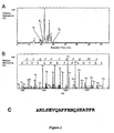
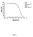
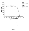


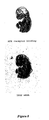
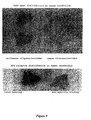


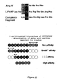
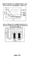


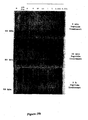
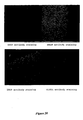


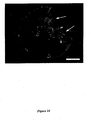
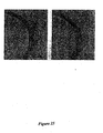
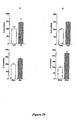

Classifications
-
- A—HUMAN NECESSITIES
- A61—MEDICAL OR VETERINARY SCIENCE; HYGIENE
- A61K—PREPARATIONS FOR MEDICAL, DENTAL OR TOILETRY PURPOSES
- A61K38/00—Medicinal preparations containing peptides
- A61K38/16—Peptides having more than 20 amino acids; Gastrins; Somatostatins; Melanotropins; Derivatives thereof
- A61K38/17—Peptides having more than 20 amino acids; Gastrins; Somatostatins; Melanotropins; Derivatives thereof from animals; from humans
- A61K38/39—Connective tissue peptides, e.g. collagen, elastin, laminin, fibronectin, vitronectin, cold insoluble globulin [CIG]
-
- A—HUMAN NECESSITIES
- A61—MEDICAL OR VETERINARY SCIENCE; HYGIENE
- A61K—PREPARATIONS FOR MEDICAL, DENTAL OR TOILETRY PURPOSES
- A61K31/00—Medicinal preparations containing organic active ingredients
- A61K31/70—Carbohydrates; Sugars; Derivatives thereof
- A61K31/7088—Compounds having three or more nucleosides or nucleotides
- A61K31/713—Double-stranded nucleic acids or oligonucleotides
-
- A—HUMAN NECESSITIES
- A61—MEDICAL OR VETERINARY SCIENCE; HYGIENE
- A61K—PREPARATIONS FOR MEDICAL, DENTAL OR TOILETRY PURPOSES
- A61K38/00—Medicinal preparations containing peptides
- A61K38/04—Peptides having up to 20 amino acids in a fully defined sequence; Derivatives thereof
- A61K38/08—Peptides having 5 to 11 amino acids
-
- A—HUMAN NECESSITIES
- A61—MEDICAL OR VETERINARY SCIENCE; HYGIENE
- A61P—SPECIFIC THERAPEUTIC ACTIVITY OF CHEMICAL COMPOUNDS OR MEDICINAL PREPARATIONS
- A61P15/00—Drugs for genital or sexual disorders; Contraceptives
- A61P15/04—Drugs for genital or sexual disorders; Contraceptives for inducing labour or abortion; Uterotonics
-
- A—HUMAN NECESSITIES
- A61—MEDICAL OR VETERINARY SCIENCE; HYGIENE
- A61P—SPECIFIC THERAPEUTIC ACTIVITY OF CHEMICAL COMPOUNDS OR MEDICINAL PREPARATIONS
- A61P15/00—Drugs for genital or sexual disorders; Contraceptives
- A61P15/06—Antiabortive agents; Labour repressants
-
- A—HUMAN NECESSITIES
- A61—MEDICAL OR VETERINARY SCIENCE; HYGIENE
- A61P—SPECIFIC THERAPEUTIC ACTIVITY OF CHEMICAL COMPOUNDS OR MEDICINAL PREPARATIONS
- A61P15/00—Drugs for genital or sexual disorders; Contraceptives
- A61P15/08—Drugs for genital or sexual disorders; Contraceptives for gonadal disorders or for enhancing fertility, e.g. inducers of ovulation or of spermatogenesis
-
- A—HUMAN NECESSITIES
- A61—MEDICAL OR VETERINARY SCIENCE; HYGIENE
- A61P—SPECIFIC THERAPEUTIC ACTIVITY OF CHEMICAL COMPOUNDS OR MEDICINAL PREPARATIONS
- A61P17/00—Drugs for dermatological disorders
- A61P17/02—Drugs for dermatological disorders for treating wounds, ulcers, burns, scars, keloids, or the like
-
- A—HUMAN NECESSITIES
- A61—MEDICAL OR VETERINARY SCIENCE; HYGIENE
- A61P—SPECIFIC THERAPEUTIC ACTIVITY OF CHEMICAL COMPOUNDS OR MEDICINAL PREPARATIONS
- A61P21/00—Drugs for disorders of the muscular or neuromuscular system
- A61P21/04—Drugs for disorders of the muscular or neuromuscular system for myasthenia gravis
-
- A—HUMAN NECESSITIES
- A61—MEDICAL OR VETERINARY SCIENCE; HYGIENE
- A61P—SPECIFIC THERAPEUTIC ACTIVITY OF CHEMICAL COMPOUNDS OR MEDICINAL PREPARATIONS
- A61P25/00—Drugs for disorders of the nervous system
-
- A—HUMAN NECESSITIES
- A61—MEDICAL OR VETERINARY SCIENCE; HYGIENE
- A61P—SPECIFIC THERAPEUTIC ACTIVITY OF CHEMICAL COMPOUNDS OR MEDICINAL PREPARATIONS
- A61P25/00—Drugs for disorders of the nervous system
- A61P25/02—Drugs for disorders of the nervous system for peripheral neuropathies
-
- A—HUMAN NECESSITIES
- A61—MEDICAL OR VETERINARY SCIENCE; HYGIENE
- A61P—SPECIFIC THERAPEUTIC ACTIVITY OF CHEMICAL COMPOUNDS OR MEDICINAL PREPARATIONS
- A61P25/00—Drugs for disorders of the nervous system
- A61P25/14—Drugs for disorders of the nervous system for treating abnormal movements, e.g. chorea, dyskinesia
- A61P25/16—Anti-Parkinson drugs
-
- A—HUMAN NECESSITIES
- A61—MEDICAL OR VETERINARY SCIENCE; HYGIENE
- A61P—SPECIFIC THERAPEUTIC ACTIVITY OF CHEMICAL COMPOUNDS OR MEDICINAL PREPARATIONS
- A61P25/00—Drugs for disorders of the nervous system
- A61P25/28—Drugs for disorders of the nervous system for treating neurodegenerative disorders of the central nervous system, e.g. nootropic agents, cognition enhancers, drugs for treating Alzheimer's disease or other forms of dementia
-
- A—HUMAN NECESSITIES
- A61—MEDICAL OR VETERINARY SCIENCE; HYGIENE
- A61P—SPECIFIC THERAPEUTIC ACTIVITY OF CHEMICAL COMPOUNDS OR MEDICINAL PREPARATIONS
- A61P25/00—Drugs for disorders of the nervous system
- A61P25/30—Drugs for disorders of the nervous system for treating abuse or dependence
-
- A—HUMAN NECESSITIES
- A61—MEDICAL OR VETERINARY SCIENCE; HYGIENE
- A61P—SPECIFIC THERAPEUTIC ACTIVITY OF CHEMICAL COMPOUNDS OR MEDICINAL PREPARATIONS
- A61P3/00—Drugs for disorders of the metabolism
-
- A—HUMAN NECESSITIES
- A61—MEDICAL OR VETERINARY SCIENCE; HYGIENE
- A61P—SPECIFIC THERAPEUTIC ACTIVITY OF CHEMICAL COMPOUNDS OR MEDICINAL PREPARATIONS
- A61P3/00—Drugs for disorders of the metabolism
- A61P3/06—Antihyperlipidemics
-
- A—HUMAN NECESSITIES
- A61—MEDICAL OR VETERINARY SCIENCE; HYGIENE
- A61P—SPECIFIC THERAPEUTIC ACTIVITY OF CHEMICAL COMPOUNDS OR MEDICINAL PREPARATIONS
- A61P3/00—Drugs for disorders of the metabolism
- A61P3/08—Drugs for disorders of the metabolism for glucose homeostasis
- A61P3/10—Drugs for disorders of the metabolism for glucose homeostasis for hyperglycaemia, e.g. antidiabetics
-
- A—HUMAN NECESSITIES
- A61—MEDICAL OR VETERINARY SCIENCE; HYGIENE
- A61P—SPECIFIC THERAPEUTIC ACTIVITY OF CHEMICAL COMPOUNDS OR MEDICINAL PREPARATIONS
- A61P35/00—Antineoplastic agents
-
- A—HUMAN NECESSITIES
- A61—MEDICAL OR VETERINARY SCIENCE; HYGIENE
- A61P—SPECIFIC THERAPEUTIC ACTIVITY OF CHEMICAL COMPOUNDS OR MEDICINAL PREPARATIONS
- A61P39/00—General protective or antinoxious agents
- A61P39/02—Antidotes
-
- A—HUMAN NECESSITIES
- A61—MEDICAL OR VETERINARY SCIENCE; HYGIENE
- A61P—SPECIFIC THERAPEUTIC ACTIVITY OF CHEMICAL COMPOUNDS OR MEDICINAL PREPARATIONS
- A61P43/00—Drugs for specific purposes, not provided for in groups A61P1/00-A61P41/00
-
- A—HUMAN NECESSITIES
- A61—MEDICAL OR VETERINARY SCIENCE; HYGIENE
- A61P—SPECIFIC THERAPEUTIC ACTIVITY OF CHEMICAL COMPOUNDS OR MEDICINAL PREPARATIONS
- A61P5/00—Drugs for disorders of the endocrine system
- A61P5/06—Drugs for disorders of the endocrine system of the anterior pituitary hormones, e.g. TSH, ACTH, FSH, LH, PRL, GH
-
- A—HUMAN NECESSITIES
- A61—MEDICAL OR VETERINARY SCIENCE; HYGIENE
- A61P—SPECIFIC THERAPEUTIC ACTIVITY OF CHEMICAL COMPOUNDS OR MEDICINAL PREPARATIONS
- A61P5/00—Drugs for disorders of the endocrine system
- A61P5/10—Drugs for disorders of the endocrine system of the posterior pituitary hormones, e.g. oxytocin, ADH
-
- A—HUMAN NECESSITIES
- A61—MEDICAL OR VETERINARY SCIENCE; HYGIENE
- A61P—SPECIFIC THERAPEUTIC ACTIVITY OF CHEMICAL COMPOUNDS OR MEDICINAL PREPARATIONS
- A61P9/00—Drugs for disorders of the cardiovascular system
-
- A—HUMAN NECESSITIES
- A61—MEDICAL OR VETERINARY SCIENCE; HYGIENE
- A61P—SPECIFIC THERAPEUTIC ACTIVITY OF CHEMICAL COMPOUNDS OR MEDICINAL PREPARATIONS
- A61P9/00—Drugs for disorders of the cardiovascular system
- A61P9/04—Inotropic agents, i.e. stimulants of cardiac contraction; Drugs for heart failure
-
- A—HUMAN NECESSITIES
- A61—MEDICAL OR VETERINARY SCIENCE; HYGIENE
- A61P—SPECIFIC THERAPEUTIC ACTIVITY OF CHEMICAL COMPOUNDS OR MEDICINAL PREPARATIONS
- A61P9/00—Drugs for disorders of the cardiovascular system
- A61P9/10—Drugs for disorders of the cardiovascular system for treating ischaemic or atherosclerotic diseases, e.g. antianginal drugs, coronary vasodilators, drugs for myocardial infarction, retinopathy, cerebrovascula insufficiency, renal arteriosclerosis
-
- A—HUMAN NECESSITIES
- A61—MEDICAL OR VETERINARY SCIENCE; HYGIENE
- A61P—SPECIFIC THERAPEUTIC ACTIVITY OF CHEMICAL COMPOUNDS OR MEDICINAL PREPARATIONS
- A61P9/00—Drugs for disorders of the cardiovascular system
- A61P9/12—Antihypertensives
-
- C—CHEMISTRY; METALLURGY
- C07—ORGANIC CHEMISTRY
- C07K—PEPTIDES
- C07K14/00—Peptides having more than 20 amino acids; Gastrins; Somatostatins; Melanotropins; Derivatives thereof
- C07K14/435—Peptides having more than 20 amino acids; Gastrins; Somatostatins; Melanotropins; Derivatives thereof from animals; from humans
- C07K14/52—Cytokines; Lymphokines; Interferons
- C07K14/54—Interleukins [IL]
-
- G—PHYSICS
- G01—MEASURING; TESTING
- G01N—INVESTIGATING OR ANALYSING MATERIALS BY DETERMINING THEIR CHEMICAL OR PHYSICAL PROPERTIES
- G01N33/00—Investigating or analysing materials by specific methods not covered by groups G01N1/00 - G01N31/00
- G01N33/48—Biological material, e.g. blood, urine; Haemocytometers
- G01N33/50—Chemical analysis of biological material, e.g. blood, urine; Testing involving biospecific ligand binding methods; Immunological testing
- G01N33/5005—Chemical analysis of biological material, e.g. blood, urine; Testing involving biospecific ligand binding methods; Immunological testing involving human or animal cells
- G01N33/5008—Chemical analysis of biological material, e.g. blood, urine; Testing involving biospecific ligand binding methods; Immunological testing involving human or animal cells for testing or evaluating the effect of chemical or biological compounds, e.g. drugs, cosmetics
- G01N33/502—Chemical analysis of biological material, e.g. blood, urine; Testing involving biospecific ligand binding methods; Immunological testing involving human or animal cells for testing or evaluating the effect of chemical or biological compounds, e.g. drugs, cosmetics for testing non-proliferative effects
- G01N33/5035—Chemical analysis of biological material, e.g. blood, urine; Testing involving biospecific ligand binding methods; Immunological testing involving human or animal cells for testing or evaluating the effect of chemical or biological compounds, e.g. drugs, cosmetics for testing non-proliferative effects on sub-cellular localization
-
- G—PHYSICS
- G01—MEASURING; TESTING
- G01N—INVESTIGATING OR ANALYSING MATERIALS BY DETERMINING THEIR CHEMICAL OR PHYSICAL PROPERTIES
- G01N2333/00—Assays involving biological materials from specific organisms or of a specific nature
- G01N2333/435—Assays involving biological materials from specific organisms or of a specific nature from animals; from humans
- G01N2333/705—Assays involving receptors, cell surface antigens or cell surface determinants
- G01N2333/72—Assays involving receptors, cell surface antigens or cell surface determinants for hormones
-
- G—PHYSICS
- G01—MEASURING; TESTING
- G01N—INVESTIGATING OR ANALYSING MATERIALS BY DETERMINING THEIR CHEMICAL OR PHYSICAL PROPERTIES
- G01N2500/00—Screening for compounds of potential therapeutic value
-
- G—PHYSICS
- G01—MEASURING; TESTING
- G01N—INVESTIGATING OR ANALYSING MATERIALS BY DETERMINING THEIR CHEMICAL OR PHYSICAL PROPERTIES
- G01N2500/00—Screening for compounds of potential therapeutic value
- G01N2500/04—Screening involving studying the effect of compounds C directly on molecule A (e.g. C are potential ligands for a receptor A, or potential substrates for an enzyme A)
Definitions
- This invention relates to methods of identifying modulators of insulin-regulated aminopeptidase activity.
- modulators have the ability to increase local concentration of a wide range of peptide hormones, thereby enhancing the activity of these hormones.
- the modulators may enhance learning and memory in both normal subjects and those with memory disorders. They also stimulate cellular proliferation while reducing apoptosis, thus stimulating tissue growth development, and affect angiogenesis. Thus they are also useful in tissue repair and regeneration.
- disorders of the central nervous system including Alzheimer's disease (AD) and other forms of dementia and memory loss, motor neurone diseases, disorders of the cardiovascular system, including cardiac hypertrophy, congestive heart failure, hypertension, and atherosclerosis, and disorders of development and growth, including disorders of glucose and fat metabolism, are all serious conditions for which effective treatment is currently unavailable. Given the importance of these conditions, it is readily apparent that effective treatments are urgently needed.
- AD Alzheimer's disease
- other forms of dementia and memory loss dementia and memory loss
- motor neurone diseases disorders of the cardiovascular system, including cardiac hypertrophy, congestive heart failure, hypertension, and atherosclerosis
- disorders of development and growth including disorders of glucose and fat metabolism
- AD Alzheimer's disease
- physicians have as yet no pharmacological agent that can be prescribed safely, either to arrest or reverse this condition.
- Angiotensin IV (Ang IV) is a hexapeptide with the sequence VYIHPF (SEQ ID NO:1), which has been shown in vitro to be produced by the consecutive actions of aminopeptidases A and N on angiotensin II. This hexapeptide was initially thought to be inactive, but has subsequently been shown to elicit actions in the brain and blood vessels. Ang IV acts via a specific binding site, designated the Angiotensin IV receptor (AT 4 receptor).
- the AT 4 receptor is defined as a high affinity binding site which selectively binds Ang IV with an affinity ranging from 1-10 nM (Swanson et al, 1992).
- the AT 4 receptor isolated from bovine adrenal membranes is a glycoprotein of 165 kDa, with a single transmembrane domain, a large extracellular domain, and a small intracellular domain (Zhang et al, 1998). In the brain, the AT 4 receptor occurs as a 150 kDa glycoprotein (Zhang et al, 1999).
- LVVYPWTQRF (SEQ ID NO:2) (Moeller et al, 1996) binds with nanomolar affinity to the AT 4 receptor, and can mimic the actions of Ang IV, and that Nle'-Ang IV (Nle'-YIHPF) (SEQ ID NO:4) binds with sub-nanomolar affinity to AT 4 receptor (Zhang et al, 1999).
- AT 4 receptors are widely distributed in the brains of guinea-pig (Miller-Wing et al, 1993), rat (Roberts et al, 1995), sheep (Moeller et al, 1996), monkey (Moeller et al, 1996), and human (Chai et al, 2000), and the distributions of the receptors are highly conserved throughout these species.
- the receptor sites occur in high abundance in the basal nucleus of Meynert, in the CA2 and dentate gyrus of the hippocampus, and throughout the neocortex, a distribution which closely resembles those of cholinergic neurones and their projections, and is consistent with the memory-enhancing effects of the receptor.
- High levels of the receptors are also found in most brain regions involved in motor control, including the motor cortex, ventral lateral thalamic nucleus, cerebellum and the cranial nerve motor nuclei. Many sensory regions also contain moderate levels of the AT 4 receptors, suggesting a more widespread role for this receptor in the brain.
- Ang IV Central infusions of Ang IV, Nle-Ang IV and Norleucinal Ang IV, which are agonists of the AT 4 receptor, were found to facilitate memory retention and retrieval in rats in the passive avoidance and Morris water maze paradigms (Braszko et al, 1988, Wright et al, 1993, Wright et al, 1999).
- the AT 4 receptor agonists reversed the performance deficits detected in the Morris water maze paradigm (Pederson et al, 1998; Wright et al, 1999).
- Ang IV was also reported to improve long-term memory in crabs (Delorenzi et al, 1999).
- WO 94/00492 discloses angiotensin AT4 receptor and AIV ligand system for binding a small N-terminal hexapeptide fragment of Angiotensin II (referred to as AIV, with amino acid sequence Val1-Tyr2-Ile3-His4-Pro5-Phe6). Also disclosed are processes for isolating angiotensin AT4 receptor and AIV angiotensinase, identifying angiotensin AIV agonists and antagonists, and constructing diagnostic assays to specifically measure AIV and AI-specific angiotensinase in biological fluids.
- Insulin-regulated aminopeptidase (IRAP) is a constituent of vesicles containing the glucose transporter molecule GLUT4.
- AT 4 receptor has a span of 18 amino acid residues which has a 95% sequence homology with the deduced amino acid sequence of human oxytocinase (TrEMBL accession # 000769), which is known to be a homologue of insulin-regulated aminopeptidase (IRAP) (Rasmussen et al, 2000).
- IRAP insulin-regulated aminopeptidase
- IRAP is the AT 4 receptor, and to hypothesise that modulation of the activity of IRAP by peptides such as Ang IV, Nle 1 -Ang IV, LVVYPWTQRF (SEQ ID NO:2) and KYLPGPLQ (SEQ ID NO:3) will activate neurones in the central nervous system to enhance learning and memory.
- peptides such as Ang IV, Nle 1 -Ang IV, LVVYPWTQRF (SEQ ID NO:2) and KYLPGPLQ (SEQ ID NO:3) will activate neurones in the central nervous system to enhance learning and memory.
- the identification of IRAP as the AT 4 receptor means that screening assays can be developed to identify compounds which have the ability to modulate IRAP enzymatic activity.
- Such agents can be used to increase serum and tissue levels of biologically active compounds such as arginine-vasopressin, oxytocin, somatostatin, angiotensin III, Lys-bradykinin, neurotensin, met-enkephalin, dynorphin A, neurokinin A, neuromedin B, cholecystokinin 8, insulin, insulin-like growth factor 1 or insulin-like growth factor 2 which may assist in preventing or treating conditions associated with deficient activity of these compounds.
- biologically active compounds such as arginine-vasopressin, oxytocin, somatostatin, angiotensin III, Lys-bradykinin, neurotensin, met-enkephalin, dynorphin A, neurokinin A, neuromedin B, cholecystokinin 8, insulin, insulin-like growth factor 1 or insulin-like growth factor 2 which may assist in preventing or treating conditions associated with deficient activity of these compounds.
- the invention provides a method as claimed in claim 1.
- DISCLOSED METHODS Also disclosed is a method of in vitro screening for compounds which have the ability to modulate the activity of AT 4 receptor/IRAP, comprising the steps of
- the candidate compound may be an intracellular or extracellular protein, or the protein which interacts with AT 4 receptor/IRAP may be tankyrase.
- IRAP is indeed the AT 4 receptor
- transfected expression plasmids containing the IRAP cDNA into HEK 293 cells and Chinese hamster ovary (CHO) cells, and found a 10-20 fold increase in sites which specifically bind 125 I-Nle 1 Ang IV.
- the binding of 125 I-Nle 1 angiotensin IV to these sites can be competitively inhibited by ligands of AT 4 receptors, including Ang IV, LVVYPWTQRF (SEQ ID NO:2) and KYLPGPLQ (SEQ ID NO:3).
- IRAP binds Ang IV with an affinity of 20 to 30 nM.
- AT 4 receptor like IRAP
- IRAP is a glycoprotein of molecular size 165 kDa.
- Nle-Ang IV and LVVYPWTQRF inhibit the aminopeptidase activity of IRAP, in vitro .
- peptides such as Ang IV, Nle 1 -Ang IV, LVVYPWTQRF (SEQ ID NO: 2) and KYLPGPLQ (SEQ ID NO:3) activates neurones in the central nervous system to enhance learning and memory.
- Structure-activity studies performed on these peptides and on known inhibitors of leucine-aminopeptidases will enable us to develop peptidomimetic and other drugs which modify the IRAP enzymatic activity.
- This invention unites findings on the effects of peptide and non-peptide ligands of the AT 4 receptor on the central nervous, cardiovascular and renal systems with the effects of
- IRAP and GLUT4 are known to translocate to the cell surface in response to insulin in adipocytes, skeletal muscles and cardiac myocytes, which is the predominant glucose-transporting mechanism in these cells. Although the roles of IRAP in these cells are unknown, its close association with GLUT4 is suggestive of a role for the enzyme in the regulation of glucose uptake and its storage.
- the activity of the AT 4 receptor may also be regulated by proteins known to interact with IRAP, such as tankyrase; proteins which are associated with GLUT4 vesicles, such as sortilin and IGF-II receptor; and proteins which regulate GLUT4 vesicle translocation, such as Munc 18c, Syntaxin 4, SNAP23 , Synip, and VAMP 2 (Pessin et al, 1999, Cheatham, 2000, Foster and Klip, 2000, Simpson et al, 2001, Holman and Sandoval, 2001).
- proteins known to interact with IRAP such as tankyrase
- proteins which are associated with GLUT4 vesicles such as sortilin and IGF-II receptor
- proteins which regulate GLUT4 vesicle translocation such as Munc 18c, Syntaxin 4, SNAP23 , Synip, and VAMP 2
- This invention relates to novel methods for the development of modulators of AT 4 receptor/IRAP activity as claimed in claim 1. These compounds may lead to an alteration in the levels of
- receptor/IRAP activity may for example be based on
- the invention provides an assay to screen for compounds which modulate the activity of IRAP as amended in claim 1. Also disclosed is a diagnostic marker for disorders related to alterations in the activity of IRAP in serum or in tissues.
- the modulators of IRAP activity preferably result in a decrease in the catalytic activity of IRAP.
- These compounds are useful for increasing serum and tissue levels of biologically active agents such as arginine-vasopressin, oxytocin, somatostatin, angiotensin III, Lys-bradykinin, neurotensin, met-enkephalin, dynorphin A, neurokinin A, neuromedin B, cholecystokinin 8, insulin, IGF-I or IGF-II. Therefore administration of modulators of IRAP activity can have the same effects or uses as administration of exogenous arginine-vasopressin, oxytocin, insulin, IGF-I or IGF-II.
- Compounds which modulate the activity of IRAP may be used in the treatment, prevention or diagnosis of conditions such as
- Such compounds may also be used to accelerate wound healing.
- the following examples use conventional molecular biology, cellular biology, and recombinant DNA techniques. Such techniques are well known to the skilled worker, and are explained fully in the literature. See, for example , Maniatis, Fritsch & Sambrook, "Molecular Cloning: A Laboratory Manual” (1982 ); “ DNA Cloning: A Practical Approach,” Volumes I and II (D.N. Glover, ed., 1985 ); “ Oligonucleotide Synthesis” (M.J. Gait, ed., 1984 ); “ Nucleic Acid Hybridisation” (B.D. Hames & S.J.
- AT 4 receptor A 4 receptor/IRAP
- IRAP IRAP
- nucleic acid and “polynucleic acid” refer herein to deoxyribonucleic acid and ribonucleic acid in all their forms, i.e., single and double-stranded DNA, cDNA, mRNA, and the like.
- antisense oligonucleotides generally refers to small, synthetic oligonucleotides resembling single-stranded DNA, which are applied to the inhibition of gene expression by inhibition of a target messenger RNA (mRNA).
- mRNA target messenger RNA
- exogenously administered antisense agents disclosed herein decrease the levels of mRNA and protein encoded by the target gene, and/or cause changes in the growth characteristics or shapes of thus treated cells. See Milligan et al., (1993); Helene, C. and Toulme, J. Biochim. Biophys, Acta 1049, 99-125 (1990 ); Cohen, J. S. D. Ed., Oligonucleotides as antisense inhibitors of gene expression; CRC Press: Boca Raton, F L (1987 ), the relevant portions of which are hereby incorporated by reference.
- antisense oligonucleotide is generally a short sequence of synthetic nucleotide which hybridises to any segment of a mRNA encoding a targeted protein under appropriate hybridisation conditions, and which upon hybridisation causes a decrease in expression of the targeted protein.
- treat refers to a treatment which decreases or abolishes the likelihood that a subject administered such treatment will manifest symptoms of an AT 4 receptor/TRAP-related disease.
- IRAP insulin-regulated aminopeptidase
- Amino acid sequence variants of the peptides disclosed herein are prepared by introducing appropriate amino changes into amino acid sequence, or by in vitro synthesis. Such variants include, for example, deletions from, or insertions or substitutions of, amino acid residues within the peptide sequences disclosed. Any combination of deletion, insertion, and substitution may be made to arrive at an amino acid sequence variant of the peptides disclosed herein, provided that such a variant possesses the desired characteristics described herein.
- Covalent modifications of the peptides disclosed herein are also included.
- covalent modifications may be intrpduced into the peptides by reacting targeted amino acid residues with an organic derivatizing agent which is capable of reacting with selected amino acid side chains or the N- or C-terminal residues.
- Cysteiny1 residues most commonly are reacted with ⁇ -haloacetates and corresponding amines, such as chloroacetic acid or chloroacetamide, to give carboxymethyl or carboxyantidomethyl derivatives. Cysteinyl residues also are derivatized by reaction with bromotrifluoroacetone, ⁇ -bromo- ⁇ -(5-imidozoyl)propionic acid, chloroacetyl phosphate N-alkylmaleimides, 3-nitro-2-pyridyl disulfide, methyl 2-pyridyl disulfide, p-chloromercuribenzoate, 2-chloromercuri-4-nitrophenol, or chloro-7-nitrobenzo-2-oxa-1,3-diazole.
- Histidyl residues are derivatized by reaction with diethyl-pyro-carbonate at pH 5.5-7.0 because this agent is relatively specific for the histidyl side chain.
- Para-bromophenacyl bromide also is useful; the reaction is preferably performed in 0.1M sodium cacodylate at pH 6.0.
- Lysinyl and amino terminal residues are reacted with succinic or other carboxylic acid anhydrides. Derivatization with these agents has the effect of reversing the charge of the lysinyl residues.
- Suitable reagents for derivatizing ⁇ -amino-containing residues include imidoesters such as methyl picolinimidate; pyridoxal phosphate; pyridoxal; chloroborohydride; trinitrobenzenesulfonic acid; O-methylisourea; 2,4-pentanedione; and transaminase-catalyzed reaction with glyoxylate.
- imidoesters such as methyl picolinimidate; pyridoxal phosphate; pyridoxal; chloroborohydride; trinitrobenzenesulfonic acid; O-methylisourea; 2,4-pentanedione; and transaminase-catalyzed reaction with glyoxylate.
- Arginyl residues are modified by reaction with one or several conventional reagents, among them phenylglyoxal, 2,3-butanedione, 1,2-cyclohexanedione, and ninhydrin. Derivatization of arginine residues requires that the reaction be performed in alkaline conditions because of the high pK a of the guanidine functional group. Furthermore, these reagents may react with the groups of lysine as well as the arginine epsilon-amino group.
- tyrosyl residues may be made, with particular interest in introducing spectral labels into tyrosyl residues by reaction with aromatic diazonium compounds or tetranitromethane.
- aromatic diazonium compounds or tetranitromethane Most commonly, N-acetylimidizole and tetranitromethane are used to form O-acetyl tyrosyl species and 3-nitro derivatives, respectively.
- aspartyl and glutamyl residues are converted to asparaginyl and glutaminyl residues by reaction with ammonium ions.
- Glutaminyl and asparaginyl residues are frequently deamidated to the corresponding glutamyl and aspartyl residues respectively. Alternatively, these residues are deamidated under mildly acidic conditions. Either form of these residues falls within the scope of this invention.
- more resistant to metabolic degradation means that the compound has been modified such that the resulting compound is more stable under acidic conditions than the "native" sequence of the peptides. For example, amino acid substitutions as discussed previously may be undertaken which produce compounds more resistant to metabolic degradation. It is well known in the art that D-amino acids, and amino acid analogues are more resistant to acidic environments. Conjugates of small peptides and cholic acid have reduced metabolic degradation problems.
- biological activity with reference to AT 4 receptor/IRAP means a biological activity which is normally promoted, either directly or indirectly, by the presence of AT 4 receptor/IRAP, and includes, but is not limited to, AT 4 receptor/IRAP binding and biochemical effects such as modulation of the enzymatic, signalling, and glucose transport activities of IRAP, and modulation of the AT4 receptor activities, including enhancement of memory, reversal of amnesia and growth promotion.
- Such biological activities can be measured by conventional in vitro and in vivo assays, including enzymatic assays using a synthetic substrate, peptide degradation assays using HPLC, behavioural assays for memory and learning such as the Barnes maze, and glucose uptake assays in cells, synaptosomes and whole animals.
- Solid phase methods such as those developed for synthesis of peptides and peptidomimetic compound, are preferred, including but not limited to the Fmoc solid phase peptide synthesis method described herein, the Boc solid phase peptide synthesis method, and pin synthesis methods (for review, see Maeji et al ., 1995) .
- Those skilled in the art will readily be able to select the most suitable method for any given compound.
- the phosphodiester residues of the antisense oligonucleotide are modified or substituted.
- Chemical analogues of oligonucleotides with modified or substituted phosphodiester residues, eg, to the methylphosphonate, which increase the in vivo stability of oligonucleotide are particularly preferred.
- the naturally occurring phosphodiester linkages of oligonucleotides are susceptible to some degree of degradation by cellular nucleases. Many of the residues proposed herein, on the contrary, are highly resistant to nuclease degradation. See Milligan et al, and Cohen, J. S D., Supra.
- the oligonucleotides may be protected from degradation by adding a "3'-end cap", by which nuclease-resistant linkages are substituted for phosphodiester linkages at the 3' end of the oligonucleotide See Tidd D. M. andressrus, H. M., Be. J. Cancer 60:343-350 (1989 ); Shaw, J.P. et al., Nucleic acids research. 19:747-750 (1991 ),
- Phosphoroamidates, phosphorothioates, and methylphosphonate linkages all function adequately in this manner.
- the number of residues which may be modified or substituted will vary, depending on the need, target and route of administration, and may be from one to all residues, or any number in between. Many different methods for replacing the entire phosphodiester backbone with novel linkages are known. See Milligan et al., Supra.
- Preferred backbone analogue residues include phosphorothioate, formacetal boranophosphate, 3'-thioformacetal, 5'-thioether, carbonate, 5'-N-carbamate, sulphate, sulfonate, sulfamate, sulfonamide, sulfone, sulfite, 2'-O methyl, sulfoxide, sulfide, hydroxylamine, methylene (methylimino) (MMI), methyleneoxy (methylimino) (MOMI) residues.
- Phosphorothioate and methylphosphate-modified oligonucleotides are particularly preferred, due to their availability through automated oligonucleotide synthesis.
- agents disclosed herein may be administered in the form of their pharmaceutically acceptable salts, or as a mixture of the antisense oligonucleotide and its salt. In a particular example, a mixture of different antisense oligonucleotides or their pharmaceutically acceptable salts is administered.
- the antisense nucleic acid molecules (agents) disclosed herein may be of any suitable length, for example about 7 to about 60 nucleotides long, preferably about 12 to about 45, more preferably up to about 30 nucleotides long, and still more preferably up to about 21, although they may be other lengths as well, depending on the particular target and the mode of delivery.
- the agents disclosed herein may be directed to any and all segments of a target RNA.
- the oligonucleotide is operatively linked to an agent or molecule which is itself internalised or taken up by living cells. In this manner, the uptake of the agent is enhanced.
- agents or molecules suitable for use with the oligonucleotides disclosed herein are transferrin asialoglycoprotein and streptavidin. Other agents will be known to those skilled in the art.
- the inventors also disclose a pharmaceutical composition
- a pharmaceutical composition comprising an antisense oligonucleotide in an amount effective to reduce expression of a target mRNA, by passing through a cell membrane and binding specifically with target mRNA in the cells, so as to prevent its translation, together with a suitable pharmaceutically acceptable carrier, eg. sterile pyrogen-free saline solution.
- a suitable pharmaceutically acceptable carrier eg. sterile pyrogen-free saline solution.
- the agent may be formulated with a hydrophobic carrier capable of passing through a cell membrane, eg in a liposome, with the liposomes carried in a pharmaceutically acceptable aqueous carrier.
- the oligonucleotides may be coupled to an agent which inactivates mRNA, such as a ribozyme.
- oligonucleotides may be administered to a subject in need of such treatment to inhibit the activation of specific receptors, enzymes and/or proteins and/or factors.
- the pharmaceutical formulation may also comprise chimeric molecules comprising antisense oligonucleotides attached to molecules which are known to be internalised by cells. These oligonucleotide conjugates utilise cellular uptake pathways to increase intercellular concentrations of the oligonucleotide. Examples of molecules used in this manner include macromolecules, including transferrin, asialoglycoprotein (bound to oligonucleotides via polylysine) and streptavidin.
- the antisense compound may be contained in the pharmaceutical formulation within a lipid particle or vesical, such as a liposome or microcrystal.
- the particles may be of any suitable structure, such as unilamellar or plurilamellar.
- the antisense oligonucleotides is comprised within the liposome.
- Positively charged lipids such as N-[1-(2,3-dioleoyloxi)propyl]-N,N,N-trimethylammoniumethylsulfate, or "DOTAP", are particularly preferred for such particles and vesicles.
- DOTAP N-[1-(2,3-dioleoyloxi)propyl]-N,N,N-trimethylammoniumethylsulfate
- DOTAP DOTAP
- the preparation of such lipid particles is well-known. See, for example, US Patent Nos.
- compositions disclosed herein may comprises the antisense oligonucleotides described above and one or more surfactants.
- Suitable surfactant components for enhancing the up-take of the antisense oligonucleotides disclosed herein include synthetic and natural as well as full length and truncated forms of surfactant protein A, surfactant protein B, surfactant protein C, surfactant protein D and surfactant protein E, di-saturated phosphatidylcholine (other than dipalmitoyl, dipalmitoyl phatidylcholine, phosphatidylcholine, phosphatidylglycerol, phosphatidylinositol, phosphatidylethanolamine, phosphatidylserine; phosphatidic acid, ubiquinones, lysophoaphatidylethanolamine, lysophosphatidylcholine, palmitoyllysophosphatidylcholine, dehydroe
- the agent disclosed herein may also provided as a composition, comprising the agent and a carrier.
- the carrier is preferably a biologically acceptable carrier, and more preferably a pharmaceutically or veterinarily acceptable carrier in the form of gaseous, liquid, or solid carriers, and mixtures thereof, which are suitable for the different routes of administration intended.
- the composition may optionally comprise other agents such as other therapeutic compounds known in the art for treatment of the condition or disease, antioxidants, flavouring and colouring agents, fillers, volatile oils, buffering agents, dispersants, surfactants, RNA-inactivating agents, antioxidants, flavouring agents, propellants and preservatives, as well as other agents known to be utilised in therapeutic compositions.
- An example of the mRNA inactivating agent is an enzyme, such as ribozyme.
- the composition generally contains the antisense oligonucleotide in an amount of about 0.01 to about 99.99 w/w, preferably about 1 to about 40 w/w, and more preferably about 5 to about 20 w/w of the composition.
- other ingredients, and other amounts of the agent are also suitable within the confines of this invention.
- the agent may be provides in various formulations which are tailored for different methods of administration and routes of delivery.
- the formulations contemplated are, for example, a transdermal formulation also containing carrier(s) and other agents suitable for delivery through the skin, mouth, nose, vagina, anus, eyes, ears, other body cavities, intradermally, as a sustained release formulation, intracranial, intrathecally, intravascularly, by inhalation, intrapulmonarily, into an organ, by implantation, including suppositories, creams, gels, and the like, as is known in the art.
- the agent may be suspended or dissolved in a solvent.
- the carrier may comprises a hydrophobic carrier, such as lipid particles or vesicles, including liposomes and micro crystals.
- lipid particles or vesicles including liposomes and micro crystals.
- the vesicles comprise liposomes containing the antisense oligonucleotdies.
- the lipid vesicles may comprise N-(1-[2,3-dioleoxyloxi]propyl)-N,N,N-trimethyl-ammonium methylsulfate as well as other lipids known in the art to provide suitable delivery of DNA to target cells.
- the formulation comprises a respirable formulation, such as an aerosol.
- a respirable formulation such as an aerosol.
- the agent, composition, and formulation disclosed herein may be provided in bulk, and in unit form, as well as in the form of an implant, a solution or suspension, a capsule or cartridge, which may be openable or piercable as is known in the art.
- a kit is also disclosed, which comprises a delivery device, and in separate containers, the agent, composition or formulation , and optionally other agents, and instructions for the use of the kit components.
- the kit may optionally also comprise in a separate container an agent selected from the group consisting of other therapeutic compounds, antioxidants, flavouring and colouring agents, fillers, volatile oils, buffering agents, dispersants, surfactants, cell internalisation or up-take agents, RNA-inactivating agents, antioxidants, flavouring agents, propellants and preservatives, among other suitable additives for the different formulations.
- an agent selected from the group consisting of other therapeutic compounds, antioxidants, flavouring and colouring agents, fillers, volatile oils, buffering agents, dispersants, surfactants, cell internalisation or up-take agents, RNA-inactivating agents, antioxidants, flavouring agents, propellants and preservatives, among other suitable additives for the different formulations.
- an agent selected from the group consisting of other therapeutic compounds, antioxidants, flavouring and colouring agents, fillers, volatile oils, buffering agents, dispersants, surfactants, cell internalisation or up-take agents, RNA-inactivating agents, antioxidants, flavouring agents, propellants
- Bovine adrenals were homogenised with a polytron homogeniser in hypotonic buffer (50mM Tris-HCl pH 7.4, 5mM EDTA, pH 7.4). Tissue debris was removed by centrifugation at 600g for 5min at 4°C. The supernatant was centrifuged at 50,000g for 20min at 20°C and the membrane pellet was resuspended in isotonic buffer consisting of 50mM Tris-HCl pH 7.4, 150mM NaCl, 5mM EDTA, 20 ⁇ M bestatin, 100 ⁇ M PMSF, 0.1% BSA.
- hypotonic buffer 50mM Tris-HCl pH 7.4, 5mM EDTA, pH 7.4
- bovine adrenal membranes (1.6 ⁇ g of protein) were incubated with 0.1 ⁇ Ci [ 125 I] -Nle-Ang-IV-BP in 20ml of isotonic buffer in Eppendorf tubes (1ml per tube) at 37°C with shaking for 90min in the dark.
- Membranes were collected by centrifugation at 60,000g for 10min at 4°C, and resuspended in 500 ⁇ l of ice cold isotonic buffer and UV irradiated at 365nm on ice for 2h. Membranes were again collected by centrifugation at 60,000g for 20 min at 4°C.
- cross-linked membranes were homogenised in 2ml solubilisation buffer (1% CHAPS, 20mM Tris-HCl, pH 7.5, 5mM EDTA) in a Dounce homogeniser, and shaken at room temperature for 48h. Insoluble material was pelleted by centrifugation at 100,000g for 1h at 4°C. Non-crosslinked membranes (48mg of protein) were solubilised and centrifuged similarly, and the supernatant combined with that from cross-linked membranes.
- solubilisation buffer 1% CHAPS, 20mM Tris-HCl, pH 7.5, 5mM EDTA
- solubilised membrane protein was applied to a 1ml DEAE fast flow anion exchange column. The column was then washed with 5ml of solubilisation buffer, then with 5ml of solubilisation buffer plus 50mM NaCl. AT 4 receptor was eluted with solubilisation buffer plus 150mM NaCl and collected in 0.5ml fractions. Proteins in 200 ⁇ l aliquots of eluted fractions were resolved by 7.5% SDS-PAGE and the gel stained first with Coomassie brilliant blue and then overstained with silver nitrate (Shevchenko et al. 1996). Radioactive bands were imaged after exposing to phophorimager plates for 16h, as illustrated in Figure 1 .
- the silver-stained band that co-migrated with the major radioactive band at 165 kDa was analysed by mass spectrometry as follows.
- the protein band was excised, reduced, alkylated and digested with trypsin in situ (Moritz et al. , 1996). Tryptic peptides were subjected to capillary-column reversed phase-high performance liquid chromatography (RP-HPLC; Hewlett Packard Model 1090A modified for capillary chromatography), coupled on-line with an electrospray ionization (ESI) ion trap (IT) mass spectrometer (LCQ Finnigan MAT, San Jose, CA, USA) for peptide sequencing.
- RP-HPLC capillary-column reversed phase-high performance liquid chromatography
- ESI electrospray ionization
- IT electrospray ion trap
- the column used was a 150 x 0.20mm ID capillary column (Brownlee RP-300, 7 ⁇ m C8) manufactured using a PVDF end frit.
- a 60 min linear gradient (flow rate 1.7 ⁇ L/min) was used from 0-100% B, where solvent A was 0.1% v/v aqueous TFA and solvent B was 0.1% v/v aqueous TFA in 60% acetonitrile.
- the ESI parameters were as follows: spray voltage, 4.5kV; sheath gas and auxiliary gas flow rates, 5 and 30 (arbitrary value), respectively; capillary temperature, 150°C; capillary voltage, 20V; and tube lens offset 16V.
- the sheath liquid used was 2-methoxyethanol (99.9% HPLC grade) delivered at a flow rate of 3 ⁇ L/min.
- the electron multiplier was set to -860 V.
- the trap was allowed a maximum injection time of up to 200ms.
- the automatic gain control parameter was turned on for all experiments, ensuring that the number of ions in the trap was automatically kept to a constant preset value.
- the range scanned in MS mode was 350-2000 Da, and in MS/MS the range varied according to the mass of the ion selected for MS/MS. After acquiring one scan in MS, the most intense ion in that spectrum above a threshold of 1x10 5 was isolated for subsequent zoom scan to determine charge state, then collision-induced dissociation or MS/MS in the following scan. The dissociation energy for MS/MS was set to 55%. All spectra were recorded in centroid mode.
- a search of the database revealed 18 of the 19 amino acids to be identical to residues 979-996 of human oxytocinase (TrEMBL accession number 000769), the human homologue of rat insulin-regulated aminopeptidase (IRAP) ( Figure 2 ).
- the derived bovine sequence has an alanine residue at the N-terminus, whereas the human sequence has a threonine.
- the amount of radioligand bound to the membranes from HEK 293 cells transfected with IRAP cDNA was expressed as a percentage of radioligand bound to membranes from HEK 293 cells transfected with control plasmids.
- Cross-linking experiments were also carried out on the membranes from HEK 293 cells transfected with the IRAP cDNA, using the method described in Example 1 ( Figure 6 ).
- Membranes from HEK 293 cells transfected with IRAP cDNA produced a 25-35 fold increase in the total number of binding sites, compared to membranes prepared from cells transfected with control plasmids.
- the affinities of Ang IV and LVVYPWTQRF (SEQ ID NO:2) in competing for the binding of 125 I-Nle Ang IV in the IRAP-transfected cells were not significantly different from that of the endogenously-occurring IRAP in HEK 293 cells ( Figures 3 and 4 ).
- AT 4 receptor/IRAP The affinity of AT 4 receptor/IRAP for its ligands varies considerably between tissues and cell types. This may be due to interactions of the ligand binding subunit with other proteins which modulate the binding affinity. Alternatively, different isoforms of the enzyme may exist.
- an IRAP-related EST (Genbank accession #BF381719) which includes intron 2 sequence following exon 1, was identified from a library made from a cell line derived from human primitive neuroectoderm. This could represent a tissue- or stage-specific variant of IRAP arising by alternative splicing.
- the existence of brain-specific variant of AT 4 receptor/IRAP may make it possible to develop modulators of AT 4 receptor/IRAP which specifically target the brain form of the receptor, and which therefore do not have unwanted side-effects in peripheral tissues.
- AT 4 receptor/IRAP influences other physiological events.
- the identification of proteins which complex with the ligand binding subunit of IRAP/AT 4 is required.
- the interactions may modulate the function of AT 4 receptor/IRAP by inhibiting or enhancing its catalytic activity.
- the binding of peptides to AT 4 receptor/IRAP may activate a second messenger system via proteins which interact with IRAP.
- AT 4 receptor/IRAP-interacting proteins might also have roles in the intracellular trafficking of AT 4 receptor/IRAP.
- Molecules which interact with the AT 4 receptor/IRAP represent potential targets for drugs designed to reverse memory deficits in conditions such as Alzheimer's disease and other forms of dementia, or to modulate other physiological actions of the IRAP/AT 4 system.
- an interacting molecule shows a more restricted distribution than does AT 4 receptor/IRAP (e.g. limited to brain or certain regions of the brain), it may provide a more suitable target than is IRAP for a drug which specifically modulates the effects of the IRAP/AT 4 system in a particular tissue or cell type.
- ion exchange purification was scaled up and fractions enriched for the receptor were further purified by wheat germ lectin affinity chromatography.
- the two-step purification was carried out as follows: [ 125 I]-Nle-Ang IV-BP was cross-linked to the AT 4 receptor in bovine adrenal membranes (93mg protein) and solubilised as described in Example 1. Non-crosslinked receptor from bovine adrenal membranes (254mg of protein) was solubilised and combined with cross-linked receptor.
- the receptor was enriched by DEAE-Sepharose fast flow anion exchange as described above.
- Fractions enriched for the receptor were then applied to a 1ml wheat-germ lectin-agarose column, the column was washed with 5ml of solubilisation buffer, and the receptor eluted with 0.5M N-acetylglucosamine, 0.5M NaCl in solubilisation buffer, and collected in 0.5ml aliquots. Proteins in 200 ⁇ l aliquots of eluted fractions were resolved by 7.5% SDS-PAGE, and the gel stained and autoradiographed as described above. The ligand-binding subunit was visualised by Coomassie blue staining, and bands which co-migrated with two of the higher molecular weight complexes were barely visible after silver staining. These bands were excised from the gel and analysed by capillary-column RP-HPLC coupled with ESI-IT-MS/MS, as described above.
- AT 4 receptor/IRAP from that tissue was cross-linked to [ 125 I]-Nle-Ang IV-BP, solubilised and purified by ion exchange chromatography and wheat germ lectin affinity,chromatography, as described above, and proteins resolved by SDS-PAGE.
- Silver or Coomassie blue-stained bands which co-migrated with radiolabelled bands corresponding to higher molecular weight complexes were excised from the gel and analysed by capillary-column RP-HPLC coupled with ESI-IT-MS/MS; as described above.
- membranes are prepared from these tissues and membrane proteins solubilised with NP40 or CHAPS as described in Example 1, and incubated with antibodies against IRAP and protein G-Sepharose beads.
- solubilised membranes are incubated with protein G-Sepharose alone.
- Precipitated proteins are resolved by SDS-PAGE and detected by Coomassie blue or silver staining. Bands appearing in the test but not in the control lanes are excised and digested with trypsin in situ , and tryptic peptides analysed by capillary-column RP-HPLC coupled with ESI-IT-MS/MS, as described in example 1.
- proteins complexing with IRAP/AT 4 are isolated by immunoaffinity chromatography using IRAP antibody coupled to a chromatography resin, and identified by tandem MS as described above.
- a Golgi-associated MAP kinase substrate has been shown to interact with IRAP in adipocytes (Chi and Lodish, 2000).
- Other potential IRAP-interacting proteins include GLUT4 and various proteins involved in GLUT4 trafficking including Syntaxin 4, SNAP23, Synip, Munc18c and VAMP2 (Pessin et al, 1999; Cheatham, 2000; Foster and Klip, 2000; Simpson et al, 2001; Holman and Sandoval, 2001).
- IRAP complexes with these proteins in brain or other tissues or cells of interest is assessed by blotting SDS-PAGE gels containing proteins purified by immunoaffinity chromatography or immunoprecipitation with IRAP antibodies, as described above, and probing with antibodies against the candidate interacting proteins.
- Yeast cells are transformed with a vector which expresses the IRAP/AT 4 intracellular or extracellular domain (the bait) as a fusion with a DNA-binding domain, such as LexA, and a cDNA library in a yeast expression vector that directs expression of cDNAs fused to a nuclear localisation signal and a transcriptional activation domain.
- a vector which expresses the IRAP/AT 4 intracellular or extracellular domain the bait
- a DNA-binding domain such as LexA
- a cDNA library in a yeast expression vector that directs expression of cDNAs fused to a nuclear localisation signal and a transcriptional activation domain.
- Cells expressing cDNAs which interact with the bait protein are then selected, based on expression of one or more reporter genes, under the control of the LexA operator.
- Candidates which activate transcription specifically in the presence of the bait-DNA binding domain hybrid are investigated further.
- the biological relevance of interactions identified in the yeast two-hybrid system is assessed by examining whether the interacting protein is co-expressed with AT 4 receptor/IRAP in relevant tissues and cell types and by determining whether the interacting protein is co-immunoprecipitated from relevant tissues or cultured cells by antibodies against IRAP/AT 4 .
- the AT 4 radioreceptor assay was used to screen the sheep brain extracts for AT 4 receptor modulatory activity.
- the membranes were prepared using bovine adrenal glands obtained from the abattoir.
- the adrenals were diced, homogenized in ice-cold hypotonic buffer (50mM Tris, 5mM EDTA, pH 7.4) and centrifuged for 10min at 500g.
- the supernatants were heated to 60°C for 15min, and centrifuged for 20min at 40,000g.
- the resulting pellets were re-homogenized in hypotonic buffer.
- Binding assay mixtures comprised 56 ⁇ g of adrenal membranes, 0.14 ⁇ Ci of [ 125 I] Ang IV (approximately 260pM), and 10 ⁇ l of column eluent in a total volume of 270 ⁇ l in 50mM Tris buffer, pH 7.4, containing 150mM sodium chloride, 5mM EDTA, 100 ⁇ m phenylmethyl-sulfonyl fluoride, 20 ⁇ M bestatin, and 0.1% (w/v) bovine serum albumin. The samples were incubated for 2h at 37°C, and the bound ligand was separated on Whatman GF/B glass fibre filters. The relative potency of the fractions in competing for 125 I-Ang IV binding was determined from a standard curve, in which known amounts of the unlabelled Ang IV were added in concentrations ranging from 10 -10 to 10 -6 M.
- the same purification procedure was carried out on sheep cerebral cortex which contained blood, and also on blood-free sheep cerebral cortex, in which the sheep brain was perfused with 10 1 of ice-cold phosphate-buffered saline solution.
- the brain tissues were homogenized in 2M acetic acid (2ml/g tissue), centrifuged and the supernatant decanted.
- a preliminary purification was carried out on the brain extracts using a preparative C18 column (55-105mm, Waters). The C18 eluant was lyophilized, reconstituted and subjected to a series of chromatographic steps in which the fractions with the highest AT 4 receptor modulatory activity were carried through to the next purification step.
- a decapeptide having the sequence LVVYPWTQRF was purified from the sheep cerebral cortex which contained blood. A search of the database revealed that this sequence was present in the internal sequence of ( ⁇ , ⁇ , ⁇ and ⁇ globin, and is known as LVV-hemorphin 7. This decapeptide may have resulted from the breakdown of haemoglobin. The purification procedure was repeated on brain which had been perfused to remove all traces of blood, and an octapeptide of sequence KYLPGPLQ (SEQ ID NO:3) was purified. This peptide is a fragment of complexin II, a cytosolic protein located in nerve terminals which is associated with the docking/fusion complex which regulates neurotransmitter release.
- Synthetic LVVYPWTQRF (SEQ ID NO:2) and KYLPGPLQ (SEQ ID NO:3) displayed comparable affinities to Ang IV in competing for 125 I-Ang IV binding to bovine adrenal membranes (approx. 1-5nM), and 125 I-KYLPGPLQ (SEQ ID NO:3) displayed identical binding patterns to 125 I-Ang IV, whereas 125 I-LVVYPWTQRF (SEQ ID NO:2) bound to a subset of 125 I-Ang IV binding sites.
- LVVYPWTQRF (SEQ ID NO:2) is released from ⁇ -globin into the circulation in uremic patients undergoing haemofiltration, and in patients undergoing open-heart surgery or cardiopulmonary bypass. Significant levels of the decapeptide are also found in ventricular cerebrospinal fluid of patients with intracerebral and subarachnoid bleeding. The half-life of the decapeptide is between 20-30min. This decapeptide may exert a modulatory effect on the activity of AT 4 receptor/IRAPs in the circulatory and central nervous systems.
- Anti-sense oligonucleotides to the human sequence of AT 4 receptor are designed to bind to the specific mRNA and target the mRNA for degradation or the prevention of translation.
- the invention also includes the ability to specifically regulate the different amino - terminus variants of AT 4 receptor which are encoded by splice variants of the gene by targeting the start site of translation for the differentially-spliced mRNAs.
- RNA walk of the IRAP mRNA was also carried out to identify regions of the RNA that contained assymmetrical bulges, short stems and included loop structures based on the predicted secondary structure of the RNA. Examples of some of the oligonucleotides identified using these methods are listed below.
- SEQ ID NOs:8 to 10 are oligonucleotides targeted to the mRNAs encoding both forms of the protein.
- SEQ ID NOs:12 and 13 are specific for the mRNAs encoding the different forms of the protein.
- An assay may be used for the discovery of compounds which modulate the AT 4 receptor/IRAP activity. This assay may also be used for the discovery of compounds for the clinical applications listed herein.
- the assay uses an aminopeptidase substrate, a source of AT 4 receptor/IRAP - containing material, and a candidate modulator.
- AT 4 receptor/IRAP-containing material include cell lines or biological tissues expressing appreciable amounts of AT 4 receptor/IRAP.
- Prokaryotic or eukaryotic cells expressing a transfected gene encoding AT 4 receptor/IRAP represent a recombinant source.
- Our results indicate that the brain AT 4 receptor/IRAP is approximately 10kDa smaller than the one expressed in the periphery. The difference in size is thought to be due to differential glycosylation.
- Using the brain-derived AT 4 receptor/IRAP as a source for screening may enable the design of modulators that are specific to the brain form of AT 4 receptor/IRAP and therefore do not interact with the peripheral form. For example, this would be important for the design of drugs for the enhancement of memory and learning and for the treatment of dementia.
- AT 4 receptor/IRAP-containing material is incubated with a synthetic aminopeptidase substrate, such as L-leucyl ⁇ -naphthylamide, and a test substance.
- a synthetic aminopeptidase substrate such as L-leucyl ⁇ -naphthylamide
- Inhibition or enhancement of the rate of degradation of the synthetic substrate is a measure of the modulatory effect of the test substance.
- Hek293 cells stably transfected with a eukaryotic expression vector expressing the AT 4 receptor gene may be utilized.
- An example of this is the vector pcDNA-3 containing the cDNA encoding the full length AT 4 receptor.
- Two assay systems are used, one for each spliced variant of the human AT 4 receptor gene.
- the assay system involves the measurement of the rate of hydrolysis of L-leucyl ⁇ -naphthylamide by the AT 4 receptor.
- the rate of hydrolysis by the transfected cells is followed by recording the increase in fluorescence (excitation at 340nm, emission at 410nm) using a purpose-designed fluorimeter.
- Test compounds are added to the assay system, and their effects on the hydrolysis of L-leucyl ⁇ -naphthylamide are a direct indicator of their modulation of AT 4 receptor activity.
- Crude membranes were prepared from HEK 293 cells stably transfected with pcDNA-3 containing the cDNA encoding the full length AT 4 receptor/IRAP, using the same method as that described for bovine adrenal membranes.
- the membranes were solubilised in 50 mM Tris-HCl containing 1% Triton-X100 at 4 °C for 16h.
- the IRAP enzymatic activity was determined by the hydrolysis of a synthetic substrate, L-leucine- ⁇ -naphthylamide, and monitored by the release of a fluorogenic product on a spectrophotometer set at excitation 340 nm and emission 410 nm.
- Ang IV or LVVYPWTQRF (SEQ ID NO:2) (10 ⁇ m) were added, and the extent of inhibition of enzymatic activity determined.
- IRAP activity could also be inhibited by the aminopeptidase inhibitors, amastatin and bestatin, but only at concentrations much higher than observed for other aminopeptidases (25% inhibition of IRAP at 100 ⁇ M amastatin, 60% at 10 uM bestatin).
- Table 1 Comparison of the apparent affinities of AT 4 ligands, as assessed by radioligand displacement and fluorescent substrate cleavage inhibition assays. IC 50 binding IC 50 inhibition 125 I-Nle-Ang IV leucine ⁇ -naphthylamide Nle 1 -Ang IV 3.32 nM 600 nM Ang IV 32.1 nM 200 nM LVV-H7 140 nM 1. 5 ⁇ M Divalinal -Ang IV 845 nM 4 ⁇ M
- Therapeutic agents which modulate the enzyme are also developed on the basis of structure-function studies on peptides known to modulate AT 4 receptor/IRAP.
- These peptides include Ang IV, Nle 1 -Ang IV, LVVYPWTQRF (SEQ ID NO:2) and the complexin fragment (KYLPGPLQ (SEQ ID NO:3)), known ligands of AT 4 receptor/IRAP such as oxytocin and vasopressin, and peptide fragments thereof, and known inhibitors of aminopeptidases, enabling the development of peptidomimetic and other compounds that modulate AT 4 receptor/IRAP activity.
- AT 4 receptor/IRAP is highly expressed in the placenta.
- the protein is cleaved from the cell surface and circulates in the maternal serum.
- the serum levels of the AT 4 receptor/IRAP can be determined using a radioimmunoassay.
- Serum and/or CSF levels of AT 4 /IRAP have the potential to be a diagnostic or prognostic marker for disorders related to the actions of AT 4 receptor/IRAP. Examples of these include central nervous system disorders, including cognitive, motor or sensory disorders such as Alzheimer's disease; motor neuron disease; cardiovascular disorders, including hypertension, congestive heart failure, cardiac hypertrophy, vasospastic disorders, and atherosclerosis; and premature or delayed labour.
- This example relates to a method of identifying and characterising peptides that are altered by the AT 4 receptor/IRAP system including the endogenous substrate of the enzyme, particularly in the brain.
- one mode of action of modulators of AT 4 receptor/IRAP is via extending the half-life of peptide substrates, by inhibiting their degradation by AT 4 receptor/IRAP.
- the endogenous substrates for AT 4 receptor/IRAP are identified, and their relationship to the clinical disorders and biological effects of modulators of the AT 4 receptor/IRAP system is determined. Therefore another approach for the treatment of these disorders is to identify the targets of the AT 4 receptor/IRAP system, and to test these compounds directly, using bioassays such as memory, learning, wound healing, neuronal regeneration etc.
- a panel of peptides is designed, synthesise and tested as potential peptide substrates for AT 4 receptor/IRAP. Based on the ability of the peptides to be altered by the AT 4 receptor/IRAP system, selective intervention such as specific blockade at the receptor level, is tested in biological systems where Ang IV and other modulators of AT 4 receptor/IRAP activity have a known effect. Therefore we can identify the peptide substrates which are directly involved in the biological systems affected by the AT 4 receptor/IRAP system.
- a peptide is incubated with AT 4 receptor/IRAP containing material and its rate of degradation measured by HPLC. If the peptide proved to be a substrate in the AT 4 receptor/IRAP system then potentially effects of modulators of AT 4 receptor/IRAP observed in vivo and in vitro may be mediated by the thus-identified substrate peptide. To confirm this hypothesis, the substrate peptide is tested in biological systems where effects of Ang IV and its analogues have been observed. Alternatively, if the peptide is known to be part of an already characterised system, antagonists to the corresponding receptor are utilized to define the observed effects with Ang IV and other modulators of AT 4 receptor/IRAP activity.
- Two candidate substrates for AT 4 receptor/IRAP in the brain are arginine-vasopressin (Herbst et al, 1997) and oxytocin (Rogi et al, 1996), two structurally related peptides which occur in relatively high abundance in the brain and have been shown to be cleaved by IRAP in vitro. These peptides have been shown to have effects on memory in certain behavioural paradigms, and thus present two potential substrates for IRAP in neurones.
- Each potential peptide substrate (30 ⁇ g) was incubated at 37°C in TBS with 10 ⁇ g solubilized membrane protein from hIRAP transfected-HEK 293T cells. At each time point (0, 0.5, 1, 2, 4 & 6 hr), an aliquot containing 5 ⁇ g peptide was removed, and the reaction stopped by addition of 4 volumes methanol/1% trifluoroacetic acid (TFA). Samples were dried on a centrifugal vacuum evaporator prior to HPLC analysis with on-line mass spectrometric detector.
- arginine-vasopressin (AVP), oxytocin (OXY) and met-enkephalin were the most rapidly cleaved by IRAP. Both Cyt 6 -AVP(2-9) and Cyt 6 -AVP(3-9) were generated by IRAP, but only the first peptide bond of OXY was cleaved. Similarly, although the first residue (Tyr 1 ) of met-enkephalin was quickly removed, only a small amount of desTyr 1 Gly 2 -met-enkephalin was detected after 6 hours.
- IRAP is found in the same vesicles as GLUT4, the insulin-dependent glucose transporter. As in the case of GLUT4, IRAP is redistributed to the cell surface in response to insulin or insulin-like growth factors. We postulate that IRAP may serve as a negative feedback control for GLUT4 translocation to the cell surface and hence glucose transport into the cell by regulating the levels the insulin and insulin-like growth factors on the cell surface and also in the GLUT4 vesicles. We are investigating the hydrolysis of insulin IGF-I and IGF-II by IRAP using HPLC with C18 reverse phase columns.
- modulation of the activity of the aminopeptidase AT 4 receptor/IRAP by a number of peptides known to be AT 4 receptor agonists results in enhancement of learning and memory in normal and amnesic animals.
- the maze comprised a raised rotatable white circular platform of diameter 1.2m, with 18 evenly spaced holes in the periphery (diameter 0.09m).
- An escape tunnel comprising a black box of internal diameter of 0.16m width, 0.29m length and 0.09m depth was positioned immediately beneath a peripheral hole.
- the maze was positioned in a large room, 6 x 4.5m, both naturally and fluorescently lit with visual cues (e.g. A4 sized posters of abstract patterns) placed on stands at various positions around the maze at a distance of 0.5m from the platform.
- the animal was placed on the maze platform under the starting chamber, which is a cylindrical chamber located in the centre of the platform and can lowered or raised, and left for 20s. While the animal was under the chamber, the floodlights were switched on. Following the twenty seconds disorientation period the chamber was raised, and the trial was begun. The animals were then allowed 240s in which to find and utilize the escape tunnel. Following 20s of light alone, the auditory buzzer was switched on, and these aversive stimuli were continued throughout the remaining time period (220s) or until the escape tunnel was found and utilized. Each rat received three consecutive trials per day over ten days.
- the starting chamber which is a cylindrical chamber located in the centre of the platform and can lowered or raised, and left for 20s. While the animal was under the chamber, the floodlights were switched on. Following the twenty seconds disorientation period the chamber was raised, and the trial was begun. The animals were then allowed 240s in which to find and utilize the escape tunnel. Following 20s of light alone, the auditory buzzer was switched on, and these
- each rat On the first day of the testing period, each rat was individually taken into the testing room in its cage, and placed directly into the escape tunnel for a 2min familiarization period. The rat was then replaced into its cage for approximately one minute, after which the animal was injected with peptide or artificial cerebrospinal fluid and then replaced into its cage for a further 5min. Three consecutive trials were then carried out, with a recovery period of 2min between each trial in its home cage.
- the Y maze consists of three identical alleys (30cm in length, 10cm in width and 17cm in height) with the three arms separated by 120° angles. To minimize stress, the maze is located in a sound-attenuated room under dim illumination, with the floor of the maze covered with sawdust. After each trial, the sawdust is mixed to eliminate olfactory cues. For spatial orientation, visual cues are placed on the walls of the testing room.
- the test consisted of two trials, separated by a time interval known as the intertrial interval.
- the animals were placed at the distal end of one arm, their heads pointing away from the centre of the maze.
- the mice were allowed to visit only two accessible arms of the maze for 3 minutes.
- the mice were replaced in their cages for the intertrial interval.
- the animals had access to all three arms for 5 minutes. The number and duration of time spent in the novel arm were documented.
- a shorter intertrial interval (2 hours) separating the acquisition phase from the retention phase enables amnesiant effects to be detected. Control mice still remember the location of the novel arm and will preferentially spend more time (45-50% of time) in that arm. A longer intertrial interval (6 hours) results in the animals not remembering the location of the novel arm and hence spending equal amount of time in all three arms.
- mice were surgically implanted with an infusion cannula in the dorsal third ventricle.
- the cannula was connected to Alzet osmotic minipumps by tygon tubing (5-6cm in length and 0.3mm in diameter).
- the minipumps were placed in the dorsal subcutaneous space in the neck.
- the minipumps were filled with artificial cerebrospinal fluid for 1 week, and delivered the fluid at a constant flow rate of 0.5 ⁇ l/h.
- the minipumps were replaced under light anaesthesia with another minipump containing either artificial cerebrospinal fluid or LVVYPWTQRF (SEQ ID NO:2).
- the peptide was delivered at a rate of 0.3pmol/h.
- mice which received LVVYPWTQRF spent more time in the novel arm (between 50-60% of time) compared to control mice (approximately 33% of time), indicating that during that time period, LVVYPWTQRF (SEQ ID NO:2) enhanced the spatial memory in this paradigm.
- mice from both treatment groups were indistinguishable from each other.
- the effect of modulation of AT 4 receptor/IRAP activity on spatial learning and memory in amnesic animals was also assessed using the Morris water maze test.
- the Morris test has been used extensively to test for spatial memory.
- the hippocampus which contains high levels of AT 4 receptors/IR.AP, is thought to play an important role in processing this form of memory.
- Scopolamine a muscarinic receptor antagonist
- This compound has been shown to block the trial-to-trial decrease in latency of this task, and this effect appeared to be due to impairment of short-term memory.
- the effect of AT 4 receptor/IRAP ligands on scopolamine-induced amnesia in a spatial memory task was tested in the Morris water maze.
- the maze consisted of a circular tank, 1.6m in diameter by 0.6m in height, containing water to a depth of 30cm.
- the room contained visual cues placed 40 cm from the edge of the tank.
- the rats were subjected to two days of familiarisation to the task, during which the escape pedestal is visible to the animals.
- the water in the tank was rendered opaque, and the escape platform was submerged under the surface of the water.
- the animals had to rely on the visual cues around the room to find the submerged escape pedestal.
- the latency of the animal in finding the pedestal during the acquisition phase was taken as a measure of the spatial memory performance of the animal.
- Rats were surgically implanted with cannulae in their lateral ventricles and allowed two to three days of recovery. During the familiarisation trials on days 1 and 2, the animals were placed in the water facing the wall of the tank from different starting positions equidistant from the escape pedestal. When the animals located and mounted the pedestal, they were allowed 30 seconds on the pedestal before the commencement of the next trial. Four consecutive trials were carried out on each animal on each day, and the mean latency period before the animal reached the pedestal was plotted.
- the scopolamine-treated animals were given 70nmole of the compound into the lateral ventricles 30min before the commencement of the trials for each day.
- the peptide (1nmole) or saline was given 5min before the trial began.
- the pedestal which remained in a constant position for each animal, was submerged for this phase, and the mean latency for each animal in locating the pedestal was documented for each day.
- the drug treatments were withdrawn on the eighth day, the pedestal once again made visible, and latency documented. The results are shown in Figure 16 .
- Intracerebroventricular infusion of 70nmole of scopolamine significantly increased the latency of the animals in locating the submerged pedestal, indicating a deficit in short-term memory.
- Rats treated with LVVYPWTQRF reversed the scopolamine-induced latency, and this group of rats was indistinguishable from saline-treated controls.
- Withdrawal of treatment on day 8 brought the latency of the scopolamine-treated group back to control levels, indicating that the effect of scopolamine was reversible, and that the latency was primarily due to the muscarinic receptor antagonist.
- Passive avoidance trials were then used to test the effect of modulation of AT 4 receptor/IRAP activity on aversive conditioning behaviour in amnesic animals.
- the passive avoidance task involves aversive conditioning behaviour to measure facilitation of memory retention and retrieval.
- the testing was carried out in an apparatus that consisted of a light and a dark compartment separated by a guillotine door.
- the floor of the dark compartment contained an electrified grid.
- Rats were surgically implanted with chronic cannulae in the lateral ventricle. On conditioning day, the animals were habituated in the dark compartment for 5min with the guillotine door closed. The animals were then placed in their home cages for 5min before being placed in the light compartment with the guillotine door opened. The latency to enter the dark compartment with all four feet was documented. When the rats entered the dark compartment within 20s of being placed in the light compartment, they were randomly divided into four treatment groups. The treatment groups were:
- Acetylcholine is a major neurotransmitter involved in the processing of cognitive function, since anti-cholinergic drugs induce memory deficit and confusion.
- neuronal loss has been reported in cholinergic-rich areas, particularly in the septohippocampal pathway.
- Angiotensin AT 4 receptors have been found in high abundance in the basal nucleus of Meynert, in the CA2 and dentate gyrus of the hippocampus, and in somatic and autonomic preganglionic motoneurones of the monkey brain. This pattern of receptor distribution closely resembles that of cholinergic neurones and their projections, and suggests that the AT 4 receptors may be associated with cholinergic pathways centrally.
- LVV-haemorphin-7 can reverse the learning deficit induced by scopolamine (a muscarinic receptor antagonist).
- scopolamine a muscarinic receptor antagonist
- the slices were washed for 60min at 37°C with Krebs buffer at a flow rate of 500 ⁇ l/min. Superfusate samples were collected at 5min intervals for the duration of the experiment.
- the hippocampal slices were depolarised by the addition of 30mM KCl to the Krebs buffer and infused for 4min (stimulation period S1). The tissue was then allowed to recover for 25min before the next stimulation period (S2).
- stimulation period S1 stimulation period 1
- S2 next stimulation period
- the peptides were added to the hippocampal slices 15min prior to and 5min during the second period of stimulation.
- LVVYPWTQRF (SEQ ID NO: 2) significantly and dose-dependently increased potassium-evoked 3 H-choline release, with a maximum effect of 180% of control observed at 10 -7 M, as shown in Figure 18 .
- Ang IV also significantly and dose-dependently increased potassium-evoked 3 H-choline release with a smaller maximum effect of 150% of control observed at 10 -7 M, as shown in Figure 18 .
- ERK/MAPK cascade involves three tiers of kinases which are sequentially activated by phosphorylation.
- the MAPK cascade is activated by multiple signals, including activation of receptor tyrosine kinases, synaptic activity and increases in intracellular Ca2+ and cAMP, and is thus capable of integrating several different signals into a graded response.
- the mechanism(s) by which IRAP/AT 4 substrates enhance memory may involve modulation of the MAPK cascade. For instance, inhibition of the aminopeptidase activity of IRAP/AT 4 by ligand binding might bring about the accumulation of a neuropeptide that activates the MAPK cascade via an interaction with a cell surface receptor.
- IRAP/AT 4 ligands to activate MAPK or CREB was initially investigated on rat hippocampal slices. Rats were killed by decapitation and the hippocampi removed, four hundred ⁇ m thick slices cut and incubated in aCSF. After four hours of stabilisation, the slices were treated with 10 -8 M Nle 1 Ang IV for 30 min, or left untreated.
- blots were probed overnight at 4°C with anti phospho-p44/42 MAP kinase (Thr202/Tyr204) antibody and detected by chemiluminescence using ECL blotting detection reagents and a Fuji LAS-1000.
- Enhanced activation of the MAPK cascade by IRAP/AT 4 ligands may only occur during learning. Therefore, animals treated IRAP/AT 4 ligands or vehicle control are subjected to varying numbers of learning trials (as described in the previous example), before analysis of the levels of phospho-MAPK phospho-CREB and phospho-Elk-1 in their hippocampi as described above.
- binding of ligands to IRAP might generate an intracellular signal which activates the MAPK cascade.
- the ability of AT 4 ligands to stimulate MAPK phosphorylation directly was investigated in HEK293 cells transfected with human IRAP.
- HEK293 cells were transfected with pCI-IRAP or with empty vector, plated on to 12-well plates at a density of 60,000 cells/well and grown in complete medium containing 0.2% fetal calf serum. The cells were then treated with 10 -6 , 10 -9 or 10 -11 M Nle 1 -Ang IV for 5,30 or 120 min.
- the medium was removed, the cells washed in phosphate-buffered saline and solubilised in 100 ⁇ l of 1xSDS loading buffer. The solubilised samples were then heated at 100°C for 5 min, centrifuged and 50 ⁇ l of the supernatant run on a 12% SDS-PAGE. The proteins were then transferred on to nitrocellulose membranes and incubated with phospho-MAPK (p44/42) antibodies and visualised with HRP-conjugated secondary antibody, using the ECL-plus western blot detection system.
- phospho-MAPK p44/42
- Nle 1 -Ang IV at concentrations of 0.001, 1 and 10 ⁇ M, induced the phosphorylation of ERK1/2 within 5 min of peptide treatment, and the phosphoERK persisted for 2 h, as shown in Figure 19b .
- the largest response was observed at 0.001 ⁇ M of Nle 1 -Ang IV.
- GLUT4 was initially found predominantly in adipocytes and skeletal muscles, where it is highly expressed in vesicles. Activation of the insulin receptor by insulin or insulin-like growth factors in these cells results in the translocation of GLUT4 vesicles to the plasma-membrane. This insulin-dependent glucose transporter was recently detected in the brain and in developing mouse embryos. In the brain, the distribution of GLUT4 protein and mRNA closely parallels the location of the AT 4 receptor, as demonstrated by 125 I-Ang IV or 125 I - Nle Ang IV binding. Since IRAP is the AT 4 receptor, we proposed that, as in adipocytes and skeletal muscles, GLUT4 is co-localised with IRAP in neurones, and subserves a similar role.
- mice C57/Black females were anaesthetised with an intraperitoneal injection of pentobarbitone, and perfused with phosphate-buffered saline (PBS) followed by 4% PFA, 2% glutaraldehyde in phosphate buffer (pH 7.4).
- PBS phosphate-buffered saline
- the brains were taken and stored in the fixative overnight at 4°C, then snap-frozen in isopentane cooled with dry ice. Forty micron thick sections were cut on a cryostat and collected into PBS. The free-floating sections were then incubated for one hour at room temperature in PBS with normal horse serum (10%) and Triton X-100 (0.3%).
- the sections were transferred to a blocking solution (NEN TSA kit) for 30min before incubation with the primary antibodies. Both antibodies (rabbit anti-IRAP and mouse anti-NeuN or rabbit anti-IRAP and mouse anti-GLUT4) were diluted 1:200 in PBS with 2% normal horse serum and 0.3% Triton X-100. The sections were incubated for 48h at 4°C.
- IRAP may have wider roles in the regulation of local neuropeptide concentration, in contrast to IRAP in adipose tissue and skeletal muscle, where the enzyme coexists with GLUT4, and may be integrally involved in insulin-regulated glucose transport.
- IRAP is involved in the regulation of the levels of insulin or insulin-like growth factors by enzymatic cleavage of these peptides, under certain physiological conditions such as during neural activation or pathophysiological states such as cerebral ischemia. Under these conditions, IRAP may play a role in the regulation of glucose uptake in neurones.
- Glucose has been found to improve memory in animals and humans, and several studies have shown that the memory-enhancing effects of glucose may not be due solely to its role in providing fuel for brain cells (Wickelgren, 1998). Glucose has been shown to facilitate acetylcholine synthesis (Kopf et al, 2001) and release (Kopf et al, 2001). Accordingly an investigation of AT 4 receptor/IRAP activity on glucose uptake in the hippocampus is undertaken.
- Male Sprague-Dawley rats are surgically implanted with chronic cannulae in their lateral ventricles and allowed to recover for at least three days.
- the rats are infused with 1nmole of either peptide over a period of 10min.
- the rats are injected with 2-deoxy-D-[1- 14 C]glucose into the peritoneal cavity at a dose of 1 ⁇ Ci/g.
- the rats are killed 45min later, the brains extracted and snap-frozen and sectioned at 14 ⁇ m thickness and exposed to Hyperfilm ⁇ max.
- Glucose utilisation is also measured on synaptosomes prepared from hippocampus. Synaptosomes are isolated from homogenised rat hippocampus using the Ficoll gradient centrifugation technique. The synaptosomes are washed in glucose-free HEPES buffer, then incubated at 37°C for 5min in the same solution containing 0.5mmole glucose. The synaptosomes are loaded with oxy-D-glucose,2-[1,2- 3 H(N)] at a concentration of 1 ⁇ Ci/ml. Uptake is assayed in the absence or presence of Ang IV or LVVYPWTQRF (SEQ ID NO:2) at concentrations of 1nM to 1 ⁇ M.
- Hexokinase I activity which is responsible for glucose-6 phosphorylation and essential for neuronal glucose utilisation, is also determined on the synaptosomal preparations, since a decrease in glucose utilisation is reflected by reduced hexokinase activity.
- Hexokinase activity is assayed on 0.2mg of freshly prepared synaptosomes suspended in MOPS-Tris buffer containing 0.1% Triton X-100, 8mM MgCl 2 , 0.4M NADP + , 5mM D-glucose, 5 units of glucose-6-phosphate dehydrogenase and 1mM ATP in the absence or presence of Ang IV or LVVYPWTQRF (SEQ ID NO:2).
- the linear rate of NADP + reduction is monitored on a spectrophotometer at 340nm absorbance and expressed as nM of product formed per mg protein.
- 3T3L1 fibroblasts were differentiated by the addition of DMEM containing 5% fetal calf serum, 4 ⁇ g/ml insulin, 0.25 mM dexamethasone and 0.5 mM 3-isobutyl-1-methyl-xanthine. After 7-14 days, more than 90% of the fibroblasts were differentiated into mature adipocytes.
- insulin and insulin-like growth factor in chaperoning glucose into neurones via translocation of the GLUT4 vesicles, these two polypeptides could have other roles in the brain which may lead to facilitation of memory.
- Insulin and IGF-I have been shown to stimulate neuronal growth and promote arborisation of dendrites (Bondy, 1991, Wickelgren, 1998, O'Kusky et al, 2000).
- Some of the memory-enhancing effects of Ang IV and LVVYPWTQRF (SEQ ID NO:2), particularly with chronic infusions, may be the result of prolonging the half-life of insulin or IGF in the brain, resulting in an increase in neurogenesis or synaptogenesis.
- This ability can be assessed by measuring neurogenesis in the hippocampus of rats subjected to the Barnes maze paradigm in the presence and absence of Nle-Ang IV or LVVYPWTQRF (SEQ ID NO:2), using a marker of dividing cells, 5-bromo-2'-deoxyuridine (BrdU).
- Male Sprague-Dawley rats are implanted with chronic intracerebroventricular cannulae and allowed two to three days recovery before the commencement of the experiment. The rats are divided into three groups:
- mice All rats are injected with bromodeoxyuracil (BrdU; 50mg/kg) twice daily for 3 days.
- the two groups of rats subjected to the behavioural paradigm are tested for 3 days on the Barnes maze, commencing the day of the first BrdU injection. During this time most of the rats which receive the peptide treatment are expected to achieve learner criteria, whereas the rats treated with artificial cerebrospinal fluid do not.
- the animals are perfused intracardially with 4% paraformaldehyde in phosphate buffered saline 24h after the last BrdU injection.
- the brains of these animals are removed and processed for BrdU immunofluorescence using a mouse anti-BrdU primary antibody and detected with an anti-mouse secondary antibody conjugated with FITC.
- the number of BrdU-labelled cells are determined on at least 24 sections (14 ⁇ m thick) of the rat hippocampus in the rostro-caudal extent.
- AT 4 receptor ligands have been shown to induce cellular proliferation in cardiac fibroblasts and neuronally-derived immortalised cell lines. This suggests that AT 4 receptor/IRAP may be abundantly expressed during development, and may have an important role in embryonic development.
- AT 4 receptor/IRAP may be abundantly expressed during development, and may have an important role in embryonic development.
- Embryonic day 13,15,17, neonatal day 1,7,21 and adult mice were killed by isofluorane inhalation.
- the embryos were embedded in mounting medium and snap-frozen whole, whereas in the neonatal and adult mice, tissues were dissected out and frozen in isopentane chilled to -40°C on dry ice.
- 10 ⁇ frozen sections were cut, thaw-mounted on to gelatine-coated slides, and dried for 2h under reduced pressure. The sections were then rinsed in buffer (pH 7.4) containing 50mM Tris, 5mM EDTA, 150mM NaCl for 30min at room temperature.
- AT 4 receptor/IRAP mRNA For the determination of AT 4 receptor/IRAP mRNA, 10 ⁇ frozen sections were cut, thaw-mounted onto silane-coated slides and stored at -20°C. Eight oligonucleotides, four in the anti-sense orientation and four in the sense orientation, were prepared from different coding regions of the AT 4 receptor/IRAP gene. The oligonucleotides were 3' end labelled with 33 P-dATP using terminal d-transferase and purified on a P6 column.
- mice sections were hybridized with 1 x 10 6 cpm of labelled oligonucleotide in a 100ml total volume of 50% formamide, 4xSSC, 1xDenhardt's solution, 2% sarcosyl, 20mM sodium phosphate buffer (pH7.0), 10% dextran sulphate, 50 ⁇ g/ml herring sperm DNA and 0.2mM dithiothreitol.
- the sections were washed four times in 1xSSC at 55°C, rinsed in distilled water and dehydrated through increasing alcohol. The sections were then exposed to X-ray film. The results are shown in Figure 8 .
- AT 4 receptor/IRAP The highest level of AT 4 receptor/IRAP protein was found in developing bone. High levels of the protein were also detected in the heart, kidney, thymus, pituitary and the subventricular zone of the brain ( Figure 8 ). These results suggest that the AT 4 receptor/IRAP system might play an important role in the development of these tissues.
- Ang IV has been reported to induce DNA synthesis in rabbit and rat cardiac fibroblasts and in rat anterior pituitary cells. Moreover, in bovine coronary venular endothelial cells, Ang IV enhanced 3 H-thymidine incorporation in response to stimulation with basic fibroblast growth factor, although Ang IV alone was a very weak stimulant.
- LVV-haemorphin 7 and Ang IV were investigated in cells of the neuronally-derived immortalised cell lines NG108-15 and SK-N-MC, as appreciable amounts of AT 4 receptors/IRAP were shown to be present in these cell lines.
- NG108-15 is a rat-mouse hybrid cell line consisting of both neurones and glia, whereas the SKN-MC cells are neuroepithelial in origin.
- SK-N-MC cells are known to display.cholinergic properties, such as the expression of choline acetyltransferase and muscarinic receptors.
- NG108-15 and SK-N-MC cells were cultured in DMEM supplemented with 10% heat-inactivated FCS, 2% HAT supplement, and 100U/ml penicillin/streptomycin. Cells were routinely used between passages 20 and 30.
- Cells were plated at a density of 3x10 4 cells/well in 24 well Falcon plates that had been coated with 0.1mg/ml poly-L-lysine hydrobromide. The cells were allowed to adhere overnight, and then serum deprived for 48h before being treated with LVVYPWTQRF (SEQ ID NO:2) and/or Ang IV (10 -10 to 10 -6 M) for 24h in medium with or without serum. Five hours before the end of the experiment 1 ⁇ Ci of 3 H-thymidine was added to each well.
- Ang IV dose-dependently inhibited the effect of 10 -6 M LVVYPWTQRF (SEQ ID NO:2) in stimulating 3 H-thymidine incorporation.
- 10 -6 M LVVYPWTQRF
- Ang IV was inactive, whereas increasing concentrations of Ang IV from 10 -9 to 10 -7 M progressively inhibited 3 H-thymidine incorporation.
- LVVYPWTQRF SEQ ID NO:2
- LVVYPWTQRF SEQ ID NO:2
- AT 4 receptors/IRAP The wide distribution of the AT 4 receptors/IRAP in the brain suggests important roles for this enzyme in the brain in the processing of several neuropeptides. Moreover, the distribution of AT 4 receptors/IRAP closely correlated with the distribution of cholinergic neurones and their processes.
- Acetylcholine the neurotransmitter in cholinergic neurones, is known to inhibit neurite outgrowth in embryonic chick ciliary ganglion cells.
- AT 4 receptor ligands Ang IV and LVVYPWTQRF (SEQ ID NO:2)
- Sympathetic ganglia from embryonic day 11 chicks were dissociated with trypsin/versene and were cultured in 24 well plates in DMEM and Ham's 12 medium which contained 1% insulin-tranferrin-selenium-X growth supplement, 100 mM putrescine, 1.67mg/ml prostaglandin F2 ⁇ , 6.67ng/ml progesterone, and 5ng/ml nerve growth factor. Neurones were allowed to adhere to the wells (approximately 2h) before being treated with the peptides. Dose response curves were performed over the concentration range 10 -11 to 10 -5 M. Culture dishes were coated with 0.1mg/ml poly-L-lysine and washed with PBS before receiving another coating of laminin (10 ⁇ g/ml).
- the culture medium was removed from the wells, the neurones fixed with 2.5% glutaraldehyde in phosphate-buffered saline for 20min, and examined under phase contrast microscope attached to an MD30 Plus Image Analysis software system. The length of neurites (longer than 50 ⁇ m) of every neurone examined was measured. A minimum of 40 neurite measurements was taken per treatment group, and each dose of peptide was repeated at least three times. At the conclusion of the experiment, the viability of the cells was confirmed by exclusion of 0.1% aniline blue. The results are shown in Figure 22 .
- Ang IV and LVVYPWTQRF inhibited neurite outgrowth in a dose dependent manner, with maximal effect achieved at concentrations between 10 -9 to 10 -8 M.
- the maximal extent of inhibition of neurite outgrowth was approximately 30%, which was comparable to the effect achieved by acetylcholine.
- Example 26 we also found a high concentration of AT 4 receptor/IRAP in the developing bone of E13, E15, E17 mouse embryos. IGF-I receptors have been shown to co-localize with GLUT4 in chondroblastic and hypertrophic cells of mouse bone growth centres. Moreover, in the streptozotocin-induced diabetic mouse model, there are severe histological changes in the mandibular condyles and humeral growth plate which are associated with decreases in GLUT4 and IGF-I and insulin receptors. We therefore consider that AT 4 receptor/IRAP co-localizes with GLUT4, and plays an important role in the regulation of IGF-I in regulating early bone growth.
- IGF-I Activated astrocytes which are glial fibrillary acidic protein positive are involved in neuronal tissue remodelling after damage (Bovolenta et al 1992). Injury-evoked plasticity is similar to events observed in the developing central nervous system (Schwartz 1992). IGF-I is a growth factor which is not only important in normal growth and development; there is strong evidence to suggest that it has an important role in neurodegenerative conditions in adult. Injury to the brain results in an up-regulation of IGFs, their receptors and binding proteins. Application of exogenous IGF-I protects the brain from hypoxic and ischemic damage.
- mice Male Sprague-Dawley rats are anaesthetized with isofluorane in O 2 and indwelling intracerebroventricular cannulae are implanted.
- the cannula are connected to Alzet minipumps containing artificial cerebrospinal fluid which are implanted subcutaneously in the dorsal neck region.
- the rats Seven days later, the rats are re-anaesthetised with halothane in O 2 , and focal cerebral ischemia induced by permanent occlusion of the left, middle cerebral artery proximal to the lenticulostriate branch, by electrocoagulation.
- the Alzet minipumps containing artificial cerebrospinal fluid are replaced with ones containing either:
- angiogenesis The vascularisation of tissues during development occurs by the process of angiogenesis, in which endothelial cells from existing capillaries form new capillaries by sprouting or by splitting from their tissue of origin. Little angiogenesis occurs in adult tissues, except during wound healing, during the female reproductive cycle and in certain pathological states such as cancer.
- Pathological angiogenesis involves the persistent proliferation of endothelial cells, as occurs in proliferative retinopathy, rheumatoid arthritis, psoriasis and tumour growth. Angiogenesis is also essential to the growth of solid tumours.
- AT 4 receptor/IRAP has a role in angiogenesis during wound healing, and possibly in the pathological conditions mentioned above. If this is so, the potential exists to use modulators of IRAP/AT 4 to interfere with angiogenesis and thereby limit tumour invasion, and/or to treat other conditions in which pathogenic angiogenesis is a feature.
- Peptidases are known to have many roles in angiogenesis. Degradation of extracellular matrix (ECM) by peptidases allows the infiltration of endothelial cells from existing blood vessels.
- ECM extracellular matrix
- ECM degradation is a tightly controlled process, and peptidases also play roles in its regulation.
- ECM-degrading proteinases mobilise growth factors such as fibroblast growth factor (FGF)from the ECM, which stimulates expression of collagenase and plasminogen activator by microvascular endothelial cells.
- FGF fibroblast growth factor
- ECM-degrading proteinases also modulate activation of latent TGF- ⁇ 1 in the ECM, which counteracts the stimulatory effects of FGF on vascular endothelial cells.
- AT 4 receptor/IRAP A number of experimental systems are used to test the role of AT 4 receptor/IRAP in angiogenesis.
- endothelial cells When endothelial cells are grown on a gel formed of reconstituted basement membrane proteins (Matrigel), they aggregate and organise into tubule-like structures (Kubota et al., 1988).
- the ability of peptides that bind to AT 4 receptor/IRAP e.g.
- Ang IV, LVVYPWTQRF (SEQ ID NO:2), KYLPGPLQ (SEQ ID NO:3) and divalina1-Ang IV) to modulate this process is assessed by culturing human umbilical vein endothelial (HUVE) cells or human dermal microvascular cells on Matrigel, with or without pre-incubation of the cells with IRAP/ AT 4 receptor -binding peptide. Alternatively the peptide is incorporated into the gel matrix. The role of AT 4 receptor/IRAP in angiogenesis is further assessed by incorporating antibodies to AT 4 receptor/IRAP into the matrigel before application of HUVE cells or HDME cells.
- HUVE human umbilical vein endothelial
- IRAP/ AT 4 receptor -binding peptide Alternatively the peptide is incorporated into the gel matrix.
- AT 4 receptor/IRAP in angiogenesis is further assessed by incorporating antibodies to AT 4 receptor/IRAP into the matrigel before application of HUVE cells or HDME cells
- IRAP/AT 4 The role of IRAP/AT 4 in tumour angiogenesis is initially investigated using receptor autoradiography with [125I]-Nle-Ang IV to examine the distribution of IRAP/AT 4 in the microvasculature in sections of human tumour tissue.
- the ability of IRAP/AT 4 ligands to modulate tumour angiogenesis is assessed in an in vivo model in which C3L5 cells,a highly metastatic murine mammary adenocarcinoma cell line, are suspended in growth factor-reduced Matrigel and implanted subcutaneously into mice. Growth factor-reduced Matrigel alone does not stimulate angiogenesis (Jadeski & Lala, 1999), but angiogenesis occurs in implants containing tumour cells.
- IRAP/AT 4 ligands are administered via osmotic minipumps over 14 days, after which their effects on angiogenesis within the implants are assessed.
- Ang IV binds to AT 4 receptors in blood vessels to induce vasodilatation and proliferation of cultured bovine endothelial cells.
- Topical application of Ang IV caused vasodilatation of pial arterioles, and infusion of Ang IV into the rat middle cerebral artery and renal artery significantly increased blood flow.
- Ang IV significantly but modestly increased DNA synthesis in cultured bovine endothelial cells, and synergistically enhanced basic fibroblast growth factor-stimulated cellular proliferation.
- Carotid arteries were sectioned at 20 ⁇ m thickness. Sections were preincubated in an isotonic buffer, 50mM Tris-HCl buffer, pH 7.4, 150mM NaCl and 5mM EDTA for 30min at 22°C and then incubated in 50mM Tris-HCl, pH 7.4, 150mM NaCl, 5mM EDTA, 100 ⁇ M phenylmethyl-sulfonyl fluoride (Sigma, Mo., U.S.A.), 20 ⁇ M bestatin (Peninsula Laboratories, Ca., U.S.A.) and 0.1% bovine serum albumin, containing approximately 200pM [ 125 I] Ang IV, for 2h at 22°C.
- an isotonic buffer 50mM Tris-HCl buffer, pH 7.4, 150mM NaCl and 5mM EDTA for 30min at 22°C and then incubated in 50mM Tris-HCl, pH 7.4, 150mM NaCl, 5mM
- Non-specific binding was determined in the presence of 1 ⁇ M Ang IV in adjacent sections. Following the incubation, the sections were given 3 x 2min washes in the isotonic buffer, at 4°C, air dried, and exposed to X-ray film for 3.5 weeks, together with a set of radioactive standards. After exposure, the sections were counterstained with haematoxylin and eosin for histological examination.
- AT 4 receptors were detected in vascular smooth muscle cells and in the vasa vasorum of the adventitia, as shown in Figure 25 , left-hand panel. Very low receptor levels were observed in the endothelial cells.
- AT 4 receptor/IRAP in the media/neointima increased to approximately 150% of control tissue, as shown in Figures 26A and 26B .
- AT 4 receptor/IRAP further increased in the media/neointima and re-endothelialized cell layer to 223% at 20 weeks after injury ( Figures 26C and 26D).
- EXAMPLE 33 Elucidation of factors which translocate AT 4 receptor/IRAP to the cell surface in neuronal cells
- AT 4 receptor/IRAP and GLUT4 are co-localised inside the cell in specialised post-endosomal vesicles, which are distinct from constitutively recycling endosomes, and also in the trans-Golgi network. Both GLUT4 and AT 4 receptor/IRAP rapidly translocate to the plasma membrane in response to insulin stimulation; GLUT4 to mediate insulin-stimulated glucose uptake(Garza et al, 2000).
- This process involves the formation of vesicles containing both GLUT4 and AT 4 receptor/IRAP, which translocate to the cell surface, and fuse with the plasma membrane by a mechanism which in part parallels synaptic vesicle trafficking and docking in the regulation of neurotransmitter release in the brain (Cheatham 2000).
- the molecular mechanism/signalling cascade regulating the trafficking of GLUT4/ AT 4 receptor/IRAP vesicles has not been fully elucidated.
- guanosine 5'-O-(3-thiotriphosphate) GTP ⁇ S
- adrenergic stimulation also result in the translocation of AT 4 receptor/IRAP and GLUT4 to the cell surface in adipocytes and/or skeletal muscle cells.
- GTP ⁇ S guanosine 5'-O-(3-thiotriphosphate)
- adrenergic stimulation also result in the translocation of AT 4 receptor/IRAP and GLUT4 to the cell surface in adipocytes and/or skeletal muscle cells.
- oxytocin receptor a substrate for AT 4 receptor/IRAP
- the results using this system are verified by performing subcellular fractionation using sucrose gradient centrifugation, followed by Western Blot analysis of AT 4 receptor/IRAP in stimulated and unstimulated cells.
- the first set of experiments measures endogenous AT 4 receptor/IRAP in untransfected cells, to verify that the results obtained are not an artefact of the transfection system used.
- the second series of experiments we determine the subcellular vesicles which are recruited to the plasma membrane following stimulation. Light density microsomes from stimulated and non-stimulated cells are immunoadsorbed with antibodies to the different vesicle marker proteins, and the level of AT 4 receptor/IRAP for each vesicle type determined by Western Blot analysis.
Landscapes
- Health & Medical Sciences (AREA)
- Life Sciences & Earth Sciences (AREA)
- Chemical & Material Sciences (AREA)
- Engineering & Computer Science (AREA)
- General Health & Medical Sciences (AREA)
- Medicinal Chemistry (AREA)
- Bioinformatics & Cheminformatics (AREA)
- Veterinary Medicine (AREA)
- Public Health (AREA)
- Animal Behavior & Ethology (AREA)
- Pharmacology & Pharmacy (AREA)
- Organic Chemistry (AREA)
- Chemical Kinetics & Catalysis (AREA)
- Nuclear Medicine, Radiotherapy & Molecular Imaging (AREA)
- General Chemical & Material Sciences (AREA)
- Biomedical Technology (AREA)
- Immunology (AREA)
- Molecular Biology (AREA)
- Neurology (AREA)
- Diabetes (AREA)
- Hematology (AREA)
- Neurosurgery (AREA)
- Gastroenterology & Hepatology (AREA)
- Endocrinology (AREA)
- Proteomics, Peptides & Aminoacids (AREA)
- Epidemiology (AREA)
- Biochemistry (AREA)
- Urology & Nephrology (AREA)
- Cardiology (AREA)
- Toxicology (AREA)
- Zoology (AREA)
- Heart & Thoracic Surgery (AREA)
- Reproductive Health (AREA)
- Gynecology & Obstetrics (AREA)
- Pregnancy & Childbirth (AREA)
- Obesity (AREA)
- Cell Biology (AREA)
- Analytical Chemistry (AREA)
- Genetics & Genomics (AREA)
- Biophysics (AREA)
Abstract
Claims (1)
- Procédé de criblage in vitro pour des composés qui ont la capacité d'améliorer la mémoire et l'apprentissage, comportant l'estimation d'une capacité d'un composé candidat à moduler une activité enzymatique du récepteur AT4, dans lequel l'activité enzymatique du récepteur AT4 est une activité aminopeptidase.
Applications Claiming Priority (5)
| Application Number | Priority Date | Filing Date | Title |
|---|---|---|---|
| AUPR677201 | 2001-08-02 | ||
| AUPR6772A AUPR677201A0 (en) | 2001-08-02 | 2001-08-02 | Modulation of aminopeptidase activity |
| US33017001P | 2001-10-17 | 2001-10-17 | |
| US330170P | 2001-10-17 | ||
| PCT/AU2002/001034 WO2003011304A1 (fr) | 2001-08-02 | 2002-08-02 | Modulation de l'activite du recepteur (at4) d'aminopeptidase (irap)/d'angiotensine iv regulee par l'insuline |
Publications (3)
| Publication Number | Publication Date |
|---|---|
| EP1420800A1 EP1420800A1 (fr) | 2004-05-26 |
| EP1420800A4 EP1420800A4 (fr) | 2008-01-23 |
| EP1420800B1 true EP1420800B1 (fr) | 2011-09-28 |
Family
ID=25646766
Family Applications (1)
| Application Number | Title | Priority Date | Filing Date |
|---|---|---|---|
| EP02747109A Expired - Lifetime EP1420800B1 (fr) | 2001-08-02 | 2002-08-02 | Modulation de l'activite du recepteur (at 4) d'aminopeptidase (irap)/d'angiotensine iv regulee par l'insuline |
Country Status (4)
| Country | Link |
|---|---|
| US (1) | US20050020519A1 (fr) |
| EP (1) | EP1420800B1 (fr) |
| JP (1) | JP2005508882A (fr) |
| WO (1) | WO2003011304A1 (fr) |
Families Citing this family (34)
| Publication number | Priority date | Publication date | Assignee | Title |
|---|---|---|---|---|
| US7117033B2 (en) | 2000-05-08 | 2006-10-03 | Brainsgate, Ltd. | Stimulation for acute conditions |
| US7146209B2 (en) * | 2000-05-08 | 2006-12-05 | Brainsgate, Ltd. | Stimulation for treating eye pathologies |
| US7640062B2 (en) | 2000-05-08 | 2009-12-29 | Brainsgate Ltd. | Methods and systems for management of alzheimer's disease |
| US7120489B2 (en) | 2000-05-08 | 2006-10-10 | Brainsgate, Ltd. | Method and apparatus for stimulating the sphenopalatine ganglion to modify properties of the BBB and cerebral blood flow |
| US20050266099A1 (en) * | 2002-04-25 | 2005-12-01 | Alon Shalev | Methods and apparatus for modifying properties of the bbb and cerebral circulation by using the neuroexcitatory and/or neuroinhibitory effects of odorants on nerves in the head |
| US7636597B2 (en) * | 2002-11-14 | 2009-12-22 | Brainsgate, Ltd. | Surgical tools and techniques for stimulation |
| US7684859B2 (en) * | 2002-04-25 | 2010-03-23 | Brainsgate Ltd. | Stimulation of the OTIC ganglion for treating medical conditions |
| US7561919B2 (en) * | 2002-11-14 | 2009-07-14 | Brainsgate Ltd. | SPG stimulation via the greater palatine canal |
| US7118747B2 (en) * | 2003-09-30 | 2006-10-10 | Pacific Northwest Biotechnology Inc. | AT4 receptor ligands as angiogenic, anti-angiogenic, and anti-tumor agents |
| US8055347B2 (en) | 2005-08-19 | 2011-11-08 | Brainsgate Ltd. | Stimulation for treating brain events and other conditions |
| US9233245B2 (en) | 2004-02-20 | 2016-01-12 | Brainsgate Ltd. | SPG stimulation |
| US8010189B2 (en) | 2004-02-20 | 2011-08-30 | Brainsgate Ltd. | SPG stimulation for treating complications of subarachnoid hemorrhage |
| US20090299418A1 (en) * | 2004-08-23 | 2009-12-03 | Brainsgate Ltd. | Concurrent bilateral spg modulation |
| JP5031567B2 (ja) * | 2004-09-09 | 2012-09-19 | ハワード・フローレイ・インスティテュート・オブ・イクスペリメンタル・フィジオロジー・アンド・メディスン | 酵素阻害剤およびその使用 |
| BRPI0615439A2 (pt) * | 2005-08-23 | 2011-05-17 | Wyeth Corp | métodos para o tratamento de ansiedade e para identificação de agentes ansiolìticos |
| US20080026409A1 (en) * | 2006-01-20 | 2008-01-31 | The Board Of Trustees Of The University Of Illinois | Salt-sensitive hypertension |
| US20080261893A1 (en) * | 2006-02-24 | 2008-10-23 | Denise Barbut | Topical corneal analgesia using neurotensin receptor agonists and synergistic neurotensin combinations without delaying wound healing |
| EP2043675A2 (fr) | 2006-07-07 | 2009-04-08 | Washington State University Research Foundation | Régulation du récepteur c-met par des ligands du récepteur de l'angiotensine iv (at4) |
| US8236761B2 (en) * | 2006-07-07 | 2012-08-07 | Washington State University Research Foundation | C-Met receptor regulation by angiotensin IV (AT4) receptor ligands |
| US20090210026A1 (en) * | 2006-08-17 | 2009-08-20 | Brainsgate Ltd. | Spg stimulation for enhancing neurogenesis and brain metabolism |
| AU2008225100A1 (en) * | 2007-03-12 | 2008-09-18 | Mayo Foundation For Medical Education And Research | Topical corneal analgesia using neurotensin receptor agonists and synergistic neurotensin combinations without delaying wound healing |
| FR2915392A1 (fr) * | 2007-04-27 | 2008-10-31 | Univ Claude Bernard Lyon I Eta | Utilisation d'un antagoniste de l'angiotensine iv dans le traitement de l'insulino resistance ou du risque cardio-vasculaire du syndrome metabolique |
| US7860569B2 (en) | 2007-10-18 | 2010-12-28 | Brainsgate, Ltd. | Long-term SPG stimulation therapy for prevention of vascular dementia |
| NZ585338A (en) | 2007-11-19 | 2012-08-31 | Howard Florey Inst | Insulin-regulated aminopeptidase (irap) inhibitors and uses thereof |
| JP2016523125A (ja) | 2013-05-30 | 2016-08-08 | グラハム エイチ. クリーシー | 局所神経性刺激 |
| US11229789B2 (en) | 2013-05-30 | 2022-01-25 | Neurostim Oab, Inc. | Neuro activator with controller |
| EP2878335B1 (fr) | 2013-11-10 | 2018-01-03 | Brainsgate Ltd. | Implant et système d'implantation pour stimulateur neural |
| US11077301B2 (en) | 2015-02-21 | 2021-08-03 | NeurostimOAB, Inc. | Topical nerve stimulator and sensor for bladder control |
| US10271907B2 (en) | 2015-05-13 | 2019-04-30 | Brainsgate Ltd. | Implant and delivery system for neural stimulator |
| US10953225B2 (en) | 2017-11-07 | 2021-03-23 | Neurostim Oab, Inc. | Non-invasive nerve activator with adaptive circuit |
| WO2019213024A1 (fr) * | 2018-04-30 | 2019-11-07 | Integral Molecular, Inc. | Anticorps transporteurs de glucose 4, leurs procédés de fabrication et leurs utilisations |
| WO2020264214A1 (fr) | 2019-06-26 | 2020-12-30 | Neurostim Technologies Llc | Activateur de nerf non invasif à circuit adaptatif |
| WO2021126921A1 (fr) | 2019-12-16 | 2021-06-24 | Neurostim Solutions, Llc | Activateur nerveux non invasif à distribution de charge amplifiée |
| US20250011792A1 (en) | 2021-12-01 | 2025-01-09 | Institut National de la Santé et de la Recherche Médicale | Irap inhibitors for use in the treatment of inflammatory diseases |
Family Cites Families (16)
| Publication number | Priority date | Publication date | Assignee | Title |
|---|---|---|---|---|
| US5972600A (en) | 1992-04-03 | 1999-10-26 | The Regents Of The University Of California | Separation of active complexes |
| AU4649293A (en) | 1992-06-24 | 1994-01-24 | Washington State University Research Foundation | Angiotensin iv peptides and receptor |
| US5968764A (en) * | 1994-09-20 | 1999-10-19 | Bayer Corporation | Glucose transporter vesicle aminopeptidase |
| EP0838471A4 (fr) * | 1995-07-07 | 2000-07-26 | Sagami Chem Res | Derives peptidiques et agoniste du recepteur de l'angiotensine iv |
| AUPO089396A0 (en) * | 1996-07-09 | 1996-08-01 | Howard Florey Institute Of Experimental Physiology And Medicine | Neuroactive peptide |
| CA2242571C (fr) * | 1996-08-06 | 2006-05-16 | Taisho Pharmaceutical Co., Ltd. | Composes amino et agonistes du recepteur de l'angiotensine iv |
| US5968784A (en) | 1997-01-15 | 1999-10-19 | Chugai Pharmaceutical Co., Ltd. | Method for analyzing quantitative expression of genes |
| US6022696A (en) * | 1998-04-02 | 2000-02-08 | Washington State University Research Foundation | Methods of identifying agonists or antagonists of angiotensin IV |
| US6277613B1 (en) * | 1998-06-10 | 2001-08-21 | The Rockefeller University | TRF1 binding protein, methods of use thereof |
| JP2002523078A (ja) * | 1998-08-26 | 2002-07-30 | トルスティーズ オブ ボストン ユニバーシティー | 新規なirap−bpポリペプチド及び核酸分子並びにこれらの利用法 |
| US6201020B1 (en) * | 1998-12-31 | 2001-03-13 | Guilford Pharmaceuticals, Inc. | Ortho-diphenol compounds, methods and pharmaceutical compositions for inhibiting parp |
| AU4475400A (en) * | 1999-04-22 | 2000-11-10 | Myriad Genetics, Inc. | Protein-protein interactions |
| WO2000077225A1 (fr) * | 1999-06-11 | 2000-12-21 | Whitehead Institute For Biomedical Research | Nouvelle molecule de signalisation d'insuline |
| US7064106B2 (en) * | 1999-12-20 | 2006-06-20 | Takeda Chemical Industries, Ltd. | Gene and use thereof |
| GB0014838D0 (en) * | 2000-06-16 | 2000-08-09 | Univ Bristol | An assay |
| US20040086510A1 (en) * | 2000-08-21 | 2004-05-06 | Hideaki Tojo | Irap-binding protein |
-
2002
- 2002-08-02 JP JP2003516534A patent/JP2005508882A/ja active Pending
- 2002-08-02 WO PCT/AU2002/001034 patent/WO2003011304A1/fr not_active Ceased
- 2002-08-02 US US10/485,518 patent/US20050020519A1/en not_active Abandoned
- 2002-08-02 EP EP02747109A patent/EP1420800B1/fr not_active Expired - Lifetime
Also Published As
| Publication number | Publication date |
|---|---|
| EP1420800A1 (fr) | 2004-05-26 |
| EP1420800A4 (fr) | 2008-01-23 |
| WO2003011304A1 (fr) | 2003-02-13 |
| US20050020519A1 (en) | 2005-01-27 |
| JP2005508882A (ja) | 2005-04-07 |
Similar Documents
| Publication | Publication Date | Title |
|---|---|---|
| US20050020519A1 (en) | Modulation of insulin-regulated aminopeptidase (irap)/angiotensin iv (at4) receptor activity | |
| US7674458B2 (en) | Lysyl oxidase-like 1 (LOXL1) and elastogenesis | |
| Neubert et al. | Inflammation-induced changes in primary afferent-evoked release of substance P within trigeminal ganglia in vivo | |
| Reyes et al. | Neurochemically distinct circuitry regulates locus coeruleus activity during female social stress depending on coping style | |
| US7550435B2 (en) | Method of modifying glucose activity using polypeptides selectively expressed in fat tissue | |
| CN101534648A (zh) | 使用mntf肽及其类似物治疗神经紊乱的方法 | |
| AU2002317634B2 (en) | Modulation of insulin-regulated aminopeptidase (IRAP)/Angiotensin IV (AT4) receptor activity | |
| McGinnis et al. | Influence of γ-aminobutyric acid on lordosis behavior and dopamine activity in estrogen primed spayed female rats | |
| US8524228B2 (en) | Netrin-1 compositions and methods of use thereof | |
| Shen et al. | Chromogranin mRNA levels in the brain as a marker for acute and chronic changes in neuronal activity: effect of treatments including seizures, osmotic stimulation and axotomy in the rat | |
| Zheng et al. | Enhanced anorexigenic signaling in lean obesity resistant syndecan-3 null mice | |
| AU2002317634A1 (en) | Modulation of insulin-regulated aminopeptidase (IRAP)/Angiotensin IV (AT4) receptor activity | |
| EP1635859B1 (fr) | Utilisation therapeutique de pam | |
| US20110105405A1 (en) | Method for alleviating pain using protein associated with myc and related compounds, and assays for identifying such compounds | |
| Young et al. | Calcitonin gene-related peptide is not essential for the development of pressure overload-induced hypertrophy in vivo | |
| Van Koughnet et al. | Proenkephalin transgene regulation in the paraventricular nucleus of the hypothalamus by lipopolysaccharide and interleukin‐1β | |
| Rahim | Experimental approach to overexpress pituitary adenylate cyclase activating polypeptide (PACAP) specifically in the hypothalamus | |
| Kim et al. | Transcriptional regulatory role of NELL2 in preproenkephalin gene expression | |
| Yang | The potential role for the EGF receptor system in the circadian regulation of locomotor activity | |
| Gibbs | An evaluation of the effects of activation of neuropeptide Y receptors on nociceptors | |
| Metzger et al. | Gqα/G11α Deficiency in Dorsomedial Hypothalamus Leads to Obesity Resulting from Decreased Energy Expenditure and Impaired Sympathetic Nerve Activity 2 | |
| Deo | Delta opioid receptor: Parasympathetic location and changing phenotypes in canine heart | |
| Club—ENC | ‘Neuropeptides 2002: From Molecule to Man’ | |
| JP2010536909A (ja) | イノシトールピロリン酸はエキソサイトーシス能を決定する | |
| Glanowska | Studies of the GnRH pulse generator. Developmental and location-‐specific regulation of GnRH secretion. |
Legal Events
| Date | Code | Title | Description |
|---|---|---|---|
| PUAI | Public reference made under article 153(3) epc to a published international application that has entered the european phase |
Free format text: ORIGINAL CODE: 0009012 |
|
| 17P | Request for examination filed |
Effective date: 20040301 |
|
| AK | Designated contracting states |
Kind code of ref document: A1 Designated state(s): AT BE BG CH CY CZ DE DK EE ES FI FR GB GR IE IT LI LU MC NL PT SE SK TR |
|
| AX | Request for extension of the european patent |
Extension state: AL LT LV MK RO SI |
|
| RIC1 | Information provided on ipc code assigned before grant |
Ipc: G01N 33/50 20060101ALI20071205BHEP Ipc: A61K 38/55 20060101ALI20071205BHEP Ipc: A61P 3/00 20060101ALI20071205BHEP Ipc: A61K 38/11 20060101ALI20071205BHEP Ipc: A61P 5/10 20060101ALI20071205BHEP Ipc: A61K 31/713 20060101AFI20030214BHEP Ipc: A61P 25/30 20060101ALI20071205BHEP Ipc: A61P 25/28 20060101ALI20071205BHEP Ipc: A61P 25/00 20060101ALI20071205BHEP |
|
| A4 | Supplementary search report drawn up and despatched |
Effective date: 20071220 |
|
| 17Q | First examination report despatched |
Effective date: 20080821 |
|
| GRAP | Despatch of communication of intention to grant a patent |
Free format text: ORIGINAL CODE: EPIDOSNIGR1 |
|
| GRAS | Grant fee paid |
Free format text: ORIGINAL CODE: EPIDOSNIGR3 |
|
| GRAA | (expected) grant |
Free format text: ORIGINAL CODE: 0009210 |
|
| AK | Designated contracting states |
Kind code of ref document: B1 Designated state(s): AT BE BG CH CY CZ DE DK EE ES FI FR GB GR IE IT LI LU MC NL PT SE SK TR |
|
| REG | Reference to a national code |
Ref country code: GB Ref legal event code: FG4D |
|
| REG | Reference to a national code |
Ref country code: CH Ref legal event code: EP |
|
| REG | Reference to a national code |
Ref country code: IE Ref legal event code: FG4D |
|
| REG | Reference to a national code |
Ref country code: DE Ref legal event code: R096 Ref document number: 60241150 Country of ref document: DE Effective date: 20111208 |
|
| REG | Reference to a national code |
Ref country code: SE Ref legal event code: TRGR |
|
| REG | Reference to a national code |
Ref country code: NL Ref legal event code: VDEP Effective date: 20110928 |
|
| PG25 | Lapsed in a contracting state [announced via postgrant information from national office to epo] |
Ref country code: FI Free format text: LAPSE BECAUSE OF FAILURE TO SUBMIT A TRANSLATION OF THE DESCRIPTION OR TO PAY THE FEE WITHIN THE PRESCRIBED TIME-LIMIT Effective date: 20110928 |
|
| PG25 | Lapsed in a contracting state [announced via postgrant information from national office to epo] |
Ref country code: GR Free format text: LAPSE BECAUSE OF FAILURE TO SUBMIT A TRANSLATION OF THE DESCRIPTION OR TO PAY THE FEE WITHIN THE PRESCRIBED TIME-LIMIT Effective date: 20111229 Ref country code: AT Free format text: LAPSE BECAUSE OF FAILURE TO SUBMIT A TRANSLATION OF THE DESCRIPTION OR TO PAY THE FEE WITHIN THE PRESCRIBED TIME-LIMIT Effective date: 20110928 Ref country code: CY Free format text: LAPSE BECAUSE OF FAILURE TO SUBMIT A TRANSLATION OF THE DESCRIPTION OR TO PAY THE FEE WITHIN THE PRESCRIBED TIME-LIMIT Effective date: 20110928 |
|
| REG | Reference to a national code |
Ref country code: AT Ref legal event code: MK05 Ref document number: 526026 Country of ref document: AT Kind code of ref document: T Effective date: 20110928 |
|
| REG | Reference to a national code |
Ref country code: CH Ref legal event code: NV Representative=s name: TROESCH SCHEIDEGGER WERNER AG |
|
| PG25 | Lapsed in a contracting state [announced via postgrant information from national office to epo] |
Ref country code: CZ Free format text: LAPSE BECAUSE OF FAILURE TO SUBMIT A TRANSLATION OF THE DESCRIPTION OR TO PAY THE FEE WITHIN THE PRESCRIBED TIME-LIMIT Effective date: 20110928 Ref country code: SK Free format text: LAPSE BECAUSE OF FAILURE TO SUBMIT A TRANSLATION OF THE DESCRIPTION OR TO PAY THE FEE WITHIN THE PRESCRIBED TIME-LIMIT Effective date: 20110928 |
|
| PG25 | Lapsed in a contracting state [announced via postgrant information from national office to epo] |
Ref country code: PT Free format text: LAPSE BECAUSE OF FAILURE TO SUBMIT A TRANSLATION OF THE DESCRIPTION OR TO PAY THE FEE WITHIN THE PRESCRIBED TIME-LIMIT Effective date: 20120130 Ref country code: NL Free format text: LAPSE BECAUSE OF FAILURE TO SUBMIT A TRANSLATION OF THE DESCRIPTION OR TO PAY THE FEE WITHIN THE PRESCRIBED TIME-LIMIT Effective date: 20110928 Ref country code: EE Free format text: LAPSE BECAUSE OF FAILURE TO SUBMIT A TRANSLATION OF THE DESCRIPTION OR TO PAY THE FEE WITHIN THE PRESCRIBED TIME-LIMIT Effective date: 20110928 |
|
| PG25 | Lapsed in a contracting state [announced via postgrant information from national office to epo] |
Ref country code: DK Free format text: LAPSE BECAUSE OF FAILURE TO SUBMIT A TRANSLATION OF THE DESCRIPTION OR TO PAY THE FEE WITHIN THE PRESCRIBED TIME-LIMIT Effective date: 20110928 |
|
| PLBE | No opposition filed within time limit |
Free format text: ORIGINAL CODE: 0009261 |
|
| STAA | Information on the status of an ep patent application or granted ep patent |
Free format text: STATUS: NO OPPOSITION FILED WITHIN TIME LIMIT |
|
| 26N | No opposition filed |
Effective date: 20120629 |
|
| REG | Reference to a national code |
Ref country code: DE Ref legal event code: R097 Ref document number: 60241150 Country of ref document: DE Effective date: 20120629 |
|
| PGFP | Annual fee paid to national office [announced via postgrant information from national office to epo] |
Ref country code: GB Payment date: 20120801 Year of fee payment: 11 Ref country code: SE Payment date: 20120813 Year of fee payment: 11 |
|
| PGFP | Annual fee paid to national office [announced via postgrant information from national office to epo] |
Ref country code: IT Payment date: 20120809 Year of fee payment: 11 Ref country code: FR Payment date: 20120823 Year of fee payment: 11 Ref country code: BE Payment date: 20120820 Year of fee payment: 11 Ref country code: DE Payment date: 20120725 Year of fee payment: 11 |
|
| PG25 | Lapsed in a contracting state [announced via postgrant information from national office to epo] |
Ref country code: MC Free format text: LAPSE BECAUSE OF NON-PAYMENT OF DUE FEES Effective date: 20120831 |
|
| PG25 | Lapsed in a contracting state [announced via postgrant information from national office to epo] |
Ref country code: ES Free format text: LAPSE BECAUSE OF FAILURE TO SUBMIT A TRANSLATION OF THE DESCRIPTION OR TO PAY THE FEE WITHIN THE PRESCRIBED TIME-LIMIT Effective date: 20120108 |
|
| REG | Reference to a national code |
Ref country code: IE Ref legal event code: MM4A |
|
| PG25 | Lapsed in a contracting state [announced via postgrant information from national office to epo] |
Ref country code: BG Free format text: LAPSE BECAUSE OF FAILURE TO SUBMIT A TRANSLATION OF THE DESCRIPTION OR TO PAY THE FEE WITHIN THE PRESCRIBED TIME-LIMIT Effective date: 20111228 |
|
| PG25 | Lapsed in a contracting state [announced via postgrant information from national office to epo] |
Ref country code: IE Free format text: LAPSE BECAUSE OF NON-PAYMENT OF DUE FEES Effective date: 20120802 |
|
| BERE | Be: lapsed |
Owner name: HOWARD FLOREY INSTITUTE OF EXPERIMENTAL PHYSIOLOG Effective date: 20130831 |
|
| REG | Reference to a national code |
Ref country code: CH Ref legal event code: PL |
|
| REG | Reference to a national code |
Ref country code: SE Ref legal event code: EUG |
|
| GBPC | Gb: european patent ceased through non-payment of renewal fee |
Effective date: 20130802 |
|
| PG25 | Lapsed in a contracting state [announced via postgrant information from national office to epo] |
Ref country code: SE Free format text: LAPSE BECAUSE OF NON-PAYMENT OF DUE FEES Effective date: 20130803 Ref country code: CH Free format text: LAPSE BECAUSE OF NON-PAYMENT OF DUE FEES Effective date: 20130831 Ref country code: LI Free format text: LAPSE BECAUSE OF NON-PAYMENT OF DUE FEES Effective date: 20130831 Ref country code: DE Free format text: LAPSE BECAUSE OF NON-PAYMENT OF DUE FEES Effective date: 20140301 Ref country code: TR Free format text: LAPSE BECAUSE OF FAILURE TO SUBMIT A TRANSLATION OF THE DESCRIPTION OR TO PAY THE FEE WITHIN THE PRESCRIBED TIME-LIMIT Effective date: 20110928 |
|
| REG | Reference to a national code |
Ref country code: FR Ref legal event code: ST Effective date: 20140430 |
|
| REG | Reference to a national code |
Ref country code: DE Ref legal event code: R119 Ref document number: 60241150 Country of ref document: DE Effective date: 20140301 |
|
| PG25 | Lapsed in a contracting state [announced via postgrant information from national office to epo] |
Ref country code: LU Free format text: LAPSE BECAUSE OF NON-PAYMENT OF DUE FEES Effective date: 20120802 Ref country code: IT Free format text: LAPSE BECAUSE OF NON-PAYMENT OF DUE FEES Effective date: 20130802 Ref country code: BE Free format text: LAPSE BECAUSE OF NON-PAYMENT OF DUE FEES Effective date: 20130831 |
|
| PG25 | Lapsed in a contracting state [announced via postgrant information from national office to epo] |
Ref country code: GB Free format text: LAPSE BECAUSE OF NON-PAYMENT OF DUE FEES Effective date: 20130802 |
|
| PG25 | Lapsed in a contracting state [announced via postgrant information from national office to epo] |
Ref country code: FR Free format text: LAPSE BECAUSE OF NON-PAYMENT OF DUE FEES Effective date: 20130902 |