EP1426021A1 - Implantierbares System mit medikamentenabgebenden Zellen zur lokalen Verabreichung von Medikamenten bei Bedarf - Google Patents
Implantierbares System mit medikamentenabgebenden Zellen zur lokalen Verabreichung von Medikamenten bei Bedarf Download PDFInfo
- Publication number
- EP1426021A1 EP1426021A1 EP04006105A EP04006105A EP1426021A1 EP 1426021 A1 EP1426021 A1 EP 1426021A1 EP 04006105 A EP04006105 A EP 04006105A EP 04006105 A EP04006105 A EP 04006105A EP 1426021 A1 EP1426021 A1 EP 1426021A1
- Authority
- EP
- European Patent Office
- Prior art keywords
- cells
- stent
- implantable system
- stimulating element
- delivery device
- Prior art date
- Legal status (The legal status is an assumption and is not a legal conclusion. Google has not performed a legal analysis and makes no representation as to the accuracy of the status listed.)
- Granted
Links
Images


Classifications
-
- A—HUMAN NECESSITIES
- A61—MEDICAL OR VETERINARY SCIENCE; HYGIENE
- A61F—FILTERS IMPLANTABLE INTO BLOOD VESSELS; PROSTHESES; DEVICES PROVIDING PATENCY TO, OR PREVENTING COLLAPSING OF, TUBULAR STRUCTURES OF THE BODY, e.g. STENTS; ORTHOPAEDIC, NURSING OR CONTRACEPTIVE DEVICES; FOMENTATION; TREATMENT OR PROTECTION OF EYES OR EARS; BANDAGES, DRESSINGS OR ABSORBENT PADS; FIRST-AID KITS
- A61F2/00—Filters implantable into blood vessels; Prostheses, i.e. artificial substitutes or replacements for parts of the body; Appliances for connecting them with the body; Devices providing patency to, or preventing collapsing of, tubular structures of the body, e.g. stents
- A61F2/82—Devices providing patency to, or preventing collapsing of, tubular structures of the body, e.g. stents
- A61F2/86—Stents in a form characterised by the wire-like elements; Stents in the form characterised by a net-like or mesh-like structure
- A61F2/88—Stents in a form characterised by the wire-like elements; Stents in the form characterised by a net-like or mesh-like structure the wire-like elements formed as helical or spiral coils
-
- A—HUMAN NECESSITIES
- A61—MEDICAL OR VETERINARY SCIENCE; HYGIENE
- A61F—FILTERS IMPLANTABLE INTO BLOOD VESSELS; PROSTHESES; DEVICES PROVIDING PATENCY TO, OR PREVENTING COLLAPSING OF, TUBULAR STRUCTURES OF THE BODY, e.g. STENTS; ORTHOPAEDIC, NURSING OR CONTRACEPTIVE DEVICES; FOMENTATION; TREATMENT OR PROTECTION OF EYES OR EARS; BANDAGES, DRESSINGS OR ABSORBENT PADS; FIRST-AID KITS
- A61F2/00—Filters implantable into blood vessels; Prostheses, i.e. artificial substitutes or replacements for parts of the body; Appliances for connecting them with the body; Devices providing patency to, or preventing collapsing of, tubular structures of the body, e.g. stents
- A61F2/02—Prostheses implantable into the body
-
- A—HUMAN NECESSITIES
- A61—MEDICAL OR VETERINARY SCIENCE; HYGIENE
- A61F—FILTERS IMPLANTABLE INTO BLOOD VESSELS; PROSTHESES; DEVICES PROVIDING PATENCY TO, OR PREVENTING COLLAPSING OF, TUBULAR STRUCTURES OF THE BODY, e.g. STENTS; ORTHOPAEDIC, NURSING OR CONTRACEPTIVE DEVICES; FOMENTATION; TREATMENT OR PROTECTION OF EYES OR EARS; BANDAGES, DRESSINGS OR ABSORBENT PADS; FIRST-AID KITS
- A61F2/00—Filters implantable into blood vessels; Prostheses, i.e. artificial substitutes or replacements for parts of the body; Appliances for connecting them with the body; Devices providing patency to, or preventing collapsing of, tubular structures of the body, e.g. stents
- A61F2/02—Prostheses implantable into the body
- A61F2/04—Hollow or tubular parts of organs, e.g. bladders, tracheae, bronchi or bile ducts
- A61F2/06—Blood vessels
-
- A—HUMAN NECESSITIES
- A61—MEDICAL OR VETERINARY SCIENCE; HYGIENE
- A61K—PREPARATIONS FOR MEDICAL, DENTAL OR TOILETRY PURPOSES
- A61K38/00—Medicinal preparations containing peptides
- A61K38/16—Peptides having more than 20 amino acids; Gastrins; Somatostatins; Melanotropins; Derivatives thereof
- A61K38/43—Enzymes; Proenzymes; Derivatives thereof
- A61K38/46—Hydrolases (3)
- A61K38/48—Hydrolases (3) acting on peptide bonds (3.4)
- A61K38/482—Serine endopeptidases (3.4.21)
- A61K38/4866—Protein C (3.4.21.69)
-
- A—HUMAN NECESSITIES
- A61—MEDICAL OR VETERINARY SCIENCE; HYGIENE
- A61K—PREPARATIONS FOR MEDICAL, DENTAL OR TOILETRY PURPOSES
- A61K38/00—Medicinal preparations containing peptides
- A61K38/16—Peptides having more than 20 amino acids; Gastrins; Somatostatins; Melanotropins; Derivatives thereof
- A61K38/43—Enzymes; Proenzymes; Derivatives thereof
- A61K38/46—Hydrolases (3)
- A61K38/48—Hydrolases (3) acting on peptide bonds (3.4)
- A61K38/49—Urokinase; Tissue plasminogen activator
-
- A—HUMAN NECESSITIES
- A61—MEDICAL OR VETERINARY SCIENCE; HYGIENE
- A61L—METHODS OR APPARATUS FOR STERILISING MATERIALS OR OBJECTS IN GENERAL; DISINFECTION, STERILISATION OR DEODORISATION OF AIR; CHEMICAL ASPECTS OF BANDAGES, DRESSINGS, ABSORBENT PADS OR SURGICAL ARTICLES; MATERIALS FOR BANDAGES, DRESSINGS, ABSORBENT PADS OR SURGICAL ARTICLES
- A61L27/00—Materials for grafts or prostheses or for coating grafts or prostheses
- A61L27/36—Materials for grafts or prostheses or for coating grafts or prostheses containing ingredients of undetermined constitution or reaction products thereof, e.g. transplant tissue, natural bone, extracellular matrix
- A61L27/38—Materials for grafts or prostheses or for coating grafts or prostheses containing ingredients of undetermined constitution or reaction products thereof, e.g. transplant tissue, natural bone, extracellular matrix containing added animal cells
- A61L27/3804—Materials for grafts or prostheses or for coating grafts or prostheses containing ingredients of undetermined constitution or reaction products thereof, e.g. transplant tissue, natural bone, extracellular matrix containing added animal cells characterised by specific cells or progenitors thereof, e.g. fibroblasts, connective tissue cells, kidney cells
-
- A—HUMAN NECESSITIES
- A61—MEDICAL OR VETERINARY SCIENCE; HYGIENE
- A61L—METHODS OR APPARATUS FOR STERILISING MATERIALS OR OBJECTS IN GENERAL; DISINFECTION, STERILISATION OR DEODORISATION OF AIR; CHEMICAL ASPECTS OF BANDAGES, DRESSINGS, ABSORBENT PADS OR SURGICAL ARTICLES; MATERIALS FOR BANDAGES, DRESSINGS, ABSORBENT PADS OR SURGICAL ARTICLES
- A61L27/00—Materials for grafts or prostheses or for coating grafts or prostheses
- A61L27/36—Materials for grafts or prostheses or for coating grafts or prostheses containing ingredients of undetermined constitution or reaction products thereof, e.g. transplant tissue, natural bone, extracellular matrix
- A61L27/38—Materials for grafts or prostheses or for coating grafts or prostheses containing ingredients of undetermined constitution or reaction products thereof, e.g. transplant tissue, natural bone, extracellular matrix containing added animal cells
- A61L27/3804—Materials for grafts or prostheses or for coating grafts or prostheses containing ingredients of undetermined constitution or reaction products thereof, e.g. transplant tissue, natural bone, extracellular matrix containing added animal cells characterised by specific cells or progenitors thereof, e.g. fibroblasts, connective tissue cells, kidney cells
- A61L27/3808—Endothelial cells
-
- A—HUMAN NECESSITIES
- A61—MEDICAL OR VETERINARY SCIENCE; HYGIENE
- A61L—METHODS OR APPARATUS FOR STERILISING MATERIALS OR OBJECTS IN GENERAL; DISINFECTION, STERILISATION OR DEODORISATION OF AIR; CHEMICAL ASPECTS OF BANDAGES, DRESSINGS, ABSORBENT PADS OR SURGICAL ARTICLES; MATERIALS FOR BANDAGES, DRESSINGS, ABSORBENT PADS OR SURGICAL ARTICLES
- A61L27/00—Materials for grafts or prostheses or for coating grafts or prostheses
- A61L27/36—Materials for grafts or prostheses or for coating grafts or prostheses containing ingredients of undetermined constitution or reaction products thereof, e.g. transplant tissue, natural bone, extracellular matrix
- A61L27/38—Materials for grafts or prostheses or for coating grafts or prostheses containing ingredients of undetermined constitution or reaction products thereof, e.g. transplant tissue, natural bone, extracellular matrix containing added animal cells
- A61L27/3839—Materials for grafts or prostheses or for coating grafts or prostheses containing ingredients of undetermined constitution or reaction products thereof, e.g. transplant tissue, natural bone, extracellular matrix containing added animal cells characterised by the site of application in the body
- A61L27/3843—Connective tissue
-
- A—HUMAN NECESSITIES
- A61—MEDICAL OR VETERINARY SCIENCE; HYGIENE
- A61L—METHODS OR APPARATUS FOR STERILISING MATERIALS OR OBJECTS IN GENERAL; DISINFECTION, STERILISATION OR DEODORISATION OF AIR; CHEMICAL ASPECTS OF BANDAGES, DRESSINGS, ABSORBENT PADS OR SURGICAL ARTICLES; MATERIALS FOR BANDAGES, DRESSINGS, ABSORBENT PADS OR SURGICAL ARTICLES
- A61L27/00—Materials for grafts or prostheses or for coating grafts or prostheses
- A61L27/50—Materials characterised by their function or physical properties, e.g. injectable or lubricating compositions, shape-memory materials, surface modified materials
- A61L27/54—Biologically active materials, e.g. therapeutic substances
-
- A—HUMAN NECESSITIES
- A61—MEDICAL OR VETERINARY SCIENCE; HYGIENE
- A61L—METHODS OR APPARATUS FOR STERILISING MATERIALS OR OBJECTS IN GENERAL; DISINFECTION, STERILISATION OR DEODORISATION OF AIR; CHEMICAL ASPECTS OF BANDAGES, DRESSINGS, ABSORBENT PADS OR SURGICAL ARTICLES; MATERIALS FOR BANDAGES, DRESSINGS, ABSORBENT PADS OR SURGICAL ARTICLES
- A61L29/00—Materials for catheters, medical tubing, cannulae, or endoscopes or for coating catheters
- A61L29/005—Ingredients of undetermined constitution or reaction products thereof
-
- A—HUMAN NECESSITIES
- A61—MEDICAL OR VETERINARY SCIENCE; HYGIENE
- A61L—METHODS OR APPARATUS FOR STERILISING MATERIALS OR OBJECTS IN GENERAL; DISINFECTION, STERILISATION OR DEODORISATION OF AIR; CHEMICAL ASPECTS OF BANDAGES, DRESSINGS, ABSORBENT PADS OR SURGICAL ARTICLES; MATERIALS FOR BANDAGES, DRESSINGS, ABSORBENT PADS OR SURGICAL ARTICLES
- A61L29/00—Materials for catheters, medical tubing, cannulae, or endoscopes or for coating catheters
- A61L29/14—Materials characterised by their function or physical properties, e.g. lubricating compositions
- A61L29/16—Biologically active materials, e.g. therapeutic substances
-
- A—HUMAN NECESSITIES
- A61—MEDICAL OR VETERINARY SCIENCE; HYGIENE
- A61L—METHODS OR APPARATUS FOR STERILISING MATERIALS OR OBJECTS IN GENERAL; DISINFECTION, STERILISATION OR DEODORISATION OF AIR; CHEMICAL ASPECTS OF BANDAGES, DRESSINGS, ABSORBENT PADS OR SURGICAL ARTICLES; MATERIALS FOR BANDAGES, DRESSINGS, ABSORBENT PADS OR SURGICAL ARTICLES
- A61L31/00—Materials for other surgical articles, e.g. stents, stent-grafts, shunts, surgical drapes, guide wires, materials for adhesion prevention, occluding devices, surgical gloves, tissue fixation devices
- A61L31/005—Ingredients of undetermined constitution or reaction products thereof
-
- A—HUMAN NECESSITIES
- A61—MEDICAL OR VETERINARY SCIENCE; HYGIENE
- A61L—METHODS OR APPARATUS FOR STERILISING MATERIALS OR OBJECTS IN GENERAL; DISINFECTION, STERILISATION OR DEODORISATION OF AIR; CHEMICAL ASPECTS OF BANDAGES, DRESSINGS, ABSORBENT PADS OR SURGICAL ARTICLES; MATERIALS FOR BANDAGES, DRESSINGS, ABSORBENT PADS OR SURGICAL ARTICLES
- A61L31/00—Materials for other surgical articles, e.g. stents, stent-grafts, shunts, surgical drapes, guide wires, materials for adhesion prevention, occluding devices, surgical gloves, tissue fixation devices
- A61L31/08—Materials for coatings
- A61L31/10—Macromolecular materials
-
- A—HUMAN NECESSITIES
- A61—MEDICAL OR VETERINARY SCIENCE; HYGIENE
- A61L—METHODS OR APPARATUS FOR STERILISING MATERIALS OR OBJECTS IN GENERAL; DISINFECTION, STERILISATION OR DEODORISATION OF AIR; CHEMICAL ASPECTS OF BANDAGES, DRESSINGS, ABSORBENT PADS OR SURGICAL ARTICLES; MATERIALS FOR BANDAGES, DRESSINGS, ABSORBENT PADS OR SURGICAL ARTICLES
- A61L31/00—Materials for other surgical articles, e.g. stents, stent-grafts, shunts, surgical drapes, guide wires, materials for adhesion prevention, occluding devices, surgical gloves, tissue fixation devices
- A61L31/14—Materials characterised by their function or physical properties, e.g. injectable or lubricating compositions, shape-memory materials, surface modified materials
- A61L31/16—Biologically active materials, e.g. therapeutic substances
-
- A—HUMAN NECESSITIES
- A61—MEDICAL OR VETERINARY SCIENCE; HYGIENE
- A61N—ELECTROTHERAPY; MAGNETOTHERAPY; RADIATION THERAPY; ULTRASOUND THERAPY
- A61N1/00—Electrotherapy; Circuits therefor
- A61N1/18—Applying electric currents by contact electrodes
- A61N1/32—Applying electric currents by contact electrodes alternating or intermittent currents
- A61N1/36—Applying electric currents by contact electrodes alternating or intermittent currents for stimulation
- A61N1/362—Heart stimulators
- A61N1/365—Heart stimulators controlled by a physiological parameter, e.g. heart potential
- A61N1/36514—Heart stimulators controlled by a physiological parameter, e.g. heart potential controlled by a physiological quantity other than heart potential, e.g. blood pressure
- A61N1/36542—Heart stimulators controlled by a physiological parameter, e.g. heart potential controlled by a physiological quantity other than heart potential, e.g. blood pressure controlled by body motion, e.g. acceleration
-
- C—CHEMISTRY; METALLURGY
- C12—BIOCHEMISTRY; BEER; SPIRITS; WINE; VINEGAR; MICROBIOLOGY; ENZYMOLOGY; MUTATION OR GENETIC ENGINEERING
- C12N—MICROORGANISMS OR ENZYMES; COMPOSITIONS THEREOF; PROPAGATING, PRESERVING, OR MAINTAINING MICROORGANISMS; MUTATION OR GENETIC ENGINEERING; CULTURE MEDIA
- C12N5/00—Undifferentiated human, animal or plant cells, e.g. cell lines; Tissues; Cultivation or maintenance thereof; Culture media therefor
- C12N5/06—Animal cells or tissues; Human cells or tissues
- C12N5/0602—Vertebrate cells
- C12N5/069—Vascular Endothelial cells
-
- A—HUMAN NECESSITIES
- A61—MEDICAL OR VETERINARY SCIENCE; HYGIENE
- A61F—FILTERS IMPLANTABLE INTO BLOOD VESSELS; PROSTHESES; DEVICES PROVIDING PATENCY TO, OR PREVENTING COLLAPSING OF, TUBULAR STRUCTURES OF THE BODY, e.g. STENTS; ORTHOPAEDIC, NURSING OR CONTRACEPTIVE DEVICES; FOMENTATION; TREATMENT OR PROTECTION OF EYES OR EARS; BANDAGES, DRESSINGS OR ABSORBENT PADS; FIRST-AID KITS
- A61F2/00—Filters implantable into blood vessels; Prostheses, i.e. artificial substitutes or replacements for parts of the body; Appliances for connecting them with the body; Devices providing patency to, or preventing collapsing of, tubular structures of the body, e.g. stents
- A61F2/82—Devices providing patency to, or preventing collapsing of, tubular structures of the body, e.g. stents
-
- A—HUMAN NECESSITIES
- A61—MEDICAL OR VETERINARY SCIENCE; HYGIENE
- A61F—FILTERS IMPLANTABLE INTO BLOOD VESSELS; PROSTHESES; DEVICES PROVIDING PATENCY TO, OR PREVENTING COLLAPSING OF, TUBULAR STRUCTURES OF THE BODY, e.g. STENTS; ORTHOPAEDIC, NURSING OR CONTRACEPTIVE DEVICES; FOMENTATION; TREATMENT OR PROTECTION OF EYES OR EARS; BANDAGES, DRESSINGS OR ABSORBENT PADS; FIRST-AID KITS
- A61F2250/00—Special features of prostheses classified in groups A61F2/00 - A61F2/26 or A61F2/82 or A61F9/00 or A61F11/00 or subgroups thereof
- A61F2250/0001—Means for transferring electromagnetic energy to implants
-
- A—HUMAN NECESSITIES
- A61—MEDICAL OR VETERINARY SCIENCE; HYGIENE
- A61F—FILTERS IMPLANTABLE INTO BLOOD VESSELS; PROSTHESES; DEVICES PROVIDING PATENCY TO, OR PREVENTING COLLAPSING OF, TUBULAR STRUCTURES OF THE BODY, e.g. STENTS; ORTHOPAEDIC, NURSING OR CONTRACEPTIVE DEVICES; FOMENTATION; TREATMENT OR PROTECTION OF EYES OR EARS; BANDAGES, DRESSINGS OR ABSORBENT PADS; FIRST-AID KITS
- A61F2250/00—Special features of prostheses classified in groups A61F2/00 - A61F2/26 or A61F2/82 or A61F9/00 or A61F11/00 or subgroups thereof
- A61F2250/0058—Additional features; Implant or prostheses properties not otherwise provided for
- A61F2250/0067—Means for introducing or releasing pharmaceutical products into the body
-
- A—HUMAN NECESSITIES
- A61—MEDICAL OR VETERINARY SCIENCE; HYGIENE
- A61K—PREPARATIONS FOR MEDICAL, DENTAL OR TOILETRY PURPOSES
- A61K35/00—Medicinal preparations containing materials or reaction products thereof with undetermined constitution
- A61K35/12—Materials from mammals; Compositions comprising non-specified tissues or cells; Compositions comprising non-embryonic stem cells; Genetically modified cells
- A61K2035/126—Immunoprotecting barriers, e.g. jackets, diffusion chambers
-
- A—HUMAN NECESSITIES
- A61—MEDICAL OR VETERINARY SCIENCE; HYGIENE
- A61K—PREPARATIONS FOR MEDICAL, DENTAL OR TOILETRY PURPOSES
- A61K48/00—Medicinal preparations containing genetic material which is inserted into cells of the living body to treat genetic diseases; Gene therapy
-
- A—HUMAN NECESSITIES
- A61—MEDICAL OR VETERINARY SCIENCE; HYGIENE
- A61K—PREPARATIONS FOR MEDICAL, DENTAL OR TOILETRY PURPOSES
- A61K9/00—Medicinal preparations characterised by special physical form
- A61K9/0002—Galenical forms characterised by the drug release technique; Application systems commanded by energy
- A61K9/0009—Galenical forms characterised by the drug release technique; Application systems commanded by energy involving or responsive to electricity, magnetism or acoustic waves; Galenical aspects of sonophoresis, iontophoresis, electroporation or electroosmosis
-
- A—HUMAN NECESSITIES
- A61—MEDICAL OR VETERINARY SCIENCE; HYGIENE
- A61K—PREPARATIONS FOR MEDICAL, DENTAL OR TOILETRY PURPOSES
- A61K9/00—Medicinal preparations characterised by special physical form
- A61K9/0012—Galenical forms characterised by the site of application
- A61K9/0019—Injectable compositions; Intramuscular, intravenous, arterial, subcutaneous administration; Compositions to be administered through the skin in an invasive manner
- A61K9/0024—Solid, semi-solid or solidifying implants, which are implanted or injected in body tissue
-
- A—HUMAN NECESSITIES
- A61—MEDICAL OR VETERINARY SCIENCE; HYGIENE
- A61L—METHODS OR APPARATUS FOR STERILISING MATERIALS OR OBJECTS IN GENERAL; DISINFECTION, STERILISATION OR DEODORISATION OF AIR; CHEMICAL ASPECTS OF BANDAGES, DRESSINGS, ABSORBENT PADS OR SURGICAL ARTICLES; MATERIALS FOR BANDAGES, DRESSINGS, ABSORBENT PADS OR SURGICAL ARTICLES
- A61L2300/00—Biologically active materials used in bandages, wound dressings, absorbent pads or medical devices
-
- A—HUMAN NECESSITIES
- A61—MEDICAL OR VETERINARY SCIENCE; HYGIENE
- A61L—METHODS OR APPARATUS FOR STERILISING MATERIALS OR OBJECTS IN GENERAL; DISINFECTION, STERILISATION OR DEODORISATION OF AIR; CHEMICAL ASPECTS OF BANDAGES, DRESSINGS, ABSORBENT PADS OR SURGICAL ARTICLES; MATERIALS FOR BANDAGES, DRESSINGS, ABSORBENT PADS OR SURGICAL ARTICLES
- A61L2300/00—Biologically active materials used in bandages, wound dressings, absorbent pads or medical devices
- A61L2300/20—Biologically active materials used in bandages, wound dressings, absorbent pads or medical devices containing or releasing organic materials
- A61L2300/22—Lipids, fatty acids, e.g. prostaglandins, oils, fats, waxes
-
- A—HUMAN NECESSITIES
- A61—MEDICAL OR VETERINARY SCIENCE; HYGIENE
- A61L—METHODS OR APPARATUS FOR STERILISING MATERIALS OR OBJECTS IN GENERAL; DISINFECTION, STERILISATION OR DEODORISATION OF AIR; CHEMICAL ASPECTS OF BANDAGES, DRESSINGS, ABSORBENT PADS OR SURGICAL ARTICLES; MATERIALS FOR BANDAGES, DRESSINGS, ABSORBENT PADS OR SURGICAL ARTICLES
- A61L2300/00—Biologically active materials used in bandages, wound dressings, absorbent pads or medical devices
- A61L2300/20—Biologically active materials used in bandages, wound dressings, absorbent pads or medical devices containing or releasing organic materials
- A61L2300/252—Polypeptides, proteins, e.g. glycoproteins, lipoproteins, cytokines
-
- A—HUMAN NECESSITIES
- A61—MEDICAL OR VETERINARY SCIENCE; HYGIENE
- A61L—METHODS OR APPARATUS FOR STERILISING MATERIALS OR OBJECTS IN GENERAL; DISINFECTION, STERILISATION OR DEODORISATION OF AIR; CHEMICAL ASPECTS OF BANDAGES, DRESSINGS, ABSORBENT PADS OR SURGICAL ARTICLES; MATERIALS FOR BANDAGES, DRESSINGS, ABSORBENT PADS OR SURGICAL ARTICLES
- A61L2300/00—Biologically active materials used in bandages, wound dressings, absorbent pads or medical devices
- A61L2300/20—Biologically active materials used in bandages, wound dressings, absorbent pads or medical devices containing or releasing organic materials
- A61L2300/252—Polypeptides, proteins, e.g. glycoproteins, lipoproteins, cytokines
- A61L2300/254—Enzymes, proenzymes
-
- A—HUMAN NECESSITIES
- A61—MEDICAL OR VETERINARY SCIENCE; HYGIENE
- A61L—METHODS OR APPARATUS FOR STERILISING MATERIALS OR OBJECTS IN GENERAL; DISINFECTION, STERILISATION OR DEODORISATION OF AIR; CHEMICAL ASPECTS OF BANDAGES, DRESSINGS, ABSORBENT PADS OR SURGICAL ARTICLES; MATERIALS FOR BANDAGES, DRESSINGS, ABSORBENT PADS OR SURGICAL ARTICLES
- A61L2300/00—Biologically active materials used in bandages, wound dressings, absorbent pads or medical devices
- A61L2300/40—Biologically active materials used in bandages, wound dressings, absorbent pads or medical devices characterised by a specific therapeutic activity or mode of action
- A61L2300/412—Tissue-regenerating or healing or proliferative agents
- A61L2300/414—Growth factors
-
- A—HUMAN NECESSITIES
- A61—MEDICAL OR VETERINARY SCIENCE; HYGIENE
- A61L—METHODS OR APPARATUS FOR STERILISING MATERIALS OR OBJECTS IN GENERAL; DISINFECTION, STERILISATION OR DEODORISATION OF AIR; CHEMICAL ASPECTS OF BANDAGES, DRESSINGS, ABSORBENT PADS OR SURGICAL ARTICLES; MATERIALS FOR BANDAGES, DRESSINGS, ABSORBENT PADS OR SURGICAL ARTICLES
- A61L2300/00—Biologically active materials used in bandages, wound dressings, absorbent pads or medical devices
- A61L2300/40—Biologically active materials used in bandages, wound dressings, absorbent pads or medical devices characterised by a specific therapeutic activity or mode of action
- A61L2300/416—Anti-neoplastic or anti-proliferative or anti-restenosis or anti-angiogenic agents, e.g. paclitaxel, sirolimus
-
- A—HUMAN NECESSITIES
- A61—MEDICAL OR VETERINARY SCIENCE; HYGIENE
- A61L—METHODS OR APPARATUS FOR STERILISING MATERIALS OR OBJECTS IN GENERAL; DISINFECTION, STERILISATION OR DEODORISATION OF AIR; CHEMICAL ASPECTS OF BANDAGES, DRESSINGS, ABSORBENT PADS OR SURGICAL ARTICLES; MATERIALS FOR BANDAGES, DRESSINGS, ABSORBENT PADS OR SURGICAL ARTICLES
- A61L2300/00—Biologically active materials used in bandages, wound dressings, absorbent pads or medical devices
- A61L2300/40—Biologically active materials used in bandages, wound dressings, absorbent pads or medical devices characterised by a specific therapeutic activity or mode of action
- A61L2300/42—Anti-thrombotic agents, anticoagulants, anti-platelet agents
-
- A—HUMAN NECESSITIES
- A61—MEDICAL OR VETERINARY SCIENCE; HYGIENE
- A61L—METHODS OR APPARATUS FOR STERILISING MATERIALS OR OBJECTS IN GENERAL; DISINFECTION, STERILISATION OR DEODORISATION OF AIR; CHEMICAL ASPECTS OF BANDAGES, DRESSINGS, ABSORBENT PADS OR SURGICAL ARTICLES; MATERIALS FOR BANDAGES, DRESSINGS, ABSORBENT PADS OR SURGICAL ARTICLES
- A61L2300/00—Biologically active materials used in bandages, wound dressings, absorbent pads or medical devices
- A61L2300/60—Biologically active materials used in bandages, wound dressings, absorbent pads or medical devices characterised by a special physical form
- A61L2300/602—Type of release, e.g. controlled, sustained, slow
-
- A—HUMAN NECESSITIES
- A61—MEDICAL OR VETERINARY SCIENCE; HYGIENE
- A61L—METHODS OR APPARATUS FOR STERILISING MATERIALS OR OBJECTS IN GENERAL; DISINFECTION, STERILISATION OR DEODORISATION OF AIR; CHEMICAL ASPECTS OF BANDAGES, DRESSINGS, ABSORBENT PADS OR SURGICAL ARTICLES; MATERIALS FOR BANDAGES, DRESSINGS, ABSORBENT PADS OR SURGICAL ARTICLES
- A61L2300/00—Biologically active materials used in bandages, wound dressings, absorbent pads or medical devices
- A61L2300/60—Biologically active materials used in bandages, wound dressings, absorbent pads or medical devices characterised by a special physical form
- A61L2300/64—Animal cells
Definitions
- the present invention relates to implantable systems that include medical devices (e.g., stents, vascular grafts, stent grafts) that function as a carrier for eukaryotic cells (e.g., genetically engineered endothelial cells).
- eukaryotic cells e.g., genetically engineered endothelial cells
- Such cells are capable of producing and releasing a therapeutic agent (e.g., tissue-type Plasminogen Activator) for on-demand localized treatment of conditions such as coronary artery disease.
- a therapeutic agent e.g., tissue-type Plasminogen Activator
- the cells or the device carrying them release the therapeutic agent upon the application of a stimulus (e.g., electrical stimulus).
- Coronary Artery Disease affects 1.5 million people in the USA annually. About 10% of these patients die within the first year and the rest suffer from myocardial infarction and develop related symptoms, such as arrhythmias, CHF, and mechanical complications (e.g., aneursym, thrombus formation, pericarditis).
- CHF cardiac arrhythmia
- mechanical complications e.g., aneursym, thrombus formation, pericarditis.
- formation of plaques under the endothelial tissue narrows the lumen of the coronary artery and increases its resistance to blood flow, thereby reducing the 02 supply.
- myocardium i.e., the middle and thickest layer of the heart wall, composed of cardiac muscle
- myocardial infarction a condition called myocardial infarction. If the ischemia due to stenosis of the coronary artery lumen could be reduced by increasing the blood circulation to the myocardium, the major cause of most of the heart disease would be eliminated.
- Current and proposed treatments for coronary artery disease typically focus on pharmacological approaches and surgical intervention. For example, angioplasty, with and without stents, is a well known technique for reducing stenosis.
- Systemically administered drugs e.g., anticoagulants
- drugs become diluted, which can reduce their potency by the time they reach the remote site.
- systemic administration can be deleterious because it can lead to complications as a result of the high dosages required upon administration to allow for dilution that occurs during transport to the remote site. Therefore, localized delivery of therapeutic agents is preferred. Local delivery is advantageous in that the effective local concentration of delivered drug is much higher than can normally be achieved by systemic administration.
- Stents have been used as delivery vehicles for therapeutic agents (i.e., drugs).
- Intravascular stents are generally permanently implanted in coronary or peripheral vessels.
- Stent designs include those of U.S. Pat. Nos. 4,733,655 (Palmaz), 4,800,882 (Gianturco), or 4,886,062 (Wiktor).
- Such designs include both metal and polymeric stents, as well as self-expanding and balloon-expandable stents.
- Stents are also used to deliver a drug (e.g., antiplatelet agents, anticoagulant agents, antimicrobial agents, antimetabolic agents) at the site of contact with the vasculature, as disclosed in U.S. Pat. No.
- Stents seeded with autologous endothelial cells are disclosed as a method for delivering recombinant protein over time to the vascular wall.
- concentration of secreted protein produced by the endothelial cells that is required to treat the surrounding vascular tissue can be significantly higher than could be tolerated if delivered systemically.
- it is not only necessary to release a high dose of the drug but it is also necessary to achieve controlled release, e.g., immediately after an occlusion.
- the present invention provides such a system.
- Patents Patent No discloses information related to the local delivery of therapeutic agents using medical devices, such as stents. Others are directed toward stent designs and other medical devices as well as genetically engineered cells, for example.
- Patents Patent No Inventor(s) 3,523,807 Gerendas 4,476,868 Thompson 4,540,573 Neurath et al. 4,548,736 Muller et al. 4,556,063 Thompson et al. 4,733,655 Palmaz 4,753,635 Sagen et al. 4,800,882 Gianturco 4,821,723 Baker et al. 4,886,062 Wiktor 4,944,659 Labbe et al.
- the present invention provides implantabe systems as described above without the sensing element, i. e. providing an implantable system comprising:
- the patient in which the system is implanted activates the stimulating element when desired (i. e., on demand).
- a physiological condition e. g., angina
- the patient can communicate with the electrical stimulating element to activate it using radio frequency, infrared, or acoustic pulses.
- the cells are capable of producing at least one therapeutic agent (i.e., drug), which is released from the delivery device upon the application of a stimulus (e.g., electrical stimulus) for on-demand (i.e., when needed) localized treatment of conditions such as coronary artery disease or cerebral vascular occlusion, for example.
- a stimulus e.g., electrical stimulus
- on-demand i.e., when needed
- drug-eluting cells.
- Release of the therapeutic agent from the delivery device is stimulated by a variety of methods, including electrical stimulation, which can be used to create an electrical field or mechanical stimulus, for example. This can be accomplished by direct action on the cells, such as by stimulating the cellular membrane to release the cellular products contained therein. Alternatively, this can be accomplished by activating the cellular products such that upon release they will function as therapeutic agents. This can also be accomplished by stimulating a microscopic containment vehicle that contains the cells, such as by stimulating a synthetic membrane of the containment vehicle, to release the cells and/or their cellular products.
- an object of the present invention is to provide a system for the treatment (including prevention) of coronary artery disease, for example, by producing and delivering locally a therapeutic agent, such as an anticoagulant.
- a therapeutic agent such as an anticoagulant.
- the local dosage can be controlled and provided on demand without worrying about the systemic effects.
- the local dosage can be administered before significant physiological damage occurs to the patient.
- the implantable system preferably includes a delivery device comprising an intraluminal stent, which includes a lumen-wall contacting surface, a lumen-exposed surface, a first polymer composition covering at least a portion of the stent (preferably, at least a portion of the lumen-exposed and the lumen-wall contacting surfaces), and endothelial cells associated with the first polymer composition covering, wherein the endothelial cells produce at least one therapeutic agent.
- An implantable system disclosed herein may include an electrical stimulating element for stimulating the release of the therapeutic agent from the delivery device, and a sensing element for monitoring at least one physiological property of a patient in which the system is implanted and communicating with the electrical stimulating element to stimulate the release of the therapeutic agent from the delivery device.
- a method of making an implantable system for local delivery of a therapeutic agent includes: providing a delivery device comprising a carrier and eukaryotic cells associated therewith that produce at least one therapeutic agent; providing a stimulating element for stimulating the release of the therapeutic agent from the delivery device; providing a sensing element for monitoring at least one physiological property of a patient in which the system is implanted; and connecting the stimulating element and sensing element such that they communicate with each other to stimulate the release of the therapeutic agent from the delivery device when implanted in the body of a patient and the sensing element detects a predetermined level of a physiological property.
- the step of providing a delivery device includes: providing a carrier; isolating eukaryotic cells from a patient; culturing the eukaryotic cells; delivering nucleic acid of a desired therapeutic agent to the eukaryotic cells to form genetically engineered eukaryotic cells; and contacting the carrier with the genetically engineered eukaryotic cells.
- the present invention provides implantable systems for the treatment (including prevention) of a variety of disorders, such as, coronary artery disease, which can be manifested by stenosis, myocardial infarction, aneurysm, angina, and/or atherosclerosis, or cerebro-vascular occlusion, which can result in a stroke, for example.
- the implantable systems of the present invention include a delivery device comprising a carrier (e.g., stents, vascular grafts, stent grafts) and eukaryotic cells (e.g., genetically engineered endothelial cells), which can optionally be enclosed within containment vehicles.
- a carrier e.g., stents, vascular grafts, stent grafts
- eukaryotic cells e.g., genetically engineered endothelial cells
- Such cells are capable of producing one or more therapeutic agents (e.g., proteins and other cellular products) that have a
- the therapeutic agents are released from the delivery device upon the application of a stimulus (e.g., electrical stimulus).
- a stimulus e.g., electrical stimulus
- This can occur as a result of the stimulus acting directly on the cells to cause them to produce, activate, and/or release the therapeutic agents.
- this can occur as a result of the stimulus acting on the optional containment vehicles within the delivery device, for example.
- the local dosage can be controlled and provided on demand without worrying about the systemic effects, and before significant damage occurs to the heart muscle, for example.
- endothelial cells 12 are obtained from a patient 10 and grown in cell culture 14. During proliferation in cell culture 14, cells are infected with a genetically engineered retrovirus which integrates the gene for the drug to be locally delivered into the chromosomes of the endothelial cells.
- a cylindrical coil shaped stent 18 is produced out of a conductive metal and coated with an insulative material, such as an organic polymer, except at its ends, and with genetically engineered endothelial cells 19.
- the stent 18 coated with endothelial cells is introduced into the patient 10. Typically, the coated stent is implanted into the appropriate coronary artery during a catheterization procedure, into the right or left coronary artery depending on the pathophysiology.
- a preferred embodiment of the implantable system 20 of the present invention includes, in addition to the coated stent 18, a stimulation device 22 that includes, for example, an implantable pulse generator or IPG implanted in the left pectoralis major area, a traditional pacemaker implant site, along with an isolated lead 24 traveling to the right ventricle via the subclavian vein, superior vena cava, and tricuspid valve, a traditional pacemaker lead placement technique.
- the stimulation device 22 can, in an alternative to that of the present invention, monitor the patient (using a sensing element) and trigger the stent (using a stimulating element) with engineered endothelial cells to release the cellular product(s).
- the stimulation device is remote from the delivery device.
- the patient can control the stimulation device and cause the stent to release the therapeutic agent(s) upon sensing a physiological change (e.g., angina) using a patient activator to communicate with the stimulating element.
- a physiological change e.g., angina
- the stimulation device of the present invention does not need a sensing element; rather, the patient acts as the sensor to detect an undesirable or a change in a physiological condition.
- patient activators can involve the use of radio frequency, infrared, or acoustic pulsed.
- a patient upon experiencing angina (i.e., chest pain), a patient can hold a hand-held device in place over the implant site of the stimulating element (e.g., an implantable pulse generator) to communicate with it and activate it to stimulate the remote endothelial cells.
- the stimulating element e.g., an implantable pulse generator
- the sensing element can be in the form of electrocardiogram (ECG) for monitoring changes in the circulatory system, such as a reduction of blood flow in the coronary sinus, for example.
- ECG electrocardiogram
- a sudden change in ECG is usually due to the onset of ischemia caused by a decrease in flow due to an occlusion resulting from the rupture of an unstable plaque (i.e., a blood clot).
- Other sensors such as blood gas sensors, for example, can alternatively or additionally be used to detect changes in a patient's circulatory system.
- an implantable pulse generator for example, will trigger the delivery device (preferably, by stimulating the endothelial cells) to release one or more cellular products (e.g., tissue-type Plasminogen Activator or t-PA) to dissolve the clot and prevent the infarction from taking place.
- tissue-type Plasminogen Activator or t-PA tissue-type Plasminogen Activator
- a radio frequency signal 26 is sent to the coil formed by the stent 18. Because the ends of the stent 18 are not insulated, electromotive force (EMF) generated by the RF signal is delivered to the drug-eluting cells. Upon that event, the cells, e.g., genetically engineered endothelial cells, can be stimulated to release their stores of thrombolytic agents, such as tissue-type plasminogen activator, immediately.
- EMF electromotive force
- tissue-type plasminogen activator t-PA
- t-PA tissue-type plasminogen activator
- Cells suitable for use in the present invention include a wide variety of eukaryotic cells that produce therapeutic agents, or can be genetically engineered to produce therapeutic agents. Ideally, such cells are also able to secrete these agents, particularly upon the application of a stimulus, such as an electrical stimulus.
- Suitable cells for use in the present invention typically include mesenchymal or mesodermal cells, including, but not limited to endothelial cells and fibroblasts, whether they are autologous or allogeneic, genetically engineered or nonengineered. Mixtures of such cells can also be used. Endothelial cells are particularly suitable for use in the present invention because they line the walls of the blood vessels. They are also particularly suitable for use in the present invention because they are capable of secreting vasodilatory, thrombolytic, or angiogenic factors, which can facilitate recovery of ischemic myocardium.
- Ex vivo gene transfer is a process by which cells are removed from the body using well known techniques, genetically manipulated, usually through transduction or transfection of nucleic acid into the cells in vitro, and then returned to the body for therapeutic purposes. This contrasts with in vivo gene therapy, where a gene transfer vector is administered to the patient resulting in genetic transfer into cells and tissues in the intact patient. Ex vivo techniques are well known to one of skill in the art.
- Ex vivo gene therapy is an effective approach because the target cells to be used in the procedure can be manipulated as needed to optimize gene transfer efficiency and thus the effectiveness of the overall procedure.
- the ex vivo approach can only be utilized for those cell types which can be readily retrieved from the body, cultured ex vivo, and then returned to the body.
- Such cells include blood and marrow cells, liver hepatocytes, skin fibroblasts, muscle myoblasts, and vascular endothelial cells.
- endothelial cells and fibroblasts which can be efficiently infected by retroviral vectors in vitro, and then transplanted back into the host to achieve gene transfer in vivo, are particularly preferred for use in the present invention.
- Autologous endothelial cells are particularly desirable.
- Vascular endothelial cells have been removed from a patient and transduced ex vivo with a retroviral vector designed for expression of b-galactosidase as a reporter gene, as disclosed in Nabel et al., Science, 244, 1342-1344 (1989).
- Such genetically engineered cells can be reintroduced into a patient.
- Nabel et al. showed that endothelial cells engineered to express b-galactosidase could be introduced into an occluded vascular site using a balloon catheter, after which the presence of engrafted endothelial cells could be detected by staining for b-galactosidase activity.
- endothelial cells are obtained from a patient and grown in cell culture. During proliferation in cell culture, they are infected with a genetically engineered retrovirus which integrates the gene for the drug to be locally delivered into the chromosomes of the endothelial cells. This can be accomplished, for example, according to the teachings of U.S. Pat. No. 5,674,722 (Mulligan et al.) and Dichek et al., Mol. Biol. Med., 8, 257-266 (1991).
- candidate genes include the gene encoding wild-type tissue plasminogen activator and the gene encoding protein C, for example.
- the isolation and characterization of the human t-PA structural gene is disclosed in Fisher et al., J. Biol. Chem., 260, 11223-11230 (1985).
- Protein C degrades the coagulation factors which are responsible for the formation of life threatening clots in the coronary arteries.
- protein C requires the presence of Ca++ (calcium ion) for its own activation, which is controlled in the cells by ion gates which, in turn, are under the control of the voltages across the cell membrane.
- Ca++ calcium ion
- electrical stimulation of the cells can activate protein C.
- endothelial cells may be depolarized, thereby altering the ion concentrations within the cells.
- changing ion concentration such as calcium ion concentration, may trigger secretion of certain cellular products, such as t-PA.
- calcium ions can activate protein C, which can start the cascade for the reduction of coagulation factors and prevent the formation of clots.
- nucleic acid can be inserted into an appropriate delivery vehicle, such as, for example, an expression plasmid, cosmid, YAC vector, and the like.
- an appropriate delivery vehicle such as, for example, an expression plasmid, cosmid, YAC vector, and the like.
- viruses live or inactive, including recombinant viruses, that can also be used.
- a retrovirus can be genetically modified to deliver any of a variety of genes.
- Adenovirus has been used in a variety of experiments to deliver nucleic acid capable of directing and expressing protein in a cell. These include, but are not limited to, superoxide dismutase, tissue plasminogen activator, and interleukin-10.
- Exemplary nucleic acid that would function as nucleic acid for incorporation into the cells include, but are not limited to, nucleic acid operably encoding a protein, polypeptide, or peptide to deliver a therapeutic effect to a cell.
- the nucleic acid can include an entire gene or a portion of a gene.
- genes include, but are not limited to, the active form of the nitric oxide synthase (a protein that is known to relax blood vessels and prevent clot formation), and prostaglandin H synthase (to restore an endogenous inhibitor of platelet aggregation and vasoconstriction following injury to endothelium).
- nitric oxide synthase a protein that is known to relax blood vessels and prevent clot formation
- prostaglandin H synthase to restore an endogenous inhibitor of platelet aggregation and vasoconstriction following injury to endothelium.
- disorders include, but are not limited to, damage associated with myocardial infarction or aneurysms (targeting fibroblast growth factor or transforming growth factor b and protease, respectively), atherosclerosis (targeting high density lipoprotein), and hypercoagulable states (targeting tissue-plasminogen activator).
- the gene sequence of the nucleic acid delivered by the virus including nucleic acid encoding proteins, polypeptide or peptide is available from a variety of sources including GenBank (Los Alamos National Laboratories, Los Alamos, New Mexico), EMBL databases (Heidelberg, Germany), and the University of Wisconsin Biotechnology Center, (Madison, WI), published journals, patents and patent publications. All of these sources are resources readily accessible to those of ordinary skill in the art.
- GenBank Los Alamos National Laboratories, Los Alamos, New Mexico
- EMBL databases Heidelberg, Germany
- the University of Wisconsin Biotechnology Center (Madison, WI)
- the nucleic acid can be obtained either by restriction endonuclease digestion and isolation of a gene fragment, or by polymerase chain reaction (PCR) using oligonucleotides as primers either to amplify cDNA copies of mRNA from cells expressing the gene of interest or to amplify cDNA copies of a gene from gene expression libraries that are commerically available.
- Oligonucleotides or shorter DNA fragments can be prepared by known nucleic acid synthesis techniques and from commercial suppliers of custom oligonucleotides such as Amitof Biotech Inc. (Boston, MA), or the like.
- the virus delivering nucleic acid to the cell can include nucleic acid encoding one or more proteins, polypeptides, or peptides.
- Oligonucleotides can be delivered by virus through the incorporation of oligonucleotides within the virus or associated with the outer surface of the virus using methods well known to one of skill in the art.
- Therapeutic agents i.e., drugs
- drugs that can be produced, stored, and secreted by eukaryotic cells, particularly genetically engineered endothelial cells, include, but are not limited to, anticoagulant agents, antiplatelet agents, antifibrinolytic agents, angiogenesis factors, etc.
- Specific examples include activated protein C, tissue plasminogen activator, prostacyclin, and vascular endothelial growth factor (VEGF).
- VEGF vascular endothelial growth factor
- the drug-eluting cells can be coated directly on the carrier of the delivery device or they can be incorporated into the carrier, as in a polymer film (i.e., sheeting) or coating on a stent. They can also be included within a microscopic containment vehicle that can be stimulated to release the cells and/or their cellular products.
- the microscopic containment vehicle can be coated on the carrier or incorporated into the carrier, as in a polymer film or coating on a stent.
- the cells can be induced to produce, activate, and/or release their cellular products (including one or more therapeutic agents) by an external stimulation device. This can occur as a result of destruction of the cellular membrane, for example. Alternatively, cells can constitutively release low levels of one or more therapeutic agents all the time. These cells would be preferentially placed in a containment vehicle, which would then be induced to release the therapeutic agent by an external stimulation device.
- the drug-eluting cells can be carried by a variety of carriers for use in the delivery devices of the present invention.
- intravascular stents and vascular grafts have been seeded with endothelial cells expressing recombinant t-PA, as disclosed in Dichek et al., Circulation, 8, 1347-1353 (1989) and Dichek et al., Circulation, 93, 301-309 (1996).
- suitable carriers for use in delivery devices of the present invention include, but are not limited to, synthetic patches, infusion sleeves, medical electrical leads and electrodes, and indwelling catheters.
- the carrier is a stent or other medical device
- the drug-eluting cells can be directly coated onto the carrier, incorporated into or onto a polymeric coating or film on the carrier, incorporated into microscopic containment vehicles and coated directly on the carrier or incorporated into or onto a polymeric coating or film on the carrier.
- a "delivery device” includes the carrier (e.g., stent), cells that produce one or more therapeutic agents, and optional containment vehicles, as well as other optional therapeutic materials.
- stent refers to any device capable of being placed into contact with a portion of a wall of a lumen. It includes classical stents used in intravascular applications, as well as any prosthesis that may be inserted and held where desired in a lumen. Such prostheses include single- or multi-filar braided mesh designs, metallic malleable designs, and others, as disclosed, for example, in International Patent Application No. WO 91/12779 (Medtronic, Inc.). Typically, the stent has a lumen wall-contacting surface and a lumen-exposed surface.
- the stent is shaped generally as a tube-like structure, including a discontinuous tube or a ring-like structure
- the lumen-wall contacting surface is the outside surface of the tube and the lumen-exposed surface is the inner surface of the tube.
- the stent can include polymeric elements, metallic elements, filametary elements, or combinations thereof. It can be coated with fibronectin or other extracellular matrix for adhering the cells to the stent.
- the stent includes a fibrin or collagen coating or film or other natural or synthetic polymeric coating or film for holding the cells in place.
- a deformable metal wire stent useful as a stent framework to support the cells of this invention is disclosed in U.S. Pat. No. 4,886,062 (Wiktor).
- Other metallic stents useful in this invention include those of U.S. Pat. Nos. 4,733,655 (Palmaz) and 4,800,882 (Gianturco).
- Figure 3 provides an elevational view of a preferred stent 30 of this invention.
- the stent comprises a stent framework 32 with a polymer film 34 (preferably, a fibrin film) extending circumferentially around at least a portion of the lumen-wall contacting surface of the stent and/or the lumen-exposed surface of the stent and preferably extending over substantially all of the lumen-wall contacting and lumen-exposed surfaces.
- a balloon 36 is positioned in Figure 3 adjacent the lumen-exposed surface of the stent to facilitate delivery of the stent.
- Polymeric stents can also be used in this invention, whether they be nonbioabsorbable or bioabsorbable in part, or total.
- the drug-eluting cells can be coated directly on the stent or they can be incorporated into a polymer film (i.e., sheeting) or coating.
- the stents of this invention preferably include a first polymer composition comprising fibrin or other polymer to provide containment for the drug-eluting cells.
- the first polymer composition of this invention can be prepared from a homopolymer, a copolymer (i.e., a polymer of two or more different monomers), or a composition (e.g., a blend) comprising fibrin with one or more polymers or copolymers, for example.
- the composition preferably forms a viscoelastic, tear-resistant, biocompatible polymer.
- viscoelastic refers to the prescribed "memory" characteristics of a molecule that allow the molecule to respond to stress as if the molecule was a combination of elastic solids and viscous fluids.
- tissue resistent indicates that when the polymer is exposed to expansion stress, the material does not substantially tear. Tearing refers to the propagation of a nick or cut in the material while under stress. When the stent of this invention is expanded on a balloon, the polymer is able to expand to accommodate the balloon expansion.
- biocompatible is used herein to refer to a material that does not have toxic or injurious effects on biological systems.
- the first polymer composition minimizes or does not exacerbate irritation to the lumen wall when the stent is in position.
- the first polymer composition is preferably nonthrombogenic when applied alone or alternatively when used with anti-thrombogenic agents such as heparin, and the like, or with anti-inflammatory agents such as cyclosporins, and the like.
- the first polymer composition is also preferably a biostable or a bioabsorbable polymer depending on the desired rate of release or the desired degree of polymer stability.
- the stents of this invention can be coated with fibrin, for example, by application of a fibrinogen solution and a solution of a fibrinogen-coagulating protein or by attachment of a fibrin preform or sleeve such as an encircling film of fibrin, including the film provided in U.S. Pat. No. 4,548,736 (Muller). As described in U.S. Pat. Nos. 4,548,736 (Muller) and 5,510,077 (Dinh et al.), fibrin is clotted by contacting fibrinogen with a fibrinogen-coagulating protein such as thrombin.
- a fibrinogen-coagulating protein such as thrombin.
- the fibrinogen is preferably used in solution with a concentration of about 10 to about 50 mg/ml with a pH of about 5.89 to about 9.0 and with an ionic strength of about 0.05 to about 0.45.
- the fibrinogen solution typically contains proteins and enzymes such as albumin, fibronectin, Factor XIII, plasminogen, antiplasmin, and Antithrombin III.
- the thrombin solution added to make the fibrin is typically at a concentration of up to about 120 NIH units/ml with a preferred concentration of calcium ions between about 0.02 M and 0.2 M.
- the fibrinogen and thrombin used to make fibrin in the present invention are from the same animal or human species as that in which the stent of the present invention will be implanted to avoid cross-species immune reactions.
- the resulting fibrin can also be subjected to heat treatment at about 150°C, for about 2 hours to reduce or eliminate antigenicity.
- the fibrin, or other polymer can be in the form of a film produced by casting the combined fibrinogen and thrombin in a film and removing moisture from the film osmotically through a moisture permeable membrane.
- a substrate can be contacted with a fibrinogen solution and with a thrombin solution resulting in a fibrin film formed by polymerization of fibrinogen on the surface of the device.
- Multiple layers of fibrin applied by this method can provide a fibrin film in a variety of thicknesses.
- the fibrin can be first clotted and then ground into a powder that is mixed with water and stamped into a desired shape in a heated mold. These methods can be used with fibrin monomers or with combinations of monomers to form the first polymer composition of this invention.
- the methods for forming the first polymer composition can be modified to include other polymers, as contemplated in this invention, without undue experimentation.
- the first polymer compositions of this invention can include one or more other synthetic or natural polymers.
- Suitable polymers include those that are compatible with the cells and therapeutic agents. These include, but are not limited to, fibrins, collagens, alginates, polylactic acids, polyglycolic acids, celluloses, hyaluronic acids, polyurethanes, silicones, polycarbonates, mixtures or copolymers thereof, and a wide variety of other polymers typically disclosed as being useful in implantable medical devices.
- An example of a copolymer with improved structural strength and improved biological performance is a fibrin and polyurethane copolymer, as disclosed in U.S. Pat. No.
- polyurethane can be used to regulate degradation of the fibrin covering the stent and to slow release of the cellular products from the stent.
- Heparin, or other anticoagulants such as polyethylene oxide, hirudin, and tissue plasminogen activator, can be incorporated into the stent prior to implantation in an amount effective to prevent or limit thrombosis.
- a heparin immersion procedure can be used to incorporate the heparin.
- a heparin solution can alternatively be added at the time that cells are loaded onto the stent.
- Heparin can also be incorporated into the polymer matrix before it has completely polymerized. For example, powdered heparin can be dusted onto the stent during the polymerization process and additional thrombin and fibrinogen can then be applied as a coating over the heparin.
- the shape of the polymer film or coating can be modified based on the methods used to cover the stent. It can be spray coated onto the stent or a film can be molded over the stent framework.
- the first polymer composition of this invention can cover both the lumen wall contacting surface and the lumen-exposed surface of the stent. Alternatively, a porous polymeric sheet material can be used into which fibrin is incorporated.
- the sheet could be prepared from polyurethane, for example, by dissolving a polyether urethane in an organic solvent such as 1-methyl-2-pyrrolidone; mixing into the resulting polyurethane solution a crystalline particulate material like salt or sugar that is not soluble in the solvent; casting the solution with particulate material into a thin film; and then applying a second solvent, such as water, to dissolve and remove the particulate material, thereby leaving a porous sheet.
- the porous sheet could then be placed into a fibrinogen solution in order to fill the pores with fibrinogen followed by application of a solution of thrombin and fibrinogen to the surface of the sheet to establish a fibrin matrix that occupies both the surface of the sheet and the pores of the sheet.
- a vacuum would be pulled on the sheet to insure that the fibrinogen applied to the sheet is received into the pores.
- the stent framework can also be positioned within a mold and the compounds forming the first polymer composition incorporated into the mold.
- the first polymer composition forming a sheet or sleeve can be prepared in an extended shape and then compressed or dehydrated into a final shape to fit over the stent. In this way, when the stent is expanded in place to fit the walls of a lumen, the first polymer composition can be readily expanded without tearing or introducing irregularities into the sleeve and/or the coating.
- the stent can be loaded with drug-eluting cells by mixing the monomer solution of the first polymer composition with cells or by directly applying the cells to the polymerized composition. In a first embodiment, the stent is loaded with cells at the time of formation of the first polymer composition.
- Figure 4 provides an example where a stent is formed over a balloon and introduced into a mold cavity to receive a solution sufficient to form the first polymer composition and including cells to be incorporated into the first polymer coating.
- the stent framework 42 is positioned over the balloon 46 and introduced into a mold 48.
- a monomer solution capable of forming a first polymer composition is introduced into the mold along with the drug-eluting cells. Once the polymer is formed over the stent framework 42, the stent is released from the mold.
- the drug-eluting cells can also be included in a monomer solution as a spray or liquid coating to be applied to the stent framework.
- the drug-eluting cells can be added to the polymer coated stent either at the time of stent manufacture or by the physician, prior to stent implantation.
- the fibrin coated stent can be supplied in a sterile, dehydrated form and cells can be loaded onto the stent by rehydration of the first polymer composition positioned on the stent framework by immersing, wicking, or spraying a liquid suspension (such as a balanced salt solution, including HBSS or a tissue culture media, and the like) containing the cells onto the stent prior to stent implantation.
- the liquid used to deliver cells to the first polymer coating should support the stability of the cells and should include pH buffered solutions, stabilizers such as albumin, glycerol, or the like, in a form that is biocompatible and preferably nonimmunogenic.
- a dehydrated first polymer covered stent can be rehydrated in a solution of calcium chloride and sodium alginate containing the drug-eluting cells.
- the solution can be sprayed onto the stent, for example. A layer of calcium alginate then precipitates onto the surface of the stent with the cells.
- a dehydrated first polymer covered stent can be rehydrated in a solution of sodium alginate containing the drug-eluting cells, and then a layer of calcium alginate is sprayed or otherwise coated over the first polymer covering and cells.
- the polymer should be of sufficiently low viscosity to facilitate the application of the solution to the stent.
- a second polymer composition can be added over the first polymer composition following, or at the time of, cell loading.
- the second polymer composition is biodegradable and the second polymer composition coats at least a portion of the first polymer composition and cells.
- the stent can be sprayed with or immersed in a solution to form a coating of a biodegradable polymer such as polylactic acid or methylcellulose, with or without heparin, or another coagulation inhibitory or anti-inflammatory compound.
- the second covering of polymer provides even greater sustained release capabilities and prevents loss of cells from the coated or covered stent during transit through the body to the target site on the body lumen.
- Suitable polymers for use in the second polymer composition include, but are not limited to, those listed above for the first polymer composition, and combinations thereof, and a wide variety of others typically disclosed as being useful in implantable medical devices.
- One or more surfaces of the stent can be coated with one or more additional coatings of a polymer that is the same or different from the second polymer composition. Additional polymer coatings on the lumen-exposed surface are used to prevent release of the cells from the lumen-exposed surface of the stent when the stent is positioned in the body. This embodiment is particularly useful where the stent is used in the blood vasculature and in particular where the stent is used in the coronary artery.
- the drug-eluting cells can also be included within a microscopic containment vehicle that can be stimulated to release the cells and/or their cellular products.
- This microscopic containment vehicle can be coated onto the delivery device directly or into or onto a polymer coating or film.
- the drug-eluting cells are enclosed within such a vehicle.
- the cells and/or cellular products e.g., therapeutic agents
- Such containment vehicles are particularly desirable for cells that continuously produce and release the therapeutic agent.
- the containment vehicle is stimulated to release the cells and/or therapeutic agents. Stimulation of the containment vehicles can be accomplished using a variety of techniques.
- microscopic containment vehicles include, but are not limited to, those described in International Patent Application Nos. WO 96/28841 (Ohman) and WO 96/34417 (Smela et al.), and in Smela et al., Science, 268, 1735-1738 (1995), which disclose micromachined structures and microactuators.
- micromachined structures include conducting bilayers made of a layer of polymer and a layer of gold that are used as hinges to connect rigid plates to each other and to a silicon substrate. The bending of the hinges is electrically controlled and reversible, allowing precise three-dimensional positioning of the plates. The use of differential adhesion allows the structures to be released from the silicon substrate.
- complex shapes can be formed, such as cubes, of micrometer size that could be used to contain drug-eluting cells and their cellular products in association with a delivery device such as a stent, until they are needed for therapeutic or preventative treatment.
- Microscopic containment vehicles can also include micropumps, reservoirs, with piezoelectric valves.
- the body of the pump and reservoir can be made of a polymeric material while the valves contain a piezoelectric material, which allows opening and closing of the valves and pumping movement by electrical stimulation.
- the reservoirs contain the drug-eluting cells and their cellular products. Upon stimulation of the piezoelectric material, the valve is opened to release the cells and/or cellular products.
- Implantable piezoelectric pumps are known (see, for example, U.S. Pat. No. 4,944,659 (Labbe et al.)) and can be modified by one of skill in the art to form containment vehicles for the present invention.
- the stents of this invention can be provided in a sterile, dehydrated form, in a hydrated form with cells (shipped frozen or on ice) or as a first polymer covered stent supplied with the necessary materials to facilitate cells loading and further coating or covering of the stent as needed.
- a kit may also be provided comprising a stent with a first polymer composition comprising fibrin, buffers suitable for rehydrating the stent and loading the cells and a container to facilitate sterile loading of the stent.
- the kit can include further coatings or coverings to be applied over the first polymer composition.
- the kit may include: a stent comprising a lumen-wall contacting surface, a lumen-exposed surface, and a first polymer composition comprising fibrin covering at least a portion of the stent; a cell-loading composition to be applied to the stent; and a container to house the stent and the composition during application of the cell-loading composition.
- Systems of the present invention include a second implantable device (i.e., a stimulation device) that includes a stimulating element, such as an implantable pulse generator of the type used in pacemakers and defibrillators, for example, which may, but not forming part of the claimed invention, be in contact with a sensing element.
- a stimulating element such as an implantable pulse generator of the type used in pacemakers and defibrillators, for example, which may, but not forming part of the claimed invention, be in contact with a sensing element.
- This device monitors the patient to detect when a stimulus needs to be sent to the cells to triggers release of one or more therapeutic agents. This monitoring can be in the form of an electrocardiogram (ECG), for example, to detect a reduction of blood flow in the coronary sinus.
- ECG electrocardiogram
- the stimulating element such as an implantable pulse generator (IPG) will trigger the release of cellular products.
- IPG implantable pulse generator
- an abnormal ECG is produced, which causes an IPG to trigger a stent to stimulate endothelial cells to release t-PA, which travels to the blood clot and dissolves it, thereby preventing myocardial infarction.
- Cellular products can be released upon the action of a stimulus directly on the cells, particularly the cellular membrane, or on the microscopic containment vehicle in which the cells are optionally located.
- the cellular products, particularly the therapeutic agents, are then distributed to the desired site through a washing action of the delivery device with body fluids.
- cellular products can also be released by erosion or bioabsorption of the coated surfaces of the stent, for example, by dissolution of fibrin or hydrolysis resulting in crosslink cleavage and concomitant release of the physically entrapped cells.
- the stimulation device can provide electrical stimulation, mechanical stimulation, acoustic stimulation, thermal stimulation, chemical stimulation, or combinations thereof.
- means of stimulation can be used to electrically, mechanically, acoustically, and/or chemically stimulate the cells and/or the containment vehicles. The following are presented as examples only and do not limit the specific mechanisms or devices by which the stimulation can occur.
- electrical stimulation causes ions to flow through cells making their membranes permeable. If sufficient stimulation is applied, then large pores are created on the cellular membrane, which are sufficient for the cellular product to pass through. Electrical stimulation can occur as described herein with reference to Figure 2.
- Mechanical stimulation can be applied by twisting the cellular membranes of the cells, for example. This causes a shear force on the cells and elongates them. Surface area is not changed but the enclosed volume is decreased so the intracellular pressure increases, causing the contents of the cells to leak out.
- mechanical stimulation can be accomplished through the use of a piezoelectric material (e.g., a piezoelectric crystal) on the surface of the carrier of the delivery device. When an electrical pulse is applied to the crystal, it changes size (e.g., longer or shorter), which can stretch or compress the carrier, and hence cause pressure on the cells.
- Acoustic stimulation is similar to mechanical stimulation, but the pressure is applied repetitively by sound waves.
- Thermal stimulation involves applying heat to the cells, which can cause them to release their cellular products. This can be accomplished by the use of resistive elements inside the carrier of the delivery device. When electrical current is applied, the resistive element applies heat to the cells.
- Chemical stimulation involves releasing a compound (e.g., a ligand) into the blood stream that acts via a lock and key mechanism to interact with the cells.
- a compound e.g., a ligand
- a ligand can be released from the stimulation device and when it reaches the cells in the delivery device, it can attach to an appropriate receptor on the cells and trigger events leading to the release of the therapeutic agent.
- IPGs implantable pulse generators
- the stimulation device can include a wide variety of microprocessor-based, implantable pulse generators such as those used in pacemakers, as disclosed in U.S. Pat. Nos. 5,158,078 (Bennett et al.), 5,312,453 (Shelton et al.), and 5,144,949 (Olson), and pacemaker/cardioverter/defibrillators (PCDs), as disclosed in U.S. Patent Nos.
- the stimulation device can include stimulating elements similar to those used in implantable nerve or muscle stimulators, such as those disclosed in U.S. Pat. Nos. 5,199,428 (Obel et al.), 5,207,218 (Carpentier et al.), and 5,330,507 (Schwartz).
- the implantable pulse generator 22 shown in Figure 2 includes a hermetically sealed enclosure that may include various elements, such as an electrochemical cell (e.g., a lithium battery), circuitry that controls device operations and records arrhythmic EGM episodes, telemetry transceiver antenna and circuit that receives downlinked telemetry commands from and transmits stored data in a telemetry uplink to an external programmer, in addition to other elements.
- an electrochemical cell e.g., a lithium battery
- circuitry that controls device operations and records arrhythmic EGM episodes
- telemetry transceiver antenna e.g., a telemetry transceiver antenna and circuit that receives downlinked telemetry commands from and transmits stored data in a telemetry uplink to an external programmer, in addition to other elements.
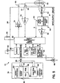
- FIG. 5 is a block diagram illustrating various components of an implantable pulse generator (IPG) 51 which is programmable by means of an external programming unit (not shown).
- IPG implantable pulse generator
- One such programmer suitable for the purposes of the present invention is the commercially available Medtronic Model 9790 programmer.
- the programmer is a microprocessor device which provides a series of encoded signals to IPG 51 by means of a programming head which transmits radio frequency encoded signals to IPG 51 according to a telemetry system, such as that described in U.S. Pat. No. 5,312,453 (Wyborny et al.), for example.
- IPG 51 is electrically coupled to the patient's heart 56 by lead 54.
- Lead 54 which includes two conductors, is coupled to a node 62 in the circuitry of IPG 51 through input capacitor 60.
- an activity sensor 63 provides a sensor output to a processing/amplifying activity circuit 65 of input/output circuit 68.
- Input/output circuit 68 also contains circuits for interfacing with heart 56, antenna 66, and circuit 74 for application of stimulating pulses to heart 56 to moderate its rate under control of software-implemented algorithms in microcomputer unit 78.
- Microcomputer unit 78 comprises on-board circuit 80 which includes system clock 82, microprocessor 83, and on-board RAM 84 and ROM 86.
- off-board circuit 88 comprises a RAM/ROM unit.
- On-board circuit 80 and off-board circuit 88 are each coupled by a data communication bus 90 to digital controller/timer circuit 92.
- the electrical components shown in Figure 5 are powered by an appropriate implantable battery power source 94 in accordance with common practice in the art. For purposes of clarity, the coupling of battery power to the various components of IPG 51 is not shown in the figures.
- Antenna 66 is connected to input/output circuit 68 to permit uplink/downlink telemetry through RF transmitter and receiver unit 55.
- Unit 55 may correspond to the telemetry and program logic disclosed in U.S. Pat. No. 4,556,063 (Thompson et al.), or to that disclosed in the above-referenced Wyborny et al. patent.
- Voltage reference (VREF) and bias circuit 61 generates a stable voltage reference and bias current for the analog circuits of input/output circuit 68.
- Analog-to-digital converter (ADC) and multiplexer unit 58 digitizes analog signals and voltages to provide "real-time" telemetry intracardiac signals and battery end-of-life (EOL) replacement functions.
- ADC analog-to-digital converter
- EOL battery end-of-life
- Operating commands for controlling the timing of IPG 51 are coupled by data bus 90 to digital controller/timer circuit 92, where digital timers and counters establish the overall escape interval of the IPG as well as various refractory, blinking, and other timing windows for controlling the operation of the peripheral components disposed within input/output circuit 68.
- Digital controller/timer circuit 92 is preferably coupled to sensing circuitry 52, including sense amplifier 53, peak sense and threshold measurement unit 57, and comparator/threshold detector 59.
- Sense amplifier 53 amplifies sensed electrocardiac signals and provides an amplified signal to peak sense and threshold measurement circuitry 57. Circuitry 57, in turn, provides an indication of peak sensed voltages and measured sense amplifier threshold voltages on path 64 to digital controller/timer circuit 92. An amplified sense amplifier signal is then provided to comparator/threshold detector 59.
- Sense amplifier 53 may correspond to that disclosed in U.S. Pat. No. 4,379,459 (Stein).
- Circuit 92 is further preferably coupled to electrogram (EGM) amplifier 76 for receiving amplified and processed signals sensed by an electrode disposed on lead 54.
- EGM electrogram
- the electrogram signal provided by EGM amplifier 76 is employed when the implanted device is being interrogated by an external programmer (not shown) to transmit by uplink telemetry a representation of an analog electrogram of the patient's electrical heart activity.
- EGM electrogram
- Such functionality is, for example, shown in previously referenced U.S. Pat. No. 4,556,063.
- Output pulse generator 74 provides pacing stimuli to the patient's heart 56 through coupling capacitor 65 in response to a pacing trigger signal provided by digital controller/timer circuit 92. For example, each time the escape interval times out, an externally transmitted pacing command is received, or such commands are received in response to other stored commands as is well known in pacing art.
- Output amplifier 74 may correspond generally to the output amplifier disclosed in U.S. Pat. No. 4,476,868 (Thompson).
- FIG. 5 is an illustration of an exemplary type of device (pacemaker) which may find application in the present invention, or which can be modified for use in the present invention by one of skill in the art, and is not intended to limit the scope of the present invention.
- alternative systems to the present invention may include a sensing element for monitoring at least one physiological property to detect a change in a physiological condition (typically, the onset of ischemia caused by a decrease in blood flow due to an occlusion resulting from the rupture of unstable plaque).
- a pseudo-surface electrocardiogram (ECG) using a subcutaneous electrode array can be used to detect a reduction in blood flow, which is represented by an abnormal morphology (e.g., inverted shape) of a T wave (i.e., the portion of an ECG pattern due to ventricular repolarization or recovery).
- ECG electrocardiogram
- an implantable pulse generator having three electrodes, each only about one centimeter apart, could be implanted into the pectoralis muscle in the chest of a patient.
- An ECG pattern similar to that of a normal ECG, would be monitored for an indication of an abnormal morphology of a T wave.
- Sensing elements can include one or more individual sensors for monitoring one or more physiological properties.
- such sensors include, for example, blood gas (e.g., CO2) sensors, pH sensors, blood flow sensors, and the like.
- Other mechanisms of detection that can be used in sensors include, for example, acoustic time of flight changes as a result of flow, acoustic doppler, which takes advantage of the doppler effect (received frequency is different that the transmitted one), thermal dilution (a clinical technique to measure blood flow and cardiac output), and venous pressure drop due to lack of driving pressure from the blocked artery. Examples of sensors or implantable monitoring devices that can be modified to be used in the stimulation devices of the present invention are disclosed, for example, in U.S.
- a telemetric communication system For electrical stimulation, because the direct wiring from the stimulation device to the delivery device is not feasible, a telemetric communication system can be used. Such systems are known and can be modified to be used in the present invention. Examples of such systems are discussed above and described in U.S. Pat. No. 5,312,453 (Wyborny et al.), for example.
- the stimulation device 22 will produce a radio frequency (RF) signal 26 using lead 24 as a transmitting antenna, which is implanted into the right ventricle of a patient's heart.
- RF radio frequency
- the lead 24 includes a coil, which transmits an H-field coupled pulse series (e.g., a 175 kilocycle, pulse position modulated, H-field signal) to the coil formed by stent 18, which is implanted into the left ventricle of a patient's heart.
- an H-field coupled pulse series e.g., a 175 kilocycle, pulse position modulated, H-field signal
- the stent For a conversion of the magnetic field (H-field) into an electrical signal, the stent includes a metal coil with all but the ends coated with an insulating coating.
- a tuned LC filter integral with the stent 18 will receive some of this signal, rectify it, and when a sufficient charge is transferred from the stimulation device to the stent, energy will be used to electrically stimulate the engineered endothelial cells in the coronary artery to trigger the release of the therapeutic agent.
- Inset Figure 2A is a representation of a circuit within the stent.
- This circuit includes an LC filter tuned to the specific frequency of the RF signal 26 for induction of electric current in the coiled stent 18 (or in a coil provided in the stent).
- a charge is stored in capacitor (Cstorage) until a threshold voltage is reached, which activates a switch to close the circuit and cause current to flow through the cells associated with the stent.
- the current density produced is preferably sufficient to stimulate the cells to release a therapeutic agent without significant damage to the cells or the therapeutic agents. Based on in vitro experiments, the current density for preferred embodiments is maintained below about 540 m-Amp/mm2 and the current threshold below about 0.9 mA/mm2 to avoid damage to the endothelial cells or the expressed product.
- the delivery device that acts as a carrier for the drug-eluting cells can be powered with RF coupling. Alternatively, it can be self powered or battery powered.
- Electrode and mechanical hardware were modified to enable the stimulation of endothelial cells cultured on inserts placed in tissue culture wells.
- the cells were stimulated with single pulses (1 milli-second long and delivered at a rate of 1 Hz) for 5 minutes a day, on 3 consecutive days. At the end of the 3 days, cells were removed from the culture wells, stained with crystal violet and photographed. Increases in the current density caused a decrease in the density of red stained endothelial cells. While a stimulation pattern that produced 540 m-Amp/mm2 of current density appears to be detrimental to the endothelial cells, another pattern which produced 270 m-Amp/mm2 of current density did not.
- Human coronary artery endothelial cells (Clonetics, San Diego, CA) were seeded in 24 well tissue culture plates at 1x10⁴ cells/cm2 (1.5 mL total volume) and grown in defined media as supplied by Clonetics. The cells were incubated at 37°C, 5% CO2 for up to four days. The media was not changed on the cells during the course of the experiment. Supernatants from duplicate wells were withdrawn on days 2, 3, and 4 post-seeding. In addition, cells plus supernatants from a second set of samples were harvested from duplicate wells on days 2, 3, and 4 post-seeding. Samples were stored at -85°C until assayed.
- the ELISA assay was performed on the test samples and on controls following the procedure outlined by the manufacturer (American Diagnostica, Inc.) The standard curve was generated using control plasma A, t-PA antigen control plasma set 2 (Product #275, American Diagnostica, Inc.). The test samples were assayed undiluted (20 uL assay volume). The results suggest that measurable t-PA was released from control endothelial cells, in the absence of stimulation. The levels of t-PA that were released were correlated to cell growth (more was measured after 4 days of culture than after 2 days).
- This experiment was performed similarly to the first experiment with the following changes.
- cells were seeded at three different densities (1x10⁴, 2x10⁴, 4x10⁴) in 0.5 mL total volume per well, and the cell supernatants were collected on day 2 and day 3 post seeding.
- cells were seeded at 2x10⁴ cells/cm2 and one group was stimulated once (on day 2 post seeding) and a second group was stimulated two times (once on day 2 and once on day 3 post seeding).
- the stimulation level was 55V (350-440 mA) for 5 minutes and supernatants were collected 15 minutes after stimulation.
- the following tables contain the data that was obtained from this experiment.
Landscapes
- Health & Medical Sciences (AREA)
- Life Sciences & Earth Sciences (AREA)
- Engineering & Computer Science (AREA)
- Biomedical Technology (AREA)
- Chemical & Material Sciences (AREA)
- General Health & Medical Sciences (AREA)
- Animal Behavior & Ethology (AREA)
- Public Health (AREA)
- Veterinary Medicine (AREA)
- Epidemiology (AREA)
- Medicinal Chemistry (AREA)
- Vascular Medicine (AREA)
- Heart & Thoracic Surgery (AREA)
- Transplantation (AREA)
- Oral & Maxillofacial Surgery (AREA)
- Cardiology (AREA)
- Cell Biology (AREA)
- Chemical Kinetics & Catalysis (AREA)
- Zoology (AREA)
- Bioinformatics & Cheminformatics (AREA)
- Dermatology (AREA)
- Botany (AREA)
- Gastroenterology & Hepatology (AREA)
- Surgery (AREA)
- Molecular Biology (AREA)
- Genetics & Genomics (AREA)
- Organic Chemistry (AREA)
- Proteomics, Peptides & Aminoacids (AREA)
- Biotechnology (AREA)
- Pharmacology & Pharmacy (AREA)
- Immunology (AREA)
- Urology & Nephrology (AREA)
- Wood Science & Technology (AREA)
- Biophysics (AREA)
- General Engineering & Computer Science (AREA)
- Biochemistry (AREA)
- Hematology (AREA)
- Microbiology (AREA)
- Nuclear Medicine, Radiotherapy & Molecular Imaging (AREA)
- Physiology (AREA)
Applications Claiming Priority (3)
| Application Number | Priority Date | Filing Date | Title |
|---|---|---|---|
| US144420 | 1998-08-31 | ||
| US09/144,420 US6206914B1 (en) | 1998-04-30 | 1998-08-31 | Implantable system with drug-eluting cells for on-demand local drug delivery |
| EP99942261A EP1027014B1 (de) | 1998-08-31 | 1999-08-23 | Implantierbares system mit medikamentenabgebenden zellen zur lokalen verabreichung von medikamenten bei bedarf |
Related Parent Applications (1)
| Application Number | Title | Priority Date | Filing Date |
|---|---|---|---|
| EP99942261A Division EP1027014B1 (de) | 1998-08-31 | 1999-08-23 | Implantierbares system mit medikamentenabgebenden zellen zur lokalen verabreichung von medikamenten bei bedarf |
Publications (2)
| Publication Number | Publication Date |
|---|---|
| EP1426021A1 true EP1426021A1 (de) | 2004-06-09 |
| EP1426021B1 EP1426021B1 (de) | 2007-07-18 |
Family
ID=22508514
Family Applications (2)
| Application Number | Title | Priority Date | Filing Date |
|---|---|---|---|
| EP99942261A Expired - Lifetime EP1027014B1 (de) | 1998-08-31 | 1999-08-23 | Implantierbares system mit medikamentenabgebenden zellen zur lokalen verabreichung von medikamenten bei bedarf |
| EP04006105A Expired - Lifetime EP1426021B1 (de) | 1998-08-31 | 1999-08-23 | Implantierbares System mit medikamentenabgebenden Zellen zur lokalen Verabreichung von Medikamenten bei Bedarf |
Family Applications Before (1)
| Application Number | Title | Priority Date | Filing Date |
|---|---|---|---|
| EP99942261A Expired - Lifetime EP1027014B1 (de) | 1998-08-31 | 1999-08-23 | Implantierbares system mit medikamentenabgebenden zellen zur lokalen verabreichung von medikamenten bei bedarf |
Country Status (4)
| Country | Link |
|---|---|
| US (3) | US6206914B1 (de) |
| EP (2) | EP1027014B1 (de) |
| DE (2) | DE69936599T2 (de) |
| WO (1) | WO2000012028A1 (de) |
Cited By (1)
| Publication number | Priority date | Publication date | Assignee | Title |
|---|---|---|---|---|
| US8123716B1 (en) | 2007-06-26 | 2012-02-28 | Pacesetter, Inc. | Pericardial delivery of treatment |
Families Citing this family (375)
| Publication number | Priority date | Publication date | Assignee | Title |
|---|---|---|---|---|
| US5981568A (en) | 1993-01-28 | 1999-11-09 | Neorx Corporation | Therapeutic inhibitor of vascular smooth muscle cells |
| US6861571B1 (en) * | 1994-11-28 | 2005-03-01 | The Procter & Gamble Company | Article having a lotioned topsheet |
| US20030036746A1 (en) | 2001-08-16 | 2003-02-20 | Avi Penner | Devices for intrabody delivery of molecules and systems and methods utilizing same |
| US20040127977A1 (en) * | 2002-09-20 | 2004-07-01 | Conor Medsystems, Inc. | Expandable medical device with openings for delivery of multiple beneficial agents |
| US6241762B1 (en) | 1998-03-30 | 2001-06-05 | Conor Medsystems, Inc. | Expandable medical device with ductile hinges |
| US7208011B2 (en) * | 2001-08-20 | 2007-04-24 | Conor Medsystems, Inc. | Implantable medical device with drug filled holes |
| US7208010B2 (en) * | 2000-10-16 | 2007-04-24 | Conor Medsystems, Inc. | Expandable medical device for delivery of beneficial agent |
| US6206914B1 (en) * | 1998-04-30 | 2001-03-27 | Medtronic, Inc. | Implantable system with drug-eluting cells for on-demand local drug delivery |
| US6692696B1 (en) * | 1998-06-18 | 2004-02-17 | ARETé ASSOCIATES | Biosensor |
| US6290728B1 (en) * | 1998-09-10 | 2001-09-18 | Percardia, Inc. | Designs for left ventricular conduit |
| US6293967B1 (en) | 1998-10-29 | 2001-09-25 | Conor Medsystems, Inc. | Expandable medical device with ductile hinges |
| GB2344053A (en) | 1998-11-30 | 2000-05-31 | Imperial College | Stents for blood vessels |
| US6371904B1 (en) * | 1998-12-24 | 2002-04-16 | Vivant Medical, Inc. | Subcutaneous cavity marking device and method |
| US6356782B1 (en) | 1998-12-24 | 2002-03-12 | Vivant Medical, Inc. | Subcutaneous cavity marking device and method |
| US9669113B1 (en) | 1998-12-24 | 2017-06-06 | Devicor Medical Products, Inc. | Device and method for safe location and marking of a biopsy cavity |
| EP2359831A3 (de) | 1999-04-30 | 2012-02-01 | The Regents of the University of Michigan | Therapeutische Anwendungen proapoptotischer Benzodiazepine |
| US6348050B1 (en) | 1999-04-30 | 2002-02-19 | Medtronic, Inc. | Infusion systems for creating microenvironments in a living body |
| WO2000066204A1 (en) * | 1999-04-30 | 2000-11-09 | University Of Southern California | Implantable microbolus infusion pump |
| US20040176358A1 (en) * | 1999-04-30 | 2004-09-09 | The Regents Of The University Of Michigan | Compositions and methods relating to novel compounds and targets thereof |
| US7144880B2 (en) * | 1999-04-30 | 2006-12-05 | Regents Of The University Of Michigan | Compositions relating to novel compounds and targets thereof |
| US20050113460A1 (en) * | 1999-04-30 | 2005-05-26 | The Regents Of The University Of Michigan | Compositions and methods relating to novel compounds and targets thereof |
| US20060025388A1 (en) | 1999-04-30 | 2006-02-02 | Glick Gary D | Compositions and methods relating to novel compounds and targets thereof |
| US6290673B1 (en) * | 1999-05-20 | 2001-09-18 | Conor Medsystems, Inc. | Expandable medical device delivery system and method |
| US6719805B1 (en) * | 1999-06-09 | 2004-04-13 | C. R. Bard, Inc. | Devices and methods for treating tissue |
| US6716242B1 (en) * | 1999-10-13 | 2004-04-06 | Peter A. Altman | Pulmonary vein stent and method for use |
| US6738661B1 (en) * | 1999-10-22 | 2004-05-18 | Biosynergetics, Inc. | Apparatus and methods for the controllable modification of compound concentration in a tube |
| US6503273B1 (en) * | 1999-11-22 | 2003-01-07 | Cyograft Tissue Engineering, Inc. | Tissue engineered blood vessels and methods and apparatus for their manufacture |
| US6379382B1 (en) * | 2000-03-13 | 2002-04-30 | Jun Yang | Stent having cover with drug delivery capability |
| US20070141107A1 (en) * | 2000-03-15 | 2007-06-21 | Orbusneich Medical, Inc. | Progenitor Endothelial Cell Capturing with a Drug Eluting Implantable Medical Device |
| US20030229393A1 (en) * | 2001-03-15 | 2003-12-11 | Kutryk Michael J. B. | Medical device with coating that promotes cell adherence and differentiation |
| US8088060B2 (en) | 2000-03-15 | 2012-01-03 | Orbusneich Medical, Inc. | Progenitor endothelial cell capturing with a drug eluting implantable medical device |
| US9522217B2 (en) | 2000-03-15 | 2016-12-20 | Orbusneich Medical, Inc. | Medical device with coating for capturing genetically-altered cells and methods for using same |
| ATE362382T1 (de) | 2000-03-15 | 2007-06-15 | Orbusneich Medical Inc | Beschichtung welche ein anhaften von endothelzellen stimuliert |
| US20070055367A1 (en) * | 2000-03-15 | 2007-03-08 | Orbus Medical Technologies, Inc. | Medical device with coating that promotes endothelial cell adherence and differentiation |
| US8460367B2 (en) * | 2000-03-15 | 2013-06-11 | Orbusneich Medical, Inc. | Progenitor endothelial cell capturing with a drug eluting implantable medical device |
| US6514195B1 (en) * | 2000-04-28 | 2003-02-04 | Medtronic, Inc. | Ischemic heart disease detection |
| US6658288B1 (en) * | 2000-05-05 | 2003-12-02 | Endovascular Technologies, Inc. | Apparatus and method for aiding thrombosis through the application of electric potential |
| US8252044B1 (en) | 2000-11-17 | 2012-08-28 | Advanced Bio Prosthestic Surfaces, Ltd. | Device for in vivo delivery of bioactive agents and method of manufacture thereof |
| AU2001280060A1 (en) | 2000-08-08 | 2002-02-18 | M.G.V.S. Ltd. | Nucleic acid constructs, vascular cells transformed therewith, pharmaceutical compositions and methods utilizing same for inducing angiogenesis |
| US7343335B1 (en) * | 2000-08-08 | 2008-03-11 | Ebay Inc. | Method for managing group finances via an electronic network |
| US20080167699A1 (en) * | 2000-09-27 | 2008-07-10 | Cvrx, Inc. | Method and Apparatus for Providing Complex Tissue Stimulation Parameters |
| US7499742B2 (en) * | 2001-09-26 | 2009-03-03 | Cvrx, Inc. | Electrode structures and methods for their use in cardiovascular reflex control |
| US8086314B1 (en) * | 2000-09-27 | 2011-12-27 | Cvrx, Inc. | Devices and methods for cardiovascular reflex control |
| US20070185542A1 (en) * | 2002-03-27 | 2007-08-09 | Cvrx, Inc. | Baroreflex therapy for disordered breathing |
| US7616997B2 (en) * | 2000-09-27 | 2009-11-10 | Kieval Robert S | Devices and methods for cardiovascular reflex control via coupled electrodes |
| US7840271B2 (en) * | 2000-09-27 | 2010-11-23 | Cvrx, Inc. | Stimulus regimens for cardiovascular reflex control |
| US7623926B2 (en) * | 2000-09-27 | 2009-11-24 | Cvrx, Inc. | Stimulus regimens for cardiovascular reflex control |
| US20080177365A1 (en) * | 2000-09-27 | 2008-07-24 | Cvrx, Inc. | Method and apparatus for electronically switching electrode configuration |
| US7158832B2 (en) * | 2000-09-27 | 2007-01-02 | Cvrx, Inc. | Electrode designs and methods of use for cardiovascular reflex control devices |
| US6764507B2 (en) | 2000-10-16 | 2004-07-20 | Conor Medsystems, Inc. | Expandable medical device with improved spatial distribution |
| ES2243556T3 (es) | 2000-10-16 | 2005-12-01 | Conor Medsystems, Inc. | Dispositivo medico expandible para proporcionar un agente beneficioso. |
| US7024248B2 (en) * | 2000-10-16 | 2006-04-04 | Remon Medical Technologies Ltd | Systems and methods for communicating with implantable devices |
| US6748954B2 (en) * | 2000-10-27 | 2004-06-15 | The Regents Of The University Of Michigan | Drug release from polymer matrices through mechanical stimulation |
| US7369890B2 (en) * | 2000-11-02 | 2008-05-06 | Cardiac Pacemakers, Inc. | Technique for discriminating between coordinated and uncoordinated cardiac rhythms |
| US8372139B2 (en) * | 2001-02-14 | 2013-02-12 | Advanced Bio Prosthetic Surfaces, Ltd. | In vivo sensor and method of making same |
| US10398830B2 (en) | 2000-11-17 | 2019-09-03 | Vactronix Scientific, Llc | Device for in vivo delivery of bioactive agents and method of manufacture thereof |
| US9107605B2 (en) * | 2000-11-17 | 2015-08-18 | Advanced Bio Prosthetic Surfaces, Ltd., A Wholly Owned Subsidiary Of Palmaz Scientific, Inc. | Device for in vivo delivery of bioactive agents and method of manufacture thereof |
| US6812217B2 (en) * | 2000-12-04 | 2004-11-02 | Medtronic, Inc. | Medical device and methods of use |
| US6689117B2 (en) * | 2000-12-18 | 2004-02-10 | Cardiac Pacemakers, Inc. | Drug delivery system for implantable medical device |
| US20020082620A1 (en) * | 2000-12-27 | 2002-06-27 | Elaine Lee | Bioactive materials for aneurysm repair |
| DE10103503A1 (de) * | 2001-01-26 | 2002-08-14 | Fraunhofer Ges Forschung | Endoluminales expandierbares Implantat mit integrierter Sensorik |
| US6964680B2 (en) * | 2001-02-05 | 2005-11-15 | Conor Medsystems, Inc. | Expandable medical device with tapered hinge |
| US20040073294A1 (en) | 2002-09-20 | 2004-04-15 | Conor Medsystems, Inc. | Method and apparatus for loading a beneficial agent into an expandable medical device |
| US20040204756A1 (en) * | 2004-02-11 | 2004-10-14 | Diaz Stephen Hunter | Absorbent article with improved liquid acquisition capacity |
| US20060094112A1 (en) * | 2001-03-07 | 2006-05-04 | Omotunde Babalola | Biological scaffold |
| US6673104B2 (en) * | 2001-03-15 | 2004-01-06 | Scimed Life Systems, Inc. | Magnetic stent |
| US6660034B1 (en) * | 2001-04-30 | 2003-12-09 | Advanced Cardiovascular Systems, Inc. | Stent for increasing blood flow to ischemic tissues and a method of using the same |
| US7209783B2 (en) * | 2001-06-15 | 2007-04-24 | Cardiac Pacemakers, Inc. | Ablation stent for treating atrial fibrillation |
| US7493162B2 (en) * | 2001-06-15 | 2009-02-17 | Cardiac Pacemakers, Inc. | Pulmonary vein stent for treating atrial fibrillation |
| US6702744B2 (en) * | 2001-06-20 | 2004-03-09 | Advanced Cardiovascular Systems, Inc. | Agents that stimulate therapeutic angiogenesis and techniques and devices that enable their delivery |
| US7727221B2 (en) * | 2001-06-27 | 2010-06-01 | Cardiac Pacemakers Inc. | Method and device for electrochemical formation of therapeutic species in vivo |
| WO2003003948A1 (en) * | 2001-07-06 | 2003-01-16 | Tricardia, L.L.C. | Anti-arrhythmia devices and methods of use |
| US7481759B2 (en) * | 2001-08-03 | 2009-01-27 | Cardiac Pacemakers, Inc. | Systems and methods for treatment of coronary artery disease |
| US8934968B2 (en) * | 2001-08-03 | 2015-01-13 | Cardiac Pacemakers, Inc. | Neurostimulation and coronary artery disease treatment |
| US7056338B2 (en) * | 2003-03-28 | 2006-06-06 | Conor Medsystems, Inc. | Therapeutic agent delivery device with controlled therapeutic agent release rates |
| US20040249443A1 (en) * | 2001-08-20 | 2004-12-09 | Shanley John F. | Expandable medical device for treating cardiac arrhythmias |
| US7842083B2 (en) * | 2001-08-20 | 2010-11-30 | Innovational Holdings, Llc. | Expandable medical device with improved spatial distribution |
| US6663615B1 (en) | 2001-09-04 | 2003-12-16 | The Ohio State University | Dual stage microvalve and method of use |
| US6790455B2 (en) * | 2001-09-14 | 2004-09-14 | The Research Foundation At State University Of New York | Cell delivery system comprising a fibrous matrix and cells |
| US8573263B2 (en) | 2001-09-24 | 2013-11-05 | Cosmetic Technologies, Llc | Apparatus and method for custom cosmetic dispensing |
| CA2461307C (en) | 2001-09-24 | 2011-01-11 | Imx Labs, Inc. | Apparatus and method for custom cosmetic dispensing |
| US7340303B2 (en) * | 2001-09-25 | 2008-03-04 | Cardiac Pacemakers, Inc. | Evoked response sensing for ischemia detection |
| US6723077B2 (en) | 2001-09-28 | 2004-04-20 | Hewlett-Packard Development Company, L.P. | Cutaneous administration system |
| US7308303B2 (en) | 2001-11-01 | 2007-12-11 | Advanced Bionics Corporation | Thrombolysis and chronic anticoagulation therapy |
| JP4458240B2 (ja) * | 2001-11-26 | 2010-04-28 | ジェネンテック インコーポレイテッド | カテーテル組成物とその用途 |
| US8608661B1 (en) | 2001-11-30 | 2013-12-17 | Advanced Cardiovascular Systems, Inc. | Method for intravascular delivery of a treatment agent beyond a blood vessel wall |
| US7585670B2 (en) | 2001-12-07 | 2009-09-08 | Cytori Therapeutics, Inc. | Automated methods for isolating and using clinically safe adipose derived regenerative cells |
| US7651684B2 (en) | 2001-12-07 | 2010-01-26 | Cytori Therapeutics, Inc. | Methods of using adipose tissue-derived cells in augmenting autologous fat transfer |
| US20060204556A1 (en) * | 2001-12-07 | 2006-09-14 | Cytori Therapeutics, Inc. | Cell-loaded prostheses for regenerative intraluminal applications |
| US20050008626A1 (en) * | 2001-12-07 | 2005-01-13 | Fraser John K. | Methods of using adipose tissue-derived cells in the treatment of cardiovascular conditions |
| US7514075B2 (en) * | 2001-12-07 | 2009-04-07 | Cytori Therapeutics, Inc. | Systems and methods for separating and concentrating adipose derived stem cells from tissue |
| US9597395B2 (en) | 2001-12-07 | 2017-03-21 | Cytori Therapeutics, Inc. | Methods of using adipose tissue-derived cells in the treatment of cardiovascular conditions |
| US20050095228A1 (en) | 2001-12-07 | 2005-05-05 | Fraser John K. | Methods of using regenerative cells in the treatment of peripheral vascular disease and related disorders |
| US7595043B2 (en) * | 2001-12-07 | 2009-09-29 | Cytori Therapeutics, Inc. | Method for processing and using adipose-derived stem cells |
| US8404229B2 (en) * | 2001-12-07 | 2013-03-26 | Cytori Therapeutics, Inc. | Methods of using adipose derived stem cells to treat acute tubular necrosis |
| US20050048036A1 (en) * | 2001-12-07 | 2005-03-03 | Hedrick Marc H. | Methods of using regenerative cells in the treatment of inherited and acquired disorders of the bone, bone marrow, liver, and other tissues |
| US7771716B2 (en) * | 2001-12-07 | 2010-08-10 | Cytori Therapeutics, Inc. | Methods of using regenerative cells in the treatment of musculoskeletal disorders |
| JP4653952B2 (ja) | 2001-12-07 | 2011-03-16 | サイトリ セラピューティクス インコーポレイテッド | 処理済み脂肪吸引細胞で患者を治療するためのシステムと方法 |
| US20050048035A1 (en) | 2001-12-07 | 2005-03-03 | Fraser John K. | Methods of using regenerative cells in the treatment of stroke and related diseases and disorders |
| US7504258B2 (en) | 2001-12-11 | 2009-03-17 | Cytograft Tissue Engineering, Inc. | Tissue engineered cellular sheets, methods of making and use thereof |
| US7008397B2 (en) * | 2002-02-13 | 2006-03-07 | Percardia, Inc. | Cardiac implant and methods |
| US7236821B2 (en) * | 2002-02-19 | 2007-06-26 | Cardiac Pacemakers, Inc. | Chronically-implanted device for sensing and therapy |
| US20030176797A1 (en) * | 2002-03-12 | 2003-09-18 | Fernando Anzellini | Thrombust; implantable delivery system sensible to self diagnosis of acute myocardial infarction for thrombolysis in the first minutes of chest pain |
| US20030181973A1 (en) * | 2002-03-20 | 2003-09-25 | Harvinder Sahota | Reduced restenosis drug containing stents |
| US7008979B2 (en) | 2002-04-30 | 2006-03-07 | Hydromer, Inc. | Coating composition for multiple hydrophilic applications |
| WO2003092791A2 (en) * | 2002-05-02 | 2003-11-13 | Scimed Life Systems, Inc. | Energetically-controlled delivery of biologically active material from an implanted medical device |
| DE10393059D2 (de) * | 2002-05-09 | 2005-05-04 | Hemoteq Gmbh | Verbindungen und Verfahren zur hemokompatiblen Beschichtung von Oberflächen |
| US7261733B1 (en) | 2002-06-07 | 2007-08-28 | Endovascular Technologies, Inc. | Endovascular graft with sensors design and attachment methods |
| US7089055B2 (en) | 2002-06-28 | 2006-08-08 | Cardiac Pacemakers, Inc. | Method and apparatus for delivering pre-shock defibrillation therapy |
| US7361368B2 (en) | 2002-06-28 | 2008-04-22 | Advanced Cardiovascular Systems, Inc. | Device and method for combining a treatment agent and a gel |
| US7588564B2 (en) * | 2002-07-03 | 2009-09-15 | Pharmaco Kinesis Corporation | Apparatus for piezoelectric layer-wise pump and valve for use in local administration of biological response modifiers and therapeutic agents |
| US7400931B2 (en) * | 2002-09-18 | 2008-07-15 | Cardiac Pacemakers, Inc. | Devices and methods to stimulate therapeutic angiogenesis for ischemia and heart failure |
| US20040127976A1 (en) * | 2002-09-20 | 2004-07-01 | Conor Medsystems, Inc. | Method and apparatus for loading a beneficial agent into an expandable medical device |
| US8303511B2 (en) * | 2002-09-26 | 2012-11-06 | Pacesetter, Inc. | Implantable pressure transducer system optimized for reduced thrombosis effect |
| AU2003287633A1 (en) * | 2002-11-08 | 2004-06-03 | Innovational Holdings, Llc | Method and apparatus for reducing tissue damage after ischemic injury |
| US20040142014A1 (en) * | 2002-11-08 | 2004-07-22 | Conor Medsystems, Inc. | Method and apparatus for reducing tissue damage after ischemic injury |
| WO2004043509A1 (en) * | 2002-11-08 | 2004-05-27 | Conor Medsystems, Inc. | Expandable medical device and method for treating chronic total occlusions with local delivery of an angiogenic factor |
| US7072711B2 (en) | 2002-11-12 | 2006-07-04 | Cardiac Pacemakers, Inc. | Implantable device for delivering cardiac drug therapy |
| US20040158289A1 (en) * | 2002-11-30 | 2004-08-12 | Girouard Steven D. | Method and apparatus for cell and electrical therapy of living tissue |
| US7627373B2 (en) * | 2002-11-30 | 2009-12-01 | Cardiac Pacemakers, Inc. | Method and apparatus for cell and electrical therapy of living tissue |
| US7189204B2 (en) * | 2002-12-04 | 2007-03-13 | Cardiac Pacemakers, Inc. | Sleep detection using an adjustable threshold |
| US7452334B2 (en) * | 2002-12-16 | 2008-11-18 | The Regents Of The University Of Michigan | Antenna stent device for wireless, intraluminal monitoring |
| EP1587450A2 (de) * | 2002-12-16 | 2005-10-26 | The Regents Of The University Of Michigan | Anordnung und planare konstruktion zur verwendung darin, die zu einer 3d-struktur aufweitbar ist, z.b. ein stent, und vorrichtung zur herstellung der planaren konstruktion |
| US7311727B2 (en) * | 2003-02-05 | 2007-12-25 | Board Of Trustees Of The University Of Arkansas | Encased stent |
| US7172624B2 (en) * | 2003-02-06 | 2007-02-06 | Boston Scientific Scimed, Inc. | Medical device with magnetic resonance visibility enhancing structure |
| WO2004075855A2 (en) | 2003-02-26 | 2004-09-10 | Biomed Solutions, Llc | Process for in vivo treatment of specific biological targets in bodily fluid |
| WO2004078065A2 (en) * | 2003-03-03 | 2004-09-16 | Sinus Rhythm Technologies, Inc. | Electrical conduction block implant device |
| WO2004078066A2 (en) * | 2003-03-03 | 2004-09-16 | Sinus Rhythm Technologies, Inc. | Primary examiner |
| US7318836B2 (en) * | 2003-03-11 | 2008-01-15 | Boston Scientific Scimed, Inc. | Covered stent |
| US20050010170A1 (en) * | 2004-02-11 | 2005-01-13 | Shanley John F | Implantable medical device with beneficial agent concentration gradient |
| US20040202692A1 (en) * | 2003-03-28 | 2004-10-14 | Conor Medsystems, Inc. | Implantable medical device and method for in situ selective modulation of agent delivery |
| AU2004226327A1 (en) * | 2003-03-28 | 2004-10-14 | Innovational Holdings, Llc | Implantable medical device with beneficial agent concentration gradient |
| US8383158B2 (en) * | 2003-04-15 | 2013-02-26 | Abbott Cardiovascular Systems Inc. | Methods and compositions to treat myocardial conditions |
| US8038991B1 (en) | 2003-04-15 | 2011-10-18 | Abbott Cardiovascular Systems Inc. | High-viscosity hyaluronic acid compositions to treat myocardial conditions |
| US8821473B2 (en) | 2003-04-15 | 2014-09-02 | Abbott Cardiovascular Systems Inc. | Methods and compositions to treat myocardial conditions |
| US7473548B2 (en) | 2003-04-25 | 2009-01-06 | Medtronic, Inc. | Optical detector for enzyme activation |
| WO2004098511A2 (en) * | 2003-05-01 | 2004-11-18 | Sinus Rhythm Technologies, Inc. | Methods and devices for creating electrical block at specific targeted sites in cardia tissue |
| US20040236416A1 (en) * | 2003-05-20 | 2004-11-25 | Robert Falotico | Increased biocompatibility of implantable medical devices |
| US20040236410A1 (en) * | 2003-05-22 | 2004-11-25 | Atrium Medical Corp. | Polymeric body formation |
| US20040236278A1 (en) * | 2003-05-22 | 2004-11-25 | Atrium Medical Corp. | Therapeutic agent delivery |
| US20040236414A1 (en) * | 2003-05-23 | 2004-11-25 | Brar Balbir S. | Devices and methods for treatment of stenotic regions |
| US7226473B2 (en) * | 2003-05-23 | 2007-06-05 | Brar Balbir S | Treatment of stenotic regions |
| US7169179B2 (en) * | 2003-06-05 | 2007-01-30 | Conor Medsystems, Inc. | Drug delivery device and method for bi-directional drug delivery |
| US8606356B2 (en) | 2003-09-18 | 2013-12-10 | Cardiac Pacemakers, Inc. | Autonomic arousal detection system and method |
| US7320675B2 (en) | 2003-08-21 | 2008-01-22 | Cardiac Pacemakers, Inc. | Method and apparatus for modulating cellular metabolism during post-ischemia or heart failure |
| US20050181018A1 (en) * | 2003-09-19 | 2005-08-18 | Peyman Gholam A. | Ocular drug delivery |
| US7371228B2 (en) * | 2003-09-19 | 2008-05-13 | Medtronic Vascular, Inc. | Delivery of therapeutics to treat aneurysms |
| US7785653B2 (en) * | 2003-09-22 | 2010-08-31 | Innovational Holdings Llc | Method and apparatus for loading a beneficial agent into an expandable medical device |
| EP1684666A4 (de) * | 2003-10-14 | 2010-04-07 | James C Peacock Iii | Aneurysma-behandlungssystem und verfahren |
| US7480532B2 (en) * | 2003-10-22 | 2009-01-20 | Cvrx, Inc. | Baroreflex activation for pain control, sedation and sleep |
| WO2005039689A2 (en) * | 2003-10-24 | 2005-05-06 | Sinus Rhythm Technologies, Inc. | Methods and devices for creating cardiac electrical blocks |
| FR2861997A1 (fr) * | 2003-11-06 | 2005-05-13 | Fred Zacouto | Stimulateur cardiaque orthorythmique inotrope |
| EP1689321B1 (de) | 2003-11-07 | 2017-01-04 | The University of Connecticut | Künstliche gewebesysteme und ihre verwendungen |
| US20050100577A1 (en) * | 2003-11-10 | 2005-05-12 | Parker Theodore L. | Expandable medical device with beneficial agent matrix formed by a multi solvent system |
| SE526861C2 (sv) | 2003-11-17 | 2005-11-15 | Syntach Ag | Vävnadslesionsskapande anordning samt en uppsättning av anordningar för behandling av störningar i hjärtrytmregleringssystemet |
| US20050137626A1 (en) * | 2003-12-19 | 2005-06-23 | Pastore Joseph M. | Drug delivery system and method employing external drug delivery device in conjunction with computer network |
| US20070027532A1 (en) * | 2003-12-22 | 2007-02-01 | Xingwu Wang | Medical device |
| JP4680939B2 (ja) * | 2004-02-10 | 2011-05-11 | シネコー・エルエルシー | 治療薬用脈管内送達システム |
| EP1713417A4 (de) * | 2004-02-12 | 2008-08-06 | Univ Akron | Verbesserter stent zur verwendung in arterien |
| US8137397B2 (en) * | 2004-02-26 | 2012-03-20 | Boston Scientific Scimed, Inc. | Medical devices |
| US7840263B2 (en) * | 2004-02-27 | 2010-11-23 | Cardiac Pacemakers, Inc. | Method and apparatus for device controlled gene expression |
| US9398967B2 (en) * | 2004-03-02 | 2016-07-26 | Syntach Ag | Electrical conduction block implant device |
| US8353896B2 (en) | 2004-04-19 | 2013-01-15 | The Invention Science Fund I, Llc | Controllable release nasal system |
| US8337482B2 (en) | 2004-04-19 | 2012-12-25 | The Invention Science Fund I, Llc | System for perfusion management |
| US7998060B2 (en) | 2004-04-19 | 2011-08-16 | The Invention Science Fund I, Llc | Lumen-traveling delivery device |
| US8092549B2 (en) | 2004-09-24 | 2012-01-10 | The Invention Science Fund I, Llc | Ciliated stent-like-system |
| US8512219B2 (en) | 2004-04-19 | 2013-08-20 | The Invention Science Fund I, Llc | Bioelectromagnetic interface system |
| US20050234545A1 (en) * | 2004-04-19 | 2005-10-20 | Yea-Yang Su | Amorphous oxide surface film for metallic implantable devices and method for production thereof |
| US9011329B2 (en) | 2004-04-19 | 2015-04-21 | Searete Llc | Lumenally-active device |
| US8019413B2 (en) | 2007-03-19 | 2011-09-13 | The Invention Science Fund I, Llc | Lumen-traveling biological interface device and method of use |
| US7850676B2 (en) | 2004-04-19 | 2010-12-14 | The Invention Science Fund I, Llc | System with a reservoir for perfusion management |
| US8361013B2 (en) | 2004-04-19 | 2013-01-29 | The Invention Science Fund I, Llc | Telescoping perfusion management system |
| US8000784B2 (en) | 2004-04-19 | 2011-08-16 | The Invention Science Fund I, Llc | Lumen-traveling device |
| US20050272723A1 (en) * | 2004-04-27 | 2005-12-08 | The Regents Of The University Of Michigan | Methods and compositions for treating diseases and conditions associated with mitochondrial function |
| EP2946666B1 (de) * | 2004-04-30 | 2017-11-15 | OrbusNeich Medical, Inc. | Medizinische vorrichtung mit beschichtung zur erfassung von genetisch veränderten zellen und anwendungsverfahren dafür |
| DE602004000646T2 (de) * | 2004-05-03 | 2007-05-16 | C.R.F. Società Consortile per Azioni, Orbassano | Vorrichtung für Elektrochemotherapie und entsprechende pharmazeutische Zusammensetzung |
| DE102004023526A1 (de) * | 2004-05-13 | 2005-12-08 | Osypka, Peter, Dr.-Ing. | Gefäßstütze |
| DE602005012950D1 (de) * | 2004-05-27 | 2009-04-09 | Medtronic Inc | Medizinprodukt mit einem biologischen wirkstoff |
| US7764995B2 (en) | 2004-06-07 | 2010-07-27 | Cardiac Pacemakers, Inc. | Method and apparatus to modulate cellular regeneration post myocardial infarct |
| US7596413B2 (en) * | 2004-06-08 | 2009-09-29 | Cardiac Pacemakers, Inc. | Coordinated therapy for disordered breathing including baroreflex modulation |
| US7747323B2 (en) | 2004-06-08 | 2010-06-29 | Cardiac Pacemakers, Inc. | Adaptive baroreflex stimulation therapy for disordered breathing |
| US20050287287A1 (en) * | 2004-06-24 | 2005-12-29 | Parker Theodore L | Methods and systems for loading an implantable medical device with beneficial agent |
| USD516723S1 (en) | 2004-07-06 | 2006-03-07 | Conor Medsystems, Inc. | Stent wall structure |
| US7167746B2 (en) | 2004-07-12 | 2007-01-23 | Ats Medical, Inc. | Anti-coagulation and demineralization system for conductive medical devices |
| WO2006020189A2 (en) * | 2004-07-19 | 2006-02-23 | Barthomolew Julie R | Customized retail point of sale dispensing methods |
| US7828711B2 (en) * | 2004-08-16 | 2010-11-09 | Cardiac Pacemakers, Inc. | Method and apparatus for modulating cellular growth and regeneration using ventricular assist device |
| US7567841B2 (en) * | 2004-08-20 | 2009-07-28 | Cardiac Pacemakers, Inc. | Method and apparatus for delivering combined electrical and drug therapies |
| US7621906B2 (en) * | 2004-08-25 | 2009-11-24 | Cardiac Pacemakers, Inc. | Method and apparatus to deliver drug and pacing therapy for treatment of cardiac disorders |
| US20060052369A1 (en) * | 2004-09-07 | 2006-03-09 | The Regents Of The University Of Michigan | Compositions and methods relating to novel compounds and targets thereof |
| US8271093B2 (en) | 2004-09-17 | 2012-09-18 | Cardiac Pacemakers, Inc. | Systems and methods for deriving relative physiologic measurements using a backend computing system |
| CN101035481A (zh) * | 2004-10-08 | 2007-09-12 | 赛恩泰克公司 | 用于治疗心房颤动的两步疤痕形成法 |
| EP1807506B1 (de) * | 2004-10-08 | 2013-04-17 | Georgia Tech Research Corporation | Mikroverkapselung von zellen in hydrogelen durch elektrostatische potentiale |
| US7156871B2 (en) * | 2004-10-28 | 2007-01-02 | Cordis Neurovascular, Inc. | Expandable stent having a stabilized portion |
| US7813808B1 (en) | 2004-11-24 | 2010-10-12 | Remon Medical Technologies Ltd | Implanted sensor system with optimized operational and sensing parameters |
| US7981065B2 (en) | 2004-12-20 | 2011-07-19 | Cardiac Pacemakers, Inc. | Lead electrode incorporating extracellular matrix |
| US9788978B2 (en) * | 2004-12-20 | 2017-10-17 | Nicholas A. Rojo | Implantable systems and stents containing cells for therapeutic uses |
| US8060219B2 (en) * | 2004-12-20 | 2011-11-15 | Cardiac Pacemakers, Inc. | Epicardial patch including isolated extracellular matrix with pacing electrodes |
| WO2006069349A2 (en) * | 2004-12-22 | 2006-06-29 | Cytori Therapeutics, Inc. | Cell-loaded prostheses for regenerative intraluminal applications |
| JP2008528448A (ja) | 2005-01-03 | 2008-07-31 | ザ リージェンツ オブ ザ ユニバーシティ オブ ミシガン | 新規化合物に関連する組成物および方法、ならびにその標的 |
| JP5138384B2 (ja) * | 2005-01-10 | 2013-02-06 | サイトリ セラピューティクス インコーポレイテッド | 医療装置を監視し、管理し、サービスするための装置及び方法 |
| US7499748B2 (en) * | 2005-04-11 | 2009-03-03 | Cardiac Pacemakers, Inc. | Transvascular neural stimulation device |
| US8187621B2 (en) * | 2005-04-19 | 2012-05-29 | Advanced Cardiovascular Systems, Inc. | Methods and compositions for treating post-myocardial infarction damage |
| US8828433B2 (en) | 2005-04-19 | 2014-09-09 | Advanced Cardiovascular Systems, Inc. | Hydrogel bioscaffoldings and biomedical device coatings |
| US9539410B2 (en) | 2005-04-19 | 2017-01-10 | Abbott Cardiovascular Systems Inc. | Methods and compositions for treating post-cardial infarction damage |
| US8303972B2 (en) * | 2005-04-19 | 2012-11-06 | Advanced Cardiovascular Systems, Inc. | Hydrogel bioscaffoldings and biomedical device coatings |
| US20080125745A1 (en) | 2005-04-19 | 2008-05-29 | Shubhayu Basu | Methods and compositions for treating post-cardial infarction damage |
| US7617003B2 (en) * | 2005-05-16 | 2009-11-10 | Cardiac Pacemakers, Inc. | System for selective activation of a nerve trunk using a transvascular reshaping lead |
| WO2007053193A2 (en) | 2005-06-01 | 2007-05-10 | The Regents Of The University Of Michigan | Unsolvated benzodiazepine compositions and methods |
| WO2006133365A2 (en) * | 2005-06-08 | 2006-12-14 | Nanoset, Llc | Medical device |
| US20070009564A1 (en) * | 2005-06-22 | 2007-01-11 | Mcclain James B | Drug/polymer composite materials and methods of making the same |
| WO2007011708A2 (en) | 2005-07-15 | 2007-01-25 | Micell Technologies, Inc. | Stent with polymer coating containing amorphous rapamycin |
| WO2007011707A2 (en) | 2005-07-15 | 2007-01-25 | Micell Technologies, Inc. | Polymer coatings containing drug powder of controlled morphology |
| US8862243B2 (en) * | 2005-07-25 | 2014-10-14 | Rainbow Medical Ltd. | Electrical stimulation of blood vessels |
| US20070036770A1 (en) * | 2005-08-12 | 2007-02-15 | Wagner Darrell O | Biologic device for regulation of gene expression and method therefor |
| US20070141106A1 (en) * | 2005-10-19 | 2007-06-21 | Bonutti Peter M | Drug eluting implant |
| US7616990B2 (en) | 2005-10-24 | 2009-11-10 | Cardiac Pacemakers, Inc. | Implantable and rechargeable neural stimulator |
| US8876763B2 (en) * | 2005-11-01 | 2014-11-04 | Boston Scientific Scimed, Inc. | Composite balloon |
| EP1948191B1 (de) | 2005-11-01 | 2013-01-16 | The Regents of the University of Michigan | Neuartige 1,4-benzodiazepin-2,5-dione mit therapeutischen eigenschaften |
| US9254256B2 (en) * | 2005-11-09 | 2016-02-09 | The Invention Science Fund I, Llc | Remote controlled in vivo reaction method |
| US7942867B2 (en) | 2005-11-09 | 2011-05-17 | The Invention Science Fund I, Llc | Remotely controlled substance delivery device |
| US8936590B2 (en) * | 2005-11-09 | 2015-01-20 | The Invention Science Fund I, Llc | Acoustically controlled reaction device |
| US20070106277A1 (en) * | 2005-11-09 | 2007-05-10 | Searete Llc, A Limited Liability Corporation Of The State Of Delaware | Remote controller for substance delivery system |
| US8273071B2 (en) | 2006-01-18 | 2012-09-25 | The Invention Science Fund I, Llc | Remote controller for substance delivery system |
| US8273075B2 (en) * | 2005-12-13 | 2012-09-25 | The Invention Science Fund I, Llc | Osmotic pump with remotely controlled osmotic flow rate |
| US9067047B2 (en) * | 2005-11-09 | 2015-06-30 | The Invention Science Fund I, Llc | Injectable controlled release fluid delivery system |
| US8992511B2 (en) | 2005-11-09 | 2015-03-31 | The Invention Science Fund I, Llc | Acoustically controlled substance delivery device |
| US8083710B2 (en) | 2006-03-09 | 2011-12-27 | The Invention Science Fund I, Llc | Acoustically controlled substance delivery device |
| US9061075B2 (en) | 2005-12-14 | 2015-06-23 | The Invention Science Fund I, Llc | Bone delivery device |
| US8900865B2 (en) * | 2005-12-14 | 2014-12-02 | The Invention Science Fund I, Llc | Blood brain barrier device |
| US8278094B2 (en) * | 2005-12-14 | 2012-10-02 | The Invention Science Fund I, Llc | Bone semi-permeable device |
| US20110172826A1 (en) * | 2005-12-14 | 2011-07-14 | Amodei Dario G | Device including altered microorganisms, and methods and systems of use |
| US9005944B2 (en) * | 2005-12-14 | 2015-04-14 | The Invention Science Fund I, Llc | Bone cell delivery device |
| US7855062B2 (en) * | 2005-12-14 | 2010-12-21 | The Invention Science Fund I, Llc | Bone cell delivery device |
| US8682619B2 (en) * | 2005-12-14 | 2014-03-25 | The Invention Science Fund I, Llc | Device including altered microorganisms, and methods and systems of use |
| US8734823B2 (en) * | 2005-12-14 | 2014-05-27 | The Invention Science Fund I, Llc | Device including altered microorganisms, and methods and systems of use |
| US8840660B2 (en) | 2006-01-05 | 2014-09-23 | Boston Scientific Scimed, Inc. | Bioerodible endoprostheses and methods of making the same |
| US8109879B2 (en) | 2006-01-10 | 2012-02-07 | Cardiac Pacemakers, Inc. | Assessing autonomic activity using baroreflex analysis |
| US8089029B2 (en) | 2006-02-01 | 2012-01-03 | Boston Scientific Scimed, Inc. | Bioabsorbable metal medical device and method of manufacture |
| US20070191816A1 (en) * | 2006-02-13 | 2007-08-16 | Boston Scientific Scimed, Inc. | Radio frequency induced drug elution |
| US20070190028A1 (en) * | 2006-02-13 | 2007-08-16 | Jihong Qu | Method and apparatus for heat or electromagnetic control of gene expression |
| US20080140057A1 (en) * | 2006-03-09 | 2008-06-12 | Searete Llc, A Limited Liability Corporation Of State Of The Delaware | Injectable controlled release fluid delivery system |
| US9844667B2 (en) * | 2006-04-12 | 2017-12-19 | Medtronic Ats Medical Inc. | System for conditioning surfaces in vivo |
| US8048150B2 (en) | 2006-04-12 | 2011-11-01 | Boston Scientific Scimed, Inc. | Endoprosthesis having a fiber meshwork disposed thereon |
| US20120035437A1 (en) * | 2006-04-12 | 2012-02-09 | Searete Llc, A Limited Liability Corporation Of The State Of Delaware | Navigation of a lumen traveling device toward a target |
| US20080058785A1 (en) | 2006-04-12 | 2008-03-06 | Searete Llc, A Limited Liability Corporation Of The State Of Delaware | Autofluorescent imaging and target ablation |
| US7751883B2 (en) * | 2006-04-25 | 2010-07-06 | Eugenio Picano | System and method for promoting coronary angiogenesis |
| CA2996768C (en) | 2006-04-26 | 2020-12-08 | Micell Technologies, Inc. | Coatings containing multiple drugs |
| US7759338B2 (en) * | 2006-04-27 | 2010-07-20 | The Regents Of The University Of Michigan | Soluble 1,4 benzodiazepine compounds and stable salts thereof |
| US20070260174A1 (en) * | 2006-05-05 | 2007-11-08 | Searete Llc | Detecting a failure to maintain a regimen |
| US20090304644A1 (en) * | 2006-05-30 | 2009-12-10 | Cytori Therapeutics, Inc. | Systems and methods for manipulation of regenerative cells separated and concentrated from adipose tissue |
| EP2029195A2 (de) * | 2006-05-30 | 2009-03-04 | Yossi Gross | Implantierbare pumpe zur arzneiabgabe zur behandlung von erektiler dysfunktion |
| EP2418206A3 (de) | 2006-06-09 | 2012-06-27 | The Regents of the University of Michigan | Benzodiazepin-derivate zur behandlung von autoimmunerkrankungen |
| EP2037999B1 (de) | 2006-07-07 | 2016-12-28 | Proteus Digital Health, Inc. | Intelligentes parenterales verabreichungssystem |
| US7955268B2 (en) * | 2006-07-21 | 2011-06-07 | Cardiac Pacemakers, Inc. | Multiple sensor deployment |
| US20100015104A1 (en) * | 2006-07-26 | 2010-01-21 | Cytori Therapeutics, Inc | Generation of adipose tissue and adipocytes |
| US20080027526A1 (en) * | 2006-07-27 | 2008-01-31 | Cardic Pacemakers, Inc. | Lead comprising a drug region shared by more than one electrode |
| US7732190B2 (en) * | 2006-07-31 | 2010-06-08 | Advanced Cardiovascular Systems, Inc. | Modified two-component gelation systems, methods of use and methods of manufacture |
| US8052743B2 (en) | 2006-08-02 | 2011-11-08 | Boston Scientific Scimed, Inc. | Endoprosthesis with three-dimensional disintegration control |
| US9242005B1 (en) | 2006-08-21 | 2016-01-26 | Abbott Cardiovascular Systems Inc. | Pro-healing agent formulation compositions, methods and treatments |
| US20080085293A1 (en) * | 2006-08-22 | 2008-04-10 | Jenchen Yang | Drug eluting stent and therapeutic methods using c-Jun N-terminal kinase inhibitor |
| US20080058772A1 (en) * | 2006-08-31 | 2008-03-06 | Robertson Timothy L | Personal paramedic |
| US8808726B2 (en) | 2006-09-15 | 2014-08-19 | Boston Scientific Scimed. Inc. | Bioerodible endoprostheses and methods of making the same |
| CA2663220A1 (en) | 2006-09-15 | 2008-03-20 | Boston Scientific Limited | Medical devices and methods of making the same |
| WO2008034048A2 (en) | 2006-09-15 | 2008-03-20 | Boston Scientific Limited | Bioerodible endoprosthesis with biostable inorganic layers |
| WO2008034066A1 (en) * | 2006-09-15 | 2008-03-20 | Boston Scientific Limited | Bioerodible endoprostheses and methods of making the same |
| US8002821B2 (en) | 2006-09-18 | 2011-08-23 | Boston Scientific Scimed, Inc. | Bioerodible metallic ENDOPROSTHESES |
| CN101678388B (zh) * | 2006-10-23 | 2013-12-11 | 米歇尔技术公司 | 用于在涂覆过程中为基底充电的保持器 |
| WO2008054260A1 (en) | 2006-10-31 | 2008-05-08 | St. Jude Medical Ab | Tissue stimulating device and method |
| WO2008070353A2 (en) | 2006-11-07 | 2008-06-12 | Genentech, Inc. | Tissue plasminogen activator variant uses |
| EP2097513A4 (de) * | 2006-11-17 | 2012-11-28 | Cytograft Tissue Engineering Inc | Herstellung und verwendung von aus zellen synthetisierten fäden |
| US8741326B2 (en) * | 2006-11-17 | 2014-06-03 | Abbott Cardiovascular Systems Inc. | Modified two-component gelation systems, methods of use and methods of manufacture |
| US9005672B2 (en) | 2006-11-17 | 2015-04-14 | Abbott Cardiovascular Systems Inc. | Methods of modifying myocardial infarction expansion |
| US8192760B2 (en) | 2006-12-04 | 2012-06-05 | Abbott Cardiovascular Systems Inc. | Methods and compositions for treating tissue using silk proteins |
| US20080139951A1 (en) * | 2006-12-08 | 2008-06-12 | Cardiac Pacemakers, Inc. | Detection of Stenosis |
| WO2008083190A2 (en) | 2006-12-28 | 2008-07-10 | Boston Scientific Limited | Bioerodible endoprostheses and methods of making same |
| CA2679712C (en) | 2007-01-08 | 2016-11-15 | Micell Technologies, Inc. | Stents having biodegradable layers |
| US11426494B2 (en) | 2007-01-08 | 2022-08-30 | MT Acquisition Holdings LLC | Stents having biodegradable layers |
| US20080188926A1 (en) * | 2007-02-01 | 2008-08-07 | Medtronic Vascular, Inc. | Stem Cell Coated Stent |
| CA2680017C (en) | 2007-03-09 | 2013-11-12 | The Regents Of The University Of Michigan | Compositions and methods relating to novel compounds and targets thereof |
| US8152711B2 (en) * | 2007-03-21 | 2012-04-10 | Yossi Gross | Implantable peristaltic pump to treat erectile dysfunction |
| US8653632B2 (en) * | 2007-03-28 | 2014-02-18 | Medtronic Ats Medical Inc. | System and method for conditioning implantable medical devices |
| WO2008121814A1 (en) | 2007-03-28 | 2008-10-09 | Ats Medical, Inc. | Method for inhibiting platelet interaction with biomaterial surfaces |
| BRPI0810370A2 (pt) * | 2007-04-17 | 2014-10-29 | Micell Technologies Inc | Stent revestido, e, método para preparar um stent |
| WO2008134297A2 (en) * | 2007-04-24 | 2008-11-06 | Johnson & Johnson Regenerative Therapeutics, Llc | Engineered renal tissue |
| US20090132002A1 (en) * | 2007-05-11 | 2009-05-21 | Cvrx, Inc. | Baroreflex activation therapy with conditional shut off |
| WO2008148013A1 (en) | 2007-05-25 | 2008-12-04 | Micell Technologies, Inc. | Polymer films for medical device coating |
| JP2010530769A (ja) * | 2007-06-14 | 2010-09-16 | カーディアック ペースメイカーズ, インコーポレイテッド | 体内圧力測定装置および方法 |
| US8594794B2 (en) | 2007-07-24 | 2013-11-26 | Cvrx, Inc. | Baroreflex activation therapy with incrementally changing intensity |
| US8052745B2 (en) | 2007-09-13 | 2011-11-08 | Boston Scientific Scimed, Inc. | Endoprosthesis |
| EP2200977B1 (de) | 2007-09-14 | 2016-11-09 | The Regents of the University of Michigan | F1f0-atpase-inhibitoren und damit in zusammenhang stehende verfahren |
| US20090088726A1 (en) * | 2007-09-28 | 2009-04-02 | Microsoft Corporation | Symbiotic biological systems as platforms for sensing, production, and intervention |
| US20100298928A1 (en) * | 2007-10-19 | 2010-11-25 | Micell Technologies, Inc. | Drug Coated Stents |
| US9125979B2 (en) | 2007-10-25 | 2015-09-08 | Proteus Digital Health, Inc. | Fluid transfer port information system |
| MX2010004857A (es) | 2007-11-06 | 2010-07-28 | Univ Michigan | Compuestos de benzodiazepinona utiles en el tratamiento de afecciones de la piel. |
| WO2009067463A1 (en) | 2007-11-19 | 2009-05-28 | Proteus Biomedical, Inc. | Body-associated fluid transport structure evaluation devices |
| US8538535B2 (en) | 2010-08-05 | 2013-09-17 | Rainbow Medical Ltd. | Enhancing perfusion by contraction |
| US7818062B2 (en) * | 2008-01-31 | 2010-10-19 | Ed Tech Medical Ltd. | Peristaltic pump for treatment of erectile dysfunction |
| US8626290B2 (en) | 2008-01-31 | 2014-01-07 | Enopace Biomedical Ltd. | Acute myocardial infarction treatment by electrical stimulation of the thoracic aorta |
| US20090198271A1 (en) * | 2008-01-31 | 2009-08-06 | Rainbow Medical Ltd. | Electrode based filter |
| US8626299B2 (en) | 2008-01-31 | 2014-01-07 | Enopace Biomedical Ltd. | Thoracic aorta and vagus nerve stimulation |
| US9005106B2 (en) * | 2008-01-31 | 2015-04-14 | Enopace Biomedical Ltd | Intra-aortic electrical counterpulsation |
| WO2009100394A2 (en) * | 2008-02-08 | 2009-08-13 | Terumo Kabushiki Kaisha | Device for local intraluminal transport of a biologically and physiologically active agent |
| SG192523A1 (en) | 2008-04-17 | 2013-08-30 | Micell Technologies Inc | Stents having bioabsorbable layers |
| US7998192B2 (en) | 2008-05-09 | 2011-08-16 | Boston Scientific Scimed, Inc. | Endoprostheses |
| US8236046B2 (en) | 2008-06-10 | 2012-08-07 | Boston Scientific Scimed, Inc. | Bioerodible endoprosthesis |
| EP2299934A1 (de) * | 2008-06-16 | 2011-03-30 | Cytograft Tissue Engineering, Inc. | Arterienimplantate |
| EP2313122B1 (de) * | 2008-07-17 | 2019-03-06 | Micell Technologies, Inc. | Medizinische wirkstofffreisetzungsvorrichtung |
| US9510856B2 (en) | 2008-07-17 | 2016-12-06 | Micell Technologies, Inc. | Drug delivery medical device |
| US7985252B2 (en) | 2008-07-30 | 2011-07-26 | Boston Scientific Scimed, Inc. | Bioerodible endoprosthesis |
| WO2010021993A1 (en) | 2008-08-19 | 2010-02-25 | Cytori Therapeutics, Inc. | Methods of using adipose tissue-derived cells in the treatment of the lymphatic system and malignant disease |
| US20100047313A1 (en) * | 2008-08-22 | 2010-02-25 | Boston Scientific Scimed, Inc. | Medical devices having a coating for electromagnetically-controlled release of therapeutic agents |
| EP2352724B1 (de) | 2008-09-11 | 2015-04-22 | The Regents of the University of Michigan | Arylguanidine-f1f0-aptase inhibitoren und deren therapeutischen verwendung |
| US8382824B2 (en) | 2008-10-03 | 2013-02-26 | Boston Scientific Scimed, Inc. | Medical implant having NANO-crystal grains with barrier layers of metal nitrides or fluorides |
| CA2741634A1 (en) | 2008-10-27 | 2010-06-03 | Baxter International Inc. | Models of thrombotic thrombocytopenic purpura and methods of use thereof |
| US8386053B2 (en) * | 2008-10-31 | 2013-02-26 | Medtronic, Inc. | Subclavian ansae stimulation |
| US8551517B2 (en) * | 2008-12-16 | 2013-10-08 | Kimberly-Clark Worldwide, Inc. | Substrates providing multiple releases of active agents |
| US8834913B2 (en) * | 2008-12-26 | 2014-09-16 | Battelle Memorial Institute | Medical implants and methods of making medical implants |
| US8685093B2 (en) * | 2009-01-23 | 2014-04-01 | Warsaw Orthopedic, Inc. | Methods and systems for diagnosing, treating, or tracking spinal disorders |
| US8126736B2 (en) * | 2009-01-23 | 2012-02-28 | Warsaw Orthopedic, Inc. | Methods and systems for diagnosing, treating, or tracking spinal disorders |
| WO2010101901A2 (en) | 2009-03-02 | 2010-09-10 | Boston Scientific Scimed, Inc. | Self-buffering medical implants |
| CA2756307C (en) * | 2009-03-23 | 2017-08-08 | Micell Technologies, Inc. | Peripheral stents having layers and reinforcement fibers |
| WO2010120552A2 (en) * | 2009-04-01 | 2010-10-21 | Micell Technologies, Inc. | Coated stents |
| CA2752905A1 (en) * | 2009-04-02 | 2010-10-07 | Cook Medical Technologies Llc | System and method for maintaining patency of a stent using a magnet |
| WO2010121164A2 (en) | 2009-04-17 | 2010-10-21 | The Regents Of The University Of Michigan | 1,4-benzodiazepinone compounds and their use in treating cancer |
| WO2010121187A2 (en) | 2009-04-17 | 2010-10-21 | Micell Techologies, Inc. | Stents having controlled elution |
| US8551749B2 (en) * | 2009-04-23 | 2013-10-08 | The Invention Science Fund I, Llc | Device including bone cage and method for treatment of disease in a subject |
| KR101738324B1 (ko) * | 2009-05-01 | 2017-05-19 | 비미니 테크놀로지스 엘엘씨 | 조직 및 세포 부유화 이식편의 최적화 시스템, 방법 및 조성물 |
| US8672855B2 (en) * | 2009-07-08 | 2014-03-18 | Pacesetter, Inc. | Methods and systems that monitor for an impending myocardial infarction |
| US20110010782A1 (en) * | 2009-07-09 | 2011-01-13 | Microsoft Corporation | Adapting parasites to combat disease |
| US20120141562A1 (en) * | 2009-08-12 | 2012-06-07 | Achneck Hardean E | Cell coated implantable device |
| US8673897B2 (en) | 2009-09-18 | 2014-03-18 | The Regents Of The University Of Michigan | Benzodiazepinone compounds and methods of treatment using same |
| WO2011038353A2 (en) * | 2009-09-28 | 2011-03-31 | Ryan Timothy J | Charged grafts and methods for using them |
| US20110091427A1 (en) * | 2009-10-02 | 2011-04-21 | Baxter International Inc. | Methods for treating a kidney injury |
| WO2011062765A2 (en) | 2009-11-17 | 2011-05-26 | The Regents Of The University Of Michigan | 1,4-benzodiazepine-2,5-diones and related compounds with therapeutic properties |
| CN102753544A (zh) | 2009-11-17 | 2012-10-24 | 密执安大学评议会 | 具有治疗性能的1,4-苯并二氮杂*-2,5-二酮和相关化合物 |
| US8945203B2 (en) | 2009-11-30 | 2015-02-03 | Endospan Ltd. | Multi-component stent-graft system for implantation in a blood vessel with multiple branches |
| KR20120114368A (ko) | 2010-02-01 | 2012-10-16 | 프로테우스 디지털 헬스, 인코포레이티드 | 양 손목을 이용한 데이터 수집 시스템 |
| KR101798128B1 (ko) | 2010-02-01 | 2017-11-16 | 프로테우스 디지털 헬스, 인코포레이티드 | 데이터 수집 시스템 |
| US11369498B2 (en) * | 2010-02-02 | 2022-06-28 | MT Acquisition Holdings LLC | Stent and stent delivery system with improved deliverability |
| US9468517B2 (en) * | 2010-02-08 | 2016-10-18 | Endospan Ltd. | Thermal energy application for prevention and management of endoleaks in stent-grafts |
| US20110202108A1 (en) * | 2010-02-18 | 2011-08-18 | Rainbow Medical Ltd. | Electrical menorrhagia treatment |
| US8668732B2 (en) | 2010-03-23 | 2014-03-11 | Boston Scientific Scimed, Inc. | Surface treated bioerodible metal endoprostheses |
| US8795762B2 (en) | 2010-03-26 | 2014-08-05 | Battelle Memorial Institute | System and method for enhanced electrostatic deposition and surface coatings |
| CA2797110C (en) | 2010-04-22 | 2020-07-21 | Micell Technologies, Inc. | Stents and other devices having extracellular matrix coating |
| US8603023B2 (en) * | 2010-05-26 | 2013-12-10 | Ethicon Endo-Surgery, Inc. | Intestinal brake inducing intraluminal therapeutic substance eluting devices and methods |
| EP2593039B1 (de) | 2010-07-16 | 2022-11-30 | Micell Technologies, Inc. | Medizinische wirkstofffreisetzungsvorrichtung |
| US8888677B2 (en) | 2010-11-12 | 2014-11-18 | Ethicon Endo-Surgery, Inc. | Pressure limiting device for gastric band adjustment |
| US8649863B2 (en) | 2010-12-20 | 2014-02-11 | Rainbow Medical Ltd. | Pacemaker with no production |
| EP2579810A4 (de) | 2011-02-03 | 2014-07-30 | Endospan Ltd | Aus einem formspeichernden material hergestellte implantierbare medizinische vorrichtungen |
| EP2680788A4 (de) | 2011-03-02 | 2014-12-10 | Endospan Ltd | Gefässaussenring mit reduzierter spannung zur behandlung von aortenaneurysmen |
| US10464100B2 (en) | 2011-05-31 | 2019-11-05 | Micell Technologies, Inc. | System and process for formation of a time-released, drug-eluting transferable coating |
| EP2729095B1 (de) | 2011-07-07 | 2016-10-26 | Endospan Ltd. | Stentfixierung mit reduzierter plastischer verformung |
| US10117972B2 (en) | 2011-07-15 | 2018-11-06 | Micell Technologies, Inc. | Drug delivery medical device |
| WO2013030818A2 (en) | 2011-08-28 | 2013-03-07 | Endospan Ltd. | Stent-grafts with post-deployment variable axial and radial displacement |
| EP2872070B1 (de) | 2011-09-09 | 2018-02-07 | Enopace Biomedical Ltd. | Elektroden für einen drahtlosen endovaskulären stent |
| US8855783B2 (en) | 2011-09-09 | 2014-10-07 | Enopace Biomedical Ltd. | Detector-based arterial stimulation |
| US10188772B2 (en) | 2011-10-18 | 2019-01-29 | Micell Technologies, Inc. | Drug delivery medical device |
| WO2013065040A1 (en) | 2011-10-30 | 2013-05-10 | Endospan Ltd. | Triple-collar stent-graft |
| US9386991B2 (en) | 2012-02-02 | 2016-07-12 | Rainbow Medical Ltd. | Pressure-enhanced blood flow treatment |
| JP2015508101A (ja) | 2012-02-21 | 2015-03-16 | バクスター・インターナショナル・インコーポレイテッドBaxter International Incorp0Rated | Cd34+細胞を含む薬学的組成物 |
| EP2943154B1 (de) | 2013-01-08 | 2017-08-23 | Endospan Ltd. | Minimierung von stentprothesenmigration während der implantierung |
| US9668892B2 (en) | 2013-03-11 | 2017-06-06 | Endospan Ltd. | Multi-component stent-graft system for aortic dissections |
| AU2014248508B2 (en) | 2013-03-12 | 2018-11-08 | Micell Technologies, Inc. | Bioabsorbable biomedical implants |
| US10405961B2 (en) | 2013-03-14 | 2019-09-10 | Cell and Molecular Tissue Engineering, LLC | Coated surgical mesh, and corresponding systems and methods |
| US10130288B2 (en) | 2013-03-14 | 2018-11-20 | Cell and Molecular Tissue Engineering, LLC | Coated sensors, and corresponding systems and methods |
| US10017551B2 (en) | 2013-03-15 | 2018-07-10 | The Regents Of The University Of California | Peptides having reduced toxicity that stimulate cholesterol efflux |
| WO2014186532A1 (en) | 2013-05-15 | 2014-11-20 | Micell Technologies, Inc. | Bioabsorbable biomedical implants |
| CN105899166B (zh) | 2013-11-06 | 2018-07-06 | 伊诺佩斯生医有限公司 | 无线型血管内基于支架的电极 |
| US10603197B2 (en) | 2013-11-19 | 2020-03-31 | Endospan Ltd. | Stent system with radial-expansion locking |
| US20160000590A1 (en) * | 2014-04-07 | 2016-01-07 | Washington University | Intravascular Device |
| US10485684B2 (en) | 2014-12-18 | 2019-11-26 | Endospan Ltd. | Endovascular stent-graft with fatigue-resistant lateral tube |
| CN107635431B (zh) | 2015-06-08 | 2022-01-11 | 化妆品科技有限责任公司 | 化妆品样品的自动分配系统 |
| KR101815783B1 (ko) * | 2016-08-26 | 2018-01-08 | 가톨릭대학교 산학협력단 | 무선전송되는 파워를 이용하는 스텐트 |
| US20210093435A1 (en) | 2019-09-27 | 2021-04-01 | Isla Technologies, Inc. | Bioartificial pancreas |
| US11400299B1 (en) | 2021-09-14 | 2022-08-02 | Rainbow Medical Ltd. | Flexible antenna for stimulator |
Citations (17)
| Publication number | Priority date | Publication date | Assignee | Title |
|---|---|---|---|---|
| US4733655A (en) | 1985-09-20 | 1988-03-29 | S.A. Faco | Electric massage appliance |
| US4753635A (en) * | 1986-05-23 | 1988-06-28 | Jacqueline Sagen | Inducing analgesia by implantation of cells releasing neuroactive substances |
| US4800882A (en) | 1987-03-13 | 1989-01-31 | Cook Incorporated | Endovascular stent and delivery system |
| US4886062A (en) | 1987-10-19 | 1989-12-12 | Medtronic, Inc. | Intravascular radially expandable stent and method of implant |
| WO1990006997A1 (en) * | 1988-12-13 | 1990-06-28 | United States Government As Represented By The Secretary Of The Department Of Health And Human Services | Genetically engineered endothelial cells and use thereof |
| WO1990013332A1 (en) | 1989-05-11 | 1990-11-15 | Cedars-Sinai Medical Center | Stent with sustained drug delivery |
| WO1991012779A1 (en) | 1990-02-28 | 1991-09-05 | Medtronic, Inc. | Intralumenal drug eluting prosthesis |
| US5102417A (en) | 1985-11-07 | 1992-04-07 | Expandable Grafts Partnership | Expandable intraluminal graft, and method and apparatus for implanting an expandable intraluminal graft |
| US5411551A (en) * | 1992-08-05 | 1995-05-02 | Ultrasonic Sensing And Monitoring Systems, Inc. | Stent assembly with sensor |
| US5419760A (en) | 1993-01-08 | 1995-05-30 | Pdt Systems, Inc. | Medicament dispensing stent for prevention of restenosis of a blood vessel |
| US5429634A (en) | 1993-09-09 | 1995-07-04 | Pdt Systems | Biogenic implant for drug delivery and method |
| US5624411A (en) * | 1993-04-26 | 1997-04-29 | Medtronic, Inc. | Intravascular stent and method |
| US5651979A (en) * | 1995-03-30 | 1997-07-29 | Gel Sciences, Inc. | Apparatus and method for delivering a biologically active compound into a biological environment |
| US5704910A (en) * | 1995-06-05 | 1998-01-06 | Nephros Therapeutics, Inc. | Implantable device and use therefor |
| US5797870A (en) * | 1995-06-07 | 1998-08-25 | Indiana University Foundation | Pericardial delivery of therapeutic and diagnostic agents |
| US5800421A (en) * | 1996-06-12 | 1998-09-01 | Lemelson; Jerome H. | Medical devices using electrosensitive gels |
| WO1999026530A1 (en) * | 1997-11-25 | 1999-06-03 | Cimochowski George E | Endoluminal implant with parameter sensing capability |
Family Cites Families (53)
| Publication number | Priority date | Publication date | Assignee | Title |
|---|---|---|---|---|
| US3523807A (en) | 1966-11-25 | 1970-08-11 | Mihaly Gerendas | Method of making a cross-linked fibrin prosthesis |
| US3893462A (en) * | 1972-01-28 | 1975-07-08 | Esb Inc | Bioelectrochemical regenerator and stimulator devices and methods for applying electrical energy to cells and/or tissue in a living body |
| US4146029A (en) * | 1974-04-23 | 1979-03-27 | Ellinwood Jr Everett H | Self-powered implanted programmable medication system and method |
| US4476868A (en) | 1978-11-06 | 1984-10-16 | Medtronic, Inc. | Body stimulator output circuit |
| US4556063A (en) | 1980-10-07 | 1985-12-03 | Medtronic, Inc. | Telemetry system for a medical device |
| SE8200751L (sv) | 1982-02-09 | 1983-08-10 | Olle Larm | Forfarande for kovalent koppling for framstellning av konjugat och hervid erhallna produkter |
| US4540573A (en) | 1983-07-14 | 1985-09-10 | New York Blood Center, Inc. | Undenatured virus-free biologically active protein derivatives |
| US4548736A (en) | 1983-08-29 | 1985-10-22 | Wisconsin Alumni Research Foundation | Preparation of protein films |
| GB8422876D0 (en) * | 1984-09-11 | 1984-10-17 | Secr Defence | Silicon implant devices |
| IT1214609B (it) | 1985-05-17 | 1990-01-18 | Opocrin Spa | Esosaminoglicani solfati depolimerizzati ad attivita'antitrombotica, fibrinolitica, antinfiammatoria, loro procedimento di preparazione e composizioni farmaceutiche che li contengono. |
| US4733665C2 (en) | 1985-11-07 | 2002-01-29 | Expandable Grafts Partnership | Expandable intraluminal graft and method and apparatus for implanting an expandable intraluminal graft |
| CH670760A5 (de) * | 1986-06-02 | 1989-07-14 | Sulzer Ag | |
| GB8701731D0 (en) | 1987-01-27 | 1987-03-04 | Patcentre Benelux Nv Sa | Pumps |
| US4821723A (en) | 1987-02-27 | 1989-04-18 | Intermedics Inc. | Biphasic waveforms for defibrillation |
| US4911717A (en) * | 1987-06-18 | 1990-03-27 | Gaskill Iii Harold V | Intravasular artificial organ |
| US5182111A (en) | 1987-11-17 | 1993-01-26 | Boston University Research Foundation | In vivo delivery of active factors by co-cultured cell implants |
| US5674722A (en) | 1987-12-11 | 1997-10-07 | Somatix Therapy Corporation | Genetic modification of endothelial cells |
| DK505588D0 (da) | 1988-02-26 | 1988-09-09 | Jesper Hamburger | Middel og anvendelse af samme |
| US5011472A (en) * | 1988-09-06 | 1991-04-30 | Brown University Research Foundation | Implantable delivery system for biological factors |
| ES2076970T3 (es) | 1988-10-28 | 1995-11-16 | Terumo Corp | Material medico antitrombotico, organo interno artificial y metodo para la produccion de material medico antitrombotico. |
| US5298255A (en) | 1988-10-28 | 1994-03-29 | Terumo Kabushiki Kaisha | Antithrombic medical material, artificial internal organ, and method for production of antithrombic medical material |
| US5674192A (en) | 1990-12-28 | 1997-10-07 | Boston Scientific Corporation | Drug delivery |
| US5158078A (en) | 1990-08-14 | 1992-10-27 | Medtronic, Inc. | Rate responsive pacemaker and methods for optimizing its operation |
| US5207218A (en) | 1991-02-27 | 1993-05-04 | Medtronic, Inc. | Implantable pulse generator |
| US5131388A (en) | 1991-03-14 | 1992-07-21 | Ventritex, Inc. | Implantable cardiac defibrillator with improved capacitors |
| US5144949A (en) | 1991-03-15 | 1992-09-08 | Medtronic, Inc. | Dual chamber rate responsive pacemaker with automatic mode switching |
| US5199428A (en) | 1991-03-22 | 1993-04-06 | Medtronic, Inc. | Implantable electrical nerve stimulator/pacemaker with ischemia for decreasing cardiac workload |
| US5372600A (en) | 1991-10-31 | 1994-12-13 | Instent Inc. | Stent delivery systems |
| US5192297A (en) | 1991-12-31 | 1993-03-09 | Medtronic, Inc. | Apparatus and method for placement and implantation of a stent |
| US5336615A (en) * | 1992-01-06 | 1994-08-09 | Yale University | Genetically engineered endothelial cells exhibiting enhanced migration and plasminogen activator activity |
| US5510077A (en) | 1992-03-19 | 1996-04-23 | Dinh; Thomas Q. | Method of making an intraluminal stent |
| US5330507A (en) | 1992-04-24 | 1994-07-19 | Medtronic, Inc. | Implantable electrical vagal stimulation for prevention or interruption of life threatening arrhythmias |
| US5312453A (en) | 1992-05-11 | 1994-05-17 | Medtronic, Inc. | Rate responsive cardiac pacemaker and method for work-modulating pacing rate deceleration |
| US5342348A (en) * | 1992-12-04 | 1994-08-30 | Kaplan Aaron V | Method and device for treating and enlarging body lumens |
| US5336620A (en) | 1993-01-27 | 1994-08-09 | American Home Products Corporation | Process for the production of an anticoagulant composition |
| US5354316A (en) | 1993-01-29 | 1994-10-11 | Medtronic, Inc. | Method and apparatus for detection and treatment of tachycardia and fibrillation |
| AU688776B2 (en) * | 1993-06-23 | 1998-03-19 | Brown University Research Foundation | Method and apparatus for sealing implantable, membrane encapsulation devices |
| US5314430A (en) | 1993-06-24 | 1994-05-24 | Medtronic, Inc. | Atrial defibrillator employing transvenous and subcutaneous electrodes and method of use |
| US6682728B1 (en) | 1993-10-13 | 2004-01-27 | The United States Of America As Represented By The Department Of Health And Human Services | Efficient and selective adenoviral-mediated gene transfer into vascular neointima |
| US5409009A (en) | 1994-03-18 | 1995-04-25 | Medtronic, Inc. | Methods for measurement of arterial blood flow |
| US6780406B1 (en) | 1994-03-21 | 2004-08-24 | The Regents Of The University Of Michigan | Inhibition of vascular smooth muscle cell proliferation administering a thymidine kinase gene |
| US5620883A (en) | 1994-04-01 | 1997-04-15 | The Johns Hopkins University | Living cells microencapsulated in a polymeric membrane having two layers |
| FR2722507B1 (fr) | 1994-07-12 | 1996-08-14 | Rhone Poulenc Rorer Sa | Adenovirus comportant un gene codant pour une no synthase |
| JPH10509696A (ja) | 1994-10-12 | 1998-09-22 | フォーカル, インコーポレイテッド | 生分解性ポリマーを介する標的化送達 |
| SE9500849D0 (sv) | 1995-03-10 | 1995-03-10 | Pharmacia Ab | Methods for the manufacturing of micromachined structures and micromachined structures manufactured using such methods |
| US5545186A (en) | 1995-03-30 | 1996-08-13 | Medtronic, Inc. | Prioritized rule based method and apparatus for diagnosis and treatment of arrhythmias |
| SE9501547D0 (sv) | 1995-04-27 | 1995-04-27 | Pharmacia Ab | Micromachined structures actuated by hinges and use thereof |
| US5702427A (en) | 1996-03-28 | 1997-12-30 | Medtronic, Inc. | Verification of capture using pressure waves transmitted through a pacing lead |
| US5683422A (en) | 1996-04-25 | 1997-11-04 | Medtronic, Inc. | Method and apparatus for treating neurodegenerative disorders by electrical brain stimulation |
| US5711316A (en) * | 1996-04-30 | 1998-01-27 | Medtronic, Inc. | Method of treating movement disorders by brain infusion |
| US5874165A (en) * | 1996-06-03 | 1999-02-23 | Gore Enterprise Holdings, Inc. | Materials and method for the immobilization of bioactive species onto polymeric subtrates |
| WO1998029030A1 (en) * | 1997-01-03 | 1998-07-09 | Biosense Inc. | Pressure-sensing stent |
| US6206914B1 (en) * | 1998-04-30 | 2001-03-27 | Medtronic, Inc. | Implantable system with drug-eluting cells for on-demand local drug delivery |
-
1998
- 1998-08-31 US US09/144,420 patent/US6206914B1/en not_active Expired - Lifetime
-
1999
- 1999-08-23 WO PCT/US1999/018719 patent/WO2000012028A1/en not_active Ceased
- 1999-08-23 EP EP99942261A patent/EP1027014B1/de not_active Expired - Lifetime
- 1999-08-23 EP EP04006105A patent/EP1426021B1/de not_active Expired - Lifetime
- 1999-08-23 DE DE69936599T patent/DE69936599T2/de not_active Expired - Lifetime
- 1999-08-23 DE DE69922143T patent/DE69922143T2/de not_active Expired - Lifetime
-
2000
- 2000-12-20 US US09/745,144 patent/US6824561B2/en not_active Expired - Fee Related
-
2004
- 2004-09-03 US US10/934,067 patent/US20050143802A1/en not_active Abandoned
Patent Citations (17)
| Publication number | Priority date | Publication date | Assignee | Title |
|---|---|---|---|---|
| US4733655A (en) | 1985-09-20 | 1988-03-29 | S.A. Faco | Electric massage appliance |
| US5102417A (en) | 1985-11-07 | 1992-04-07 | Expandable Grafts Partnership | Expandable intraluminal graft, and method and apparatus for implanting an expandable intraluminal graft |
| US4753635A (en) * | 1986-05-23 | 1988-06-28 | Jacqueline Sagen | Inducing analgesia by implantation of cells releasing neuroactive substances |
| US4800882A (en) | 1987-03-13 | 1989-01-31 | Cook Incorporated | Endovascular stent and delivery system |
| US4886062A (en) | 1987-10-19 | 1989-12-12 | Medtronic, Inc. | Intravascular radially expandable stent and method of implant |
| WO1990006997A1 (en) * | 1988-12-13 | 1990-06-28 | United States Government As Represented By The Secretary Of The Department Of Health And Human Services | Genetically engineered endothelial cells and use thereof |
| WO1990013332A1 (en) | 1989-05-11 | 1990-11-15 | Cedars-Sinai Medical Center | Stent with sustained drug delivery |
| WO1991012779A1 (en) | 1990-02-28 | 1991-09-05 | Medtronic, Inc. | Intralumenal drug eluting prosthesis |
| US5411551A (en) * | 1992-08-05 | 1995-05-02 | Ultrasonic Sensing And Monitoring Systems, Inc. | Stent assembly with sensor |
| US5419760A (en) | 1993-01-08 | 1995-05-30 | Pdt Systems, Inc. | Medicament dispensing stent for prevention of restenosis of a blood vessel |
| US5624411A (en) * | 1993-04-26 | 1997-04-29 | Medtronic, Inc. | Intravascular stent and method |
| US5429634A (en) | 1993-09-09 | 1995-07-04 | Pdt Systems | Biogenic implant for drug delivery and method |
| US5651979A (en) * | 1995-03-30 | 1997-07-29 | Gel Sciences, Inc. | Apparatus and method for delivering a biologically active compound into a biological environment |
| US5704910A (en) * | 1995-06-05 | 1998-01-06 | Nephros Therapeutics, Inc. | Implantable device and use therefor |
| US5797870A (en) * | 1995-06-07 | 1998-08-25 | Indiana University Foundation | Pericardial delivery of therapeutic and diagnostic agents |
| US5800421A (en) * | 1996-06-12 | 1998-09-01 | Lemelson; Jerome H. | Medical devices using electrosensitive gels |
| WO1999026530A1 (en) * | 1997-11-25 | 1999-06-03 | Cimochowski George E | Endoluminal implant with parameter sensing capability |
Cited By (1)
| Publication number | Priority date | Publication date | Assignee | Title |
|---|---|---|---|---|
| US8123716B1 (en) | 2007-06-26 | 2012-02-28 | Pacesetter, Inc. | Pericardial delivery of treatment |
Also Published As
| Publication number | Publication date |
|---|---|
| DE69936599T2 (de) | 2007-11-22 |
| DE69922143T2 (de) | 2005-11-10 |
| WO2000012028A1 (en) | 2000-03-09 |
| DE69922143D1 (de) | 2004-12-30 |
| EP1426021B1 (de) | 2007-07-18 |
| US20050143802A1 (en) | 2005-06-30 |
| EP1027014A1 (de) | 2000-08-16 |
| US6824561B2 (en) | 2004-11-30 |
| US6206914B1 (en) | 2001-03-27 |
| US20010000802A1 (en) | 2001-05-03 |
| EP1027014B1 (de) | 2004-11-24 |
| DE69936599D1 (de) | 2007-08-30 |
Similar Documents
| Publication | Publication Date | Title |
|---|---|---|
| EP1027014B1 (de) | Implantierbares system mit medikamentenabgebenden zellen zur lokalen verabreichung von medikamenten bei bedarf | |
| EP1015017B1 (de) | Implantierbares system, das medikamente freisetzende zellen enthält, für eine bedarfsgerechte, lokale verabreichung von medikamenten | |
| US7031775B2 (en) | Method and system for myocardial infarction repair | |
| US6151525A (en) | Method and system for myocardial identifier repair | |
| AU2001215860C1 (en) | Method and system for myocardial infarction repair | |
| AU2001215860A1 (en) | Method and system for myocardial infarction repair | |
| US6228845B1 (en) | Therapeutic intraluminal stents | |
| US8874204B2 (en) | Implantable medical devices comprising isolated extracellular matrix | |
| US20050271631A1 (en) | Material compositions and related systems and methods for treating cardiac conditions | |
| US7981065B2 (en) | Lead electrode incorporating extracellular matrix | |
| US5957972A (en) | Implants possessing a surface of endothelial cells genetically-modified to inhibit intimal thickening | |
| US20070106201A1 (en) | Method and System for Myocardial Infarction Repair | |
| US9393351B2 (en) | Cloned biological material medical device and method thereof |
Legal Events
| Date | Code | Title | Description |
|---|---|---|---|
| PUAI | Public reference made under article 153(3) epc to a published international application that has entered the european phase |
Free format text: ORIGINAL CODE: 0009012 |
|
| AC | Divisional application: reference to earlier application |
Ref document number: 1027014 Country of ref document: EP Kind code of ref document: P |
|
| AK | Designated contracting states |
Kind code of ref document: A1 Designated state(s): CH DE FR LI NL SE |
|
| RIN1 | Information on inventor provided before grant (corrected) |
Inventor name: DONOVAN, MAURA, G Inventor name: SOYKAN, ORHAN |
|
| 17P | Request for examination filed |
Effective date: 20041125 |
|
| AKX | Designation fees paid |
Designated state(s): CH DE FR LI NL SE |
|
| RIC1 | Information provided on ipc code assigned before grant |
Ipc: A61F 2/90 20060101AFI20061204BHEP Ipc: A61F 2/02 20060101ALI20061204BHEP |
|
| GRAP | Despatch of communication of intention to grant a patent |
Free format text: ORIGINAL CODE: EPIDOSNIGR1 |
|
| GRAS | Grant fee paid |
Free format text: ORIGINAL CODE: EPIDOSNIGR3 |
|
| GRAA | (expected) grant |
Free format text: ORIGINAL CODE: 0009210 |
|
| AC | Divisional application: reference to earlier application |
Ref document number: 1027014 Country of ref document: EP Kind code of ref document: P |
|
| AK | Designated contracting states |
Kind code of ref document: B1 Designated state(s): CH DE FR LI NL SE |
|
| REG | Reference to a national code |
Ref country code: CH Ref legal event code: EP |
|
| REF | Corresponds to: |
Ref document number: 69936599 Country of ref document: DE Date of ref document: 20070830 Kind code of ref document: P |
|
| ET | Fr: translation filed | ||
| PG25 | Lapsed in a contracting state [announced via postgrant information from national office to epo] |
Ref country code: NL Free format text: LAPSE BECAUSE OF FAILURE TO SUBMIT A TRANSLATION OF THE DESCRIPTION OR TO PAY THE FEE WITHIN THE PRESCRIBED TIME-LIMIT Effective date: 20070718 |
|
| REG | Reference to a national code |
Ref country code: CH Ref legal event code: PL |
|
| NLV1 | Nl: lapsed or annulled due to failure to fulfill the requirements of art. 29p and 29m of the patents act | ||
| PG25 | Lapsed in a contracting state [announced via postgrant information from national office to epo] |
Ref country code: CH Free format text: LAPSE BECAUSE OF FAILURE TO SUBMIT A TRANSLATION OF THE DESCRIPTION OR TO PAY THE FEE WITHIN THE PRESCRIBED TIME-LIMIT Effective date: 20070718 Ref country code: LI Free format text: LAPSE BECAUSE OF FAILURE TO SUBMIT A TRANSLATION OF THE DESCRIPTION OR TO PAY THE FEE WITHIN THE PRESCRIBED TIME-LIMIT Effective date: 20070718 |
|
| PLBE | No opposition filed within time limit |
Free format text: ORIGINAL CODE: 0009261 |
|
| STAA | Information on the status of an ep patent application or granted ep patent |
Free format text: STATUS: NO OPPOSITION FILED WITHIN TIME LIMIT |
|
| 26N | No opposition filed |
Effective date: 20080421 |
|
| PG25 | Lapsed in a contracting state [announced via postgrant information from national office to epo] |
Ref country code: SE Free format text: LAPSE BECAUSE OF FAILURE TO SUBMIT A TRANSLATION OF THE DESCRIPTION OR TO PAY THE FEE WITHIN THE PRESCRIBED TIME-LIMIT Effective date: 20071018 |
|
| PGFP | Annual fee paid to national office [announced via postgrant information from national office to epo] |
Ref country code: DE Payment date: 20120829 Year of fee payment: 14 Ref country code: FR Payment date: 20120830 Year of fee payment: 14 |
|
| PG25 | Lapsed in a contracting state [announced via postgrant information from national office to epo] |
Ref country code: DE Free format text: LAPSE BECAUSE OF NON-PAYMENT OF DUE FEES Effective date: 20140301 |
|
| REG | Reference to a national code |
Ref country code: FR Ref legal event code: ST Effective date: 20140430 |
|
| REG | Reference to a national code |
Ref country code: DE Ref legal event code: R119 Ref document number: 69936599 Country of ref document: DE Effective date: 20140301 |
|
| PG25 | Lapsed in a contracting state [announced via postgrant information from national office to epo] |
Ref country code: FR Free format text: LAPSE BECAUSE OF NON-PAYMENT OF DUE FEES Effective date: 20130902 |