EP1432990B1 - Modele diabetique - Google Patents
Modele diabetique Download PDFInfo
- Publication number
- EP1432990B1 EP1432990B1 EP02775020A EP02775020A EP1432990B1 EP 1432990 B1 EP1432990 B1 EP 1432990B1 EP 02775020 A EP02775020 A EP 02775020A EP 02775020 A EP02775020 A EP 02775020A EP 1432990 B1 EP1432990 B1 EP 1432990B1
- Authority
- EP
- European Patent Office
- Prior art keywords
- diabetes
- mammal
- ljungan virus
- virus
- bank
- Prior art date
- Legal status (The legal status is an assumption and is not a legal conclusion. Google has not performed a legal analysis and makes no representation as to the accuracy of the status listed.)
- Expired - Lifetime
Links
- 206010012601 diabetes mellitus Diseases 0.000 title claims abstract description 174
- 241000708386 Ljungan virus Species 0.000 claims abstract description 121
- 241000466360 Myodes glareolus Species 0.000 claims description 113
- NOESYZHRGYRDHS-UHFFFAOYSA-N insulin Chemical compound N1C(=O)C(NC(=O)C(CCC(N)=O)NC(=O)C(CCC(O)=O)NC(=O)C(C(C)C)NC(=O)C(NC(=O)CN)C(C)CC)CSSCC(C(NC(CO)C(=O)NC(CC(C)C)C(=O)NC(CC=2C=CC(O)=CC=2)C(=O)NC(CCC(N)=O)C(=O)NC(CC(C)C)C(=O)NC(CCC(O)=O)C(=O)NC(CC(N)=O)C(=O)NC(CC=2C=CC(O)=CC=2)C(=O)NC(CSSCC(NC(=O)C(C(C)C)NC(=O)C(CC(C)C)NC(=O)C(CC=2C=CC(O)=CC=2)NC(=O)C(CC(C)C)NC(=O)C(C)NC(=O)C(CCC(O)=O)NC(=O)C(C(C)C)NC(=O)C(CC(C)C)NC(=O)C(CC=2NC=NC=2)NC(=O)C(CO)NC(=O)CNC2=O)C(=O)NCC(=O)NC(CCC(O)=O)C(=O)NC(CCCNC(N)=N)C(=O)NCC(=O)NC(CC=3C=CC=CC=3)C(=O)NC(CC=3C=CC=CC=3)C(=O)NC(CC=3C=CC(O)=CC=3)C(=O)NC(C(C)O)C(=O)N3C(CCC3)C(=O)NC(CCCCN)C(=O)NC(C)C(O)=O)C(=O)NC(CC(N)=O)C(O)=O)=O)NC(=O)C(C(C)CC)NC(=O)C(CO)NC(=O)C(C(C)O)NC(=O)C1CSSCC2NC(=O)C(CC(C)C)NC(=O)C(NC(=O)C(CCC(N)=O)NC(=O)C(CC(N)=O)NC(=O)C(NC(=O)C(N)CC=1C=CC=CC=1)C(C)C)CC1=CN=CN1 NOESYZHRGYRDHS-UHFFFAOYSA-N 0.000 claims description 103
- 241000282414 Homo sapiens Species 0.000 claims description 64
- 229940125396 insulin Drugs 0.000 claims description 52
- 102000004877 Insulin Human genes 0.000 claims description 51
- 108090001061 Insulin Proteins 0.000 claims description 51
- 238000000034 method Methods 0.000 claims description 37
- WQZGKKKJIJFFOK-GASJEMHNSA-N Glucose Natural products OC[C@H]1OC(O)[C@H](O)[C@@H](O)[C@@H]1O WQZGKKKJIJFFOK-GASJEMHNSA-N 0.000 claims description 36
- 241000124008 Mammalia Species 0.000 claims description 36
- 239000008103 glucose Substances 0.000 claims description 36
- 239000008280 blood Substances 0.000 claims description 33
- 210000004369 blood Anatomy 0.000 claims description 32
- 238000011161 development Methods 0.000 claims description 22
- 208000007976 Ketosis Diseases 0.000 claims description 19
- 238000012360 testing method Methods 0.000 claims description 17
- 210000000987 immune system Anatomy 0.000 claims description 16
- 241000282326 Felis catus Species 0.000 claims description 13
- 238000010171 animal model Methods 0.000 claims description 12
- 206010023379 Ketoacidosis Diseases 0.000 claims description 8
- 241000283984 Rodentia Species 0.000 claims description 7
- 230000002829 reductive effect Effects 0.000 claims description 7
- 230000001010 compromised effect Effects 0.000 claims description 5
- 208000015181 infectious disease Diseases 0.000 claims description 5
- 239000012678 infectious agent Substances 0.000 claims description 2
- 241000009328 Perro Species 0.000 claims 1
- 239000002773 nucleotide Substances 0.000 abstract description 8
- 125000003729 nucleotide group Chemical group 0.000 abstract description 8
- 108020004707 nucleic acids Proteins 0.000 abstract description 5
- 102000039446 nucleic acids Human genes 0.000 abstract description 5
- 150000007523 nucleic acids Chemical class 0.000 abstract description 5
- 241000708385 Ljungan virus 87-012 Species 0.000 abstract description 4
- 241000708381 Ljungan virus 145SL Species 0.000 abstract description 3
- 241000708382 Ljungan virus 174F Species 0.000 abstract description 3
- 108090000765 processed proteins & peptides Proteins 0.000 abstract description 3
- 102000004196 processed proteins & peptides Human genes 0.000 abstract description 2
- 108091028043 Nucleic acid sequence Proteins 0.000 abstract 1
- 239000012634 fragment Substances 0.000 abstract 1
- 229920001184 polypeptide Polymers 0.000 abstract 1
- 229960005486 vaccine Drugs 0.000 abstract 1
- 206010067584 Type 1 diabetes mellitus Diseases 0.000 description 73
- 108091022930 Glutamate decarboxylase Proteins 0.000 description 46
- 102000008214 Glutamate decarboxylase Human genes 0.000 description 45
- 239000000427 antigen Substances 0.000 description 45
- 108091007433 antigens Proteins 0.000 description 45
- 102000036639 antigens Human genes 0.000 description 45
- 102100034091 Receptor-type tyrosine-protein phosphatase-like N Human genes 0.000 description 35
- 101000591210 Homo sapiens Receptor-type tyrosine-protein phosphatase-like N Proteins 0.000 description 34
- 241001465754 Metazoa Species 0.000 description 27
- 210000000227 basophil cell of anterior lobe of hypophysis Anatomy 0.000 description 26
- 241000700605 Viruses Species 0.000 description 21
- 238000000338 in vitro Methods 0.000 description 21
- 210000002966 serum Anatomy 0.000 description 20
- 208000024891 symptom Diseases 0.000 description 18
- 201000010099 disease Diseases 0.000 description 17
- 208000037265 diseases, disorders, signs and symptoms Diseases 0.000 description 17
- 210000000496 pancreas Anatomy 0.000 description 17
- 208000001072 type 2 diabetes mellitus Diseases 0.000 description 17
- 241000699666 Mus <mouse, genus> Species 0.000 description 15
- 210000004027 cell Anatomy 0.000 description 12
- 210000004153 islets of langerhan Anatomy 0.000 description 12
- 101100223318 Homo sapiens GAD2 gene Proteins 0.000 description 11
- 241000699729 Muridae Species 0.000 description 11
- 230000006378 damage Effects 0.000 description 11
- 206010018473 Glycosuria Diseases 0.000 description 10
- 150000001875 compounds Chemical class 0.000 description 10
- 230000035780 glucosuria Effects 0.000 description 10
- 241000282412 Homo Species 0.000 description 9
- 229960004666 glucagon Drugs 0.000 description 9
- 201000001421 hyperglycemia Diseases 0.000 description 9
- 239000013598 vector Substances 0.000 description 9
- 241000282472 Canis lupus familiaris Species 0.000 description 8
- 102000051325 Glucagon Human genes 0.000 description 8
- 108060003199 Glucagon Proteins 0.000 description 8
- MASNOZXLGMXCHN-ZLPAWPGGSA-N glucagon Chemical compound C([C@@H](C(=O)N[C@H](C(=O)N[C@@H](CCC(N)=O)C(=O)N[C@@H](CC=1C2=CC=CC=C2NC=1)C(=O)N[C@@H](CC(C)C)C(=O)N[C@@H](CCSC)C(=O)N[C@@H](CC(N)=O)C(=O)N[C@@H]([C@@H](C)O)C(O)=O)C(C)C)NC(=O)[C@H](CC(O)=O)NC(=O)[C@H](CCC(N)=O)NC(=O)[C@H](C)NC(=O)[C@H](CCCNC(N)=N)NC(=O)[C@H](CCCNC(N)=N)NC(=O)[C@H](CO)NC(=O)[C@H](CC(O)=O)NC(=O)[C@H](CC(C)C)NC(=O)[C@H](CC=1C=CC(O)=CC=1)NC(=O)[C@H](CCCCN)NC(=O)[C@H](CO)NC(=O)[C@H](CC=1C=CC(O)=CC=1)NC(=O)[C@H](CC(O)=O)NC(=O)[C@H](CO)NC(=O)[C@@H](NC(=O)[C@H](CC=1C=CC=CC=1)NC(=O)[C@@H](NC(=O)CNC(=O)[C@H](CCC(N)=O)NC(=O)[C@H](CO)NC(=O)[C@@H](N)CC=1NC=NC=1)[C@@H](C)O)[C@@H](C)O)C1=CC=CC=C1 MASNOZXLGMXCHN-ZLPAWPGGSA-N 0.000 description 8
- 230000008595 infiltration Effects 0.000 description 8
- 238000001764 infiltration Methods 0.000 description 8
- 238000003653 radioligand binding assay Methods 0.000 description 8
- 230000009385 viral infection Effects 0.000 description 8
- 241000699800 Cricetinae Species 0.000 description 7
- 108010076181 Proinsulin Proteins 0.000 description 7
- 239000002299 complementary DNA Substances 0.000 description 7
- 230000000694 effects Effects 0.000 description 7
- 238000010166 immunofluorescence Methods 0.000 description 7
- 238000012744 immunostaining Methods 0.000 description 7
- YBJHBAHKTGYVGT-ZKWXMUAHSA-N (+)-Biotin Chemical compound N1C(=O)N[C@@H]2[C@H](CCCCC(=O)O)SC[C@@H]21 YBJHBAHKTGYVGT-ZKWXMUAHSA-N 0.000 description 6
- 206010000410 Acetonaemia Diseases 0.000 description 6
- 208000031226 Hyperlipidaemia Diseases 0.000 description 6
- 241001147360 Microtus agrestis Species 0.000 description 6
- 241000710799 Rubella virus Species 0.000 description 6
- 150000001413 amino acids Chemical group 0.000 description 6
- 238000003364 immunohistochemistry Methods 0.000 description 6
- 230000003902 lesion Effects 0.000 description 6
- 238000004458 analytical method Methods 0.000 description 5
- 238000003556 assay Methods 0.000 description 5
- 230000005784 autoimmunity Effects 0.000 description 5
- 230000009260 cross reactivity Effects 0.000 description 5
- 238000010790 dilution Methods 0.000 description 5
- 239000012895 dilution Substances 0.000 description 5
- 210000005087 mononuclear cell Anatomy 0.000 description 5
- 238000012552 review Methods 0.000 description 5
- 230000003612 virological effect Effects 0.000 description 5
- 241000283707 Capra Species 0.000 description 4
- 241000700198 Cavia Species 0.000 description 4
- 206010022489 Insulin Resistance Diseases 0.000 description 4
- 101100116170 Mus musculus Gad2 gene Proteins 0.000 description 4
- 241000699670 Mus sp. Species 0.000 description 4
- 208000008589 Obesity Diseases 0.000 description 4
- 241000283973 Oryctolagus cuniculus Species 0.000 description 4
- 210000001744 T-lymphocyte Anatomy 0.000 description 4
- 230000037396 body weight Effects 0.000 description 4
- HVYWMOMLDIMFJA-DPAQBDIFSA-N cholesterol Chemical compound C1C=C2C[C@@H](O)CC[C@]2(C)[C@@H]2[C@@H]1[C@@H]1CC[C@H]([C@H](C)CCCC(C)C)[C@@]1(C)CC2 HVYWMOMLDIMFJA-DPAQBDIFSA-N 0.000 description 4
- 230000002596 correlated effect Effects 0.000 description 4
- 230000007613 environmental effect Effects 0.000 description 4
- 235000020824 obesity Nutrition 0.000 description 4
- 102000002260 Alkaline Phosphatase Human genes 0.000 description 3
- 108020004774 Alkaline Phosphatase Proteins 0.000 description 3
- 108090001008 Avidin Proteins 0.000 description 3
- 241000700199 Cavia porcellus Species 0.000 description 3
- 206010023388 Ketonuria Diseases 0.000 description 3
- 229930040373 Paraformaldehyde Natural products 0.000 description 3
- 241000700159 Rattus Species 0.000 description 3
- 230000002159 abnormal effect Effects 0.000 description 3
- 229960002685 biotin Drugs 0.000 description 3
- 235000020958 biotin Nutrition 0.000 description 3
- 239000011616 biotin Substances 0.000 description 3
- 239000003795 chemical substances by application Substances 0.000 description 3
- 230000001086 cytosolic effect Effects 0.000 description 3
- 230000001419 dependent effect Effects 0.000 description 3
- 238000007490 hematoxylin and eosin (H&E) staining Methods 0.000 description 3
- 230000003345 hyperglycaemic effect Effects 0.000 description 3
- 238000005259 measurement Methods 0.000 description 3
- 230000007246 mechanism Effects 0.000 description 3
- 210000004923 pancreatic tissue Anatomy 0.000 description 3
- 239000012188 paraffin wax Substances 0.000 description 3
- 229920002866 paraformaldehyde Polymers 0.000 description 3
- 230000007170 pathology Effects 0.000 description 3
- 206010036067 polydipsia Diseases 0.000 description 3
- 108090000623 proteins and genes Proteins 0.000 description 3
- 239000000243 solution Substances 0.000 description 3
- 238000010186 staining Methods 0.000 description 3
- 238000010561 standard procedure Methods 0.000 description 3
- XLYOFNOQVPJJNP-UHFFFAOYSA-N water Substances O XLYOFNOQVPJJNP-UHFFFAOYSA-N 0.000 description 3
- 208000016261 weight loss Diseases 0.000 description 3
- CSCPPACGZOOCGX-UHFFFAOYSA-N Acetone Chemical compound CC(C)=O CSCPPACGZOOCGX-UHFFFAOYSA-N 0.000 description 2
- 241000283690 Bos taurus Species 0.000 description 2
- 208000003606 Congenital Rubella Syndrome Diseases 0.000 description 2
- 206010010619 Congenital rubella infection Diseases 0.000 description 2
- 208000035751 Epidemic nephropathy Diseases 0.000 description 2
- LFQSCWFLJHTTHZ-UHFFFAOYSA-N Ethanol Chemical compound CCO LFQSCWFLJHTTHZ-UHFFFAOYSA-N 0.000 description 2
- 208000011688 Generalised anxiety disease Diseases 0.000 description 2
- WZUVPPKBWHMQCE-UHFFFAOYSA-N Haematoxylin Chemical compound C12=CC(O)=C(O)C=C2CC2(O)C1C1=CC=C(O)C(O)=C1OC2 WZUVPPKBWHMQCE-UHFFFAOYSA-N 0.000 description 2
- 208000032982 Hemorrhagic Fever with Renal Syndrome Diseases 0.000 description 2
- 206010020710 Hyperphagia Diseases 0.000 description 2
- 206010070070 Hypoinsulinaemia Diseases 0.000 description 2
- 108020004711 Nucleic Acid Probes Proteins 0.000 description 2
- 241000709664 Picornaviridae Species 0.000 description 2
- 108010076039 Polyproteins Proteins 0.000 description 2
- 229920001213 Polysorbate 20 Polymers 0.000 description 2
- 241000702670 Rotavirus Species 0.000 description 2
- 208000036142 Viral infection Diseases 0.000 description 2
- 230000005856 abnormality Effects 0.000 description 2
- 230000001363 autoimmune Effects 0.000 description 2
- 230000000903 blocking effect Effects 0.000 description 2
- 230000000747 cardiac effect Effects 0.000 description 2
- 235000012000 cholesterol Nutrition 0.000 description 2
- 230000000120 cytopathologic effect Effects 0.000 description 2
- 230000007850 degeneration Effects 0.000 description 2
- 230000000994 depressogenic effect Effects 0.000 description 2
- 238000001514 detection method Methods 0.000 description 2
- 238000006073 displacement reaction Methods 0.000 description 2
- 238000002474 experimental method Methods 0.000 description 2
- 235000013305 food Nutrition 0.000 description 2
- 208000029364 generalized anxiety disease Diseases 0.000 description 2
- 230000002068 genetic effect Effects 0.000 description 2
- 230000000265 glucosuric effect Effects 0.000 description 2
- 230000035860 hypoinsulinemia Effects 0.000 description 2
- 230000028993 immune response Effects 0.000 description 2
- 238000001114 immunoprecipitation Methods 0.000 description 2
- PBGKTOXHQIOBKM-FHFVDXKLSA-N insulin (human) Chemical compound C([C@@H](C(=O)N[C@@H](CC(C)C)C(=O)N[C@H]1CSSC[C@H]2C(=O)N[C@H](C(=O)N[C@@H](CO)C(=O)N[C@H](C(=O)N[C@H](C(N[C@@H](CO)C(=O)N[C@@H](CC(C)C)C(=O)N[C@@H](CC=3C=CC(O)=CC=3)C(=O)N[C@@H](CCC(N)=O)C(=O)N[C@@H](CC(C)C)C(=O)N[C@@H](CCC(O)=O)C(=O)N[C@@H](CC(N)=O)C(=O)N[C@@H](CC=3C=CC(O)=CC=3)C(=O)N[C@@H](CSSC[C@H](NC(=O)[C@H](C(C)C)NC(=O)[C@H](CC(C)C)NC(=O)[C@H](CC=3C=CC(O)=CC=3)NC(=O)[C@H](CC(C)C)NC(=O)[C@H](C)NC(=O)[C@H](CCC(O)=O)NC(=O)[C@H](C(C)C)NC(=O)[C@H](CC(C)C)NC(=O)[C@H](CC=3NC=NC=3)NC(=O)[C@H](CO)NC(=O)CNC1=O)C(=O)NCC(=O)N[C@@H](CCC(O)=O)C(=O)N[C@@H](CCCNC(N)=N)C(=O)NCC(=O)N[C@@H](CC=1C=CC=CC=1)C(=O)N[C@@H](CC=1C=CC=CC=1)C(=O)N[C@@H](CC=1C=CC(O)=CC=1)C(=O)N[C@@H]([C@@H](C)O)C(=O)N1[C@@H](CCC1)C(=O)N[C@@H](CCCCN)C(=O)N[C@@H]([C@@H](C)O)C(O)=O)C(=O)N[C@@H](CC(N)=O)C(O)=O)=O)CSSC[C@@H](C(N2)=O)NC(=O)[C@H](CCC(N)=O)NC(=O)[C@H](CCC(O)=O)NC(=O)[C@H](C(C)C)NC(=O)[C@@H](NC(=O)CN)[C@@H](C)CC)[C@@H](C)CC)[C@@H](C)O)NC(=O)[C@H](CCC(N)=O)NC(=O)[C@H](CC(N)=O)NC(=O)[C@@H](NC(=O)[C@@H](N)CC=1C=CC=CC=1)C(C)C)C1=CN=CN1 PBGKTOXHQIOBKM-FHFVDXKLSA-N 0.000 description 2
- 238000002955 isolation Methods 0.000 description 2
- 150000002576 ketones Chemical class 0.000 description 2
- 230000004140 ketosis Effects 0.000 description 2
- 210000001165 lymph node Anatomy 0.000 description 2
- 230000002101 lytic effect Effects 0.000 description 2
- 238000004519 manufacturing process Methods 0.000 description 2
- 201000010756 nephropathia epidemica Diseases 0.000 description 2
- 239000002853 nucleic acid probe Substances 0.000 description 2
- 230000036961 partial effect Effects 0.000 description 2
- 235000010486 polyoxyethylene sorbitan monolaurate Nutrition 0.000 description 2
- 239000000256 polyoxyethylene sorbitan monolaurate Substances 0.000 description 2
- 230000008569 process Effects 0.000 description 2
- 230000000750 progressive effect Effects 0.000 description 2
- 102000004169 proteins and genes Human genes 0.000 description 2
- 238000011160 research Methods 0.000 description 2
- 230000003938 response to stress Effects 0.000 description 2
- 241000894007 species Species 0.000 description 2
- 230000002269 spontaneous effect Effects 0.000 description 2
- 230000004006 stereotypic behavior Effects 0.000 description 2
- 238000013518 transcription Methods 0.000 description 2
- 230000035897 transcription Effects 0.000 description 2
- 238000013519 translation Methods 0.000 description 2
- 230000029812 viral genome replication Effects 0.000 description 2
- 230000004580 weight loss Effects 0.000 description 2
- QRXMUCSWCMTJGU-UHFFFAOYSA-N 5-bromo-4-chloro-3-indolyl phosphate Chemical compound C1=C(Br)C(Cl)=C2C(OP(O)(=O)O)=CNC2=C1 QRXMUCSWCMTJGU-UHFFFAOYSA-N 0.000 description 1
- 206010000087 Abdominal pain upper Diseases 0.000 description 1
- 208000037259 Amyloid Plaque Diseases 0.000 description 1
- 241000209527 Arum Species 0.000 description 1
- 208000023275 Autoimmune disease Diseases 0.000 description 1
- 206010006326 Breath odour Diseases 0.000 description 1
- 208000031229 Cardiomyopathies Diseases 0.000 description 1
- 241000867607 Chlorocebus sabaeus Species 0.000 description 1
- 108010044226 Class 8 Receptor-Like Protein Tyrosine Phosphatases Proteins 0.000 description 1
- 235000006481 Colocasia esculenta Nutrition 0.000 description 1
- 241000709687 Coxsackievirus Species 0.000 description 1
- 241000699802 Cricetulus griseus Species 0.000 description 1
- NBSCHQHZLSJFNQ-GASJEMHNSA-N D-Glucose 6-phosphate Chemical compound OC1O[C@H](COP(O)(O)=O)[C@@H](O)[C@H](O)[C@H]1O NBSCHQHZLSJFNQ-GASJEMHNSA-N 0.000 description 1
- 206010013786 Dry skin Diseases 0.000 description 1
- 238000002965 ELISA Methods 0.000 description 1
- 208000032163 Emerging Communicable disease Diseases 0.000 description 1
- 241000709661 Enterovirus Species 0.000 description 1
- 206010014909 Enterovirus infection Diseases 0.000 description 1
- 102000004190 Enzymes Human genes 0.000 description 1
- 108090000790 Enzymes Proteins 0.000 description 1
- 206010016825 Flushing Diseases 0.000 description 1
- 208000034826 Genetic Predisposition to Disease Diseases 0.000 description 1
- VFRROHXSMXFLSN-UHFFFAOYSA-N Glc6P Natural products OP(=O)(O)OCC(O)C(O)C(O)C(O)C=O VFRROHXSMXFLSN-UHFFFAOYSA-N 0.000 description 1
- 229920002527 Glycogen Polymers 0.000 description 1
- 208000032843 Hemorrhage Diseases 0.000 description 1
- 102000005548 Hexokinase Human genes 0.000 description 1
- 108700040460 Hexokinases Proteins 0.000 description 1
- 101000976075 Homo sapiens Insulin Proteins 0.000 description 1
- 241000709694 Human parechovirus 1 Species 0.000 description 1
- 241001529465 Human parechovirus 2 Species 0.000 description 1
- 208000001953 Hypotension Diseases 0.000 description 1
- 241000710770 Langat virus Species 0.000 description 1
- 241000699673 Mesocricetus auratus Species 0.000 description 1
- 206010028813 Nausea Diseases 0.000 description 1
- 241001428748 Ockelbo virus Species 0.000 description 1
- 241000991583 Parechovirus Species 0.000 description 1
- 102000001105 Phosphofructokinases Human genes 0.000 description 1
- 108010069341 Phosphofructokinases Proteins 0.000 description 1
- 208000004880 Polyuria Diseases 0.000 description 1
- 241000150264 Puumala orthohantavirus Species 0.000 description 1
- 102000013009 Pyruvate Kinase Human genes 0.000 description 1
- 108020005115 Pyruvate Kinase Proteins 0.000 description 1
- 241000713124 Rift Valley fever virus Species 0.000 description 1
- 241000710960 Sindbis virus Species 0.000 description 1
- 229920002472 Starch Polymers 0.000 description 1
- 108010090804 Streptavidin Proteins 0.000 description 1
- 206010047700 Vomiting Diseases 0.000 description 1
- 230000001133 acceleration Effects 0.000 description 1
- 238000009825 accumulation Methods 0.000 description 1
- 230000001154 acute effect Effects 0.000 description 1
- 230000001780 adrenocortical effect Effects 0.000 description 1
- 206010002022 amyloidosis Diseases 0.000 description 1
- 210000000612 antigen-presenting cell Anatomy 0.000 description 1
- 239000003443 antiviral agent Substances 0.000 description 1
- 238000013459 approach Methods 0.000 description 1
- WQZGKKKJIJFFOK-VFUOTHLCSA-N beta-D-glucose Chemical compound OC[C@H]1O[C@@H](O)[C@H](O)[C@@H](O)[C@@H]1O WQZGKKKJIJFFOK-VFUOTHLCSA-N 0.000 description 1
- 230000000740 bleeding effect Effects 0.000 description 1
- 238000009395 breeding Methods 0.000 description 1
- 230000001488 breeding effect Effects 0.000 description 1
- 210000004899 c-terminal region Anatomy 0.000 description 1
- 239000002775 capsule Substances 0.000 description 1
- 230000006727 cell loss Effects 0.000 description 1
- 230000006037 cell lysis Effects 0.000 description 1
- 230000007541 cellular toxicity Effects 0.000 description 1
- 238000000546 chi-square test Methods 0.000 description 1
- 238000004140 cleaning Methods 0.000 description 1
- 230000000875 corresponding effect Effects 0.000 description 1
- 230000001461 cytolytic effect Effects 0.000 description 1
- 230000003247 decreasing effect Effects 0.000 description 1
- 230000002950 deficient Effects 0.000 description 1
- 230000001904 diabetogenic effect Effects 0.000 description 1
- 238000003745 diagnosis Methods 0.000 description 1
- 235000005911 diet Nutrition 0.000 description 1
- 230000037213 diet Effects 0.000 description 1
- KAKKHKRHCKCAGH-UHFFFAOYSA-L disodium;(4-nitrophenyl) phosphate;hexahydrate Chemical compound O.O.O.O.O.O.[Na+].[Na+].[O-][N+](=O)C1=CC=C(OP([O-])([O-])=O)C=C1 KAKKHKRHCKCAGH-UHFFFAOYSA-L 0.000 description 1
- 238000009826 distribution Methods 0.000 description 1
- 229940079593 drug Drugs 0.000 description 1
- 239000003814 drug Substances 0.000 description 1
- 230000037336 dry skin Effects 0.000 description 1
- 230000004064 dysfunction Effects 0.000 description 1
- 230000002124 endocrine Effects 0.000 description 1
- SEACYXSIPDVVMV-UHFFFAOYSA-L eosin Y Chemical compound [Na+].[Na+].[O-]C(=O)C1=CC=CC=C1C1=C2C=C(Br)C(=O)C(Br)=C2OC2=C(Br)C([O-])=C(Br)C=C21 SEACYXSIPDVVMV-UHFFFAOYSA-L 0.000 description 1
- MHMNJMPURVTYEJ-UHFFFAOYSA-N fluorescein-5-isothiocyanate Chemical compound O1C(=O)C2=CC(N=C=S)=CC=C2C21C1=CC=C(O)C=C1OC1=CC(O)=CC=C21 MHMNJMPURVTYEJ-UHFFFAOYSA-N 0.000 description 1
- 231100000853 glomerular lesion Toxicity 0.000 description 1
- 206010061989 glomerulosclerosis Diseases 0.000 description 1
- 230000004153 glucose metabolism Effects 0.000 description 1
- 229940096919 glycogen Drugs 0.000 description 1
- 230000002414 glycolytic effect Effects 0.000 description 1
- 244000144993 groups of animals Species 0.000 description 1
- 230000036541 health Effects 0.000 description 1
- 229940088597 hormone Drugs 0.000 description 1
- 239000005556 hormone Substances 0.000 description 1
- 244000052637 human pathogen Species 0.000 description 1
- 206010020718 hyperplasia Diseases 0.000 description 1
- 238000003365 immunocytochemistry Methods 0.000 description 1
- 239000012133 immunoprecipitate Substances 0.000 description 1
- 230000002621 immunoprecipitating effect Effects 0.000 description 1
- 230000001506 immunosuppresive effect Effects 0.000 description 1
- 210000004969 inflammatory cell Anatomy 0.000 description 1
- 230000000977 initiatory effect Effects 0.000 description 1
- 238000002347 injection Methods 0.000 description 1
- 239000007924 injection Substances 0.000 description 1
- 230000003914 insulin secretion Effects 0.000 description 1
- 239000007928 intraperitoneal injection Substances 0.000 description 1
- 238000001990 intravenous administration Methods 0.000 description 1
- 238000010253 intravenous injection Methods 0.000 description 1
- 210000003292 kidney cell Anatomy 0.000 description 1
- 230000000670 limiting effect Effects 0.000 description 1
- 150000002632 lipids Chemical class 0.000 description 1
- 230000004807 localization Effects 0.000 description 1
- 208000012866 low blood pressure Diseases 0.000 description 1
- 239000000463 material Substances 0.000 description 1
- 230000001404 mediated effect Effects 0.000 description 1
- 239000012528 membrane Substances 0.000 description 1
- 208000030159 metabolic disease Diseases 0.000 description 1
- DWCZIOOZPIDHAB-UHFFFAOYSA-L methyl green Chemical compound [Cl-].[Cl-].C1=CC(N(C)C)=CC=C1C(C=1C=CC(=CC=1)[N+](C)(C)C)=C1C=CC(=[N+](C)C)C=C1 DWCZIOOZPIDHAB-UHFFFAOYSA-L 0.000 description 1
- 206010062198 microangiopathy Diseases 0.000 description 1
- 230000003278 mimic effect Effects 0.000 description 1
- 239000000203 mixture Substances 0.000 description 1
- 230000002107 myocardial effect Effects 0.000 description 1
- 230000008693 nausea Effects 0.000 description 1
- 230000003472 neutralizing effect Effects 0.000 description 1
- 238000011587 new zealand white rabbit Methods 0.000 description 1
- 230000036285 pathological change Effects 0.000 description 1
- 230000002093 peripheral effect Effects 0.000 description 1
- 238000001558 permutation test Methods 0.000 description 1
- 230000002085 persistent effect Effects 0.000 description 1
- 229920003023 plastic Polymers 0.000 description 1
- 230000035935 pregnancy Effects 0.000 description 1
- 230000001681 protective effect Effects 0.000 description 1
- 230000002285 radioactive effect Effects 0.000 description 1
- 238000011552 rat model Methods 0.000 description 1
- 230000009467 reduction Effects 0.000 description 1
- 230000001850 reproductive effect Effects 0.000 description 1
- 230000029058 respiratory gaseous exchange Effects 0.000 description 1
- 230000004044 response Effects 0.000 description 1
- 239000000523 sample Substances 0.000 description 1
- 230000001932 seasonal effect Effects 0.000 description 1
- 230000028327 secretion Effects 0.000 description 1
- 230000000405 serological effect Effects 0.000 description 1
- 235000019698 starch Nutrition 0.000 description 1
- 230000002459 sustained effect Effects 0.000 description 1
- 230000009182 swimming Effects 0.000 description 1
- 208000011580 syndromic disease Diseases 0.000 description 1
- MUUHXGOJWVMBDY-UHFFFAOYSA-L tetrazolium blue Chemical compound [Cl-].[Cl-].COC1=CC(C=2C=C(OC)C(=CC=2)[N+]=2N(N=C(N=2)C=2C=CC=CC=2)C=2C=CC=CC=2)=CC=C1[N+]1=NC(C=2C=CC=CC=2)=NN1C1=CC=CC=C1 MUUHXGOJWVMBDY-UHFFFAOYSA-L 0.000 description 1
- 230000001225 therapeutic effect Effects 0.000 description 1
- 150000003626 triacylglycerols Chemical class 0.000 description 1
- UFTFJSFQGQCHQW-UHFFFAOYSA-N triformin Chemical compound O=COCC(OC=O)COC=O UFTFJSFQGQCHQW-UHFFFAOYSA-N 0.000 description 1
- 230000002485 urinary effect Effects 0.000 description 1
- 238000002255 vaccination Methods 0.000 description 1
- 210000003934 vacuole Anatomy 0.000 description 1
- 230000002477 vacuolizing effect Effects 0.000 description 1
- 230000008673 vomiting Effects 0.000 description 1
Images


Classifications
-
- A—HUMAN NECESSITIES
- A01—AGRICULTURE; FORESTRY; ANIMAL HUSBANDRY; HUNTING; TRAPPING; FISHING
- A01K—ANIMAL HUSBANDRY; AVICULTURE; APICULTURE; PISCICULTURE; FISHING; REARING OR BREEDING ANIMALS, NOT OTHERWISE PROVIDED FOR; NEW BREEDS OF ANIMALS
- A01K67/00—Rearing or breeding animals, not otherwise provided for; New or modified breeds of animals
- A01K67/027—New or modified breeds of vertebrates
-
- A—HUMAN NECESSITIES
- A61—MEDICAL OR VETERINARY SCIENCE; HYGIENE
- A61P—SPECIFIC THERAPEUTIC ACTIVITY OF CHEMICAL COMPOUNDS OR MEDICINAL PREPARATIONS
- A61P3/00—Drugs for disorders of the metabolism
- A61P3/08—Drugs for disorders of the metabolism for glucose homeostasis
- A61P3/10—Drugs for disorders of the metabolism for glucose homeostasis for hyperglycaemia, e.g. antidiabetics
-
- A—HUMAN NECESSITIES
- A61—MEDICAL OR VETERINARY SCIENCE; HYGIENE
- A61P—SPECIFIC THERAPEUTIC ACTIVITY OF CHEMICAL COMPOUNDS OR MEDICINAL PREPARATIONS
- A61P31/00—Antiinfectives, i.e. antibiotics, antiseptics, chemotherapeutics
- A61P31/12—Antivirals
- A61P31/14—Antivirals for RNA viruses
-
- G—PHYSICS
- G01—MEASURING; TESTING
- G01N—INVESTIGATING OR ANALYSING MATERIALS BY DETERMINING THEIR CHEMICAL OR PHYSICAL PROPERTIES
- G01N33/00—Investigating or analysing materials by specific methods not covered by groups G01N1/00 - G01N31/00
- G01N33/48—Biological material, e.g. blood, urine; Haemocytometers
- G01N33/50—Chemical analysis of biological material, e.g. blood, urine; Testing involving biospecific ligand binding methods; Immunological testing
- G01N33/53—Immunoassay; Biospecific binding assay; Materials therefor
- G01N33/564—Immunoassay; Biospecific binding assay; Materials therefor for pre-existing immune complex or autoimmune disease, i.e. systemic lupus erythematosus, rheumatoid arthritis, multiple sclerosis, rheumatoid factors or complement components C1-C9
-
- G—PHYSICS
- G01—MEASURING; TESTING
- G01N—INVESTIGATING OR ANALYSING MATERIALS BY DETERMINING THEIR CHEMICAL OR PHYSICAL PROPERTIES
- G01N33/00—Investigating or analysing materials by specific methods not covered by groups G01N1/00 - G01N31/00
- G01N33/48—Biological material, e.g. blood, urine; Haemocytometers
- G01N33/50—Chemical analysis of biological material, e.g. blood, urine; Testing involving biospecific ligand binding methods; Immunological testing
- G01N33/53—Immunoassay; Biospecific binding assay; Materials therefor
- G01N33/569—Immunoassay; Biospecific binding assay; Materials therefor for microorganisms, e.g. protozoa, bacteria, viruses
- G01N33/56983—Viruses
-
- G—PHYSICS
- G01—MEASURING; TESTING
- G01N—INVESTIGATING OR ANALYSING MATERIALS BY DETERMINING THEIR CHEMICAL OR PHYSICAL PROPERTIES
- G01N2333/00—Assays involving biological materials from specific organisms or of a specific nature
- G01N2333/005—Assays involving biological materials from specific organisms or of a specific nature from viruses
- G01N2333/08—RNA viruses
- G01N2333/085—Picornaviridae, e.g. coxsackie virus, echovirus, enterovirus
-
- G—PHYSICS
- G01—MEASURING; TESTING
- G01N—INVESTIGATING OR ANALYSING MATERIALS BY DETERMINING THEIR CHEMICAL OR PHYSICAL PROPERTIES
- G01N2333/00—Assays involving biological materials from specific organisms or of a specific nature
- G01N2333/435—Assays involving biological materials from specific organisms or of a specific nature from animals; from humans
- G01N2333/46—Assays involving biological materials from specific organisms or of a specific nature from animals; from humans from vertebrates
-
- G—PHYSICS
- G01—MEASURING; TESTING
- G01N—INVESTIGATING OR ANALYSING MATERIALS BY DETERMINING THEIR CHEMICAL OR PHYSICAL PROPERTIES
- G01N2800/00—Detection or diagnosis of diseases
- G01N2800/04—Endocrine or metabolic disorders
- G01N2800/042—Disorders of carbohydrate metabolism, e.g. diabetes, glucose metabolism
Definitions
- the present invention relates to a method for obtaining an animal model for diabetes.
- Diabetes is a disease in which the body does not produce or use insulin correctly.
- Insulin is a hormone that is required to convert sugar, starches and other food into energy needed for daily life. Insulin is produced by the beta cells in the islets of Langerhans in the pancreas. Partial or total loss of these cells will result in partial or total loss of insulin production.
- diabetes There are two major types of diabetes.
- Type 1 diabetes is an autoimmune disease in which the body actually fails to produce any insulin. Type 1 disease most often occurs in children and young adults but can develop at any age. Type 1 diabetes is characterized by total loss of beta cells so that the patient requires insulin by injection. Type 1 diabetes accounts for 10-15% of all diabetes. Type 1 diabetes is strongly associated with auto-antibodies and this association has become part of the definition/classification of type 1 diabetes. Type 1 diabetes is discussed in greater detail below.
- Type 2 diabetes is a metabolic disorder resulting from the body's inability to make enough, or properly use, insulin. It is the most common form of the disease. Type 2 diabetes accounts for 85-90% of diabetes.
- type 1 and 2 diabetes are changing slowly. Auto-antibodies are found in type 2 diabetes patients and type 2 diabetes is found in increasing numbers in children. As a result, the traditional view of type 1 and 2 diabetes as two different diseases both resulting in increased blood glucose levels is shifting to the view that there is a large grey zone with patients in between the two extremes. This view is important when evaluating the usefulness of different animal models.
- T lymphocytes recognizing viral antigens may potentially contribute to islet autoimmunity by cross-reactivity or molecular mimicry.
- cross-reactive GAD65 and rubella virus peptides were recognized by T cells in type 1 diabetes patients ( Ou et al., Diabetologia, 43,.750-62, 2000 ).
- viruses cause beta cell destruction directly by a cytolytic infection in the islets or indirectly by initiating autoimmunity ( Vreugdenhil et al., Clin. Infect. Dis., 31, 1025-31, 2000 ; and Kukreja et al., Cell Mol. Life Sci., 57, 534-41, 2000 ).
- Puumala virus causing Nephropathia Epidemica Myhrman, Nordisk Medicinsk Tidskrift, 7,739-794, 1934 ; and Niklasson et al., Lancet, 1, 1012-3,1984 ) is one example of an important human pathogen carried by bank voles. It has been demonstrated that the incidence rate of human Nephropathia Epidemica correlates with the vole population density during the previous year ( Niklasson et al., Am. J. Trop. Med. Hyg., 53,134-40, 1995 ).
- type 1 diabetes in humans also tracks the 3-to 4-year population density cycles of the bank vole with a similar time lag ( Niklasson etal., Emerg. Infect. Dis. , 4, 187-93,1998 ). It also was observed that a high frequency of bank voles trapped in the wild and kept in the laboratory for studies of stereotypic behavior ( Schoenecker et al., Appl. Anim. Behav. Sci., 68.349-357, 2000 ) develop symptoms of type 1 diabetes, i. e. , polydipsia and glucosuria, at a high frequency.
- the invention provides a method for obtaining an animal model for human diabetes.
- Figure 1 shows the histology of the pancreas in bank voles without diabetes (panels a and b as well as e and f) and with diabetes (panels c and d as well as g and h).
- the histology of non-diabetic bank voles demonstrates well-defined islets of Langerhans surrounded by a conspicuous and delicate capsule.
- Diabetic bank voles have dramatic islet cytopathology characterized by distinct vacuoles or fatty infiltration of the pancreatic islets. Hematoxylin and Eosin staining are shown in panels a-d while immunostaining for insulin and glucagon are shown in panels e-h.
- the immunostained sections demonstrate that the cytopathology affected only insulin positive cells, which were lost resulting in a redistribution of glucagon immunoreactive cells. Size bars are indicated in each panel.
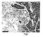
- Figure 2 shows the histology of the pancreas of a diabetic bank vole demonstrating islet infiltration of mononuclear cells following Hematoxylin and Eosin staining.
- Figure 3 shows the histology of the pancreas in bank voles without (panels a and b) and with diabetes (panels c and d) following immunostaining with the mouse 87-012 or 145L antiserum against Ljungan virus.
- the binding of the mouse antiserum was revealed with red vector staining.
- the sections were double stained with glucagon antiserum revealed with alkaline phosphatase and tetrazolium blue.
- the non-diabetic voles did not show binding of mouse anti-Ljungan virus antibodies while the immunostaining against glucagon stained cells in the periphery of the islets (panel a).
- the islets in diabetic bank voles showed varying degree of vacuolization or fatty infiltration of the pancreatic islets (panels b, c and d).
- the edges of these lesions are stained indicating the presence of Ljungan virus antigen.
- the glucagon immunostaining showed redistribution of cells that became more pronounced the greater the lesions.
- Figure 4 shows that the bank voles have autoantibodies against islet cell autoantigens and against Ljungan virus in vitro translated antigens.
- Autoantibodies to GAD65 (panel a), IA-2 (panel b), insulin (panel c) as well as Ljungan virus in vitro translated antigens are shown as in-house relative Units on a log scale (wherein a 1/25 dilution of standard serum is equal to 100 units/ml).
- Group A animals were caught and bled in the wild and only 4% had diabetes.
- Group B bank voles were captive and 33% of the animals shown had diabetes.
- GAD65 p ⁇ 0.0001
- IA-2 p ⁇ 0.0001
- insulin p ⁇ 0.03
- the autoantibody levels of both GAD65 and IA-2 were higher in diabetic as compared to non-diabetic Group B bank voles as indicated in the Figure.
- the data in panel d demonstrate that antibodies to Ljungan virus in vitro translated antigens were also increased in diabetic compared to non-diabetic bank voles. Data for individual bank voles are shown.
- Figure 5 shows the sequence similarities and cross-reactivity between GAD65 autoantibodies and mouse or human anti-Ljungah virus antibodies. Sequence comparisons between the predicted amino acid sequence of Ljungan virus (serotype 87-012) and type 1 diabetes associated autoantigens are shown in panel a. The data compares the 87-012 Ljungan virus sequence and regions of potential molecular mimicry to GAD65, IA-2 and insulin. Areas of homology are boxed, with identical amino acids indicated by a dot, similar amino acids are boxed, and non-similar amino acids are plain type. Antibodies against Ljungan virus raised in mice (antiserum 87-012) showed cross-reactivity with human GAD65 (panel b).
- Radiobinding analysis to the 87-012 antiserum showed concentration-dependent binding of 35 S-labeled Ljungan virus in vitro translated antigen (x-x) and human (o-o) but not mouse ( ⁇ - ⁇ ) GAJD65.
- the competition at half maximal binding of the 87-012 antiserum between binding of 35 S-labelled human GAD65 and unlabelled Ljungan virus antigen (x-x), human GAD65 (o-o) or human proinsulin ( ⁇ - ⁇ ) (panel c) demonstrates displacement by unlabelled Ljungan virus in vitro translated antigens as well as by recombinant human GAD65.
- Fig. 5 d The competition at half maximal binding of the 87-012 antiserum between binding of 35 S-labelled human GAD65 and unlabelled Ljungan virus antigen (x-x), human GAD65 (o-o) or human proinsulin ( ⁇ - ⁇ ) (panel c) demonstrates displacement by unlabelled Ljungan virus in vitro translated antigens as well as by
- Figure 6 shows the results of tests on sera from children with new onset type 1 diabetes indicating the presence of anti-Ljungan virus antibodies.
- Ljungan virus antibodies were determined in two independent tests by either indirect immunofluorescence of cells used to propagate the virus or by the radioligand binding assay with Ljungan virus in vitro translated antigens. The radioligand binding assay correlated to the indirect immunofluorescence test (p ⁇ 0.001 at 95 confidence interval).
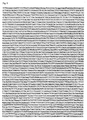
- Figure 7 shows the nucleotide sequence of Ljungan virus 87-012.
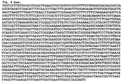
- Figure 8 shows the nucleotide sequence of Ljungan virus 145SL.
- Figure 9 shows the nucleotide sequence of Ljungan virus 174F.
- a method for obtaining an animal model for human diabetes comprising obtaining a non-human mammal; determining whether the mammal is infected with a Ljungan virus; and compromising the immune system of the mammal to facilitate the development of diabetes.
- the non-human mammal may be any non-human mammal including rodents such as rats, mice, hamsters, guinea pigs, rabbits, bank voles and field voles; cattle such as cows; cats; dogs; and non-human primates.
- rodents such as rats, mice, hamsters, guinea pigs, rabbits, bank voles and field voles
- cattle such as cows; cats; dogs; and non-human primates.
- the mammal is a rodent, a cat or a dog, more preferably the mammal is a rodent, most preferably the mammal is a bank vole.
- bank voles having type 1 diabetes are all infected with Ljungan virus and that the presence of Ljungan virus causes or at least contributes to the development of type 1 diabetes.
- Ljungan virus refers to any Ljungan picornavirus as defined in International PCT patent application WO 98/11133 , the disclosure of which is incorporated herein by reference.
- the Ljungan virus is Ljungan virus 87-012, the nucleotide sequence of which is shown in Figure 7 ; Ljungan virus 145SL, the nucleotide sequence of which is shown in Figure 8 ; or Ljungan virus 174F, the nucleotide sequence of which is shown in Figure 9 .
- Ljungan virus can be determined using any standard procedure including, but not limited to, virus isolation, detection of Ljungan virus antigen by ELISA or immunohistochemistry using antibody molecules having affinity for Ljungan virus or detection of Ljungan virus specific RNA sequences using PCR or by a labeled nucleic acid probe capable of specifically hybridizing to Ljungan virus nucleic acid.
- the presence of Ljungan virus also can be determined by detecting for the presence of Ljungan virus antibodies using a suitable test. Suitable techniques for determining the presence of Ljungan virus or anti-Ljungan virus antibodies are described in the examples below.
- non-human mammal obtained using the method according to the first embodiment of the present invention has features that mimic the human disease means that it closely represents the human disease and is therefore a particularly useful model of diabetes.
- the method according to the first' embodiment of the present invention also comprises determining whether the non-human mammal has high blood glucose levels that can be reduced by insulin and signs of ketoacidosis.
- the non-human mammal is a bank vole.
- the bank vole may be any species of bank vole.
- the bank vole is Clethrionomys glareolus .
- the non-human mammal can be male or female.
- the bank vole may be obtained from the wild or may be the progeny of a bank vole obtained from the wild. It is preferred that the bank vole obtained from the wild is obtained from Denmark, Sweden or Finland. Alternatively, the bank vole may be a laboratory bred bank vole.
- diabetes means type 1 or type 2 diabetes or diabetes having a combination of symptoms of both type 1 and type 2 diabetes.
- the type of diabetes developed by the non-human mammal will defend on the type of mammal.
- bank voles infected with Ljungan virus develop type 1 diabetes
- cats and dogs can develop type 1 or type 2 diabetes or diabetes having a combination of symptoms of both type 1 and type 2 diabetes. It is currently believed that diabetes in humans is not always type 1 or type 2 diabetes but that diabetes can fall somewhere between the two defined types wherein the individual has some symptoms of both type 1 and type 2 diabetes.
- diabetes in humans is not always type 1 or type 2 diabetes but that diabetes can fall somewhere between the two defined types wherein the individual has some symptoms of both type 1 and type 2 diabetes.
- diabetes refers to diabetes characterized by high blood glucose levels that can be reduced by insulin and signs of ketoacidosis.
- the presence of auto-antibodies to at least one of GAD65, IA-2 and insulin is an additionally preferred characteristic of type 1 diabetes according to the present invention. Additional features of diabetes such as hyperlipidemia, slowly progressive increase of hyperglycemia and variable glucosuria as well as symptoms of hyperphagia and obesity also may be present in addition to the characteristics of type 1 diabetes defined above in accordance with the situation in humans.
- high blood glucose levels means blood glucose levels that are at least 1.5 times as high, more preferably at least 3 times as high and most preferably at least 5 times as high as the mean level of blood glucose found in the corresponding non-diabetic mammals.
- Non-diabetic mammals are mammals that do not show any symptoms of diabetes such as increased glucosuria. It is particularly preferred that a high blood glucose level is at least 150 mg/dl, more preferably at least 200 mg/dl.
- the term "reduced by insulin” as used herein means that the high blood glucose levels can be reduced by the addition of insulin.
- the blood glucose levels can be reduced by about 30%, more preferably 60% and most preferably to approximately the level of a non-diabetic mammals by the addition of insulin.
- the reduction in blood glucose levels will vary depending on the amount of insulin given to the mammal.
- Ketoacidosis can be determined by the measurement of keton bodies in the blood or in plasma or serum.
- the method according to the first embodiment of the present invention includes compromising the immune system of the non-human mammal to facilitate the development of diabetes.
- the immune system can be compromised by suppressing the immune system.
- the immune system of the non-human mammal can be compromised by any method including administrating immunosuppressing agents, altering the diet of the non-human mammal or subjecting the non-human mammal to stress.
- the immune system of the non-human mammal is compromised by subjecting the mammal to stress.
- the non-human mammal can be subjected to any form of stress that affects the immune system of the mammal including keeping the mammal in a cage.
- the mammal is a bank vole
- it preferably is kept in a cage for at least 2 months, more preferably at least 3 months.
- the mammal is kept isolated in its own cage.
- a non-human mammal obtained by the method according to the first embodiment of the present invention can be used as a model of diabetes in order to investigate the development and etiology of diabetes.
- the non-human mammal can also be used to test candidate compounds for their effects on symptoms of diabetes.
- a candidate compound can be administered to the non-human mammal and the effects of the compound on symptoms of diabetes, such as blood glucose levels, signs of ketoacidosis and glucosuria can be measured.
- the non-human mammal can also be used to screen for compounds having an effect on the development of diabetes.
- the mammal can be used to screen for compounds that prevent the development of, or reduce the symptoms of, diabetes.
- a method for obtaining an animal model for human diabetes comprising the steps of: infecting a non-human mammal with a Ljungan virus; and suppressing the immune system of the mammal.
- the non-human mammal infected with Ljungan virus can be used as a model of diabetes whether or not symptoms of diabetes can be detected.
- the non-human mammal can be infected using any standard technique, including, but not limited to, parenteral routes such as intravenous injection and intraperitoneal injection. Methods for determining the necessary viral dose leading to the development of diabetes can be easily determined by those skilled in the art. In making such a determination, a number of factors are considered including the species of mammal, the rate of viral replication, the route of infection, the age and sex of the mammal. Preferably about 1,000 infection units are given to the non-human mammal.
- the non-human mammal infected with Ljungan virus may develop type 1 or type 2 diabetes or diabetes having a combination of symptoms of both type 1 and type 2 diabetes.
- the method according to the second embodiment of the present invention comprises suppressing the immune system of the non-human mammal as described above with respect to the first embodiment of the present invention.
- the immune system of the non-human mammal is compromised by subjecting the mammal to stress as described above with respect to the first embodiment of the present invention subsequent to infection with the infectious agent.
- the non-human mammal infected with a Ljungan virus can be used as a model to investigate the development and etiology of diabetes.
- the non-human mammal also can be used to test candidate compounds for their effects on diabetes.
- a candidate compound can be administered to a non-human mammal infected with a Ljungan virus and the effects of the compound on symptoms of diabetes, such as blood glucose levels, signs of ketoacidosis, glucosuria, hyperlipidemia, a slowly progressive increase in hyperglycemia, symptoms of hyperphagia, obesity and insulin resistance, can be measured.
- the non-human mammal infected with a Ljungan virus can also be used to screen for compounds having an effect on the development of diabetes.
- the non-human mammal may be used to screen for compounds which prevent the development of, or reduce the symptoms of, diabetes.
- Ljungan virus can be determined using any standard procedure including immunohistochemistry using antibody molecules having affinity for Ljungan virus or by using a labeled nucleic acid probe capable of specifically hybridizing to Ljungan virus nucleic acid.
- the presence of Ljungan virus can be determined by detecting the presence of anti-Ljungan virus antibodies using a suitable test. Suitable techniques for determining the presence of Ljungan virus or anti-Ljungan virus antibodies are described in the examples below.
- Wild caught bank voles (Group A). Group A bank voles represent 101 animals from a single trapping session. These bank voles were tested at the trap for glucosuria and then euthanized. Heart-blood samples for blood glucose, ketosis, lipids and antibody analyses were taken immediately after the voles were killed. Blood samples were either immediately analyzed for blood glucose and ketones or centrifuged for 25 minutes at 1,000xg and plasma stored at -30°C. Pancreas was dissected and fixed in 4% paraformaldehyde followed by ethanol before being embedded in paraffin.
- the cages were supplied with a woodcutting bed, and food (standard rat chow) and water were available ad libitum . Cage cleaning and body weight measurements were performed once every week. A portion of grain mixture was given when the cages were cleaned. Diabetes development was followed by measurements of water intake, glucosuria, and blood glucose and ketonemia determined after bleeding from the retro orbital plexus. Polydipsic voles were characterized by >21 ml/day water intake compared with non-polydipsic voles for which daily intake did not exceed 12 ml.
- pancreas fixed in 4% paraformaldehyde and embedded in paraffin were cut into 5 micron thick sections, affixed to slides, deparaffinized and rehydrated.
- the sections were blocked for 30 min at RT in PBS containing 0.05% Tween 20 (Sigma, St. Louis, MO), 1% BSA (Sigma), 2% normal horse (in the case of staining for Ljungan virus antisera) or 2% normal goat (in the case of insulin or glucagon staining) serum (Vector Laboratories, Burlingame, CA), and 4 drops/ml Avidin solution (Avidin/Biotin blocking kit, Vector Laboratories).
- the primary antibody was diluted in PBS with 0.05% Tween 20, 1% BSA, 2% normal serum, and 4 drops/ml Biotin solution (from Avidin/Biotin blocking kit, Vector Labs) to 1:100 (guinea pig anti-insulin and rabbit anti-glucagon, Zymed Laboratories, S. San Francisco, CA) or 1:500 (mouse Ljungan virus antiserum). Slides were exposed to the primary antibody solution for 60 minutes at room temperature or overnight at 4°C.
- Immunfluorescence assay for Ljungan virus antibodies Sera from children with type 1 diabetes and controls were tested for presence of antibodies to Ljungan virus using an indirect immunofluorescence test (IFT).
- IFT indirect immunofluorescence test
- a previously described IFT protocol Niklasson et al., J. Infect. Dis., 155, 369-76, 1987 ) was used to test antibody titers. Briefly, spot slides were prepared by incubating virus in Green Monkey Kidney cells for 8-10 days. At signs of discrete cytopathic effects (CPE), cells were removed from the flask with a rubber policeman and applied onto microscope slides, air dried, fixed in cold (4°C) acetone and stored at - 70°C until used.
- CPE discrete cytopathic effects
- the titer was determined after incubating the serum, diluted in PBS, on the slides at 37°C for 1 h in a moist chamber and bound antibodies were detected by incubating FITC-conjugated goat antihuman IgG (Sigma, St Louis, MO) for 1 h at 37 °C.
- Patient and control sera was first tested at a 1:8 dilution using three Ljungan virus isolates (87-012, 145SL, 174F). Any sera scoring positive for any of the three isolates were titrated again using all three isolates separately. Patients and controls positive to one or several isolates at a titer of 32 or higher was considered positive.
- GAD65 and IA-2 antibodies Radioligand binding assays for GAD65 and IA-2 antibodies .
- GAD65 and IA-2 antibodies were analyzed as described ( Grubin et al., Diabetologia, 37, 344-350, 1994 ; Hampe et al., J. Clin. Endocrinol. Metab., 85, 4671-9, 2000 ; Vandewalle et al., Diabetes Care, 20, 1547-1552, 1997 ).
- GAD65 and IA-2 antibody levels were expressed in U/ml for GAD65 and IA-2 antibodies using the WHO/JDF standard ( Mire-Sluis et al., Diabetologia, 43, 1282-1292, 2000 ).
- IAA Insulin autoantibodies
- Competition experiments Competition in binding between radioactive and cold antigens was carried out at half maximal binding of either the Ljungan virus 87-012 mouse antiserum or the 591 human standard serum found to be positive for antibodies against both Ljungan virus in vitro translated antigens and GAD65 (Mire-Sluis et al. , ( supra ), 2000). Competition for binding of 35 S-labeled Ljungan virus in vitro translated antigens was carried out with different concentrations of unlabeled Ljungan virus in vitro translated antigens, recombinant human GAD65 (DiamydAB, Sweden) or human proinsulin (Elli Lilly Company, Indianapolis, Indiana).
- Type 1 diabetes patients and controls A total of 53 children with a median age of 10.1 years (range 2.3-16.4 years of age) were diagnosed with type 1 diabetes at the St Göran Hospital and Astid Lindgren's Children's Hospital between 1992 and 1995. Within two days of diagnosis, blood samples were drawn for antibody analysis. Healthy children (7 boys, median age 12.6 (7.8 - 16.8 years and 10 girls, median age 13.5 (6.7 - 16.6 years) were recruited from school classes in central Swedish and children to personnel at the hospital. All children'were previously healthy and without present medication. The Ethics Committee at the Karolinska Institute, Sweden, approved the study.
- Bioinformatics To identify regions of high local homology between the virus polyprotein and known diabetes autoantigens, we created a local database of GAD65, IA-2 and insulin sequences and ran stand alone BLAST using software from the NCBI ( Altschul et al., Nucleic Acids Res., 25, 3389-402, 1997 ). Alignments were compiled manually to align regions of similarity onto the Ljungan protein sequence using CLUSTALW to determine similarity between non-homologous residues.
- Bank voles were trapped from May to November in a forest habitat on the island of Zealand, Denmark. In different continuous trapping sessions of 30 days duration, 100 traditional live traps were set and inspected twice a day. Two groups of bank voles were analyzed for diabetes and pancreas histology in addition to type 1 diabetes associated autoantibodies against insulin (Williams et al. , ( supra ), 1997), GAD65 (Grubin et al, ( supra ), 1994), and IA-2 ( Lan et al., DNA and Cell Biology, 13, 505-514, 1994 ) also known as ICA512 ( Rabin et al., J.
- Group A bank voles represents 101 trapped bank voles that were euthanized in the forest for immediate examination of blood glucose, glucosuria, body weight, pancreas histology and antibodies.
- Group B represents 67 bank voles that were trapped and kept in the laboratory for one month as previously described ( Schoenecker et al., Appl. Anim. Behav. Sci., 68, 349-357, 2000 ).
- An additional group of 54 animals were examined in Sweden for insulin sensitivity and pancreas histology before and after diabetes development.
- the data in Table 1 shows the occurrence of diabetes in the two groups of bank voles.
- the mean blood glucose ⁇ S.D. was normally distributed at 101 ⁇ 28 mg/dl.
- the body weight of the trapped voles from group A ranged from 8.5 - 28.4g, the mean value ⁇ S.D. was 19 ⁇ 5g. Occasional hyperglycemic and glucosuric bank voles may therefore be trapped in the wild. Whether these four animals had stress-induced hyperglycemia or overt diabetes remains to be established.
- Group A bank voles differ markedly from the 67 Group B bank voles that were trapped and kept in standard laboratory mouse cages for one month before they were tested for diabetes.
- 22/67 (33%) of these Group B bank voles had a blood glucose above 200 mg/dl, the range being 211-540 mg/dl.
- 18/22 (82%) had ketones and were polydipsic. Gender differences are common in both humans ( Harris, Diabetes in America, (ed. Harris, M.
- Table 1 The frequency of diabetes in wild caught bank voles and in bank voles kept in the laboratory. Group of bank voles A. Analyzed at trap B. Trapped and captive N 101 67 M/F ratio 42/59 29/38 Blood glucose (mg/dl) Non-diabetic 101 ⁇ 27 86 ⁇ 24 Diabetic 293 ⁇ 54 346 ⁇ 88 Diabetes n (%) 4 (4%) 22 (33%) M/F ratio 0/4 14/8
- the pancreas of all 101 Group A bank voles showed normal islets as did those of non-diabetic Group B bank voles ( Fig. 1 ).
- the four hyperglycemic Group A bank voles had no appreciable islet lesions.
- Immunostaining with insulin and glucagon antibodies showed a normal islet cell distribution with beta cells located in the center surrounded by glucagon-positive cells ( Fig. 1 a and 1 b ).
- Group B bank voles with diabetes had an almost complete loss of centrally located insulin-positive cells that were replaced by prominent vacuolization or fatty infiltration ( Fig. 1 c and I d ).
- Islets with infiltrating mononuclear cells were occasionally observed ( Fig. 2 ) but insulitis was conspicuously absent in the majority of the bank voles.
- the beta cell destruction was unique to bank voles with diabetes and indicate that the animal should be classified as having type 1 diabetes.
- diabetic group B bank voles had higher GAD65 ( P ⁇ 0.001), IA-2 ( P ⁇ 0.001) and insulin ( P ⁇ 0.0346) autoantibody levels than the non-diabetic voles ( Fig. 4 ).
- the increased levels of GAD65, IA-2 and insulin autoantibodies further indicates that diabetes in these bank voles should be classified as type 1 diabetes.
- the mean levels of Ljungan virus antibodies in'the non-diabetic Group B bank voles were significantly increased ( P ⁇ 0.001).
- antibody levels against Ljungan virus antigens correlated with levels of GAD65 ( P ⁇ 0.0001), IA-2 ( P ⁇ 0.0001) and insulin (P ⁇ 0.03) autoantibodies.
- mouse GAD65 While labeled mouse GAD65 was not recognized, the mouse Ljungan virus polyclonal antiserum 87-012 (Niklasson et al ., ( supra ), 1999) was capable of immunoprecipitating human GAD65 ( Fig. 5b ), indicating significant epitope specificity (Hampe et al. , (supra), 2000).
- the Ljungan virus in vitro translated antigens immunoprecipitated by the 87-012 Ljungan virus antiserum was reciprocally displaced by both cold Ljungan virus in vitro translated antigens and human GAD65 but not by human proinsulin ( Fig. 5c ).
- the human type 1 diabetes GAD65 antibody-positive serum #591 showed concentration dependent immunoprecipitation of Ljungan virus in vitro translated antigens and both human and mouse GAD65 ( Fig 5 d ).
- the examples provide evidence that wild caught bank voles may develop type 1 diabetes associated with specific beta-cell destruction, insulitis and autoantibodies to GAD65 and IA-2.
- Our observation that it was possible to detect Ljungan virus antigen in affected pancreatic islets showing gradual destruction and end-stage fatty degeneration also indicates that this virus causes or at least contributes to the loss of beta cells.
- the diabetic bank voles had increased levels of antibodies to Ljungan virus cDNA in vitro translated virus antigens. The levels of these antibodies were also found to correlate to the levels of autoantibodies to both GAD65 and IA-2.
- Diabetes in bank voles was first described during a study of stereotypic behavior in bank voles ( Schoenecker et al., Appl. Anim. Behav. Sci., 68, 349-357, 2000 ) When captured in the wild, brought to the laboratory to be kept in standard laboratory mouse cages, and fed laboratory chow, bank voles developed polydipsia and glucosuria. Diabetes was detected in 4/101 Group A animals that were euthanized in the forest at the site of the trap which was different from the 22/67 bank voles kept in the laboratory. Our data suggest that the diabetes symptoms in these animals fulfill current classification criteria for autoimmune type 1 diabetes in humans ( Mellitus et al., Diabetes Care, 20, 1183-1197, 1997 ).
- the diabetic bank voles sustain hyperglycemia, ketonemia, ketonuria, hyperlipidemia and weight loss, all criteria that are consistent with type 1 diabetes classification.
- the diabetic bank voles had increased levels of both GAD65, IA-2 and insulin autoantibodies. These autoantibodies predict type 1 diabetes in humans ( Verge et al., Diabetes, 45, 926-933, 1996 ); Bingley et al., Diabetes Care, 22, 1796-801, 1999 ) and confirm the type 1 diabetes classification.
- bank voles develop diabetes that fulfills the criteria for type 1 diabetes: diabetic animals showed persistent hyperglycemia associated with weight loss, ketosis and hyperlipidemia (data not shown) as well as insulin dependence associated with specific beta-cell destruction and insulitis.
- diabetic voles had increased levels of autoantibodies to GAD65 and IA-2, and that these autoantibodies correlated to Ljungan virus antigen antibodies.
- the association between Ljungan virus and bank vole diabetes was supported by the presence of Ljungan virus antigen detected by irnmunocytochemistry in affected diabetic bank vole islets.
- the field vole develops clinical diabetes with symptoms of polydipsia and polyuria identical to bank voles.
- Ljungan virus has been isolated from field voles with type 1 diabetes trapped in Sweden. It is likely that field voles are just as good an animal model for type 1 diabetes as the bank vole.
- the clinical disease mimics in some animals type 1 diabetes and in some animals type 2 diabetes.
- Amyloid deposits localized to the islets of Langerhans are typical of type 2 diabetes mellitus.
- diabetic cats most commonly have pancreatic islet destruction associated with pancreatic amyloidosis and are insulin deficient like type 1 diabetes.
- the disease occurs in all ages of the cats but the majority of diabetes affect older cats ( Westermark et al., PNAS USA, 84, 3881-5, 1987 ; Johnson et al., Veterinary Pathology, 22(5):463-8, 1985 ; and Yano et al., Veterinary Pathology, 18(3): 310-5, 1981 ).
- Spontaneous diabetes mellitus in guinea pigs parallels in many ways the syndrome known as juvenile diabetes mellitus in man: elevated blood glucose levels; reproductive dysfunction in the female; degranulation and severe cytoplasmic vacuolation of beta cells, severe fatty degeneration of acinar cells, and hyperplasia of the islets of the pancreas; and a high frequency of abnormal pancreatic secretions ( Lang et al., Diabetes, 25(5):434-43, 1976 ).
- the severity of pathologic changes in the pancreatic islets parallel, in general, the severity of the clinical symptoms.
- the other clinical parameters of note are elevated serum triglycerides, normal serum but elevated aortic cholesterol, and absence of ketonemia or ketonuria.
- Microangiopathy another characteristic of juvenile diabetes mellitus in man was demonstrated as a significant increase in the thickness of the basal membranes in peripheral capillaries.
- a glomerular lesion encountered in some of the diabetic guinea pigs was shown to be similar to the glomerular sclerosis seen in human diabetics. Although a definitive etiologic agent was not identified, the disease was clearly contagious in origin.
- Spontaneous diabetes mellitus has been observed in a female New Zealand white rabbit. Three groups of animals could be identified. Some animals had overt diabetes characterized by fasting hyperglycemia and depressed intravenous glucose stimulated serum insulin levels ( Conaway et al., Clinical & Experimental, 30(1):50-6, 1981 ). This abnormality is seen between 1 and 3 years of life. Another group of animals developed abnormal glucose disposal with normal or slight elevations in fasting serum glucose levels. Glucose stimulated insulin levels are also significantly lower in the rabbits with abnormal glucose disposal. The remaining animals exhibit no apparent abnormalities of glucose metabolism. Despite marked increases in serum and urinary glucose, only mild ketonemia was observed. The relatively late onset of diabetic symptoms, lack of obesity, severe hyperglycemia, and depressed insulin secretion without ketoacidosis make this a model with many of the characteristics of insulin responsive diabetes as seen in non-obese human adults.
- the diabetic hamsters shows body weight loss, hyperglycemia (mean fasting plasma glucose 402 mg/dl), hypoinsulinemia, hyperlipidemia and ketonemia.
- the diabetic hamsters showed reduced activities of cytoplasmic glycolytic key enzymes: hexokinase, pyruvate kinase and phosphofructokinase; increases in cardiac glycogen and glucose-6-phosphate contents; and a 40% decrease in cardiac ATP content, indicating decreased energy production.
- An accumulation of myocardial triglyceride and cholesterol was found in the diabetic hamsters ( Eto et al., Diabetes Research & Clinical Practice, 3 (6) :297-305, 1987 ).
Landscapes
- Health & Medical Sciences (AREA)
- Life Sciences & Earth Sciences (AREA)
- Immunology (AREA)
- Engineering & Computer Science (AREA)
- Hematology (AREA)
- Chemical & Material Sciences (AREA)
- Molecular Biology (AREA)
- Urology & Nephrology (AREA)
- Biomedical Technology (AREA)
- General Health & Medical Sciences (AREA)
- Virology (AREA)
- Medicinal Chemistry (AREA)
- Food Science & Technology (AREA)
- Pathology (AREA)
- Environmental Sciences (AREA)
- Animal Behavior & Ethology (AREA)
- General Physics & Mathematics (AREA)
- Biochemistry (AREA)
- Analytical Chemistry (AREA)
- Physics & Mathematics (AREA)
- Microbiology (AREA)
- Biotechnology (AREA)
- Cell Biology (AREA)
- Diabetes (AREA)
- Rheumatology (AREA)
- Animal Husbandry (AREA)
- Organic Chemistry (AREA)
- Nuclear Medicine, Radiotherapy & Molecular Imaging (AREA)
- General Chemical & Material Sciences (AREA)
- Tropical Medicine & Parasitology (AREA)
- Veterinary Medicine (AREA)
- Public Health (AREA)
- Zoology (AREA)
- Chemical Kinetics & Catalysis (AREA)
- Biodiversity & Conservation Biology (AREA)
- Pharmacology & Pharmacy (AREA)
- Rehabilitation Therapy (AREA)
- Oncology (AREA)
- Communicable Diseases (AREA)
- Emergency Medicine (AREA)
Claims (14)
- Procédé pour obtenir un modèle animal pour le diabète humain, comprenant les étapes consistant à se procurer un mammifère non humain ; à déterminer si le mammifère est infecté par le virus Ljungan ; et à compromettre le système immunitaire du mammifère afin de faciliter le développement du diabète.
- Procédé selon la revendication 1, dans lequel le procédé comprend en outre l'étape consistant à déterminer si le mammifère possède des taux de glycémie élevés pouvant être réduits par insuline et des signes d'acidocétose.
- Procédé selon la revendication 1 ou 2, dans lequel le procédé comprend en outre l'étape consistant à détecter la présence d'auto-anticorps contre au moins l'une parmi GAD65, IA-2 et l'insuline.
- Procédé selon la revendication 1, dans lequel le système immunitaire du mammifère est compromis en soumettant le mammifère à un stress pendant au moins 2 mois.
- Procédé selon la revendication 4, dans lequel le mammifère est soumis à un stress en le maintenant dans une cage.
- Procédé selon l'une quelconque des revendications précédentes, dans lequel le mammifère est un rongeur.
- Procédé selon l'une quelconque des revendications précédentes, dans lequel le mammifère est un campagnol roussâtre.
- Procédé selon la revendication 7, dans lequel le campagnol roussâtre est Clethrionomys glareolus.
- Procédé selon la revendication 7, dans lequel le campagnol roussâtre est prélevé dans la nature ou fait partie de la progéniture d'un campagnol roussâtre prélevé dans la nature.
- Procédé selon la revendication 9, dans lequel le campagnol roussâtre est originaire du Danemark, de Suède ou de Finlande.
- Procédé pour obtenir un modèle animal pour le diabète humain comprenant les étapes consistant à : infecter un mammifère non humain avec le virus Ljungan ; et à supprimer le système immunitaire du mammifère.
- Procédé selon la revendication 11, dans lequel le système immunitaire est supprimé en soumettant l'animal à un stress à la suite d'une infection avec l'agent infectieux.
- Procédé selon la revendication 11 ou 12, dans lequel le mammifère est un chat, un chien ou un campagnol roussâtre.
- Procédé selon la revendication 11 ou 12, dans lequel le mammifère est un campagnol roussâtre.
Applications Claiming Priority (3)
| Application Number | Priority Date | Filing Date | Title |
|---|---|---|---|
| GB0120437 | 2001-08-22 | ||
| GBGB0120437.9A GB0120437D0 (en) | 2001-08-22 | 2001-08-22 | Diabetic model |
| PCT/IB2002/003957 WO2003019197A2 (fr) | 2001-08-22 | 2002-08-21 | Modele diabetique |
Publications (2)
| Publication Number | Publication Date |
|---|---|
| EP1432990A2 EP1432990A2 (fr) | 2004-06-30 |
| EP1432990B1 true EP1432990B1 (fr) | 2008-11-19 |
Family
ID=9920833
Family Applications (1)
| Application Number | Title | Priority Date | Filing Date |
|---|---|---|---|
| EP02775020A Expired - Lifetime EP1432990B1 (fr) | 2001-08-22 | 2002-08-21 | Modele diabetique |
Country Status (7)
| Country | Link |
|---|---|
| US (2) | US7560609B2 (fr) |
| EP (1) | EP1432990B1 (fr) |
| AT (1) | ATE414905T1 (fr) |
| AU (1) | AU2002341238A1 (fr) |
| DE (1) | DE60229966D1 (fr) |
| GB (1) | GB0120437D0 (fr) |
| WO (1) | WO2003019197A2 (fr) |
Families Citing this family (5)
| Publication number | Priority date | Publication date | Assignee | Title |
|---|---|---|---|---|
| WO2004073710A1 (fr) * | 2003-02-21 | 2004-09-02 | Apodemus Ab | Methode de traitement a l'aide de pleconaril de maladies induites par le virus ljungan |
| GB0505321D0 (en) * | 2005-03-15 | 2005-04-20 | Apodemus Ab | Detection method |
| EP1743522A1 (fr) * | 2005-07-13 | 2007-01-17 | ID-Lelystad, Instituut voor Dierhouderij en Diergezondheid B.V. | Modèle animal pour diabète de type II et pour syndrome x |
| CA2651123C (fr) * | 2006-05-01 | 2016-04-19 | William Nix | Procedes et agents permettant de detecter le parechovirus |
| WO2013083753A2 (fr) * | 2011-12-07 | 2013-06-13 | Institut Pasteur | Identification d'un virus porcin de type paréchovirus et applications |
Family Cites Families (1)
| Publication number | Priority date | Publication date | Assignee | Title |
|---|---|---|---|---|
| SE9603305D0 (sv) | 1996-09-11 | 1996-09-11 | Bo Niklasson | New picornaviruses, vaccines and diagnostic kits |
-
2001
- 2001-08-22 GB GBGB0120437.9A patent/GB0120437D0/en not_active Ceased
-
2002
- 2002-08-21 US US10/487,134 patent/US7560609B2/en not_active Expired - Fee Related
- 2002-08-21 DE DE60229966T patent/DE60229966D1/de not_active Expired - Lifetime
- 2002-08-21 AT AT02775020T patent/ATE414905T1/de not_active IP Right Cessation
- 2002-08-21 AU AU2002341238A patent/AU2002341238A1/en not_active Abandoned
- 2002-08-21 WO PCT/IB2002/003957 patent/WO2003019197A2/fr not_active Ceased
- 2002-08-21 EP EP02775020A patent/EP1432990B1/fr not_active Expired - Lifetime
-
2009
- 2009-06-01 US US12/475,704 patent/US20120027776A1/en not_active Abandoned
Also Published As
| Publication number | Publication date |
|---|---|
| AU2002341238A1 (en) | 2003-03-10 |
| WO2003019197A3 (fr) | 2004-05-06 |
| ATE414905T1 (de) | 2008-12-15 |
| WO2003019197A2 (fr) | 2003-03-06 |
| US20120027776A1 (en) | 2012-02-02 |
| US7560609B2 (en) | 2009-07-14 |
| GB0120437D0 (en) | 2001-10-17 |
| EP1432990A2 (fr) | 2004-06-30 |
| DE60229966D1 (de) | 2009-01-02 |
| US20040265793A1 (en) | 2004-12-30 |
Similar Documents
| Publication | Publication Date | Title |
|---|---|---|
| Niklasson et al. | Development of type 1 diabetes in wild bank voles associated with islet autoantibodies and the novel ljungan virus | |
| Fukata et al. | Toll-like receptor-4 is required for intestinal response to epithelial injury and limiting bacterial translocation in a murine model of acute colitis | |
| Hermes et al. | Neurological and behavioral abnormalities, ventricular dilatation, altered cellular functions, inflammation, and neuronal injury in brains of mice due to common, persistent, parasitic infection | |
| Flodström et al. | Target cell defense prevents the development of diabetes after viral infection | |
| Allegretti et al. | Broad MICA/B expression in the small bowel mucosa: a link between cellular stress and celiac disease | |
| US20120027776A1 (en) | Diabetic model | |
| Douxfils et al. | Does domestication process affect stress response in juvenile Eurasian perch Perca fluviatilis? | |
| MacDonald et al. | Maternal high-fat diet in mice leads to innate airway hyperresponsiveness in the adult offspring | |
| BRPI0612419A2 (pt) | método para tratar infecção por rhabdovirus em um sujeito mamìfero suspeito de ter uma infecção, método para tratar uma doença autoimune em um sujeito mamìfero, método para tratar inflamação em um sujeito mamìfero, método para identificar um modulador de sinalização em células via uma trajetória de recpetor "tool-like" 4, método para selecionar um composto para tratar uma doença infecciosa, método para selecionar um composto para tratar uma doença autoimune, método para selecionar um composto para tratar inflamação, animal não humano transgênico, célula ou linha de células, método para seleção in vitro de um modulador de atividade de sinalização de receptor "tool-like" 4 ou de tram-trif e método para seleção in vivo de um modulador de atividade de sinalização de receptor "tool-like" 4 ou de tram-trif | |
| Oz et al. | Atovaquone ameliorate gastrointestinal toxoplasmosis complications in a pregnancy model | |
| Starich et al. | A morphological and immunohistochemical investigation of endocrine pancreata from obese ob+/ob+ mice | |
| Wang et al. | The TIR/BB‐loop mimetic AS‐1 prevents non‐alcoholic steatohepatitis and hepatic insulin resistance by inhibiting NLRP3‐ASC inflammasome activation | |
| KR20080031436A (ko) | 포유류 대상에서 그람 양성 박테리아 감염 치료를 위한조성물 및 방법 | |
| Svensson et al. | Neurokinin 1 receptor signaling affects the local innate immune defense against genital herpes virus infection | |
| Kimura et al. | Increased thyroidal fat and goitrous hypothyroidism induced by interferon‐γ | |
| Kim et al. | A relationship between Alzheimer's disease and type 2 diabetes mellitus through the measurement of serum amyloid-β autoantibodies | |
| US11974552B2 (en) | Transgenic C57BL6-BTBR mouse with a humanized MHC II gene that expresses GAD65 | |
| Wiedosari et al. | Host differences in response to trickle infection with Fasciola gigantica in buffalo, Ongole and Bali calves | |
| Rasilainen et al. | Mechanisms of beta cell death during restricted and unrestricted enterovirus infection | |
| Alhalwani | A review of lactoferrin inflammatory role in type 2 diabetes mellitus with neutrophil dysfunction | |
| Cruickshank et al. | Infection of inbred and nude (athymic) rats with Brugia spp. | |
| Behnke et al. | The response of hamsters to primary and secondary infection with Trichinella spiralis and to vaccination with parasite antigens | |
| US7745689B2 (en) | Nephropathy-associated gene | |
| Prendergast et al. | Generating a reproducible model of mid-gestational maternal immune activation utilizing the viral mimic poly (I: C) to study susceptibility and resilience in offspring | |
| Schønecker et al. | Diabetes in Danish bank voles (M. glareolus): survivorship, influence on weight, and evaluation of polydipsia as a screening tool for hyperglycaemia |
Legal Events
| Date | Code | Title | Description |
|---|---|---|---|
| PUAI | Public reference made under article 153(3) epc to a published international application that has entered the european phase |
Free format text: ORIGINAL CODE: 0009012 |
|
| 17P | Request for examination filed |
Effective date: 20040312 |
|
| AK | Designated contracting states |
Kind code of ref document: A2 Designated state(s): AT BE BG CH CY CZ DE DK EE ES FI FR GB GR IE IT LI LU MC NL PT SE SK TR |
|
| AX | Request for extension of the european patent |
Extension state: AL LT LV MK RO SI |
|
| GRAP | Despatch of communication of intention to grant a patent |
Free format text: ORIGINAL CODE: EPIDOSNIGR1 |
|
| TPAC | Observations filed by third parties |
Free format text: ORIGINAL CODE: EPIDOSNTIPA |
|
| RIN1 | Information on inventor provided before grant (corrected) |
Inventor name: LERNMARK, AKE Inventor name: NIKLASSON, BO |
|
| GRAJ | Information related to disapproval of communication of intention to grant by the applicant or resumption of examination proceedings by the epo deleted |
Free format text: ORIGINAL CODE: EPIDOSDIGR1 |
|
| GRAP | Despatch of communication of intention to grant a patent |
Free format text: ORIGINAL CODE: EPIDOSNIGR1 |
|
| TPAC | Observations filed by third parties |
Free format text: ORIGINAL CODE: EPIDOSNTIPA |
|
| GRAS | Grant fee paid |
Free format text: ORIGINAL CODE: EPIDOSNIGR3 |
|
| GRAA | (expected) grant |
Free format text: ORIGINAL CODE: 0009210 |
|
| AK | Designated contracting states |
Kind code of ref document: B1 Designated state(s): AT BE BG CH CY CZ DE DK EE ES FI FR GB GR IE IT LI LU MC NL PT SE SK TR |
|
| REG | Reference to a national code |
Ref country code: GB Ref legal event code: FG4D |
|
| REG | Reference to a national code |
Ref country code: CH Ref legal event code: EP |
|
| REG | Reference to a national code |
Ref country code: IE Ref legal event code: FG4D |
|
| REF | Corresponds to: |
Ref document number: 60229966 Country of ref document: DE Date of ref document: 20090102 Kind code of ref document: P |
|
| PG25 | Lapsed in a contracting state [announced via postgrant information from national office to epo] |
Ref country code: AT Free format text: LAPSE BECAUSE OF FAILURE TO SUBMIT A TRANSLATION OF THE DESCRIPTION OR TO PAY THE FEE WITHIN THE PRESCRIBED TIME-LIMIT Effective date: 20081119 Ref country code: ES Free format text: LAPSE BECAUSE OF FAILURE TO SUBMIT A TRANSLATION OF THE DESCRIPTION OR TO PAY THE FEE WITHIN THE PRESCRIBED TIME-LIMIT Effective date: 20090301 |
|
| NLV1 | Nl: lapsed or annulled due to failure to fulfill the requirements of art. 29p and 29m of the patents act | ||
| PG25 | Lapsed in a contracting state [announced via postgrant information from national office to epo] |
Ref country code: NL Free format text: LAPSE BECAUSE OF FAILURE TO SUBMIT A TRANSLATION OF THE DESCRIPTION OR TO PAY THE FEE WITHIN THE PRESCRIBED TIME-LIMIT Effective date: 20081119 Ref country code: FI Free format text: LAPSE BECAUSE OF FAILURE TO SUBMIT A TRANSLATION OF THE DESCRIPTION OR TO PAY THE FEE WITHIN THE PRESCRIBED TIME-LIMIT Effective date: 20081119 |
|
| PG25 | Lapsed in a contracting state [announced via postgrant information from national office to epo] |
Ref country code: EE Free format text: LAPSE BECAUSE OF FAILURE TO SUBMIT A TRANSLATION OF THE DESCRIPTION OR TO PAY THE FEE WITHIN THE PRESCRIBED TIME-LIMIT Effective date: 20081119 Ref country code: BE Free format text: LAPSE BECAUSE OF FAILURE TO SUBMIT A TRANSLATION OF THE DESCRIPTION OR TO PAY THE FEE WITHIN THE PRESCRIBED TIME-LIMIT Effective date: 20081119 Ref country code: DK Free format text: LAPSE BECAUSE OF FAILURE TO SUBMIT A TRANSLATION OF THE DESCRIPTION OR TO PAY THE FEE WITHIN THE PRESCRIBED TIME-LIMIT Effective date: 20081119 Ref country code: BG Free format text: LAPSE BECAUSE OF FAILURE TO SUBMIT A TRANSLATION OF THE DESCRIPTION OR TO PAY THE FEE WITHIN THE PRESCRIBED TIME-LIMIT Effective date: 20090219 |
|
| PG25 | Lapsed in a contracting state [announced via postgrant information from national office to epo] |
Ref country code: CZ Free format text: LAPSE BECAUSE OF FAILURE TO SUBMIT A TRANSLATION OF THE DESCRIPTION OR TO PAY THE FEE WITHIN THE PRESCRIBED TIME-LIMIT Effective date: 20081119 Ref country code: SE Free format text: LAPSE BECAUSE OF FAILURE TO SUBMIT A TRANSLATION OF THE DESCRIPTION OR TO PAY THE FEE WITHIN THE PRESCRIBED TIME-LIMIT Effective date: 20090219 Ref country code: PT Free format text: LAPSE BECAUSE OF FAILURE TO SUBMIT A TRANSLATION OF THE DESCRIPTION OR TO PAY THE FEE WITHIN THE PRESCRIBED TIME-LIMIT Effective date: 20090420 |
|
| PLBE | No opposition filed within time limit |
Free format text: ORIGINAL CODE: 0009261 |
|
| STAA | Information on the status of an ep patent application or granted ep patent |
Free format text: STATUS: NO OPPOSITION FILED WITHIN TIME LIMIT |
|
| PG25 | Lapsed in a contracting state [announced via postgrant information from national office to epo] |
Ref country code: SK Free format text: LAPSE BECAUSE OF FAILURE TO SUBMIT A TRANSLATION OF THE DESCRIPTION OR TO PAY THE FEE WITHIN THE PRESCRIBED TIME-LIMIT Effective date: 20081119 |
|
| 26N | No opposition filed |
Effective date: 20090820 |
|
| PG25 | Lapsed in a contracting state [announced via postgrant information from national office to epo] |
Ref country code: MC Free format text: LAPSE BECAUSE OF NON-PAYMENT OF DUE FEES Effective date: 20090831 |
|
| REG | Reference to a national code |
Ref country code: CH Ref legal event code: PL |
|
| PG25 | Lapsed in a contracting state [announced via postgrant information from national office to epo] |
Ref country code: LI Free format text: LAPSE BECAUSE OF NON-PAYMENT OF DUE FEES Effective date: 20090831 Ref country code: CH Free format text: LAPSE BECAUSE OF NON-PAYMENT OF DUE FEES Effective date: 20090831 |
|
| REG | Reference to a national code |
Ref country code: IE Ref legal event code: MM4A |
|
| PG25 | Lapsed in a contracting state [announced via postgrant information from national office to epo] |
Ref country code: IE Free format text: LAPSE BECAUSE OF NON-PAYMENT OF DUE FEES Effective date: 20090821 |
|
| PG25 | Lapsed in a contracting state [announced via postgrant information from national office to epo] |
Ref country code: GR Free format text: LAPSE BECAUSE OF FAILURE TO SUBMIT A TRANSLATION OF THE DESCRIPTION OR TO PAY THE FEE WITHIN THE PRESCRIBED TIME-LIMIT Effective date: 20090220 |
|
| PG25 | Lapsed in a contracting state [announced via postgrant information from national office to epo] |
Ref country code: IT Free format text: LAPSE BECAUSE OF FAILURE TO SUBMIT A TRANSLATION OF THE DESCRIPTION OR TO PAY THE FEE WITHIN THE PRESCRIBED TIME-LIMIT Effective date: 20081119 |
|
| PG25 | Lapsed in a contracting state [announced via postgrant information from national office to epo] |
Ref country code: LU Free format text: LAPSE BECAUSE OF NON-PAYMENT OF DUE FEES Effective date: 20090821 |
|
| PG25 | Lapsed in a contracting state [announced via postgrant information from national office to epo] |
Ref country code: TR Free format text: LAPSE BECAUSE OF FAILURE TO SUBMIT A TRANSLATION OF THE DESCRIPTION OR TO PAY THE FEE WITHIN THE PRESCRIBED TIME-LIMIT Effective date: 20081119 |
|
| PG25 | Lapsed in a contracting state [announced via postgrant information from national office to epo] |
Ref country code: CY Free format text: LAPSE BECAUSE OF FAILURE TO SUBMIT A TRANSLATION OF THE DESCRIPTION OR TO PAY THE FEE WITHIN THE PRESCRIBED TIME-LIMIT Effective date: 20081119 |
|
| PGFP | Annual fee paid to national office [announced via postgrant information from national office to epo] |
Ref country code: DE Payment date: 20140821 Year of fee payment: 13 |
|
| PGFP | Annual fee paid to national office [announced via postgrant information from national office to epo] |
Ref country code: GB Payment date: 20140801 Year of fee payment: 13 Ref country code: FR Payment date: 20140821 Year of fee payment: 13 |
|
| REG | Reference to a national code |
Ref country code: DE Ref legal event code: R119 Ref document number: 60229966 Country of ref document: DE |
|
| GBPC | Gb: european patent ceased through non-payment of renewal fee |
Effective date: 20150821 |
|
| REG | Reference to a national code |
Ref country code: FR Ref legal event code: ST Effective date: 20160429 |
|
| PG25 | Lapsed in a contracting state [announced via postgrant information from national office to epo] |
Ref country code: DE Free format text: LAPSE BECAUSE OF NON-PAYMENT OF DUE FEES Effective date: 20160301 Ref country code: GB Free format text: LAPSE BECAUSE OF NON-PAYMENT OF DUE FEES Effective date: 20150821 |
|
| PG25 | Lapsed in a contracting state [announced via postgrant information from national office to epo] |
Ref country code: FR Free format text: LAPSE BECAUSE OF NON-PAYMENT OF DUE FEES Effective date: 20150831 |