EP1434592B1 - Acide nucleique et proteine correspondante appele 121p2a3 utile pour le traitement et la detection des cancers - Google Patents
Acide nucleique et proteine correspondante appele 121p2a3 utile pour le traitement et la detection des cancers Download PDFInfo
- Publication number
- EP1434592B1 EP1434592B1 EP02762038A EP02762038A EP1434592B1 EP 1434592 B1 EP1434592 B1 EP 1434592B1 EP 02762038 A EP02762038 A EP 02762038A EP 02762038 A EP02762038 A EP 02762038A EP 1434592 B1 EP1434592 B1 EP 1434592B1
- Authority
- EP
- European Patent Office
- Prior art keywords
- protein
- cancer
- expression
- cells
- antibodies
- Prior art date
- Legal status (The legal status is an assumption and is not a legal conclusion. Google has not performed a legal analysis and makes no representation as to the accuracy of the status listed.)
- Expired - Lifetime
Links
- 108090000623 proteins and genes Proteins 0.000 title claims abstract description 321
- 102000004169 proteins and genes Human genes 0.000 title claims abstract description 231
- 206010028980 Neoplasm Diseases 0.000 title claims abstract description 202
- 201000011510 cancer Diseases 0.000 title claims abstract description 102
- 150000007523 nucleic acids Chemical class 0.000 title description 20
- 238000011282 treatment Methods 0.000 title description 20
- 238000001514 detection method Methods 0.000 title description 19
- 108020004707 nucleic acids Proteins 0.000 title description 17
- 102000039446 nucleic acids Human genes 0.000 title description 17
- 239000012634 fragment Substances 0.000 claims abstract description 32
- 230000014509 gene expression Effects 0.000 claims description 124
- 238000000034 method Methods 0.000 claims description 98
- 125000003275 alpha amino acid group Chemical group 0.000 claims description 53
- 108090000765 processed proteins & peptides Proteins 0.000 claims description 43
- 210000002307 prostate Anatomy 0.000 claims description 26
- 238000012360 testing method Methods 0.000 claims description 20
- 108091007433 antigens Proteins 0.000 claims description 18
- 102000036639 antigens Human genes 0.000 claims description 18
- 239000000427 antigen Substances 0.000 claims description 17
- 230000027455 binding Effects 0.000 claims description 15
- 210000001072 colon Anatomy 0.000 claims description 13
- 239000003550 marker Substances 0.000 claims description 9
- 102000004190 Enzymes Human genes 0.000 claims description 4
- 108090000790 Enzymes Proteins 0.000 claims description 4
- 150000001875 compounds Chemical class 0.000 claims description 3
- 239000007850 fluorescent dye Substances 0.000 claims description 3
- 229910052751 metal Inorganic materials 0.000 claims description 3
- 239000002184 metal Substances 0.000 claims description 3
- 239000002738 chelating agent Substances 0.000 claims description 2
- 230000002285 radioactive effect Effects 0.000 claims 1
- 230000003053 immunization Effects 0.000 abstract description 13
- 238000002649 immunization Methods 0.000 abstract description 11
- 210000001744 T-lymphocyte Anatomy 0.000 abstract description 6
- 230000000069 prophylactic effect Effects 0.000 abstract description 5
- 230000024932 T cell mediated immunity Effects 0.000 abstract 1
- 230000028996 humoral immune response Effects 0.000 abstract 1
- 235000018102 proteins Nutrition 0.000 description 210
- 210000004027 cell Anatomy 0.000 description 136
- 210000001519 tissue Anatomy 0.000 description 88
- 235000001014 amino acid Nutrition 0.000 description 63
- 229940024606 amino acid Drugs 0.000 description 58
- 241000282414 Homo sapiens Species 0.000 description 54
- 150000001413 amino acids Chemical class 0.000 description 54
- 239000002299 complementary DNA Substances 0.000 description 53
- 239000000523 sample Substances 0.000 description 41
- 208000000236 Prostatic Neoplasms Diseases 0.000 description 38
- 239000013598 vector Substances 0.000 description 37
- 108091032973 (ribonucleotides)n+m Proteins 0.000 description 36
- 206010060862 Prostate cancer Diseases 0.000 description 36
- 238000003556 assay Methods 0.000 description 33
- 201000010099 disease Diseases 0.000 description 28
- 208000037265 diseases, disorders, signs and symptoms Diseases 0.000 description 28
- 102000004196 processed proteins & peptides Human genes 0.000 description 27
- 108010072866 Prostate-Specific Antigen Proteins 0.000 description 26
- 102100038358 Prostate-specific antigen Human genes 0.000 description 26
- 239000000047 product Substances 0.000 description 25
- 241000699670 Mus sp. Species 0.000 description 24
- 229920001184 polypeptide Polymers 0.000 description 24
- 206010027476 Metastases Diseases 0.000 description 22
- 108700026244 Open Reading Frames Proteins 0.000 description 22
- 208000007097 Urinary Bladder Neoplasms Diseases 0.000 description 22
- 239000012472 biological sample Substances 0.000 description 22
- 206010005003 Bladder cancer Diseases 0.000 description 21
- 239000003098 androgen Substances 0.000 description 21
- 108020004999 messenger RNA Proteins 0.000 description 21
- 239000002773 nucleotide Substances 0.000 description 21
- 125000003729 nucleotide group Chemical group 0.000 description 21
- 201000005112 urinary bladder cancer Diseases 0.000 description 21
- 230000002163 immunogen Effects 0.000 description 20
- 102000037865 fusion proteins Human genes 0.000 description 19
- 108020001507 fusion proteins Proteins 0.000 description 19
- 230000001225 therapeutic effect Effects 0.000 description 19
- 230000009401 metastasis Effects 0.000 description 18
- 230000034994 death Effects 0.000 description 17
- 231100000517 death Toxicity 0.000 description 17
- 210000004962 mammalian cell Anatomy 0.000 description 17
- 208000008839 Kidney Neoplasms Diseases 0.000 description 16
- 108010008281 Recombinant Fusion Proteins Proteins 0.000 description 16
- 102000007056 Recombinant Fusion Proteins Human genes 0.000 description 16
- 238000004458 analytical method Methods 0.000 description 16
- 230000000875 corresponding effect Effects 0.000 description 16
- 239000002671 adjuvant Substances 0.000 description 15
- 201000010982 kidney cancer Diseases 0.000 description 15
- 239000013612 plasmid Substances 0.000 description 15
- 206010006187 Breast cancer Diseases 0.000 description 14
- 208000026310 Breast neoplasm Diseases 0.000 description 14
- 206010038389 Renal cancer Diseases 0.000 description 14
- 206010009944 Colon cancer Diseases 0.000 description 13
- 238000000636 Northern blotting Methods 0.000 description 13
- 230000010261 cell growth Effects 0.000 description 13
- 238000002474 experimental method Methods 0.000 description 13
- 210000002966 serum Anatomy 0.000 description 13
- 238000001356 surgical procedure Methods 0.000 description 13
- 108020004414 DNA Proteins 0.000 description 12
- 206010033128 Ovarian cancer Diseases 0.000 description 12
- 206010061535 Ovarian neoplasm Diseases 0.000 description 12
- 238000006243 chemical reaction Methods 0.000 description 12
- 238000010367 cloning Methods 0.000 description 12
- 230000004927 fusion Effects 0.000 description 12
- 210000004072 lung Anatomy 0.000 description 12
- 108020004705 Codon Proteins 0.000 description 11
- 102000005720 Glutathione transferase Human genes 0.000 description 11
- 108010070675 Glutathione transferase Proteins 0.000 description 11
- 206010058467 Lung neoplasm malignant Diseases 0.000 description 11
- 238000002512 chemotherapy Methods 0.000 description 11
- 239000000203 mixture Substances 0.000 description 11
- 238000001262 western blot Methods 0.000 description 11
- 241000701022 Cytomegalovirus Species 0.000 description 10
- 238000002965 ELISA Methods 0.000 description 10
- FFEARJCKVFRZRR-BYPYZUCNSA-N L-methionine Chemical compound CSCC[C@H](N)C(O)=O FFEARJCKVFRZRR-BYPYZUCNSA-N 0.000 description 10
- 201000005202 lung cancer Diseases 0.000 description 10
- 208000020816 lung neoplasm Diseases 0.000 description 10
- 210000001165 lymph node Anatomy 0.000 description 10
- 229930182817 methionine Natural products 0.000 description 10
- 241000588724 Escherichia coli Species 0.000 description 9
- 229960000723 ampicillin Drugs 0.000 description 9
- AVKUERGKIZMTKX-NJBDSQKTSA-N ampicillin Chemical compound C1([C@@H](N)C(=O)N[C@H]2[C@H]3SC([C@@H](N3C2=O)C(O)=O)(C)C)=CC=CC=C1 AVKUERGKIZMTKX-NJBDSQKTSA-N 0.000 description 9
- 208000029742 colonic neoplasm Diseases 0.000 description 9
- 238000005516 engineering process Methods 0.000 description 9
- 238000002864 sequence alignment Methods 0.000 description 9
- 210000003932 urinary bladder Anatomy 0.000 description 9
- 201000009030 Carcinoma Diseases 0.000 description 8
- 108020004635 Complementary DNA Proteins 0.000 description 8
- 241000124008 Mammalia Species 0.000 description 8
- 210000000481 breast Anatomy 0.000 description 8
- 230000000694 effects Effects 0.000 description 8
- 239000013604 expression vector Substances 0.000 description 8
- 238000003384 imaging method Methods 0.000 description 8
- 238000001114 immunoprecipitation Methods 0.000 description 8
- 238000000338 in vitro Methods 0.000 description 8
- 210000003734 kidney Anatomy 0.000 description 8
- 230000036210 malignancy Effects 0.000 description 8
- 239000011159 matrix material Substances 0.000 description 8
- 230000002018 overexpression Effects 0.000 description 8
- 210000002784 stomach Anatomy 0.000 description 8
- 108091026890 Coding region Proteins 0.000 description 7
- 241000699666 Mus <mouse, genus> Species 0.000 description 7
- 241000283973 Oryctolagus cuniculus Species 0.000 description 7
- 101710120463 Prostate stem cell antigen Proteins 0.000 description 7
- 102100036735 Prostate stem cell antigen Human genes 0.000 description 7
- 240000004808 Saccharomyces cerevisiae Species 0.000 description 7
- 230000001580 bacterial effect Effects 0.000 description 7
- 230000006870 function Effects 0.000 description 7
- 238000004519 manufacturing process Methods 0.000 description 7
- 206010061289 metastatic neoplasm Diseases 0.000 description 7
- 238000001959 radiotherapy Methods 0.000 description 7
- 230000009257 reactivity Effects 0.000 description 7
- 238000003757 reverse transcription PCR Methods 0.000 description 7
- 238000010200 validation analysis Methods 0.000 description 7
- 102000007469 Actins Human genes 0.000 description 6
- 108010085238 Actins Proteins 0.000 description 6
- 241000894006 Bacteria Species 0.000 description 6
- 108700024394 Exon Proteins 0.000 description 6
- 101710175625 Maltose/maltodextrin-binding periplasmic protein Proteins 0.000 description 6
- 108091028043 Nucleic acid sequence Proteins 0.000 description 6
- 230000004075 alteration Effects 0.000 description 6
- 230000033115 angiogenesis Effects 0.000 description 6
- 108010006025 bovine growth hormone Proteins 0.000 description 6
- 230000021164 cell adhesion Effects 0.000 description 6
- 239000003153 chemical reaction reagent Substances 0.000 description 6
- 238000009396 hybridization Methods 0.000 description 6
- 239000003446 ligand Substances 0.000 description 6
- 239000000463 material Substances 0.000 description 6
- 238000012544 monitoring process Methods 0.000 description 6
- 238000010606 normalization Methods 0.000 description 6
- 210000000496 pancreas Anatomy 0.000 description 6
- 230000001105 regulatory effect Effects 0.000 description 6
- 241000701447 unidentified baculovirus Species 0.000 description 6
- 208000001333 Colorectal Neoplasms Diseases 0.000 description 5
- 241001529936 Murinae Species 0.000 description 5
- 206010061902 Pancreatic neoplasm Diseases 0.000 description 5
- 208000015634 Rectal Neoplasms Diseases 0.000 description 5
- 208000006265 Renal cell carcinoma Diseases 0.000 description 5
- 238000011579 SCID mouse model Methods 0.000 description 5
- 235000014680 Saccharomyces cerevisiae Nutrition 0.000 description 5
- 238000012300 Sequence Analysis Methods 0.000 description 5
- 210000000988 bone and bone Anatomy 0.000 description 5
- 230000001413 cellular effect Effects 0.000 description 5
- 230000008859 change Effects 0.000 description 5
- 230000001419 dependent effect Effects 0.000 description 5
- 238000003745 diagnosis Methods 0.000 description 5
- 238000010195 expression analysis Methods 0.000 description 5
- 238000000684 flow cytometry Methods 0.000 description 5
- 102000054766 genetic haplotypes Human genes 0.000 description 5
- HNDVDQJCIGZPNO-UHFFFAOYSA-N histidine Natural products OC(=O)C(N)CC1=CN=CN1 HNDVDQJCIGZPNO-UHFFFAOYSA-N 0.000 description 5
- 239000012216 imaging agent Substances 0.000 description 5
- 238000001727 in vivo Methods 0.000 description 5
- 239000007924 injection Substances 0.000 description 5
- 238000002347 injection Methods 0.000 description 5
- 108010045069 keyhole-limpet hemocyanin Proteins 0.000 description 5
- 210000004185 liver Anatomy 0.000 description 5
- 238000012423 maintenance Methods 0.000 description 5
- 208000015486 malignant pancreatic neoplasm Diseases 0.000 description 5
- 210000001672 ovary Anatomy 0.000 description 5
- 201000002528 pancreatic cancer Diseases 0.000 description 5
- 208000008443 pancreatic carcinoma Diseases 0.000 description 5
- 238000000746 purification Methods 0.000 description 5
- 238000006467 substitution reaction Methods 0.000 description 5
- 238000002560 therapeutic procedure Methods 0.000 description 5
- 238000013518 transcription Methods 0.000 description 5
- 230000035897 transcription Effects 0.000 description 5
- 210000004881 tumor cell Anatomy 0.000 description 5
- YBJHBAHKTGYVGT-ZKWXMUAHSA-N (+)-Biotin Chemical compound N1C(=O)N[C@@H]2[C@H](CCCCC(=O)O)SC[C@@H]21 YBJHBAHKTGYVGT-ZKWXMUAHSA-N 0.000 description 4
- 108010043121 Green Fluorescent Proteins Proteins 0.000 description 4
- 102000004144 Green Fluorescent Proteins Human genes 0.000 description 4
- 241000282412 Homo Species 0.000 description 4
- 101000776447 Homo sapiens Centrosomal protein of 55 kDa Proteins 0.000 description 4
- 101000638154 Homo sapiens Transmembrane protease serine 2 Proteins 0.000 description 4
- -1 LEU2 Proteins 0.000 description 4
- 241001195348 Nusa Species 0.000 description 4
- 108091034117 Oligonucleotide Proteins 0.000 description 4
- 108010076504 Protein Sorting Signals Proteins 0.000 description 4
- IQFYYKKMVGJFEH-XLPZGREQSA-N Thymidine Chemical compound O=C1NC(=O)C(C)=CN1[C@@H]1O[C@H](CO)[C@@H](O)C1 IQFYYKKMVGJFEH-XLPZGREQSA-N 0.000 description 4
- 102100031989 Transmembrane protease serine 2 Human genes 0.000 description 4
- ISAKRJDGNUQOIC-UHFFFAOYSA-N Uracil Chemical compound O=C1C=CNC(=O)N1 ISAKRJDGNUQOIC-UHFFFAOYSA-N 0.000 description 4
- 230000004913 activation Effects 0.000 description 4
- 238000000246 agarose gel electrophoresis Methods 0.000 description 4
- 230000003321 amplification Effects 0.000 description 4
- 230000000890 antigenic effect Effects 0.000 description 4
- 238000013459 approach Methods 0.000 description 4
- 230000015572 biosynthetic process Effects 0.000 description 4
- 230000007423 decrease Effects 0.000 description 4
- 230000003247 decreasing effect Effects 0.000 description 4
- 238000012217 deletion Methods 0.000 description 4
- 230000037430 deletion Effects 0.000 description 4
- 238000011161 development Methods 0.000 description 4
- 230000018109 developmental process Effects 0.000 description 4
- 208000028715 ductal breast carcinoma in situ Diseases 0.000 description 4
- 239000000499 gel Substances 0.000 description 4
- 239000005090 green fluorescent protein Substances 0.000 description 4
- 229940124452 immunizing agent Drugs 0.000 description 4
- 238000002991 immunohistochemical analysis Methods 0.000 description 4
- 238000003780 insertion Methods 0.000 description 4
- 230000037431 insertion Effects 0.000 description 4
- 230000003993 interaction Effects 0.000 description 4
- 230000004807 localization Effects 0.000 description 4
- 238000005259 measurement Methods 0.000 description 4
- 208000010658 metastatic prostate carcinoma Diseases 0.000 description 4
- 238000000386 microscopy Methods 0.000 description 4
- 230000035772 mutation Effects 0.000 description 4
- 238000003199 nucleic acid amplification method Methods 0.000 description 4
- 238000002823 phage display Methods 0.000 description 4
- 102000054765 polymorphisms of proteins Human genes 0.000 description 4
- 230000004850 protein–protein interaction Effects 0.000 description 4
- 238000011160 research Methods 0.000 description 4
- 230000028327 secretion Effects 0.000 description 4
- 238000012163 sequencing technique Methods 0.000 description 4
- 150000003384 small molecules Chemical class 0.000 description 4
- 239000006228 supernatant Substances 0.000 description 4
- 210000001550 testis Anatomy 0.000 description 4
- 230000007704 transition Effects 0.000 description 4
- 238000011277 treatment modality Methods 0.000 description 4
- 230000004614 tumor growth Effects 0.000 description 4
- 102000002260 Alkaline Phosphatase Human genes 0.000 description 3
- 108020004774 Alkaline Phosphatase Proteins 0.000 description 3
- 108700028369 Alleles Proteins 0.000 description 3
- 102100031219 Centrosomal protein of 55 kDa Human genes 0.000 description 3
- 108010047041 Complementarity Determining Regions Proteins 0.000 description 3
- 108091035707 Consensus sequence Proteins 0.000 description 3
- LFQSCWFLJHTTHZ-UHFFFAOYSA-N Ethanol Chemical compound CCO LFQSCWFLJHTTHZ-UHFFFAOYSA-N 0.000 description 3
- 108091060211 Expressed sequence tag Proteins 0.000 description 3
- 102100031181 Glyceraldehyde-3-phosphate dehydrogenase Human genes 0.000 description 3
- PEDCQBHIVMGVHV-UHFFFAOYSA-N Glycerine Chemical compound OCC(O)CO PEDCQBHIVMGVHV-UHFFFAOYSA-N 0.000 description 3
- DHMQDGOQFOQNFH-UHFFFAOYSA-N Glycine Chemical compound NCC(O)=O DHMQDGOQFOQNFH-UHFFFAOYSA-N 0.000 description 3
- 208000037396 Intraductal Noninfiltrating Carcinoma Diseases 0.000 description 3
- 206010073094 Intraductal proliferative breast lesion Diseases 0.000 description 3
- 101710128836 Large T antigen Proteins 0.000 description 3
- 229930193140 Neomycin Natural products 0.000 description 3
- 238000012408 PCR amplification Methods 0.000 description 3
- 101000820656 Rattus norvegicus Seminal vesicle secretory protein 4 Proteins 0.000 description 3
- 102100036407 Thioredoxin Human genes 0.000 description 3
- 108091023040 Transcription factor Proteins 0.000 description 3
- 102000040945 Transcription factor Human genes 0.000 description 3
- 108010084455 Zeocin Proteins 0.000 description 3
- 230000001594 aberrant effect Effects 0.000 description 3
- 230000006907 apoptotic process Effects 0.000 description 3
- 230000033228 biological regulation Effects 0.000 description 3
- 210000004369 blood Anatomy 0.000 description 3
- 239000008280 blood Substances 0.000 description 3
- 210000004556 brain Anatomy 0.000 description 3
- 231100000504 carcinogenesis Toxicity 0.000 description 3
- 239000013592 cell lysate Substances 0.000 description 3
- 239000003795 chemical substances by application Substances 0.000 description 3
- 238000002405 diagnostic procedure Methods 0.000 description 3
- 229940079593 drug Drugs 0.000 description 3
- 239000003814 drug Substances 0.000 description 3
- 201000007273 ductal carcinoma in situ Diseases 0.000 description 3
- 210000002889 endothelial cell Anatomy 0.000 description 3
- 230000010502 episomal replication Effects 0.000 description 3
- 108020004445 glyceraldehyde-3-phosphate dehydrogenase Proteins 0.000 description 3
- 230000012010 growth Effects 0.000 description 3
- 210000004408 hybridoma Anatomy 0.000 description 3
- 230000028993 immune response Effects 0.000 description 3
- 238000010324 immunological assay Methods 0.000 description 3
- 230000003902 lesion Effects 0.000 description 3
- 238000007726 management method Methods 0.000 description 3
- 229960004927 neomycin Drugs 0.000 description 3
- 238000004806 packaging method and process Methods 0.000 description 3
- 230000007170 pathology Effects 0.000 description 3
- 230000037361 pathway Effects 0.000 description 3
- CWCMIVBLVUHDHK-ZSNHEYEWSA-N phleomycin D1 Chemical compound N([C@H](C(=O)N[C@H](C)[C@@H](O)[C@H](C)C(=O)N[C@@H]([C@H](O)C)C(=O)NCCC=1SC[C@@H](N=1)C=1SC=C(N=1)C(=O)NCCCCNC(N)=N)[C@@H](O[C@H]1[C@H]([C@@H](O)[C@H](O)[C@H](CO)O1)O[C@@H]1[C@H]([C@@H](OC(N)=O)[C@H](O)[C@@H](CO)O1)O)C=1N=CNC=1)C(=O)C1=NC([C@H](CC(N)=O)NC[C@H](N)C(N)=O)=NC(N)=C1C CWCMIVBLVUHDHK-ZSNHEYEWSA-N 0.000 description 3
- 230000008488 polyadenylation Effects 0.000 description 3
- 230000008569 process Effects 0.000 description 3
- 238000004393 prognosis Methods 0.000 description 3
- 208000023958 prostate neoplasm Diseases 0.000 description 3
- 238000011002 quantification Methods 0.000 description 3
- 230000005855 radiation Effects 0.000 description 3
- 238000003753 real-time PCR Methods 0.000 description 3
- 108020003175 receptors Proteins 0.000 description 3
- 206010038038 rectal cancer Diseases 0.000 description 3
- 210000000664 rectum Anatomy 0.000 description 3
- 201000001275 rectum cancer Diseases 0.000 description 3
- 208000015347 renal cell adenocarcinoma Diseases 0.000 description 3
- 230000004044 response Effects 0.000 description 3
- 239000013606 secretion vector Substances 0.000 description 3
- 230000019491 signal transduction Effects 0.000 description 3
- 230000000391 smoking effect Effects 0.000 description 3
- 238000010186 staining Methods 0.000 description 3
- 230000004083 survival effect Effects 0.000 description 3
- 108060008226 thioredoxin Proteins 0.000 description 3
- 238000001890 transfection Methods 0.000 description 3
- 230000014616 translation Effects 0.000 description 3
- 238000013519 translation Methods 0.000 description 3
- 230000032258 transport Effects 0.000 description 3
- 230000005751 tumor progression Effects 0.000 description 3
- 239000013603 viral vector Substances 0.000 description 3
- XLYOFNOQVPJJNP-UHFFFAOYSA-N water Substances O XLYOFNOQVPJJNP-UHFFFAOYSA-N 0.000 description 3
- DWRXFEITVBNRMK-UHFFFAOYSA-N Beta-D-1-Arabinofuranosylthymine Natural products O=C1NC(=O)C(C)=CN1C1C(O)C(O)C(CO)O1 DWRXFEITVBNRMK-UHFFFAOYSA-N 0.000 description 2
- 108091003079 Bovine Serum Albumin Proteins 0.000 description 2
- HEDRZPFGACZZDS-UHFFFAOYSA-N Chloroform Chemical compound ClC(Cl)Cl HEDRZPFGACZZDS-UHFFFAOYSA-N 0.000 description 2
- KCXVZYZYPLLWCC-UHFFFAOYSA-N EDTA Chemical compound OC(=O)CN(CC(O)=O)CCN(CC(O)=O)CC(O)=O KCXVZYZYPLLWCC-UHFFFAOYSA-N 0.000 description 2
- 208000028782 Hereditary disease Diseases 0.000 description 2
- 241000238631 Hexapoda Species 0.000 description 2
- 101000663000 Homo sapiens TNFAIP3-interacting protein 1 Proteins 0.000 description 2
- 108060003951 Immunoglobulin Proteins 0.000 description 2
- 108700005091 Immunoglobulin Genes Proteins 0.000 description 2
- DCXYFEDJOCDNAF-REOHCLBHSA-N L-asparagine Chemical compound OC(=O)[C@@H](N)CC(N)=O DCXYFEDJOCDNAF-REOHCLBHSA-N 0.000 description 2
- KDXKERNSBIXSRK-UHFFFAOYSA-N Lysine Natural products NCCCCC(N)C(O)=O KDXKERNSBIXSRK-UHFFFAOYSA-N 0.000 description 2
- TWRXJAOTZQYOKJ-UHFFFAOYSA-L Magnesium chloride Chemical compound [Mg+2].[Cl-].[Cl-] TWRXJAOTZQYOKJ-UHFFFAOYSA-L 0.000 description 2
- 108700018351 Major Histocompatibility Complex Proteins 0.000 description 2
- 206010027452 Metastases to bone Diseases 0.000 description 2
- 108700019961 Neoplasm Genes Proteins 0.000 description 2
- 102000048850 Neoplasm Genes Human genes 0.000 description 2
- 206010061309 Neoplasm progression Diseases 0.000 description 2
- PXHVJJICTQNCMI-UHFFFAOYSA-N Nickel Chemical compound [Ni] PXHVJJICTQNCMI-UHFFFAOYSA-N 0.000 description 2
- 244000061176 Nicotiana tabacum Species 0.000 description 2
- 235000002637 Nicotiana tabacum Nutrition 0.000 description 2
- ISWSIDIOOBJBQZ-UHFFFAOYSA-N Phenol Chemical compound OC1=CC=CC=C1 ISWSIDIOOBJBQZ-UHFFFAOYSA-N 0.000 description 2
- 208000037062 Polyps Diseases 0.000 description 2
- 108010029485 Protein Isoforms Proteins 0.000 description 2
- 102000001708 Protein Isoforms Human genes 0.000 description 2
- FAPWRFPIFSIZLT-UHFFFAOYSA-M Sodium chloride Chemical compound [Na+].[Cl-] FAPWRFPIFSIZLT-UHFFFAOYSA-M 0.000 description 2
- 241000256251 Spodoptera frugiperda Species 0.000 description 2
- 208000005718 Stomach Neoplasms Diseases 0.000 description 2
- NKANXQFJJICGDU-QPLCGJKRSA-N Tamoxifen Chemical compound C=1C=CC=CC=1C(/CC)=C(C=1C=CC(OCCN(C)C)=CC=1)/C1=CC=CC=C1 NKANXQFJJICGDU-QPLCGJKRSA-N 0.000 description 2
- 108010073929 Vascular Endothelial Growth Factor A Proteins 0.000 description 2
- 102000005789 Vascular Endothelial Growth Factors Human genes 0.000 description 2
- 108010019530 Vascular Endothelial Growth Factors Proteins 0.000 description 2
- 241000700605 Viruses Species 0.000 description 2
- JLCPHMBAVCMARE-UHFFFAOYSA-N [3-[[3-[[3-[[3-[[3-[[3-[[3-[[3-[[3-[[3-[[3-[[5-(2-amino-6-oxo-1H-purin-9-yl)-3-[[3-[[3-[[3-[[3-[[3-[[5-(2-amino-6-oxo-1H-purin-9-yl)-3-[[5-(2-amino-6-oxo-1H-purin-9-yl)-3-hydroxyoxolan-2-yl]methoxy-hydroxyphosphoryl]oxyoxolan-2-yl]methoxy-hydroxyphosphoryl]oxy-5-(5-methyl-2,4-dioxopyrimidin-1-yl)oxolan-2-yl]methoxy-hydroxyphosphoryl]oxy-5-(6-aminopurin-9-yl)oxolan-2-yl]methoxy-hydroxyphosphoryl]oxy-5-(6-aminopurin-9-yl)oxolan-2-yl]methoxy-hydroxyphosphoryl]oxy-5-(6-aminopurin-9-yl)oxolan-2-yl]methoxy-hydroxyphosphoryl]oxy-5-(6-aminopurin-9-yl)oxolan-2-yl]methoxy-hydroxyphosphoryl]oxyoxolan-2-yl]methoxy-hydroxyphosphoryl]oxy-5-(5-methyl-2,4-dioxopyrimidin-1-yl)oxolan-2-yl]methoxy-hydroxyphosphoryl]oxy-5-(4-amino-2-oxopyrimidin-1-yl)oxolan-2-yl]methoxy-hydroxyphosphoryl]oxy-5-(5-methyl-2,4-dioxopyrimidin-1-yl)oxolan-2-yl]methoxy-hydroxyphosphoryl]oxy-5-(5-methyl-2,4-dioxopyrimidin-1-yl)oxolan-2-yl]methoxy-hydroxyphosphoryl]oxy-5-(6-aminopurin-9-yl)oxolan-2-yl]methoxy-hydroxyphosphoryl]oxy-5-(6-aminopurin-9-yl)oxolan-2-yl]methoxy-hydroxyphosphoryl]oxy-5-(4-amino-2-oxopyrimidin-1-yl)oxolan-2-yl]methoxy-hydroxyphosphoryl]oxy-5-(4-amino-2-oxopyrimidin-1-yl)oxolan-2-yl]methoxy-hydroxyphosphoryl]oxy-5-(4-amino-2-oxopyrimidin-1-yl)oxolan-2-yl]methoxy-hydroxyphosphoryl]oxy-5-(6-aminopurin-9-yl)oxolan-2-yl]methoxy-hydroxyphosphoryl]oxy-5-(4-amino-2-oxopyrimidin-1-yl)oxolan-2-yl]methyl [5-(6-aminopurin-9-yl)-2-(hydroxymethyl)oxolan-3-yl] hydrogen phosphate Polymers Cc1cn(C2CC(OP(O)(=O)OCC3OC(CC3OP(O)(=O)OCC3OC(CC3O)n3cnc4c3nc(N)[nH]c4=O)n3cnc4c3nc(N)[nH]c4=O)C(COP(O)(=O)OC3CC(OC3COP(O)(=O)OC3CC(OC3COP(O)(=O)OC3CC(OC3COP(O)(=O)OC3CC(OC3COP(O)(=O)OC3CC(OC3COP(O)(=O)OC3CC(OC3COP(O)(=O)OC3CC(OC3COP(O)(=O)OC3CC(OC3COP(O)(=O)OC3CC(OC3COP(O)(=O)OC3CC(OC3COP(O)(=O)OC3CC(OC3COP(O)(=O)OC3CC(OC3COP(O)(=O)OC3CC(OC3COP(O)(=O)OC3CC(OC3COP(O)(=O)OC3CC(OC3COP(O)(=O)OC3CC(OC3COP(O)(=O)OC3CC(OC3CO)n3cnc4c(N)ncnc34)n3ccc(N)nc3=O)n3cnc4c(N)ncnc34)n3ccc(N)nc3=O)n3ccc(N)nc3=O)n3ccc(N)nc3=O)n3cnc4c(N)ncnc34)n3cnc4c(N)ncnc34)n3cc(C)c(=O)[nH]c3=O)n3cc(C)c(=O)[nH]c3=O)n3ccc(N)nc3=O)n3cc(C)c(=O)[nH]c3=O)n3cnc4c3nc(N)[nH]c4=O)n3cnc4c(N)ncnc34)n3cnc4c(N)ncnc34)n3cnc4c(N)ncnc34)n3cnc4c(N)ncnc34)O2)c(=O)[nH]c1=O JLCPHMBAVCMARE-UHFFFAOYSA-N 0.000 description 2
- 238000003277 amino acid sequence analysis Methods 0.000 description 2
- 239000002246 antineoplastic agent Substances 0.000 description 2
- 238000013528 artificial neural network Methods 0.000 description 2
- IQFYYKKMVGJFEH-UHFFFAOYSA-N beta-L-thymidine Natural products O=C1NC(=O)C(C)=CN1C1OC(CO)C(O)C1 IQFYYKKMVGJFEH-UHFFFAOYSA-N 0.000 description 2
- 238000001574 biopsy Methods 0.000 description 2
- 229960002685 biotin Drugs 0.000 description 2
- 235000020958 biotin Nutrition 0.000 description 2
- 239000011616 biotin Substances 0.000 description 2
- 210000003443 bladder cell Anatomy 0.000 description 2
- 239000000872 buffer Substances 0.000 description 2
- DEGAKNSWVGKMLS-UHFFFAOYSA-N calcein Chemical compound O1C(=O)C2=CC=CC=C2C21C1=CC(CN(CC(O)=O)CC(O)=O)=C(O)C=C1OC1=C2C=C(CN(CC(O)=O)CC(=O)O)C(O)=C1 DEGAKNSWVGKMLS-UHFFFAOYSA-N 0.000 description 2
- 208000035269 cancer or benign tumor Diseases 0.000 description 2
- 238000009566 cancer vaccine Methods 0.000 description 2
- 229940022399 cancer vaccine Drugs 0.000 description 2
- 239000002775 capsule Substances 0.000 description 2
- 230000022131 cell cycle Effects 0.000 description 2
- 230000030833 cell death Effects 0.000 description 2
- 238000001516 cell proliferation assay Methods 0.000 description 2
- 230000010307 cell transformation Effects 0.000 description 2
- 210000000349 chromosome Anatomy 0.000 description 2
- 238000003776 cleavage reaction Methods 0.000 description 2
- 238000004590 computer program Methods 0.000 description 2
- 230000002596 correlated effect Effects 0.000 description 2
- YPHMISFOHDHNIV-FSZOTQKASA-N cycloheximide Chemical compound C1[C@@H](C)C[C@H](C)C(=O)[C@@H]1[C@H](O)CC1CC(=O)NC(=O)C1 YPHMISFOHDHNIV-FSZOTQKASA-N 0.000 description 2
- 108010038204 cytoplasmic linker protein 190 Proteins 0.000 description 2
- 238000010494 dissociation reaction Methods 0.000 description 2
- 230000005593 dissociations Effects 0.000 description 2
- 239000012636 effector Substances 0.000 description 2
- ZMMJGEGLRURXTF-UHFFFAOYSA-N ethidium bromide Chemical compound [Br-].C12=CC(N)=CC=C2C2=CC=C(N)C=C2[N+](CC)=C1C1=CC=CC=C1 ZMMJGEGLRURXTF-UHFFFAOYSA-N 0.000 description 2
- 229960005542 ethidium bromide Drugs 0.000 description 2
- 210000003527 eukaryotic cell Anatomy 0.000 description 2
- 238000011156 evaluation Methods 0.000 description 2
- 230000002349 favourable effect Effects 0.000 description 2
- 238000000799 fluorescence microscopy Methods 0.000 description 2
- 238000003205 genotyping method Methods 0.000 description 2
- 230000013595 glycosylation Effects 0.000 description 2
- 238000006206 glycosylation reaction Methods 0.000 description 2
- 102000043697 human TNIP1 Human genes 0.000 description 2
- 210000005260 human cell Anatomy 0.000 description 2
- 230000001900 immune effect Effects 0.000 description 2
- 230000008073 immune recognition Effects 0.000 description 2
- 210000000987 immune system Anatomy 0.000 description 2
- 102000018358 immunoglobulin Human genes 0.000 description 2
- 238000009169 immunotherapy Methods 0.000 description 2
- 208000015181 infectious disease Diseases 0.000 description 2
- 230000003834 intracellular effect Effects 0.000 description 2
- 230000010189 intracellular transport Effects 0.000 description 2
- 230000009545 invasion Effects 0.000 description 2
- 238000002955 isolation Methods 0.000 description 2
- 210000003292 kidney cell Anatomy 0.000 description 2
- 239000006166 lysate Substances 0.000 description 2
- 230000003211 malignant effect Effects 0.000 description 2
- 230000001394 metastastic effect Effects 0.000 description 2
- 230000003278 mimic effect Effects 0.000 description 2
- 230000004048 modification Effects 0.000 description 2
- 238000012986 modification Methods 0.000 description 2
- 238000010369 molecular cloning Methods 0.000 description 2
- 229960002378 oftasceine Drugs 0.000 description 2
- 210000000056 organ Anatomy 0.000 description 2
- 230000008520 organization Effects 0.000 description 2
- 230000036961 partial effect Effects 0.000 description 2
- 239000013610 patient sample Substances 0.000 description 2
- 230000010412 perfusion Effects 0.000 description 2
- 210000001322 periplasm Anatomy 0.000 description 2
- 239000000546 pharmaceutical excipient Substances 0.000 description 2
- 229940124531 pharmaceutical excipient Drugs 0.000 description 2
- 230000002974 pharmacogenomic effect Effects 0.000 description 2
- 230000026731 phosphorylation Effects 0.000 description 2
- 238000006366 phosphorylation reaction Methods 0.000 description 2
- 230000035479 physiological effects, processes and functions Effects 0.000 description 2
- 108091033319 polynucleotide Proteins 0.000 description 2
- 102000040430 polynucleotide Human genes 0.000 description 2
- 239000002157 polynucleotide Substances 0.000 description 2
- 230000004481 post-translational protein modification Effects 0.000 description 2
- 238000002360 preparation method Methods 0.000 description 2
- 125000002924 primary amino group Chemical group [H]N([H])* 0.000 description 2
- 210000005267 prostate cell Anatomy 0.000 description 2
- 238000000159 protein binding assay Methods 0.000 description 2
- 230000000284 resting effect Effects 0.000 description 2
- 238000007894 restriction fragment length polymorphism technique Methods 0.000 description 2
- 230000007017 scission Effects 0.000 description 2
- 238000012216 screening Methods 0.000 description 2
- 239000007787 solid Substances 0.000 description 2
- 238000010561 standard procedure Methods 0.000 description 2
- 239000000126 substance Substances 0.000 description 2
- 230000001629 suppression Effects 0.000 description 2
- 230000020382 suppression by virus of host antigen processing and presentation of peptide antigen via MHC class I Effects 0.000 description 2
- 238000009121 systemic therapy Methods 0.000 description 2
- 229940094937 thioredoxin Drugs 0.000 description 2
- 229940104230 thymidine Drugs 0.000 description 2
- 210000001541 thymus gland Anatomy 0.000 description 2
- 230000005030 transcription termination Effects 0.000 description 2
- 230000002103 transcriptional effect Effects 0.000 description 2
- 230000014621 translational initiation Effects 0.000 description 2
- 241001430294 unidentified retrovirus Species 0.000 description 2
- 229940035893 uracil Drugs 0.000 description 2
- 230000002485 urinary effect Effects 0.000 description 2
- 230000003612 virological effect Effects 0.000 description 2
- HDTRYLNUVZCQOY-UHFFFAOYSA-N α-D-glucopyranosyl-α-D-glucopyranoside Natural products OC1C(O)C(O)C(CO)OC1OC1C(O)C(O)C(O)C(CO)O1 HDTRYLNUVZCQOY-UHFFFAOYSA-N 0.000 description 1
- OIXLLKLZKCBCPS-RZVRUWJTSA-N (2s)-2-azanyl-5-[bis(azanyl)methylideneamino]pentanoic acid Chemical compound OC(=O)[C@@H](N)CCCNC(N)=N.OC(=O)[C@@H](N)CCCNC(N)=N OIXLLKLZKCBCPS-RZVRUWJTSA-N 0.000 description 1
- FDSYTWVNUJTPMA-UHFFFAOYSA-N 2-[3,9-bis(carboxymethyl)-3,6,9,15-tetrazabicyclo[9.3.1]pentadeca-1(15),11,13-trien-6-yl]acetic acid Chemical compound C1N(CC(O)=O)CCN(CC(=O)O)CCN(CC(O)=O)CC2=CC=CC1=N2 FDSYTWVNUJTPMA-UHFFFAOYSA-N 0.000 description 1
- JKMHFZQWWAIEOD-UHFFFAOYSA-N 2-[4-(2-hydroxyethyl)piperazin-1-yl]ethanesulfonic acid Chemical compound OCC[NH+]1CCN(CCS([O-])(=O)=O)CC1 JKMHFZQWWAIEOD-UHFFFAOYSA-N 0.000 description 1
- QKNYBSVHEMOAJP-UHFFFAOYSA-N 2-amino-2-(hydroxymethyl)propane-1,3-diol;hydron;chloride Chemical compound Cl.OCC(N)(CO)CO QKNYBSVHEMOAJP-UHFFFAOYSA-N 0.000 description 1
- YVOOPGWEIRIUOX-UHFFFAOYSA-N 2-azanyl-3-sulfanyl-propanoic acid Chemical compound SCC(N)C(O)=O.SCC(N)C(O)=O YVOOPGWEIRIUOX-UHFFFAOYSA-N 0.000 description 1
- WOVKYSAHUYNSMH-RRKCRQDMSA-N 5-bromodeoxyuridine Chemical compound C1[C@H](O)[C@@H](CO)O[C@H]1N1C(=O)NC(=O)C(Br)=C1 WOVKYSAHUYNSMH-RRKCRQDMSA-N 0.000 description 1
- ZCYVEMRRCGMTRW-UHFFFAOYSA-N 7553-56-2 Chemical compound [I] ZCYVEMRRCGMTRW-UHFFFAOYSA-N 0.000 description 1
- HRPVXLWXLXDGHG-UHFFFAOYSA-N Acrylamide Chemical compound NC(=O)C=C HRPVXLWXLXDGHG-UHFFFAOYSA-N 0.000 description 1
- 208000003200 Adenoma Diseases 0.000 description 1
- 229920001817 Agar Polymers 0.000 description 1
- 102100033312 Alpha-2-macroglobulin Human genes 0.000 description 1
- 108091093088 Amplicon Proteins 0.000 description 1
- 108020005544 Antisense RNA Proteins 0.000 description 1
- 208000019901 Anxiety disease Diseases 0.000 description 1
- 206010003445 Ascites Diseases 0.000 description 1
- DCXYFEDJOCDNAF-UHFFFAOYSA-N Asparagine Natural products OC(=O)C(N)CC(N)=O DCXYFEDJOCDNAF-UHFFFAOYSA-N 0.000 description 1
- 108090001008 Avidin Proteins 0.000 description 1
- 108010077805 Bacterial Proteins Proteins 0.000 description 1
- 206010061728 Bone lesion Diseases 0.000 description 1
- 241000283690 Bos taurus Species 0.000 description 1
- 238000010599 BrdU assay Methods 0.000 description 1
- 208000011691 Burkitt lymphomas Diseases 0.000 description 1
- 102100029348 CDGSH iron-sulfur domain-containing protein 2 Human genes 0.000 description 1
- 241000282472 Canis lupus familiaris Species 0.000 description 1
- 208000005623 Carcinogenesis Diseases 0.000 description 1
- 102000014914 Carrier Proteins Human genes 0.000 description 1
- 108010078791 Carrier Proteins Proteins 0.000 description 1
- 102000011632 Caseins Human genes 0.000 description 1
- 108010076119 Caseins Proteins 0.000 description 1
- 229920001076 Cutan Polymers 0.000 description 1
- 108090000695 Cytokines Proteins 0.000 description 1
- 102000004127 Cytokines Human genes 0.000 description 1
- 102000053602 DNA Human genes 0.000 description 1
- 102000012410 DNA Ligases Human genes 0.000 description 1
- 108010061982 DNA Ligases Proteins 0.000 description 1
- 108010014303 DNA-directed DNA polymerase Proteins 0.000 description 1
- 102000016928 DNA-directed DNA polymerase Human genes 0.000 description 1
- 206010061818 Disease progression Diseases 0.000 description 1
- 241000255581 Drosophila <fruit fly, genus> Species 0.000 description 1
- 208000030453 Drug-Related Side Effects and Adverse reaction Diseases 0.000 description 1
- 239000006144 Dulbecco’s modified Eagle's medium Substances 0.000 description 1
- UPEZCKBFRMILAV-JNEQICEOSA-N Ecdysone Natural products O=C1[C@H]2[C@@](C)([C@@H]3C([C@@]4(O)[C@@](C)([C@H]([C@H]([C@@H](O)CCC(O)(C)C)C)CC4)CC3)=C1)C[C@H](O)[C@H](O)C2 UPEZCKBFRMILAV-JNEQICEOSA-N 0.000 description 1
- YQYJSBFKSSDGFO-UHFFFAOYSA-N Epihygromycin Natural products OC1C(O)C(C(=O)C)OC1OC(C(=C1)O)=CC=C1C=C(C)C(=O)NC1C(O)C(O)C2OCOC2C1O YQYJSBFKSSDGFO-UHFFFAOYSA-N 0.000 description 1
- 241000283086 Equidae Species 0.000 description 1
- 241000206602 Eukaryota Species 0.000 description 1
- 208000006168 Ewing Sarcoma Diseases 0.000 description 1
- 108010037362 Extracellular Matrix Proteins Proteins 0.000 description 1
- 102000010834 Extracellular Matrix Proteins Human genes 0.000 description 1
- 108010040476 FITC-annexin A5 Proteins 0.000 description 1
- 108010074860 Factor Xa Proteins 0.000 description 1
- 108091006020 Fc-tagged proteins Proteins 0.000 description 1
- 241000282326 Felis catus Species 0.000 description 1
- 238000012413 Fluorescence activated cell sorting analysis Methods 0.000 description 1
- 101710113436 GTPase KRas Proteins 0.000 description 1
- 102100039554 Galectin-8 Human genes 0.000 description 1
- 208000034826 Genetic Predisposition to Disease Diseases 0.000 description 1
- 239000004471 Glycine Substances 0.000 description 1
- 244000068988 Glycine max Species 0.000 description 1
- 235000010469 Glycine max Nutrition 0.000 description 1
- 108060003393 Granulin Proteins 0.000 description 1
- 241000989913 Gunnera petaloidea Species 0.000 description 1
- 101150009006 HIS3 gene Proteins 0.000 description 1
- 241000175212 Herpesvirales Species 0.000 description 1
- 101000989662 Homo sapiens CDGSH iron-sulfur domain-containing protein 2 Proteins 0.000 description 1
- 101000608769 Homo sapiens Galectin-8 Proteins 0.000 description 1
- 101000628547 Homo sapiens Metalloreductase STEAP1 Proteins 0.000 description 1
- 241000700588 Human alphaherpesvirus 1 Species 0.000 description 1
- 208000001953 Hypotension Diseases 0.000 description 1
- XQFRJNBWHJMXHO-RRKCRQDMSA-N IDUR Chemical compound C1[C@H](O)[C@@H](CO)O[C@H]1N1C(=O)NC(=O)C(I)=C1 XQFRJNBWHJMXHO-RRKCRQDMSA-N 0.000 description 1
- 102000009786 Immunoglobulin Constant Regions Human genes 0.000 description 1
- 108010009817 Immunoglobulin Constant Regions Proteins 0.000 description 1
- 108010021625 Immunoglobulin Fragments Proteins 0.000 description 1
- 102000008394 Immunoglobulin Fragments Human genes 0.000 description 1
- 102100034343 Integrase Human genes 0.000 description 1
- 108091092195 Intron Proteins 0.000 description 1
- ONIBWKKTOPOVIA-BYPYZUCNSA-N L-Proline Chemical compound OC(=O)[C@@H]1CCCN1 ONIBWKKTOPOVIA-BYPYZUCNSA-N 0.000 description 1
- QNAYBMKLOCPYGJ-REOHCLBHSA-N L-alanine Chemical compound C[C@H](N)C(O)=O QNAYBMKLOCPYGJ-REOHCLBHSA-N 0.000 description 1
- CKLJMWTZIZZHCS-REOHCLBHSA-N L-aspartic acid Chemical compound OC(=O)[C@@H](N)CC(O)=O CKLJMWTZIZZHCS-REOHCLBHSA-N 0.000 description 1
- AGPKZVBTJJNPAG-WHFBIAKZSA-N L-isoleucine Chemical compound CC[C@H](C)[C@H](N)C(O)=O AGPKZVBTJJNPAG-WHFBIAKZSA-N 0.000 description 1
- ROHFNLRQFUQHCH-YFKPBYRVSA-N L-leucine Chemical compound CC(C)C[C@H](N)C(O)=O ROHFNLRQFUQHCH-YFKPBYRVSA-N 0.000 description 1
- COLNVLDHVKWLRT-QMMMGPOBSA-N L-phenylalanine Chemical compound OC(=O)[C@@H](N)CC1=CC=CC=C1 COLNVLDHVKWLRT-QMMMGPOBSA-N 0.000 description 1
- QIVBCDIJIAJPQS-VIFPVBQESA-N L-tryptophane Chemical compound C1=CC=C2C(C[C@H](N)C(O)=O)=CNC2=C1 QIVBCDIJIAJPQS-VIFPVBQESA-N 0.000 description 1
- OUYCCCASQSFEME-QMMMGPOBSA-N L-tyrosine Chemical compound OC(=O)[C@@H](N)CC1=CC=C(O)C=C1 OUYCCCASQSFEME-QMMMGPOBSA-N 0.000 description 1
- KZSNJWFQEVHDMF-BYPYZUCNSA-N L-valine Chemical compound CC(C)[C@H](N)C(O)=O KZSNJWFQEVHDMF-BYPYZUCNSA-N 0.000 description 1
- ROHFNLRQFUQHCH-UHFFFAOYSA-N Leucine Natural products CC(C)CC(N)C(O)=O ROHFNLRQFUQHCH-UHFFFAOYSA-N 0.000 description 1
- 108060001084 Luciferase Proteins 0.000 description 1
- 239000005089 Luciferase Substances 0.000 description 1
- 208000007433 Lymphatic Metastasis Diseases 0.000 description 1
- 239000004472 Lysine Substances 0.000 description 1
- 101150028955 MBP gene Proteins 0.000 description 1
- 208000035346 Margins of Excision Diseases 0.000 description 1
- 108091027974 Mature messenger RNA Proteins 0.000 description 1
- 108090000301 Membrane transport proteins Proteins 0.000 description 1
- 102000003939 Membrane transport proteins Human genes 0.000 description 1
- 102100026712 Metalloreductase STEAP1 Human genes 0.000 description 1
- 206010027459 Metastases to lymph nodes Diseases 0.000 description 1
- 206010050513 Metastatic renal cell carcinoma Diseases 0.000 description 1
- 241001465754 Metazoa Species 0.000 description 1
- 241000699660 Mus musculus Species 0.000 description 1
- 101100350375 Mus musculus Rho gene Proteins 0.000 description 1
- 206010029113 Neovascularisation Diseases 0.000 description 1
- 238000010222 PCR analysis Methods 0.000 description 1
- 101150102573 PCR1 gene Proteins 0.000 description 1
- 102100035593 POU domain, class 2, transcription factor 1 Human genes 0.000 description 1
- 101710084414 POU domain, class 2, transcription factor 1 Proteins 0.000 description 1
- 102000035195 Peptidases Human genes 0.000 description 1
- 108091005804 Peptidases Proteins 0.000 description 1
- 108010010677 Phosphodiesterase I Proteins 0.000 description 1
- 206010035226 Plasma cell myeloma Diseases 0.000 description 1
- 241000276498 Pollachius virens Species 0.000 description 1
- 208000006664 Precursor Cell Lymphoblastic Leukemia-Lymphoma Diseases 0.000 description 1
- 108010015078 Pregnancy-Associated alpha 2-Macroglobulins Proteins 0.000 description 1
- ONIBWKKTOPOVIA-UHFFFAOYSA-N Proline Natural products OC(=O)C1CCCN1 ONIBWKKTOPOVIA-UHFFFAOYSA-N 0.000 description 1
- 239000004365 Protease Substances 0.000 description 1
- 229940123573 Protein synthesis inhibitor Drugs 0.000 description 1
- 206010037660 Pyrexia Diseases 0.000 description 1
- 108091034057 RNA (poly(A)) Proteins 0.000 description 1
- 238000002123 RNA extraction Methods 0.000 description 1
- 108010092799 RNA-directed DNA polymerase Proteins 0.000 description 1
- 241000700159 Rattus Species 0.000 description 1
- 108020004511 Recombinant DNA Proteins 0.000 description 1
- 108050009454 Reduced Folate Carrier Proteins 0.000 description 1
- 101100394989 Rhodopseudomonas palustris (strain ATCC BAA-98 / CGA009) hisI gene Proteins 0.000 description 1
- 108091028664 Ribonucleotide Proteins 0.000 description 1
- 235000004789 Rosa xanthina Nutrition 0.000 description 1
- 241000109329 Rosa xanthina Species 0.000 description 1
- MTCFGRXMJLQNBG-UHFFFAOYSA-N Serine Natural products OCC(N)C(O)=O MTCFGRXMJLQNBG-UHFFFAOYSA-N 0.000 description 1
- 206010041067 Small cell lung cancer Diseases 0.000 description 1
- 238000002105 Southern blotting Methods 0.000 description 1
- AYFVYJQAPQTCCC-UHFFFAOYSA-N Threonine Natural products CC(O)C(N)C(O)=O AYFVYJQAPQTCCC-UHFFFAOYSA-N 0.000 description 1
- 239000004473 Threonine Substances 0.000 description 1
- 108010034949 Thyroglobulin Proteins 0.000 description 1
- 102000009843 Thyroglobulin Human genes 0.000 description 1
- 101710120037 Toxin CcdB Proteins 0.000 description 1
- 206010066901 Treatment failure Diseases 0.000 description 1
- HDTRYLNUVZCQOY-WSWWMNSNSA-N Trehalose Natural products O[C@@H]1[C@@H](O)[C@@H](O)[C@@H](CO)O[C@@H]1O[C@@H]1[C@H](O)[C@@H](O)[C@@H](O)[C@@H](CO)O1 HDTRYLNUVZCQOY-WSWWMNSNSA-N 0.000 description 1
- 101710162629 Trypsin inhibitor Proteins 0.000 description 1
- 229940122618 Trypsin inhibitor Drugs 0.000 description 1
- QIVBCDIJIAJPQS-UHFFFAOYSA-N Tryptophan Natural products C1=CC=C2C(CC(N)C(O)=O)=CNC2=C1 QIVBCDIJIAJPQS-UHFFFAOYSA-N 0.000 description 1
- KZSNJWFQEVHDMF-UHFFFAOYSA-N Valine Natural products CC(C)C(N)C(O)=O KZSNJWFQEVHDMF-UHFFFAOYSA-N 0.000 description 1
- 230000003187 abdominal effect Effects 0.000 description 1
- 238000002679 ablation Methods 0.000 description 1
- 238000010317 ablation therapy Methods 0.000 description 1
- 238000009825 accumulation Methods 0.000 description 1
- 239000002253 acid Substances 0.000 description 1
- 230000009471 action Effects 0.000 description 1
- 239000012190 activator Substances 0.000 description 1
- 108091005764 adaptor proteins Proteins 0.000 description 1
- 102000035181 adaptor proteins Human genes 0.000 description 1
- 230000001464 adherent effect Effects 0.000 description 1
- 238000011360 adjunctive therapy Methods 0.000 description 1
- 238000001042 affinity chromatography Methods 0.000 description 1
- 238000000787 affinity precipitation Methods 0.000 description 1
- 238000001261 affinity purification Methods 0.000 description 1
- 239000008272 agar Substances 0.000 description 1
- 239000000556 agonist Substances 0.000 description 1
- 235000004279 alanine Nutrition 0.000 description 1
- 108010017659 albumin Banks Peninsula Proteins 0.000 description 1
- HDTRYLNUVZCQOY-LIZSDCNHSA-N alpha,alpha-trehalose Chemical compound O[C@@H]1[C@@H](O)[C@H](O)[C@@H](CO)O[C@@H]1O[C@@H]1[C@H](O)[C@@H](O)[C@H](O)[C@@H](CO)O1 HDTRYLNUVZCQOY-LIZSDCNHSA-N 0.000 description 1
- UPEZCKBFRMILAV-UHFFFAOYSA-N alpha-Ecdysone Natural products C1C(O)C(O)CC2(C)C(CCC3(C(C(C(O)CCC(C)(C)O)C)CCC33O)C)C3=CC(=O)C21 UPEZCKBFRMILAV-UHFFFAOYSA-N 0.000 description 1
- VREFGVBLTWBCJP-UHFFFAOYSA-N alprazolam Chemical compound C12=CC(Cl)=CC=C2N2C(C)=NN=C2CN=C1C1=CC=CC=C1 VREFGVBLTWBCJP-UHFFFAOYSA-N 0.000 description 1
- 125000000539 amino acid group Chemical group 0.000 description 1
- 238000010171 animal model Methods 0.000 description 1
- 239000005557 antagonist Substances 0.000 description 1
- 230000001093 anti-cancer Effects 0.000 description 1
- 229940041181 antineoplastic drug Drugs 0.000 description 1
- 230000036506 anxiety Effects 0.000 description 1
- 235000009582 asparagine Nutrition 0.000 description 1
- 229960001230 asparagine Drugs 0.000 description 1
- 235000003704 aspartic acid Nutrition 0.000 description 1
- 238000002820 assay format Methods 0.000 description 1
- 238000000376 autoradiography Methods 0.000 description 1
- 210000003719 b-lymphocyte Anatomy 0.000 description 1
- 210000002469 basement membrane Anatomy 0.000 description 1
- 230000009286 beneficial effect Effects 0.000 description 1
- 230000008901 benefit Effects 0.000 description 1
- OQFSQFPPLPISGP-UHFFFAOYSA-N beta-carboxyaspartic acid Natural products OC(=O)C(N)C(C(O)=O)C(O)=O OQFSQFPPLPISGP-UHFFFAOYSA-N 0.000 description 1
- 108050002883 beta-defensin Proteins 0.000 description 1
- 102000012265 beta-defensin Human genes 0.000 description 1
- 230000004071 biological effect Effects 0.000 description 1
- 230000008827 biological function Effects 0.000 description 1
- 230000008236 biological pathway Effects 0.000 description 1
- 239000012888 bovine serum Substances 0.000 description 1
- 229940098773 bovine serum albumin Drugs 0.000 description 1
- 239000006172 buffering agent Substances 0.000 description 1
- 210000004899 c-terminal region Anatomy 0.000 description 1
- 238000010804 cDNA synthesis Methods 0.000 description 1
- 230000036952 cancer formation Effects 0.000 description 1
- 230000005907 cancer growth Effects 0.000 description 1
- 150000001718 carbodiimides Chemical class 0.000 description 1
- 125000003178 carboxy group Chemical group [H]OC(*)=O 0.000 description 1
- 238000000423 cell based assay Methods 0.000 description 1
- 238000004113 cell culture Methods 0.000 description 1
- 230000032823 cell division Effects 0.000 description 1
- 230000004709 cell invasion Effects 0.000 description 1
- 230000009087 cell motility Effects 0.000 description 1
- 230000004663 cell proliferation Effects 0.000 description 1
- 230000017455 cell-cell adhesion Effects 0.000 description 1
- 230000005754 cellular signaling Effects 0.000 description 1
- 229940106189 ceramide Drugs 0.000 description 1
- 208000019065 cervical carcinoma Diseases 0.000 description 1
- 210000003679 cervix uteri Anatomy 0.000 description 1
- 239000013522 chelant Substances 0.000 description 1
- 210000000038 chest Anatomy 0.000 description 1
- 238000004587 chromatography analysis Methods 0.000 description 1
- 230000002759 chromosomal effect Effects 0.000 description 1
- 230000014107 chromosome localization Effects 0.000 description 1
- 108010060348 citron-kinase Proteins 0.000 description 1
- 230000008045 co-localization Effects 0.000 description 1
- 238000000975 co-precipitation Methods 0.000 description 1
- 238000012761 co-transfection Methods 0.000 description 1
- 230000001332 colony forming effect Effects 0.000 description 1
- 201000010989 colorectal carcinoma Diseases 0.000 description 1
- 230000000295 complement effect Effects 0.000 description 1
- 239000003184 complementary RNA Substances 0.000 description 1
- 230000021615 conjugation Effects 0.000 description 1
- 239000000470 constituent Substances 0.000 description 1
- 208000029078 coronary artery disease Diseases 0.000 description 1
- 238000004132 cross linking Methods 0.000 description 1
- 238000007821 culture assay Methods 0.000 description 1
- 239000012228 culture supernatant Substances 0.000 description 1
- 206010052015 cytokine release syndrome Diseases 0.000 description 1
- 230000002380 cytological effect Effects 0.000 description 1
- 210000000805 cytoplasm Anatomy 0.000 description 1
- 229940127089 cytotoxic agent Drugs 0.000 description 1
- 230000002950 deficient Effects 0.000 description 1
- 238000004925 denaturation Methods 0.000 description 1
- 230000036425 denaturation Effects 0.000 description 1
- 238000003935 denaturing gradient gel electrophoresis Methods 0.000 description 1
- 238000013461 design Methods 0.000 description 1
- 238000002059 diagnostic imaging Methods 0.000 description 1
- 235000005911 diet Nutrition 0.000 description 1
- 230000037213 diet Effects 0.000 description 1
- 238000007865 diluting Methods 0.000 description 1
- 239000003085 diluting agent Substances 0.000 description 1
- 238000010790 dilution Methods 0.000 description 1
- 239000012895 dilution Substances 0.000 description 1
- 230000005750 disease progression Effects 0.000 description 1
- 230000008482 dysregulation Effects 0.000 description 1
- UPEZCKBFRMILAV-JMZLNJERSA-N ecdysone Chemical compound C1[C@@H](O)[C@@H](O)C[C@]2(C)[C@@H](CC[C@@]3([C@@H]([C@@H]([C@H](O)CCC(C)(C)O)C)CC[C@]33O)C)C3=CC(=O)[C@@H]21 UPEZCKBFRMILAV-JMZLNJERSA-N 0.000 description 1
- 238000002330 electrospray ionisation mass spectrometry Methods 0.000 description 1
- 230000008030 elimination Effects 0.000 description 1
- 238000003379 elimination reaction Methods 0.000 description 1
- 210000002472 endoplasmic reticulum Anatomy 0.000 description 1
- 230000010595 endothelial cell migration Effects 0.000 description 1
- 239000003623 enhancer Substances 0.000 description 1
- 230000007613 environmental effect Effects 0.000 description 1
- 210000002919 epithelial cell Anatomy 0.000 description 1
- VJJPUSNTGOMMGY-MRVIYFEKSA-N etoposide Chemical compound COC1=C(O)C(OC)=CC([C@@H]2C3=CC=4OCOC=4C=C3[C@@H](O[C@H]3[C@@H]([C@@H](O)[C@@H]4O[C@H](C)OC[C@H]4O3)O)[C@@H]3[C@@H]2C(OC3)=O)=C1 VJJPUSNTGOMMGY-MRVIYFEKSA-N 0.000 description 1
- 229960005420 etoposide Drugs 0.000 description 1
- 210000002744 extracellular matrix Anatomy 0.000 description 1
- 239000000284 extract Substances 0.000 description 1
- 201000010255 female reproductive organ cancer Diseases 0.000 description 1
- 210000004996 female reproductive system Anatomy 0.000 description 1
- 239000012894 fetal calf serum Substances 0.000 description 1
- 238000009093 first-line therapy Methods 0.000 description 1
- 239000012530 fluid Substances 0.000 description 1
- MHMNJMPURVTYEJ-UHFFFAOYSA-N fluorescein-5-isothiocyanate Chemical compound O1C(=O)C2=CC(N=C=S)=CC=C2C21C1=CC=C(O)C=C1OC1=CC(O)=CC=C21 MHMNJMPURVTYEJ-UHFFFAOYSA-N 0.000 description 1
- 238000001943 fluorescence-activated cell sorting Methods 0.000 description 1
- 239000003269 fluorescent indicator Substances 0.000 description 1
- 229960002074 flutamide Drugs 0.000 description 1
- MKXKFYHWDHIYRV-UHFFFAOYSA-N flutamide Chemical compound CC(C)C(=O)NC1=CC=C([N+]([O-])=O)C(C(F)(F)F)=C1 MKXKFYHWDHIYRV-UHFFFAOYSA-N 0.000 description 1
- 238000001502 gel electrophoresis Methods 0.000 description 1
- 238000003500 gene array Methods 0.000 description 1
- 230000002068 genetic effect Effects 0.000 description 1
- ZDXPYRJPNDTMRX-UHFFFAOYSA-N glutamine Natural products OC(=O)C(N)CCC(N)=O ZDXPYRJPNDTMRX-UHFFFAOYSA-N 0.000 description 1
- 239000003102 growth factor Substances 0.000 description 1
- 239000001963 growth medium Substances 0.000 description 1
- 208000019622 heart disease Diseases 0.000 description 1
- 238000010438 heat treatment Methods 0.000 description 1
- 238000004128 high performance liquid chromatography Methods 0.000 description 1
- 230000003054 hormonal effect Effects 0.000 description 1
- 239000005556 hormone Substances 0.000 description 1
- 229940088597 hormone Drugs 0.000 description 1
- 238000001794 hormone therapy Methods 0.000 description 1
- 210000003917 human chromosome Anatomy 0.000 description 1
- 210000002758 humerus Anatomy 0.000 description 1
- 206010020718 hyperplasia Diseases 0.000 description 1
- 230000036543 hypotension Effects 0.000 description 1
- 238000009802 hysterectomy Methods 0.000 description 1
- 230000036039 immunity Effects 0.000 description 1
- 238000003018 immunoassay Methods 0.000 description 1
- 238000003119 immunoblot Methods 0.000 description 1
- 230000009851 immunogenic response Effects 0.000 description 1
- 230000005847 immunogenicity Effects 0.000 description 1
- 230000002055 immunohistochemical effect Effects 0.000 description 1
- 238000012151 immunohistochemical method Methods 0.000 description 1
- 238000003364 immunohistochemistry Methods 0.000 description 1
- 239000012133 immunoprecipitate Substances 0.000 description 1
- 239000012535 impurity Substances 0.000 description 1
- 238000007901 in situ hybridization Methods 0.000 description 1
- 238000011065 in-situ storage Methods 0.000 description 1
- 238000011534 incubation Methods 0.000 description 1
- 238000001802 infusion Methods 0.000 description 1
- 230000002401 inhibitory effect Effects 0.000 description 1
- 230000000977 initiatory effect Effects 0.000 description 1
- 230000010354 integration Effects 0.000 description 1
- 230000008611 intercellular interaction Effects 0.000 description 1
- 238000007912 intraperitoneal administration Methods 0.000 description 1
- 239000007928 intraperitoneal injection Substances 0.000 description 1
- 238000001990 intravenous administration Methods 0.000 description 1
- 208000030776 invasive breast carcinoma Diseases 0.000 description 1
- 238000011835 investigation Methods 0.000 description 1
- 229910052740 iodine Inorganic materials 0.000 description 1
- 239000011630 iodine Substances 0.000 description 1
- AGPKZVBTJJNPAG-UHFFFAOYSA-N isoleucine Natural products CCC(C)C(N)C(O)=O AGPKZVBTJJNPAG-UHFFFAOYSA-N 0.000 description 1
- 229960000310 isoleucine Drugs 0.000 description 1
- 238000011862 kidney biopsy Methods 0.000 description 1
- 231100000518 lethal Toxicity 0.000 description 1
- 230000001665 lethal effect Effects 0.000 description 1
- 230000000670 limiting effect Effects 0.000 description 1
- 238000009630 liquid culture Methods 0.000 description 1
- 230000033001 locomotion Effects 0.000 description 1
- 230000005923 long-lasting effect Effects 0.000 description 1
- 230000007774 longterm Effects 0.000 description 1
- 238000004020 luminiscence type Methods 0.000 description 1
- 201000005296 lung carcinoma Diseases 0.000 description 1
- 210000005265 lung cell Anatomy 0.000 description 1
- 208000003747 lymphoid leukemia Diseases 0.000 description 1
- 229910001629 magnesium chloride Inorganic materials 0.000 description 1
- 238000013507 mapping Methods 0.000 description 1
- 230000001404 mediated effect Effects 0.000 description 1
- 201000001441 melanoma Diseases 0.000 description 1
- 210000004379 membrane Anatomy 0.000 description 1
- 239000012528 membrane Substances 0.000 description 1
- 230000009061 membrane transport Effects 0.000 description 1
- 208000037819 metastatic cancer Diseases 0.000 description 1
- 208000011575 metastatic malignant neoplasm Diseases 0.000 description 1
- 238000002493 microarray Methods 0.000 description 1
- 239000002480 mineral oil Substances 0.000 description 1
- 235000010446 mineral oil Nutrition 0.000 description 1
- 230000009149 molecular binding Effects 0.000 description 1
- 229940035032 monophosphoryl lipid a Drugs 0.000 description 1
- 201000000050 myeloid neoplasm Diseases 0.000 description 1
- 230000003472 neutralizing effect Effects 0.000 description 1
- 238000011587 new zealand white rabbit Methods 0.000 description 1
- 229910052759 nickel Inorganic materials 0.000 description 1
- 231100000252 nontoxic Toxicity 0.000 description 1
- 230000003000 nontoxic effect Effects 0.000 description 1
- 210000004940 nucleus Anatomy 0.000 description 1
- 230000030648 nucleus localization Effects 0.000 description 1
- 231100000590 oncogenic Toxicity 0.000 description 1
- 230000002246 oncogenic effect Effects 0.000 description 1
- 230000011164 ossification Effects 0.000 description 1
- 230000001582 osteoblastic effect Effects 0.000 description 1
- 230000000010 osteolytic effect Effects 0.000 description 1
- 230000002611 ovarian Effects 0.000 description 1
- 210000003101 oviduct Anatomy 0.000 description 1
- 239000003002 pH adjusting agent Substances 0.000 description 1
- 230000001575 pathological effect Effects 0.000 description 1
- 210000004197 pelvis Anatomy 0.000 description 1
- 210000003200 peritoneal cavity Anatomy 0.000 description 1
- COLNVLDHVKWLRT-UHFFFAOYSA-N phenylalanine Natural products OC(=O)C(N)CC1=CC=CC=C1 COLNVLDHVKWLRT-UHFFFAOYSA-N 0.000 description 1
- 230000035790 physiological processes and functions Effects 0.000 description 1
- 229920000642 polymer Polymers 0.000 description 1
- 230000002980 postoperative effect Effects 0.000 description 1
- 239000003755 preservative agent Substances 0.000 description 1
- 230000002335 preservative effect Effects 0.000 description 1
- 230000003449 preventive effect Effects 0.000 description 1
- 230000037452 priming Effects 0.000 description 1
- 210000001236 prokaryotic cell Anatomy 0.000 description 1
- XJMOSONTPMZWPB-UHFFFAOYSA-M propidium iodide Chemical compound [I-].[I-].C12=CC(N)=CC=C2C2=CC=C(N)C=C2[N+](CCC[N+](C)(CC)CC)=C1C1=CC=CC=C1 XJMOSONTPMZWPB-UHFFFAOYSA-M 0.000 description 1
- 238000011471 prostatectomy Methods 0.000 description 1
- 238000002331 protein detection Methods 0.000 description 1
- 239000000007 protein synthesis inhibitor Substances 0.000 description 1
- 230000006337 proteolytic cleavage Effects 0.000 description 1
- 238000009801 radical cystectomy Methods 0.000 description 1
- 238000011472 radical prostatectomy Methods 0.000 description 1
- 238000002601 radiography Methods 0.000 description 1
- 238000003127 radioimmunoassay Methods 0.000 description 1
- 238000011363 radioimmunotherapy Methods 0.000 description 1
- 239000011535 reaction buffer Substances 0.000 description 1
- 239000011541 reaction mixture Substances 0.000 description 1
- 230000006798 recombination Effects 0.000 description 1
- 238000005215 recombination Methods 0.000 description 1
- 230000000306 recurrent effect Effects 0.000 description 1
- 239000013074 reference sample Substances 0.000 description 1
- 230000022983 regulation of cell cycle Effects 0.000 description 1
- 230000014493 regulation of gene expression Effects 0.000 description 1
- 230000037425 regulation of transcription Effects 0.000 description 1
- 230000022532 regulation of transcription, DNA-dependent Effects 0.000 description 1
- 208000010639 renal pelvis urothelial carcinoma Diseases 0.000 description 1
- 238000002271 resection Methods 0.000 description 1
- 230000001177 retroviral effect Effects 0.000 description 1
- 238000012552 review Methods 0.000 description 1
- 201000009410 rhabdomyosarcoma Diseases 0.000 description 1
- 239000002336 ribonucleotide Substances 0.000 description 1
- 125000002652 ribonucleotide group Chemical group 0.000 description 1
- 102200082402 rs751610198 Human genes 0.000 description 1
- 230000003248 secreting effect Effects 0.000 description 1
- 210000000582 semen Anatomy 0.000 description 1
- 210000001625 seminal vesicle Anatomy 0.000 description 1
- 230000036299 sexual function Effects 0.000 description 1
- 239000013605 shuttle vector Substances 0.000 description 1
- 210000003625 skull Anatomy 0.000 description 1
- 208000000587 small cell lung carcinoma Diseases 0.000 description 1
- 239000011780 sodium chloride Substances 0.000 description 1
- 238000002415 sodium dodecyl sulfate polyacrylamide gel electrophoresis Methods 0.000 description 1
- 239000000243 solution Substances 0.000 description 1
- 241000894007 species Species 0.000 description 1
- 238000002798 spectrophotometry method Methods 0.000 description 1
- 210000000952 spleen Anatomy 0.000 description 1
- 238000011255 standard chemotherapy Methods 0.000 description 1
- 238000011301 standard therapy Methods 0.000 description 1
- 230000004936 stimulating effect Effects 0.000 description 1
- 238000007920 subcutaneous administration Methods 0.000 description 1
- 239000007929 subcutaneous injection Substances 0.000 description 1
- 238000002198 surface plasmon resonance spectroscopy Methods 0.000 description 1
- 208000024891 symptom Diseases 0.000 description 1
- 238000003786 synthesis reaction Methods 0.000 description 1
- 229960001603 tamoxifen Drugs 0.000 description 1
- 230000002123 temporal effect Effects 0.000 description 1
- 208000001608 teratocarcinoma Diseases 0.000 description 1
- 230000002381 testicular Effects 0.000 description 1
- 229960002175 thyroglobulin Drugs 0.000 description 1
- 210000001685 thyroid gland Anatomy 0.000 description 1
- 231100000419 toxicity Toxicity 0.000 description 1
- 230000001988 toxicity Effects 0.000 description 1
- 238000012546 transfer Methods 0.000 description 1
- 230000009466 transformation Effects 0.000 description 1
- 230000009261 transgenic effect Effects 0.000 description 1
- 238000011830 transgenic mouse model Methods 0.000 description 1
- 206010044412 transitional cell carcinoma Diseases 0.000 description 1
- 239000002753 trypsin inhibitor Substances 0.000 description 1
- 239000000439 tumor marker Substances 0.000 description 1
- OUYCCCASQSFEME-UHFFFAOYSA-N tyrosine Natural products OC(=O)C(N)CC1=CC=C(O)C=C1 OUYCCCASQSFEME-UHFFFAOYSA-N 0.000 description 1
- 238000009808 unilateral salpingo-oophorectomy Methods 0.000 description 1
- 210000000689 upper leg Anatomy 0.000 description 1
- 230000003827 upregulation Effects 0.000 description 1
- 210000000626 ureter Anatomy 0.000 description 1
- 201000000334 ureter transitional cell carcinoma Diseases 0.000 description 1
- 210000002700 urine Anatomy 0.000 description 1
- 210000004291 uterus Anatomy 0.000 description 1
- 229960005486 vaccine Drugs 0.000 description 1
- 239000004474 valine Substances 0.000 description 1
- 210000005166 vasculature Anatomy 0.000 description 1
- 230000028973 vesicle-mediated transport Effects 0.000 description 1
- 201000010653 vesiculitis Diseases 0.000 description 1
- 230000007998 vessel formation Effects 0.000 description 1
- 238000011179 visual inspection Methods 0.000 description 1
- 238000005406 washing Methods 0.000 description 1
- 239000002699 waste material Substances 0.000 description 1
- 239000000080 wetting agent Substances 0.000 description 1
- 210000005253 yeast cell Anatomy 0.000 description 1
- 229910052727 yttrium Inorganic materials 0.000 description 1
- VWQVUPCCIRVNHF-UHFFFAOYSA-N yttrium atom Chemical compound [Y] VWQVUPCCIRVNHF-UHFFFAOYSA-N 0.000 description 1
Images

























Classifications
-
- C—CHEMISTRY; METALLURGY
- C07—ORGANIC CHEMISTRY
- C07K—PEPTIDES
- C07K14/00—Peptides having more than 20 amino acids; Gastrins; Somatostatins; Melanotropins; Derivatives thereof
- C07K14/435—Peptides having more than 20 amino acids; Gastrins; Somatostatins; Melanotropins; Derivatives thereof from animals; from humans
- C07K14/46—Peptides having more than 20 amino acids; Gastrins; Somatostatins; Melanotropins; Derivatives thereof from animals; from humans from vertebrates
- C07K14/47—Peptides having more than 20 amino acids; Gastrins; Somatostatins; Melanotropins; Derivatives thereof from animals; from humans from vertebrates from mammals
Definitions
- a gene and its encoded protein, termed 121P2A3, expressed in certain cancers is described herein.
- the invention relates to diagnostic methods useful in the management of cancers that express 121P2A3.
- Cancer is the second leading cause of human death next to coronary disease. Worldwide, millions of people die from cancer every year. In the United States alone, as reported by the American Cancer Society, cancer causes the death of well over a half-million people annually, with over 1.2 million new cases diagnosed per year. While deaths from heart disease have been declining significantly, those resulting from cancer generally are on the rise. In the early part of the next century, cancer is predicted to become the leading cause of death.
- carcinomas of the lung, prostate, breast, colon, pancreas, and ovary represent the primary causes of cancer death. These and virtually all other carcinomas share a common lethal feature. With very few exceptions, metastatic disease from a carcinoma is fatal. Moreover, even for those cancer patients who initially survive their primary cancers, common experience has shown that their lives are dramatically altered. Many cancer patients experience strong anxieties driven by the awareness of the potential for recurrence or treatment failure. Many cancer patients experience physical debilitations following treatment. Furthermore, many cancer patients experience a recurrence.
- prostate cancer is the fourth most prevalent cancer in men. In North America and Northern Europe, it is by far the most common cancer in males and is the second leading cause of cancer death in men. In the United States alone, well over 30,000 men die annually of this disease - second only to lung cancer. Despite the magnitude of these figures, there is still no effective treatment for metastatic prostate cancer. Surgical prostatectomy, radiation therapy, hormone ablation therapy, surgical castration and chemotherapy continue to be the main treatment modalities. Unfortunately, these treatments are ineffective for many and are often associated with undesirable consequences.
- PSA serum prostate specific antigen
- the LAPC L os A ngeles P rostate C ancer
- the LAPC L os A ngeles P rostate C ancer
- the LAPC L os A ngeles P rostate C ancer
- the LAPC L os A ngeles P rostate C ancer
- the LAPC L os A ngeles P rostate C ancer
- the LAPC L os A ngeles P rostate C ancer
- SCID severe combined immune deficient
- More recently identified prostate cancer markers include PCTA-1 ( Su et al., 1996, Proc. Natl. Acad. Sci.
- PSM prostate-specific membrane
- PSCA prostate stem cell antigen
- Renal cell carcinoma accounts for approximately 3 percent of adult malignancies. Once adenomas reach a diameter of 2 to 3 cm, malignant potential exists. In the adult, the two principal malignant renal tumors are renal cell adenocarcinoma and transitional cell carcinoma of the renal pelvis or ureter. The incidence of renal cell adenocarcinoma is estimated at more than 29,000 cases in the United States, and more than 11,600 patients died of this disease in 1998. Transitional cell carcinoma is less frequent, with an incidence of approximately 500 cases per year in the United States.
- bladder cancer represents approximately 5 percent in men (fifth most common neoplasm) and 3 percent in women (eighth most common neoplasm). The incidence is increasing slowly, concurrent with an increasing older population. In 1998, there was an estimated 54,500 cases, including 39,500 in men and 15,000 in women. The age-adjusted incidence in the United States is 32 per 100,000 for men and 8 per 100,000 in women. The historic male/female ratio of 3:1 may be decreasing related to smoking patterns in women. There were an estimated 11,000 deaths from bladder cancer in 1998 (7,800 in men and 3,900 in women). Bladder cancer incidence and mortality strongly increase with age and will be an increasing problem as the population becomes more elderly.
- bladder cancers recur in the bladder.
- Bladder cancer is managed with a combination of transurethral resection of the bladder (TUR) and intravesical chemotherapy or immunotherapy.
- TUR transurethral resection of the bladder
- the multifocal and recurrent nature of bladder cancer points out the limitations of TUR.
- Most muscle-invasive cancers are not cured by TUR alone. Radical cystectomy and urinary diversion is the most effective means to eliminate the cancer but carry an undeniable impact on urinary and sexual function. There continues to be a significant need for treatment modalities that are beneficial for bladder cancer patients.
- Treatment options for lung and bronchial cancer are determined by the type and stage of the cancer and include surgery, radiation therapy, and chemotherapy. For many localized cancers, surgery is usually the treatment of choice. Because the disease has usually spread by the time it is discovered, radiation therapy and chemotherapy are often needed in combination with surgery. Chemotherapy alone or combined with radiation is the treatment of choice for small cell lung cancer; on this regimen, a large percentage of patients experience remission, which in some cases is long lasting. There is however, an ongoing need for effective treatment and diagnostic approaches for lung and bronchial cancers.
- treatment of breast cancer may involve lumpectomy (local removal of the tumor) and removal of the lymph nodes under the arm; mastectomy (surgical removal of the breast) and removal of the lymph nodes under the arm; radiation therapy; chemotherapy; or hormone therapy.
- lumpectomy local removal of the tumor
- mastectomy surgical removal of the breast
- radiation therapy chemotherapy
- hormone therapy chemotherapy
- two or more methods are used in combination.
- Numerous studies have shown that, for early stage disease, long-term survival rates after lumpectomy plus radiotherapy are similar to survival rates after modified radical mastectomy.
- Significant advances in reconstruction techniques provide several options for breast reconstruction after mastectomy. Recently, such reconstruction has been done at the same time as the mastectomy.
- DCIS ductal carcinoma in situ
- Surgery, radiation therapy, and chemotherapy are treatment options for ovarian cancer.
- Surgery usually includes the removal of one or both ovaries, the fallopian tubes (salpingo-oophorectomy), and the uterus (hysterectomy).
- the fallopian tubes salivary-oophorectomy
- the uterus hematoma-oophorectomy
- pancreatic cancer There were an estimated 28,300 new cases of pancreatic cancer in the United States in 2000. Over the past 20 years, rates of pancreatic cancer have declined in men. Rates among women have remained approximately constant but may be beginning to decline. Pancreatic cancer caused an estimated 28,200 deaths in 2000 in the United States. Over the past 20 years, there has been a slight but significant decrease in mortality rates among men (about -0.9% per year) while rates have increased slightly among women.
- pancreatic cancer Surgery, radiation therapy, and chemotherapy are treatment options for pancreatic cancer. These treatment options can extend survival and/or relieve symptoms in many patients but are not likely to produce a cure for most. There is a significant need for additional therapeutic and diagnostic options for pancreatic cancer.
- a gene designated 121P2A3, has now been found to be over-expressed in the cancer(s) listed in Table I.
- Northern blot expression analysis of 121 P2A3 gene expression in normal tissues shows a restricted expression pattern in adult tissues.
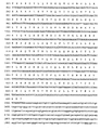
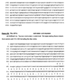
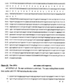
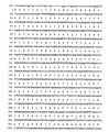
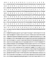
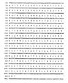
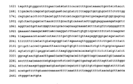
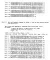
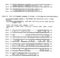
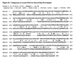
- the nucleotide ( Figure 2 ) and amino acid ( Figure 2 , and Figure 3 ) sequences of 121P2A3 are provided.
- Antibodies that bind to 121P2A3 proteins and polypeptide fragments thereof including polyclonal and monoclonal antibodies, murine and other mammalian antibodies, chimeric antibodies, humanized and fully human antibodies, and antibodies labelled with a detectable marker are disclosed herein.
- the invention provides a method for diagnosing cancer in a test prostate or colon sample, comprising:
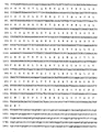
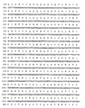
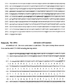
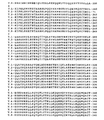
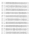
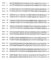
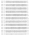
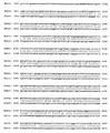
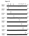
- a reference to 121P2A3 includes all variants thereof, including those shown in Figure 10 and Figure 12 , unless a variant is specified.
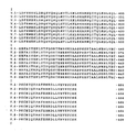
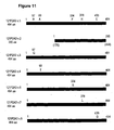
- a reference to 121P2A3 includes all variants thereof, including those shown in Figure 11 , unless a variant is specified.
- prostate cancer means prostate cancers that have extended through the prostate capsule, and are meant to include stage C disease under the American Urological Association (AUA) system, stage C1- C2 disease under the Whitmore-Jewett system, and stage T3 - T4 and N+ disease under the TNM (tumor, node, metastasis) system.
- AUA American Urological Association
- stage C1- C2 disease under the Whitmore-Jewett system
- stage T3 - T4 and N+ disease under the TNM (tumor, node, metastasis) system stage C disease under the American Urological Association (AUA) system
- stage C1- C2 disease under the Whitmore-Jewett system
- TNM tumor, node, metastasis
- analog refers to a molecule which is structurally similar or shares similar or corresponding attributes with another molecule (e.g. a 121P2A3-related protein).
- a 121P2A3-related protein e.g. an analog of a 121P2A3 protein can be specifically bound by an antibody or T cell that specifically binds to 121P2A3.
- Antibody is used in the broadest sense. Therefore an “antibody” can be naturally occuring or man-made such as monoclonal antibodies produced by conventional hybridoma technology.
- Anti-121P2A3 antibodies comprise monoclonal and polyclonal antibodies as well as fragments containing the antigen-binding domain and/or one or more complementarity determining regions of these antibodies.
- an “antibody fragment” is defined as at least a portion of the variable region of the immunoglobulin molecule that binds to its target, i.e., the antigen-binding region. In one embodiment if specifically covers single anti-121P2A3 antibodies and clones thereof (including agonist, antagonist and neutralizing antibodies) and anti-121P2A3 antibody compositions with polyepitopic specificity.
- homolog refers to a molecule which exhibits homology to another molecule, by for example, having sequences of chemical residues that are the same or similar at corresponding positions.
- mammal refers to any organism classified as a mammal, including mice, rats, rabbits, dogs, cats, cows, horses and humans. In one embodiment of the invention, the mammal is a mouse. In another embodiment of the invention, the mammal is a human.
- metastatic prostate cancer and “metastatic disease” mean prostate cancers that have spread to regional lymph nodes or to distant sites, and are meant to include stage D disease under the AUA system and stage TxNxM+ under the TNM system.
- surgery is generally not indicated for patients with metastatic disease, and hormonal (androgen ablation) therapy is a preferred treatment modality.
- Patients with metastatic prostate cancer eventually develop an androgen-refractory state within 12 to 18 months of treatment initiation. Approximately half of these androgen-refractory patients die within 6 months after developing that status.
- the most common site for prostate cancer metastasis is bone. Prostate cancer bone metastases are often osteoblastic rather than osteolytic (i.e., resulting in net bone formation).
- Bone metastases are found most frequently in the spine, followed by the femur, pelvis, rib cage, skull and humerus. Other common sites for metastasis include lymph nodes, lung, liver and brain. Metastatic prostate cancer is typically diagnosed by open or laparoscopic pelvic lymphadenectomy, whole body radionuclide scans, skeletal radiography, and/or bone lesion biopsy.
- the term "monoclonal antibody” refers to an antibody obtained from a population of substantially homogeneous antibodies, i.e., the antibodies comprising the population are identical except for possible naturally occurring mutations that are present in minor amounts.
- a “motif”, as in biological motif of a 121P2A3-related protein, refers to any pattern of amino acids forming part of the primary sequence of a protein, that is associated with a particular function (e.g. protein-protein interaction, protein-DNA interaction, etc) or modification (e.g. that is phosphorylated, glycosylated or amidated), or localization (e.g. secretory sequence, nuclear localization sequence, etc.) or a sequence that is correlated with being immunogenic, either humorally or cellularly.
- a motif can be either contiguous or capable of being aligned to certain positions that are generally correlated with a certain function or property.
- a “pharmaceutical excipient” comprises a material such as an adjuvant, a carrier, pH-adjusting and buffering agents, tonicity adjusting agents, wetting agents, preservative, and the like.
- “Pharmaceutically acceptably refers to a non-toxic, inert, and/or composition that is physiologically compatible with humans or other mammals.
- polynucleotide means a polymeric form of nucleotides of at least 10 bases or base pairs in length, either ribonucleotides or deoxynucleotides or a modified form of either type of nucleotide, and is meant to include single and double stranded forms of DNA and/or RNA. In the art, this term if often used interchangeably with “oligonucleotide”.
- a polynucleotide can comprises a nucleotide sequence disclosed herein wherein thymidine (T), as shown for example in Figure 2 , can also be uracil (U); this definition pertains to the differences between the chemical structures of DNA and RNA, in particular the observation that one of the four major bases in RNA is uracil (U) instead of thymidine (T).
- T thymidine
- U uracil
- polypeptide means a polymer of at least about 4, 5, 6, 7, or 8 amino acids. Throughout the specification, standard three letter or single letter designations for amino acids are used. In the art, this term is often used interchangeably with “peptide” or "protein”.
- variant refers to a molecule that exhibits a variation from a described type or norm, such as a protein that has one or more different amino acid residues in the corresponding position(s) of a specifically described protein (e.g. the 121P2A3 protein shown in Figure 2 or Figure 3 .
- An analog is an example of a variant protein.
- Splice isoforms and single nucleotides polymorphisms (SNPs) are further examples of variants.
- the "121P2A3 proteins” are those specifically identified in figure 3 . Allelic variants, conservative substitution variants, analogs and homologs can be isolated/generated and characterized without undue experimentation following the methods outlined herein or readily available in the art.
- 121P2A3 proteins are polypeptides having all of the amino acid sequence of human 121P2A3 as shown in Figure 2 or Figure 3 .
- 121P2A3 can be conveniently expressed in cells (such as 293T cells) transfected with a commercially available expression vector such as a CMV-driven expression vector encoding 121P2A3 with a C-terminal 6XHis and MYC tag (pcDNA3.1/mycHIS, Invitrogen or Tag5, GenHunter Corporation, Nashville TN).
- the Tag5 vector provides an IgGK secretion signal that can be used to facilitate the production of a secreted 121P2A3 protein in transfected cells.
- the secreted HIS-tagged 121P2A3 in the culture media can be purified, e.g., using a nickel column using standard techniques.
- 121P2A3 protein fragments/subsequences are particularly useful in generating and characterizing domain-specific antibodies (e.g., antibodies recognizing an extracellular or intracellular epitope of a 121P2A3 protein), for identifying agents or cellular factors that bind to 121P2A3 or a particular structural domain thereof, and in various therapeutic and diagnostic contexts, including but not limited to diagnostic essays, cancer vaccines and methods of preparing such vaccines.
- domain-specific antibodies e.g., antibodies recognizing an extracellular or intracellular epitope of a 121P2A3 protein
- Proteins encoded by the 121P2A3 genes, or by analogs, homologs or fragments thereof, have a variety of uses, including but not limited to generating antibodies and in methods for identifying ligands and other agents and cellular constituents that bind to a 121P2A3 gene product.
- Antibodies raised against a 121P2A3 protein or fragment thereof are useful in diagnostic and prognostic assays, and imaging methodologies in the management of human cancers characterized by expression of 121P2A3 protein, such as those listed in Table I.
- immunological assays useful for the detection of 121P2A3 proteins are used, including but not limited to various types of radioimmonoassays, enzyme-linked immunosorbent assays (ELISA), enzyme-linked immunofluorescent assays (ELIFA), immunocytochemical methods, and the like.
- Antibodies can be labeled and used as immunonological imaging reagents capable of detecting 121P2A3-expressing cells (e.g., in radioscintigraphic imaging methods)- 121P2A3 proteins are also particularly useful in generating cancer vaccines, as further described herein.
- Antibodies that bind to 121P2A3 proteins are disclosed herein.
- the antibodies used in the method of the invention specifically bind to a 121P2A3 protein and do not bind (or bind weakly) to peptides or proteins that are not 121P2A3 proteins or fragments thereof.
- 121P2A3 antibodies are particularly useful in cancer (see, e.g., Table I) diagnostic and prognostic assays, and imaging methodologies. Similarly, such antibodies are useful in the diagnosis and/or prognosis of other cancers, to the extent 121P2A3 is also expressed or overexpressed in these other cancers.
- Immunological assays useful for the detection and quantification of 121P2A3 proteins are also disclosed herein.
- Such assays can comprise one or more 121P2A3 antibodies capable of recognizing and binding a 121P2A3 protein, as appropriate.
- These assays are performed within various immunological assay formats well know in the art, including but not limited to various types of radioimmunoassays, enzyme-linked immunosorbent assays (ELISA), enzyme-linked immunofluorescent assays (ELIFA), and the like.
- Immunological non-antibody assays also comprise T cell immunogenicity assays (inhibitory or stimulatory) as well as major histocompatibility complex (MHC) binding assays.
- T cell immunogenicity assays inhibitory or stimulatory
- MHC major histocompatibility complex
- immunological imaging methods capable of detecting prostate cancer and other cancers expressing 121P2A3 are also provided by the invention, including but not limited to riadioscintigraphic imaging methods using labeled 121P2A3 antibodies. Such assays are clinically useful in the detection, monitoring, and prognosis of 121P2A3 expressing cancers such as prostate cancer.
- 121P2A3 antibodies are also used in methods for purifying a 121P2A3 protein and for isolating 121P2A3 homologues and related molecules.
- a method of purifying a 121P2A3 protein comprises incubating a 121P2A3 antibody, which has been coupled to a solid matrix, with a lysate or other solution containing a 121P2A3 protein under conditions that permit the 121P2A3 antibody to bind to the 121P2A3 protein; washing the solid matrix to eliminate impurities; and eluting the 121P2A3 protein from the coupled antibody.
- Other uses of 121P2A3 antibodies include generating anti-idiotypic antibodies that mimic a 121P2A3 protein.
- antibodies can be prepared by immunizing a suitable mammalian host using a 121P2A3 protein, peptide, or fragment, in isolated or immunoconjugated form ( Antibodies: A Laboratory Manual, CSH Press, Eds., Harlow, and Lane (1988 ); Harlow, Antibodies, Cold Spring Harbor Press, NY (1989 )).
- fusion proteins of 121P2A3 can also be used, such as a 121P2A3 GST-fusion protein.
- a GST fusion protein comprising all or most of the amino acid sequence of Figure 2 or Figure 3 is produced, then used as an immunogen to generate appropriate antibodies.
- a 1211P2A3 protein is synthesized and used as an immunogen.
- the amino acid sequence of a 121P2A3 protein as shown in Figure 2 or Figure 3 can be analyzed to select specific regions of the 121P2A3 protein for generating antibodies.
- hydrophobicity and hydrophilicity analyses of a 121P2A3 amino acid sequence are used to identify hydrophilic regions in the 121P2A3 structure.
- Regions of a 121P2A3 protein that show immunogenic structure, as well as other regions and domains can readily be identified using various other methods known in the art, such as Chou-Fasman, Garnier-Robson, Kyte-Doolittle, Eisenberg, Karplus-Schultz or Jameson-Wolf analysis.
- Hydrophilicity profiles can be generated using the method of Hopp, T.P.
- Hydropathicity profiles can be generated using the method of Kyte, J. and Doolittle, R.F., 1982, J. Mol. Biol. 157:105-132 . Percent (%) Accessible Residues profiles can be generated using the method of Janin. J., 1979, Nature 277:491-492 . Average Flexibility profiles can be generated using the method of Bhaskaran R., Ponnuswamy P.K., 1988, Inc. J. Pept. Protein Res. 32:242-255 . Beta-turn profiles can be generated using the method of Deleage, G., Roux B.
- Methods for the generation of 121P2A3 antibodies are further illustrated by way of the examples provided herein.
- Methods for preparing a protein or polypeptide for use as an immunogen are well known in the art.
- methods for preparing immunogenic conjugates of a protein with a carrier such as BSA, KLH or other carrier protein.
- a carrier such as BSA, KLH or other carrier protein.
- direct conjugation using, for example, carbodiimide reagents are used; in other instances linking reagents such as those supplied by Pierce Chemical Co., Rockford, IL, are effective.
- a 121P2A3 immunogen is often conducted by injection over a suitable time period and with use of a suitable adjuvant, as is understood in the art.
- titers of antibodies can be taken to determine adequacy of antibody formation.
- 121P2A3 monoclonal antibodies can be produced by various means well known in the art.
- immortalized cell lines that secrete a desired monoclonal antibody are prepared using the standard hybridoma technology of Kohler and Milstein or modifications that immortalize antibody-producing B cells, as is generally known.
- Immortalized cell lines that secrete the desired antibodies are screened by immunoassay in which the antigen is a 121P2A3 protein.
- the cells can be expanded and antibodies produced either from in vitro cultures or from ascites fluid.
- the antibodies or fragments can also be produced, by recombinant means: Regions that bind specifically to the desired regions of a 121P2A3 protein can also be produced in the context of chimeric or complementarity determining region (CDR) grafted antibodies of multiple species origin. Humanized or human 121P2A3 antibodies can also be produced, and are preferred for use in therapeutic contexts.
- CDR complementarity determining region
- Fully human 121P2A3 monoclonal antibodies can be generated using cloning technologies employing large human Ig gene combinatorial libraries (i.e., phage display) ( Griffiths and Hoogenboom, Building an in vitro immune system: human antibodies from phage display libraries.
- phage display large human Ig gene combinatorial libraries
- Griffiths and Hoogenboom Building an in vitro immune system: human antibodies from phage display libraries.
- Fully human 121P2A3 monoclonal antibodies can also be produced using transgenic mice engineered to contain human immunoglobulin gene loci as described in PCT Patent Application WO98/24893 , Kucherlapati and Jakobovits et al. , published December 3, 1997 (see also, Jakobovits, 1998, Exp. Opin. Invest. Drugs 7(4): 607-614 ; U.S. patents 6,162,963 issued 19 December 2006 ; 6,150,584 issued 12 November 2000 ; and, 6,114598 issued 5 September 2000 ). This method avoids the in vitro manipulation required with phage display technology and efficiently produces high affinity authentic human antibodies.
- Reactivity of 121P2A3 antibodies with a 121P2A3 protein can be established by a number of well known means, including Western blot, immunoprecipitation, ELISA, and FACS analyses using, as appropriate, 121P2A3-related proteins, 121P2A3-expressing cells or extracts thereof.
- a 121P2A3 antibody or fragment thereof can be labeled with a detectable marker or conjugated to a second molecule. Suitable detectable markers include, but are not limited to, a radioisotope, a fluorescent compound, a bioluminescent compound, chemiluminescent compound, a metal chelator or an enzyme.
- bi-specific antibodies specific for two or more 121P2A3 epitopes are generated using methods generally known in the art.
- Homodimeric antibodies can also be generated by cross-linking techniques known in the art (e.g., Wolff et al., Cancer Res. 53; 2560-2565 ).
- 121P2A3 proteins Methods for detecting 121P2A3 proteins, are disclosed herein.
- the Expression profile of 121P2A3 makes it a diagnostic marker for metastasized disease. Accordingly, the status of 121P2A3 gene products provides information useful for predicting a variety of factors including susceptibility to advanced stage disease, rate of progression, and/or tumor aggressiveness.
- Assays for detecting the presence of a 121P2A3 protein in a tissue or other biological sample such as serum, semen, bone, prostate, urine, cell preparations, and the like are also disclosed herein.
- Methods for detecting a 121P2A3 protein are also well known and include, for example, immunoprecipitation, immunohistochemical analysis, Western blot analysis, molecular binding assays, ELISA, ELIFA and the like.
- a method of detecting the presence of a 121P2A3 protein in a biological sample comprises first contacting the sample with a 121P2A3 antibody, a 121P2A3-reactive fragment thereof, or a recombinant protein containing an antigen binding region of a 121P2A3 antibody; and then detecting the binding of 121P2A3 protein in the sample.
- Oncogenesis is known to be a multistep process where cellular growth becomes progressively dysregulated and cells progress from a normal physiological state to precancerous and then cancerous states (see, e.g., Alers et al., Lab Invest. 77(5): 437-438 (1997) and Isaacs et al., Cancer Surv. 23: 19-32 (1995) ).
- examining a biological sample for evidence of dysregulated cell growth allows for early detection of such aberrant physiology, before a pathologic state such as cancer has progressed to a stage that therapeutic options are more limited and or the prognosis is worse.
- the status of 121P2A3 in a biological sample of interest can be compared, for example, to the status of 121P2A3 in a corresponding normal sample (e.g. a sample from that individual or alternatively another individual that is not affected by a pathology).
- a corresponding normal sample e.g. a sample from that individual or alternatively another individual that is not affected by a pathology.
- An alteration in the status of 121P2A3 in the biological sample provides evidence of dysregulated cellular growth.
- a predetermined Normative value such as a predetermined normal level of mRNA expression (see, e.g., Grever et al., J. Comp. Neurol. 1996 Dec 9; 376(2): 306-14 and U.S. Patent No. 5,837,501 ) to compare 121P2A3 status in a sample.
- status in this context is used according to its art accepted meaning and refers to the condition or state of a gene and its products.
- skilled artisans use a number of parameters to evaluate the condition or state of a gene and its products. These include, but are not limited to the location of expressed gene products (including the location of 121P2A3 expressing cells) as well as the level, and biological activity of expressed gene products (such as 121P2A3 polypeptides).
- an alteration in the status of 121P2A3 comprises a change in the location of 121P2A3 and/or 121P2A3 expressing cells and/or an increase in 121P2A3 protein expression.
- 121P2A3 status in a sample can be analyzed by a number of means well known in the art, including without limitation , immunohistochemical analysis, Western blot analysis, and tissue array analysis. Typical protocols for evaluating the status of a 121P2A3 protein are found, for example in Ausubel et al. eds., 1995, Current Protocols In Molecular Biology , Units 2 (Northern Blotting), 4 (Southern Blotting), 15 (Immunoblotting) and 18 (PCR Analysis).
- the status of 121P2A3 in a biological sample is evaluated by various methods utilized by skilled artisans including, but not limited to Western and/or immunohistochemical analysis (to examine, for example alterations in polypeptide sequences, alterations in polypeptide localization within a sample, alterations in expression levels of 121P2A3 proteins and/or associations of 121P2A3 proteins with polypeptide binding partners).
- Western and/or immunohistochemical analysis to examine, for example alterations in polypeptide sequences, alterations in polypeptide localization within a sample, alterations in expression levels of 121P2A3 proteins and/or associations of 121P2A3 proteins with polypeptide binding partners.
- the Expression profile of 121P2A3 makes it a diagnostic marker for local and/or metastasized disease, and provides information on the growth or oncogenic potential of a biological sample.
- the status of 121P2A3 provides information useful for predicting susceptibility to particular disease stages, progression, and/or tumor aggressiveness.
- the invention provides methods and assays for determining 121P2A3 status and diagnosing cancers that express 121P2A3, such as cancers of the tissues listed in Table I.
- 121P2A3 mRNA is so highly expressed in prostate and other cancers relative to normal prostate tissue
- assays that evaluate the levels of 121P2A3 mRNA transcripts or proteins in a biological sample can be used to diagnose a disease associated with 121P2A3 dysregulation, and can provide prognostic information useful in defining appropriate therapeutic options.
- the expression status of 121P2A3 provides information including the presence, stage and location of dysplastic, precancerous and cancerous cells, predicting susceptibility to various stages of disease, and/or for gauging tumor aggressiveness. Moreover, the expression profile makes it useful as an imaging reagent for metastasized disease. Consequently, an aspect of the invention is directed to the various molecular prognostic and diagnostic methods for examining the status of 121P2A3 in biological samples such as those from individuals suffering from, or suspected of suffering from a pathology characterized by dysregulated cellular growth, such as cancer.
- the status of 121P2A3 in a biological sample can be examined by a number of well-known procedures in the art.
- the status of 121P2A3 in a biological sample taken from a specific location in the body can be examined by evaluating the sample for the presence or absence of 121P2A3 expressing cells (e.g. those that express 121P2A3 mRNAs or proteins).
- This examination can provide evidence of dysregulated cellular growth, for example, when 121P2A3-expressing cells are found in a biological sample that does not normally contain such cells (such as a lymph node), because such alterations in the status of 121P2A3 in a biological sample are often associated with dysregulated cellular growth.
- one indicator of dysregulated cellular growth is the metastases of cancer cells from an organ of origin (such as the prostate) to a different area of the body (such as a lymph node).
- evidence of dysregulated cellular growth is important for example because occult lymph node metastases can be detected in a substantial proportion of patients with prostate cancer, and such metastases are associated with known predictors of disease progression (see, e.g., Murphy et al., Prostate 42(4): 315-317 (2000) ; Su et al., Semin. Surg. Oncol. 18(1): 17-28 (2000) and Freeman et al., J Urol 1995 Aug 154(2 Pt 1):474-8 ).
- methods for monitoring 121P2A3 gene products by determining the status of 121P2A3 gene products expressed by cells from an individual suspected of having a disease associated with dysregulated cell growth (such as hyperplasia or cancer) and then comparing the status so determined to the status of 121P2A3 gene products in a corresponding normal sample are disclosed herein.
- the presence of aberrant 121P2A3 gene products in the test sample relative to the normal sample provides an indication of the presence of dysregulated cell growth within the cells of the individual.
- Assays useful in determining the presence of cancer in an individual comprising detecting a significant increase in 121P2A3 protein expression in a test cell or tissue sample relative to expression levels in the corresponding normal cell or tissue are described herein.
- the presence of significant 121P2A3 expression in any of the tissues listed in table I is useful to indicate the emergence, presence and/or severity of a cancer, since the corresponding normal tissues do not express 121P2A3 mRNA or express it at lower levels.
- 121P2A3 status is determined at the protein level rather than at the nucleic acid level.
- a method comprises determining the level of 121P2A3 protein expressed by cells in a test tissue sample and comparing the level so determined to the level of 121P2A3 expressed in a corresponding normal sample.
- the presence of 121P2A3 protein is evaluated, for example, using immunohistochemical methods.
- 121P2A3 antibodies or binding partners capable of detecting 121P2A3 protein expression are used in a variety of assay formats well known in the art for this purposes.
- These perturbations can include insertions, deletions, substitutions and the like.
- Such evaluations are useful because perturbations in the nucleotide and amino acid sequences are observed in a large number of proteins associated with a growth dysregulated phenotype (see, e.g., Marrogi et al., 1999, J. Cutan, Pathol. 26(8):369-378 ).
- a mutation in the sequence of 121P2A3 may be indicative of the presence or promotion of a tumor.
- Such assays therefore have diagnostic and predictive value where a mutation in 121P2A3 indicates a potential loss of function or increase in tumor growth.
- a method for predicting susceptibility to cancer comprises detecting
- 121P2A3 protein in a tissue sample its presence indicating susceptibility to cancer, wherein the degree of 121P2A3 expression correlates to the degree of susceptibility.
- the presence of 121P2A3 in prostate or other tissue is examined, with the presence of 121P2A3 in the sample providing an indication of prostate cancer susceptibility (or the emergence or existence of a prostate tumor).
- the presence of one or more perturbations in 121P2A3 gene products in the sample is an indication of cancer susceptibility (or the emergence or existence of tumor).
- a method for gauging aggressiveness of a tumor comprises determining the level of 121P2A3 protein expressed by tumor cells, comparing the level so determined to the level of 121P2A3 protein expressed in a corresponding normal tissue taken from the same individual or a normal tissue reference sample, wherein the degree of 121P2A3 protein expression in the tumor sample relative to the normal sample indicates the degree of aggressiveness.
- aggressiveness of a tumor is evaluated by determining the extent to which 121P2A3 is expressed in the tumor cells, with higher expression levels indicating more aggressive tumors.
- Another embodiment is the evaluation of the integrity of 121P2A3 amino acid sequences in a biological sample, in order to identify perturbations in the structure of these molecules such as insertions, deletions, substitutions and the like. The presence of one or more perturbations indicates more aggressive tumors.
- methods for observing the progression of a malignancy in an individual over time comprise determining the level of 121P2A3 protein expressed by cells in a sample of the tumor, comparing the level so determined to the level of 121P2A3 protein expressed in an equivalent tissue sample taken from the same individual at a different time, wherein the degree of 121P2A3 protein expression in the tumor sample over time provides information on the progression of the cancer.
- the progression of a cancer is evaluated by determining 121P2A3 expression in the tumor cells over time, where increased expression over time indicates a progression of the cancer.
- the above diagnostic approaches can be combined with any one of a wide variety of prognostic and diagnostic protocols known in the art. For example, methods for observing a coincidence between the expression of 121P2A3 gene products (or perturbations in 121P2A3 gene products) and a factor that is associated with malignancy, as a means for diagnosing and prognosticating the status of a tissue sample are also disclosed herein.
- a wide variety of factors associated with malignancy can be utilized, such as the expression of genes associated with malignancy (e.g. PSA, PSCA and PSM expression for prostate cancer etc.) as well as gross cytological observations (see, e.g., Bocking et al.,1984, Anal. Quant. Cytol.
- methods for observing a coincidence between the expression of 121P2A3 gene products (or perturbations in 121P2A3 gene products) and another factor associated with malignancy entails detecting the overexpression of 121P2A3 protein in a tissue sample, detecting the overexpression of PSA mRNA or protein in a tissue sample (or PSCA or PSM expression), and observing a coincidence of 121P2A3 protein and PSA mRNA or protein overexpression (or PSCA or PSM expression).
- the expression of 121P2A3 and PSA mRNA in prostate tissue is examined, where the coincidence of 121P2A3 and PSA mRNA overexpression in the sample indicates the existence of prostate cancer, prostate cancer susceptibility or the emergence or status of a prostate tumor.
- polyclonal or monoclonal antibodies specifically reactive with the wild-type 121P2A3 protein can be used in an immunohistochemical assay of biopsied tissue.
- 121P2A3 anti-polypeptide antibodies arc used in well known diagnostics and prognostic assays that examine conditions associated with dysregulated cell growth such as cancer, in particular the cancers listed in Table I (see, e.g., both its specific pattern of tissue expression as well as its overexpression in certain cancers as described for example in the Example entitled "Expression analysis of 121P2A3 in normal tissues, and patient specimens").
- 121P2A3 can be analogized to a prostate associated antigen PSA, the archetypal marker that has been used by medical practitioners for years to identify and monitor the presence of prostate cancer (see, e.g., Merrill et al., J. Urol. 163(2): 503-5120 (2000) ; Polascik et al., J. Urol. Aug; 162(2):293-306 (1999) and Fortier et al., J. Nat. Cancer lost. 91(19): 1635-1640(1999) ).
- a variety of other diagnostic markers are also used in similar contexts including p53 and K-ras (see, e.g., Tulchinsky et al., Int.
- Typical embodiments of diagnostic methods which utilize the 121P2A3 antibodies are analogous to those methods from well-established diagnostic assays which employ, e.g., PSA antibodies.
- PSA polypeptides are used to generate antibodies specific for PSA which can then be used to observe the presence and/or the level of PSA proteins in methods to monitor PSA protein overexpression (see, e.g., Stephan et al., Urology 55(4):560-3 (2000) ) or the metastasis of prostate cells (see, e.g., Alanen et al., Pathol. Res.
- 121P2A3 polypeptides described herein can be utilized to generate antibodies for use in detecting 121P2A3 overexpression or the metastasis of prostate cells and cells of other cancers expressing this gene.
- metastases involves the movement of cancer cells from an organ of origin (such as the lung or prostate gland etc.) to a different area of the body (such as a lymph node)
- assays which examine a biological sample for the presence of cells expressing 121P2A3 polypeptides can be used to provide evidence of metastasis.
- tissue that does not normally contain 121P2A3-expressing cells lymph node
- 121P2A3-expressing cells such as the 121P2A3 expression seen in LAPC4 and LAPC9
- 121P2A3 polypeptides can be used to provide evidence of cancer, for example, when cells in a biological sample that do not normally express 121P2A3 or express 121P2A3 at a different level are found to express 121P2A3 or have an increased expression of 121P2A3 (see, e.g., the 121P2A3 expression in the cancers listed in Table I and in patient samples etc. shown in the accompanying Figures).
- artisans may further wish to generate supplementary evidence of metastasis by testing the biological sample for the presence of a second tissue restricted marker (in addition to 121P2A3) such as PSA, PSCA etc. (see, e.g., Alanen et al., Pathol. Res. Pract. 192(3): 233-237 (1996) ).
- PSA polypeptides which contain an epitope that can be recognized by an antibody or T cell that specifically blinds to that epitope are used in methods of monitoring PSA.
- 121P2A3 polypeptide fragments and polypeptide analogs or variants can also be used in an analogous manner.
- This practice of using polypeptide fragments or polypeptide variants to generate antibodies is typical in the art with a wide variety of systems such as fusion proteins being used by practitioners (see, e.g., Current Protocols In Molecular Biology, Volume 2, Unit 16, Frederick M. Ausubel et al. eds., 1995 ).
- each epitope(s) functions to provide the architecture with which an antibody or T cell is reactive.
- polypeptide fragments that can be used in order to generate immune responses specific for different portions of a polypeptide of interest (see, e.g., U. S. Patent No. 5,840,501 and U.S. Patent No. 5,939,533 ).
- a polypeptide comprising one of the 121P2A3 biological motifs discussed herein or a motif-bearing subsequence which is readily identified by one of skill in the art based on motifs available in the art
- Polypeptide fragments, variants or analogs are useful in this context as long as they comprise an epitope capable of generating an antibody specific for a 121P2A3 polypeptide shown in Figure 3 .
- the 121P2A3 polypeptides, as well as the anti-121P2A3 antibodies used to identify the presence of these molecules exhibit specific properties that make them useful in diagnosing cancers such as those listed in Table I. Diagnostic assays that measure the presence of 121P2A3 gene products, in order to evaluate the presence or onset of a disease condition described herein, such as prostate cancer, arc used to identify patients for preventive measures or further monitoring, as has been done so successfully with PSA.
- these materials satisfy a need in the art for molecules having similar or complementary characteristics to PSA in situations where, for example, a definite diagnosis of metastasis of prostatic origin cannot be made on the basis of a test for PSA alone (see, e.g., Alanen et al., Pathol. Res. Pract. 192(3): 233-237 (1996) ), and consequently, materials such as anti-121P2A3 antibodies, need to be employed to confirm a metastases of prostatic origin.
- kits are also disclosed herein.
- Such kits can comprise a carrier, package or container that is compartmentalized to receive one or more containers such as vials, tubes, and the like, each of the container(s) comprising one of the separate elements to be used in the method.
- the container(s) can comprise a probe that is or can be detectably labeled.
- Such probe can be an antibody specific for a 121P2A3
- the kit can include all or part of the amino acid sequence of Figure 2 or Figure 3 or analogs thereof, or a nucleic acid molecules that encodes such amino acid sequences.
- the kit of the invention will typically comprise the container described above and one or more other containers comprising materials desirable from a commercial and user standpoint, including buffers, diluents, filters, needles, syringes, and package inserts with instructions for use.
- a label can be present on the container to indicate that the composition is used for a specific therapy or non-therapeutic application, and can also indicate directions for either in vivo or in vitro use, such as those described above. Directions and or other information can also be included on an insert which is included with the kit.
- Example 1 SSH-Generated Isolation of a cDNA Fragment of the 121P2A3 Gene
- the gene 121P2A3 was derived from an LAPC-9 AD minus LAPC-9 AD (28 days post-castration) subtraction.
- the SSH DNA sequence of 259 bp is listed in Figure 1 .
- a cDNA (121P2A3 clone 5) of 2473 bp was isolated from a LAPC-9AD cDNA library, revealing an ORF of 464 amino acids ( Figures 2 and 3 ).
- Variants of 121P2A3 v.1 were also identified, and these are listed in Figures 2 and 3 .
- the 121P2A3 protein shows homology to a novel hypothetical protein FLJ10540 isolated from the human teratocarcinoma cell line NT2 ( Figure 4B and 4D ). The two proteins are 98% identical over a 223 amino acid region starting from position 170 of 121P2A3 v.1.
- the 121P2A3 protein also shows homology to the XM_005908 (similar to RIKEN cDNA 1200008012) gene.
- the gene XM_005908 was isolated from rhabdomyosarcoma cDNA library, validating the expression of 121P2A3 in human cancers.
- 121P2A3 v.1 and XM_005908 proteins are 99% identical over 464 amino acids ( Figure 4E ).
- 121P2A3 Amino acid sequence analysis of 121P2A3 reveals 75% identity over 464 amino acid region to a mouse putative protein ( Figure 4F ). 121P2A3 v.1 also shows homology to the human nef-associated factor-1 (naf-1), The two proteins are 23% identical over a 339 amino acid region ( Figure 4G ).
- LAPC Xenografts and Human Tissues
- LAPC xenografts were obtained from Dr. Charles Sawyers (UCLA) and generated as described ( Klein et al, 1997, Nature Med. 3: 402-408 ; Craft et al., 1999, Cancer Res. 59: 5030-5036 ). Androgen dependent and independent LAPC-9 AD and AI xenografts were grown in male SCID mice and were passaged as small tissue chunks in recipient males.
- LAPC-9 AI xenografts were derived from LAPC-9 AD tumors, respectively. To generate the AI xenografts, male mice bearing AD tumors were castrated and maintained for 2-3 months. After the tumors re-grew, the tumors were harvested and passaged in castrated males or in female SCID mice.
- Human cell lines e.g., HeLa
- DMEM fetal calf serum
- the patient cancer and normal tissues were purchased from different sources such as the NDRI (Philadelphia, PA).
- mRNA for some normal tissues were purchased from Clontech, Palo Alto, CA.
- Tissues were homogenized in Trizol reagent (Life Technologies, Gibco BRL) using 10 ml/ g tissue isolate total RNA. Poly A RNA was purified from total RNA using Qiagen's Oligotex mRNA Mini and Midi kits. Total and mRNA were quantified by spectrophotometric analysis (O.D. 260/280 nm) and analyzed by gel electrophoresi
- SSH Suppression Subtractive Hybridization
- the gene 121P2A3 was derived from an LAPC-9 AD tumor (grown in intact male mouse) minus an LAPC-9 AD tumor (28 days post-castration) subtraction.
- the SSH DNA sequence 121P2A3 ( Figure 1 ) was identified.
- the cDNA derived from an LAPC-9 AD tumor (28 days post-castration) was used as the source of the "driver” cDNA, while the cDNA from the LAPC-9 AD tumor (grown in intact male mouse) was used as the source of the "tester” cDNA.
- Double stranded cDNAs corresponding to tester and driver cDNAs were synthesized from 2 ⁇ g of poly(A) + RNA isolated from the relevant xenograft tissue, as described above, using CLONTECH's PCR-Select cDNA Subtraction Kit and 1 ng of oligonucleotide DPNCDN as primer. First-and second-strand synthesis were carried out as described in the Kit's user manual protocol (CLONTECH Protocol No. PT1117-1, Catalog No. K1804-1). The resulting cDNA was digested with Dpn II for 3 hrs at 37°C. Digested cDNA was extracted with phenol/chloroform (1:1) and ethanol precipitated.
- Driver cDNA was generated by combining in a 1:1 ratio Dpn II digested cDNA from the relevant xenograft source (see above) with a mix of digested cDNAs derived from the human cell lines HeLa, 293, A431, Colo205, and mouse liver.
- Tester cDNA was generated by diluting 1 ⁇ l of Dpn II digested cDNA from the relevant xenograft source (see above) (400 ng) in 5 ⁇ l of water. The diluted cDNA (2 ⁇ l, 160 ng) was then ligated to 2 ⁇ l of Adaptor 1 and Adaptor 2 (10 ⁇ M), in separate ligation reactions, in a total volume of 10 ⁇ l at 16°C overnight, using 400 u of T4 DNA ligase (CLONTECH). Ligation was terminated with 1 ⁇ l of 0.2 M EDTA and heating at 72°C for 5 min.
- the first hybridization was performed by adding 1.5 ⁇ l (600 ng) of driver cDNA to each of two tubes containing 1.5 ⁇ l (20 ng) Adaptor 1- and Adaptor 2- ligated tester cDNA. In a final volume of 4 ⁇ l, the samples were overlaid with mineral oil, denatured in an MJ Research thermal cycler at 98°C for 1.5 minutes, and then were allowed to hybridize for 8 hrs at 68°C. The two hybridizations were then mixed together with an additional 1 ⁇ l of fresh denatured driver cDNA and were allowed to hybridize overnight at 68°C. The second hybridization was then diluted in 200 ⁇ l of 20 mM Hepes, pH 8.3, 50 mM NaCl, 0.2 mM EDTA, heated at 70°C for 7 min. and stored at -20°C.
- PCR 1 was conducted using the following conditions: 75°C for 5 min., 94°C for 25 sec., then 27 cycles of 94°C for 10 sec, 66°C for 30 sec, 72°C for 1.5 min. Five separate primary PCR reactions were performed for each experiment.
- the products were pooled and diluted 1:10 with water.
- 1 ⁇ l from the pooled and diluted primary PCR reaction was added to the same reaction mix as used for PCR 1, except that primers NP1 and NP2 (10 ⁇ M) were used instead of PCR primer 1, PCR 2 was performed using 10-12 cycles of 94°C for 10 sec, 68°C for 30 sec, and 72°C for 1.5 minutes.
- the PCR products were analyzed using 2% agarose gel electrophoresis.
- PCR products were inserted into pCR2.1 using the T/A vector cloning kit (Invitrogen). Transformed E. coli were subjected to blue/white and ampicillin selection. White colonies were picked and arrayed into 96 well plates and were grown in liquid culture overnight. To identify inserts, PCR amplification was performed on 1 ml of bacterial culture using the conditions of PCR1 and MP1 and NP2 as primers. PCR products were analyzed using 2% agarose gel electrophoresis.
- Bacterial clones were stored in 20% glycerol in a 96 well format. Plasmid DNA was prepared, sequenced, and subjected to nucleic acid homology searches of the GenBank, dBest, and NCR-CGAP databases.
- First strand cDNAs can be generated from 1 ⁇ g of mRNA with oligo (dT)12-18 priming using the Gibco-BRL Superscript Preamplification system. The manufacturer's protocol was used which included an incubation for 50 min at 42°C with reverse transcriptase followed by RNAse H treatment at 37°C for 20 min. After completing the reaction, the volume can be increased to 200 ⁇ l with water prior to normalization. First strand cDNAs from 16 different normal human tissues can be obtained from Clontech,
- First strand cDNA (5 ⁇ l) were amplified in a total volume of 50 ⁇ l containing 0.4 ⁇ M primers, 0.2 ⁇ M each dNTPs, 1XPCR buffer (Clontech, 10 mM Tris-HCL, 15 mM MgCl 2 , 50 mM KCl, pH8.3) and 1X Klentaq DNA polymerase (Clontech).
- PCR Five ⁇ l of the PCR reaction can be removed at 18, 20, and 22 cycles and used for agarose gel electrophoresis.
- PCR was performed using an MJ Research thermal cycler under the following conditions: initial denaturation can be at 94°C for 15 sec, followed by a 18, 20, and 22 cycles of 94°C for 15, 65°C for 2 min, 72°C for 5 sec. A final extension at 72°C was carried out for 2 min.
- the band intensities of the 283 b.p. ⁇ -actin bands from multiple tissues were compared by visual inspection. Dilution factors for the first strand cDNAs were calculated to result in equal ⁇ -actin band intensities in all tissues after 22 cycles of PCR. Three rounds of normalization can be required to achieve equal band intensities in all tissues after 22 cycles of PCR.
- First strand cDNA was prepared from vital po 1 (liver, lung and kidney), vital pool 2 (pancreas, colon and stomach), LAPC xenograft pool (LAPC-4AD, LAPC-4AL, LAPC-9AD and LAPC-9AI), prostate cancer pool, bladder cancer pool, kidney cancer pool, colon cancer pool lung cancer pool, ovary cancer pool, breast cancer pool, and cancer metastasis pool. Normalization was performed t PCR using primers to actin and GAPDH. Semi-quantitative PCR, using primers to 121P2A3, was performed at 26 and 30 cycles of amplification.
- Results show strong expression of 121P2A3 in LAPC xenograft pool, bladder caner pool, kidney cancer pool, colon cancer pool, lung cancer pool, ovary cancer pool, breast cancer pool, and cancer metastasis pool. Expression of 121P2A3 was also detected in prostate cancer pooL Very low expression was detect in vital pool 1 and 2.
- the gene 121P2A3 was derived from an LAPC-9 AD (no castration) minus LAPC-9AD (28 days post-castration) subtraction.
- the SSH DNA sequence ( Figure 1 ) was designated 121P2A3.
- cDNA clone 121P2A3-clone 5 ( Figure 4 ) was identified by screening an LAPC-9AD cDNA library (Lambda ZAP Express, Stratagene) using the 121P2A3 SSH DNA as a probe.
- 121P2A3 clone 5 cDNA was deposited under the terms of the Budapest Treaty on 1 March 2001, with the American Type Culture Collection (ATCC; 10801 University Boulevard., Manassas, VA 20110-2209 USA) as plasmid 121P2A3-5, and bas been assigned Accession No. PTA-3138.
- the mapping program placed 121P2A3 on chromosome 10q23.32, a genomic region found to be rearranged in certain cancers.
- Example 4 Expression analysis of 121P2A3 in normal tissues, cancer cell lines and patient samples
- First strand cDNA was prepared from vital pool 1 (liver, lung and kidney), vital pool 2 (pancreas, colon and stomach), LAPC xenograft pool (LAPC-4AD, LAPC-4AI, LAPC-9AD and LAPC-9AI), prostate cancer pool, bladder cancer pool, kidney cancer pool, colon cancer pool, lung cancer pool, ovary cancer pool, breast cancer pool, and cancer metastasis pool. Normalization was performed by PCR using primers to actin and GAPDH. Semi-quantitative PCR, using primers to 121P2A3, was performed at 26 and 30 cycles of amplification.
- Results show strong expression of 121P2A3 in LAPC xenograft pool, bladder cancer pool, kidney cancer pool, colon cancer pool, lung cancer pool, ovary cancer pool, breast cancer pool, and cancer metastasis pool. Expression of 121P2A3 was also detected in prostate cancer pool. Very low expression was detected in vital pool I and 2.
- 121P2A3 expression was detected in all cell lines tested ( Figure 16 ).
- RNA was extracted from a number of human cancer cell lines. Northern blots with 10 ug of total RNA/lane were probed with the 121P2A3 SSH fragment.
- results show expression in prostate (LAPC4AD, LAPC4AI, LAPC9AD, LAPC 9AI, LNCaP, PC-3, DUI45, Tsu-Prl and LAPC-4 CL), bladder (HT1197, SCaBER, UM-UC-3, TCCSUP, J82, 5637), 293T cell line, Ewing's sarcoma (RD-ES), pancreas (PANC-1, Bx PC-3, HPAC, Capan-1) colon (SK-CO-1, Caco-2, LoVo, T84, Colo205), breast (CAMA-1, DU4475, MCF-7, MDA-MB435s), testicular (NTERRA-2, NCCIT, TERA-1, TERA-2), cervical (A431), ovarian (OV-1063, PA-1, SW 626), brain (PFSK-1, T98G) and bone (SK-ES-1, HOS, U-2 OS, RD-ES) cancer cell lines.
- Figure 13 shows that 121P2A3 was expressed in kidney cancer patient specimens.
- RNA was extracted from kidney cancer cell lines (CL: 769-P, A498, SW839), normal kidney (NK), kidney cancer patient tumors (T) and their normal adjacent tissues (N).
- Northern blots with 10 ug of total RNA were probed with the 121P2A3 SSH sequence. Size standards in kilobases are on the side. Results show expression of 121P2A3 in patient kidney tumor tissues and in all kidney cancer cell lines tested, but not in normal kidney.
- 121P2A3 is also expressed in stomach, and rectum patient cancer samples ( Figure 19 ).
- the expression detected in normal adjacent tissues (isolated from diseased tissues) but not in normal tissues (isolated from healthy donors) indicates that these tissues are not fully normal and that 121P2A3 can be expressed in early stage tumors.
- 121P2A3 was also found to be highly expressed in the nine human cancer cell lines tested, the cervical carcinoma HeLa, the CML line K562, the PML line HL-60, the melanoma line G361, the lung carcinoma line A549, the lymphoblastic leukemia line MOLT-4, the colorectal carcinoma SW480, and Burkitt's lymphoma lines Daudi and Raji.
- LAPC-9AD tumor cells were injected in male mice ( Figure 20 ). When tumor reached a palpable size (0.3-0.5cm in diameter), mice were castrated and tumors harvested at different time points following castration. RNA was isolated from the xenograft tissues. Northern blots with 10 ug of total RNA/lanc were probed with the 121P2A3 SSH fragment. Size standards in kilobases (kb) are indicated on the side. Results show expression of 121P2A3 is downregulated within 7 days of castration.
- 121P2A3 expression is reminiscent of a cancer-testis gene. Its restricted normal tissue expression and the upregulation detected in human cancers indicate that 121P2A3 is therapeutic and prophylactic target and a diagnostic and prognostic marker for human cancers.
- Transcript variants are variants of mature mRNA from the same gene which arise by alternative transcription or alternative splicing, Alternative transcripts are transcripts from the same gene but start transcription at different points. Splice variants are mRNA variants spliced differently from the same transcript. In eukaryotes, when a multi-exon gene is transcribed from genomic DNA, the initial RNA is spliced to produce functional mRNA, which has only exons and is used for translation into an amino acid sequence. Accordingly, a given gene can have zero to many Alternative transcripts and each transcript can have zero to many splice variants. Each transcript variant has a unique exon makeup, and can have different coding and/or non-coding (5' or 3' end) portions, from the original transcript.
- Transcript variants can code for similar or different proteins with the same or a similar function or can encode proteins with different functions, and can be expressed in the same tissue at the same time, or in different tissues at the same time, or in the same tissue, at different times, or in different tissues at different times. Proteins encoded by transcript variants can have similar or different cellular or extracellular localizations, e.g., secreted versus intracellular.
- Transcript variants are identified by a variety of art-accepted methods. For example, alternative transcripts and splice variants are identified by full-length cloning experiment or by use of full-length transcript and EST sequences. First, all human ESTs were grouped into clusters which show direct or indirect identity with each other. Second, ESTs in the same cluster were further grouped into sub-clusters and assembled into a consensus sequence. The original gene sequence is compared to the consensus sequence(s) or other full-length sequences. Each consensus sequence is a potential splice variant for that gene (see, e.g., URL www.doubletwist.com/products/c11_agentsOverview.jhtml). Even when a variant is identified that is not a full-length clone, that portion of the variant is very useful for antigen generation and for further cloning of the full-length splice variant, using technique known in the art,
- Genomic-based transcript variant identification programs include FgenesH ( A. Salamov and V. Solovyev, "Ab initio gene finding in Drosophila genomic DNA,” Genome Research. 2000 April; 10(4):516-22 ); Grail (URL compbio.ornl.gov/Grail-bin/EmptyGrailForm) and GenScan (URL genes.mit.edu/GENSCAN.html).
- FgenesH A. Salamov and V. Solovyev, "Ab initio gene finding in Drosophila genomic DNA," Genome Research. 2000 April; 10(4):516-22
- Grail URL compbio.ornl.gov/Grail-bin/EmptyGrailForm
- GenScan URL genes.mit.edu/GENSCAN.html
- PCR-based Validation Wellmann S, et al., Specific reverse transcription-PCR quantification of vascular endothelial growth factor (VEGF) splice variants by LightCycler technology, Glin Chem. 2001 Apr,47(4)-654-60 ; Jia, H.P., et al., Discovery of new human beta-defensins using a genomics-based approach, Gene. 2001 Jan 24; 263(1-2):211-8 .
- PCR-based and 5' RACE Validation Brigle, K.E., et al., Organization of the marine reduced folate carrier gene and identification of variant splice forms, Biochem Biopbys Acta. 1997 Aug 7; 1353(2): 191-8 ).
- genomic regions are modulated in cancers.
- the alternative transcripts or splice variants of the gene are modulated as well.
- 121P2A3 has a particular expression profile related to cancer.
- Alternative transcripts and splice variants of 121P2A3 may also be involved in cancers in the same or different tissues, thus serving as tumor-associated markers/antigens.
- the exon composition of the original transcript designated as 121P2A3 v.1, is shown in Table LIII.
- 121P2A3 v.2 Using the full-length gene and EST sequences, one transcript variant was identified, designated as 121P2A3 v.2. Compared with 121P2A3 v.1, transcript variant 121P2A3 v.2 has a shorter exon 2, as shown in Figure 12 . All other exons are the same corresponding exons of 121P2A3 v.1.
- each different combination of exons in spatial order e.g. exons 2 and 3 is a potential splice variant
- Figure 12 shows the schematic alignment of exons of the two transcript variants.
- Table LIV shows nucleotide sequence of the transcript variant.
- Table LV shows the alignment of the transcript variant with nucleic acid sequence of 121P2A3 v-1.
- Table LVI lays out amino acid translation-of the transcript variant for the identified reading frame orientation.
- Table LVII displays alignments of the amino acid sequence encoded by the splice variant with that of 121P2A3 v.1.
- a Single Nucleotide Polymorphism is a single base pair variation in a nucleotide sequence at a specific location.
- SNP Single Nucleotide Polymorphism
- A/T refers to the specific base pair sequence of one or more locations in the genome of an individual.
- Haplotype refers to the base pair sequence of more than one location on the same DNA molecule (or the same chromosome in higher organisms), often in the context of one gene or in the context of several tightly linked genes.
- SNPs that occur on a cDNA are called cSNPs.
- cSNPs may change amino acids, of the protein encoded by the gene and thus change the functions of the protein.
- Some SNPs cause inherited diseases; others contribute to quantitative variations in phenotype and reactions to environmental factors including diet and drugs among individuals. Therefore, SNPs and/or combinations of alleles (called haplotypes) have many applications, including diagnosis of inherited diseases, determination of drug reactions and dosage, identification of genes responsible for diseases, and analysis of the genetic relationship between individuals ( P. Nowotny, J. M. Kwon and A. M. Goate, " SNP analysis to dissect human traits," Curr. Opin. Neurobiol. 2001 Oct; 11(5):637-641; M. Pirmohamed and B. K.
- SNPs are identified by a variety of art-accepted method ( P. Bean, "The promising voyage of SNP target discovery," Am. Clin. Lab. 2001 Oct-Nov; 20(9):18-20 ; K. M. Weiss, "In search of human variation,” Genome Res. 1998 Jul; 8(7):691-697 ; M. M. She, “Enabling large-scale pharmacogenetic studies by high-throughput mutation detection and genotyping technologies,” Clin. Chem. 2001 Feb; 47(2):164-172 ).
- SNPs are identified by sequencing DNA fragments that show polymorphism by gel-based methods such as restriction fragment length polymorphism (RFLP) and denaturing gradient gel electrophoresis (DGGE).
- RFLP restriction fragment length polymorphism
- DGGE denaturing gradient gel electrophoresis
- SNPs can also be discovered by direct sequencing of DNA samples pooled from different individuals or by comparing sequences from different DNA samples. With the rapid accumulation of sequence data in public and private databases, one can discover SNPs by comparing sequences using computer programs ( Z. Gu, L. Hillier and P. Y. Kwok, "Single nucleotide polymorphism hunting in cyberspace,” Hum. Mutat 1998; 12(4):221-225 ). SNPs can be verified and genotype or haplotype of an individual can be determined by a variety of methods including direct sequencing and high throughput microarrays ( P. Y. Kwok, "Methods for genotyping single nucleotide polymorphisms," Annu. Rev. Genomics Hum. Genet.
- transcripts or proteins with alternative alleles were designated as variants 121P2A3 v.3, v.4, v.5, v.6, v.7, v.8 and v.9.
- Figure 10 and Figure 12 show the schematic alignment of the nucleotide variants.
- Figure 11 shows the schematic alignment of protein variants, corresponding to nucleotide variants.
- Nucleotide variants that code for the same amino acid sequence as variant 1 are not shown in Figure 11 , These alleles of the SNPs, though shown separately here, can occur in different combinations (haplotypes) and in any one of the transcript variants (such as 121P2A3 v.2) that contains the sequence context of the SNPs.
- Figure 4A and Table LVIII show detailed sequence alignments of the variant proteins; variant locations are shaded.
- 121P2A3 and 121P2A3 variants are cloned into any one of a variety of expression vectors known in the art
- One or more of the following regions of 121P2A3 variants are expressed: the full length sequence presented in Figures 2 and 3 , or any 8, 9, 10, 11, 12, 13, 14, 15, 16, 17, 18, 19, 20, 21, 22, 23, 24, 25, 26, 27, 28, 29, 30 or more contiguous amino acids from 121P2A3, variants, or analogs thereof.
- pCRII To generate 121P2A3 sense and anti-sense RNA probes for RNA in situ investigations, pCRII constructs (Invitrogen, Carlsbad CA) are generated encoding either all or fragments of the 121P2A3 cDNA.
- the pCRII vector has Sp6 and T7 promoters flanking the insert to drive the transcription of 121P2A3 RNA for use as probes in RNA in situ hybridization experiments. These probes are used to analyze the cell and tissue expression of 121P2A3 at the RNA levels.
- Transcribed 121P2A3 RNA representing the cDNA amino acid coding region of the 121P2A3 gene is used in in vitro translation systems such as the TnT TM Coupled Reticulolysate System (Promega, Corp., Madison, WI) to synthesize 121P2A3 protein.
- TnT TM Coupled Reticulolysate System Promega, Corp., Madison, WI
- pGEX Constructs To generate recombinant 121P2A3 proteins in bacteria that are fused to the Glutathione S-transferase (GST) protein, all or parts of the 121P2A3 cDNA protein coding sequence are cloned into the pGEX family of GST-fusion vectors (Amersham Pharmacia Biotech, Piscataway, NJ). These constructs allow controlled expression of recombinant 121P2A3 protein sequences with GST fused at the amino-terminus and a six histidine epitope (6X His) at the carboxyl-terminus.
- GST Glutathione S-transferase
- the GST and 6X His tags permit purification of the recombinant fusion protein from induced bacteria with the appropriate affinity matrix and allow recognition of the fusion protein with anti-GST and anti-His antibodies.
- the 6X His tag is generated by adding 6 histidine codons to the cloning primer at the 3' end, e.g., of the open reading frame (ORF).
- a proteolytic cleavage site such as the PreScission TM recognition site in pGEX-6P-1, may be employed such that it permits cleavage of the GST tag from 121P2A3-related protein.
- the ampicillin resistance gene and pBR322 origin permits selection and maintenance of the pGEX plasmids in E . coli.
- pMAL Constructs To generate, in bacteria, recombinant 121P2A3 proteins that are fused to maltose-binding protein (MBP), all or parts of the 121P2A3 cDNA protein coding sequence arc fused to the MBP gene by cloning into the pMAL-c2X and pMAL-p2X vectors (New England Biolabs, Beverly, MA). These constructs allow controlled expression of recombinant 121P2A3 protein sequences with MBP fused at the amino-terminus and a 6X His epitope tag at the carboxyl-terminus.
- MBP maltose-binding protein
- the MBP and 6X His tags permit purification of the recombinant protein from induced bacteria with the appropriate affinity matrix and allow recognition of the fusion protein with anti-MBP and anti-His antibodies.
- the 6X His epitope tag is generated by adding 6 histidine codons to the 3' cloning primer.
- a Factor Xa recognition site permits cleavage of the pMAL tag from 121P2A3.
- the pMAL-c2X and pMAL-p2X vectors are optimized to express the recombinant protein in the cytoplasm or periplasm respectively. Periplasm expression enhances folding of proteins with disulfide bonds.
- pET Constructs To express 121P2A3 in bacterial cells, all or parts of the 121P2A3 cDNA protein coding sequence are cloned into the pET family of vectors (Novagen, Madison, WI). These vectors allow tightly controlled expression of recombinant 121P2A3 protein in bacteria with and without fusion to proteins that enhance solubility, such as NusA and thioredoxin (Trx), and epitope tags, such as 6X His and S-Tag TM that aid purification and detection of the recombinant protein. For example, constructs are made utilizing pET NusA fusion system 43.1 such that regions of the 121P2A3 protein are expressed as amino-terminal fusions to NusA.
- pESC Constructs To express 121P2A3 in the yeast species Saccharomyces cerevisiae for generation of recombinant protein and functional studies, all or parts of the 121P2A3 cDNA protein coding sequence arc cloned into the pESC family of vectors each of which contain 1 of 4 selectable markers, HIS3, TRP1, LEU2, and URA3 (Stratagene, La Jolla, CA). These vectors allow controlled expression from the same plasmid of up to 2 different genes or cloned sequences containing either Flag TM or Myc epitope tags in the same yeast cell. This system is useful to confirm protein-protein interactions of 121P2A3. In addition, expression in yeast yields similar post-translational modifications, such as glycosylations and phosphorylations, that are found when expressed in eukaryotic cells.
- pESP Constructs To express 121P2A3 in the yeast species Saccharomyces pombe , all or parts of the 121P2A3 cDNA protein coding sequence are cloned into the pESP family of vectors. These vectors allow controlled high level of expression of a 121P2A3 protein sequence that is fused at either the amino terminus or at the carboxyl terminus to GST which aids purification of the recombinant protein. A Flag TM epitope tag allows detection of the recombinant protein with anti- Flag TM antibody.
- Example 8 Production of Recombinant 121P2A3 in Eukaryotic Systems
- 121P2A3 in eukaryotic cells, the full or partial length 121P2A3 cDNA sequences can be cloned into any one of a variety of expression vectors known in the art
- One or more of the following regions of 121P2A3 are expressed in these constructs, amino acids 1 to 464 of 121P2A3 v.1, v.3, v.4, v.6, v.7 and v.8, amino acids 1 to 295 of 121P2A3 v.2, or any 8, 9, 10, 11, 12, 13, 14, 15, 16, 17, 18, 19, 20, 21, 22, 23, 24, 25, 26, 27, 28, 29, 30, 31, 32, 33, 34, 35, 36, 37, 38, 39, 40, 41, 42, 43, 44, 45, 46, 47, 48, 49, 50 or more contiguous amino acids from 121P2A3, variants, or analogs thereof.
- a region of a specific variant of 121P2A3 is expressed that encodes an amino acid at a specific position which differs from the amino acid of any other variant found at that position. In other embodiments, a region of a variant of 121P2A3 is expressed that lies partly or entirely within a sequence that is unique to that variant.
- constructs can be transfected into any one of a wide variety of mammalian cells such as 293T cells.
- Transfected 293T cell lysates can be probed with the anti-121P2A3 polyclonal serum, described herein.
- pcDNA4/HisMax Constructs To express 121P2A3 in mammalian cells, a 121P2A3 ORF, or portions thereof, of 121P2A3 are cloned into pcDNA4/HisMax Version A (Invitrogen, Carlsbad, CA). Protein expression is driven from the cytomegalovirus (CMV) promoter and the SP16 translational enhancer. The recombinant protein has Xpress TM and six histidine (6X His) epitopes fused to the amino-terminus.
- CMV cytomegalovirus
- 6X His six histidine
- the pcDNA4/HisMax vector also contains the bovine growth hormone (BGH) polyadenylation signal and transcripton termination sequence to enhance mRNA stability along with the SV40 origin for episomal replication and simple vector rescue in cell lines expressing the large T antigen.
- BGH bovine growth hormone
- the Zeocin resistance gene allows for selection of mammalian cells expressing the protein and the ampicillin resistance gene and ColE1 origin permits selection and maintenance of the plasmid in E. coli .
- telomere sequence is cloned into pcDNA3.1/MycHis Version A (Invitrogen, Carlsbad, CA). Protein expression is driven from the cytomegalovirus (CMV) promoter. The recombinant protein has the myc epitope and 6X His epitope fused to the carboxyl-terminus.
- CMV cytomegalovirus
- the pcDNA3.1/MycHis vector also contains the bovine growth hormone (BGH) polyadenylation signal and transcription termination sequence to enhance mRNA stability, along with the SV40 origin for episomal replication and simple vector rescue in cell lines expressing the large T antigen.
- BGH bovine growth hormone
- the Neomycin resistance gene was used, as it allows for selection of mammalian cells expressing the protein and the ampicillin resistance gene and ColE1 origin permits selection and maintenance of the plasmid in E . coli. Results of expression from 121P2A3.pcDNA3.1/MycHis construct are shown in Figure 21 .
- pcDNA3.1/CT-GFP-TOPO Construct To express 121P2A3 in mammalian cells and to allow detection of the recombinant proteins using fluorescence, a 121P2A3 ORF, or portions thereof, with a consensus Kozak translation initiation site are cloned into pcDNA3.1/CT-GFP-TOPO (Invitrogen, CA). Protein expression is driven from the cytomegalovirus (CMV) promoter, The recombinant proteins have the Green Fluorescent Protein (GFP) fused to the carboxyl-terminus facilitating non-invasive, in vivo detection and cell biology studies.
- CMV cytomegalovirus
- GFP Green Fluorescent Protein
- the pcDNA3.1CT-GFP-TOPO vector also contains the bovine growth hormone (BGH) polyadenylation signal and transcription termination sequence to enhance mRNA stability along with the SV40 origin for episomal replication and simple vector rescue in cell lines expressing the large T antigen.
- BGH bovine growth hormone
- the Neomycin resistance gene allows for selection of mammalian cells that express the protein, and the ampicillin resistance gene and ColE1 origin permits selection and maintenance of the plasmid in E. coli. Additional constructs with an amino-terminal GFP fusion are made in pcDNA3.1/NT-GFP-TOPO spanning the entire length of a 121P2A3 protein.
- PAPtag A 121P2A3 ORF, or portions thereof, is cloned into pAPtag-5 (GenHunter Corp. Nashville, TN). This construct generates an alkaline phosphatase fusion at the carboxyl-terminus of a 121P2A3 protein while fusing the IgG ⁇ signal sequence to the amino-terminus. Constructs are also generated in which alkaline phosphatase with an amino-terminal IgG ⁇ signal sequence is fused to the amino-terminus of a 121P2A3 protein.
- the resulting recombinant 121P2A3 proteins are optimized for secretion into the media of transfected mammalian cells and can be used to identify proteins such as ligands or receptors that interact with 121P2A3 proteins.
- Protein expression is driven from the CMV promoter and the recombinant proteins also contain myc and 6X His epitopes fused at the carboxyl-terminus that facilitates detection and purification.
- the Zeocin resistance gene present in the vector allows for selection of mammalian cells expressing the recombinant protein and the ampicillin resistance gene permits selection of the plasmid in E . coli.
- ptag5 A 121P2A3 ORF, or portions thereof, is cloned into pTag-5.
- This vector is similar to pAPtag but without the alkaline phosphatase fusion.
- This construct generates 121P2A3 protein with an amino-terminal IgG ⁇ signal sequence and myc and 6X His epitope tags at the carboxyl-terminus that facilitate detection and affinity purification.
- the resulting recombinant 121P2A3 protein is optimized for secretion into the media of transfected mammalian cells, and is used as immunogen or ligand to identify proteins such as ligands or receptors that interact with the 121P2A3 proteins. Protein expression is driven from the CMV promoter.
- the Zeocin resistance gene present in the vector allows for selection of mammalian cells expressing the protein, and the ampicillin resistance gene permits selection of the plasmid in E . coli.
- PsecFc A 121P2A3 ORF, or portions thereof, is also cloned into psecFc.
- the psecFc vector was assembled by cloning the human immunoglobulin G1 (IgG) Fc (hinge, CH2, CH3 regions) into pSecTag2 (Invitrogen, California). This construct generate an IgG1 Fc fusion at the carboxyl-terminus of the 121P2A3 proteins, while fusing the IgGK signal sequence to N-terminus.
- IgG Fc human immunoglobulin G1
- 121P2A3 fusions utilizing the murine IgG1 Fc region are also used.
- the resulting recombinant 121P2A3 proteins are optimized for secretion into the media of transfected mammalian cells, and can be used as immunogens or to identify proteins such as ligands or receptors that interact with 121P2A3 protein. Protein expression is driven from the CMV promoter.
- the hygromycin resistance gene present in the vector allows for selection of mammalian cells that express the recombinant protein, and the ampicillin resistance gene permits selection of the plasmid in E . coli.
- pSR ⁇ Constructs To generate mammalian cell lines that express 121P2A3 constitutively, 121P2A3 ORF, or portions thereof, of 121P2A3 are cloned into pSR ⁇ constructs. Amphotropic and ecotropic retroviruses are generated by transfection of pSR ⁇ constructs into the 293T-10A1 packaging line or co-transfection of pSR ⁇ and a helper plasmid (containing deleted packaging sequences) into the 293 cells, respectively. The retrovirus is used to infect a variety of mammalian cell lines, resulting in the integration of the cloned gene, 121P2A3, into the host cell-lines. Protein expression is driven from a long terminal repeat (LTR).
- LTR long terminal repeat
- Neomycin resistance gene present in the vector allows for selection of mammalian cells that express the protein, and the ampicillin resistance gene and ColE1 origin permit selection and maintenance of the plasmid in E . coli.
- the retroviral vectors can thereafter be used for infection and generation of various cell lines using, for example, PC3, NIH 3T3, TsuPr1, 293 or rat-1 cells.
- Additional pSR ⁇ constructs are made that fuse an epitope tag such as the FLAG TM tag to the carboxyl-terminus of 121P2A3 sequences to allow detection using anti-Flag antibodies.
- the FLAG TM sequence 5' gat tac aag gat gac gac gat aag 3' is added to cloning primer at the 3' end of the ORF.
- Additional pSR ⁇ constructs are made to produce both amino-terminal and carboxyl-terminal GFP and myc/6X His fusion proteins of the full-length 121P2A3 proteins.
- Additional Viral Vectors Additional constructs are made for viral-mediated delivery and expression of 121P2A3. High virus titer leading to high level expression of 121P2A3 is achieved in viral delivery systems such as adenoviral vectors and herpes amplicon vectors.
- a 121P2A3 coding sequences or fragments thereof are amplified by PCR and subcloned into the AdEasy shuttle vector (Stratagene). Recombination and virus packaging arc performed according to the manufacturer's instructions to generate adenoviral vectors.
- 121P2A3 coding sequences or fragments thereof are closed into the HSV-1 vector (Imgenex) to generate herpes viral vectors.
- the viral vectors are thereafter used for inflection of various cell lines such as PC3, NIH 3T3, 293 or rat-1 cells.
- 121P2A3 To control expression of 121P2A3 in mammalian cells, coding sequences of 121P2A3, or portions thereof, are cloned into regulated mammalian expression systems such as the T-ReX System (Invitrogen), the GeneSwitch System (Invitrogen) and the tightly-regulated Ecdysone System (Sratagene). These systems allow the study of the temporal and concentration dependent effects of recombinant 121P2A3. These vectors are thereafter used to control expression of 121P2A3 in various cell lines such as PC3, NIH 3T3, 293 or rat-1 cells.
- T-ReX System Invitrogen
- GeneSwitch System Invitrogen
- Sratagene Ecdysone System
- 121P2A3 ORF To generate recombinant 121P2A3 proteins in a baculovirus expression system, 121P2A3 ORF, or portions thereof, are cloned into the baculovirus transfer vector pBlueBac 4.5 (Invitrogen), which provides a His-tag at the N-terminus.
- pBlueBac-121P2A3 is co-transfected with helper plasmid pBac-N-Blue (Invitrogen) into SF9 ( Spodoptera frugiperda ) insect cells to generate recombinant baculovirus (see Invitrogen instruction manual for details). Baculovirus is then collected from cell supernatant and purified by plaque assay.
- Recombinant 121P2A3 protein is then generated by infection of HighFive insect cells (Invitrogen) with purified baculovirus. Recombinant 121P2A3 protein can be detected using anti-121P2A3 or anti-His-tag antibody. 121P2A3 protein can be purified and used in various cell-based assays or as immunogen to generate polyclonal and monoclonal antibodies specific for 121P2A3.
- Figure 5 , Figure 6 , Figure 7 , Figure 8 , and Figure 9 depict graphically five amino acid profiles of 121P2A3 variants 1 and 2, each assessment available by accessing the ProtScale website (URL www.expasy.ch/cgi-bin/protscale.pl) on the ExPasy molecular biology server.
- Figure 5 Hydrophilicity, ( Hopp T.P., Woods K.R., 1981. Proc. Natl. Acad. Sci. U.S.A. 78:3824-3828 );
- Figure 6 Hydropathicity, ( Kyte J., Doolittle R.F., 1982. J. Mol. BioL 157:105-132 );
- Figure 7 Percentage Accessible Residues ( Janin J., 1979 Nature 277:491-492 );
- Figure 8 Average Flexibility, ( Bhaskaran R., and Ponnuswarny P.K., 1988. Int. J. Pept. Protein Res. 32:242-255 );
- Figure 9 Beta-turn ( Deltage, G., Roux B.
- Hydrophilicity ( Figure 5 ), Hydropathicity ( Figure 6 ) and Percentage Accessible Residues ( Figure 7 ) profiles were used to determine stretches of hydrophilic amino acids (i.e., values greater than 0.5 on the Hydrophilicity and Percentage Accessible Residues profiles, and values less than 0.5 on the Hydropathicity profile). Such regions are likely to be exposed to the aqueous environment, be present on the surface of the protein, and thus available for immune recognition, such as by antibodies.
- Average Flexibility ( Figure 8 ) and Beta-turn ( Figure 9 ) profiles determine stretches of amino acids (i.e., values greater than 0.5 on the Beta-turn profile and the Average Flexibility profile) that are not constrained in secondary structures such as beta sheets and alpha helices. Such regions are also more likely to be exposed on the protein and thus accessible to immune recognition, such as by antibodies.
- Antigenic sequences of the 121P2A3 protein indicated, e.g., by the profiles set forth in Figure 5 , Figure 6 , Figure 7 , Figure 8 , and/or Figure 9 are used to prepare immunogens, either peptides or nucleic acids that encode them, to generate therapeutic and diagnostic anti-121P2A3 antibodies.
- the immunogen can be any 5, 6, 7, 8, 9, 10, 11, 12, 13, 14, 15, 16, 17, 18, 19, 20, 21, 22, 23, 24, 25, 30, 35, 40, 45, 50 or more than 50 contiguous amino acids, or the corresponding nucleic acids that encode them, from the 121P2A3 protein or variants listed in Figures 2 and 3 .
- peptide immunogens can comprise, a peptide region of at least 5 amino acids of Figures 2 and 3 in any whole number increment that includes an amino acid position having a value greater than 0.5 in the Hydrophilicity profiles of Figure 5 ; a peptide region of at least 5 amino acids of Figures 2 and 3 in any whole number increment that includes an amino acid position having a value less than 0.5 in the Hydropathicity profile of Figures 6 ; a peptide region of at least 5 amino acids of Figures 2 and 3 in any whole number increment that includes an amino acid positions having a value greater than 0.5 in the Percent Accessible Residues profiles of Figure 7 ; a peptide region of at least 5 amino acids of Figures 2 and 3 in any whole number increment that includes an amino acid position having a value greater than 0.5 in the Average Flexibility profiles on Figure 8 ; and, a peptide region of at least 5 amino acids of Figures 2 and 3 in any whole number increment that includes an amino acid position having a value greater than 0.5 in the Beta-
- All immunogens, peptide or nucleic acid can be embodied in human unit dose form, or comprised by a composition that includes a pharmaceutical excipient compatible with human physiology.
- the analysis indicates that 121P2A3 protein is composed of 63.79% alpha helix, 4.74% extended strand, and 31.47% random coil ( Figure 13 ).
- Polyclonal antibodies can be raised in a mammal, for example, by one or more injections of an immunizing agent and, if desired, an adjuvant.
- the immunizing agent and/or adjuvant will be injected in the mammal by multiple subcutaneous or intraperitoneal injection.
- computer algorithms are employed in design of immunogens that, based on amino acid sequence analysis contain characteristics of being antigenic and available for recognition by the immune system of the immunized host (see the Example entitled "Antigenicity Profiles").
- Such regions would be predicted to be hydrophilic, flexible, in beta-turn conformation, and be exposed on the surface of the protein (see, e.g., Figure 5 , Figure 6 , Figure 7 , Figure 8 , or Figure 9 for amino acid profiles that indicate such regions of 121P2A3 protein).
- recombinant bacterial fusion proteins or peptides containing hydrophilic, flexible, beta-turn regions of 121P2A3 protein are used as antigens to generate polyclonal antibodies in New Zealand White rabbits.
- regions include, but are not limited to, amino acids 1-38, amino acids 97-12, amino acids, 213-238, and amino acids 284-330.
- immunogenic proteins include, but are not limited to, keyhole limpet hemocyanin (KLH), serum albumin, bovine thyroglobulin, and soybean trypsin inhibitor.
- a peptide encoding amino acids 1-38 of 121P2A3 variant 1 is conjugated to KLH and used to immunize the rabbit.
- the immunizing agent may include all or portions of the 121P2A3 variant proteins, analogs or fusion proteins thereof.
- the 121P2A3 variant 1 amino acid sequence can be fused using recombinant DNA techniques to any one of a variety of fusion protein partners that are well known in the art, such as glutathione-S-transferase (GST) and HIS tagged fusion proteins.
- GST glutathione-S-transferase
- HIS HIS tagged fusion proteins
- a GST-fusion proteins encoding amino acids 1-150 of 121P2A3 variant 1 is produced, purified and used as immunogen.
- Other recombinant bacterial fusion proteins that may be employed include maltose binding protein, LacZ, thioredoxin, NusA, or an immunoglobulin constant region (see the section entitled " Production of 121P2A3 in Prokaryotic Systems” and Current Protocols In Molecular Biology, Volume 2, Unit 16, Frederick M. Ausubul et al. eds., 1995 ; Linsley, P.S., Brady, W., Urnes, M., Grosmaire, L., Damle, N., and Ledbetter, L.(1991) J.Exp. Med. 174, 561-566 ).
- mammalian expressed protein antigens are also used. These antigens are expressed from mammalian expression vectors such as the Tag5 and Fc-fusion vectors (see the section entitled "Production of Recombinant 121P2A3 in Eukaryotic Systems"), and retain post-translational modifications such as glycosylations found in native protein.
- amino acids 1-464 of variant 1 is cloned into the Tag5 mammalian secretion vector.
- the recombinant protein is purified by metal chelate chromatography from tissue culture supernatants of 293T cells stably expressing the recombinant vector.
- the purified Tag5 121P2A3 protein is then used as immunogen.
- adjuvants include, but are not limited to, complete Freund's adjuvant (CFA) and MPL-TDM adjuvant (monophosphoryl Lipid A, synthetic trehalose dicorynomycolate).
- CFA complete Freund's adjuvant
- MPL-TDM adjuvant monophosphoryl Lipid A, synthetic trehalose dicorynomycolate
- rabbits are initially immunized subcutaneously with up to 200 ⁇ g, typically 100-200 ⁇ g, of fusion protein or peptide conjugated to KLH mixed in complete Freund's adjuvant (CFA). Rabbits are then injected subcutaneously every two weeks with up to 200 ⁇ g, typically 100-200 ⁇ g, of the immunogen in incomplete Freund's adjuvant (IFA). Test bleeds are taken approximately 7-10 days following each immunization and used to monitor the titer of the antiserum by ELISA.
- CFA complete Freund's adjuvant
- 121P2A3 cDNA is cloned into pCDNA 3.1 myc-his expression vector (Invitrogen, see the Example entitled "Production of Recombinant 121P2A3 in Eukaryotic. Systems").
- cell lysates are probed with the anti-121P2A3 serum and with anti-His antibody. (Santa Cruz Biotechnologies, Santa Cruz, CA) to determine specific reactivity to denatured 121P2A3 protein using the Western blot technique.
- Figure 21 shows expression of Myc His epitope tagged 121P2A3 variant 1 protein in 293T cells as detected by an anti-His antibody.
- the immune serum is tested by fluorescence microscopy, flow cytometry and immunoprecipitation against 293T and other recombinant 121P2A3-expressing cells to determine specific recognition of native protein.
- Western blot, immunoprecipitation, fluorescent microscopy, and flow cytometric techniques using cells that endogenously express 121P2A3 are also carried out to test reactivity and specificity.
- Anti-serum from rabbits immunized with 121P2A3 variant fusion proteins are purified by depletion of antibodies reactive to the fusion partner sequence by passage over an affinity column containing the fusion partner either alone or in the context of an irrelevant fusion protein.
- antiserum derived from a GST-121P2A3 variant 1 fusion protein encoding amino acids 1-150 is first purified by passage over a column of GST protein covalently coupled to AffiGel matrix (BioRad, Hercules, Calif.). The antiserum is then affinity purified by passage over a column composed of a MBP-fusion protein also encoding amino acids 1-150 covalently coupled to Affigel matrix.
- the serum is then further purified by protein G affinity chromatography to isolate the IgG fraction.
- Sera from other His-tagged antigens and peptide immunized rabbits as well as fusion partner depleted sera are affinity purified by passage over a column matrix composed of the original protein immunogen or free peptide.
- therapeutic mAbs to 121P2A3 variants comprise those that react with epitopes specific for each variant protein or specific to sequences in common between the variants that would disrupt or modulate the biological function of the 121P2A3 variants, for example those that would disrupt the interaction with ligands and binding partners.
- Immunogens for generation of such mAbs include those designed to encode or contain the entire 121P2A3 protein variant sequence, regions of the 121P2A3 protein variants predicted to be antigenic from computer analysis of the amino acid sequence (see, e.g., Figure 5 , Figure 6 , Figure 7 , Figure 8 , or Figure 9 , and the Example entitled "Antigenicity Profiles").
- Immunogens include peptides, recombinant bacterial proteins, and mammalian expressed Tag 5 proteins and human and murine IgG FC fusion proteins.
- cells engineered to express high levels of a respective 121P2A3 variant such as 293T-121P2A3 variant 1 or 300.19-121P2A3 variant Imurine Pre-B cells, are used to immunize mice.
- mice are first immunized intraperitoneally (IP) with, typically, 10-50 ⁇ g of protein immunogen or 10 7 121P2A3-expressing cells mixed in complete Freund's adjuvant. Mice are then subsequently immunized IP every 2-4 weeks with, topically, 10-50 ⁇ g of protein immunogen or 10 7 cells mixed in incomplete Freund's adjuvant.
- IP intraperitoneally
- MPL-TDM adjuvant is used in immunizations.
- a DNA-based immunization protocol in which a mammalian expression vector encoding a 121P2A3 variant sequence is used to immune mice by direct injection of the plasmid DNA.
- amino acids 1-464 is cloned into the Tag5 mammalian secretion vector and the recombinant vector is used as immunogen.
- the same amino acids are cloned into an Fc-fusion secretion vector in which the 121P2A3 variant 1 sequence is fused at the amino-terminus to an IgK leader sequence and at the carboxyl-terminus to the coding sequence of the human or murine IgG Fc region.
- This recombinant vector is then used as immunogen.
- the plasmid immunization protocols are used in combination with purified proteins expressed from the same vector and with cells expressing the respective 121P2A3 variant.
- test bleeds are taken 7-10 days following an injection to monitor titer and specificity of the immune response. Once appropriate reactivity and specificity is obtained as determined by ELISA, Western blotting, immunoprecipitation, fluorescence microscopy, and flow cytometric analyses, fusion and hybridoma generation is then carried out with established procedures well known in the art (see, e.g., Harlow and Lane, 1988).
- a Tag5-121P2A3 variant 1 antigen encoding amino acids 1-464 is expressed and purified from stably transfected 293T cells.
- Balb C mice are initially immunized intraperitoneally with 25 ⁇ g of the Tag5-121P2A3 variant 1 protein mixed in complete Freund's adjuvant. Mice are subsequently immunized every two weeks with 25 ⁇ g of the antigen mixed in incomplete Freund's adjuvant for a total of three Immunizations.
- ELISA using the Tag5 antigen determines the titer of serum from immunized mice.
- Reactivity and specificity of serum to full length 121P2A3 variant 1 protein is monitored by Western blotting, immunoprecipitation and flow cytometry using 293T cells transfected with an expression vector encoding the 121P2A3 variant 1 cDNA (see e.g., the Example entitled "Production of Recombinant 121P2A3 in Eukaryotic Systems" and Figure 21 ).
- Other recombinant 121P2A3 variant 1-expressing cells or cells endogenously expressing 121P2A3 variant 1 are also used. Mice showing the strongest reactivity are rested and given a final injection of Tag5 antigen in PBS and then sacrificed four days later.
- mice The spleens of the sacrificed mice are harvested and fused to SPO/2 myeloma cells using standard procedures (Harlow and Lane, 1988). Supernatants from HAT selected growth wells are screened by ELISA, Western blot, immunoprecipitation, fluorescent microscopy, and flow cytometry to identify 121P2A3 specific antibody-producing clones.
- the binding affinity of a 121P2A3 monoclonal antibody is determined using standard technologies. Affinity measurements quantify the strength of antibody to epitope binding and are used to help define which 121P2A3 monoclonal antibodies preferred for diagnostic or therapeutic use, as appreciated by one of skill in the art.
- the BIAcore system (Uppsala, Sweden) is a preferred method for determining binding affinity.
- the BIAcore system uses surface plasmon resonance (SPR, Welford K. 1991, Opt Quant. Elect. 23:1 ; Morton and Myszka, 1998, Methods in Enzymology 295: 268 ) to monitor biomolecular interactions in real time. BIAcore analysis conveniently generates association rate constants, dissociation rate constants, equilibrium dissociation constants, and affinity constants.
- Example 12 Diagnostic use of Anti-121P2A3 Antibodies in humans.
- Anti-121P2A3 monoclonal antibodies are safely and effectively used for diagnostic, prophylactic, and/or prognostic purposes in humans.
- Western blot and immunohistochemical analysis of cancer tissues and cancer xenografts with anti-121P2A3 mAb show strong extensive staining in carcinoma but significantly lower or undetectable levels in normal tissues.
- Detection of 121P2A3 in carcinoma and in metastatic disease demonstrates the usefulness of the mAb as a diagnostic and/or prognostic indicator.
- Anti-121P2A3 antibodies are therefore used in diagnostic applications such as immunohistochemistry of kidney biopsy specimens to detect cancer from suspect patients.
- anti-121P2A3 mAb specifically binds to carcinoma cells.
- anti-121P2A3 antibodies are used in diagnostic whole body imaging applications, such as radioimmunoscintigraphy and radioimmunotherapy, (see, e.g., Potamianos S-, et. al. Anticancer Res 20(2A):925-948 (2000 )) for the detection of localized and metastatic cancers that exhibit expression of 121P2A3.
- Shedding or release of an extracellular domain of 121P2A3 into the extracellular milieu such as that seen for alkaline phosphodiesterase B10 ⁇ Meerson, N. R., Hepatology 37:563-568 (1998) ), allows diagnostic detection of 121P2A3 by anti-121P2A3 antibodies in serum and l or mine samples from suspect patients.
- Example 13 Human Clinical Trials for the Diagnostic of Human Carcinomas trough use of Human Anti-121P2A3 Antibodies In vivo
- Antibodies are used in accordance with the present invention which recognize an epitope on 121P2A3, and are used in the treatment of certain tumors such as those listed in Table I. Based upon a number of factors, including 121P2A3 expression levels, tumors such as those listed in Table I are presently preferred indications.
- the radiolabeled antibodies are utilized as a a diagnostic and l or imaging agent.
- the labeled antibodies localize to both solid tumors, as well as, metastatic lesions of cells expressing 121P2A3.
- the antibodies are used as an adjunct to surgical treatment of solid tumors, as both a pre-surgical screen as well as a postoperative follow-up to determine what tumor remains and/or returns.
- a ( 111 In)-121P2A3 antibody is used as an imaging agent in a Phase I human clinical trial in patients having a carcinoma that expresses 121P2A3 (by analogy see, e . g ., Divgi et al. J. Natl. Cancer Inst. 83:97-104 (1991) ). Patients are followed with standard anterior and posterior gamma camera. The results indicate that primary lesions and metastatic lesions are identified
- anti-121P2A3 antibodies can be administered with doses in the range of 5 to 400 mg/m 2 , with the lower doses used, e.g., in connection with safety studies.
- the affinity of anti-121P2A3 antibodies relative to the affinity of a known antibody for its target is one parameter used by those of skill in the art for determining analogous dose regimens.
- anti-121P2A3 antibodies that are fully human antibodies, as compared to the chimeric antibody, have slower clearance; accordingly, dosing in patients with such fully human anti-121P2A3 antibodies can be lower, perhaps in the range of 50 to 300 mg/m 2 , and still remain efficacious.
- Dosing in mg l m 2 is a measurement based on surface area and is a convenient dosing measurement that is designed to include patients of all sizes from infants to adults.
- intravenous delivery is one standard delivery technique for many tumors.
- tumors in the peritoneal cavity such as tumors of the ovaries, biliary duct, other ducts, and the like
- intraperitoneal administration may prove favorable for obtaining high dose of antibody at the tumor and to also minimize antibody clearance.
- certain solid tumors possess vasculature that is appropriate for regional perfusion. Regional perfusion allows for a high dose of antibody at the site of a tumor and minimizes short term clearance of the antibody.
- the CDP follows and develops treatments of anti-121P2A3 antibodies as an imaging agent. Trials initially demonstrate safety and thereafter confirm efficacy in repeat doses. Trails are open label comparing standard chemotherapy with standard therapy plus anti- 121P2A3 antibodies. As will be appreciated, one criteria that can be utilized in connection with enrollment of patients is 121P2A3 expression levels in their tumors as determined by biopsy.
- safety concerns are related primarily to (i) cytokine release syndrome, i.e., hypotension, fever, shaking, chills; (ii) the development of an immunogenic response to the material (i.e., development of human antibodies by the patient to the antibody therapeutic, or HAHA response); and, (iii) toxicity to normal cells that express 121P2A3. Standard tests and follow-up are utilized to monitor each of these safety concerns.
- Anti-121P2A3 antibodies are found to be safe upon human administration.
- a human clinical trial is conducted concerning the use of anti-121P2A3 antibodies as a diagnostic imaging agent.
- the protocol is designed in a substantially similar manner to those described in the art, such as in Divgi et al. J, Natl. Cancer Inst. 83:97-104 (1991) .
- the antibodies are found to be both safe and efficacious when used as a diagnostic modality. and luminescence of the reaction is monitored in a luminometer.
- the 121P2A3 protein contains several phosphorylation sites (Table XX), indicating its association with specific signaling cascades.
- 121P2A3 Signaling pathways activated by 121P2A3 are mapped and used for the identification and validation of therapeutic targets. When 121P2A3 is involved in cell signaling, it is used as target for diagnostic, prognostic, preventative and therapeutic purposes.
- the 121P2A3 gene can contribute to the growth of cancer cells.
- the role of 121P2A3 in tumor growth is investigated in a variety of primary and transfected cell lines including prostate, colon, bladder and kidney cell lines as well as NIH 3T3 cells engineered to stably express 121P2A3.
- Parental cells lacking 121P2A3 and cells expressing 121P2A3 are evaluated for cell growth using a well-documented proliferation assay ( Fraser SP, Grimes IA, Djamgoz MB. Prostate. 2000;44:61 , Johnson DE, Ochieng J, Evans SL. Anticancer Drugs. 1996, 7:288 ).
- 121P2A3 In the transformation process, its effect in colony forming assays is investigated.
- Parental NIH3T3 cells lacking 121P2A3 are compared to NHI-3T3 cells expressing 121P2A3, using a soft agar assay under stringent and more permissive conditions ( Song Z. et al. Cancer Res. 2000;60:6730 ).
- a well-established assay is used, e.g., a Transwell Insert System assay (Becton Dickinson) ( Cancer Res. 1999; 59:6014 ).
- Control cells including prostate, colon, bladder and kidney cell lines lacking 121P2A3 are compared to cells expressing 121P2A3.
- Cells are loaded with the fluorescent dye, calcein, and plated in the top well of the Transwell insert coated with a basement membrane analog. Invasion is determined by fluorescence of cells in the lower chamber relative to the fluorescence of the entire cell population.
- 121P2A3 can also play a role in cell cycle and apoptosis
- Parental cells and cells expressing t21P2A3 are compared for differences in cell cycle regulation using a well-established BrdU assay ( Abdel-Malek ZA. J Cell Physiol. 1988, 136:247 ).
- BrdU assay Abdel-Malek ZA. J Cell Physiol. 1988, 136:247 .
- cells are grown under both optimal (full serum) and limiting (low serum) conditions, then are labeled with BrdU and stained with anti-BrdU Ab and propidium iodide. Cells are analyzed for entry into the G1, S, and G2M phases of the cell cycle.
- the effect of stress on apoptosis is evaluated in control parental cells and cells expressing 121P2A3, including normal and tumor prostate, colon and lung cells.
- Engineered and parental cells are treated with various chemotherapeutic agents, such as etoposide, flutamide, etc, and protein synthesis inhibitors, such as cycloheximide.
- Cells are stained with annexin V-FITC and cell death is measured by FACS analysis.
- the modulation of cell death by 121P2A3 can play a critical role in regulating tumor progression and tumor load.
- 121 P2A3 plays a role in cell growth, transformation, invasion or apoptosis, it is used as a target for diagnostic or prognostic purposes.
- endothelial cells engineered to express 121P2A3 are evaluated using tube formation and proliferation assays.
- the effect of 121P2A3 can also be evaluated in animal models in vivo .
- cells either expressing or lacking 121P2A3 are implanted subcutaneously in ⁇ mmunocompromised mice. Endothelial cell migration and angiogenesis are evaluated 5-15 days later using ummunohistochemistry techniques.
- 121P2A3 affects angiogenesis, it is used as a target for diagnostic or prognostic purposes
- 121P2A3 plays a role in the transcriptional regulation of eukaryotic genes. Regulation of gene expression is evaluated, e.g., by studying gene expression in cells expressing or lacking 121P2A3. For this purpose, two types of experiments are performed.
- RNA from parental and 121P2A3-expressing cells are extracted and hybridized to commercially available gene arrays (Clontech) ( Smid-Koopman E et al. Br J Cancer. 2000. 83:246 ). Resting cells as well as cells treated with FBS or androgen are compared. Differentially expressed genes are identified in accordance with procedures known in the art. The differentially expressed genes are then mapped to biological pathways ( Chen K. et al. Thyroid. 2001.11:41 .).
- luciferase reporter constructs including: NFkB-luc, SRE-luc, ELK1-luc, ARE-luc, p53-luc, and CRE-luc. These transcriptional reporters contain consensus binding sites for known transcription factors that lie downstream of well-characterized signal transduction pathways, and represent a good tool to ascertain pathway activation and screen for positive and negative modulators of pathway activation.
- 121P2A3 plays a role in gene regulation, it is used as a target for diagnostic or prognostic, purposes.
- 121P2A3 can participate in cellular organization, and as a consequence cell adhesion and motility.
- control cells lacking 121P2A3 are compared to cells expressing 121P2A3, using techniques previously described (see, e.g., Haier et al, Br. J. Cancer. 1999, 80:1867 ; Lehr and Pienta J. Natl. Cancer lnst. 1998, 90,118 ).
- cells labeled with a fluorescent indicator such as calcein are incubated on tissue culture wells coated with media alone or with matrix proteins. Adherent cells are detected by fluorimetric analysis and percent adhesion is calculated.
- cells lacking or expressing 121P2A3 are analyzed for their ability to mediate cell-cell adhesion using similar experimental techniques as described above. Both of these experimental systems are used to identify proteins, antibodies and/or small molecules that modulate cell adhesion to extracellular matrix and cell-cell interaction. Since cell adhesion plays a critical role in tumor growth, progression, and, colonization, when 121 P2A3 is involved in this processes it serves as a diagnostic, preventative and therapeutic modality
- Example 19 Involvement of 121P2A3 in Protein Trafficking .
- 121P2A3 can regulate intracellular trafficking. Trafficking of proteins can be studied using well-established methods ( valetti C. et al. Mol Biol Cell. 1999, 10:4107 ). For example, FITC-conjugated ⁇ 2-macroglobulin is incubated with 121P2A3-expressing and 121P2A3-negative cells. The location and uptake of FITC- ⁇ 2-macroglobulin is visualized using a fluorescent microscope. In another set of experiments, the co-localization of 121P2A3 with vesicular proteins is evaluated by coprecipitation and Western blotting techniques and fluorescent microscopy.
- 121P2A3-expresing and 121P2A3-lacking cells are compared using bodipy-ceramide labeled bovine serum albumine ( Huber L et al. Mol. Cell. Biol. 1995, 15:918 ). Briefly, cells are allowed to injest the labeled BSA and are placed intermittently at 4°C and 18°C to allow for trafficking to take place. Cells are examined under fluorescent microscopy at different time points for the presence of labeled BSA in specific vesicular compartments, including Golgi, endoplasmic reticulum, etc. In another embodiment, the effect of 121P2A3 on membrane transport is examined using biotin-avidin complexes.
- 121P2A3 Cells either expressing or lacking 121P2A3 are transiently incubated with biotin. The cells are placed at 4°C or transiently warmed to 37°C for various periods of time. The cells are fractionated and examined by avidin affinity precipitation for the presence of biotin in specific cellular compartments. Using such assay systems, proteins, antibodies and small molecules are identified that modify the effect of 121P2A3 on vesicular transport. When 121P2A3 plays a role in intracellular trafficking, 121P2A3 is a target for diagnostic or prognostic purposes
- the Naf-1 protein homologous to 121P2A3 has been shown to interact with other proteins, thereby forming a protein complex that can regulate cell division, gene transcription, and cell transformation ( Renkema GH et al, Curr Biol. 1999, 9:1407 ; Baur AS et al, Immunity. 1997, 6:283 ; Karakesisoglou I, Yang Y, Fuchs E. J Cell Biol. 2000, 149:195 .).
- proteins are identified that associate with 121P2A3.
- immunoprecipitates from cells expressing 121P2A3 and cells lacking 121P2A3 are compared for specific protein-protein associations.
- 121 P2A3 associates with effector molecules, such as adaptor proteins and SH2-containing proteins.
- effector molecules such as adaptor proteins and SH2-containing proteins.
- Studies comparing 121P2A3 positive and 121P2A3 negative cells as well as studies comparing unstimulated/resting cells and cells treated with epithelial cell activators, such as cytokines, growth factors, androgen and anti-integrin Ab reveal unique interactions.
- protein-protein interactions are studied using two yeast hybrid methodology ( Curr Opin Chem Biol. 1999, 3:64 ), A vector carrying a library of proteins fused to the activation domain of a transcription factor is introduced into yeast expressing a 121P2A3 -DNA-binding domain fusion protein and a reporter construct.
- Protein-protein interaction is detected by calorimetric reporter activity. Specific association with effector molecules and transcription factors indicates the mode of action of 121P2A3, and thus identifies therapeutic, preventative and/or diagnostic targets for cancer. This and similar assays can also be used to identify and screen for small molecules that interact with 121P2A3.
Landscapes
- Chemical & Material Sciences (AREA)
- Health & Medical Sciences (AREA)
- Life Sciences & Earth Sciences (AREA)
- Organic Chemistry (AREA)
- Medicinal Chemistry (AREA)
- Toxicology (AREA)
- Biochemistry (AREA)
- Biophysics (AREA)
- General Health & Medical Sciences (AREA)
- Genetics & Genomics (AREA)
- Zoology (AREA)
- Molecular Biology (AREA)
- Proteomics, Peptides & Aminoacids (AREA)
- Gastroenterology & Hepatology (AREA)
- Peptides Or Proteins (AREA)
- Medicines That Contain Protein Lipid Enzymes And Other Medicines (AREA)
- Measuring Or Testing Involving Enzymes Or Micro-Organisms (AREA)
- Medicines Containing Antibodies Or Antigens For Use As Internal Diagnostic Agents (AREA)
- Investigating Or Analysing Biological Materials (AREA)
- Pharmaceuticals Containing Other Organic And Inorganic Compounds (AREA)
- Saccharide Compounds (AREA)
- Medicines Containing Material From Animals Or Micro-Organisms (AREA)
Claims (3)
- Méthode pour diagnostiquer un cancer dans un échantillon de test de prostate ou de côlon comprenant :la mise en contact de l'échantillon de test avec un anticorps ou un fragment de celui-ci se liant à un antigène qui se lie spécifiquement à une protéine ayant une séquence d'acides aminés identique à celle présentée à la figure 3A, 3B, 3C, 3D, 3E, 3F, ou 3G et qui ne se lie pas à un peptide ou à une protéine qui (a) n'a pas une séquence d'acides aminés identique à celle présentée à la figure 3A, 3B, 3C, 3D, 3E, 3F, ou 3G ou (b) n'est pas un fragment d'une séquence présentée à la figure 3A, 3B, 3C, 3D, 3E, 3F, ou 3G ;la détermination du niveau d'expression de la protéine dans l'échantillon de test ; etla comparaison du niveau ainsi déterminé avec le niveau d'expression de la protéine dans un échantillon normal correspondant ;
dans laquelle un taux plus élevé d'expression de la protéine dans l'échantillon de test par rapport à l'échantillon normal indique la présence d'un cancer. - Méthode de la revendication 1, dans laquelle l'anticorps ou un fragment de celui-ci est marqué avec un marqueur détectable.
- Méthode de la revendication 2, dans laquelle le marqueur détectable est un isotope radioactif, un composé fluorescent, un composé bioluminescent, un chélateur de métaux ou une enzyme.
Priority Applications (2)
| Application Number | Priority Date | Filing Date | Title |
|---|---|---|---|
| EP07001331A EP1790662A1 (fr) | 2001-04-10 | 2002-04-09 | Acide nucléique et protéine correspondante 121P2A3 utilisés dans le traitement et la détection du cancer |
| CY20091100173T CY1108768T1 (el) | 2001-04-10 | 2009-02-18 | Νουκλεϊνικο οξυ και αντιστοιχη πρωτεϊνη ονομαζομενη 121ρ2α3 χρησιμη για την αγωγη και την ανιχνευση του καρκινου |
Applications Claiming Priority (7)
| Application Number | Priority Date | Filing Date | Title |
|---|---|---|---|
| US28273901P | 2001-04-10 | 2001-04-10 | |
| US282739P | 2001-04-10 | ||
| US28663001P | 2001-04-25 | 2001-04-25 | |
| US286630P | 2001-04-25 | ||
| US30037301P | 2001-06-22 | 2001-06-22 | |
| US300373P | 2001-06-22 | ||
| PCT/US2002/011359 WO2002083068A2 (fr) | 2001-04-10 | 2002-04-09 | Acide nucleique et proteine correspondante appele 121p2a3 utile pour le traitement et la detection des cancers |
Related Child Applications (1)
| Application Number | Title | Priority Date | Filing Date |
|---|---|---|---|
| EP07001331A Division EP1790662A1 (fr) | 2001-04-10 | 2002-04-09 | Acide nucléique et protéine correspondante 121P2A3 utilisés dans le traitement et la détection du cancer |
Publications (3)
| Publication Number | Publication Date |
|---|---|
| EP1434592A1 EP1434592A1 (fr) | 2004-07-07 |
| EP1434592A4 EP1434592A4 (fr) | 2005-05-04 |
| EP1434592B1 true EP1434592B1 (fr) | 2008-11-26 |
Family
ID=27403333
Family Applications (2)
| Application Number | Title | Priority Date | Filing Date |
|---|---|---|---|
| EP07001331A Withdrawn EP1790662A1 (fr) | 2001-04-10 | 2002-04-09 | Acide nucléique et protéine correspondante 121P2A3 utilisés dans le traitement et la détection du cancer |
| EP02762038A Expired - Lifetime EP1434592B1 (fr) | 2001-04-10 | 2002-04-09 | Acide nucleique et proteine correspondante appele 121p2a3 utile pour le traitement et la detection des cancers |
Family Applications Before (1)
| Application Number | Title | Priority Date | Filing Date |
|---|---|---|---|
| EP07001331A Withdrawn EP1790662A1 (fr) | 2001-04-10 | 2002-04-09 | Acide nucléique et protéine correspondante 121P2A3 utilisés dans le traitement et la détection du cancer |
Country Status (11)
| Country | Link |
|---|---|
| US (3) | US20040018189A1 (fr) |
| EP (2) | EP1790662A1 (fr) |
| AT (1) | ATE415418T1 (fr) |
| AU (3) | AU2002307250B2 (fr) |
| CA (1) | CA2443088C (fr) |
| CY (1) | CY1108768T1 (fr) |
| DE (1) | DE60230051D1 (fr) |
| DK (1) | DK1434592T3 (fr) |
| ES (1) | ES2315391T3 (fr) |
| PT (1) | PT1434592E (fr) |
| WO (1) | WO2002083068A2 (fr) |
Families Citing this family (2)
| Publication number | Priority date | Publication date | Assignee | Title |
|---|---|---|---|---|
| WO2005011592A2 (fr) | 2003-08-01 | 2005-02-10 | Janssen Pharmaceutica N.V. | Indazoles-o-glucosides substitues |
| WO2026068517A1 (fr) * | 2024-09-24 | 2026-04-02 | Universiteit Gent | Immunothérapie anticancéreuse |
Family Cites Families (11)
| Publication number | Priority date | Publication date | Assignee | Title |
|---|---|---|---|---|
| US5350836A (en) * | 1989-10-12 | 1994-09-27 | Ohio University | Growth hormone antagonists |
| WO2000073801A2 (fr) * | 1999-05-28 | 2000-12-07 | Ludwig Institute For Cancer Research | Antigenes associes au cancer du sein, de la prostate et de l'estomac et leur utilisation |
| EP1074617A3 (fr) * | 1999-07-29 | 2004-04-21 | Research Association for Biotechnology | Amorces pour la synthèse de cADN de pleine longueur et leur utilisation |
| CA2384713A1 (fr) * | 1999-09-29 | 2001-04-05 | Human Genome Sciences, Inc. | Polynucleotides et polypeptides associes au colon et au cancer du colon |
| WO2001060860A2 (fr) * | 2000-02-17 | 2001-08-23 | Millennium Predictive Medicine, Inc. | Genes, compositions, kits, et procedes d'identification, d'evaluation, de prevention, et de traitement du cancer de la prostate |
| GB2367593B (en) * | 2000-10-06 | 2004-05-05 | Abb Offshore Systems Ltd | Control of hydrocarbon wells |
| WO2002031111A2 (fr) * | 2000-10-12 | 2002-04-18 | Hyseq, Inc. | Acides nucleiques et polypeptides |
| AU2002255478A1 (en) * | 2001-01-10 | 2002-09-12 | Pe Corporation (Ny) | Kits, such as nucleic acid arrays, comprising a majority of human exons or transcripts, for detecting expression and other uses thereof |
| WO2002059271A2 (fr) * | 2001-01-25 | 2002-08-01 | Gene Logic, Inc. | Profils d'expression genetique dans des tissus mammaires |
| US20020192678A1 (en) * | 2001-02-09 | 2002-12-19 | Huei-Mei Chen | Genes expressed in senescence |
| WO2002072757A2 (fr) * | 2001-03-08 | 2002-09-19 | Curagen Corporation | Proteines et acides nucleiques codant pour ces proteines |
-
2002
- 2002-04-09 PT PT02762038T patent/PT1434592E/pt unknown
- 2002-04-09 ES ES02762038T patent/ES2315391T3/es not_active Expired - Lifetime
- 2002-04-09 EP EP07001331A patent/EP1790662A1/fr not_active Withdrawn
- 2002-04-09 CA CA2443088A patent/CA2443088C/fr not_active Expired - Fee Related
- 2002-04-09 AU AU2002307250A patent/AU2002307250B2/en not_active Ceased
- 2002-04-09 EP EP02762038A patent/EP1434592B1/fr not_active Expired - Lifetime
- 2002-04-09 DK DK02762038T patent/DK1434592T3/da active
- 2002-04-09 DE DE60230051T patent/DE60230051D1/de not_active Expired - Lifetime
- 2002-04-09 WO PCT/US2002/011359 patent/WO2002083068A2/fr not_active Ceased
- 2002-04-09 US US10/120,835 patent/US20040018189A1/en not_active Abandoned
- 2002-04-09 AT AT02762038T patent/ATE415418T1/de active
-
2005
- 2005-03-03 US US11/073,349 patent/US20050214211A1/en not_active Abandoned
-
2006
- 2006-12-12 AU AU2006252015A patent/AU2006252015B2/en not_active Ceased
-
2007
- 2007-02-06 US US11/704,092 patent/US20080233598A1/en not_active Abandoned
-
2009
- 2009-02-18 CY CY20091100173T patent/CY1108768T1/el unknown
-
2010
- 2010-05-03 AU AU2010201755A patent/AU2010201755B2/en not_active Ceased
Also Published As
| Publication number | Publication date |
|---|---|
| US20080233598A1 (en) | 2008-09-25 |
| US20050214211A1 (en) | 2005-09-29 |
| AU2006252015A1 (en) | 2007-01-11 |
| WO2002083068A8 (fr) | 2003-03-06 |
| DK1434592T3 (da) | 2009-03-09 |
| US20040018189A1 (en) | 2004-01-29 |
| WO2002083068A2 (fr) | 2002-10-24 |
| DE60230051D1 (de) | 2009-01-08 |
| EP1434592A4 (fr) | 2005-05-04 |
| EP1790662A1 (fr) | 2007-05-30 |
| ATE415418T1 (de) | 2008-12-15 |
| CA2443088C (fr) | 2013-02-19 |
| EP1434592A1 (fr) | 2004-07-07 |
| ES2315391T3 (es) | 2009-04-01 |
| CA2443088A1 (fr) | 2002-10-24 |
| AU2002307250B2 (en) | 2006-09-14 |
| CY1108768T1 (el) | 2014-04-09 |
| AU2006252015B2 (en) | 2010-02-04 |
| AU2010201755A1 (en) | 2010-05-20 |
| AU2010201755B2 (en) | 2013-05-23 |
| PT1434592E (pt) | 2009-02-25 |
Similar Documents
| Publication | Publication Date | Title |
|---|---|---|
| EP1629088B1 (fr) | Variants de l'antigene de cellules souches de la prostate (psca) et utilisations subsequentes de ceux-ci | |
| EP1576170B1 (fr) | Acides nucleiques et proteines correspondantes intitulees 191p4d12(b) utilises dans le traitement et la detection du cancer | |
| JP4994224B2 (ja) | Steap−1タンパク質に結合する抗体とそれから由来する分子 | |
| US7834156B2 (en) | 125P5C8: tissue specific protein highly expressed in various cancers | |
| JP2006204290A (ja) | ヒト前立腺癌において発現されるヘビ状膜貫通抗原およびその使用方法 | |
| JP2011152132A (ja) | 癌の処置および検出において有用な109p1d4と称される、核酸および対応タンパク質 | |
| JP2005516593A (ja) | 癌の処置および検出において有用な193p1e1bと称される、核酸および対応タンパク質 | |
| US7172898B2 (en) | 103P2D6: tissue specific protein highly expressed in various cancers | |
| EP1778284B1 (fr) | Acides nucleiques et proteines correspondantes appeles 58p1d12 utiles dans la detection du cancer | |
| US20020055478A1 (en) | GTP-binding protein useful in treatment and detection of cancer | |
| US7892548B2 (en) | 121P1F1: a tissue specific protein highly expressed in various cancers | |
| EP1257644B1 (fr) | 103p2d6: proteine specifique de certains tissus, fortement exprimee dans divers cancers | |
| JP5840351B2 (ja) | 癌の処置および検出において有用な98p4b6と称される、核酸および対応タンパク質 | |
| EP1434592B1 (fr) | Acide nucleique et proteine correspondante appele 121p2a3 utile pour le traitement et la detection des cancers | |
| JP2005512509A (ja) | 癌の処置および検出において有用な101p3a11またはphor−1と名称が与えられる核酸および対応するタンパク質 | |
| JP2006508163A (ja) | 癌の処置および検出において有用な24p4c12と称される、核酸および対応タンパク質 | |
| US20020150972A1 (en) | 34P3D7: a tissue specific protein highly expressed in prostate cancer | |
| US20020102640A1 (en) | 83P5G4: a tissue specific protein highly expressed in prostate cancer | |
| EP1553980B1 (fr) | Acide nucleique et proteine correspondante appelee 238p1b2 utile dans le traitement et la detection du cancer | |
| JP2010151822A (ja) | 癌の処置および検出において有用な24p4c12と称される、核酸および対応タンパク質 | |
| HK1088635B (en) | Prostate stem cell antigen (psca) variants and subsequences thereof |
Legal Events
| Date | Code | Title | Description |
|---|---|---|---|
| PUAI | Public reference made under article 153(3) epc to a published international application that has entered the european phase |
Free format text: ORIGINAL CODE: 0009012 |
|
| 17P | Request for examination filed |
Effective date: 20031027 |
|
| AK | Designated contracting states |
Kind code of ref document: A1 Designated state(s): AT BE CH CY DE DK ES FI FR GB GR IE IT LI LU MC NL PT SE TR |
|
| AX | Request for extension of the european patent |
Extension state: AL LT LV MK RO SI |
|
| RIC1 | Information provided on ipc code assigned before grant |
Ipc: 7C 07K 16/00 B Ipc: 7A 61K 38/00 B Ipc: 7A 61K 39/00 B Ipc: 7C 07K 14/47 A Ipc: 7C 12N 15/12 B |
|
| A4 | Supplementary search report drawn up and despatched |
Effective date: 20050317 |
|
| 17Q | First examination report despatched |
Effective date: 20050818 |
|
| 17Q | First examination report despatched |
Effective date: 20050818 |
|
| GRAP | Despatch of communication of intention to grant a patent |
Free format text: ORIGINAL CODE: EPIDOSNIGR1 |
|
| GRAS | Grant fee paid |
Free format text: ORIGINAL CODE: EPIDOSNIGR3 |
|
| GRAA | (expected) grant |
Free format text: ORIGINAL CODE: 0009210 |
|
| AK | Designated contracting states |
Kind code of ref document: B1 Designated state(s): AT BE CH CY DE DK ES FI FR GB GR IE IT LI LU MC NL PT SE TR |
|
| REG | Reference to a national code |
Ref country code: GB Ref legal event code: FG4D |
|
| REG | Reference to a national code |
Ref country code: CH Ref legal event code: EP |
|
| REG | Reference to a national code |
Ref country code: IE Ref legal event code: FG4D |
|
| REF | Corresponds to: |
Ref document number: 60230051 Country of ref document: DE Date of ref document: 20090108 Kind code of ref document: P |
|
| REG | Reference to a national code |
Ref country code: SE Ref legal event code: TRGR |
|
| REG | Reference to a national code |
Ref country code: CH Ref legal event code: NV Representative=s name: R. A. EGLI & CO. PATENTANWAELTE |
|
| REG | Reference to a national code |
Ref country code: GR Ref legal event code: EP Ref document number: 20090400202 Country of ref document: GR |
|
| REG | Reference to a national code |
Ref country code: PT Ref legal event code: SC4A Free format text: AVAILABILITY OF NATIONAL TRANSLATION Effective date: 20090217 |
|
| REG | Reference to a national code |
Ref country code: DK Ref legal event code: T3 |
|
| REG | Reference to a national code |
Ref country code: ES Ref legal event code: FG2A Ref document number: 2315391 Country of ref document: ES Kind code of ref document: T3 |
|
| PLBE | No opposition filed within time limit |
Free format text: ORIGINAL CODE: 0009261 |
|
| STAA | Information on the status of an ep patent application or granted ep patent |
Free format text: STATUS: NO OPPOSITION FILED WITHIN TIME LIMIT |
|
| 26N | No opposition filed |
Effective date: 20090827 |
|
| PGFP | Annual fee paid to national office [announced via postgrant information from national office to epo] |
Ref country code: TR Payment date: 20120326 Year of fee payment: 11 |
|
| PGFP | Annual fee paid to national office [announced via postgrant information from national office to epo] |
Ref country code: ES Payment date: 20120426 Year of fee payment: 11 |
|
| PGFP | Annual fee paid to national office [announced via postgrant information from national office to epo] |
Ref country code: AT Payment date: 20120321 Year of fee payment: 11 |
|
| PGFP | Annual fee paid to national office [announced via postgrant information from national office to epo] |
Ref country code: MC Payment date: 20130319 Year of fee payment: 12 |
|
| PGFP | Annual fee paid to national office [announced via postgrant information from national office to epo] |
Ref country code: PT Payment date: 20130321 Year of fee payment: 12 |
|
| PGFP | Annual fee paid to national office [announced via postgrant information from national office to epo] |
Ref country code: BE Payment date: 20130429 Year of fee payment: 12 Ref country code: GB Payment date: 20130429 Year of fee payment: 12 Ref country code: LU Payment date: 20130502 Year of fee payment: 12 Ref country code: CY Payment date: 20130404 Year of fee payment: 12 Ref country code: DE Payment date: 20130429 Year of fee payment: 12 Ref country code: CH Payment date: 20130429 Year of fee payment: 12 Ref country code: SE Payment date: 20130429 Year of fee payment: 12 Ref country code: DK Payment date: 20130429 Year of fee payment: 12 Ref country code: IE Payment date: 20130425 Year of fee payment: 12 |
|
| PGFP | Annual fee paid to national office [announced via postgrant information from national office to epo] |
Ref country code: FI Payment date: 20130429 Year of fee payment: 12 Ref country code: NL Payment date: 20130426 Year of fee payment: 12 Ref country code: IT Payment date: 20130422 Year of fee payment: 12 Ref country code: FR Payment date: 20130506 Year of fee payment: 12 Ref country code: GR Payment date: 20130426 Year of fee payment: 12 |
|
| REG | Reference to a national code |
Ref country code: PT Ref legal event code: MM4A Free format text: LAPSE DUE TO NON-PAYMENT OF FEES Effective date: 20141009 |
|
| REG | Reference to a national code |
Ref country code: DE Ref legal event code: R119 Ref document number: 60230051 Country of ref document: DE |
|
| REG | Reference to a national code |
Ref country code: DK Ref legal event code: EBP Effective date: 20140430 |
|
| REG | Reference to a national code |
Ref country code: NL Ref legal event code: V1 Effective date: 20141101 |
|
| PG25 | Lapsed in a contracting state [announced via postgrant information from national office to epo] |
Ref country code: LU Free format text: LAPSE BECAUSE OF NON-PAYMENT OF DUE FEES Effective date: 20140409 Ref country code: MC Free format text: LAPSE BECAUSE OF NON-PAYMENT OF DUE FEES Effective date: 20140430 |
|
| REG | Reference to a national code |
Ref country code: CH Ref legal event code: PL |
|
| REG | Reference to a national code |
Ref country code: SE Ref legal event code: EUG |
|
| REG | Reference to a national code |
Ref country code: AT Ref legal event code: MM01 Ref document number: 415418 Country of ref document: AT Kind code of ref document: T Effective date: 20140409 |
|
| GBPC | Gb: european patent ceased through non-payment of renewal fee |
Effective date: 20140409 |
|
| REG | Reference to a national code |
Ref country code: GR Ref legal event code: ML Ref document number: 20090400202 Country of ref document: GR Effective date: 20141104 |
|
| REG | Reference to a national code |
Ref country code: DE Ref legal event code: R119 Ref document number: 60230051 Country of ref document: DE Effective date: 20141101 |
|
| REG | Reference to a national code |
Ref country code: FR Ref legal event code: ST Effective date: 20141231 |
|
| REG | Reference to a national code |
Ref country code: IE Ref legal event code: MM4A |
|
| PG25 | Lapsed in a contracting state [announced via postgrant information from national office to epo] |
Ref country code: GB Free format text: LAPSE BECAUSE OF NON-PAYMENT OF DUE FEES Effective date: 20140409 Ref country code: DE Free format text: LAPSE BECAUSE OF NON-PAYMENT OF DUE FEES Effective date: 20141101 Ref country code: SE Free format text: LAPSE BECAUSE OF NON-PAYMENT OF DUE FEES Effective date: 20140410 Ref country code: PT Free format text: LAPSE BECAUSE OF NON-PAYMENT OF DUE FEES Effective date: 20141009 Ref country code: LI Free format text: LAPSE BECAUSE OF NON-PAYMENT OF DUE FEES Effective date: 20140430 Ref country code: FI Free format text: LAPSE BECAUSE OF NON-PAYMENT OF DUE FEES Effective date: 20140409 Ref country code: GR Free format text: LAPSE BECAUSE OF NON-PAYMENT OF DUE FEES Effective date: 20141104 Ref country code: CH Free format text: LAPSE BECAUSE OF NON-PAYMENT OF DUE FEES Effective date: 20140430 |
|
| PG25 | Lapsed in a contracting state [announced via postgrant information from national office to epo] |
Ref country code: AT Free format text: LAPSE BECAUSE OF NON-PAYMENT OF DUE FEES Effective date: 20140409 Ref country code: CY Free format text: LAPSE BECAUSE OF NON-PAYMENT OF DUE FEES Effective date: 20140409 Ref country code: NL Free format text: LAPSE BECAUSE OF NON-PAYMENT OF DUE FEES Effective date: 20141101 Ref country code: FR Free format text: LAPSE BECAUSE OF NON-PAYMENT OF DUE FEES Effective date: 20140430 |
|
| PG25 | Lapsed in a contracting state [announced via postgrant information from national office to epo] |
Ref country code: IT Free format text: LAPSE BECAUSE OF NON-PAYMENT OF DUE FEES Effective date: 20140409 |
|
| PG25 | Lapsed in a contracting state [announced via postgrant information from national office to epo] |
Ref country code: DK Free format text: LAPSE BECAUSE OF NON-PAYMENT OF DUE FEES Effective date: 20140430 Ref country code: IE Free format text: LAPSE BECAUSE OF NON-PAYMENT OF DUE FEES Effective date: 20140409 |
|
| REG | Reference to a national code |
Ref country code: ES Ref legal event code: FD2A Effective date: 20150529 |
|
| PG25 | Lapsed in a contracting state [announced via postgrant information from national office to epo] |
Ref country code: ES Free format text: LAPSE BECAUSE OF NON-PAYMENT OF DUE FEES Effective date: 20140410 |
|
| PG25 | Lapsed in a contracting state [announced via postgrant information from national office to epo] |
Ref country code: BE Free format text: LAPSE BECAUSE OF NON-PAYMENT OF DUE FEES Effective date: 20140430 |
|
| PG25 | Lapsed in a contracting state [announced via postgrant information from national office to epo] |
Ref country code: TR Free format text: LAPSE BECAUSE OF NON-PAYMENT OF DUE FEES Effective date: 20140409 |