EP1456645B1 - Methodes permettant d'etablir un pronostic pour des patients atteints d'un cancer sur la base du niveau de tucan - Google Patents
Methodes permettant d'etablir un pronostic pour des patients atteints d'un cancer sur la base du niveau de tucan Download PDFInfo
- Publication number
- EP1456645B1 EP1456645B1 EP02736682A EP02736682A EP1456645B1 EP 1456645 B1 EP1456645 B1 EP 1456645B1 EP 02736682 A EP02736682 A EP 02736682A EP 02736682 A EP02736682 A EP 02736682A EP 1456645 B1 EP1456645 B1 EP 1456645B1
- Authority
- EP
- European Patent Office
- Prior art keywords
- tucan
- level
- levels
- patient
- biomarker
- Prior art date
- Legal status (The legal status is an assumption and is not a legal conclusion. Google has not performed a legal analysis and makes no representation as to the accuracy of the status listed.)
- Expired - Lifetime
Links
Images






Classifications
-
- G—PHYSICS
- G01—MEASURING; TESTING
- G01N—INVESTIGATING OR ANALYSING MATERIALS BY DETERMINING THEIR CHEMICAL OR PHYSICAL PROPERTIES
- G01N2800/00—Detection or diagnosis of diseases
- G01N2800/52—Predicting or monitoring the response to treatment, e.g. for selection of therapy based on assay results in personalised medicine; Prognosis
Definitions
- This invention relates generally to colon cancer and, more specifically, to biomarkers that can be used to diagnose or prognose cancer.
- Cancer remains a major public health problem that profoundly affects the more than 1 million people diagnosed each year, as well as their families and friends. As our National's population grows and ages, more people will get cancer. The use of screening tests to detect cancers early often leads to more effective treatment with fewer side effects. Patients whose cancers are found early also are less likely to die from these cancers than are those whose cancers are not found until symptoms appear.
- Tumor markers are substances produced by cancer cells that are not typically produced by normal cells. These substances generally can be detected in the body fluids or tissues of patients with cancer. Unfortunately, some tumor markers also can be detected in significant amounts in the body fluids or tissues of people who do not have cancer, making certain markers less reliable for diagnosis. Nevertheless, tumor markers remain an important tool for diagnosing cancer.
- tumor markers Another important use for tumor markers is for monitoring patients being treated for advanced cancer. Measuring tumor markers for this purpose can be less invasive, less time-consuming, as well as less expensive, than repeating chest x-rays, computed tomography (CT) scans, bone scans, or other complicated tests, to determine if a therapy is reducing the cancer.
- CT computed tomography
- a further important use for tumor markers is for determining a prognosis of survival of a cancer patient.
- Such prognostic methods can be used to identify surgically treated patients likely to experience cancer recurrence so that they can be offered additional therapeutic options.
- Biomarkers useful for prognosis of survival also can be especially effective for determining the risk of metastasis in patients who demonstrate no measurable metastasis at the time of examination or surgery.
- Knowledge of the likelihood of metastasis in a cancer patient can be an important factor in selecting a treatment option. For example, a cancer patient likely to experience metastasis may be advantageously treated using a modality that is particularly aggressive.
- WO 01/30813 discloses CARD-8, which is a synonym for TUCAN; it also discloses, that methods of detecting CARD-8 can be used as prognostic assays.
- WO 01/22920 discloses that a variant of TUCAN (SEQ ID NOs:2105 and 6382) is enhanced in human colon and colon cancer tissue.
- the invention provides methods for determining a prognosis for survival for a colon cancer patient.
- a first method involves (a) measuring a level of TUCAN in a neoplastic cell-containing sample from the cancer patient, and (b) comparing the level of TUCAN in the sample to a reference level of TUCAN, wherein a level of TUCAN lower than the reference level in the sample correlates with increased survival of the patient.
- Another method for determining a prognosis for survival for a cancer patient involves in addition to the steps of the first method (a) measuring levels of one or more biomarkers selected from the group consisting of cIAP2, Apaf1, Bcl-2 and Smac in the neoplastic cell-containing sample from the cancer patient, and (b) comparing the level of TUCAN and the one or more selected biomarkers in the sample to a reference level of TUCAN and the one or more selected biomarkers, wherein a level of TUCAN lower than the reference level and a level of any of Apaf1, Bcl-2 or Smac higher than the reference level, or a level of TUCAN and a level of cIAP2 lower than the reference level, in said sample correlate with increased survival of said patient.
- a further method of determining a prognosis for survival for a cancer patient involves in addition to the steps of the first method classifying the patient as belonging to either a first or second group of patients, wherein the first group of patients having levels of TUCAN lower than the reference level is classified as having an increased likelihood of survival than the second group of patients having levels of TUCAN higher than the reference level.
- the invention also provides a method for monitoring the effectiveness of a course of treatment for a patient with colon cancer.
- the method involves (a) determining a level of TUCAN in a neoplastic cell-containing sample from the cancer patient prior to treatment, and (b) determining the level of TUCAN in a neoplastic cell-containing sample from the patient after treatment, whereby comparison of the TUCAN level prior to treatment with the TUCAN level after treatment indicates the effectiveness of the treatment wherein a decrease in the level of TUCAN after treatment indicates that treatment is effective.
- This invention relates to the finding that expression of the CARD domain containing protein, TUCAN (Tumor Up-regulated CARD-containing Antagonist of Caspase Nine), formerly known as CARD-X in PCT publication WO .01/16170 , can be used to effectively predict clinical outcome for patients with colon cancer, either independently, or in combination with other biomarkers.
- TUCAN Tumor Up-regulated CARD-containing Antagonist of Caspase Nine
- the prognostic methods of the invention are useful for determining if a patient is at risk for relapse.
- Cancer relapse is a concern relating to a variety of types of cancer. For example, of patients undergoing complete surgical removal of colon cancer, 25-40% of patients with stage II colon carcinoma and about 50% of patients with stage III colon carcinoma experience cancer recurrence.
- One explanation for cancer recurrence is that patients with relatively early stage disease (for example, stage II or stage III) already have small amounts of cancer spread outside the affected organ that were not removed by surgery. These cancer cells, referred to as micrometastases, cannot typically be detected with currently available tests.
- the prognostic methods of the invention can be used to identify surgically treated patients likely to experience colon cancer recurrence so that they can be offered additional therapeutic options, including preoperative or postoperative adjuncts such as chemotherapy, radiation, biological modifiers and other suitable therapies.
- the methods are especially effective for determining the risk of metastasis in patients who demonstrate no measurable metastasis at the time of examination or surgery.
- the prognostic methods of the invention also are useful for determining a proper course of treatment for a patient having colon cancer.
- a course of treatment refers to the therapeutic measures taken for a patient after diagnosis or after treatment for cancer. For example, a determination of the likelihood for cancer recurrence, spread, or patient survival, can assist in determining whether a more conservative or more radical approach to therapy should be taken, or whether treatment modalities should be combined. For example, when cancer recurrence is likely, it can be advantageous to precede or follow surgical treatment with chemotherapy, radiation, immunotherapy, biological modifier therapy, gene therapy, vaccines, and the like, or adjust the span of time during which the patient is treated.
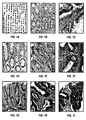
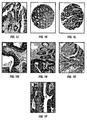
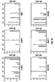
- Example IV 78% (39/50) of patients whose tumors contained low levels of TUCAN remained alive and disease-free during the time covered by this study, compared to only 44% (21/48) of those with high expression of this protein.
- Example VIII also indicates that TUCAN immunostaining was significantly higher among patients who died of colon cancer, as compared to patients who remained alive.
- TUCAN can be used alone or in combination with other prognostic indicators as a specific biomarker for prognosing survival of patients suffering from colon cancer.
- level refers to mean the amount, accumulation or rate of a biomarker molecule, such as TUCAN.
- a level can be represented, for example, by the amount or synthesis rate of messenger RNA (mRNA) encoded by a gene, the amount or synthesis rate of polypeptide corresponding to a given amino acid sequence encoded by a gene, or the amount or synthesis rate, of a biochemical form of a molecule accumulated in a cell, including, for example, the amount of particular post-synthetic modifications of a molecule such as a polypeptide, nucleic acid or small molecule.
- mRNA messenger RNA
- the term can be used to refer to an absolute amount of a molecule in a sample or to a relative amount of the molecule, including amounts determined under steady-state or non-steady-state conditions.
- the expression level of a molecule can be determined relative to a control molecule in a sample.
- the term level refers to the extent, amount or rate of synthesis of the nucleic acid sequence shown as SEQ ID NO:1 or the TUCAN polypeptide shown as SEQ ID NO:2, or substantially the same nucleotide or amino acid sequences.
- the nucleic acid sequence and amino acid sequence of TUCAN are also described in PCT publication WO 01/16170 .
- the term level refers to the extent, amount or rate of synthesis of the nucleic acid sequence shown as SEQ ID NO:5 or the CIAP2 polypeptide shown as SEQ ID NO:6, or substantially the same nucleotide or amino acid sequences.
- the term level refers to the extent, amount or rate of synthesis of the nucleic acid sequence shown as SEQ ID NO:7 or the ⁇ -catenin polypeptide shown as SEQ ID NO:8, or substantially the same nucleotide or amino acid sequences.
- the term level refers to the extent, amount or rate of synthesis of the nucleic acid sequence shown as SEQ ID NO:9 or the Apaf1 polypeptide shown as SEQ ID NO:10, or substantially the same nucleotide or amino acid sequences.
- the term level refers to the extent, amount or rate of synthesis of the nucleic acid sequence shown as SEQ ID NO:11 or the Bcl-2 polypeptide shown as SEQ ID NO:12, or substantially the same nucleotide or amino acid sequences.
- the term level refers to the extent, amount or rate of synthesis of the nucleic acid sequence shown as SEQ ID NO:13 or the Smac polypeptide shown as SEQ ID NO:14, or substantially the same nucleotide or amino acid sequences.
- a level of these and other biomarkers of cancer can be a gene expression level or a polypeptide expression level.
- amino acid sequence that has substantially the same amino acid sequence as a reference amino acid sequence contains a considerable degree of sequence identity or similarity, such as at least 70%, 80%, 90%, 95%, 98%, or 100% sequence identity or similarity, to a reference amino acid sequence.
- sequence identity or similarity such as at least 70%, 80%, 90%, 95%, 98%, or 100% sequence identity or similarity, to a reference amino acid sequence.
- Such changes, gaps and insertions can be naturally occurring mutations, or can result from processing a sample containing the polypeptide.
- a nucleotide sequence that is substantially the same as a reference nucleotide sequences contains a considerable degree of sequence identity or similarity, such as at least 70%, 80%, 90%, 95%, 98%, or 100% sequence identity or similarity, to the reference nucleotide sequence.
- differences can be due to genetic differences between individuals, such as mutations and polymorphisms of a gene.
- nucleotide and amino acid sequences can be determined using available algorithms and programs such as the Smith-Waterman algorithm and the BLAST homology search program ( Altschul et al., J. Mol. Biol. 215:403-410 (1990) ).
- a gene expression level of a molecule is intended to mean the amount, accumulation or rate of synthesis of a biomarker gene.
- the gene expression level can be represented by, for example, the amount or transcription rate of hnRNA or mRNA encoded by a gene.
- a gene expression level similarly refers to an absolute or relative amount or a synthesis rate determined, for example, under steady-state or non-steady-state conditions.
- a polypeptide expression level is intended to mean the amount, accumulation or rate of synthesis of a biomarker polypeptide.
- the polypeptide expression level can be represented by, for example, the amount or rate of synthesis of the polypeptide, a precursor form or a post-translationally modified form of the polypeptide.
- Various biochemical forms of a polypeptide resulting from post-synthetic modifications can be present in cell contained in a sample. Such modifications include post-translational modifications, proteolysis, and formation of macromolecular complexes.
- Post-translational modifications of polypeptides include, for example, phosphorylation, lipidation, prenylation, sulfation, hydroxylation, acetylation, addition of carbohydrate, addition of prosthetic groups or cofactors, formation of disulfide bonds and the like.
- fragments of a polypeptide are included within the definition of a polypeptide expression level. Fragments can include, for example, amino terminal, carboxyl terminal, or internal deletions of a full length polypeptide. Accumulation or synthesis rate with or without such modifications is included with in the meaning of the term.
- a polypeptide expression level also refers to an absolute amount or a synthesis rate of the polypeptide determined, for example, under steady-state or non-steady-state conditions.
- the term "reference level” refers to a control level of expression of a biomarker used to evaluate a test level of expression of a biomarker in a neoplastic cell-containing sample of a patient. For example, when the level of TUCAN in the neoplastic cells of a patient are higher than the reference level of TUCAN, the cells will be considered to have a high level of expression, or overproduction, of TUCAN. Conversely, when the level of TUCAN in the neoplastic cells of a patient are lower than the reference level, the cells will be considered to have a low level of expression, or underproduction, of TUCAN.
- the reference level can be determined by a plurality of methods, provided that the resulting reference level accurately provides a level of a biomarker above which exists a first group of patients having a different probability of survival than that of a second group of patients having levels of the biomarker below the reference level.
- the reference level can be determined by, for example, measuring the level of expression of a biomarker in non-tumorous cancer cells from the same tissue as the tissue of the neoplastic cells to be tested.
- the reference level can also be a level of a biomarker of in vitro cultured cells which can be manipulated to simulate tumor cells, or can be manipulated in any other manner which yields expression levels which accurately determine the reference level.
- the reference level can also be determined by comparison of the level of a biomarker, such as TUCAN, in populations of patients having the same cancer. This can be accomplished, for example, by histogram analysis, in which an entire cohort of patients are graphically presented, wherein a first axis represents the level of the biomarker, and a second axis represents the number of patients in the cohort whose neoplastic cells express the biomarker at a given level. Two or more separate groups of patients can be determined by identification of subsets populations of the cohort which have the same or similar levels of the biomarker. Determination of the reference level can then be made based on a level which best distinguishes these separate groups.
- a reference level also can represent the levels of two or more markers. Two or more markers can be represented, for example, by a ratio of values for levels of each biomarker.
- the reference level can be a single number, equally applicable to every patient, or the reference level can vary, according to specific subpopulations of patients. For example, older men might have a different reference level than younger men for the same cancer, and women might have a different reference level than men for the same cancer.
- the reference level can be some level determined for each patient individually.
- the reference level might be a certain ratio of a biomarker in the neoplastic cells of a patient relative to the biomarker levels in non-tumor cells within the same patient.
- the reference level for each patient can be proscribed by a reference ratio of one or more biomarkers, such as TUCAN, wherein the reference ratio can be determined by any of the methods for determining the reference levels described herein.
- neoplastic cell refers to any cell that is transformed such that it proliferates without normal homeostatic growth control. Such cells can result in a benign or malignant lesion of proliferating cells. Such a lesion can be located in a variety of tissues and organs of the body. Table 1, below, provides a list of exemplary types of cancers from which a neoplastic cell can be derived.
- cancer is intended to mean a class of diseases characterized by the uncontrolled growth of aberrant cells, including all known cancers, and neoplastic conditions, whether characterized as malignant, benign, soft tissue or solid tumor.
- Specific cancers include digestive and gastrointestinal cancers, such as anal cancer, bile duct cancer, gastrointestinal carcinoid tumor, colon cancer, esophageal cancer, gallbladder cancer, liver cancer, pancreatic cancer, rectal cancer, appendix cancer, small intestine cancer and stomach (gastric) cancer; breast cancer; ovarian cancer; lung cancer; renal cancer; CNS cancer; leukemia and melanoma.
- a list of known cancers is provided below in Table 1.
- the term "specifically reactive" when used in reference to an antibody refers to the discriminatory binding of the antibody to the indicated target polypeptide. For such binding to be discriminating, the antibody will not substantially cross react with other polypeptides. Specific reactivity can include binding properties such as binding specificity, binding affinity and binding avidity. For example, an antibody can bind a target polypeptide with a binding affinity (Kd) of about 10 -4 M or more, 10 -6 M or more, 10 -7 M or more, 10 -8 M or more, 10 -9 M or more, or 10 -10 M or more.
- Kd binding affinity
- sample is intended to mean any biological fluid, cell, tissue, organ or portion thereof, that includes or potentially includes a neoplastic cell, such as a cell from the colon, rectum, breast, ovary, prostate, kidney, lung, blood, brain or other organ or tissue that contains or is suspected to contain a neoplastic cell.
- a sample includes samples present in an individual as well as samples obtained or derived from the individual.
- a sample can be a histologic section of a specimen obtained by biopsy, or cells that are placed in or adapted to tissue culture.
- a sample further can be a subcellular fraction or extract, or a crude or substantially pure nucleic acid molecule or protein preparation.
- disease-free survival refers to the lack of tumor recurrence and/or spread and the fate of a patient after diagnosis, for example, a patient who is alive without tumor recurrence.
- all survival refers to the fate of the patient after diagnosis, regardless of whether the patient has a recurrence of the tumor.
- risk of recurrence refers to the probability of tumor recurrence or spread in a patient subsequent to diagnosis of cancer, wherein the probability is determined according to the process of the invention.
- Tumor recurrence refers to further growth of neoplastic or cancerous cells after diagnosis of cancer. Particularly, recurrence can occur when further cancerous cell growth occurs in the cancerous tissue.
- Tumor spread refers to dissemination of cancer cells into local or distant tissues and organs, for example during tumor metastasis. Tumor recurrence, in particular, metastasis, is a significant cause of mortality among patients who have undergone surgical treatment for cancer. Therefore, tumor recurrence or spread is correlated with disease-free and overall patient survival.
- the invention relates to the use of TUCAN as a biomarker for prognosing survival and monitoring the effectiveness of a treatment for a colon cancer patient.
- TUCAN is a CARD domain-containing protein that has a role in regulating apoptosis.
- Apoptosis is a physiologic process that ensures homeostasis is maintained between cell production and cell turnover in essentially all self-renewing tissues.
- apoptosis also occurs in response to a variety of external stimuli, including growth factor deprivation, alterations in calcium levels, free-radicals, cytotoxic lymphokines, infection by some viruses, radiation and most chemotherapeutic agents.
- apoptosis is an inducible event that likely is subject to similar mechanisms of regulation as occur, for example, in a metabolic pathway.
- dysregulation of apoptosis also can occur and is observed, for example, in some types of cancer cells, which survive for a longer time than corresponding normal cells, and in neurodegenerative diseases where neurons die prematurely.
- induction of apoptosis can figure prominently in the pathophysiology of the disease process, because immune-based eradication of viral infections depends on elimination of virus-producing host cells by immune cell attack resulting in apoptosis.
- Caspases The principal effectors of apoptosis are a family of intracellular proteases known as Caspases, representing an abbreviation for C ysteine Asp artyl Prote ases . Caspases are found as inactive zymogens in essentially all animal cells. During apoptosis, the caspases are activated by proteolytic processing at specific aspartic acid residues, resulting in the production of subunits that assemble into an active protease typically consisting of a heterotetramer containing two large and two small subunits ( Thornberry and Lazebnik, Science 281:1312-1316 (1998) ). The phenomenon of apoptosis is produced directly or indirectly by the activation of caspases in cells, resulting in the proteolytic cleavage of specific substrate proteins.
- TUCAN contains at least two protein domains, one of which is a CARD (Caspase-Associated Recruitment Domain).
- CARDs are protein interaction motifs found in the N-terminal prodomains of several caspases and in apoptosis- regulatory proteins that either activate or suppress activation of CARD-containing pro-caspases. In mammals, eight CARD-carrying caspases have been identified, including pro-caspases-1, 2, 4, 5, 9, 11, 12 and 13.
- CARD4 Apaf1, Nod1 (CARD4), NAC (DEPCAP), Raidd (CRADD), Cardiak (Rip2, RICK), Bc110 (CIPER), ARC (Nop30), Asc, CARD9, CARD10, CARD11, CARD14, cIAP1, cIAP2, and CLAN.
- CARD domains of many of these proteins are capable of binding the CARD-containing prodomains of specific CARD-carrying caspases, either facilitating or inhibiting protease activation.
- the CARD domain of TUCAN selectively binds to its own CARD and to pro-Caspase-9 (see Example IX).
- the binding of TUCAN to pro-caspase-9 has been shown to interfere with the ability of pro-caspase-9 to interact with Apaf1.
- TUCAN inhibits apoptosis signaling in the mitochondrial/cytochrome c pathway. Consistent with this observation is that finding that over-expression of TUCAN reduces apoptosis induced by stimuli that are known to activate the mitochondrial pathway for caspase-activation, including Bax, DNA-damaging drugs, and staurosporine.
- apoptosis induced via alternative pathways is not inhibited by TUCAN.
- over-expression of TUCAN in cells by stable or transient transfection inhibits apoptosis and caspase activation induced by Apaf1/caspase-9-dependent stimuli, including Bax, VP16, and Staurosporine, but not by Apafl/caspase-9-independent stimuli, Fas and Granzyme B.
- Apaf1/caspase-9-dependent stimuli including Bax, VP16, and Staurosporine
- TUCAN also contains an N-terminal domain that shares amino-acid similarity with a segment of the NAC protein, a CARD-carrying regulator of the Apaf1 apoptosome Chu et al. J Biol Chem 276:9239-9245 (2001) and Hlaing et al. J Biol Chem 276:9230-9238 (2001) ).
- the TUCAN N-terminal domain contains several candidate phosphorylation sites, including PKC (S/T-x-R/K) sites at amino-acids 72, 286, 313, and 416, Casein kinase II (S/T-x-D/E) sites at 289, 376, 398, 414, and 416 and MAP kinase/CDK (S/T-P) sites at 187 and 289.
- PKC S/T-x-R/K
- Casein kinase II S/T-x-D/E
- S/T-P MAP kinase/CDK
- Example VII relatively high levels of TUCAN are found in several human cancer cell lines. Moreover, as disclosed in Examples II and VIII, compared to normal colonic mucosa, TUCAN immunostaining was pathologically elevated in roughly two-thirds of early-stage colon cancers, indicating abnormal over-expression of this anti-apoptotic protein in association with malignant transformation. Studies of cells derived from pro-caspase-9 knock-out mice have indicated that pro-caspase-9 functions as a tumor suppressor in a p53-dependent pathway ( Soengas et al. Science 284:156-159 (1999) ).
- the invention provides a method for determining a prognosis for survival for a colon cancer patient using TUCAN.
- the method involves (a) measuring a level of TUCAN in a neoplastic cell-containing sample from the cancer patient, and (b) comparing the level of TUCAN in the sample to a reference level of TUCAN, wherein levels of TUCAN lower than the reference level in the sample correlate with increased survival of the patient.
- a level of TUCAN in a neoplastic cell-containing sample that exceeds a determined basal level, or reference level, of TUCAN can be a significant factor in tumor recurrence or spread.
- the level of TUCAN is characterized as high or overproduced. High or overproduced TUCAN can be indicative of increased risk of tumor recurrence or spread. Low or underproduced TUCAN can be indicative of decreased risk or tumor recurrence or spread.
- the methods of the invention for prognosing survival for a colon cancer patient involve measuring the level of one or more biomarkers, at least TUCAN.
- the level of the biomarker TUCAN is used to determine the prognosis for disease-free or overall survival of a cancer patient based on the correlations provided herein.
- Such prognosis is possible because the likelihood of tumor recurrence or spread correlates with the level of TUCAN in a tumor cell'. For example, as shown in Examples VI and VIII, it has been found that when the levels of TUCAN expression are low, the likelihood of cancer recurrence is low.
- the level of TUCAN in a neoplastic-cell containing sample from a patient can be used as the sole factor in assessing disease status or can be used in addition to other predictive methods.
- TUCAN can be used to prognose survival or monitor the effectiveness of a course of treatment for patients suffering from a variety of types of cancer.
- TUCAN is present in multiple different cancer cell types, including leukemia, melanoma and breast, ovarian, lung, CNS, prostate and renal cancers.
- a cellular function of TUCAN is suppression of mitochondrial signaling pathway-induced apoptosis.
- Mitochondrial signaling pathway-induced apoptosis is an apoptotic mechanism that can occur in any cell type, and that can become dysregulated or suppressed in any type cell, resulting in transformation of a cell such that it proliferates without normal homeostatic growth control.
- a level of TUCAN can be correlated with tumor recurrence or survival of a patient having any type of cancer.
- those skilled in the art will be able to determine if a level of TUCAN in a particular tumor cell type correlates with patient survival. Having determined a correlation between a reference level of TUCAN and survival of a cancer patient, those skilled in the art can practice the methods for determining the prognosis for survival for a cancer patient and the method for monitoring the effectiveness of a course of treatment for a patient with cancer described herein.
- a sample can be, for example, a cell or tissue obtained using a biopsy procedure or can be a fluid sample containing cells, such as blood, serum, semen, urine, or stool.
- a fluid sample containing cells such as blood, serum, semen, urine, or stool.
- Those skilled in the art will be able tb determine an appropriate sample, which will depend on cancer type, and an appropriate method for obtaining a biopsy sample, if necessary.
- a level of TUCAN can be determined by measuring the amount of a TUCAN using a selective binding agent, such as an antibody specifically reactive with a TUCAN polypeptide.
- selective binding agents include polypeptides that bind to a TUCAN polypeptide, such as a TUCAN polypeptide that contains the TUCAN CARD domain (amino acids 345-431 (SEQ ID NO:3)), and a caspase 9 polypeptide, the amino acid sequence of which (SEQ ID NO:4) is referenced as P55211 in the Prosite database, or modifications thereof that bind to a TUCAN polypeptide.
- affinity binding assays are applicable for use in determining a level of TUCAN, or another biomarker polypeptide, such as cIAP2, Apaf1, Smac, ⁇ -catenin, Bcl-2 or p53, in a sample. Such methods are rapid, efficient and sensitive. Moreover, affinity binding methods are simple and can be modified to be performed under a variety of clinical settings and conditions to suit a variety of particular needs.
- Affinity binding assays that are known and can be used in the methods of the invention include both soluble and solid phase formats. A specific example of a soluble phase affinity binding assay is immunoprecipitation using a biomarker selective antibody or other binding agent. Solid phase formats are advantageous for the methods of the invention since they are rapid and ,can be performed more easily on multiple different samples simultaneously without losing sensitivity or accuracy. Moreover, solid phase affinity binding assays are further amenable to high throughput screening and automation.
- solid phase affinity binding assays include immunohistochemical binding assays, immunoaffinity binding assays such as an ELISA and radioimmune assay (RIA).
- Other solid phase affinity binding assays are known to those skilled in the art and are applicable to the methods of the invention.
- affinity binding assays are generally formatted for use with an antibody binding molecules that is selective for the analyte or ligand of interest, essentially any binding agent can be alternatively substituted for the selectively binding antibody.
- binding agents include, for example, macromolecules such as polypeptides, peptides, nucleic acid molecules, lipids and sugars as well as small molecule compounds.
- the various modes of affinity binding assays include, for example, immunohistochemistry methods, solid phase ELISA and RIA as well as modifications thereof. Such modifications thereof include, for example, capture assays and sandwich assays as well as the use of either mode in combination with a competition assay format.
- the choice of which mode or format of immunoaffinity binding assay to use will depend on the intent of the user. Such methods can be found described in common laboratory manuals such as Harlow and Lane, Using Antibodies: A Laboratory Manual, Cold Spring Harbor Laboratory Press, New York (1999 ).
- An antibody useful in the methods of the invention includes a polyclonal and monoclonal antibody, as well as an antigen binding fragment of such antibodies.
- Methods of preparing polyclonal or monoclonal antibodies are well known to those skilled in the art and are described in Example I and in Harlow and Lane, Antibodies: A Laboratory Manual, Cold Spring Harbor Laboratory Press (1988 ).
- An antibody useful in the methods of the invention also includes naturally occurring antibodies as well as non-naturally occurring antibodies, including, for example, single chain antibodies, chimeric, bifunctional and humanized antibodies, as well as antigen-binding fragments thereof.
- non-naturally occurring antibodies can be constructed using solid phase peptide synthesis, can be produced recombinantly or can be obtained, for example, by screening combinatorial libraries consisting of variable heavy chains and variable light chains as described by Huse et al. (Science 246:1275-1281 (1989) ).
- These and other methods of making, for example, chimeric, humanized, CDR-grafted, single chain, and bifunctional antibodies are well known to those skilled in the art ( Winter and Harris, Immunol.
- Detection systems include the detection of bound biomarker by both direct and indirect means.
- Direct detection methods include labeling of the biomarker-specifically reactive antibody or binding agent.
- Indirect detection systems include, for example, the use of labeled secondary antibodies and binding agents.
- Detectable labels can be essentially any label that can be quantitated or measured by analytical methods.
- labels include, for example, enzymes, radioisotopes, fluorochromes as well as chemi- and bioluminescent compounds.
- enzyme labels include horseradish peroxidase (HRP), alkaline phosphatase (AP), ⁇ -galactosidase, urease and luciferase.
- a horseradish-peroxidase detection system can be used, for example, with the chromogenic substrate tetramethylbenzidine (TMB), which yields a soluble product in the presence of hydrogen peroxide that is detectable by measuring absorbance at 450 nm.
- TMB tetramethylbenzidine
- An alkaline phosphatase detection system can be used with the chromogenic substrate p-nitrophenyl phosphate, for example, which yields a soluble product readily detectable by measuring absorbance at 405 nm.
- a ⁇ -galactosidase detection system can be used with the chromogenic substrate o-nitrophenyl- ⁇ -D-galactopyranoside (ONPG), which yields a soluble product detectable by measuring absorbance at 410 nm, or a urease detection system can be used with a substrate such as urea-bromocresol purple (Sigma Immunochemicals, St. Louis, MO). Luciferin is the substrate compound for luciferase which emits light following ATP-dependent oxidation.
- ONPG o-nitrophenyl- ⁇ -D-galactopyranoside
- a urease detection system can be used with a substrate such as urea-bromocresol purple (Sigma Immunochemicals, St. Louis, MO).
- Luciferin is the substrate compound for luciferase which emits light following ATP-dependent oxidation.
- Fluorochrome detection labels are rendered detectable through the emission of light of ultraviolet or visible wavelength after excitation by light or another energy source.
- DAPI fluorescein, Hoechst 33258, R-phycocyanin, B-phycoerythrin, R-phycoerythrin, rhodamine, Texas red and lissamine are specific examples of fluorochrome detection labels that can be utilized in the affinity binding formats of the invention.
- a particularly useful fluorochrome is fluorescein or rhodamine.
- Chemiluminescent as well as bioluminescent detection labels are convenient for sensitive, non-radioactive detection of a biomarker and can be obtained commercially from various sources such as Amersham Lifesciences, Inc. (Arlington Heights, IL).
- radioisotopes can be used as detectable labels in the methods of the invention.
- Iodine-125 is a specific example of a radioisotope useful as a detectable label.
- Signals from detectable labels can be analyzed, for example, using a spectrophotometer to detect color from a chromogenic substrate; a fluorometer to detect fluorescence in the presence of light of a certain wavelength; or a radiation counter to detect radiation, such as a gamma counter for detection of iodine-125.
- a quantitative analysis of the amount of bound agent can be made using a spectrophotometer such as an EMAX Microplate Reader (Molecular Devices, Menlo Park, CA) in accordance with the manufacturer's instructions.
- the assays of the invention can be automated or performed robotically, and the signal from multiple samples can be detected simultaneously.
- the prognostic formats of the present invention can be forward, reverse or simultaneous as described in U.S. Patent No. 4,376,110 and No. 4,778,751 .
- Separation steps for the various assay formats described herein, including the removal of unbound secondary antibody, can be performed by methods known in the art (Harlow and Lane, supra). For example, washing with a suitable buffer can be followed by filtration, aspiration, vacuum or magnetic separation as well as by centrifugation.
- a binding agent selective for a biomarker also can be utilized in imaging methods that are targeted at biomarker- expressing neoplastic cells. These imaging techniques will have utility in identification of residual neoplastic cells at the primary site following standard treatments including, for example, surgical resection of an organ of the gastrointestinal system, such as the colon, and radiation therapy. In addition, imaging techniques that detect neoplastic cells have utility in detecting secondary sites of metastasis.
- the biomarker specific binding agent can be radiolabeled with, for example, 111 indium and infused intravenously as described by Kahn et al., Journal of Urology 152:1952-1955 (1994) .
- the binding agent selective for a biomarker can be, for example, a monoclonal antibody specifically reactive with TUCAN or another biomarker, such as cIAP2, Apaf1, Smac, ⁇ -catenin, Bcl-2 or p53. Imaging can be accomplished by, for example, radioimmunoscintigraphy as described by Kahn et al., supra.
- the level of TUCAN, or another biomarker, such as cIAP2, Apaf1, Smac, ⁇ -catenin, Bcl-2 or p53, also can be determined by measuring the amount of a biomarker mRNA or DNA using a binding agent selective for the biomarker, such as a nucleic acid probe.
- the methods used to detect mRNA levels include detection of hybridization or amplification of mRNA encoding the biomarker. This detection can be carried out by analysis of mRNA either in vitro or in situ using one of the methods known to one of ordinary skill in the art as exemplified in the Current Protocols in Molecular Biology (John Wiley & Sons, 1999 ); in U.S. Patent No.
- a TUCAN mRNA, or other biomarker mRNA, detected will be any RNA transcript of a TUCAN gene, or fragment thereof, or cIAP2, Bcl-2, p53, b- catenin, survivin or Apaf1 gene, or fragment thereof.
- TUCAN nucleic acid probe complementary to a TUCAN gene having a detectable label is added to a neoplastic cell-containing sample obtained from the individual having, or suspected of having cancer under conditions which allow annealing of the probe to TUCAN RNA.
- Methods for detecting TUCAN RNA in a sample can include the use of, for example, RT-PCR.
- Conditions are well known in the art for both solution and solid phase hybridization procedures.
- optimization of hybridization conditions can be performed, if desired, by hybridization of an aliquot of the sample at different temperatures, durations and in different buffer conditions.
- the sample is washed and the signal is measured and compared with a suitable control or standard value.
- the magnitude of the hybridization signal is directly proportional to the mRNA level of TUCAN.
- a level of TUCAN mRNA in a neoplastic cell-containing sample is compared to a suitable reference level for TUCAN mRNA.
- the levels of other biomarker mRNA such as cIAP2, Apaf1, Smac, ⁇ -catenin, Bcl-2 or p53, can be similarly determined and compared to a suitable reference level for the particular biomarker.
- PCR and other amplification methods such as RT-PCR, 5' or 3' RACE, RNase protection, RNA blot, dot blot or other membrane-based technologies, dip stick, pin, ELISA or two-dimensional arrays immobilized onto a solid support. These methods can be performed using either qualitative or quantitative measurements, all of which are well known to those skilled in the art.
- PCR or RT-PCR can be used with isolated RNA or crude cell lysate preparations. PCR is advantageous when there is limiting amounts of starting material. A further description of PCR methods can be found in, for example, Dieffenbach, C.W., and Dveksler, G.S., PCR Primer: A Laboratory Manual, Cold Spring Harbor Press, Plainview, New York (1995 ). Multisample formats such as microarrays offer the advantage of analyzing numerous, different.samples in a single assay. In contrast, solid-phase dip stick-based methods offer the advantage of being able to rapidly analyze a patient's fluid sample for an immediate result.
- Nucleic acid probes useful for measuring the expression level of a biomarker, such as cIAP2, TUCAN, Apaf1, ⁇ -catenin, Bcl-2, or Smac by hybridization include, for example, probes prepared using the nucleotide sequences provided herein. Nucleic acid molecules corresponding to the entire cDNA sequences and fragments thereof, including oligonucleotides corresponding to cIAP2, TUCAN, Apaf1, ⁇ -catenin, Bcl-2, or Smac nucleotide sequences and which are capable of specifically or selectively hybridizing to cIAP2, TUCAN, Apaf1, ⁇ -catenin, Bcl-2, or Smac RNA, are useful for hybridization methods.
- a reference level is a level a biomarker, such as cIAP2, TUCAN, Apaf1, Smac, ⁇ -catenin, of Bcl-2, used to evaluate the level of the biomarker in cancerous cells of a patient.
- a biomarker such as cIAP2, TUCAN, Apaf1, Smac, ⁇ -catenin, of Bcl-2.
- the cells will be considered to have a high level of, or overproduction, of the biomarker.
- the level of biomarker in the cancerous cells of a patient are lower than the reference level, the cells will be considered to have a low level of, or underproduction, of the biomarker.
- a high level of a biomarker such as cIAP2, TUCAN, Apaf1, Smac, ⁇ -catenin, Bcl-2 or p53, or overproduction of a biomarker gene is related to a level of the biomarker above a determined basal level.
- a reference or basal level of a biomarker such as cIAP2, TUCAN, Apaf1, Smac, ⁇ -catenin, Bcl-2 or p53, in a cancer cell is identified as a "cutoff" value, above which there is a significant correlation between the presence of the biomarker and increased or decreased tumor recurrence or spread.
- Such overproduction is not typically calculated in terms of absolute biomarker levels, but is determined using relative measurements. These relative measurements are illustrated for quantitation purposes with an internal standard; however, it will be appreciated that other standards or methods of determination can be used, such as comparison with external standards, biomarker polypeptide measurements, biomarker mRNA measurements, absolute values of protein, mRNA or DNA levels, and the like.
- a reference level can also be determined by comparison of biomarker levels in populations of patients having cancer, such as patients having cancer of the same stage. This can be accomplished by histogram analysis, in which the entire cohort of patients tested are graphically presented, wherein a first axis represents the level of a biomarker, and a second axis represents the number of patients in the cohort whose tumor cells contain the biomarker at a given level. Two or more separate groups of patients can be determined by identification of subsets populations of the cohort which have the same or similar levels of the biomarker. Determination of the reference level can then be made based on a biomarker level that best distinguishes these separate groups.
- Verification that the reference level distinguishes the likelihood of tumor recurrence or spread in cancer patients expressing below-reference biomarker levels versus cancer patients expressing above-reference biomarker levels can be carried out using single variable or multi-variable analysis. These methods determine the likelihood of a correlation between one or more variables and a given outcome. In the specific case, the methods will determine the likelihood of a correlation between a biomarker levels (or biomarker level coupled with another variable) and disease-free or overall survival of cancer patients. Any one of a plurality of methods well known to those of ordinary skill in the art for carrying out these analyses can be used. Examples of single variable analysis is the Kaplan-Meir method or the log-rank test. An example of multi-variable analysis is the Cox proportional-hazards regression model (see, for example, Example VI).
- Population-based determination of reference levels for example, by histogram analysis can be carried out using a cohort of patients sufficient in size in order to determine two or more separate groups of patients having different biomarker levels.
- a cohort comprises at least 25 patients, such as at least 50 patients, including at least 75 patients, and at least 100 patients.
- verification of determined reference levels can also comprise at least 25 patients, such as at least 50 patients, including at least 75 patients, and at least 100 patients.
- the reference level can be a single number, equally applicable to every patient, or the reference level can vary according to specific subpopulations of patients. For example, men might have a different reference level than women for the same cancer.
- the reference level can be a level determined for each patient individually.
- the reference level might be a certain ratio of a biomarker level in the tumor cells of a patient relative to the biomarker level in non-tumor cells within the same patient.
- the reference level for each patient can be proscribed by a reference ratio of biomarker levels, wherein the reference ratio can be determined by any of the methods for determining the reference levels described above.
- a reference level can separate two groups of patients, it is within the scope of the invention that numerous reference values might exist which separate a plurality of populations.
- two reference values can separate a first group of patients with high levels of a biomarker from a second group of patients with intermediate levels the biomarker, and from a third group of patients with low levels of the biomarker.
- the number of different reference levels can be sufficient to proscribe a curve, such as a continuous line, which describes the likelihood of disease-free or overall survival in a patient as a function of the biomarker level in that patient.
- Such a curve will constitute a "continuous" biomarker level, where the likelihood of disease free or overall survival in a patient is proportional to the biomarker level in that patient.
- Two or more biomarker levels also can be represented by such a curve.
- the reference level can also represent the level of a biomarker protein, such as cIAP2, TUCAN, Apaf1, Smac, ⁇ -catenin, Bcl-2 or p53, in one or more compartments of the cell.
- the reference level will represent the level of biomarker protein in (a) the whole cell, (b) the nucleus, or (c) the cytosol. This level will be useful when cell compartmentalization of the protein correlates with the risk of tumor recurrence or spread of a certain cancer.
- the reference level can be a ratio of levels of biomarker protein in the different compartments (for example, the ratio of nuclear biomarker protein to whole cell biomarker protein, or the ratio of nuclear to cytosolic biomarker protein).
- the reference level of a biomarker such as cIAP2, TUCAN, Apaf1, or Smac, can further be used in conjunction with another variable found to be a statistically significant indicator of the likelihood of disease-free or overall survival for cancer.
- Such indicators include the presence or levels of known cancer markers (for example, colon cancer markers include sialosyl-TnCEA, CA19-9, and LASA), or can be clinical or pathological indicators (for example, age, tumor size, tumor histology, clinical stage, family history and the like).
- clinical stage of the cancer is also a statistically significant indicator of disease-free or overall survival, wherein the reference level of a biomarker can vary according to the clinical stage of the cancer.
- the level of a biomarker such as a low level of TUCAN, in conjunction with clinical stage II of a cancer for a given patient, together are indicators for increased likelihood of disease free or overall survival.
- the reference level of a biomarker can vary as a function of another statistically significant indicator of disease-free or overall survival for cancer.
- the levels of biomarkers, such as cIAP2, Apaf1, TUCAN, Bcl-2 and Smac, in a cancer cell can correlate with each other and with other molecules because these molecules participate in common dysregulated molecular pathways that contribute to the hyperproliferative state of a cancer cell. Therefore a combination of TUCAN with one or more additional biomarkers can be used in the methods of the invention for determining a prognosis for survival for a colon cancer patient.
- a second or additional biomarker can be, for example, Apaf1, cIAP1, cIAP2, survivin, AIF, Bcl-2, Bcl-XL, Bax, Bid, BAG1, p53, mutant p53, ⁇ -catenin, MIB-1 or another well-known tumor marker, such as the exemplary commercially available tumor markers described below.
- the use of a combination of TUCAN with one or more biomarkers can provide increased prognostic significance or confidence in a prognostic determination.
- the invention provides a method for determining a prognosis for survival for a colon cancer patient that involves the use of two or more biomarkers.
- the method is practiced by (a) measuring the levels of TUCAN and one or more biomarkers selected from the group consisting of cIAP2, Apaf1, Bcl-2 and Smac in a neoplastic cell-containing sample from the cancer patient, and (b) comparing the level of TUCAN and the one or more selected biomarkers in the sample to a reference level of TUCAN and the biomarkers, wherein a level of TUCAN lower than the reference level a level of any of Apaf1, Bcl-2 or Smac higher than the reference level or a level of TUCAN and a level of cIAP2 lower than the reference level, in said sample correlate with increased survival of said patient.
- the methods of the invention can be practiced, for example, by selecting a combination of TUCAN and one or more biomarkers for which increased or decreased expression correlates with improved survival, such as any of cIAP2, Apaf1, Bcl-2, Smac, or another known or standard biomarker for colon cancer.
- biomarkers for which increased or decreased expression correlates with improved survival such as any of cIAP2, Apaf1, Bcl-2, Smac, or another known or standard biomarker for colon cancer.
- diagnostic or prognostic markers useful for multiple types of cancer such as CA 125, CEA or LDH, diagnostic or prognostic colon cancer markers (for example, sialosyl-TnCEA, CA19-9, or LASA), breast cancer markers (for example, CA 15-2.
- Her-2/neu and CA 27.29 Her-2/neu and CA 27.29), ovarian cancer markers (for example, CA72-4), lung cancer (for example, neuron-specific enolase (NSE) and tissue polypeptide antigen (TPA)), prostate cancer (for example, PSA, prostate-specific membrane antigen and prostatic acid phosphatase), melanoma (for example, S-100 and TA-90), as well as other biomarkers specific for other types of cancer.
- NSE neuron-specific enolase
- TPA tissue polypeptide antigen
- PSA prostate-specific membrane antigen and prostatic acid phosphatase
- melanoma for example, S-100 and TA-90
- biomarkers specific for other types of cancer for example, S-100 and TA-90
- biomarkers can provide increased confidence in prognostic outcome.
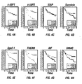
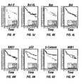
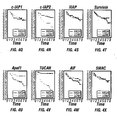
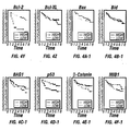
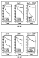
- combinations of low cIAP and low TUCAN, and high Apaf1 and low TUCAN were correlated with increased disease-free survival (see Example V).
- Combinations of biomarkers useful in the prognostic methods of the invention include, for example, cIAP2 and TUCAN, Apaf1 and TUCAN, and a multiplicity of other combination of TUCAN with biomarkers such as cIAP2, Apaf1, Bcl-2 and Smac and other molecules, including AIF, Bcl-2, Bcl-XL, Bax, Bid, BAG1, p53, mutant p53, ⁇ -catenin, MIB-1 and a variety of other general and tumor-specific biomarkers, such as commercially available diagnostic markers described herein above.
- the invention also provides a method for monitoring the effectiveness of a course of treatment for a patient with colon cancer.
- the method involves (a) determining the level of TUCAN in a neoplastic cell-containing sample from the cancer patient prior to treatment, and (b) determining the level of TUCAN in a neoplastic cell-containing sample from the patient after treatment, whereby comparison of the TUCAN level prior to treatment with the biomarker level after treatment indicates the effectiveness of the treatment wherein a decrease in the level of TUCAN after treatment indicates that treatment is effective.
- ⁇ refers to the ability of the course of treatment to decrease the risk of tumor recurrence or spread and therefore to increase the likelihood of disease-free or overall survival of the patient.
- This method will have particular utility when the level a biomarker, such as cIAP, TUCAN, Apaf1 or Smac, in the tumor cells of a patient is abnormal compared to the level of cIAP, TUCAN, Apaf1 and Smac in the non-tumor cells of the patient.
- Comparison of biomarker levels in a neoplastic cell-containing sample from a patient before and after treatment will thereby serve to indicate whether a biomarker level is returning to that of non-tumor cells, implying a more effective course of treatment, or whether a biomarker level is remaining abnormal or increasing in abnormality, implying a less effective course of treatment.
- an increase in the level of Apaf1, Bcl-2 or Smac in a patient sample after treatment indicates that treatment is effective because high levels of Apaf1 or Smac correlate with a lower incidence of colon cancer recurrence.
- a low in the level of ⁇ -catenin, cIAP2 or TUCAN in a patient sample after treatment indicates that treatment is effective because low levels of ⁇ -catenin, cIAP2 or TUCAN correlate with a lower incidence of colon cancer recurrence.
- Patients having cancer can be classified according to whether a high level of a particular biomarker, or a low level of the biomarker, is measured in a neoplastic cell-containing sample obtained from the patient. Determination of the prognosis for the patient can be made by determining whether the group to which the patient has been assigned correlates with a higher or lower likelihood of disease-free or overall survival with respect to the group to which the patient was not assigned.
- the invention also provides a method of determining a prognosis for survival for a colon cancer patient that involves patient classification.
- the method is practiced by (a) measuring a level of TUCAN in a neoplastic cell-containing sample from the cancer patient, and (b) classifying the patient as belonging to either a first or second group of patients, wherein the first group of patients having levels of TUCAN lower than the reference level is classified as having an increased likelihood of survival compared to the second group of patients having levels of TUCAN higher than the reference level.
- a level of TUCAN higher than the reference level, or overproduction of TUCAN correlates with patients having an increased risk of tumor recurrence or spread.
- patients belonging to a first group having levels of TUCAN higher than the reference level are classified as having an increased risk of tumor recurrence or spread compared to a second group of patients having levels TUCAN lower than the reference level.
- Patients belonging to a first group having levels of TUCAN lower than the reference level are classified as having increased likelihood of survival compared to a second group of patients having levels of TUCAN higher than the reference level.
- the method of determining a prognosis for survival for a colon cancer patient can be practiced using one or more additional biomarkers.
- biomarkers including known cancer markers and the prognostic biomarkers disclosed herein, can be used in combination with TUCAN to determine a prognosis for survival for a cancer patient.
- the method involves (a) determining a level of cIAP2 the neoplastic cell-containing sample from the cancer patient, and (b) classifying the patient as belonging to either a first or second group of patient, wherein the first group of patients having levels of TUCAN and levels of cIAP2 lower than the reference level is classified as having increased likelihood of survival compared to the second group of patients having levels of TUCAN and levels of cIAP2 higher than the reference level.
- the method involves (a) determining a level of a biomarker selected from the group consisting of Apaf1, Smac and Bcl-2 in the neoplastic cell-containing sample from the cancer patient, and (b) classifying the patient as belonging to either a first or second group of patient, wherein the first group of patients having levels of TUCAN lower than the reference level and levels of any of Apaf1, Smac or Bc1-2 higher than the reference level is classified as having increased likelihood of survival compared to the second group of patients having levels of TUCAN higher than the reference level and levels of any of Apaf1, Smac or Bcl-2 lower than the reference level.
- the patient After the levels of one or more biomarker in patient sample have been determined and compared to a reference level, the patient is then classified into a group having a certain likelihood of disease free or overall survival. Then the likelihood of disease-free or overall survival for the patient is assessed based on the likelihood of disease-free or overall survival for patients in that group.
- a neoplastic cell containing sample from a colon cancer patient can be determined to have high levels of Apaf1, Bcl-2 or Smac relative to a reference level. This patient would then be classified into a group of patients having levels of Apaf1, Bcl-2 or Smac higher than the reference level. Because it has been discovered that there is an increased likelihood of disease-free or overall survival for the group of patients expressing levels of Apaf1, Bcl-2 or Smac higher than the reference level in cancer cells (relative to those expressing levels of Apaf1, Bcl-2 or Smac lower than the reference level in cancer cells), the specific cancer patient would be considered to have an increased likelihood of disease free or overall survival.
- a neoplastic cell containing sample from a colon cancer patient can be determined to have high levels of cIAP2, ⁇ -catenin or TUCAN relative to a reference level. This patient would then be classified into a group of patients having levels of cIAP2, ⁇ -catenin or TUCAN higher than the reference level. Because it has been discovered that there is a decreased likelihood of disease-free or overall survival for the group of patients expressing levels of cIAP2, ⁇ -catenin or TUCAN higher than the reference level in cancer cells (relative to those expressing levels of cIAP2, ⁇ -catenin or TUCAN lower than the reference level in cancer cells), the specific cancer patient would be considered to have an decreased likelihood of disease free or overall survival.
- the methods of the invention are applicable to determining the susceptibility of an individual for developing colon cancer.
- the methods could be applicable to a variety of other cancers, including gastrointestinal, lung, prostate, breast, ovarian, skin, blood and kidney cancers.
- Colon cancers develop from premalignant precursor lesions known as adenomatous colon polyps.
- Multiple epidemiological studies have demonstrated that once one member of a family has developed an adenomatous colon polyp, his or her siblings are at markedly elevated risk for developing both colon adenomas and colon cancers.
- the method of the invention is practiced as described herein for colon neoplastic conditions, such as adenomatous colon polyps, for example, using an appropriately obtained biopsy sample.
- the methods of the invention for determining a prognosis for survival for a colon cancer patient are applicable to patients at any stage of tumor progression, and might be used to determine a stage of tumor progress.
- a stage of a tumor refers to the degree of progression of a tumor.
- Various stages of tumor development are well known to those of skill in the art, as exemplified in Markman, "Basic Cancer Medicine,” Saunders, (ed. Zorab, R.) (1997).
- cancers can be staged into three general stages -- localized, regional spread, and distant spread. Cancers also can be staged using the TNM system, which considers the extent of direct spread within affected and nearby tissues, the extent of spread to nearby lymph nodes, and the extent of spread to distant organs. Based on these features, spread of cancers can be summarized by assigning Roman numerals from 0 through IV. Those skilled in the art can select an appropriate staging system for a particular type of cancer.
- colon cancer can be staged using the Dukes, Astler-Coller and AJCC/TNM systems, which describe the spread of the cancer in relation to the layers of the wall of the colon or rectum, organs next to the colon and rectum, and other organs farther away.
- Dukes stage A is equivalent to AJCC/TNM stage I and Astler-Coller stage A, B1;
- Duke's stage B is equivalent to AJCC/TNM stage II and Astler-Coller stage B2, B3.
- Dukes stage C is equivalent to AJCC/TNM stage III and Astler-Coller stage C1, C2, C3.
- Stage 0 the cancer has not grown beyond the inner layer (mucosa) of the colon or rectum. This stage is also known as carcinoma in situ or intramucosal carcinoma; Stage I: the cancer has grown through the mucosa into the submucosa, or can also have grown into the muscularis intestinal, but it has not spread outside the wall itself into nearby tissue such as lymph nodes; Stage II: the cancer has grown through the wall of the colon or rectum, into the outermost layers and may have invaded other nearby tissues, but has not yet spread to the nearby lymph nodes; Stage III: the cancer can be of any size, but has spread to 3 or fewer nearby lymph nodes, or has spread to 4 or more nodes but it has not spread to other parts of the body; Stage IV: the cancer has spread to distant organs such as the liver, lung, peritoneum or ovary.
- stages of tumor development shall be understood to refer to stages in tumor development in which the tumor has detectably spread no further than the lymph nodes local to the organ of the primary tumor. Typically, early stages will be considered to be stages I and II.
- the predictive value of the method of the invention will be particularly effective in the case of patients in the early stages of colon cancer. This is because the method of the invention is advantageously effective in determining the risk of metastasis in patients who demonstrate no measurable metastasis at the time of examination.
- the prognostic indicators of survival for cancer patients suffering from stage I cancer may be different from those for cancer patients suffering from stage IV cancer.
- prognosis for stage I cancer patients may be oriented toward the likelihood of continued growth and/or metastasis of the cancer
- prognosis for stage IV cancer patients may be oriented toward the likely effectiveness of therapeutic methods for treating the cancer.
- a stage of cancer progression can be correlated with a level of one or more biomarkers, such as a level of TUCAN, Apaf1 or an IAP, such as cIAP2 or Smac. Therefore, a determination of a level of a biomarker in a sample from a cancer patient can be used to determine a stage of the tumor from which the sample was derived by comparing the sample with a reference level of the biomarker indicative of a particular stage of cancer.
- samples applicable for use in one or more prognostic formats of the invention include tissue and cell samples.
- a tissue or cell sample can be obtained, for example, from a fluid sample obtained from the patient, by biopsy or surgery.
- a tissue sample from the surgically removed tumor can be obtained and prepared for testing by conventional techniques.
- a sample can be removed from a patient, for example, using well-known biopsy procedures.
- a colonoscope can be fitted with a snare to remove a polyp without damage to the wall of the colon (polypectomy); or to obtain small, flatter polyps, a biopsy forceps can be attached to a colonoscope to collect a small sample of tissue.
- the tissue can be used whole or subjected to various methods known in the art to disassociate the sample into smaller pieces, cell aggregates or individual cells.
- amplification methods such as polymerase chain reaction (PCR)
- PCR polymerase chain reaction
- a single cell sample is sufficient for use in prognostic assays of the invention which employ hybridization detection methods.
- amplification of the signal with enzymatic coupling or photometric enhancement can be employed using only a few or a small number of cells.
- Tumor tissue cell samples can be assayed employing any of the formats described below.
- the tumor tissue sample can be mounted and hybridized in situ with biomarker nucleic acid probes.
- Similar histological formats employing protein detection methods and in situ activity assays also can be used to detect a biomarker polypeptide in whole tissue tumor cell samples.
- Protein detection methods include, for example, staining with a biomarker specific antibody, as described herein, in Example II.
- Such histological methods as well as others well known to those skilled in the art are applicable for use in the prognostic methods of the invention using whole tissue as the source of a neoplastic cell-containing sample. Methods for preparing and mounting the samples are similarly well known in the art.
- neoplastic cell-containing sample that can be analyzed for increased or decreased expression of biomarker RNA or polypeptide.
- the cells can be grown in culture and analyzed using procedures such as those described above.
- Whole cell samples expressing cell surface markers associated with biomarker expression can be rapidly tested using fluorescent or magnetic activated cell sorting (FACS or MACS) with labeled binding agents selective for the surface marker or using binding agents selective for specific cell populations, for example, and then determining a level of a biomarker within this population.
- FACS or MACS fluorescent or magnetic activated cell sorting
- a level of a biomarker can be determined using, for example, binding specifically reacting agents for a biomarker or by hybridization to a biomarker specific probe. Other methods for measuring the level of a biomarker in whole cell samples are known in the art and are similarly applicable in any of the prognostic formats described below.
- the tissue or whole cell tumor cell sample obtained from an individual also can be analyzed for increased or decreased biomarker levels by lysing the cell and measuring the level of a biomarker in the lysate, a fractionated portion thereof or a purified component thereof using any of formats described herein.
- biomarker RNA can be amplified directly from the lysate using PCR, or other amplification procedures well known in the art such as RT-PCR, 5' or 3' RACE to directly measure the level of a biomarker nucleic acid molecules.
- RNA also can be isolated and probed directly such as by solution hybridization or indirectly by hybridization to immobilized RNA.
- lysates can be assayed directly, or they can be further fractionated to enrich for a biomarker.
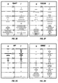
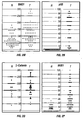
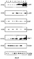
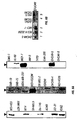
- an immunochemical method such as immunoblot analysis (see Example III) can be performed using a neoplastic cell-containing sample. Numerous other methods applicable for use with whole tumor cell samples are well known to those skilled in the art and can accordingly be used in the methods of the invention.
- the tumor tissue or cell sample can be obtained directly from the individual or, alternatively, it can be obtained from other sources for testing.
- a cell sample can be tested when it is freshly isolated or it can be tested following short or prolonged periods of cryopreservation without substantial loss in accuracy or sensitivity. If the sample is to be tested following an indeterminate period of time, it can be obtained and then cryopreserved, or stored at 4°C for short periods of time, for example.
- An advantage of the prognostic methods of the invention is that they do not require histological analysis of the sample. As such, the sample can be initially disaggregated, lysed, fractionated or purified and the active component stored for later diagnosis.
- the prognostic methods of the invention are applicable for use with a variety of different types of samples other than tumor cell samples.
- a biomarker polypeptide or fragment thereof that is released into the extracellular space including circulatory fluids as well as other bodily fluids, can be used in prognostic methods to detect a secreted polypeptide or fragment related to a biomarker polypeptide.
- the methods of the invention are applicable with fluid samples collected from an individual having, or suspected of having a colon neoplastic condition or colon cancer.
- Fluid samples which can be measured for biomarker levels, include, for example, blood, serum, lymph, urine and stool.
- Other bodily fluids are known to those skilled in the art and are similarly applicable for use as a sample in the prognostic methods of the invention.
- One advantage of analyzing fluid samples is that they are readily obtainable, in sufficient quantity, without invasive procedures as required by biopsy and surgery. Analysis of fluid samples such as blood, serum and urine will generally be in the prognostic formats described herein which measure biomarker polypeptide levels. As the biomarker related polypeptide is circulating in a soluble form, the methods will be similar to those which measure expression levels from cell lysates, fractionated portions thereof or purified components.
- Neoplastic conditions and cancer can be diagnosed, predicted or prognosed by measuring a level of a biomarker in a neoplastic cell-containing sample, circulating fluid or other bodily fluid obtained from the individual.
- levels of a biomarker can be measured by a variety methods known in the art.
- RNA or polypeptide can readily determine an appropriate assay system given the teachings and guidance provided herein and choose a method based on measuring RNA or polypeptide. Considerations such as the sample type, availability and amount will also influence selection of a particular prognostic format. For example, if the sample is a tumor cell sample and there is only a small amount available, then prognostic formats which measure the amount of biomarker RNA by, for example, PCR amplification, or which measure biomarker polypeptide by, for example, FACS analysis can be appropriate choices for determining the level of a biomarker.
- a multisample format such as an Enzyme Linked Immunoabsorbant Assay (ELISA), which measures the amount of a biomarker polypeptide can be an appropriate choice for determining the level of a biomarker.
- ELISA Enzyme Linked Immunoabsorbant Assay
- biomarker nucleic acid molecules released into bodily fluids from the neoplastic or pathological cells can also be analyzed by, for example, PCR or RT-PCR.
- Nucleic acid probes can be produced recombinantly or chemically synthesized using methods well known in the art. Additionally, hybridization probes can be labeled with a variety of detectable labels including, for example, radioisotopes, fluorescent tags, reporter enzymes, biotin and other ligands. Such detectable labels can additionally be coupled with, for example, colorimetric or photometric indicator substrate for spectrophotometric detection.
- Nucleic acid probes useful for detecting a biomarker in a sample can be hybridized under various stringency conditions readily determined by one skilled in the art. Depending on the particular assay, one skilled in the art can readily vary the stringency conditions to optimize detection of a particular biomarker in a particular sample type.
- the stability of a hybrid is a function of the ion concentration and temperature.
- a hybridization reaction is performed under conditions of lower stringency, followed by washes of varying, but higher, stringency.
- Moderately stringent hybridization refers to conditions that permit a nucleic acid molecule such as a probe to bind a complementary nucleic acid molecule.
- the hybridized nucleic acid molecules generally have at least 60% identity, at least 75% identity, at least 85% identity; or at least 90% identity.
- Moderately stringent conditions are conditions equivalent to hybridization in 50% formamide, 5X Denhart's solution, 5X SSPE, 0.2% SDS at 42°C, followed by washing in 0.2X SSPE, 0.2% SDS, at 42°C.
- High stringency conditions can be provided, for example, by hybridization in 50% formamide, 5X Denhart's solution, 5X SSPE, 0.2% SDS at 42°C, followed by washing in 0.1X SSPE, and 0.1% SDS at 65°C.
- Low stringency hybridization refers to conditions equivalent to hybridization in 10% formamide, 5X Denhart's solution, 6X SSPE, 0.2% SDS at 22°C, followed by washing in 1X SSPE, 0.2% SDS, at 37°C.
- Denhart's solution contains 1% Ficoll, 1% polyvinylpyrolidone, and 1% bovine serum albumin (BSA).
- BSA bovine serum albumin
- 20X SSPE sodium chloride, sodium phosphate, ethylene diamide tetraacetic acid (EDTA) contains 3M sodium chloride, 0.2M sodium phosphate, and 0.025 M (EDTA).
- nucleic acid molecules encoding polypeptides hybridize under moderately stringent or high stringency conditions to substantially the entire sequence, or substantial portions, for example, typically at least 15-30 nucleotides of the nucleic acid sequences of cIAP2, TUCAN, Apaf1, Bcl-2, Smac, ⁇ -catenin or another biomarker.
- the invention relates to the discovery that amounts of particular biomarkers higher or lower than the reference level, including cIAP2, TUCAN, Apaf1, Bcl-2, ⁇ -catenin and Smac are predictive of survival of patients having colon cancer.
- the over-expression or under-expression of these biomarkers can contribute to the genetic malfunction of cancer cells that leads to uncontrolled proliferation. Therefore, modulation of the level of a biomarker in a cancer cell to a level consistent with a normal cell can be used to return a cancer cell to a more normal proliferation state.
- over-expressed biomarker genes such as cIAP2, TUCAN and b-catenin a variety of strategies can be employed to reduce gene expression.
- inhibition of transcription or translation of cIAP2, TUCAN and b-catenin, or reduction in the amount of active cIAP2, TUCAN and b-catenin polypeptide can be used to reduce the levels of these biomarkers to a level representative of a normal cell.
- under-expressed biomarker genes such as Apaf1, Bcl-2 and Smac
- a variety of strategies can be employed to increase gene expression.
- introduction of Apaf1, Bcl-2 and Smac from an exogenous nucleic acid molecule, promotion of transcription or translation of Apaf1, Bcl-2 or Smac, or promotion in the amount of active Apaf1, Bcl-2 or Smac polypeptide can be used to increase the levels of these biomarkers to a level representative of a normal cell.
- a method for treating or reducing the progression of a neoplastic condition such as cancer by reducing neoplastic cell proliferation could be provided.
- the method could involve administering a nucleic acid encoding Apaf1, Bcl-2 or Smac into a neoplastic cell and expressing the Apaf1, Bcl-2 or Smac polypeptide in an amount effective to reduce neoplastic cell proliferation.
- the method of reducing neoplastic cell proliferation could involve contacting a neoplastic cell with an effective amount of an agent that, under sufficient conditions, increases the amount of Apaf1, Bcl-2 or Smac in the cell.
- Such an agent can increase the amount of a biomarker directly or indirectly, for example, by increasing the amount of a biomarker polypeptide in a cell, such as by stimulating increased mRNA expression.
- Apaf1, Bcl-2 or Smac mRNA expression can be increased, for example, by inducing or derepressing transcription of Apaf1, Bcl-2 or Smac genes and by regulating the expression of a cellular protein that acts as a transcription factor to regulate gene expression.
- An agent can act to increase the amount of Apaf1, Bcl-2 or Smac by increasing the stability of a Apaf1, Bcl-2 or Smac mRNA or polypeptide, for example, by decreasing a cellular degradation activity, such as a protease activity.
- Molecules that mediate the regulation of Apaf1, Bcl-2 or Smac expression can also be targets of agents that increase the amount of Apaf1, Bcl-2 or Smac in a cell.
- a signal transduction pathway that stimulates the expression of Apaf1, Bcl-2 or Smac can be modulated to increase the level of Apaf1, Bcl-2 or Smac expression, for example, by increasing the rate of Apaf1, Bcl-2 or Smac synthesis or the length of time that gene expression remains active.
- a decrease in the amount of a biomarker in a cell can be affected by inducing changes in biomarker transcription, translation or protein stability opposite to those described above.
- the method of reducing neoplastic cell proliferation could involve contacting a neoplastic cell with an effective amount of an agent that, under sufficient conditions, decreases the amount of cIAP2, b-catenin or TUCAN in the cell.
- the amount of a biomarker in a cell can be modulated, for example, by increasing expression of the biomarker from an exogenous nucleic acid molecule, by introducing a biomarker polypeptide or functional analog thereof into a cell, by introducing inhibitor of a biomarker polypeptide into a cell, and by modulating the expression or activity of a gene or protein product that regulates the level of a biomarker in a cell.
- the amount of a biomarker in a cell also can be modulated using an antisense molecule to block transcription or translation of the biomarker mRNA.
- cells can be transformed with sequences complementary to cIAP2, ⁇ -catenin or TUCAN nucleic acid molecules.
- sense or antisense oligonucleotides or larger fragments can be designed from various locations along the coding or control regions of sequences encoding biomarkers.
- antisense molecules can be used to modulate biomarker activity, or to achieve regulation of gene function.
- Ribozymes enzymatic RNA molecules
- Ribozymes can also be used to catalyze the specific cleavage of a biomarker mRNA, such as cIAP2, ⁇ -catenin or TUCAN.
- the mechanism of ribozyme action involves sequence-specific hybridization of the ribozyme molecule to complementary target biomarker RNA, followed by endonucleolytic cleavage.
- Specific ribozyme cleavage sites within any potential RNA target are identified by scanning the biomarker RNA for ribozyme cleavage sites which include the following sequences: GUA, GUU, and GUC.
- RNA sequences of between 15 and 20 ribonucleotides corresponding to the region of the target gene containing the cleavage site can be evaluated for secondary structural features which can,render the oligonucleotide inoperable.
- the suitability of candidate targets can also be evaluated by testing accessibility to hybridization with complementary oligonucleotides using ribonuclease protection assays.
- Antisense molecules and ribozymes can be prepared by any method known in the art for the synthesis of nucleic acid molecules.
- RNA interference can also be used to modulate the amount of a biomarker mRNA, such as cIAP2, ⁇ -catenin or TUCAN.
- RNAi is a process of sequence-specific gene silencing by post-transcriptional RNA degradation, which is initiated by double-stranded RNA (dsRNA) homologous in sequence to the silenced gene.
- dsRNA double-stranded RNA
- RNAi double-stranded RNA
- dsRNA double-stranded RNA
- RNAi contains sense and antisense strands of about 21 contiguous nucleotides corresponding to the gene to be targeted that form 19 RNA base pairs, leaving overhangs of two nucleotides at each 3' end ( Elbashir et al., Nature 411:494-498 (2001) ; Bass, Nature 411:428-429 (2001) ; Zamore, Nat. Struct. Biol. 8:746-750 (2001) ).
- dsRNAs of about 25-30 nucleotides have also been used successfully for RNAi ( Karabinos et al., Proc. Natl. Acad. Sci. 98:7863-7868 (2001) .
- dsRNA can be synthesized in vitro and introduced into a cell by methods known in the art.
- a variety of methods are known in the art for introducing a nucleic acid molecule into a cell, including a cancer cell. Such methods include microinjection, electroporation, lipofection, calcium-phosphate mediated transfection, DEAE-Dextran-mediated transfection, polybrene- or polylysine-mediated transfection, and conjugation to an antibody, gramacidinS, artificial viral envelopes or other intracellular carriers such as TAT.
- cells can be transformed by microinjection as described in Cibelli et al., Nat. Biotech. 16:642-646 (1998) or Lamb and Gearhart, Cur. Opin. Gen. Dev. 5:342-348 (1995) ; by lipofection as described in Choi (U.S.
- a nucleic acid encoding a biomarker polypeptide, such as Apaf1, Bcl-2 or Smac, or other molecule useful for reducing proliferation of a cancer cell can be delivered into a mammalian cell, either in vivo or in vitro using suitable vectors well-known in the art.
- suitable vectors for delivering a nucleic acid encoding a biomarker polypeptide to a mammalian cell include viral vectors and non-viral vectors such as plasmid vectors. Such vectors are useful for providing therapeutic amounts of a biomarker polypeptide, such as Apaf1, Bcl-2 or Smac, as well as for delivering antisense nucleic acid molecules and ribozymes.
- Viral based systems provide the advantage of being able to introduce relatively high levels of the heterologous nucleic acid into a variety of cells.
- Suitable viral vectors for introducing a nucleic acid encoding a biomarker polypeptide, such as Bcl-2, Smac or Apaf1, into a mammalian cell are well known in the art. These viral vectors include, for example, Herpes simplex virus vectors ( Geller et al., Science, 241:1667-1669 (1988) ); vaccinia virus vectors ( Piccini et al., Meth. Enzymology, 153:545-563 (1987) ); cytomegalovirus vectors ( Mocarski et al., in Viral Vectors, Y.
- Moloney murine leukemia virus vectors Danos et al., Proc. Natl. Acad. Sci. USA, 85:6460-6464 (1988) ; Blaese et al., Science, 270:475-479 (1995) ; Onodera et al., J. Virol., 72:1769-1774 (1998) ); adenovirus vectors ( Berkner, Biotechniques, 6:616-626 (1988) ; Cotten et al., Proc. Natl. Acad. Sci. USA.
- An Apaf1, Bcl-2 or Smac polypeptide-encoding recombinant nucleic acid can be directed into a particular tissue or organ system, for example, by vector targeting or tissue-restricted gene expression. Therefore, a vector useful for therapeutic administration of a nucleic acid encoding an Apaf1, Bcl-2 or Smac polypeptide can contain a regulatory element that provides tissue specific expression of the polypeptide. For example, a nucleic acid sequence encoding a Apaf1, Bcl-2 or Smac polypeptide can be operatively linked to a cell specific promoter.
- inducible promoters or enhancers can also be included in a nucleic acid or vector to allow control of expression of a Apaf1, Bcl-2 or Smac polypeptide, or another molecule useful for modulating cell proliferation, such as an antisense nucleic acid molecule or ribozyme, by added stimuli or molecules.
- inducible-systems include, for example, tetracycline inducible system ( Gossen & Bizard, Proc. Natl. Acad. Sci.
- An inducible system particularly useful for therapeutic administration utilizes an inducible promoter that can be regulated to deliver a level of therapeutic product in response to a given level of drug administered to an individual and to have little or no expression of the therapeutic product in the absence of the drug.
- One such system utilizes a Gal4 fusion that is inducible by an antiprogestin such as mifepristone in a modified adenovirus vector ( Burien et al., Proc. Natl. Acad. Sci. USA, 96:355-360 (1999) .
- the GENE SWITCH inducible expression system US Patent Nos. 5,935,934 and 5,874,534 ) is an example of such a system.
- inducible systems use the drug rapamycin to induce reconstitution of a transcriptional activator containing rapamycin binding domains of FKBP12 and FRAP in an adeno-associated virus vector ( Ye et al., Science, 283:88-91 (1999) ), use tetracycline to control transcription ( Baron Nucleic Acids Res. 25:2723-2729, (1997) ) and use synthetic dimerizers to regulate gene expression ( Pollock et al., Methods Enzymol. 306:263-281 (1999) ).
- a regulatable inducible system is advantageous because the level of expression of the therapeutic product can be controlled by the amount of drug administered to the individual or, if desired, expression of the therapeutic product can be terminated by stopping administration of the drug.
- This example shows preparation and characterization of antibodies useful for detecting IAPs and Apaf1.
- FIG. 3A shows in vitro translated Survivin, XIAP, cIAP1, cIAP2, NAIP, BRUCE, and baculovirus Cp-IAP proteins were subjected to SDS-PAGE/immunoblot analysis, using polyclonal XIAP antiserum (AR-27A).
- XIAP antiserum detected only XIAP in vitro translated protein.
- Detergent lysates were prepared from various normal human tissues, normalized for total protein content (50 ug), and subjected to SDS-PAGE/immunoblot assay using antisera specific for Survivin (B), Apaf1 (C), SMAC (D) or AIF (E); molecular weight markers are indicated in kilo-Daltons (F).
- lysates from 5 matched pairs of colon carcinoma (T) and normal colonic mucosa (N) specimens were analyzed for total protein content (100 mg per lane) and subjected to SDS-PAGE/immunoblot analysis, using the antisera specific for c-IAP1, c-IAP2, XIAP, Survivin, Apaf-1, and TUCAN (G).
- Antibody detection was accomplished by an ECL method. Immunoblot data were quantified by scanning densitometry using Pro-Image software system.
- the anti-XIAP antiserum reacted specifically with the expected - 57 kDa XIAP protein, but not with other IAP-family members including Survivin, cIAP1, cIAP2, NAIP, BRUCE, and baculovirus Cp-IAP - which were all produced by in vitro transcription and translation from cDNAs ( Figure 3A).
- monospecificity of the anti-Survivin antiserum was demonstrated by SDS-PAGE/immunoblot analysis of recombinant IAP-family proteins and lysates from normal tissues which lack Survivin mRNA and protein versus tumor cell lines which express Survivin protein (Figure 3B).
- the anti-Smac antiserum displayed specific reactivity against GST-Smac recombinant protein (Figure 3C).
- the antibody detected abundant amounts of Smac protein in RS11 and Jurkat cells, and several human tissues, such as epidermis, brain and testis. Barely detectable Smac levels in normal colon contrasted with relatively high amount of this protein in a colon cancer lysate.
- Polyclonal antisera for Survivin, Apaf1, XIAP and Smac were generated in rabbits using recombinant protein immunogens.
- Survivin full-length protein
- Apaf1 residues 264 -282
- the protein purification method has been described previously.
- An additional anti-Apaf-1 serum was generated in rabbits using a synthetic peptide as the immunogen.
- a peptide (NH2 -CGPKYVVPVESSLGKEKGLE -amide (SEQ ID NO:15)) corresponding to residues 264-282 of human (hu) Apaf-1, was synthesized with an N-terminal cysteine appended to permit conjugation to maleimide-activated carrier proteins KLH and OVA (Pierce, Inc.). This peptide conjugate was used to generate a polyclonal antiserum (AR-23) in rabbits. Affinity-purified His 6 -tagged - XIAP BIR2 recombinant protein was produced using published methods and was used as an immunogen to produce XIAP-specific antiserum (AR-27A).
- An anti-AIF serum was produced in rabbits using a synthetic peptide corresponding to residues 151-170 of human AIF.
- New Zealand white female rabbits were injected subcutaneously with a mixture of 0.25 ml KLH-peptide (1 mg/ml), 0.25 ml OVA-peptide (1 mg/ml), or recombinant protein (0.1 - 0.25 ⁇ g protein per immunization) and 0.5 ml Freund's complete adjuvant (dose divided over 10 injections sites) and then boosted 3 times at weekly intervals, followed by another 3 - 20 boostings at monthly intervals with 0.25 mg each of KLH-peptide, OVA-peptide, or recombinant protein immunogens in Freund's incomplete adjuvant, collecting blood at 1-3 weeks after each boosting to obtain immune serum.
- Anti-c-IAP1 and c-IAP2 antibodies were obtained from Santa Cruz Biotechnology Inc., CA and R&D Systems, Inc., ⁇ -Catenin antiserum from BD Transduction Laboratories, and p53 clone DO-7, MIB-1, and BAG1 clone KS-6C8 from DAKO.
- This example shows immunohistochemical analysis of IAPs and other biomarkers in a tissue microarray representing tissue samples obtained from 102 individuals.
- a tissue microarray was constructed using primary tumor specimens derived from a relatively homogenous cohort of 102 patients presenting with stage II disease (Dukes' B stage) to a single institution, and who were treated by surgical resection with curative intent.
- Colon carcinoma specimens were obtained from Department of Pathology, Yonsei University, College of Medicine, Seoul, Korea. Samples were taken from primary tumors derived from patients who presented between 1986 and 1996 with Dukes' B stage [stage II disease, as defined by American Joint Committee on Cancer and Union Internationale Contre le Cancer (AJCC/UICC) criteria].
- Patients with Dukes' stage B2 (T3N0M0) constituted 91% of the cohort, whereas 9% suffered from a Dukes'B3 (T4N0M0) cancer. All patients were treated by surgical resection of the involved segment of colon. No postoperative adjuvant chemotherapy was performed initially in all cases. However, chemotherapy was administered for some patients after relapse. Clinical data represent a median follow up of 60 months.
- colon cancer microarrays 2-5 cylinders of 1 mm diameter tissue were taken from representative areas of archival paraffin blocks containing 8% formalin-fixed tumor and arrayed into a new recipient paraffin block with a custom-built precision instrument (Beecher Instruments, Silver Spring, MD). Serial sections (4 ⁇ m) were applied to 3-aminopropyltriethoxysilane (APES)-coated slides (Sigma).
- APES 3-aminopropyltriethoxysilane
- Microarrays were immunostained using antisera specific for the IAP family members Survivin, XIAP, cIAP1, and cIAP2 ( Figure 1A), and other markers such as Apaf1, Smac, AIF, Bcl-2, Bcl-XL, Bax, BAG1, ⁇ -Catenin, MIB-1 and p53.
- Dewaxed tissue sections were immunostained using a diaminobenzidine (DAB)-based detection method as described in detail, employing the Envision-Plus-Horse Radish Peroxidase (HRP) system (DAKO) using an automated immunostainer (Dako Universal Staining System).
- DAB diaminobenzidine
- Antisera specific for Survivin, XIAP, Apaf1, TUCAN, AIF, Smac, Bax, and Bid were applied at 1:3000 to 10000 (v/v), for Bcl-2 and Bcl-XL at 1:2000 (v/v).
- the dilutions of c-IAP1, c-IAP2 and ⁇ -Catenin antibodies were 1:600 (v/v), BAG1 and MIB-1 1:100, and p53 1:50.
- the immunostaining procedure was performed in parallel using preimmune serum to verify specificity of the results.
- FIG. 1A shows a colon cancer microarray slide stained for cIAP2 (x5 magnification). Examples of normal colonic epithelium immunostaining are presented for cIAP1 (B; x100), Survivin (D; x150), Smac (E; x150), AIF (G; x150), and Tucan (K; x20).
- Immunostaining results in regions of invasive cancer are shown for Smac (F; x400), AIF (H; x250), Apaf1 (I, J; x200), TUCAN (L x20; M x400), and Bcl-2 (N; x150).
- Smac F; x400
- AIF H; x250
- Apaf1 I, J; x200
- TUCAN L x20; M x400
- Bcl-2 N; x150
- Examples of malignant and the adjacent normal colonic epithelium are presented for cIAP2 (C; x40), p53 (O; x150) and MIB-1 (P; x400).
- cIAP2 immunoreactivity was more evenly distributed along the crypt-villus axis, though a slight increase in immunointensity in the upper regions of the villi was sometimes evident.
- XIAP was also expressed in non-malignant colonic epithelium (63/63 [100%]) and was distributed in a gradient similar to cIAP1, with XIAP immunoreactivity highest in the upper portions of the villi.
- Low intensity Survivin immunostaining was present in 60/62 (97%) of specimens containing normal colonic epithelium.
- FIG 1 shows some examples of the immunostaining results for tumor specimens.
- the mean intensity of immunostaining was significantly higher in the invasive cancer compared to normal colonic epithelium for all investigated proteins ( Figure 1C, E-F, K-M, O, P) with the exception of Bcl-2, Bax, and AIF ( Figure 1G, H).
- Figure 1C, E-F, K-M, O, P the immunostaining results varied widely among specimens examined, the immunoscores for a portion of the cancer specimens clustered into groups displaying clear elevations in immunoreactivity when compared to normal specimens ( Figure 2).
- An increased nuclear concentration of p53 which in 80% is related to p53 point missense mutation correlated with increased expression of Bcl-XL (p ⁇ .0001).
- This example shows immunoblot analysis of IAPs, Apaf1 and other apoptosis-regulators in five frozen colon cancer specimens.
- the protein lysates were prepared without additional microdissection or fractionation.
- the tumor lysates and the samples of the normal mucosa from the same patients were prepared using modified RIPA buffer (50 mM Tris [pH 7.4], 150 mM NaCl, 0.25% Na-deoxycholate, 1% NP40, 1 mM EDTA, 1 mM Na3VO4, 1 mM NaF, 1 mM PMSF) containing complete protease inhibitor cocktail (SIGMA), Pan-Caspase inhibitor z-Asp-2.6-dichlorobenzoyloxy-methylketone and ZVAD-fmk, normalized for total protein content (100 ug) and resolved by SDS-PAGE (12% and 15% gels).
- modified RIPA buffer 50 mM Tris [pH 7.4], 150 mM NaC
- Protein quantification was performed using the Bio-Rad Protein Assay Kit (Bio-Rad). Proteins were transferred (overnight 150 mA, 4°C) to PVDF membranes (Amersham Pharmacia). After blocking with 5% skim milk in TBST (50 mM Tris [pH 7.6], 150 mM NaCl, 0.05% Tween 20) at room temperature for 2 hours, blots were incubated overnight with antisera specific for particular IAP family members, Apaf1, and TUCAN, using 1:1,000-1:10,000 (v/v) dilutions at 4°C.
- cIAP2 Higher levels of cIAP2, XIAP, Survivin, and Apaf1 were detected in every specimen evaluated, compared to case-matched normal tissue. Levels of cIAP1 protein, as well as the anti-apoptotic protein TUCAN, were elevated in some tumor specimens compared to normal, but not others. A nonspecific band obtained during preblocking procedure with a secondary ECL antibody (Biorad), served as a loading control.
- Clinical data were available for all patient specimens included on the tissue microarray with respect to relapse and overall survival, with a median follow-up of 5 years. Patients were categorized as: (i) Alive without disease (A); (ii) Alive with recurrent disease (R); or (iii) Dead (D). As shown in Table 2, no significant differences in the immunoscores for cIAP1, XIAP, or Survivin were observed when comparing the A, R, and D groups of patients. cIAP2 immunostaining was significantly higher in colon cancer patients who had either died of disease (D) or who had relapsed after surgery (R) (p ⁇ 0.0001).
- Table 2 Summary of immunostaining results for colon cancer patients XIAP Survivin cIAP1 cIAP2 Apaf1 Patient Status Mean ⁇ SE Median Mean ⁇ SE Median Mean ⁇ SE Median Mean ⁇ SE Median Mean ⁇ SE Median A 168 ⁇ 9 160 152 ⁇ 10 160 166 ⁇ 9 190 81 ⁇ 9 70 132 ⁇ 8 140 R 176 ⁇ 22 170 125 ⁇ 25 95 169 ⁇ 23 180 162 ⁇ 22 160 46 ⁇ 20 50 D 177 ⁇ 13 180 135 ⁇ 13 120 174 ⁇ 13 180 146 ⁇ 13 130 77 ⁇ 12 70 p-values A vs R+D 0.5 0.2 0.7 ⁇ .0001 ⁇ .0001
- immunostaining data for these proteins were dichotomized into high- versus low-expression groups.
- the comparisons of the immunoscores obtained for normal colonic epithelium and colon cancers shown in Figure 2 were used to set logical cut-offs for dichotomization of data.
- Immunoscores for normal and malignant colon epithelium were depicted in a graphic form in Figure 2. Based on comparisons with normal colonic epithelium, cutoffs for dichotomizing immunostaining data were selected.
- the range of immunoscore for 95% of normal specimens defined a group of tumors with low immunoscore for cIAP1, cIAP2, XIAP, Survivin, Bcl-XL, and BAG1.
- Bimodal distribution of proteins helped to identify cut-offs for Bax, Apaf1 and TUCAN.
- Figure 4 shows correlations of biomarkers immunostaining data with disease-free (A-P) survival and overall survival (Q-F-1) for colon carcinoma patients. All biomarkers data and outcome measures were entered into a database using STATISTICA software system (StatSoft). The log rank test was used to for correlation of immunoscore data with the patient survival. The Kaplan-Meier curves illustrate correlations of the investigated biomarkers with survival for this cohort of patients.
- This example shows combined analysis of cIAP2 and Apaf1 expression data.
- Figure 5 shows correlations of biomarkers and their combinations with disease-free (A, B) survival and overall survival (C, D) for colon carcinoma patients.
- panels A and B illustrate a combination of biomarkers [low cIAP2 and high Apaf1 (A); low cIAP2 and low TUCAN (B)] with positive impact on disease-free survival.
- the two combinations of markers with an adverse effect on survival are presented in panel C (low Apaf1 and high TUCAN), and panel D (low Bcl-2 and high cIAP2).
- Multivariate Cox proportional hazards models were fitted to assess whether elevated levels of biomarkers were associated with disease-free survival (DFS) and overall survival (OS).
- the variables were not stratified into T3N0M0 and T4N0M0 subgroups due to a small number of patients involved in this study.
- the data mining system LERS Learning from Examples based on Rough Sets was employed to perform a multivariate analysis of immunohistochemical staining data.
- LEM2 was determined to be the most applicable to the data and therefore was employed for multivariate analysis.
- Table 3 A-B shows multivariate analysis of DFS (A) and OS (B) in stage II colon carcinoma patients using backward stepwise Cox proportional hazards regression analysis to assess whether elevated levels of biomarkers were associated with disease-free survival or overall survival.
- Table 3 Multivariate analysis of DFS (A) and OS (B) in stage II colon carcinoma patients A.
- OS BIOMARKER coeffifcient HR (95% CI) p cIAP2 0.98 2.66 0.01 (1.04 - 5.42) Apaf1 -1.36 0.26 0.004 (0.10 - 0.65) TUCAN 2.84 17.19 0.000004 (5.12 - 57.48) Bcl-2 -1.69 0.18 0.00006 (0.08 - 0.43)
- This example shows that TUCAN is expressed in several tumor cell lines.
- Table 4 NCI panel of 60 human tumor cell lines Cell Line Name Cell Type CCRF-CEM Leukemia HL-60(TB) Leukemia K-562 Leukemia MOLT-4 Leukemia RPMI-8226 Leukemia SR Leukemia A549/ATCC Non-Small Cell Lung EKVX Non-Small Cell Lung HOP-62 Non-Small Cell Lung HOP-92 Non-Small Cell Lung NCI-H226 Non-Small Cell Lung NCI-H23 Non-Small Cell Lung NCI-H322M Non-Small Cell Lung NCI-H460 Non-Small Cell Lung NCI-H522 Non-Small Cell Lung COLO 205 Colon HCC-2998 Colon HCT-116 Colon HCT-15 Colon HT29 Colon KM12 Colon SW-620 Colon SF-268 CNS SF-295 CNS SF-539 CNS SNB-19 CNS
- TUCAN protein Lysates were normalized for total protein content prior to analysis. Relative levels of TUCAN protein varied widely among the tumor lines tested, with some cell lines containing especially abundant levels of this protein (for example, MCF7 breast cancer cells, OVCAR5 ovarian cancer cells, and NCI-H322M lung cancer cells). TUCAN protein also was present in HL-60 leukemia cells, SNB-19 CNS cancer cells, MDA-MB-231 breast cancer cells, IGROV1 ovarian cancer cells, NCI-H226 non-small cell lung cancer cells, NCI-H23 non small cell lung cancer cells, M14 melanoma cells, Du-145 prostate cancer cells, UO-31 renal cancer cells, and K562 leukemia cells. In some of these tumor lines, TUCAN migrated in SDS-PAGE as a broad band or as an apparent doublet, indicating that multiple forms of TUCAN protein can be present in cancer cells (Figure 6A).
- the levels of endogenous TUCAN protein in some of these cancer cell lines were compared with the transfected HEK293T and Jurkat cells.
- the levels of plasmid-derived TUCAN produced in transiently transfected HEK293T cells were comparable to the endogenous levels of TUCAN found in MCF7 breast cancer cells ( Figure 6B).
- Levels of plasmid-derived TUCAN produced in the stably transfected Jurkat cells were comparable in amount to endogenous TUCAN measured in OVCAR5 ovarian and NCI-H322M lung cancer cell lines.
- TUCAN is expressed in a variety of tumor cell lines, including cancer cells obtained from human breast, ovarian, lung, CNS, leukemia, kidney, prostate, skin and colon tumors.
- This example shows that TUCAN expression is elevated in colon cancers and that TUCAN elevation correlates with reduced colon cancer patient survival.
- TUCAN protein was analyzed by immunohistochemical methods in a collection of 102 archival paraffin-embedded colon cancer specimens derived from patients with uniform clinical stage (Duke's B; Stage II) and treatment (surgery without adjuvant chemotherapy).
- a tissue microarray was constructed so that all 102 tumor specimens could be analyzed on a single glass slide, thus minimizing differences in immuno-intensity due to technical artifacts (Figure 7).
- dewaxed tissue sections were immunostained using a diaminobenzidine (DAB)-based detection method, employing the Envision-Plus-Horse Radish Peroxidase (HRP) system (DAKO) using an automated immunostainer (Dako Universal Staining System) ( Krajewski et al. Proc. Natl. Acad. Sci. USA 96:5752-5757 (1999) ).
- Anti-TUCAN antibody was applied at 1:5000 (v/v).
- Incubation with antiserum preabsorbed with 5 ug/ml of either synthetic peptide (BUR215) or recombinant GST-CARD/TUCAN protein (BUR206) immunogen was used to verify specificity of the results.
- the scoring of tumor immunostaining was based on the percentage of immunopositive cells (0-100) multiplied by staining intensity score (0/1/2/3), yielding scores of 0-300.
- TUCAN immuno-intensity was stronger in the invasive cancer cells compared to normal colonic epithelial cells in 42 of 66 (64%) of these specimens, indicating that roughly two-thirds of colon cancers have up-regulated levels of TUCAN protein.
- TUCAN immunoreactivity was present diffusely through the cytosol of these cells ( Figure 7). Control staining performed with either preimmune serum or with anti-TUCAN antiserum that had been preabsorbed with TUCAN immunogen confirmed that these results were specific for anti-TUCAN.
- Tumor immunostaining results were scored with respect to immuno-intensity (ranked on a scale of 0-3), percentage immunopositivity (0-100%), and immunoscore (which is the product of immuno-intensity and immuno-percentage), and these data were correlated with patient survival information.
- a summary of TUCAN immunostaining results is shown below in Table 5.
- Table 5 Summary of TUCAN Immunostaining Results Patient %Immunopositivity Immunointensity Immunoscore Status n Mean Median ⁇ SE Mean Median ⁇ SE Mean Median ⁇ SE Alive 61 58 ⁇ 3 60 1.4 ⁇ 0.1 92 ⁇ 9 80 without disease 1 Alive with 10 54 ⁇ 7 55 1.3 ⁇ 0.2 73 ⁇ 21 65 disease 1 Dead from 31 90 ⁇ 4 95 2.5 ⁇ 0.1 224 ⁇ 12 240 disease 3 p-values 10 2 p ⁇ .0001 p ⁇ .0001 p ⁇ .0001
- TUCAN expression is abnormally elevated in a substantial proportion of early-stage colon cancers. Furthermore, elevated TUCAN expression correlates with reduced patient survival.
- TUCAN binds selectively to Pro-Caspase-9 and to itself. Since CARDS are known for their ability to bind each other, TUCAN was tested for interactions with the CARD-containing proteins pro-Caspase-1, pro-Caspase-2, pro-Caspase-9, Apaf1, Nod1 (CARD4), CED4, NAC (DEFCAP), Cardiak (RIP2), Raidd (CRADD), Bcl10 (CIPER; huE10), cIAP1, cIAP2, CLAN, CARD9, and itself. Among these, TUCAN associated only with pro-Caspase-9 and itself.
- Figure 8 shows representative results from co-immunoprecipitation experiments performed using TUCAN containing either Flag or Myc epitope tags.
- the TUCAN polypeptides were expressed by transient transfection in HEK293T cells together with epitope-tagged pro-caspase-9 or other proteins.
- An inactive mutant of pro-caspase-9 in which the catalytic cysteine was substitute with alanine (Cys287/Ala) was employed for these experiments to avoid induction of apoptosis ( Cardone et al. Science 282:1318-1321 (1998) ).
- FIG. 8A shows that TUCAN co-immunoprecipitated with pro-Caspase-9 but not the CARD-containing protein Apaf1.
- TUCAN also did not co-immunoprecipitate with the CARD-containing proteins pro-Caspase-1, pro-Caspase-2, Nodl, CED4, NAC, Cardiak, Raidd, Bcl10, CLAN, CARD9, cIAP1 and cIAP2.
- TUCAN did not associate non-specifically with caspases, as co-immunoprecipitation experiments did not reveal interactions with the DED-containing caspases, pro-caspase-8 and-10 ( Figure 8A).
Landscapes
- Investigating Or Analysing Biological Materials (AREA)
- Measuring Or Testing Involving Enzymes Or Micro-Organisms (AREA)
- Polysaccharides And Polysaccharide Derivatives (AREA)
- Medicines Containing Antibodies Or Antigens For Use As Internal Diagnostic Agents (AREA)
Claims (28)
- Procédé pour la détermination d'un pronostic pour la survie d'un patient ayant un cancer du côlon, comprenant :(a) la mesure d'un niveau de TUCAN, "tumor up-regulated CARD-containing antagonist of caspase nine" (antagoniste de la caspase-9, contenant un domaine CARD (dont l'expression est régulée positivement dans les tumeurs)), dans un échantillon contenant des cellules néoplasiques provenant dudit patient ayant un cancer, et(b) la comparaison du niveau de TUCAN dans ledit échantillon à un niveau de référence de TUCAN,dans lequel un niveau de TUCAN inférieur au niveau de référence dans ledit échantillon est en corrélation avec une survie prolongée dudit patient.
- Procédé selon la revendication 1, dans lequel ledit niveau de TUCAN est utilisé pour déterminer si ledit patient est à risque de récidive.
- Procédé selon la revendication 1, dans lequel ledit niveau de TUCAN est utilisé pour déterminer une évolution appropriée du traitement pour ledit patient.
- Procédé selon la revendication 1, comprenant en outre:(a) la mesure de niveaux de un ou plusieurs biomarqueurs choisis dans le groupe constitué d'un inhibiteur 2 de l'apoptose (cIAP2), d'un facteur 1 d'activation des protéases apoptotiques (Apaf1), d'une protéine 2 d'un lymphome malin à cellules B (Bcl-2) et d'un second activateur de caspase dérivé des mitochondries (Smac) dans l'échantillon contenant des cellules néoplasiques provenant dudit patient ayant un cancer et,(b) la comparaison du niveau de TUCAN et d'un ou plusieurs biomarqueurs choisis dans ledit échantillon à un niveau de référence de TUCAN et dudit un ou plusieurs biomarqueurs choisis, dans lequel un niveau de TUCAN inférieur au niveau de référence et un niveau de l'un quelconque de Apaf1, Bcl-2 ou Smac supérieur aux niveaux de référence, ou un niveau de TUCAN et un niveau de cIAP2 inférieur aux niveaux de référence, dans ledit échantillon, sont en corrélation avec une survie prolongée dudit patient.
- Procédé selon la revendication 4, dans lequel cIAP2 est un biomarqueur choisi.
- Procédé selon la revendication 4 ou 5, dans lequel Apaf1 est un biomarqueur choisi.
- Procédé selon les revendications 4 à 6, dans lequel Bcl-2 est un biomarqueur choisi.
- Procédé selon les revendications 4 à 7, dans lequel Smac est un biomarqueur choisi.
- Procédé selon les revendications 4 à 8, dans lequel un niveau de TUCAN ou d'un polypeptide biomarqueur est mesuré.
- Procédé selon la revendication 9, dans lequel un niveau de TUCAN ou d'un polypeptide biomarqueur est mesuré en utilisant un anticorps spécifiquement réactif vis-à-vis de TUCAN ou du polypeptide biomarqueur.
- Procédé selon les revendications 4 à 8, dans lequel un niveau de TUCAN ou d'un acide nucléique de biomarqueur est mesuré.
- Procédé selon la revendication 4, dans lequel les niveaux de TUCAN et d'un ou plusieurs biomarqueurs sont utilisés pour déterminer si ledit patient présente un risque de récidive.
- Procédé selon la revendication 4, dans lequel les niveaux de TUCAN et d'un ou plusieurs biomarqueurs sont utilisés pour déterminer une évolution appropriée du traitement pour ledit patient.
- Procédé selon la revendication 4, comprenant en outre le choix de deux ou plusieurs biomarqueurs dans le groupe constitué de cIAP2, Apaf1, Bcl-2 et Smac.
- Procédé pour la surveillance de l'efficacité d'un traitement pour un patient ayant un cancer du côlon, comprenant(a) la détermination d'un niveau de TUCAN dans un échantillon contenant des cellules néoplasiques provenant d'un patient ayant un cancer avant traitement, et(b) la détermination du niveau de TUCAN dans un échantillon contenant des cellules néoplasiques provenant dudit patient après traitement, de sorte que la comparaison dudit niveau de TUCAN avant traitement avec le niveau de TUCAN après traitement indique l'efficacité dudit traitement, dans lequel une diminution du niveau de TUCAN après traitement indique que le traitement est efficace.
- Procédé selon la revendication 1, comprenant :(c) la classification dudit patient comme appartenant soit à un premier groupe soit à un second groupe de patients, dans lequel ledit premier groupe de patients ayant des niveaux de TUCAN inférieurs au niveau de référence est classé comme ayant une probabilité accrue de survie en comparaison audit second groupe de patients ayant des niveaux de TUCAN supérieurs au niveau de référence.
- Procédé selon les revendications 1 à 14 et 16, dans lequel ladite survie est une survie en rémission.
- Procédé selon les revendications 1 à 14 et 16, dans lequel ladite survie est une survie sans maladie.
- Procédé selon les revendications 1 à 18, dans lequel ledit échantillon est un tissu tumoral du côlon.
- Procédé selon les revendications 1 à 18, dans lequel ledit échantillon est un fluide choisi dans le groupe constitué de sang, sérum, urine, sperme et selles.
- Procédé selon les revendications 1 à 3, 15 et 16, dans lequel un niveau d'un polypeptide de TUCAN est mesuré.
- Procédé selon les revendications 1 à 3, 15 et 16, dans lequel un niveau d'un polypeptide de TUCAN est mesuré en utilisant un anticorps spécifiquement réactif vis-à-vis d'un polypeptide de TUCAN.
- Procédé selon les revendications 1 à 3, 15 et 16, dans lequel un niveau d'un acide nucléique de TUCAN est mesuré.
- Procédé selon les revendications 1 à 23, dans lequel ledit patient a un cancer à un stade précoce.
- Procédé selon la revendication 16, comprenant en outre:(a) la détermination d'un niveau de cIAP2 dans ledit échantillon contenant des cellules néoplasiques provenant dudit patient ayant un cancer, et(b) la classification dudit patient comme appartenant soit à un premier soit à un second groupe de patients, dans lequel ledit premier groupe de patients ayant des niveaux de TUCAN et des niveaux de cIAP2 inférieurs aux niveaux de référence est classé comme ayant une probabilité accrue de survie en comparaison audit second groupe de patients ayant des niveaux de TUCAN et des niveaux de cIAP2 supérieurs aux niveaux de référence.
- Procédé selon la revendication 16, comprenant en outre :(a) la détermination d'un niveau d'un biomarqueur choisi dans le groupe constitué de Apaf1, Smac et Bcl-2 dans ledit échantillon contenant des cellules néoplasiques provenant dudit patient ayant un cancer, et(b) la classification dudit patient comme appartenant soit à un premier soit à un second groupe de patients, dans lequel ledit premier groupe de patients ayant des niveaux de TUCAN inférieurs au niveau de référence et des niveaux de l'un quelconque de Apaf1, Smac ou Bcl-2 supérieurs aux niveaux de référence est classé comme ayant une probabilité accrue de survie en comparaison audit second groupe de patients ayant des niveaux de TUCAN supérieurs au niveau de référence et des niveaux de l'un quelconque de Apaf1, Smac ou Bcl-2 inférieurs aux niveaux de référence.
- Procédé selon la revendication 16, comprenant en outre la détermination d'un niveau d'un biomarqueur choisi dans le groupe constitué de cIAP2, Apaf1, Smac et Bcl-2 dans ledit échantillon contenant des cellules néoplasiques provenant dudit patient ayant un cancer avant et après traitement, dans lequel les niveaux du biomarqueur choisi et de TUCAN avant traitement sont comparés aux niveaux du biomarqueur choisi de TUCAN après traitement pour indiquer l'efficacité dudit traitement.
- Procédé selon les revendications 1 à 27, dans lequel le patient a un carcinome du côlon de stade II.
Applications Claiming Priority (5)
| Application Number | Priority Date | Filing Date | Title |
|---|---|---|---|
| US28922301P | 2001-05-07 | 2001-05-07 | |
| US289223P | 2001-05-07 | ||
| US35693402P | 2002-02-12 | 2002-02-12 | |
| US356934P | 2002-02-12 | ||
| PCT/US2002/014487 WO2002090931A2 (fr) | 2001-05-07 | 2002-05-07 | Methodes permettant d'etablir un pronostic pour des patients atteints d'un cancer sur la base du niveau de tucan |
Publications (3)
| Publication Number | Publication Date |
|---|---|
| EP1456645A2 EP1456645A2 (fr) | 2004-09-15 |
| EP1456645A4 EP1456645A4 (fr) | 2005-02-23 |
| EP1456645B1 true EP1456645B1 (fr) | 2007-07-11 |
Family
ID=30117968
Family Applications (1)
| Application Number | Title | Priority Date | Filing Date |
|---|---|---|---|
| EP02736682A Expired - Lifetime EP1456645B1 (fr) | 2001-05-07 | 2002-05-07 | Methodes permettant d'etablir un pronostic pour des patients atteints d'un cancer sur la base du niveau de tucan |
Country Status (5)
| Country | Link |
|---|---|
| EP (1) | EP1456645B1 (fr) |
| JP (1) | JP4256169B2 (fr) |
| AT (1) | ATE366938T1 (fr) |
| CA (1) | CA2446825A1 (fr) |
| DE (1) | DE60221151T2 (fr) |
Families Citing this family (2)
| Publication number | Priority date | Publication date | Assignee | Title |
|---|---|---|---|---|
| SG169395A1 (en) | 2006-02-09 | 2011-03-30 | Univ South Florida | Detection of cancer by elevated levels of bcl-2 |
| IT201700003916A1 (it) * | 2017-01-16 | 2018-07-16 | Univ Degli Studi Di Verona | Tampone di lisi per cellule e suoi usi in saggi per la rilevazione di una proteina cellulare o dell’attività di una proteina cellulare. |
Family Cites Families (3)
| Publication number | Priority date | Publication date | Assignee | Title |
|---|---|---|---|---|
| CA2384713A1 (fr) * | 1999-09-29 | 2001-04-05 | Human Genome Sciences, Inc. | Polynucleotides et polypeptides associes au colon et au cancer du colon |
| WO2001030813A1 (fr) * | 1999-10-27 | 2001-05-03 | Millennium Pharmaceuticals, Inc. | Nouvelles molecules de la famille de la proteine apparentee a card, et utilisation desdites molecules |
| US6110691A (en) * | 2000-01-06 | 2000-08-29 | Board Of Regents, The University Of Texas System | Activators of caspases |
-
2002
- 2002-05-07 JP JP2002588143A patent/JP4256169B2/ja not_active Expired - Fee Related
- 2002-05-07 DE DE60221151T patent/DE60221151T2/de not_active Expired - Fee Related
- 2002-05-07 AT AT02736682T patent/ATE366938T1/de not_active IP Right Cessation
- 2002-05-07 CA CA002446825A patent/CA2446825A1/fr not_active Abandoned
- 2002-05-07 EP EP02736682A patent/EP1456645B1/fr not_active Expired - Lifetime
Non-Patent Citations (1)
| Title |
|---|
| None * |
Also Published As
| Publication number | Publication date |
|---|---|
| EP1456645A2 (fr) | 2004-09-15 |
| JP4256169B2 (ja) | 2009-04-22 |
| JP2005501225A (ja) | 2005-01-13 |
| EP1456645A4 (fr) | 2005-02-23 |
| ATE366938T1 (de) | 2007-08-15 |
| DE60221151T2 (de) | 2008-03-20 |
| DE60221151D1 (de) | 2007-08-23 |
| CA2446825A1 (fr) | 2002-11-14 |
Similar Documents
| Publication | Publication Date | Title |
|---|---|---|
| US7163801B2 (en) | Methods for determining the prognosis for cancer patients using tucan | |
| US7700307B2 (en) | Mitochondrial stress-70 protein markers for colorectal cancer | |
| Ueki et al. | Ubiquitination and downregulation of BRCA1 by ubiquitin-conjugating enzyme E2T overexpression in human breast cancer cells | |
| US20100279300A1 (en) | Methods for determining the prognosis for patients with a prostate neoplastic condition | |
| JP5167464B2 (ja) | 肝細胞癌または結腸直腸癌に関連する遺伝子およびポリペプチド | |
| US8759004B2 (en) | Compounds and methods for detection of carcinomas and their precursor lesions | |
| US20090136508A1 (en) | Method of diagnosing colon and gastric cancers | |
| JP2012024089A (ja) | ヒト前立腺癌の同定、評価、予防および治療のための遺伝子、組成物、キットおよび方法 | |
| CN101835894A (zh) | Ebi3、dlx5、nptx1和cdkn3用作肺癌治疗和诊断的靶基因 | |
| US20160245817A1 (en) | Mucin 5b as a pancreatic cyst fluid specific biomarker for accurate diagnosis of mucinous cysts and other markers useful for detection of pancreatic malignancy | |
| Pyle-Chenault et al. | VSGP/F-spondin: a new ovarian cancer marker | |
| CA2687997A1 (fr) | Procedes de diagnostic et marqueurs | |
| Li et al. | Increased expression of stomatin-like protein 2 (STOML2) predicts decreased survival in gastric adenocarcinoma: a retrospective study | |
| EP1456645B1 (fr) | Methodes permettant d'etablir un pronostic pour des patients atteints d'un cancer sur la base du niveau de tucan | |
| US9822184B2 (en) | DCLK1 short form specific binding agents | |
| CA2500531A1 (fr) | Methode de traitement ou de prevention de metastases de cancers colorectaux | |
| WO2002090931A2 (fr) | Methodes permettant d'etablir un pronostic pour des patients atteints d'un cancer sur la base du niveau de tucan | |
| US20150004621A1 (en) | Biological marker for early cancer detection and methods for cancer detection (bf819) | |
| EP1426442A1 (fr) | Marqueurs associes au lesions colorectales | |
| EP1672079A1 (fr) | Méthode de diagnostic de cancer |
Legal Events
| Date | Code | Title | Description |
|---|---|---|---|
| PUAI | Public reference made under article 153(3) epc to a published international application that has entered the european phase |
Free format text: ORIGINAL CODE: 0009012 |
|
| 17P | Request for examination filed |
Effective date: 20031204 |
|
| AK | Designated contracting states |
Kind code of ref document: A2 Designated state(s): AT BE CH CY DE DK ES FI FR GB GR IE IT LI LU MC NL PT SE TR |
|
| AX | Request for extension of the european patent |
Extension state: AL LT LV MK RO SI |
|
| RIC1 | Information provided on ipc code assigned before grant |
Ipc: 7G 01N 33/574 A |
|
| A4 | Supplementary search report drawn up and despatched |
Effective date: 20050110 |
|
| GRAP | Despatch of communication of intention to grant a patent |
Free format text: ORIGINAL CODE: EPIDOSNIGR1 |
|
| GRAS | Grant fee paid |
Free format text: ORIGINAL CODE: EPIDOSNIGR3 |
|
| GRAA | (expected) grant |
Free format text: ORIGINAL CODE: 0009210 |
|
| AK | Designated contracting states |
Kind code of ref document: B1 Designated state(s): AT BE CH CY DE DK ES FI FR GB GR IE IT LI LU MC NL PT SE TR |
|
| DAX | Request for extension of the european patent (deleted) | ||
| REG | Reference to a national code |
Ref country code: GB Ref legal event code: FG4D |
|
| REG | Reference to a national code |
Ref country code: CH Ref legal event code: EP |
|
| REF | Corresponds to: |
Ref document number: 60221151 Country of ref document: DE Date of ref document: 20070823 Kind code of ref document: P |
|
| REG | Reference to a national code |
Ref country code: IE Ref legal event code: FG4D |
|
| REG | Reference to a national code |
Ref country code: CH Ref legal event code: NV Representative=s name: E. BLUM & CO. AG PATENT- UND MARKENANWAELTE VSP |
|
| ET | Fr: translation filed | ||
| PG25 | Lapsed in a contracting state [announced via postgrant information from national office to epo] |
Ref country code: PT Free format text: LAPSE BECAUSE OF FAILURE TO SUBMIT A TRANSLATION OF THE DESCRIPTION OR TO PAY THE FEE WITHIN THE PRESCRIBED TIME-LIMIT Effective date: 20071211 Ref country code: NL Free format text: LAPSE BECAUSE OF FAILURE TO SUBMIT A TRANSLATION OF THE DESCRIPTION OR TO PAY THE FEE WITHIN THE PRESCRIBED TIME-LIMIT Effective date: 20070711 Ref country code: FI Free format text: LAPSE BECAUSE OF FAILURE TO SUBMIT A TRANSLATION OF THE DESCRIPTION OR TO PAY THE FEE WITHIN THE PRESCRIBED TIME-LIMIT Effective date: 20070711 Ref country code: ES Free format text: LAPSE BECAUSE OF FAILURE TO SUBMIT A TRANSLATION OF THE DESCRIPTION OR TO PAY THE FEE WITHIN THE PRESCRIBED TIME-LIMIT Effective date: 20071022 |
|
| NLV1 | Nl: lapsed or annulled due to failure to fulfill the requirements of art. 29p and 29m of the patents act | ||
| PG25 | Lapsed in a contracting state [announced via postgrant information from national office to epo] |
Ref country code: AT Free format text: LAPSE BECAUSE OF FAILURE TO SUBMIT A TRANSLATION OF THE DESCRIPTION OR TO PAY THE FEE WITHIN THE PRESCRIBED TIME-LIMIT Effective date: 20070711 |
|
| PG25 | Lapsed in a contracting state [announced via postgrant information from national office to epo] |
Ref country code: BE Free format text: LAPSE BECAUSE OF FAILURE TO SUBMIT A TRANSLATION OF THE DESCRIPTION OR TO PAY THE FEE WITHIN THE PRESCRIBED TIME-LIMIT Effective date: 20070711 |
|
| PG25 | Lapsed in a contracting state [announced via postgrant information from national office to epo] |
Ref country code: DK Free format text: LAPSE BECAUSE OF FAILURE TO SUBMIT A TRANSLATION OF THE DESCRIPTION OR TO PAY THE FEE WITHIN THE PRESCRIBED TIME-LIMIT Effective date: 20070711 Ref country code: GR Free format text: LAPSE BECAUSE OF FAILURE TO SUBMIT A TRANSLATION OF THE DESCRIPTION OR TO PAY THE FEE WITHIN THE PRESCRIBED TIME-LIMIT Effective date: 20071012 |
|
| PLBE | No opposition filed within time limit |
Free format text: ORIGINAL CODE: 0009261 |
|
| STAA | Information on the status of an ep patent application or granted ep patent |
Free format text: STATUS: NO OPPOSITION FILED WITHIN TIME LIMIT |
|
| 26N | No opposition filed |
Effective date: 20080414 |
|
| PG25 | Lapsed in a contracting state [announced via postgrant information from national office to epo] |
Ref country code: SE Free format text: LAPSE BECAUSE OF FAILURE TO SUBMIT A TRANSLATION OF THE DESCRIPTION OR TO PAY THE FEE WITHIN THE PRESCRIBED TIME-LIMIT Effective date: 20071011 |
|
| PGFP | Annual fee paid to national office [announced via postgrant information from national office to epo] |
Ref country code: CH Payment date: 20080530 Year of fee payment: 7 |
|
| PGFP | Annual fee paid to national office [announced via postgrant information from national office to epo] |
Ref country code: DE Payment date: 20080630 Year of fee payment: 7 |
|
| PG25 | Lapsed in a contracting state [announced via postgrant information from national office to epo] |
Ref country code: MC Free format text: LAPSE BECAUSE OF NON-PAYMENT OF DUE FEES Effective date: 20080531 |
|
| PGFP | Annual fee paid to national office [announced via postgrant information from national office to epo] |
Ref country code: GB Payment date: 20080529 Year of fee payment: 7 |
|
| PG25 | Lapsed in a contracting state [announced via postgrant information from national office to epo] |
Ref country code: IE Free format text: LAPSE BECAUSE OF NON-PAYMENT OF DUE FEES Effective date: 20080507 |
|
| PG25 | Lapsed in a contracting state [announced via postgrant information from national office to epo] |
Ref country code: CY Free format text: LAPSE BECAUSE OF FAILURE TO SUBMIT A TRANSLATION OF THE DESCRIPTION OR TO PAY THE FEE WITHIN THE PRESCRIBED TIME-LIMIT Effective date: 20070711 |
|
| REG | Reference to a national code |
Ref country code: CH Ref legal event code: PL |
|
| GBPC | Gb: european patent ceased through non-payment of renewal fee |
Effective date: 20090507 |
|
| PG25 | Lapsed in a contracting state [announced via postgrant information from national office to epo] |
Ref country code: LI Free format text: LAPSE BECAUSE OF NON-PAYMENT OF DUE FEES Effective date: 20090531 Ref country code: CH Free format text: LAPSE BECAUSE OF NON-PAYMENT OF DUE FEES Effective date: 20090531 |
|
| REG | Reference to a national code |
Ref country code: FR Ref legal event code: ST Effective date: 20100129 |
|
| PG25 | Lapsed in a contracting state [announced via postgrant information from national office to epo] |
Ref country code: FR Free format text: LAPSE BECAUSE OF NON-PAYMENT OF DUE FEES Effective date: 20090602 |
|
| PGFP | Annual fee paid to national office [announced via postgrant information from national office to epo] |
Ref country code: FR Payment date: 20080519 Year of fee payment: 7 |
|
| PG25 | Lapsed in a contracting state [announced via postgrant information from national office to epo] |
Ref country code: GB Free format text: LAPSE BECAUSE OF NON-PAYMENT OF DUE FEES Effective date: 20090507 |
|
| PG25 | Lapsed in a contracting state [announced via postgrant information from national office to epo] |
Ref country code: DE Free format text: LAPSE BECAUSE OF NON-PAYMENT OF DUE FEES Effective date: 20091201 |
|
| PG25 | Lapsed in a contracting state [announced via postgrant information from national office to epo] |
Ref country code: LU Free format text: LAPSE BECAUSE OF NON-PAYMENT OF DUE FEES Effective date: 20080507 |
|
| PG25 | Lapsed in a contracting state [announced via postgrant information from national office to epo] |
Ref country code: TR Free format text: LAPSE BECAUSE OF FAILURE TO SUBMIT A TRANSLATION OF THE DESCRIPTION OR TO PAY THE FEE WITHIN THE PRESCRIBED TIME-LIMIT Effective date: 20070711 |
|
| PG25 | Lapsed in a contracting state [announced via postgrant information from national office to epo] |
Ref country code: IT Free format text: LAPSE BECAUSE OF NON-PAYMENT OF DUE FEES Effective date: 20080531 |