EP1465109A2 - Verfahren zur automatischen Analyse von Burströntgenaufnahmen - Google Patents
Verfahren zur automatischen Analyse von Burströntgenaufnahmen Download PDFInfo
- Publication number
- EP1465109A2 EP1465109A2 EP03078741A EP03078741A EP1465109A2 EP 1465109 A2 EP1465109 A2 EP 1465109A2 EP 03078741 A EP03078741 A EP 03078741A EP 03078741 A EP03078741 A EP 03078741A EP 1465109 A2 EP1465109 A2 EP 1465109A2
- Authority
- EP
- European Patent Office
- Prior art keywords
- lung
- image
- regions
- chest
- region
- Prior art date
- Legal status (The legal status is an assumption and is not a legal conclusion. Google has not performed a legal analysis and makes no representation as to the accuracy of the status listed.)
- Withdrawn
Links
Images







Classifications
-
- G—PHYSICS
- G06—COMPUTING OR CALCULATING; COUNTING
- G06T—IMAGE DATA PROCESSING OR GENERATION, IN GENERAL
- G06T7/00—Image analysis
- G06T7/0002—Inspection of images, e.g. flaw detection
- G06T7/0012—Biomedical image inspection
-
- G—PHYSICS
- G06—COMPUTING OR CALCULATING; COUNTING
- G06T—IMAGE DATA PROCESSING OR GENERATION, IN GENERAL
- G06T7/00—Image analysis
- G06T7/10—Segmentation; Edge detection
- G06T7/12—Edge-based segmentation
-
- G—PHYSICS
- G06—COMPUTING OR CALCULATING; COUNTING
- G06T—IMAGE DATA PROCESSING OR GENERATION, IN GENERAL
- G06T7/00—Image analysis
- G06T7/10—Segmentation; Edge detection
- G06T7/149—Segmentation; Edge detection involving deformable models, e.g. active contour models
-
- G—PHYSICS
- G06—COMPUTING OR CALCULATING; COUNTING
- G06V—IMAGE OR VIDEO RECOGNITION OR UNDERSTANDING
- G06V10/00—Arrangements for image or video recognition or understanding
- G06V10/20—Image preprocessing
- G06V10/28—Quantising the image, e.g. histogram thresholding for discrimination between background and foreground patterns
-
- G—PHYSICS
- G06—COMPUTING OR CALCULATING; COUNTING
- G06T—IMAGE DATA PROCESSING OR GENERATION, IN GENERAL
- G06T2207/00—Indexing scheme for image analysis or image enhancement
- G06T2207/10—Image acquisition modality
- G06T2207/10116—X-ray image
-
- G—PHYSICS
- G06—COMPUTING OR CALCULATING; COUNTING
- G06T—IMAGE DATA PROCESSING OR GENERATION, IN GENERAL
- G06T2207/00—Indexing scheme for image analysis or image enhancement
- G06T2207/20—Special algorithmic details
- G06T2207/20004—Adaptive image processing
- G06T2207/20008—Globally adaptive
-
- G—PHYSICS
- G06—COMPUTING OR CALCULATING; COUNTING
- G06T—IMAGE DATA PROCESSING OR GENERATION, IN GENERAL
- G06T2207/00—Indexing scheme for image analysis or image enhancement
- G06T2207/30—Subject of image; Context of image processing
- G06T2207/30004—Biomedical image processing
- G06T2207/30061—Lung
Definitions
- the present invention relates to techniques for automated analysis of digital chest radiographs, and more specifically to a method for automated detection of lung regions in digital chest radiographs.
- Identification of lung regions in chest radiographs is an important pre-processing step for most types of computer analysis of digital chest radiographs, such as interstitial disease, pneumothorax, cardiomegaly and pulmonary nodules.
- image processing methods can be basically classified into two categories.
- One is feature-based pixel classification and the other is ruled-based reasoning.
- pixel classification systems each pixel in the image is represented by a set of features, such as density, histogram, entropy, and gradients etc, and is classified into a region type based on the output of Neural Networks or Markov Random Field Modeling.
- Prior work in this subject area includes the work of McNitt-Gray et al.
- Lung segmentation by rule-based reasoning consists of a series of steps, each containing specific processing and, usually, certain adjustable parameters.
- Armato et al. Automated Registration of ventilation/perfusion images with digital chest radiographs., Acad. Radiology, 1997, 4, 183-192, used a combination of a global and local gray-level thresholding to extract lung regions and then smoothed the lung contours by a rolling ball technique.
- Duryea et al. A fully automated algorithm for the segmentation of lung fields in digital chest radiographic images, Med.
- Brown et al. Knowledge-based method for segmentation and analysis of lung boundaries in chest x-ray images, Computerized medical Imaging and Graphics, 1998, 22, pp 463-477, presented a knowledge-based system which matches image edges to an anatomical model of the lung boundary using parametric features and reasoning mechanisms.
- Ginneken et al. Computer-Aided Diagnosis in Chest Radiography PhD thesis, Utrecht University, March 2001, used a hybrid system that combines the strength of a rule-based approach and pixel classification to detect lung regions. Although the latter methods demonstrate improved performance, to automatically and accurately detect lung regions is still a difficult problem.
- the present invention provides a solution to the problems of the prior art and employs a robust means to automatically segment lung regions in digital chest radiographs by using a knowledge-based model, which not only encapsulates the properties of anatomic structures, but also specifies an efficient way to detect them and evaluate their relationships.
- an automated method for detecting lung regions in chest radiographs there is provided an automated method for detecting lung regions in chest radiographs.
- a further object of this invention is to improve the image display quality based on the anatomical structures in chest radiographs.
- the method includes pre-processing chest radiographs, extracting the chest body midline and lung centerlines, locating the chest body model, the spine column model and the lung models in chest radiographs, and deforming the lung shape model to coverage to the true boundaries of the lung regions.
- Pre-processing chest radiographs comprises analyzing the histogram of a chest radiograph, deriving two thresholds from the histogram, segmenting the chest radiograph by the two thresholds, estimating the two lung regions and the mediastinum region from the segmented image, and normalizing the radiograph based on the properties extracted from the estimated lung regions and mediastinum region.
- Extracting the chest body midline and lung centerlines makes use of the 0 th -order X direction derivative image and the estimated lung regions and mediastinum region to detect three feature lines, one corresponding to the chest body midline and the other two to the lung centerlines.
- Locating the knowledge model starts from the chest body model, then the spine model and finally the lung models with the help of three feature lines.
- Deforming the lung shape model includes determining a target point for each landmark, adjusting the pose and size of the shape model and finally deforming the shape model locally to best fit the target points.
- the invention has the following advantages.
- Fig. 1 is a block diagram illustrating an embodiment of the automated method for detection of lung regions in chest radiographs according to the invention.
- Figs. 2A and 2B are diagrammatic views demonstrating the structure of the knowledge model.
- Fig. 2A shows that the knowledge model includes the following components: the chest body model (21), the spine model (23) and the left and right lung models (22, 24).
- Fig. 2B illustrates the spatial relations between the models
- Fig. 3 is a block diagram illustrating the method for preprocessing of chest radiographs
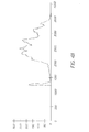
- Figs. 4A, 4B and 4C are graphical views illustrating the histogram analysis for chest radiographs.
- Fig. 4A is the gray-level histogram of the original chest radiograph.
- Fig. 4B shows the smoothed histogram.
- Fig. 4C displays the two detected thresholds in the histogram.
- Figs. 5A, 5B, 5C and 5D illustrating the pre-process of chest radiographs.
- Fig.5a shows the original chest radiograph.
- Fig. 5b displays the segmented image by using the two thresholds, and
- Fig. 5C shows the processed image after the background removal.
- Fig. 5D demonstrates the estimate of the lung regions.
- Fig. 6 is diagrammatic view illustrating the determination of the mediastinum region (the gray region).
- Fig. 7A and 7B are diagrammatic views showing the original chest radiograph and the normalized chest radiograph, respectively.
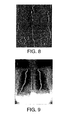
- Fig. 8 is a diagrammatic view showing the 0 th -order X direction derivative image of the chest radiograph (I 0 0 (x,y, ⁇ )) .
- Fig. 9 is a diagrammatic view displaying the search result of the chest body midline and two lung centerlines.
- Fig. 10 is a diagrammatic view which demonstrates the initial location of the knowledge model in the chest radiograph.
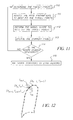
- Fig. 11 is a flow chart illustrating the deforming scheme of lung shape model.
- Fig. 12 is a diagrammatic view which illustrates the construction of the local search region.
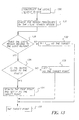
- Fig. 13 is a flow chart illustrating the determination of a target point to a landmark.
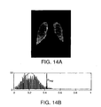
- Figs. 14A and 14B are diagrammatic views which respectively show the gray-level distribution of the detected lung regions and the determination of the lung region threshold.
- Fig. 14A displays the detected lung regions and
- Fig. 14B is the gray-level histogram of the detected lung region.
- Fig. 15 is a diagrammatic view which illustrates how to select the different edge information for each landmark.
- Image 150 shows that the edge information from the 1 st -order X-direction derivative image is chosen for the landmark points closed to the mediastinum.
- Image 151 demonstrates that the edges in the 2 nd ⁇ order Y-direction derivative image are associated to the landmark points near the lung top.
- Image 152 displays the lung shape models and their landmark points, which are grouped based on their positions.
- Image 153 shows the edge information from the 2 nd -order X-direction derivative image is used for the landmark points closed to the rib cage.
- Image 154 shows that the edges in the 1 st -order Y derivative image are related to the landmark points near the lung bottom.
- Figs. 16A and 16B are diagrammatic views which demonstrate the detected lung regions in the chest radiograph
- Fig. 17 is a block diagram of a radiographic imaging system incorporating the present invention.
- Fig. 17 is a block diagram of a radiographic system incorporating the present invention.
- a radiographic image such as a chest radiographic image is acquired by an image acquisition system 1600.
- Image acquisition system 1600 can include one of the following: (1) a conventional radiographic film/screen system in which a body part (chest) of a patient is exposed to x-radiation from an x-ray source and a radiographic image is formed in the radiographic image is formed in the radiographic film. The film is developed and digitized to produce a digital radiographic image. (2) A computed radiography system in which the radiographic image of the patient's body part is formed in a storage phosphor plate.
- the storage phosphor plate is scanned to produce a digital radiographic image.
- the storage phosphor plate is erased and reused.
- the digital radiographic image is processed according to the present invention by image processing system 1602.
- System 1602 is preferably a digital computer or digital microprocessor by can include hardware and firmware to carry out the various image processing operations.
- the processed digital radiographic image is provided to image output 1604, such as a high resolution electronic display or a printer which produces a hard copy (film) of the processed radiographic image.
- image output 1604 such as a high resolution electronic display or a printer which produces a hard copy (film) of the processed radiographic image.
- the original as well as the processed image can be transmitted to a remote location, can be stored in a radiographic image storage system (PACS), etc.
- PES radiographic image storage system
- the present invention discloses a method for automatically segmenting lung regions in chest radiographic images, which is based on the combination of six processing steps as shown in Fig 1.
- the input chest radiograph image (box 9) is pre-processed (box 10).
- the chest body midline and lung centerlines are extracted from the image (box 11).
- the chest body model (box 12), the spine model (box 13) and two lung models (box 14) are located in the chest radiograph image one by one based on these extracted feature lines.
- the lung contours (box 16) are detected by deforming the lung shape models to converge to the true lung boundaries with the help of the region and edge information (box 15).
- a knowledge model is used for lung segmentation, which encapsulates the following characteristics:
- a novelty of the present invention is that all input images are normalized based on Regions of Interests (ROIs), instead of the image gray-level histogram. This is reasonable because most image histograms contain the gray levels from foreground, background, and ROIs. Foreground is the area that is occluded by x-ray collimation during the exposure.
- ROIs Regions of Interests
- Backgrounds are areas that have received direct x-ray exposure, and ROIs is taken to be the remainder of the image that normally contains the anatomical regions of interest for diagnosis. If image normalization is simply based on its histogram, the result is inevitably biased by the image foreground and background.
- a block diagram illustrating the pre-processing of a chest radiograph as shown to implement the pre-processing a gray-level histogram of a chest radiographs 30 is produced and analyzed (box 31), and two thresholds are detected to separate the background, the lung regions and the rest of body parts (box 32), respectively.
- the lung regions can be estimated in a chest radiograph (box 34) and further the mediastinum region can be derived (box 35).
- the image is normalized based on the gray-level properties of the detected lung and mediastinum regions (box 36).
- the output of this step includes not only the normalized image (box 37), but also the estimate of lung regions (box 38).
- a gray-level histogram as shown in Fig. 4A is generated, and the maximal and minimal gray-level of the image are detected. Then two thresholds are derived to segment the original image based on the histogram.
- the first threshold ( th1 ) is used to separate the background from the image, and the second one ( th2 ) extracts the lung regions from the rest of chest body parts.
- the way to detect the first threshold is as follows: the histogram of Fig. 4A is first smoothed to reduce the noise interference, as shown in Fig. 4B. Then the derivative of the histogram is computed. Finally the position with the maximal derivative is detected and set as the first threshold, and the second threshold is obtained by Where h(i) is the histogram of the chest radiograph.
- Fig. 4C indicates the two detected thresholds.
- Figs. 5A and 5B respectively show the chest radiograph and its result by using the first threshold. After image thresholding, all regions with the gray-level below the first threshold are labeled and checked to ensure if they belong to background, then the background regions are removed from the segmented image as shown in Fig. 5C. Those speckles and noise left after the background removal can be easily eliminated by using morphological operations.
- Fig. 5D shows the final result image, in which two lung regions are apparent and can be used as an estimate of lung regions.
- the next step is to detect the mediastinum region which is located between the two lung fields. Its extraction can be simply completed by detecting the region between the two lung regions, as shown in Fig. 6. Once the mediastinum region is obtained, the input chest radiograph can be normalized by using the minimal gray-level of lung regions and the maximal gray-level of the mediastinum, as given in Eq. 2.
- I(x,y) is the grey-level of the original chest radiographic image at the pixel (x,y) .
- Figs. 7A-7B display both the original and the normalized chest radiograph. It is obvious that the image quality is greatly improved after the normalization.
- Derivative images are computed by convoluting the normalized image I new (x,y) with the derivative of a Gaussian G(x,y, ⁇ ) at a particular scale ⁇ .
- I ⁇ n ( x , y , ⁇ ) G ⁇ n ( x , y , ⁇ ) ⁇ I new ( x , y )
- G ( x,y, ⁇ ) 1 2 ⁇ 2 exp( -( x 2 + y 2 ) 2 ⁇ 2 )
- ⁇ denotes convolution and G n ⁇ is the n th -order derivative of the Gaussian kernel in the direction ⁇ .
- the white and black pixels in the derivative images correspond the maximum and minimum in the direction ⁇ , respectively, which are obtained by comparing each pixel with its neighboring regions using the non-maximal suppression algorithm.
- the two lung centerlines can be detected by finding two start points, one on each black line, then tracing them in both directions, and finally stopping at the pixel whose gray-level is greater than the second threshold (th2).
- the chest body midline is found by the same technique in the mediastinum region.
- Fig. 9 displays the search result of the chest body midline and two lung centerlines.
- the chest body model can be located by aligning its centerline with the chest body midline, and its model size is derived from the distance between the two lung centerlines, then the spine model is placed in the middle of the chest body model according to their anatomic spatial relationship.
- the locating of lung models is a little complicated, since their size, position and orientation have to be firstly derived from the lung centerlines, then the lung shape models are adjusted by these parameters and finally the models are aligned along the lung centerlines.
- Fig. 10 demonstrates the initial location of the knowledge model in the chest radiograph.
- the lung shape model used in the present invention is a 2D statistical shape model from H. Luo et al., Method for automatic construction of 2D statistical shape model for the lung regions, which consists of a mean shape vector ( X ) to represent the general shape and variation modes in the form of the eigenvectors (P t ) .
- Deforming the lung shape models is performed in three stages, as illustrated in Fig. 11. In the first stage, a proper target point is selected for each landmark based on the initial location of the lung mean shape model (box 10). In the second stage, the lung mean shape models are deformed rigidly to optimize the objective function (box 111). This step allows for any small adjustments in the position, orientation and size of shape models.
- the shape model is deformed locally in order to best fit the target points (box 112) and the current shape is updated (box 113).
- the process is repeated ideally, after several iterations, the change of shape will become negligible (diamond 114) and the shape model tends to be stable (box 115).
- region and edge information are employed for the detection.
- the region information is obtained from a small local search region around the landmark, and used to indicate the landmark location, such as inside or outside of the lung regions, or close to the boundaries.
- the edge information is extracted from different orders and direction derivative images for each landmark, which gives an accurate representation of the boundary properties of the lung regions.
- a local search region is constructed at each landmark point (box 130), which is a narrow band perpendicular to the local contour of the landmark.
- the direction perpendicular to a landmarks (x n ,y n ) is computed by rotating the vector that runs from landmark (x n-1 , y n-1 ) to landmark (x n+1 , y n+1 ) over 90° . Since the lung shape models are closed contours, the first landmark uses the last landmark and the second landmark as two adjacent landmarks and the last landmark uses the second last landmark and the first landmark.
- Fig. 12 illustrates the construction of the local search region, where t in represents the inside target point of the local search region, and t out represents the outside target point of the local search region.
- t in represents the inside target point of the local search region
- t out represents the outside target point of the local search region.
- th reg the threshold th reg
- the landmark belongs to lung regions (diamond 135), and its target point is set to be t out (box 132). If most of them are above the threshold (diamond 136), the landmark lies outside of lung regions, and its target point is set to be t in (box 133). Otherwise, the edge information is needed to determine the target point (box 134) (box 137).
- the lung region threshold used here is adaptive and updated during each iteration of deformation. It is first initialized as th2 , detected in the pre-processing. Then after an iteration of deformation, the lung region threshold is updated based on a weighted mean of the newly detected lung region. Eq. 5 gives the way to compute the weighted region mean. For those pixels less than th2 in the detected lung region, it is sure that they belong to the lung region, thus their weights w(x,y) are set higher to emphasize their contribution to the region properties.
- the gray level distribution of lung regions is modeled as a Guassian distribution over the region weighted mean I reg with variance ⁇ reg , as illustrated in Fig 14.
- edge information is based on the position of a landmark and its contour shape.
- Fig. 15 indicates the way to determine edge information in the present invention.
- the landmark points on the lung shape contours are classified into three or four groups depending on their positions, as demonstrated in (image 154).
- the landmark points closed to the mediastinum (group1) select their edge information from the 1 st -order X derivative image (image 150). More precisely, the landmarks on the left side of the mediastinum try to converge to the maximal edges (the white pixels) in the derivative image and those on the right side of the mediastinum try to converage to the minimal edges (the black pixels) in the derivative image.
- the landmark point on the top of the shape (group2), they are more interested in the minimal edges (the black pixels) in the 2 nd ⁇ order Y derivative image (image 151).
- the landmark points near the rib cages (group 3) use the minimal edges (the black pixels) in the 2 nd -order X derivative image (image 152).
- the landmark points on the lung bottom (group 4) search for the maximal edges (the white pixels) from the 1 st -order Y derivative image (image 153). Since the target point comes from the local search region of the landmark, it is possible that no edge information is found in the local search region. In such situation, the landmark point is left where it is, and later the model constraints will eventually pull it into a reasonable position.
- a set of suitable target points (X T ) are given, and the best fit pose parameters can be computed by minimizing the sum of squares of distances between the landmarks from the lung mean shape and their corresponding target points.
- E ( X T - M(s d , ⁇ d ) X - t d ) T ( X T - M(s d , ⁇ d ) X - t d )
- ⁇ d is an appropriate rotation
- s d is a scale
- t d ( t dx , t dy ) represents translation.
- any shape vector X can be approximated using the mean shape X and a weighted sum of its variation, as given in Eq. (9).
- X X + P t b t
- dx M -1 ( s d , ⁇ d )( X T + t d )- X
- the shape parameters (components in the vector b t ) have to be checked by pre-defined limits to avoid implausible shape. This may cause the deformed shape not match the target points precisely sometimes. However, such imprecision can be eventually minimized or disappear after enough iteration, and finally the lung shape models will converge to the true boundaries of lung regions, as shown in Fig. 16.
Landscapes
- Engineering & Computer Science (AREA)
- General Physics & Mathematics (AREA)
- Physics & Mathematics (AREA)
- Theoretical Computer Science (AREA)
- Computer Vision & Pattern Recognition (AREA)
- General Health & Medical Sciences (AREA)
- Software Systems (AREA)
- Health & Medical Sciences (AREA)
- Multimedia (AREA)
- Medical Informatics (AREA)
- Nuclear Medicine, Radiotherapy & Molecular Imaging (AREA)
- Radiology & Medical Imaging (AREA)
- Quality & Reliability (AREA)
- Apparatus For Radiation Diagnosis (AREA)
- Image Processing (AREA)
- Image Analysis (AREA)
Applications Claiming Priority (2)
| Application Number | Priority Date | Filing Date | Title |
|---|---|---|---|
| US315884 | 2002-12-10 | ||
| US10/315,884 US7221787B2 (en) | 2002-12-10 | 2002-12-10 | Method for automated analysis of digital chest radiographs |
Publications (2)
| Publication Number | Publication Date |
|---|---|
| EP1465109A2 true EP1465109A2 (de) | 2004-10-06 |
| EP1465109A3 EP1465109A3 (de) | 2007-08-01 |
Family
ID=32468822
Family Applications (1)
| Application Number | Title | Priority Date | Filing Date |
|---|---|---|---|
| EP03078741A Withdrawn EP1465109A3 (de) | 2002-12-10 | 2003-11-27 | Verfahren zur automatischen Analyse von Burströntgenaufnahmen |
Country Status (2)
| Country | Link |
|---|---|
| US (2) | US7221787B2 (de) |
| EP (1) | EP1465109A3 (de) |
Cited By (8)
| Publication number | Priority date | Publication date | Assignee | Title |
|---|---|---|---|---|
| WO2006123272A3 (en) * | 2005-05-18 | 2007-05-03 | Koninkl Philips Electronics Nv | Automatic organ mapping method an device |
| GB2468164A (en) * | 2009-02-27 | 2010-09-01 | Medicsight Plc | Characterising of image geometry using derivatives |
| US7860283B2 (en) | 2006-10-25 | 2010-12-28 | Rcadia Medical Imaging Ltd. | Method and system for the presentation of blood vessel structures and identified pathologies |
| US7873194B2 (en) | 2006-10-25 | 2011-01-18 | Rcadia Medical Imaging Ltd. | Method and system for automatic analysis of blood vessel structures and pathologies in support of a triple rule-out procedure |
| US7940977B2 (en) | 2006-10-25 | 2011-05-10 | Rcadia Medical Imaging Ltd. | Method and system for automatic analysis of blood vessel structures to identify calcium or soft plaque pathologies |
| US7940970B2 (en) | 2006-10-25 | 2011-05-10 | Rcadia Medical Imaging, Ltd | Method and system for automatic quality control used in computerized analysis of CT angiography |
| US8103074B2 (en) | 2006-10-25 | 2012-01-24 | Rcadia Medical Imaging Ltd. | Identifying aorta exit points from imaging data |
| US8369927B2 (en) | 2005-11-21 | 2013-02-05 | Koninklijke Philips Electronics N.V. | Method for creating a model of a structure |
Families Citing this family (56)
| Publication number | Priority date | Publication date | Assignee | Title |
|---|---|---|---|---|
| US7391893B2 (en) * | 2003-06-27 | 2008-06-24 | Siemens Medical Solutions Usa, Inc. | System and method for the detection of shapes in images |
| US7352888B2 (en) * | 2004-07-30 | 2008-04-01 | Carestream Health, Inc. | Method for computer recognition of projection views and orientation of chest radiographs |
| US7736313B2 (en) * | 2004-11-22 | 2010-06-15 | Carestream Health, Inc. | Detecting and classifying lesions in ultrasound images |
| DE102004061507B4 (de) * | 2004-12-21 | 2007-04-12 | Siemens Ag | Verfahren zur Korrektur von Inhomogenitäten in einem Bild sowie bildgebende Vorrichtung dazu |
| US7263346B2 (en) * | 2005-05-10 | 2007-08-28 | Sbc Knowledge Ventures, L.P. | Method and system for enabling mobile users to manage cells |
| CN1879553B (zh) * | 2005-06-15 | 2010-10-06 | 佳能株式会社 | 在胸部图像中检测边界的方法及装置 |
| US20070058862A1 (en) * | 2005-09-09 | 2007-03-15 | Meier Michael R | Histogram equalization method for a vision-based occupant sensing system |
| US20070092864A1 (en) * | 2005-09-30 | 2007-04-26 | The University Of Iowa Research Foundation | Treatment planning methods, devices and systems |
| US7672497B2 (en) * | 2005-12-29 | 2010-03-02 | Carestream Health, Inc. | Computer aided disease detection system for multiple organ systems |
| DE102006012943B4 (de) * | 2006-03-21 | 2009-11-19 | Siemens Ag | Verfahren zur automatischen Auswertung eines Abbildungsdatensatzes eines Objektes und medizinisches Abbildungssystem |
| US8014602B2 (en) * | 2006-03-29 | 2011-09-06 | Seiko Epson Corporation | Backlight image determining apparatus, backlight image determining method, backlight image correction apparatus, and backlight image correction method |
| WO2007135913A1 (ja) * | 2006-05-19 | 2007-11-29 | Hitachi Medical Corporation | 医用画像表示装置及びプログラム |
| US7916943B2 (en) * | 2006-06-02 | 2011-03-29 | Seiko Epson Corporation | Image determining apparatus, image determining method, image enhancement apparatus, and image enhancement method |
| US7916942B1 (en) * | 2006-06-02 | 2011-03-29 | Seiko Epson Corporation | Image determining apparatus, image enhancement apparatus, backlight image enhancement apparatus, and backlight image enhancement method |
| DE102006028646A1 (de) * | 2006-06-22 | 2007-12-27 | Siemens Ag | Auswertungsverfahren für Bilddatensätze mit selbsttätiger Bestimmung von Auswertungsbereichen |
| US20080058642A1 (en) * | 2006-09-05 | 2008-03-06 | Board Of Regents Of The University Of Texas System | Method for detecting coronary endothelial dysfunction and early atherosclerosis |
| US8165376B2 (en) * | 2006-12-11 | 2012-04-24 | Siemens Corporation | System and method for automatic detection of rib metastasis in computed tomography volume |
| US8989468B2 (en) | 2007-05-25 | 2015-03-24 | Definiens Ag | Generating an anatomical model using a rule-based segmentation and classification process |
| US8107699B2 (en) * | 2007-08-08 | 2012-01-31 | Siemens Medical Solutions Usa, Inc. | Feature processing for lung nodules in computer assisted diagnosis |
| CN101502420B (zh) * | 2008-02-05 | 2013-05-22 | Ge医疗系统环球技术有限公司 | 图像处理方法和设备 |
| JP2009285145A (ja) * | 2008-05-29 | 2009-12-10 | Fujifilm Corp | 放射線画像補正装置および方法ならびにプログラム |
| US8189886B2 (en) * | 2008-08-13 | 2012-05-29 | Carestream Health, Inc. | Method for detecting anatomical structures |
| US8577108B2 (en) | 2008-08-13 | 2013-11-05 | Carestream Health, Inc. | Method for detecting anatomical structures |
| EP2353147B1 (de) | 2008-11-28 | 2021-05-19 | Fujifilm Medical Systems U.S.A. Inc. | System und verfahren zur propagation des spine-labeling |
| CN102711626B (zh) * | 2010-01-07 | 2014-12-10 | 株式会社日立医疗器械 | 医用图像诊断装置和医用图像的轮廓提取处理方法 |
| US8553957B2 (en) | 2010-12-22 | 2013-10-08 | Samsung Electronics Co., Ltd. | Method of processing image and image processing apparatus using the method |
| US8781187B2 (en) * | 2011-07-13 | 2014-07-15 | Mckesson Financial Holdings | Methods, apparatuses, and computer program products for identifying a region of interest within a mammogram image |
| US9659390B2 (en) | 2011-10-28 | 2017-05-23 | Carestream Health, Inc. | Tomosynthesis reconstruction with rib suppression |
| US8913817B2 (en) * | 2011-10-28 | 2014-12-16 | Carestream Health, Inc. | Rib suppression in radiographic images |
| US9269139B2 (en) * | 2011-10-28 | 2016-02-23 | Carestream Health, Inc. | Rib suppression in radiographic images |
| KR20130072073A (ko) * | 2011-12-21 | 2013-07-01 | 한국전자통신연구원 | 영상 윤곽선 추출 장치 및 방법 |
| US20130243077A1 (en) * | 2012-03-13 | 2013-09-19 | Canon Kabushiki Kaisha | Method and apparatus for processing moving image information, and method and apparatus for identifying moving image pattern |
| JP5844187B2 (ja) * | 2012-03-23 | 2016-01-13 | 富士フイルム株式会社 | 画像解析装置および方法並びにプログラム |
| US9230320B2 (en) * | 2012-03-30 | 2016-01-05 | University Of Louisville Research Foundation, Inc. | Computer aided diagnostic system incorporating shape analysis for diagnosing malignant lung nodules |
| US9025086B2 (en) | 2012-04-13 | 2015-05-05 | Red.Com, Inc. | Video projector system |
| EP2842330B1 (de) | 2012-04-13 | 2018-08-29 | Red.Com, Llc | Videoprojektorsystem |
| CA2910376C (en) | 2012-05-09 | 2020-06-30 | Laboratoires Bodycad Inc. | Segmentation of magnetic resonance imaging data |
| EP2876607B1 (de) * | 2012-07-23 | 2019-07-31 | Fujitsu Limited | Formdatenerzeugungsverfahren und vorrichtung |
| US9672600B2 (en) * | 2012-11-19 | 2017-06-06 | Carestream Health, Inc. | Clavicle suppression in radiographic images |
| US9940545B2 (en) * | 2013-09-20 | 2018-04-10 | Change Healthcare Llc | Method and apparatus for detecting anatomical elements |
| US9501498B2 (en) * | 2014-02-14 | 2016-11-22 | Nant Holdings Ip, Llc | Object ingestion through canonical shapes, systems and methods |
| US9626476B2 (en) | 2014-03-27 | 2017-04-18 | Change Healthcare Llc | Apparatus, method and computer-readable storage medium for transforming digital images |
| CN106163404B (zh) * | 2014-04-08 | 2019-04-23 | Icad股份有限公司 | 用于射线图像的肺分割和骨抑制技术 |
| US10061979B2 (en) | 2014-09-28 | 2018-08-28 | Koninklijke Philips N.V. | Image processing apparatus and method |
| US11160528B2 (en) * | 2015-12-18 | 2021-11-02 | General Electric Company | System and method for visualization of ultrasound volumes |
| US9801601B2 (en) | 2015-12-29 | 2017-10-31 | Laboratoires Bodycad Inc. | Method and system for performing multi-bone segmentation in imaging data |
| JP6833444B2 (ja) * | 2016-10-17 | 2021-02-24 | キヤノン株式会社 | 放射線撮影装置、放射線撮影システム、放射線撮影方法、及びプログラム |
| US10957045B2 (en) * | 2016-12-12 | 2021-03-23 | University Of Notre Dame Du Lac | Segmenting ultrasound images |
| JP2018157884A (ja) * | 2017-03-22 | 2018-10-11 | コニカミノルタ株式会社 | X線動画像処理装置 |
| US10431000B2 (en) * | 2017-07-18 | 2019-10-01 | Sony Corporation | Robust mesh tracking and fusion by using part-based key frames and priori model |
| JP7047574B2 (ja) * | 2018-04-26 | 2022-04-05 | コニカミノルタ株式会社 | 動態画像解析装置、動態画像解析システム、動態画像解析プログラム及び動態画像解析方法 |
| CN108888284A (zh) * | 2018-05-18 | 2018-11-27 | 沈阳东软医疗系统有限公司 | 图像调整方法、装置及设备、存储介质 |
| EP3866107A1 (de) * | 2020-02-14 | 2021-08-18 | Koninklijke Philips N.V. | Modellbasierte bildsegmentierung |
| CN111816314B (zh) * | 2020-07-01 | 2023-10-20 | 深圳市职业病防治院 | 一种用于人工智能筛查尘肺病胸片遴选、标注及验证方法 |
| CN113516677B (zh) * | 2021-04-13 | 2022-02-22 | 推想医疗科技股份有限公司 | 一种结构化分级管状结构血管的方法、装置及电子设备 |
| CN120707428B (zh) * | 2025-06-25 | 2025-11-21 | 中国人民解放军总医院第八医学中心 | 一种面向肺炎影像的ct图像自适应增强方法 |
Family Cites Families (7)
| Publication number | Priority date | Publication date | Assignee | Title |
|---|---|---|---|---|
| US5359513A (en) * | 1992-11-25 | 1994-10-25 | Arch Development Corporation | Method and system for detection of interval change in temporally sequential chest images |
| US5570430A (en) * | 1994-05-31 | 1996-10-29 | University Of Washington | Method for determining the contour of an in vivo organ using multiple image frames of the organ |
| US6282307B1 (en) * | 1998-02-23 | 2001-08-28 | Arch Development Corporation | Method and system for the automated delineation of lung regions and costophrenic angles in chest radiographs |
| US6594378B1 (en) * | 1999-10-21 | 2003-07-15 | Arch Development Corporation | Method, system and computer readable medium for computerized processing of contralateral and temporal subtraction images using elastic matching |
| US6577752B2 (en) * | 2001-06-15 | 2003-06-10 | Arch Development Corporation | Automated method and system for the delineation of the chest wall in computed tomography scans for the assessment of pleural disease |
| US7058210B2 (en) * | 2001-11-20 | 2006-06-06 | General Electric Company | Method and system for lung disease detection |
| US20050033139A1 (en) * | 2002-07-09 | 2005-02-10 | Deus Technologies, Llc | Adaptive segmentation of anatomic regions in medical images with fuzzy clustering |
-
2002
- 2002-12-10 US US10/315,884 patent/US7221787B2/en not_active Expired - Lifetime
-
2003
- 2003-11-27 EP EP03078741A patent/EP1465109A3/de not_active Withdrawn
-
2006
- 2006-12-01 US US11/565,709 patent/US20070086640A1/en not_active Abandoned
Non-Patent Citations (3)
| Title |
|---|
| ARMATO S G ET AL: "AUTOMATED LUNG SEGMENTATION IN DIGITIZED POSTEROANTERIOR CHEST RADIOGRAPHS" ACADEMIC RADIOLOGY, RESTON, VA, US, vol. 5, no. 4, April 1998 (1998-04), pages 245-255, XP008055922 ISSN: 1076-6332 * |
| BROWN MATTHEW S ET AL: "Knowledge-based segmentation of thoracic computed tomography images for assessment of split lung function" MEDICAL PHYSICS, AIP, MELVILLE, NY, US, vol. 27, no. 3, March 2000 (2000-03), pages 592-598, XP012011085 ISSN: 0094-2405 * |
| LELIEVELDT B P F ET AL: "Anatomical Modeling with Fuzzy Implicit Surface Templates: Application to Automated Localization of the Heart and Lungs in Thoracic MR Volumes" October 2000 (2000-10), COMPUTER VISION AND IMAGE UNDERSTANDING, ACADEMIC PRESS, SAN DIEGO, CA, US, PAGE(S) 1-20 , XP004439256 ISSN: 1077-3142 * abstract * * page 9, paragraph 4 * * |
Cited By (10)
| Publication number | Priority date | Publication date | Assignee | Title |
|---|---|---|---|---|
| WO2006123272A3 (en) * | 2005-05-18 | 2007-05-03 | Koninkl Philips Electronics Nv | Automatic organ mapping method an device |
| US8369927B2 (en) | 2005-11-21 | 2013-02-05 | Koninklijke Philips Electronics N.V. | Method for creating a model of a structure |
| US7860283B2 (en) | 2006-10-25 | 2010-12-28 | Rcadia Medical Imaging Ltd. | Method and system for the presentation of blood vessel structures and identified pathologies |
| US7873194B2 (en) | 2006-10-25 | 2011-01-18 | Rcadia Medical Imaging Ltd. | Method and system for automatic analysis of blood vessel structures and pathologies in support of a triple rule-out procedure |
| US7940977B2 (en) | 2006-10-25 | 2011-05-10 | Rcadia Medical Imaging Ltd. | Method and system for automatic analysis of blood vessel structures to identify calcium or soft plaque pathologies |
| US7940970B2 (en) | 2006-10-25 | 2011-05-10 | Rcadia Medical Imaging, Ltd | Method and system for automatic quality control used in computerized analysis of CT angiography |
| US8103074B2 (en) | 2006-10-25 | 2012-01-24 | Rcadia Medical Imaging Ltd. | Identifying aorta exit points from imaging data |
| GB2468164A (en) * | 2009-02-27 | 2010-09-01 | Medicsight Plc | Characterising of image geometry using derivatives |
| GB2468164B (en) * | 2009-02-27 | 2014-08-13 | Samsung Electronics Co Ltd | Computer-aided detection of lesions |
| US9014447B2 (en) | 2009-02-27 | 2015-04-21 | Samsung Electronics Co., Ltd. | System and method for detection of lesions in three-dimensional digital medical image |
Also Published As
| Publication number | Publication date |
|---|---|
| US20070086640A1 (en) | 2007-04-19 |
| US20040109595A1 (en) | 2004-06-10 |
| EP1465109A3 (de) | 2007-08-01 |
| US7221787B2 (en) | 2007-05-22 |
Similar Documents
| Publication | Publication Date | Title |
|---|---|---|
| US7221787B2 (en) | Method for automated analysis of digital chest radiographs | |
| Raba et al. | Breast segmentation with pectoral muscle suppression on digital mammograms | |
| Timp et al. | A new 2D segmentation method based on dynamic programming applied to computer aided detection in mammography | |
| Sluimer et al. | Toward automated segmentation of the pathological lung in CT | |
| JP3110045B2 (ja) | 医用コンピュータ断層撮影スキャンで病変を検出するための自動化された方法およびシステム | |
| EP0757544B1 (de) | Rechnergestützte bestimmung von parenchymatösen massen und verformungen | |
| Vujovic et al. | Establishing the correspondence between control points in pairs of mammographic images | |
| US7876938B2 (en) | System and method for whole body landmark detection, segmentation and change quantification in digital images | |
| EP2916738B1 (de) | Lungen-, lappen- und spaltenbildgebungssysteme und -verfahren | |
| Xu et al. | An edge-region force guided active shape approach for automatic lung field detection in chest radiographs | |
| US20050207630A1 (en) | Lung nodule detection and classification | |
| US9269139B2 (en) | Rib suppression in radiographic images | |
| Hogeweg et al. | Clavicle segmentation in chest radiographs | |
| US8913817B2 (en) | Rib suppression in radiographic images | |
| CN102947841A (zh) | 放射摄影图像中的毛刺恶性肿块检测和分级 | |
| Farag et al. | Automatic detection and recognition of lung abnormalities in helical CT images using deformable templates | |
| US20120099771A1 (en) | Computer aided detection of architectural distortion in mammography | |
| Timp et al. | Interval change analysis to improve computer aided detection in mammography | |
| US20050033139A1 (en) | Adaptive segmentation of anatomic regions in medical images with fuzzy clustering | |
| Hooda et al. | Tuberculosis detection from chest radiographs: a comprehensive survey on computer-aided diagnosis techniques | |
| Farag et al. | Detection and recognition of lung abnormalities using deformable templates | |
| US20060098854A1 (en) | Abnormal pattern candidate detecting method and apparatus | |
| van Ginneken et al. | Automatic delineation of ribs in frontal chest radiographs | |
| Schilham et al. | Multi-scale nodule detection in chest radiographs | |
| JP2004188202A (ja) | デジタル胸部放射線写真の自動分析方法 |
Legal Events
| Date | Code | Title | Description |
|---|---|---|---|
| PUAI | Public reference made under article 153(3) epc to a published international application that has entered the european phase |
Free format text: ORIGINAL CODE: 0009012 |
|
| AK | Designated contracting states |
Kind code of ref document: A2 Designated state(s): AT BE BG CH CY CZ DE DK EE ES FI FR GB GR HU IE IT LI LU MC NL PT RO SE SI SK TR |
|
| AX | Request for extension of the european patent |
Extension state: AL LT LV MK |
|
| PUAL | Search report despatched |
Free format text: ORIGINAL CODE: 0009013 |
|
| AK | Designated contracting states |
Kind code of ref document: A3 Designated state(s): AT BE BG CH CY CZ DE DK EE ES FI FR GB GR HU IE IT LI LU MC NL PT RO SE SI SK TR |
|
| AX | Request for extension of the european patent |
Extension state: AL LT LV MK |
|
| 17P | Request for examination filed |
Effective date: 20070913 |
|
| RAP1 | Party data changed (applicant data changed or rights of an application transferred) |
Owner name: CARESTREAM HEALTH, INC. |
|
| AKX | Designation fees paid |
Designated state(s): DE FR GB IT NL |
|
| STAA | Information on the status of an ep patent application or granted ep patent |
Free format text: STATUS: THE APPLICATION IS DEEMED TO BE WITHDRAWN |
|
| 18D | Application deemed to be withdrawn |
Effective date: 20090603 |