EP1475442A2 - Polypeptides de l'antivirus lymphotropiques de cellules t du chat - Google Patents
Polypeptides de l'antivirus lymphotropiques de cellules t du chat Download PDFInfo
- Publication number
- EP1475442A2 EP1475442A2 EP04015677A EP04015677A EP1475442A2 EP 1475442 A2 EP1475442 A2 EP 1475442A2 EP 04015677 A EP04015677 A EP 04015677A EP 04015677 A EP04015677 A EP 04015677A EP 1475442 A2 EP1475442 A2 EP 1475442A2
- Authority
- EP
- European Patent Office
- Prior art keywords
- fiv
- polypeptide
- polypeptide fragment
- polypeptides
- gag
- Prior art date
- Legal status (The legal status is an assumption and is not a legal conclusion. Google has not performed a legal analysis and makes no representation as to the accuracy of the status listed.)
- Withdrawn
Links
Images


Classifications
-
- C—CHEMISTRY; METALLURGY
- C07—ORGANIC CHEMISTRY
- C07K—PEPTIDES
- C07K14/00—Peptides having more than 20 amino acids; Gastrins; Somatostatins; Melanotropins; Derivatives thereof
- C07K14/005—Peptides having more than 20 amino acids; Gastrins; Somatostatins; Melanotropins; Derivatives thereof from viruses
-
- A—HUMAN NECESSITIES
- A61—MEDICAL OR VETERINARY SCIENCE; HYGIENE
- A61P—SPECIFIC THERAPEUTIC ACTIVITY OF CHEMICAL COMPOUNDS OR MEDICINAL PREPARATIONS
- A61P31/00—Antiinfectives, i.e. antibiotics, antiseptics, chemotherapeutics
- A61P31/12—Antivirals
-
- A—HUMAN NECESSITIES
- A61—MEDICAL OR VETERINARY SCIENCE; HYGIENE
- A61P—SPECIFIC THERAPEUTIC ACTIVITY OF CHEMICAL COMPOUNDS OR MEDICINAL PREPARATIONS
- A61P31/00—Antiinfectives, i.e. antibiotics, antiseptics, chemotherapeutics
- A61P31/12—Antivirals
- A61P31/14—Antivirals for RNA viruses
-
- A—HUMAN NECESSITIES
- A61—MEDICAL OR VETERINARY SCIENCE; HYGIENE
- A61K—PREPARATIONS FOR MEDICAL, DENTAL OR TOILETRY PURPOSES
- A61K39/00—Medicinal preparations containing antigens or antibodies
-
- C—CHEMISTRY; METALLURGY
- C12—BIOCHEMISTRY; BEER; SPIRITS; WINE; VINEGAR; MICROBIOLOGY; ENZYMOLOGY; MUTATION OR GENETIC ENGINEERING
- C12N—MICROORGANISMS OR ENZYMES; COMPOSITIONS THEREOF; PROPAGATING, PRESERVING, OR MAINTAINING MICROORGANISMS; MUTATION OR GENETIC ENGINEERING; CULTURE MEDIA
- C12N2740/00—Reverse transcribing RNA viruses
- C12N2740/00011—Details
- C12N2740/10011—Retroviridae
- C12N2740/15011—Lentivirus, not HIV, e.g. FIV, SIV
- C12N2740/15022—New viral proteins or individual genes, new structural or functional aspects of known viral proteins or genes
Definitions
- This invention relates to purification of polypeptides from feline T-cell lymphotropic lentivirus (FIV).
- FIV is a retrovirus originally isolated from cats which exhibit an AIDS-like syndrome.
- Pederson et al. 235 Science 790, 1987.
- the virus belongs to the same group as the human immunodeficiency virus (HIV), the causative agent of human AIDS.
- HIV human immunodeficiency virus
- Pederson et al. describe detection of antibody to FIV by an immunofluorescent assay, and by western blots, using virus purified by centrifugation on sucrose gradients in Tris base, pH 7.4, containing 0.1M NaCl and 1mM EDTA. A few protein bands were detected and, although antigenic comparison was not made, the positions of these bands were tentatively said to correspond to the major core protein, p24 the gag precursor protein, p55, and the endonuclease protein, p32, of HIV.
- Pederson et al. U.S. Patent Application Filed August 26, 1987 entitled "Feline T-Lymphotropic Lentivirus” (which is not admitted to be prior art to the present application) describes the results presented by Pederson et al., supra , and states that Western blotting of FIV infected cell lysates yielded major protein bands at approximately 22-26 kD, usually about 24 kD; 50-60 kD, usually about 55 kD; and 28-36 kD, usually about 32 kD.
- the invention features a purified polypeptide having an epitope of an antigenic polypeptide of FIV.
- the polypeptide may be glycosylated or unglycosylated.
- antigenic polypeptide is meant a polypeptide which is able to raise (with the aid of an adjuvant if necessary) an antibody response in cats.
- the polypeptide may be a polypeptide fragment of at least 5 amino acids, or a polypeptide naturally occurring in FIV particles.
- the fragment may be obtained from a naturally-occurring polypeptide, for example, by enzymatic digestion of a naturally occurring polypeptide, or may be produced by isolation or synthesis of a gene encoding a desired polypeptide and expression of that polypeptide within a desired expression system, for example, a bacterial, yeast, or mammalian expression system.
- epitope is meant a single antigenic site of an antigenic polypeptide. Such an epitope is recognized specifically by a monoclonal antibody to an antigenic polypeptide of FIV.
- purified is meant that the polypeptide is separated from other cell components with which it naturally occurs, for example, FIV polypeptides.
- the polypeptide is sufficiently pure to permit its use to prepare a monoclonal antibody ro the polypeptide, and even more preferably, pure enough to allow the amino acid sequence of the polypeptide to be determined by standard procedure.
- the purified, polypeptide is biologically active in that it is suitable for preparation of a monoclonal antibody, or is suitable for defection of naturally-occurring antibodies within the serum of a cat.
- the purified polypeptide has at least 75% amino acid homology to a polypeptide fragment of at least 20 amino acids obtained from an FIV gag or env polypeptide, most preferably the purified polypeptide includes an amino acid sequence having at least 75% homology to a whole of a gag or env polypeptide, even more preferably, the purified polypeptide is an entire gag or env amino acid sequence.
- gag and env polypeptides include p10, p15, p26, gp40, gp100, and gp130.
- the invention features a method for detecting an antibody to FIV within a sample, including the steps of providing a purified polypeptide as described above, and contacting that polypeptide under conditions suitable to allow an antibody/polypeptide complex to form between antibodies within the sample and the purified polypeptide, and detecting the formation of such complexes. The presence of antibody/polypeptide complexes is indicative of antibody to FIV present within the sample.
- the invention features a purified nucleic acid including a 50 nucleotide sequence having at least 90% homology with a 50 nucleotide sequence naturally occurring in an FIV carticle.
- purified is meant that the nucleic acid is substantially separated away from all of the components with which it naturally occurs, e.g., polypeptides and other nucleic acids.
- the nucleic acid is completely separated from such components, and is a pure solution of nucleic acid, or is held within a cell in which it does not naturally occur, e.g, a bacterial cell, another viral particle or a non-feline eucaryotic cell.
- 90% homology is meant that the nucleotide sequence is identical at at least 45 of the 50 nucleotides.
- the nucleic acid encodes a polypeptide including an epitope of an antigenic polypeptide of FIV, e.g., an epitope of a gag or env polypeptide of FIV.
- the nucleic acid is carried in an expression vector and can be expressed in a bacterial, fungal or other eucaryotic cell, e.g., a mammalian cell.
- the invention features purified polypeptide including ten or more contiguous amino acids taken from the sequence (using standard letters to represent amino acids)
- polypeptides suitable for specific detection of FIV antibodies and thus have allowed accurate detection of infection with FIV.
- polypeptides useful for production of vaccines to prevent disease caused by FIV in cats are also provided.
- FIV polypeptide antigens useful in this invention are generally described above.
- Polypeptides useful in this invention may be purified from virus isolated as described below and fragments of the purified polypeptides isolated by enzymatic treatment or other standard procedures. Further, the polypeptides may be synthesized by standard in vitro expression systems in which DNA encoding for the FIV polypeptide is cloned and expressed in a bacterial, yeast, or mammalian cell expression system. Such DNA may be isolated and expressed as described below.
- the polypeptides may also be synthesized by standard chemical methods, for example, the polypeptide segments of various FIV polypeptides given below can be synthesized.
- FIV polypeptides were obtained directly from FIV-infected cells and took the form of polypeptides naturally occurring in an FIV virus particle. This example is not limiting to the present invention, and those skilled in the art will recognize the many alternative methods for obtaining polypeptides of this invention.
- the polypeptides are referred to according to molecular weight, thus a polypeptide of 30kD is termed p30, and a glycoprotein of this weight is termed gp30.
- virus cultures can be obtained as described by both Pederson et al. references, supra , or by Harbour et al., 122 The Veterinary Record 84, 1988. Seed stocks of virus producing cell cultures were obtained by freeze-downs of FIV-infectec master seed cell cultures following at least 19 post infection passages in culture. Additional seed stocks of virus producing cultures were obtained by either infection of the continuous feline cell culture with FIV master seed virus or by single cell microwell cloning of high level FIV producers from the original FIV infected master seed cell culture. For propogation, master seed virus infected feline cell cultures were inoculated into tissue cell culture flasks. Following growth to a confluent monolayer of cells, tissue culture fluid was harvested at intervals of 2-5 days.
- Working seed virus was produced by propogation by the master seed cell line permanently infected with FIV.
- An inoculum was added to tissue culture flasks, in Dulbecco's Modified Eagles medium containing 2 mM L-glutamine and 4.5 g per liter/glucose (DME) containing 100 units per ml. penicillin and streptomycin and 2 mM glutamine.
- An inoculum was added to tissue culture flasks, incubated, and the spent tissue culture fluid harvested when the cells were grown to confluence. The cells were released from the culture vessel with tryps in/EDTA and diluted between 1:5 and 1:25 (typically 1:8) in medium.
- the flasks were incubated at 36°C-38°C for a maximum of 7 days (between 3 and 7 days) before fluid and cell harvest.
- the harvested fluid including cell material, was centrifuged in a high speed centrifuge (Sorval RC-5B cr Beckman J2-21) leading to separation of supernatant and cell pellet material.
- the cell pellet was discarded, and the supernatant culture fluid used to prepare working virus.
- the clarified supernatant was made 0.5 M in NaCl and 4%-10% (usually 7%) in polyethylene glycol (PEG 8000, Sigma).
- virus was pelleted (at 13,000 x g for 30 min.) and resuspended in buffer (10 mM Tris, pH 7.6 300 mM NaCl, 1mM EDTA, at 2°C-7°C) . After overnight incubation the virus, was centrifuged at 13,000 x g for 15 min., the pellet discarded and the supernatant centrifuged on a 50%/80% discontinuous glycerol step gradient in 10 mM Tris 300 mM NaCl, 1 mM EDTA at pH 7.6. Centrifugation was at 100,000 x g for 3 hrs. at 4°C and the FIV viral band at the 50%-80% interface collected.
- buffer 10 mM Tris, pH 7.6 300 mM NaCl, 1mM EDTA, at 2°C-7°C
- the band was suspended in 10 mM Tris, 0.3 M NaCl and 1 mM EDTA and diluted 1:3 in the buffer and repelleted at 100,000 x g for 1 hr.
- the resulting pellet was purified virus and was resuspended in the above buffer and stored at -70°C.
- the resulting virus was substantially free from FIV host cell proteins and was composed of at least 5% p26 (the major nucleocapsid protein, as measured by densitometric scans of Commassie Blue 250 stained SDS/PAGE as total protein).
- Such purified virus may be obtained by other techniques, however, applicants have found that high molecular weight contaminants present in virus preparations may be eliminated by use of the high salt (i.e., greater than physiological range salt concentration) used in the gradient centrifugation procedure.
- high salt i.e., greater than physiological range salt concentration
- polypeptides associated with purified FIV were analyzed by SDS/PAGE and compared with polypeptides isolated in an identical manner from the spent culture medium of uninfected cells.
- Analysis of the Commassie Blue stained gels revealed three major polypeptides with molecular weights of about 10, 15, and 26 kD, named p10, p15 and p26, respectively.
- each of the cars had antibodies which reacted with one or more polypeptides of molecular weight p10, 15, 26, 40 and 65 kD under the conditions used.
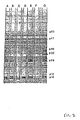
- a standard Western immuno blot was performed as described by Towbin et al., 76 Proc. Natl. Acad. Sci USA 4350, 1979. Briefly, FIV was disrupted with SDS and proteins transferred to a sheet of nitrocellulose. The nitrocellulose sheet was blocked with 30% calf serum, 1% bovine serum albumin (BSA), and 0.05% Tween 20 in Dulbecco's phosphate buffer saline. The sheets were cut into 0.5 cm strips and incubated with a 1:100 dilution of serum sample in blocking buffer for 2 hrs. for 20-22°C.
- BSA bovine serum albumin
- Strips were repeatedly washed with washing buffer (0.05% Tween 20 in Dulbecco's phosphate buffer saline) and then incubated with a second antibody (specific for feline heavy and light chain Ig) horseradish peroxidase conjugate (obtained from kirkguard and Perry Laboratories Inc. Gaithersburg, MD). After 1 hr. incubation, the strips were repeatedly washed with washing buffer and incubated with the precipitating substrate 4-chloronaphthol for 10 min. The strips were partially dried and the results interpreted immediately.
- the serum in each of the lanes A-G was obtained from various cats infected with FIV. Predominant cross-reactivity is detected with p26 and p15 and to a lesser extent with p10. Other proteins of 32, 40, 47 and 65 kD molecular weight are also detected.
- viral polypeptides such as the gag (e.g., p26) antigenic polypeptides
- gag e.g., p26
- viral envelope polypeptides e.g., gpl30
- RIPA immunoprecipitation
- the cell cultures were then incubated for 4 hrs. in 8 ml of the same medium containing 100 microCuries per ml of 35 S-methionine and 35 S-cysteine (specific activity 1200 Curies per mM, New England Nuclear Corporation, Boston, MA).
- the radioactive tissue culture fluids were removed and the cells lysed with 5 ml of 10 mM sodium phosphate buffer pH 7.5 containing 100 mM NaCl, 1% Triton X 100, 0.5% sodium deoxycholate, 0.1% SDS, 0.1mM phenylmethylsulfonylfluoride, and 100 Kallikren inactivator units of aprotenin per ml. (Sigma Chemical Co., St. Louis, MO).
- the cell lysates were clarified by centrifugation 100,000 x g for 30 min. and the pellet discarded. Aliquots of the labelled cell lysates (0.1 ml) and 5 ⁇ l of serum being tested were mixed in a microcentrifuge tube and incubated for 1 hr. at 37°C and then overnight at 4°C. The next day, 0.2 ml of a 5% suspension of protein A Sepharose CL-4B beads (Pharmacia, Piscataway, NJ) in 10 mM of phosphate buffer, pH 7.5 containing 100 mM NaCl, 1% Triton X-100 and 0.1% SDS was added to each tube and mixed for 30 min. at 4°C.
- Sepharose beads were collected by centrifugation (2 min. at 20,000 x g) and washed 3 times in lysing buffer. The final pellet was resuspended in 25 ⁇ l SDS/PAGE loading buffer and heated and 100°C for 3 minutes. The Sepharose beads were removed by centrifugation and the supernatant applied to a PAGE. Gels were processed for fluorography using enlighteningTM (New England Nuclear Corporation, Boston, MA) and exposed at -70° to Kodak XR-5 film. Sera from experimentally infected cats recognize polypeptides of 15, 22, 36, 40, 47, 110 and 130 kD. Although there were some quantitative and qualititative differences all cats appear to mount a response to p22, gp40, gp47 and gn130.
- RIPA-PAGE analysis was carried out using monoclonal antibodies which reacted with p26 as determined by Western blotting. This monoclonal immunoprecipited proteins p47, p36, p22 and p15. High molecular weight polypeptides (130 kD) of FIV which were not detected by the p26 specific monoclonal antibody, were identified by RIPA-PAGE. A protein of molecular weight 100 kD was also detectable utilizing serum antibodies obtained from some infected cats.
- antigenic glycopeptides of FIV can be obtained as follows. Actively growing CRFK FTLV infected cells were scraped from roller bottles, gently washed with phosphate-buffered saline (PBS), and pelleted. The cell pellet was gently resuspended in 10 mM sodium phosphate, pH 7.2, at a ratio of 1 ml buffer to 0.1 ml of cell pellet. This suspension was incubated on ice or refrigerated for 5-10 min., vigorously vortex mixed for 30 seconds, and four volumes of PBS with 1mM PMSF added. The mixture was then vigorously homogenized for 90-120 seconds with a Brinkmann Homogenizer PT10/35 with a PTA 20 generator.
- PBS phosphate-buffered saline
- the resulting homogenate was clarified for 20 minutes at 5,000 x g.
- the supernatant fraction was discarded and the cell membrane pellet resuspended in PBS + 0.2% Triton X-100 at a ratio of 2.5 ml buffer to 0.1 ml original cell pellet.
- the mixture was then vigorously homogenized for 90-120 seconds with a Brinkmann Homogenizer ?T 10/35 with a PTA 20 generator.
- the resulting homogenate was clarified at 100,000 x g for 1 hr, the supernatant decanted off and batch bound overnight at 20-23°C on Pharmacia Lentil Lectin Sepharose 4B at a ratio of 6 ml of resin to 5 ml of original cell pellet.
- the Lentil Lectin Suspension was poured through a column, the resin collected, and washed with 15 column volumes of PBS - 0.2% Triton X-100.
- the glycoproteins were then eluted from the resin by subjecting the resin to 5-10 column volumes of PBS + 0.2% Triton X-100 + 200 mM methyl ⁇ -D mannopyranoside, collecting fractions of 1 column volume/tube.
- glycoproteins isolation was verified by 3% SDS-PAGE electrophorisis, and checked using 35 S-radiolabeled cell preparations in conjunction with RIPA data.
- Further purification of viral glycoprotein from host cell glycoprotein includes use of HPLC, or of a polyclonal or monoclonal antibody for affinity chromotography.
- Isolated virus 250-500 microliters was combined with two volumes of 6M guanidine hydrochloride, pH 3 (adjusted with 20% trifluoro acidic acid in water) .
- the mixture was vortexed and incubated at 37°C-40°C in a water bath for 20-25 minutes.
- the filtered sample was loaded onto an HPLC column for purification.
- the HPLC system consisted of a Beckman HPLC having three 110V pumps, a 421 controller, a 166 variable wavelength detector, a 427 integrator, a 210A injector with dynamic mixer, and a 1000 microliter sample.
- the column was a Waters radial compression cartridge held in an RCM-100 column holder with a modified inlet connector (Waters MicroBond-A-Pak Fenile 10 MU 8 mm x 10 cm cartridge No. 85722 with a guard pak resolve CN cartridge, No. 85826).
- the system was set such that two levers were compressed and the pressure was in the mid-yellow zone.
- Fractions were collected and condensed by a Savant Speed Vac to remove all solvents to a final volume ranging from 50-100 microliters.
- the condensed fractions where neutralized by addition of 200-300 microliters of 50 mM or 100 mM sodium phosphate buffer, pH 7.2.
- the buffered fraction pHs were then checked with pH paper and, if the pH was still below 6, 1N NaOH added until the pH was brought within the range of 6.5-7.5.
- the neutralized fractions were frozen at -20°C until use.
- Fig. 3 the printout from an HPLC column described above is shown.
- the flow rate was at 1 ml per minute starting with 100% of solvent A. After 15 min. the solvent was changed to contain 26% solvent 3, 10 min. later to 31% solvent B, 12 min. later to 37.5% solvent B, 5 min. later to 40% solvent B, 6 min. later to 43% solvent B, 8 min. later to 45% solvent B, 15 min. later to 60% solvent, and 20 min. later to 100% solvent B. Peaks corresponding to p10, p15, and p26 are indicated in Fig. 3. Fractions containing these peaks were collected.
- fractions corresponding to p10, p15 and p26 were run in an SDS-polyacrylamide gel containing 15% bis-acrylamide at 70V. The gel was stained with silver using a Biorad Silver Staining Kit 161-0443. The isolated fractions corresponding to p15, p10, and p26 were essentially homogeneous solutions of the FIV polypeptides.
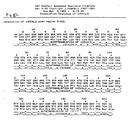
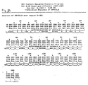
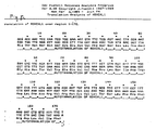
- the isolated FIV polypeptides were analyzed by Standard technique for their amino acid sequence, yielding the following results (? indicates uncertainty in the actual amino acid, or no knowledge at all).
- any standard procedure can be used. For example an ELISA test can be performed using a polyclonal antibody from cat serum to determine whether the polypeptide is cross-reactive.
- the polypeptide can be injected with or without an adjuvant, into an animal, e.g., a mouse, to determine if it causes antibodies to be raised to it.
- These polypeptides are useful for production of vaccines to prevent FIV-caused disease symptoms and FIV infection in cats. These vaccines are produced by standard procedure.
- gag or env polypeptides are provided in a standard inoculation medium and injected intravenously, intrarterially or otherwise into a cat at a level of 1-100 ⁇ g/kg animal at intervals of 3-4 weeks until immunity to FIV is produced.
- Antibodies to FIV polypeptides are useful aids for identification of a purified polypeptide (as described above), and for purifying polypeptides. They are also useful to determine the antigenicity of any polypeptide.
- An example of preparation of useful antibodies follows. These antibodies are monoclonal antibodies which allow specific detection and purification of either individual or a small number of FIV polypeptides.
- mice Balb/CJ (Jackson Labs) mice were immunized with an initial injection of 50 micrograms of FIV virus (prepared as described above) per mouse mixed 1:1 with Difco Bacto adjuvant complete. After two weeks a booster injection of 100 micrograms of FIV virus was injected into each mouse intravenously without adjuvant. Three days after the booster injection a fusion was performed with mouse myeloma cell lines FO or p3X63-Ag8.653. Mid log phase myeloma lines were harvested on the day of fusion and checked for viability. The cells were-spun at 300 x g for 8 min., separated from the growth medium, and resuspended in serum free DME.
- an FIV-inoculated mouse was killed by cervical dislocation and the spleen aseptically removed.
- the spleen was washed three times in serum free DME and placed in a sterile Petri dish containing 20 mls of complete medium (DME containing 20% bovine fetal serum, 100 units per ml. of penicillin and streptomycin, and 1 mM sodium pyruvate). To release cells, the spleen was perfused with a 23 gauge needle.
- Cells were placed in a 50 mi conical centrifuge tube and pelleted at 300 x g for 8 min. The pellet resuspended in 5 ml of 0.17M ammonium chloride and placed on ice for 8 min. 5 ml of bovine fetal serum (20%) was added and the cells pelleted again at 300 x g for 8 min. After resuspension in 10 ml DME the cells were counted and the spleen and myeloma cells mixed in a ratio of 3:1. The cell mixture was pelleted at 200 x g for 10 minutes, the supernatant decanted, and the pellet allowed to stand for 5 min.
- the cells were diluted in complete medium and normal mouse spleen cells at concentrations of hybridoma cells of 5, 10 and 50 cells per ml added.
- the cells were plated into 96-well plates for each dilution. After 10 days the cloning plates were screened for growth. About 37% of all wells showed growth.
- the growth-positive wells were screened for antibody and those testing positive expanded to 2 cm 2 cultures and provided with normal mouse spleen cells. The cloning procedure was repeated 2 times until a stable antibody-producing hybridoma was obtained. At this point the cell culture was expanded from 2 to 9 to 75 to 150 cm 2 culture vessels, at which point ascite production could be commenced.
- pristane primed IRCFl female mice were used. 0.5 ml of pristane was injected intraperitoneally (IP) to each mouse, and the mouse allowed to rest for 10-60 days. At this time 4.5 x 10 6 cells were injected IP into each mouse and ascites formed in 7-14 days. Ascites fluid was harvested with a pasteur pipette through a hole in the peritoneum.
- Antibodies to glycoproteins can also be isolated and detected.
- antibodies to two glycoproteins of molecular weight 40 kD (gp40) and 130 kD (gp130) which are detected using PAGE and RIPA respectively.
- Monoclonals useful in this invention for purification and identification of specific polypeptides of FIV include those which are specific for FIV and form a sufficiently strong interation with an FIV epitope, and an FIV antigen, to be useful in an immunoassay, for example, an ELISA, to detect an FIV antigen.
- an immunoassay for example, an ELISA
- Another test is to perform a Western blot to determine whether the monoclonal antibody has good reactivity with one or more FIV antigens. Generally, those monoclonals which show poor reactivity, that is, produce faint bands on the Western blot, are not suitable in this invention.
- Yet another test involves radioimmunoprecipitation assay (RIPA) where FIV virus labeled with 35 S-methionine is reacted with a monoclonal antibody to form within immunoprecipitate, and the immunoprecipitate run in a SDS-PAGE and autoradiographed to detect the labelled proteins. This analysis determines which of the monoclonal antibodies is able to detect precursor FIV antigenic polypeptides and not just mature polypeptides.
- RIPA radioimmunoprecipitation assay
- antigenic polypeptides can be used to detect naturally occurring antibodies produced by cats. Such detection can be any standard immunoassay procedure, for example, by an ELISA test, as described above. One example of such a test follows. This example is not limiting to the present invention and those skilled in the art will recognize that any of many other standard procedures can be used.
- test antigen e.g., p26, p15, or p10
- the test wells were coated with 100 ⁇ l of a solution containing 0.15 micrograms antigen in 0.25 molar sodium citrate, pH 7.5.
- the wells were covered with parafilm, incubated overnight at 4°C, and tapped until dry.
- the antigen was then overcoated by adding 200 ⁇ l 1% BSA in 0.25 molar sodium citrate, incubating at room temperature (20-25°C) for 1 hour, and tapping the wells dry. 200 microliters of 7.5% sucrose in 0.25 molar sodium citrate was then added to each well and incubated at room temperature for 30 minutes. The resulting strips were used immediately, or dried under vacuum for 6 hours at room temperature for later use.
- Assays were performed by adding 100 ⁇ l of feline serum sample (positive control, negative control, or test sera) diluted 1 to 100 in Dulbecco s PBS containing 0.1% Bovine serum albumin, 30% calf serum, and 0.05% Tween-20 (Sigma Chemical, St. Louis, MO) to a well, incubating at room temperature for 30 minutes, and tapping the well dry. The wells were washed immediately two times with Dulbecco's PBS containing 0.05% Tween-20, tapping the wells dry after the second wash.
- a solution containing 0.1% 3',3',5,5' Tetramethylbenzidene in 40% Glycerol and 50 % methanol was mixed with an equal volume of a solution containing 22.82 grams dibasic potassium phosphate, 19.2 grams citric acid and 1.34 milliliters 30% hydrogen peroxide solution per liter.
- One hundred microliters of this solution was added to each well, and then incubated at room temperature for 15 minutes. At the end of the incubation period, 100 ⁇ l of 0.25% hydrofluoric acid was added to each well.
- the optical density at 650 nanometer of the solution in each well was then determined with a microtiter plate reader.
- An immune response to p10, p15 and p26 was detectable either following experimental infection of a cat with FIV, or in feline sera possessing antibodies to FIV.
- FIV nucleic acid is useful for production of large amounts of FIV polypeptides, or fragments thereof, and also for detection of homologous nucleic acid in vivo , using standard techniques. There follows an example of cloning of FIV viral DNA. The specific FIV strain chosen is not meant to be limiting in this invention and those skilled in the art will recognize that equivalent nucleic acid may be isolated by use of the cloned sequences which are provided as specific deposits, or by techniques similar to those described in this example.
- a Crandal feline kidney cell line productively infected with FIV strain 2428 (Pentaluma isolate) was used as a source of unintegrated viral DNA.
- the unintegrated viral DNA was prepared by Hirt extraction and CsCl-ethidium bromide centrifugation to resolve linear and supercoiled viral DNA. (Hirt, 26 J Mol. Biol. 365, 1967; Canaani et al., 282 Nature 378, 1979).
- the supercoiled viral DNA was used to construct libraries which contain overlapping viral DNA sequences.
- the procedures used to construct these libraries were similar to those described by Maniatis et al., Molecular Cloning, A Laboratory Manual, Cold Spring Harbor Lab, Cold Spring Harbor, N.Y.) and Glover, DNA Cloning, Vol. 1, A Practical Approach, IRL Press, Washington, DC) and are familiar to those of ordinary skill in the art.
- Two viral DNA libraries were generated by cleavage or supercoiled viral DNA by one of two restriction endonucleases. Partial cleavage by the restriction endonuclease Rsa I, which recognizes the DNA sequence 5'GTAC3', or cleavage by the restriction endonuclease Hae III, which recognizes the DNA sequence 5'GGCC3' , of the supercoiled viral DNA generates two sets of blunt-ended DNA molecules whose sequences overlap. The blunt-ended DNA molecules were then treated with Eco RI methylase, according to the manufacturer's directions, to modify the 3' adenine residue of the Eco RI recognition sequence 5'-GAATCC-3'.
- Methylation at this site inhabits the cleavage of treated DNA by the restriction endonuclease Eco RI.
- the methylated DNA molecules were then ligated to linker DNA molecules which contained a cleavage site for the restriction endonuclease Eco RI.
- the linker containing DNA molecules were then treated with the restriction endonuclease Eco RI to generate molecules whose termini were compatible with the Eco RI cloning site in the recombinant DNA phage vector ⁇ ZAPTM (Stratagene, La Jolla, California). Linker fragments resulting from this cleavage were separated from the large DNA molecules by size separation on a quickspin column (Boehringer, Mannheim, Indianapolis, IN).
- the DNA molecules were then ligated into the Eco RI cleaved ⁇ ZAP vector using T4 DNA ligase (New England Biolabs, Beverly, MA). Ligated DNA molecules were then packaged into phage using Gigapack gold (Stratagene, La Jolla, California). Viable phage from the packaging reaction were then amplified by infecting BB4 cells (Stratagene) and harvesting plate lysates of those infected cells in order to obtain a stock of recombinant ⁇ ZAP clones.
- the insert DNA of the recombinant ⁇ ZAP clones contain host cellular DNA as well as FIV proviral DNA sequences, it was necessary to screen each library with an FIV DNA probe that contains a readily detectable label.
- FIV DNA probe was made from RNA isolated from FIV.
- a radioactive complementary DNA was synthesized from total viral RNA essentially as described by Maniatis et al. 1982, supra except that selection of poly A-containing RNA was not performed, and methymercuric hydroxide was omitted from the protocol.
- the bacteriophage libraries were plated at a density of 10,000 bacteriophage per 150 mm dish. They were then screened by hybridization of the radioactively labeled probe to phage DNA which was immobilized on nitrocellulose filters (Maniatis, et al., 1982 supra ). Each hybridizing bacteriophage plaque was then picked, replaced, and hybridized as described, until a single well was isolated which contained a ⁇ ZAP recombinant clone.
- XL-1-Blue cells (Stratagene) were then infected with recombinant ⁇ ZAP phage, and plasmids containing the insert DNA were obtained following superinfection with R408 helper phage according to the manufacturer's directions (Stratagene, La Jolla, California). This procedure also provides both recombinant plasmids (which can be isolated from the cell) and single stranded phage stock which can be isolated from the medium for DNA sequence analysis.
- the above recombinant plasmids were analyzed for inserts by preparing plasmid DNAs from overnight culture of bacteria replicating these plasmids as follows. One and one half ml. of. an overnight culture was placed in a microcentrifuge tube and spun for four minutes at 4000xg. The supernatant was removed and the tube respun for four minutes at 4000xg. The supernatant was removed and the tube respun for a few seconds, and residual liquid removed carefully with a pasteur pipet. The bacterial pellet was then thoroughtly resuspended in 200 microliters of a solution containing 8% sucrose, 50 mm EDTA, 5% Triton X-100 and 50 MM Tris/HCl, pH 8.05.
- Plasmid DNAs thus isolated were anlayzed for inserts by restriction enconuclease cleavage and electrophoresis in 0.8% agarose gels (Maniatis et al., 1982 supra ).
- Nucleic acid probes derived from the 2BY DNA sequence hybridize to DNA isolated from FIV infected but not uninfected cells. These probes can be used to isolate other FIV genes from other strains and can be expressed by standard procedures to provide the purified polypeptides described above.
- Strains 10CX, 2B4 and R5X have been deposited with the ATCC and assigned numbers 67937, 67938, and 67939, respectively.
Landscapes
- Health & Medical Sciences (AREA)
- Life Sciences & Earth Sciences (AREA)
- Chemical & Material Sciences (AREA)
- Virology (AREA)
- Organic Chemistry (AREA)
- General Health & Medical Sciences (AREA)
- Medicinal Chemistry (AREA)
- Pharmacology & Pharmacy (AREA)
- Public Health (AREA)
- Veterinary Medicine (AREA)
- Molecular Biology (AREA)
- Animal Behavior & Ethology (AREA)
- Nuclear Medicine, Radiotherapy & Molecular Imaging (AREA)
- Communicable Diseases (AREA)
- Oncology (AREA)
- Chemical Kinetics & Catalysis (AREA)
- General Chemical & Material Sciences (AREA)
- Biochemistry (AREA)
- Gastroenterology & Hepatology (AREA)
- Proteomics, Peptides & Aminoacids (AREA)
- Genetics & Genomics (AREA)
- Biophysics (AREA)
- Peptides Or Proteins (AREA)
- Preparation Of Compounds By Using Micro-Organisms (AREA)
- Medicines That Contain Protein Lipid Enzymes And Other Medicines (AREA)
- Medicines Containing Antibodies Or Antigens For Use As Internal Diagnostic Agents (AREA)
- Micro-Organisms Or Cultivation Processes Thereof (AREA)
Applications Claiming Priority (6)
| Application Number | Priority Date | Filing Date | Title |
|---|---|---|---|
| US34878489A | 1989-05-08 | 1989-05-08 | |
| US348784 | 1989-05-08 | ||
| US44781089A | 1989-12-08 | 1989-12-08 | |
| US447810 | 1989-12-08 | ||
| EP19900907657 EP0471752A4 (en) | 1989-05-08 | 1990-04-30 | Polypeptides of feline t-cell lymphotropic lentivirus |
| EP98104238A EP0860504A1 (fr) | 1989-05-08 | 1990-04-30 | Polypeptides de l'antivirus lymphptropiques de cellules T du chat |
Related Parent Applications (1)
| Application Number | Title | Priority Date | Filing Date |
|---|---|---|---|
| EP98104238A Division EP0860504A1 (fr) | 1989-05-08 | 1990-04-30 | Polypeptides de l'antivirus lymphptropiques de cellules T du chat |
Publications (2)
| Publication Number | Publication Date |
|---|---|
| EP1475442A2 true EP1475442A2 (fr) | 2004-11-10 |
| EP1475442A3 EP1475442A3 (fr) | 2004-11-17 |
Family
ID=26995886
Family Applications (3)
| Application Number | Title | Priority Date | Filing Date |
|---|---|---|---|
| EP19900907657 Withdrawn EP0471752A4 (en) | 1989-05-08 | 1990-04-30 | Polypeptides of feline t-cell lymphotropic lentivirus |
| EP04015677A Withdrawn EP1475442A3 (fr) | 1989-05-08 | 1990-04-30 | Polypeptides de l'antivirus lymphotropiques de cellules t du chat |
| EP98104238A Ceased EP0860504A1 (fr) | 1989-05-08 | 1990-04-30 | Polypeptides de l'antivirus lymphptropiques de cellules T du chat |
Family Applications Before (1)
| Application Number | Title | Priority Date | Filing Date |
|---|---|---|---|
| EP19900907657 Withdrawn EP0471752A4 (en) | 1989-05-08 | 1990-04-30 | Polypeptides of feline t-cell lymphotropic lentivirus |
Family Applications After (1)
| Application Number | Title | Priority Date | Filing Date |
|---|---|---|---|
| EP98104238A Ceased EP0860504A1 (fr) | 1989-05-08 | 1990-04-30 | Polypeptides de l'antivirus lymphptropiques de cellules T du chat |
Country Status (4)
| Country | Link |
|---|---|
| EP (3) | EP0471752A4 (fr) |
| JP (2) | JP3058685B2 (fr) |
| CA (1) | CA2016183A1 (fr) |
| WO (1) | WO1990013573A1 (fr) |
Cited By (1)
| Publication number | Priority date | Publication date | Assignee | Title |
|---|---|---|---|---|
| WO2006098849A3 (fr) * | 2005-03-09 | 2007-04-19 | Idexx Lab Inc | Procede et dispositif destines a detecter le virus de l'immunodeficience feline |
Families Citing this family (27)
| Publication number | Priority date | Publication date | Assignee | Title |
|---|---|---|---|---|
| US5118602A (en) * | 1987-08-26 | 1992-06-02 | The Regents Of The University Of California | Feline T-lymphotropic lentivirus assay |
| US6107077A (en) * | 1987-08-26 | 2000-08-22 | Yamamoto; Janet K. | Feline lymphoid cell lines capable of producing FIV for FIV diagnostics and vaccines |
| CA1341439C (fr) * | 1987-08-26 | 2003-09-23 | Niels C. Pedersen | Lentivirus t-lymphotrope felin |
| NZ240558A (en) | 1990-11-14 | 1994-11-25 | Smithkline Beecham Corp | Recombinant feline coronavirus s proteins useful in diagnosis and vaccination against feline peritonitis virus disease |
| FR2669338A1 (fr) * | 1990-11-21 | 1992-05-22 | Centre Nat Rech Scient | Fragments peptidiques issus de la proteine externe du vif, anticorps antifragments, application de ceux-ci au diagnostic et/ou au traitement de l'immunodeficience feline. |
| US6228608B1 (en) * | 1991-02-28 | 2001-05-08 | Aquila Biopharmaceuticals, Inc. | Recombinant FIV glycoprotein 160 and P24 gag protein |
| ATE160358T1 (de) * | 1991-06-14 | 1997-12-15 | St Vincents Inst Med Res | Detektion des immunschwäche-virus aus felis |
| US5413927A (en) * | 1991-09-03 | 1995-05-09 | North Carolina State University | Feline immunodeficiency virus isolate NCSU1Lb |
| US5736378A (en) * | 1991-09-12 | 1998-04-07 | The Scripps Research Institute | Molecular cloning and characterization of the feline immunodeficiency virus isolate PPR |
| GB9125024D0 (en) * | 1991-11-25 | 1992-01-22 | Kirby Julian | Rheumatoid arthritus treatment |
| US5837686A (en) * | 1991-11-25 | 1998-11-17 | Peptide Therapeutics Limited | Peptides and antibodies for treatment of rheumatoid arthritis |
| FR2692279B1 (fr) * | 1992-06-16 | 1995-05-19 | Centre Nat Rech Scient | Séquences nucléotidiques issues de la souche WO du vif et leurs fragments, applications desdites séquences à l'expression de peptides immunogènes et au diagnostic de l'immunodéficience féline. |
| AU2234995A (en) * | 1994-04-29 | 1995-11-29 | Pharmacia & Upjohn Company | Feline immunodeficiency virus vaccine |
| DE69535669T2 (de) | 1994-05-09 | 2008-12-04 | Oxford Biomedica (Uk) Ltd. | Retrovirale vektoren mit verminderter rekombinationsrate |
| US6013517A (en) * | 1994-05-09 | 2000-01-11 | Chiron Corporation | Crossless retroviral vectors |
| US6300118B1 (en) | 1995-06-07 | 2001-10-09 | American Home Products Corporation | Plasmids comprising a genetically altered feline immunodeficiency virus genome |
| US5820869A (en) * | 1995-06-07 | 1998-10-13 | American Home Products Corporation | Recombinant raccoon pox viruses and their use as an effective vaccine against feline immunodeficiency virus infection |
| US6458528B1 (en) * | 1998-05-15 | 2002-10-01 | Idexx Laboratories, Inc. | Diagnosis of feline immunodeficiency virus infection using ENV/GAG polypeptide markers |
| US6764676B1 (en) | 1998-08-24 | 2004-07-20 | Pfizer Inc. | Compositions and methods for protecting animals from lentivirus-associated disease such as feline immunodeficiency virus |
| AU2002244103A1 (en) | 2001-02-22 | 2002-09-12 | University Of Florida | Materials and methods for detecting, preventing, and treating hiv and fiv retroviral infection |
| ATE435425T1 (de) | 2003-09-11 | 2009-07-15 | Idexx Lab Inc | Verfahren zum nachweis des felinen immunschwächevirus |
| AU2005267607B8 (en) | 2003-09-11 | 2009-07-16 | Idexx Laboratories, Inc. | Method and device for detecting feline immunodeficiency virus |
| WO2005062053A2 (fr) | 2003-12-18 | 2005-07-07 | Idexx Laboratories, Inc. | Procede et dispositif pour detecter le virus d'immunideficience feline |
| WO2005080939A2 (fr) | 2004-02-19 | 2005-09-01 | Idexx Laboratories, Inc. | Methode et dispositif de detection du virus de l'immunodeficience feline |
| JP4866848B2 (ja) | 2004-06-30 | 2012-02-01 | アイデックス ラボラトリーズ インコーポレイテッド | ネコ免疫不全ウイルス(fiv)のenvタンパク質のv3領域に由来するペプチドの使用を含むfivを検出するための方法および装置 |
| AU2005267605B2 (en) * | 2004-06-30 | 2009-03-26 | Idexx Laboratories, Inc. | Method and device for detecting feline immunodeficiency virus |
| WO2011123781A1 (fr) | 2010-04-02 | 2011-10-06 | Idexx Laboratories, Inc. | Détection du virus de l'immunodéficience féline |
Family Cites Families (5)
| Publication number | Priority date | Publication date | Assignee | Title |
|---|---|---|---|---|
| GB8324800D0 (en) * | 1983-09-15 | 1983-10-19 | Pasteur Institut | Antigens |
| AU6713287A (en) * | 1986-01-06 | 1987-07-09 | F. Hoffmann-La Roche & Co. | Expression of htlv-iii gag-gene |
| NZ220200A (en) * | 1986-05-19 | 1990-08-28 | Viral Technologies Inc | Hiv related peptides, antibodies to the peptides and vaccines therefrom |
| CA1341439C (fr) * | 1987-08-26 | 2003-09-23 | Niels C. Pedersen | Lentivirus t-lymphotrope felin |
| US5219725A (en) * | 1988-12-05 | 1993-06-15 | Idexx Laboratories Incorporated | Monoclonal antibodies to feline-t-lymphotropic lentivirus |
-
1990
- 1990-04-30 JP JP2507656A patent/JP3058685B2/ja not_active Expired - Lifetime
- 1990-04-30 WO PCT/US1990/002338 patent/WO1990013573A1/fr not_active Ceased
- 1990-04-30 EP EP19900907657 patent/EP0471752A4/en not_active Withdrawn
- 1990-04-30 EP EP04015677A patent/EP1475442A3/fr not_active Withdrawn
- 1990-04-30 EP EP98104238A patent/EP0860504A1/fr not_active Ceased
- 1990-05-07 CA CA002016183A patent/CA2016183A1/fr not_active Abandoned
-
1999
- 1999-12-01 JP JP11342237A patent/JP2000204100A/ja active Pending
Cited By (1)
| Publication number | Priority date | Publication date | Assignee | Title |
|---|---|---|---|---|
| WO2006098849A3 (fr) * | 2005-03-09 | 2007-04-19 | Idexx Lab Inc | Procede et dispositif destines a detecter le virus de l'immunodeficience feline |
Also Published As
| Publication number | Publication date |
|---|---|
| JPH04507094A (ja) | 1992-12-10 |
| WO1990013573A1 (fr) | 1990-11-15 |
| EP0471752A1 (fr) | 1992-02-26 |
| CA2016183A1 (fr) | 1990-11-08 |
| JP3058685B2 (ja) | 2000-07-04 |
| EP1475442A3 (fr) | 2004-11-17 |
| JP2000204100A (ja) | 2000-07-25 |
| EP0471752A4 (en) | 1992-09-02 |
| EP0860504A1 (fr) | 1998-08-26 |
Similar Documents
| Publication | Publication Date | Title |
|---|---|---|
| EP1475442A2 (fr) | Polypeptides de l'antivirus lymphotropiques de cellules t du chat | |
| US6383765B1 (en) | Polypeptides of feline T-cell lymphotropic lentivirus | |
| Slamon et al. | Identification of the putative transforming protein of the human T-cell leukemia viruses HTLV-I and HTLV-II | |
| US6406841B1 (en) | Methods for the detection of HTLV-II antibodies employing novel HTLV-II NRA envelope peptides | |
| US5177014A (en) | Monoclonal antibodies to feline-T-lymphotropic lentivirus | |
| JP4612071B2 (ja) | エプスタイン−バールウイルスペプチド及び該ペプチドに対する抗体 | |
| WO1995001457A9 (fr) | Compositions de virus htlv-iinra et essais utiles pour detecter l'infection par des virus htlv | |
| JP2679973B2 (ja) | 免疫学的に反応性のウイルス蛋白質の発現 | |
| KR100624517B1 (ko) | 인간 사이토메갈로바이러스 검출용 펩티드 시약 | |
| CA1339363C (fr) | Anticorps monoclonaux pour des regions antigeniques specifiques du virus de l'immunodeficience humaine; methodes d'utilisation | |
| US20050186560A1 (en) | Retrovirus isolated from mantle histiocytes in mantle cell lymphoma | |
| WO1993017341A1 (fr) | Titrage de htlv-i/htlv-ii et procede | |
| JP4155591B2 (ja) | エプスタイン・バールウイルスのペプチド及びこれらのペプチドに対する抗体 | |
| AU642886B2 (en) | T-lymphotropic retrovirus monoclonal antibodies | |
| JP3470899B2 (ja) | 診断に使用するためのhtlv−▲i▼およびhtlv−▲ii▼gp21由来ペプチド | |
| KR910000748B1 (ko) | 사람 면역결핍증 바이러스의 gag-암호화된 단백질 | |
| EP0848820A1 (fr) | Detection d'anticorps du htlv a l'aide de proteines de recombinaison | |
| JPH0475583A (ja) | ハイブリドーマ系細胞とその細胞により産生されるモノクローナル抗体 | |
| AU7026887A (en) | Sor gene product from human t-cell lymphotropic virus iii |
Legal Events
| Date | Code | Title | Description |
|---|---|---|---|
| PUAI | Public reference made under article 153(3) epc to a published international application that has entered the european phase |
Free format text: ORIGINAL CODE: 0009012 |
|
| PUAL | Search report despatched |
Free format text: ORIGINAL CODE: 0009013 |
|
| 17P | Request for examination filed |
Effective date: 20040708 |
|
| AC | Divisional application: reference to earlier application |
Ref document number: 0471752 Country of ref document: EP Kind code of ref document: P Ref document number: 0860504 Country of ref document: EP Kind code of ref document: P |
|
| AK | Designated contracting states |
Kind code of ref document: A2 Designated state(s): AT BE CH DE DK ES FR GB IT LI LU NL SE |
|
| AK | Designated contracting states |
Kind code of ref document: A3 Designated state(s): AT BE CH DE DK ES FR GB IT LI LU NL SE |
|
| RIN1 | Information on inventor provided before grant (corrected) |
Inventor name: TONELLI, QUENTIN J. Inventor name: O'CONNOR, THOMAS P. Inventor name: ANDERSEN, PHILIP R. |
|
| 17Q | First examination report despatched |
Effective date: 20050114 |
|
| AKX | Designation fees paid |
Designated state(s): AT BE CH DE DK ES FR GB IT LI LU NL SE |
|
| STAA | Information on the status of an ep patent application or granted ep patent |
Free format text: STATUS: THE APPLICATION IS DEEMED TO BE WITHDRAWN |
|
| 18D | Application deemed to be withdrawn |
Effective date: 20050929 |