EP1487323B1 - Procede d'examen du fond de l'oeil - Google Patents
Procede d'examen du fond de l'oeil Download PDFInfo
- Publication number
- EP1487323B1 EP1487323B1 EP20030717242 EP03717242A EP1487323B1 EP 1487323 B1 EP1487323 B1 EP 1487323B1 EP 20030717242 EP20030717242 EP 20030717242 EP 03717242 A EP03717242 A EP 03717242A EP 1487323 B1 EP1487323 B1 EP 1487323B1
- Authority
- EP
- European Patent Office
- Prior art keywords
- camera
- data
- lighting unit
- parameters
- ocular fundus
- Prior art date
- Legal status (The legal status is an assumption and is not a legal conclusion. Google has not performed a legal analysis and makes no representation as to the accuracy of the status listed.)
- Expired - Lifetime
Links
- 238000000034 method Methods 0.000 title claims description 27
- 201000010099 disease Diseases 0.000 claims description 16
- 208000037265 diseases, disorders, signs and symptoms Diseases 0.000 claims description 16
- 238000003909 pattern recognition Methods 0.000 claims description 7
- 238000004422 calculation algorithm Methods 0.000 claims description 6
- 238000004458 analytical method Methods 0.000 claims description 4
- 238000010191 image analysis Methods 0.000 claims description 4
- 230000000875 corresponding effect Effects 0.000 claims description 3
- 230000001105 regulatory effect Effects 0.000 claims description 3
- 238000007635 classification algorithm Methods 0.000 claims description 2
- 230000002596 correlated effect Effects 0.000 claims description 2
- 238000005457 optimization Methods 0.000 claims description 2
- 208000010412 Glaucoma Diseases 0.000 description 12
- 238000012216 screening Methods 0.000 description 12
- 238000005286 illumination Methods 0.000 description 8
- 210000003128 head Anatomy 0.000 description 6
- 238000002560 therapeutic procedure Methods 0.000 description 6
- 230000008859 change Effects 0.000 description 5
- 210000001328 optic nerve Anatomy 0.000 description 5
- 208000037849 arterial hypertension Diseases 0.000 description 4
- 238000001514 detection method Methods 0.000 description 4
- 230000004438 eyesight Effects 0.000 description 4
- 230000002792 vascular Effects 0.000 description 4
- 208000017442 Retinal disease Diseases 0.000 description 3
- 230000006978 adaptation Effects 0.000 description 3
- 230000008901 benefit Effects 0.000 description 3
- 206010012601 diabetes mellitus Diseases 0.000 description 3
- 238000010586 diagram Methods 0.000 description 3
- 238000011156 evaluation Methods 0.000 description 3
- 230000000977 initiatory effect Effects 0.000 description 3
- 230000007774 longterm Effects 0.000 description 3
- 230000003287 optical effect Effects 0.000 description 3
- 238000012545 processing Methods 0.000 description 3
- 210000001525 retina Anatomy 0.000 description 3
- 238000011282 treatment Methods 0.000 description 3
- 208000018152 Cerebral disease Diseases 0.000 description 2
- 206010012689 Diabetic retinopathy Diseases 0.000 description 2
- 210000002565 arteriole Anatomy 0.000 description 2
- 230000036772 blood pressure Effects 0.000 description 2
- 238000013399 early diagnosis Methods 0.000 description 2
- 230000036541 health Effects 0.000 description 2
- 238000005259 measurement Methods 0.000 description 2
- 230000002207 retinal effect Effects 0.000 description 2
- 210000001210 retinal vessel Anatomy 0.000 description 2
- 201000004569 Blindness Diseases 0.000 description 1
- 206010020772 Hypertension Diseases 0.000 description 1
- 206010020852 Hypertonia Diseases 0.000 description 1
- 208000001344 Macular Edema Diseases 0.000 description 1
- 206010025415 Macular oedema Diseases 0.000 description 1
- 206010025421 Macule Diseases 0.000 description 1
- 230000005856 abnormality Effects 0.000 description 1
- 230000003044 adaptive effect Effects 0.000 description 1
- 230000003276 anti-hypertensive effect Effects 0.000 description 1
- 239000012503 blood component Substances 0.000 description 1
- 210000004556 brain Anatomy 0.000 description 1
- 238000004364 calculation method Methods 0.000 description 1
- 230000001276 controlling effect Effects 0.000 description 1
- 238000003745 diagnosis Methods 0.000 description 1
- 230000000694 effects Effects 0.000 description 1
- 239000005555 hypertensive agent Substances 0.000 description 1
- 230000001631 hypertensive effect Effects 0.000 description 1
- 238000003384 imaging method Methods 0.000 description 1
- 238000011835 investigation Methods 0.000 description 1
- 230000002427 irreversible effect Effects 0.000 description 1
- 210000003734 kidney Anatomy 0.000 description 1
- 238000013532 laser treatment Methods 0.000 description 1
- 230000004446 light reflex Effects 0.000 description 1
- 201000010230 macular retinal edema Diseases 0.000 description 1
- 210000005036 nerve Anatomy 0.000 description 1
- 230000001575 pathological effect Effects 0.000 description 1
- 230000003449 preventive effect Effects 0.000 description 1
- 238000011321 prophylaxis Methods 0.000 description 1
- 230000003304 psychophysiological effect Effects 0.000 description 1
- 210000001747 pupil Anatomy 0.000 description 1
- 230000010344 pupil dilation Effects 0.000 description 1
- 230000009467 reduction Effects 0.000 description 1
- 238000012552 review Methods 0.000 description 1
- 230000002123 temporal effect Effects 0.000 description 1
- 238000003325 tomography Methods 0.000 description 1
- 210000003462 vein Anatomy 0.000 description 1
- 230000004304 visual acuity Effects 0.000 description 1
Images
Classifications
-
- A—HUMAN NECESSITIES
- A61—MEDICAL OR VETERINARY SCIENCE; HYGIENE
- A61B—DIAGNOSIS; SURGERY; IDENTIFICATION
- A61B3/00—Apparatus for testing the eyes; Instruments for examining the eyes
- A61B3/10—Objective types, i.e. instruments for examining the eyes independent of the patients' perceptions or reactions
- A61B3/14—Arrangements specially adapted for eye photography
- A61B3/15—Arrangements specially adapted for eye photography with means for aligning, spacing or blocking spurious reflection ; with means for relaxing
- A61B3/152—Arrangements specially adapted for eye photography with means for aligning, spacing or blocking spurious reflection ; with means for relaxing for aligning
-
- A—HUMAN NECESSITIES
- A61—MEDICAL OR VETERINARY SCIENCE; HYGIENE
- A61B—DIAGNOSIS; SURGERY; IDENTIFICATION
- A61B3/00—Apparatus for testing the eyes; Instruments for examining the eyes
- A61B3/10—Objective types, i.e. instruments for examining the eyes independent of the patients' perceptions or reactions
- A61B3/12—Objective types, i.e. instruments for examining the eyes independent of the patients' perceptions or reactions for looking at the eye fundus, e.g. ophthalmoscopes
Definitions
- the invention relates to a method for examining the fundus, according to the features indicated in the preamble of patent claim 1.
- the known methods and devices for diagnostic recording of the fundus do not allow feedback of the recording sensor due to the image quality of the recording. Therefore, when screening large populations with automatic assessment of the ocular fundus, there may be many exposures that are not recoverable because the image quality is insufficient for automatic assessment.
- the prevalence of a disease of diabetes mellitus, arterial hypertension with vascular retinal diseases, glaucoma or cerebral diseases with involvement of the optic nerve in the population is high. In the early detection of these diseases, which result in changes in the fundus, However, there are very good treatment options. For screening screening examinations are required, as an examination by ophthalmologists is usually carried out at a later date. Screening studies of the fundus are only promising if they can be carried out automatically and thus free of observer bias.
- the examination of the ocular fundus is carried out with ophthalmoscopes, the images of which are so far only usable for the human observer if, after a manual adjustment of the optics, the ocular fundus is well illuminated and the correct section is visible.
- a change according to a predetermined criterion, according to physical parameters such as brightness or illumination, can easily cause an existing abnormality, which is reflected in the image as a bright area, to become even less noticeable in the case of increasing the brightness. If necessary, an increase in the contrast or a reduction in the brightness or a change in the angle of illumination would be the appropriate solution that an observer or doctor must make.
- the duration of the period of undetected glaucoma is one of the most important risk factors, and the longer the treatment is missed, the greater the risk to the patient for the patient, and careful screening for early glaucoma detection is necessary to begin therapy in time for irreversible damage can. Since only a portion of patients with diabetes mellitus, arterial hypertension with vascular retinal diseases, glaucoma and cerebral disease involving the optic nerve in the initial stages of these diseases is examined by ophthalmologists, especially since the patient has no subjective vision problems in the initial stage, there is a great need for preventive care Screening of the fundus.
- the object of the invention is to further develop the method for examining the ocular fundus in such a way that the examination can be carried out in a simple manner, the decision reliability being optimized.
- the method is intended to allow a quick and reliable review, with subjective ratings to be avoided. Furthermore, the method should be suitable for screening of the fundus.
- the inventive method allows a complete automatic assessment of the eye image due to the feedback of the data obtained in the image analysis.
- the camera in particular its optics and / or positioning, and / or the lighting unit to the effect that the safety of the automatic assessment is maximized in particular according to a classification system.
- the camera and / or the lighting or the lighting unit is controlled such that the detection reliability is maximized.
- the image data determined in the image evaluation are correlated with provided data which are characteristic for a disease, wherein parameters for setting the camera and / or illumination unit are generated in such a way that the recognition reliability is optimized.
- the acquired image data are compared with characteristic data for a disease, which are provided in a database or a computer, wherein the characteristic data here are in particular the constriction of the vessels, altered light reflexes, larger light or dark areas in the ocular fundus.
- a correlation method is preferably used which, on the basis of established and / or significant coincidence of the image data on the one hand and the data provided, on the other hand provides parameters for controlling the camera and / or the illumination unit in that the recognition reliability is optimized.
- the findings of the so-called active vision are used, with adaptation of the acquisition parameters to the image analysis problem and tasks adapted and fed back via the sensor information image processing modules are used.
- the most important aspects of active seeing are selection in space, in time, in accuracy and in resolution.
- the processing limited in the spatial selection on the part of the image, which is relevant to the solution of the task.
- the algorithms used are limited in the selection in time to those parts of the image sequence that are necessary for the solution of the task, whereby redundant calculations are avoided.
- a selection is carried out in space, time and resolution, wherein a selection is achieved, in particular, by spatially variant sensors with varying resolution.
- the optimization of the positioning of the fundus camera and / or the illumination unit is specified by the fact that the detection for safety according to automatic classification is controlled by a given algorithm of the pattern recognition.
- the method is based on a pattern recognition and / or classification algorithm.
- the method of classification uses the data known for this disease, for example glaucoma, in particular a linear discriminant analysis is performed on the basis of six parameters of a retina tomograph. Furthermore, a linear discriminant analysis can be performed on the basis of parameters of a surface which is approximated in the data of said retina tomograph for the classification of glaucoma.
- measurement data from the optical tomography of the ocular fundus are also suitably taken into account.
- classification methods can be carried out which enable automatic recognition of the disease, such as glaucoma, based on image data.
- the image data generated in the examination of the fundus contains a pattern and, on the basis of the comparison and / or the classification by the pattern recognition algorithm, the determination of the parameter (s) of the feedback measuring system takes place.
- the benefits to the treating physician and patient of the proposed automatic screening method are manifold. It is advantageous for the patients that a screening examination of the ocular fundus, via "telebefundung" is carried out quickly and without additional appointments and without pupil dilation by an ophthalmologist.
- the patient is sensitized with regard to the significance of ocular fundus examinations in certain medical conditions.
- the general medical examination center quickly receives a screening report on the status of the retinal vessels and the optic nerve via telephoto.
- the advice is to present a detailed ophthalmological or internal examination with pupil dilatation and possible therapy by the local ophthalmologist / internist.
- the attending physician (ophthalmologist / internist / general practitioner) quickly receives an additional important finding (status of the retinal vessels, the macula and the optic nerve) in these high-risk patients.
- the decisive advantage for this procedure is the diagnostic and clinical gain for all participants.
- a screening system realized with the proposed method leads in the long term due to an earlier diagnosis and an earlier onset of therapy (timely initiation of increased antihypertensive therapy in hypertensives for the prophylaxis of strokes, timely glaucoma treatment on incidence of glaucoma on the optic nerve, timely initiation of laser treatment of diabetic retinopathy) to a significant cost savings in the health system.
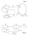
- Fig. 1 a laser containing or formed from such a camera 2, wherein the laser beam 4 is directed to the eye 6 of the schematically indicated head of a patient.
- the laser beam 4 is directed to the eye 6 of the schematically indicated head of a patient.
- the fundus is scanned and illuminated in a known manner.
- the reflected light is detected by means of the camera and given corresponding data for image processing on a computer 10.
- the movement of the camera and / or its optics and / or the illumination by means of the camera or the laser is regulated in a closed control loop via an actuating unit 12.
- Fig. 2 schematically shows a block diagram of the control loop of the automatically adaptive image acquisition to optimize the classification security with the essential function blocks.
- Such characteristic of a disease data are provided in a database or a computer and used according to the invention for evaluation.
- the image evaluation takes place in accordance with block 22.
- a comparison with a known and one or more diseases corresponding image pattern is performed according to the data detected by the camera or the image pattern.
- analysis parameters are determined for a renewed adjustment according to block 26.
- the parameters are supplied to the actuator or actuator and finally a new image acquisition according to block 20 is performed.
- ophthalmological, in particular tele-ophthalmological screening examinations can be carried out in order to diagnose diseases of the ocular fundus, such as diabetic retinopathy, vascular retinal diseases in arterial hypertension and glaucoma, early diagnosis and initiation of therapy to enable.
- diseases of the ocular fundus such as diabetic retinopathy, vascular retinal diseases in arterial hypertension and glaucoma
- this leads to significant cost savings in the health care system due to early diagnosis and early onset of therapy by avoiding inpatient care.
- the decisive advantage is that the parameter to be optimized is the classification certainty.
Landscapes
- Life Sciences & Earth Sciences (AREA)
- Health & Medical Sciences (AREA)
- Medical Informatics (AREA)
- Biophysics (AREA)
- Ophthalmology & Optometry (AREA)
- Engineering & Computer Science (AREA)
- Biomedical Technology (AREA)
- Heart & Thoracic Surgery (AREA)
- Physics & Mathematics (AREA)
- Molecular Biology (AREA)
- Surgery (AREA)
- Animal Behavior & Ethology (AREA)
- General Health & Medical Sciences (AREA)
- Public Health (AREA)
- Veterinary Medicine (AREA)
- Eye Examination Apparatus (AREA)
Claims (8)
- Procédé pour l'examen du fond de l'oeil, dans lequel, à l'aide d'une caméra et d'une unité d'illumination, des images du fond de l'oeil sont générées et soumises à une évaluation, la position de la caméra pouvant en outre être modifiée à l'aide d'une unité de réglage, caractérisé en ce que des données sont mises à disposition, lesquelles sont caractéristiques pour une maladie,
en ce que les données de l'évaluation d'image sont mises en corrélation avec les données mises à disposition, une classification automatique étant effectuée à l'aide d'un algorithme de reconnaissance de motif et en ce que, en fonction du constat d'une coïncidence des données mentionnées lors de la corrélation et la classification effectuée, des paramètres sont générés, lesquels sont donnés dans un circuit de régulation fermé sur l'unité de réglage de la caméra en ce sens que la coïncidence constatée est plus claire et où la sécurité de reconnaissance est optimisée. - Procédé selon la revendication 1, caractérisé en ce que les paramètres générés sont donnés de manière supplémentaire ou en variante sur une unité de réglage de l'unité d'illumination.
- Procédé selon la revendication 1 ou 2, caractérisé en ce que des motifs contenus dans l'image générée sont comparés avec des motifs connus correspondant à au moins une maladie, en ce que, en fonction des données déterminées ce faisant, les paramètres pour un nouveau réglage de la caméra et/ou de l'unité d'illumination sont déterminés et en ce que l'on modifie ensuite, à l'aide du dispositif de réglage, le réglage de la caméra et/ou de l'unité d'illumination à l'aide des paramètres dans le circuit de régulation fermé.
- Procédé selon l'une des revendications 1 à 3, caractérisé en ce qu'une optimisation est commandée et/ou réglée, concernant la sécurité de reconnaissance, grâce au réglage de la position de la caméra et/ou de l'unité d'illumination après la classification automatique, grâce à l'algorithme de reconnaissance de motif donné.
- Procédé selon l'une des revendications 1 à 4, caractérisé en ce que le réglage de la caméra et/ou de l'unité d'illumination est commandé et/ou réglé via un algorithme de classification.
- Procédé selon l'une des revendications 1 à 5, caractérisé en ce qu'un laser est utilisé en guise de caméra et d'unité d'illumination, avec le faisceau duquel le fond de l'oeil est balayé, la position du laser et/ou de la zone de balayage du faisceau laser et/ou l'intensité du faisceau laser étant modifiés à l'aide de l'unité de réglage.
- Procédé selon la revendication 6, caractérisé en ce que l'on modifie la zone de balayage et/ou le foyer du faisceau laser à l'aide d'une optique.
- Procédé selon l'une des revendications 1 à 7, caractérisé en ce que les données pour la reconnaissance de motif sont mises à disposition et/ou en ce que les motifs ou données caractéristiques pour une maladie sont mis à disposition au moyen d'un ordinateur.
Applications Claiming Priority (3)
| Application Number | Priority Date | Filing Date | Title |
|---|---|---|---|
| DE10214358 | 2002-03-28 | ||
| DE10214358 | 2002-03-28 | ||
| PCT/EP2003/003216 WO2003082082A2 (fr) | 2002-03-28 | 2003-03-27 | Procede d'examen du fond de l'oeil |
Publications (2)
| Publication Number | Publication Date |
|---|---|
| EP1487323A2 EP1487323A2 (fr) | 2004-12-22 |
| EP1487323B1 true EP1487323B1 (fr) | 2015-04-22 |
Family
ID=28458493
Family Applications (1)
| Application Number | Title | Priority Date | Filing Date |
|---|---|---|---|
| EP20030717242 Expired - Lifetime EP1487323B1 (fr) | 2002-03-28 | 2003-03-27 | Procede d'examen du fond de l'oeil |
Country Status (6)
| Country | Link |
|---|---|
| US (1) | US7404641B2 (fr) |
| EP (1) | EP1487323B1 (fr) |
| JP (1) | JP4137799B2 (fr) |
| AU (1) | AU2003221527A1 (fr) |
| DE (1) | DE10313975B4 (fr) |
| WO (1) | WO2003082082A2 (fr) |
Families Citing this family (8)
| Publication number | Priority date | Publication date | Assignee | Title |
|---|---|---|---|---|
| DE10357734B4 (de) * | 2003-12-05 | 2009-10-01 | Imedos Gmbh | Universelles ophthalmologisches Untersuchungsgerät und ophthalmologisches Untersuchungsverfahren |
| WO2006069089A2 (fr) | 2004-12-21 | 2006-06-29 | Packaging Service Corporation Of Kentucky | Systeme de plaque cervicale |
| JP4790295B2 (ja) * | 2005-03-31 | 2011-10-12 | 株式会社ニデック | 解析装置の作動方法 |
| DE102006054774A1 (de) * | 2006-11-17 | 2008-05-21 | Carl Zeiss Meditec Ag | Ophthalmologisches Untersuchungsgerät |
| JP4919862B2 (ja) * | 2007-03-30 | 2012-04-18 | 株式会社トーメーコーポレーション | 角膜内皮撮影装置 |
| US8348429B2 (en) * | 2008-03-27 | 2013-01-08 | Doheny Eye Institute | Optical coherence tomography device, method, and system |
| JP5607640B2 (ja) * | 2008-10-15 | 2014-10-15 | オプティブランド リミティド,リミティド ライアビリティ カンパニー | 眼の特徴の画像を得る方法と装置 |
| JP5388765B2 (ja) * | 2009-09-01 | 2014-01-15 | キヤノン株式会社 | 眼底カメラ |
Family Cites Families (9)
| Publication number | Priority date | Publication date | Assignee | Title |
|---|---|---|---|---|
| JPS60137347A (ja) * | 1983-12-27 | 1985-07-20 | キヤノン株式会社 | 自動露光量制御眼底カメラ |
| US5625408A (en) * | 1993-06-24 | 1997-04-29 | Canon Kabushiki Kaisha | Three-dimensional image recording/reconstructing method and apparatus therefor |
| US6022108A (en) | 1996-06-28 | 2000-02-08 | Nidek Co., Ltd. | Opthalmic apparatus for judging alignment conditions based on target images |
| US6033076A (en) * | 1996-07-31 | 2000-03-07 | Virtual-Eye.Com, Inc. | Visual field testing via telemedicine |
| US6386706B1 (en) | 1996-07-31 | 2002-05-14 | Virtual-Eye.Com | Visual function testing with virtual retinal display |
| US6247812B1 (en) * | 1997-09-25 | 2001-06-19 | Vismed | System and method for diagnosing and treating a target tissue |
| WO2001060241A1 (fr) | 2000-02-15 | 2001-08-23 | Ian Marshall | Ophthalmoscope comportant plusieurs groupes interchangeables de composants optiques |
| AU5892701A (en) | 2000-05-18 | 2001-11-26 | Michael David Abramoff | Screening system for inspecting a patient's retina |
| CA2458815A1 (fr) | 2001-08-30 | 2003-03-13 | Philadelphia Ophthalmic Imaging Systems | Systeme et procede de depistage de la retinopathie diabetique chez des patients |
-
2003
- 2003-03-27 EP EP20030717242 patent/EP1487323B1/fr not_active Expired - Lifetime
- 2003-03-27 WO PCT/EP2003/003216 patent/WO2003082082A2/fr not_active Ceased
- 2003-03-27 US US10/509,265 patent/US7404641B2/en not_active Expired - Lifetime
- 2003-03-27 AU AU2003221527A patent/AU2003221527A1/en not_active Abandoned
- 2003-03-27 DE DE10313975A patent/DE10313975B4/de not_active Expired - Lifetime
- 2003-03-27 JP JP2003579631A patent/JP4137799B2/ja not_active Expired - Lifetime
Also Published As
| Publication number | Publication date |
|---|---|
| DE10313975B4 (de) | 2007-08-23 |
| DE10313975A1 (de) | 2003-11-27 |
| WO2003082082A3 (fr) | 2004-06-03 |
| JP2006508701A (ja) | 2006-03-16 |
| US7404641B2 (en) | 2008-07-29 |
| AU2003221527A8 (en) | 2003-10-13 |
| EP1487323A2 (fr) | 2004-12-22 |
| JP4137799B2 (ja) | 2008-08-20 |
| US20050270489A1 (en) | 2005-12-08 |
| WO2003082082A2 (fr) | 2003-10-09 |
| AU2003221527A1 (en) | 2003-10-13 |
Similar Documents
| Publication | Publication Date | Title |
|---|---|---|
| EP2301424B1 (fr) | Ophtalmoscope doté d'un dispositif laser | |
| EP1327412B1 (fr) | Dispositif pour illuminer la lentille de l'oeil humain | |
| DE102010017837B4 (de) | Anordnung zur Erzielung hochgenauer Messwerte am Auge | |
| DE10290005B4 (de) | Vorrichtung und Verfahren zur Bildgebung, Stimulierung, Messung und Therapie insbesondere am Auge | |
| WO2018083323A1 (fr) | Procédé d'auto-examen d'un œil et dispositif d'auto-examen ophtalmologique | |
| DE112011103923B4 (de) | Verfahren und Vorrichtung zur mehrstufigen Augenregistrierung | |
| DE69902906T2 (de) | Bilddarstellung und untersuchung der bewegung von einzelnen erythrozyten in blutgefässen | |
| EP2254457A1 (fr) | Appareil ophtalmologique et procédé d'observation, d'examen, de diagnostic et/ou de traitement d'un oeil | |
| EP1065966A1 (fr) | Dispositif et procede d'examen de vaisseaux biologiques | |
| EP3773143B1 (fr) | Dispositif et procédé d'analyse de la fonction endothéliale vasculaire rétinienne | |
| DE112020000880T5 (de) | Steuervorrichtung, ophthalmisches mikroskopsystem, ophthalmisches mikroskop und bildverarbeitungsvorrichtung | |
| DE102018103334A1 (de) | Verfahren zur nicht-invasiven videookulographischen Messung von Augenbewegungen als Diagnoseunterstützung für eine (Früh-) Erkennung von neuropsychiatrischen Erkrankungen | |
| WO2005084526A1 (fr) | Procédé et instrument de mesure optique du fonctionnnement de la rétine | |
| WO2019185096A1 (fr) | Procédé et dispositif pour analyser le couplage neurovasculaire dans l'œil d'un patient | |
| DE10313975B4 (de) | Verfahren zur Untersuchung des Augenhintergrundes | |
| DE102004030904A1 (de) | Vorrichtung zum Erfassen der räumlichen Lage der optischen Achse eines Auges sowie zum Zentrieren eines Bezugssystems relativ zur optischen Achse | |
| DE112019002013T5 (de) | Bildverarbeitungsverfahren, Programm und Bildverarbeitungsvorrichtung | |
| EP1200026B1 (fr) | Dispositif de traitement ophtalmologique des yeux par faisceau lumineux de fixation | |
| DE10196226B3 (de) | Vorrichtung zur Bestimmung und Verminderung dynamischer Positionierungsfehler eines Ablations-Lasers während refraktiver Laserchirurgie | |
| EP2194840A1 (fr) | Dispositif et procédé d'analyse du fond de l'oeil, en particulier des photorécepteurs | |
| DE10357734B4 (de) | Universelles ophthalmologisches Untersuchungsgerät und ophthalmologisches Untersuchungsverfahren | |
| EP4248924A1 (fr) | Procédé de détermination des paramètres de rayonnement, ainsi que dispositif de rayonnement | |
| WO2019063763A1 (fr) | Dispositif pour la projection par réflexion de paramètres et/ou de données d'images dans un trajet optique stéréoscopique d'observation d'appareils ophtalmologiques | |
| EP4633446A1 (fr) | Dispositif et système pour initier un phénomène entoptique perceptible par un patient | |
| EP4574110A1 (fr) | Dispositif de traitement de la rétine d'un il |
Legal Events
| Date | Code | Title | Description |
|---|---|---|---|
| PUAI | Public reference made under article 153(3) epc to a published international application that has entered the european phase |
Free format text: ORIGINAL CODE: 0009012 |
|
| 17P | Request for examination filed |
Effective date: 20040428 |
|
| AK | Designated contracting states |
Kind code of ref document: A2 Designated state(s): AT BE BG CH CY CZ DE DK EE ES FI FR GB GR HU IE IT LI LU MC NL PT RO SE SI SK TR |
|
| AX | Request for extension of the european patent |
Extension state: AL LT LV MK |
|
| 17Q | First examination report despatched |
Effective date: 20070213 |
|
| RAP1 | Party data changed (applicant data changed or rights of an application transferred) |
Owner name: HEIDELBERG ENGINEERING GMBH |
|
| GRAP | Despatch of communication of intention to grant a patent |
Free format text: ORIGINAL CODE: EPIDOSNIGR1 |
|
| INTG | Intention to grant announced |
Effective date: 20150204 |
|
| GRAS | Grant fee paid |
Free format text: ORIGINAL CODE: EPIDOSNIGR3 |
|
| GRAA | (expected) grant |
Free format text: ORIGINAL CODE: 0009210 |
|
| AK | Designated contracting states |
Kind code of ref document: B1 Designated state(s): AT BE BG CH CY CZ DE DK EE ES FI FR GB GR HU IE IT LI LU MC NL PT RO SE SI SK TR |
|
| REG | Reference to a national code |
Ref country code: GB Ref legal event code: FG4D Free format text: NOT ENGLISH |
|
| REG | Reference to a national code |
Ref country code: CH Ref legal event code: EP |
|
| REG | Reference to a national code |
Ref country code: AT Ref legal event code: REF Ref document number: 722690 Country of ref document: AT Kind code of ref document: T Effective date: 20150515 |
|
| REG | Reference to a national code |
Ref country code: IE Ref legal event code: FG4D Free format text: LANGUAGE OF EP DOCUMENT: GERMAN |
|
| REG | Reference to a national code |
Ref country code: DE Ref legal event code: R096 Ref document number: 50315254 Country of ref document: DE Effective date: 20150603 |
|
| REG | Reference to a national code |
Ref country code: NL Ref legal event code: VDEP Effective date: 20150422 |
|
| PG25 | Lapsed in a contracting state [announced via postgrant information from national office to epo] |
Ref country code: NL Free format text: LAPSE BECAUSE OF FAILURE TO SUBMIT A TRANSLATION OF THE DESCRIPTION OR TO PAY THE FEE WITHIN THE PRESCRIBED TIME-LIMIT Effective date: 20150422 |
|
| PG25 | Lapsed in a contracting state [announced via postgrant information from national office to epo] |
Ref country code: ES Free format text: LAPSE BECAUSE OF FAILURE TO SUBMIT A TRANSLATION OF THE DESCRIPTION OR TO PAY THE FEE WITHIN THE PRESCRIBED TIME-LIMIT Effective date: 20150422 Ref country code: FI Free format text: LAPSE BECAUSE OF FAILURE TO SUBMIT A TRANSLATION OF THE DESCRIPTION OR TO PAY THE FEE WITHIN THE PRESCRIBED TIME-LIMIT Effective date: 20150422 Ref country code: PT Free format text: LAPSE BECAUSE OF FAILURE TO SUBMIT A TRANSLATION OF THE DESCRIPTION OR TO PAY THE FEE WITHIN THE PRESCRIBED TIME-LIMIT Effective date: 20150824 |
|
| PG25 | Lapsed in a contracting state [announced via postgrant information from national office to epo] |
Ref country code: GR Free format text: LAPSE BECAUSE OF FAILURE TO SUBMIT A TRANSLATION OF THE DESCRIPTION OR TO PAY THE FEE WITHIN THE PRESCRIBED TIME-LIMIT Effective date: 20150723 |
|
| REG | Reference to a national code |
Ref country code: DE Ref legal event code: R097 Ref document number: 50315254 Country of ref document: DE |
|
| PG25 | Lapsed in a contracting state [announced via postgrant information from national office to epo] |
Ref country code: DK Free format text: LAPSE BECAUSE OF FAILURE TO SUBMIT A TRANSLATION OF THE DESCRIPTION OR TO PAY THE FEE WITHIN THE PRESCRIBED TIME-LIMIT Effective date: 20150422 Ref country code: EE Free format text: LAPSE BECAUSE OF FAILURE TO SUBMIT A TRANSLATION OF THE DESCRIPTION OR TO PAY THE FEE WITHIN THE PRESCRIBED TIME-LIMIT Effective date: 20150422 |
|
| PLBE | No opposition filed within time limit |
Free format text: ORIGINAL CODE: 0009261 |
|
| STAA | Information on the status of an ep patent application or granted ep patent |
Free format text: STATUS: NO OPPOSITION FILED WITHIN TIME LIMIT |
|
| PG25 | Lapsed in a contracting state [announced via postgrant information from national office to epo] |
Ref country code: SK Free format text: LAPSE BECAUSE OF FAILURE TO SUBMIT A TRANSLATION OF THE DESCRIPTION OR TO PAY THE FEE WITHIN THE PRESCRIBED TIME-LIMIT Effective date: 20150422 Ref country code: CZ Free format text: LAPSE BECAUSE OF FAILURE TO SUBMIT A TRANSLATION OF THE DESCRIPTION OR TO PAY THE FEE WITHIN THE PRESCRIBED TIME-LIMIT Effective date: 20150422 Ref country code: RO Free format text: LAPSE BECAUSE OF NON-PAYMENT OF DUE FEES Effective date: 20150422 |
|
| REG | Reference to a national code |
Ref country code: FR Ref legal event code: PLFP Year of fee payment: 14 |
|
| 26N | No opposition filed |
Effective date: 20160125 |
|
| PG25 | Lapsed in a contracting state [announced via postgrant information from national office to epo] |
Ref country code: IT Free format text: LAPSE BECAUSE OF FAILURE TO SUBMIT A TRANSLATION OF THE DESCRIPTION OR TO PAY THE FEE WITHIN THE PRESCRIBED TIME-LIMIT Effective date: 20150422 |
|
| PG25 | Lapsed in a contracting state [announced via postgrant information from national office to epo] |
Ref country code: SI Free format text: LAPSE BECAUSE OF FAILURE TO SUBMIT A TRANSLATION OF THE DESCRIPTION OR TO PAY THE FEE WITHIN THE PRESCRIBED TIME-LIMIT Effective date: 20150422 |
|
| PG25 | Lapsed in a contracting state [announced via postgrant information from national office to epo] |
Ref country code: BE Free format text: LAPSE BECAUSE OF NON-PAYMENT OF DUE FEES Effective date: 20160331 |
|
| PG25 | Lapsed in a contracting state [announced via postgrant information from national office to epo] |
Ref country code: LU Free format text: LAPSE BECAUSE OF FAILURE TO SUBMIT A TRANSLATION OF THE DESCRIPTION OR TO PAY THE FEE WITHIN THE PRESCRIBED TIME-LIMIT Effective date: 20160327 Ref country code: MC Free format text: LAPSE BECAUSE OF FAILURE TO SUBMIT A TRANSLATION OF THE DESCRIPTION OR TO PAY THE FEE WITHIN THE PRESCRIBED TIME-LIMIT Effective date: 20150422 |
|
| REG | Reference to a national code |
Ref country code: CH Ref legal event code: PL |
|
| REG | Reference to a national code |
Ref country code: IE Ref legal event code: MM4A |
|
| PG25 | Lapsed in a contracting state [announced via postgrant information from national office to epo] |
Ref country code: CH Free format text: LAPSE BECAUSE OF NON-PAYMENT OF DUE FEES Effective date: 20160331 Ref country code: LI Free format text: LAPSE BECAUSE OF NON-PAYMENT OF DUE FEES Effective date: 20160331 Ref country code: IE Free format text: LAPSE BECAUSE OF NON-PAYMENT OF DUE FEES Effective date: 20160327 |
|
| REG | Reference to a national code |
Ref country code: DE Ref legal event code: R082 Ref document number: 50315254 Country of ref document: DE Representative=s name: REBLE KLOSE SCHMITT PARTNERSCHAFTSGESELLSCHAFT, DE |
|
| REG | Reference to a national code |
Ref country code: FR Ref legal event code: PLFP Year of fee payment: 15 |
|
| REG | Reference to a national code |
Ref country code: AT Ref legal event code: MM01 Ref document number: 722690 Country of ref document: AT Kind code of ref document: T Effective date: 20160327 |
|
| PG25 | Lapsed in a contracting state [announced via postgrant information from national office to epo] |
Ref country code: SE Free format text: LAPSE BECAUSE OF FAILURE TO SUBMIT A TRANSLATION OF THE DESCRIPTION OR TO PAY THE FEE WITHIN THE PRESCRIBED TIME-LIMIT Effective date: 20150422 |
|
| PG25 | Lapsed in a contracting state [announced via postgrant information from national office to epo] |
Ref country code: AT Free format text: LAPSE BECAUSE OF NON-PAYMENT OF DUE FEES Effective date: 20160327 |
|
| REG | Reference to a national code |
Ref country code: FR Ref legal event code: PLFP Year of fee payment: 16 |
|
| PG25 | Lapsed in a contracting state [announced via postgrant information from national office to epo] |
Ref country code: CY Free format text: LAPSE BECAUSE OF FAILURE TO SUBMIT A TRANSLATION OF THE DESCRIPTION OR TO PAY THE FEE WITHIN THE PRESCRIBED TIME-LIMIT Effective date: 20150422 Ref country code: HU Free format text: LAPSE BECAUSE OF FAILURE TO SUBMIT A TRANSLATION OF THE DESCRIPTION OR TO PAY THE FEE WITHIN THE PRESCRIBED TIME-LIMIT; INVALID AB INITIO Effective date: 20030327 |
|
| PG25 | Lapsed in a contracting state [announced via postgrant information from national office to epo] |
Ref country code: TR Free format text: LAPSE BECAUSE OF FAILURE TO SUBMIT A TRANSLATION OF THE DESCRIPTION OR TO PAY THE FEE WITHIN THE PRESCRIBED TIME-LIMIT Effective date: 20150422 |
|
| PG25 | Lapsed in a contracting state [announced via postgrant information from national office to epo] |
Ref country code: BG Free format text: LAPSE BECAUSE OF FAILURE TO SUBMIT A TRANSLATION OF THE DESCRIPTION OR TO PAY THE FEE WITHIN THE PRESCRIBED TIME-LIMIT Effective date: 20150422 |
|
| PGFP | Annual fee paid to national office [announced via postgrant information from national office to epo] |
Ref country code: GB Payment date: 20220324 Year of fee payment: 20 Ref country code: DE Payment date: 20220322 Year of fee payment: 20 |
|
| PGFP | Annual fee paid to national office [announced via postgrant information from national office to epo] |
Ref country code: FR Payment date: 20220322 Year of fee payment: 20 |
|
| REG | Reference to a national code |
Ref country code: DE Ref legal event code: R071 Ref document number: 50315254 Country of ref document: DE |
|
| REG | Reference to a national code |
Ref country code: GB Ref legal event code: PE20 Expiry date: 20230326 |
|
| PG25 | Lapsed in a contracting state [announced via postgrant information from national office to epo] |
Ref country code: GB Free format text: LAPSE BECAUSE OF EXPIRATION OF PROTECTION Effective date: 20230326 |