EP1491894A2 - Verfahren zur Bestimmung der Zellaktivierung - Google Patents
Verfahren zur Bestimmung der Zellaktivierung Download PDFInfo
- Publication number
- EP1491894A2 EP1491894A2 EP04014730A EP04014730A EP1491894A2 EP 1491894 A2 EP1491894 A2 EP 1491894A2 EP 04014730 A EP04014730 A EP 04014730A EP 04014730 A EP04014730 A EP 04014730A EP 1491894 A2 EP1491894 A2 EP 1491894A2
- Authority
- EP
- European Patent Office
- Prior art keywords
- proteins
- phosphorylated
- arrays
- cells
- level
- Prior art date
- Legal status (The legal status is an assumption and is not a legal conclusion. Google has not performed a legal analysis and makes no representation as to the accuracy of the status listed.)
- Withdrawn
Links
Images




Classifications
-
- C—CHEMISTRY; METALLURGY
- C12—BIOCHEMISTRY; BEER; SPIRITS; WINE; VINEGAR; MICROBIOLOGY; ENZYMOLOGY; MUTATION OR GENETIC ENGINEERING
- C12Q—MEASURING OR TESTING PROCESSES INVOLVING ENZYMES, NUCLEIC ACIDS OR MICROORGANISMS; COMPOSITIONS OR TEST PAPERS THEREFOR; PROCESSES OF PREPARING SUCH COMPOSITIONS; CONDITION-RESPONSIVE CONTROL IN MICROBIOLOGICAL OR ENZYMOLOGICAL PROCESSES
- C12Q1/00—Measuring or testing processes involving enzymes, nucleic acids or microorganisms; Compositions therefor; Processes of preparing such compositions
- C12Q1/48—Measuring or testing processes involving enzymes, nucleic acids or microorganisms; Compositions therefor; Processes of preparing such compositions involving transferase
- C12Q1/485—Measuring or testing processes involving enzymes, nucleic acids or microorganisms; Compositions therefor; Processes of preparing such compositions involving transferase involving kinase
-
- C—CHEMISTRY; METALLURGY
- C12—BIOCHEMISTRY; BEER; SPIRITS; WINE; VINEGAR; MICROBIOLOGY; ENZYMOLOGY; MUTATION OR GENETIC ENGINEERING
- C12Q—MEASURING OR TESTING PROCESSES INVOLVING ENZYMES, NUCLEIC ACIDS OR MICROORGANISMS; COMPOSITIONS OR TEST PAPERS THEREFOR; PROCESSES OF PREPARING SUCH COMPOSITIONS; CONDITION-RESPONSIVE CONTROL IN MICROBIOLOGICAL OR ENZYMOLOGICAL PROCESSES
- C12Q1/00—Measuring or testing processes involving enzymes, nucleic acids or microorganisms; Compositions therefor; Processes of preparing such compositions
- C12Q1/34—Measuring or testing processes involving enzymes, nucleic acids or microorganisms; Compositions therefor; Processes of preparing such compositions involving hydrolase
- C12Q1/42—Measuring or testing processes involving enzymes, nucleic acids or microorganisms; Compositions therefor; Processes of preparing such compositions involving hydrolase involving phosphatase
Definitions
- This invention relates to a method for determining whether a cell is in an activated state or not, which method comprises assessing the equilibrium between kinase and phosphatase activities inside a cell.
- the present invention also pertains to a method for the quantification of the level of phosphorylation of enzymes and proteins involved in the pathways of cell activation or being the products of these activations.
- a method is provided for the simultaneous quantification of the phosphorylation level of multiple proteins of the same or different pathways present in the same cell.
- Phosphorylation/dephosphorylation are performed by kinase and phosphatase enzymes, which may exhibit an inter-correlated performance. Following a stimulating event, cell kinases and phosphatases are both transiently activated, leading to time-dependent fluctuations in the phosphorylation level of their cellular targets. These targets include the kinases and phosphatases themselves, or other enzymes, adaptor proteins, structural proteins and transcription factors.
- Western blotting comprises separation of the proteins via electrophoresis and then transfer onto a membrane. Subsequently, proteins of interest are detected with antibodies specific for each of the proteins, in the case of phosphorylated proteins, phospho-specific antibodies. However, this process is quite time consuming and may be performed on one protein at a time only on each of the membranes.
- IP immunoprecipitation
- Kinase activity assays are another tool to study post-translational modifications of proteins, specifically phosphorylation events. These assays are generally performed by mixing a substrate with the enzyme containing solution in the presence of radio-labeled ATP for a certain period of time. The phosphorylated substrate is subsequently separated from the non-phosphorylated one and from other proteins via electrophoresis. The kinase activity is reflected by the amount of radioactivity incorporated into the substrate during a given incubation time.
- ELISA assays are widely used particularly so as to follow receptor activation and/or to detect specific post-translational modifications of proteins, such as tyrosine phosphorylation. Examples include detection of the Heregulin-induced ErbB2 phosphorylation (Sadick et al. Analytical Chemistry 235 (1996), 207-214) and assay for VEGF-induced Flk-1/KDR phosphorylation.
- the cell lysate from each sample is added into wells of a 96-well plate pre-coated with an antibody against a desired antigen to allow binding of the desired protein onto the surface of plate.
- the cell lysate is removed from the well and replaced with a specific antibody that is directly or indirectly conjugated with an enzyme.
- the amount of antigen of interest is then determined by the activity of the antibody-conjugate enzyme activity. Even though ELISA assays may be performed on 96-well plates and are designed to screen a large number of samples for a single antigen they are not well suited for an analysis of multiple antigens on a single 96-well plate.
- proteomics In the recent past a new burgeoning field has emerged, functional proteomics. Here, the difference in the status of a cell is directly related to variations in the levels of proteins expressed.
- the methods being used for proteomic analyses combine 2D gel electrophoresis for separation with mass spectroscopy for identification of the respective proteins.
- these methods are quite time consuming and not optimized for high throughput analysis. They are also limited in that they are not suitable for use with transmembrane proteins, are technically difficult to perform, and have limited ability to detect low abundance proteins, such as those present in signal transduction pathways.
- a general and flexible technology, that can detect proteins and their interactions is currently not available in proteomics.
- Protein arrays In the past, protein analysis has also been performed on protein arrays, which in general represent a tool for performing assays in high throughput. Protein arrays are patterned arrays of known biomolecules that can undergo a molecular recognition with specific proteins among a complex mixture of proteins in solution. So far, various concepts have been proposed for protein arrays to screen for antibodies, ligands and receptors that interact with the proteins of the arrays. Various protein chips are exemplified here under.
- US-P-6,475,809 and 6,406,921 describe protein arrays and protein-coated substrates for the parallel, in vitro screening of biomolecular activity. These protein arrays are described to be particularly useful in high throughput drug screening and clinical diagnostics.
- US-P-6,325,904 discloses microelectronic chips for the binding of protein-specific receptors without the use of any specific binding agents or molecules.
- the chip is described to be usable to detect, characterize and to quantify single molecules in solution such as individual proteins, complex protein mixtures, DNA or other molecules.
- US-P-6,491,871 describes a synthetic strategy for the creation of large scale chemical diversity and the use of this solid-phase array for determining receptor-ligand binding affinity.
- Solid-phase chemistry, photolabile protecting groups, and photolithography are used to achieve light-directed spatially-addressable parallel chemical synthesis of a plurality of ligands.
- Ligand-arrays are used to screen fluorescently-labeled receptors.
- US-A-20020177242 provides methods for adsorbing proteins to a substrate under a plurality of different selectivity conditions, and detecting the proteins retained on the substrate by desorption spectrometry.
- the methods are useful in biology and medicine, including clinical diagnostics and drug discovery.
- WO-02084249 provides a method for determining the antibody specificity profile in an individual through the binding of the patient's antibodies comprising samples to the arrays that contain antigens and epitopes. This specificity profile reveals the individual's immune response to multiple antigens and/or epitopes of auto-antigens, allergens, graft antigens, etc..
- US-A-2002168640 is directed to the formation of protein arrays through the use of nucleic acid/protein (NAP) conjugates, which allow the covalent attachment of proteins and the nucleic acids encoding them.
- NAP conjugates By using vectors that include capture sequences that will hybridize to capture molecules on a nucleic acid array, the NAP conjugates including the proteins of interest are arrayed and used in a wide variety of applications.
- WO-0214864 describes methods to elucidate signal transduction pathways and other biological processes governed by enzyme-mediated post-translational modifications.
- the array systems described are adapted for the investigation of the activity of enzymes involved in signal transduction pathways.
- WO-0157530 discloses a method to assess the status of a selected signal transduction pathway, i.e. the protein interactions and the phosphorylation events. This method includes applying a cell lysate to a series of binding reagents immobilized on a membrane in order to investigate the protein-protein interactions and the protein interaction network.
- WO-0127624 provides methods for detecting protein modification and enzyme activity. The methods comprise contacting a sample containing a target protein with a plurality of capture molecules immobilized on a porous solid support and assessing the modification status of the immobilized target protein.
- an object of the present invention is to provide a method for the evaluation, whether a cell is in an activated state.
- Another object of the invention resides in providing a kit for determining, whether a sample cell is in a particular activated state or not.
- the invention provides a method for evaluating the activation level of cells via the quantification of phosphorylated versus non-phosphorylated proteins belonging to the kinase cascades which is the result of the equilibrium between the kinase/phosphatase activities in a cell at a given time.
- the invention also provides a method for the quantification of the level of phosphorylation of multiple enzymes and proteins involved in the pathways of cell activation or being the products of theses activations.
- the invention provides a method for the simultaneous quantification of the phosphorylation level of a variety of proteins of the same or different pathways present in the same cell extract.
- None of the prior art documents discloses an evaluation of the level of cell activation in relation to specific pathways via the absolute quantitative determination of the phosphorylated levels of proteins participating in the cascade of activation.
- protein encompasses polypeptides, oligopeptides and peptides.
- protein modification refers to addition of a peptidic or non-peptidic moiety to a protein that cannot be considered as the elongation of the peptidic chain of the protein.
- the addition of the peptidic or non-peptidic moiety can be performed in vivo or in vitro.
- the peptidic or non-peptidic moiety can be added to a pure protein or a protein or peptidic component of a complex containing such protein or peptide.
- Protein phosphorylation refers to post-translational protein phosphorylation.
- Phosphorylation includes phosphorylation on a protein residue being either a tyrosine, serine, threonine and/or histidine.
- Antibodies that can be used to detect these modifications can include phosphotyrosine-specific antibody, phosphoserine-specific antibody, and phospho-threonine-proline antibody, for example.
- Antibodies that may be used to detect these modifications also include antibodies specific to (a) phosphorylated residue(s) of a protein, or a fragment of the protein containing the phosphorylated residue(s).
- Bio sample includes a biological fluid or a biological tissue or an extract thereof.
- biological fluids include urine, blood, plasma, serum, saliva, semen, stool, sputum, cerebral spinal fluid, tears, mucus, amniotic fluid or the like.
- Biological tissues are aggregates of cells, usually of a particular kind together with their intercellular substance that form one of the structural materials of a human, animal, plant, bacterial, fungal or viral structure, including connective, epithelium, muscle and nerve tissues. Examples of biological tissues also include organs, tumors, lymph nodes, arteries and individual cell(s).
- a control biological sample is any biological sample taken as a reference compared to a "test biological sample”.
- the control biological sample can be derived from a normal tissue and be compared to the test biological sample being a diseased tissue.
- Diseased tissue refers to a pathological condition in an organism resulting e.g. from infection or genetic defect, and is characterized by identifiable symptoms.
- the control and the test biological sample can be the same tissue but are derived from different organisms, or from the same tissue at different developmental or differentiation stages.
- the control biological sample can also correspond to untreated cells and the test biological sample to treated cells , wherein the treatment is physical, chemical, physiological or drug administration.
- drug as used herein, should be interpreted broadly to include compounds of known efficacy and safety, drug candidates picked randomly from libraries of compounds, and drugs at every level of investigation therebetween.
- Drugs include compounds which affects the level of activation of cells and could be modulators of kinase/phosphatase activities.
- capture molecule refers to a molecule, or complex or combination thereof, that is capable of specifically binding to one target protein, or to a family of proteins, or to one or more member (s) of a plurality of target proteins, or portion(s) thereof.
- the capture molecules are peptides, proteins, e.g., antibodies or part of it containing various parts of the heavy and/or the light chains of the antibodies, scaffold proteins or receptors or even nucleotides e.g. octamers being able to specifically recognize proteins.
- Detection molecule has similar properties as the capture molecule since it can recognize the presence of the target protein and lead to its detection through direct of indirect labeling.
- the capture molecule is capable of specifically binding to both a phosphorylated and a non phosphorylated form of a target protein" means that the capture molecule can bind to the target protein through specific interaction between the capture molecule and the peptidic portion of the target protein.
- antibody includes immunoglobulins which are composed of two light chains and two heavy chains linked by disulfide bounds and also fragments, such as Fab, (Fab)2, Fv or single variable region fragments (scFv).
- receptor refers to a molecule that has an affinity for a given ligand. Receptors may be naturally-occurring or synthetic molecules.
- assessing is intended to include quantitative and qualitative determination in the sense of obtaining an absolute value for the amount or concentration of the analyte present in the sample, and also of obtaining an index, ratio, percentage, visual and/or other value indicative of the level of analyte in the sample. Assessment may be direct or indirect and the chemical species actually detected need not of course be the analyte itself but may for example be a derivative thereof or some further substance.
- Array means a support on which capture molecules are immobilized in order to be able to bind to the given specific protein or target.
- the array is preferentially composed of spots of capture molecules deposited at a given location on the surface or within the support or on the substrate covering the support.
- arrays of capture molecules are also provided on different supports as long as the different supports contain specific capture molecules and may be distinguished from each other in order to be able to quantify the specific target proteins. This can be achieved by using a mixture of beads having particular features and being able to be recognized from each other in order to quantify the bound molecules.
- a group of structurally and/or functionally related proteins refers to a group of proteins, at their natural status, that are structurally linked, located at the same cellular locations, e. g., cellular organelles, located in the same tissues or organs, expressed and/or be functional in the same biological stages, e. g., a particular cell cycle stage or developmental stage, or expressed and/or be functional in the same biological pathway, e. g., a particular metabolic pathway, signal transduction pathway, etc. or act as a regulator for a pathway activation or a biological function etc..
- tissue refers to a collection of similar cells and the intracellular substances surrounding them. There are four basic tissues in the body: 1) epithelium; 2) connective tissues, including blood, bone, and cartilage; 3) muscle tissue; and 4) nerve tissue.
- organ refers to any part of the body exercising a specific function, as of respiration, secretion or digestion.
- disease or disorder refer to a pathological condition in an organism resulting from, e. g., infection or a genetic defect, and characterized by identifiable symptoms.
- the activation state of a cell may be determined by quantitatively measuring the level of phosphorylated versus non-phosphorylated proteins belonging to the kinase cascades which is the result of the equilibrium between the kinase/phosphatase activities in a cell, and/or by quantifying the level of phosphorylation of multiple enzymes and proteins involved in the pathways of cell activation or being the products of theses activations.
- the invention provides a method for the simultaneous quantification of the phosphorylation level of a variety of proteins of the same or different pathways present in the same cell extract.
- the target proteins considered in the present invention are involved in one or several particular biological pathway(s), and/or belong to a group of proteins with identical or similar biological function(s), and/or are expressed at a particular stage of the cell cycle, expressed in a specific cell type, expressed in a tissue type, expressed in an organ type, or expressed in a developmental stage, proteins whose expression and/or activity is altered in a disease or disorder type or stage, proteins whose expression and/or activity is altered by drug or other treatments.
- the method of the present invention is used to quantitatively monitor the activity level of kinases and phosphatases of a given cascade resulting from an activation by an external stimulus.
- a list of such cascade proteins is presented in tables 1, 2 and 3.
- the specific proteins to be detected are at least 3 proteins from the serine/threonine kinase cascade provided in table 1.
- the proteins to be detected are at least 3 proteins from the tyrosine kinase cascade provided in table 2.
- the specific proteins to be detected is at least one protein, preferably at least 3 proteins, selected from table 3.
- the specific proteins to be detected is at least one protein, preferably at least 3 proteins, selected from figure 3
- the invention may also be used for the detection of a multitude of proteins from different kinase cascades and the resulting phosphoproteins.
- protein phosphorylation occurs on an amino acid residue selected from the group consisting of tyrosine, serine, threonine and histidine.
- either the activities of specific kinase/phosphatase enzymes are determined, which will give an overview of the cell's present activity to activate the cellular components, and/or the quantitative phosphorylation level of a variety of different cellular targets of the kinases/phosphatases is determined, resulting from the equilibrium between these kinases/phosphatases and providing a general overview about the present cell activation state. In consequence, by evaluating either of these information or by combining these data the actual activation of a cellular system may be determined.
- the method comprises quantitative determination of the level of phosphorylation at specific kinase and phosphatase residues acting on the same activation pathway or on the same target proteins of a specific activation pathway.
- the level of phosphorylation of the kinase/phosphatase is then converted into their level of activity and the ratio between the two kinase and phosphatase activities is an estimation of the cell activation level.
- Typical example of such invention is the estimation of the activities of the MAP kinase p44/42 (ERK1/2).
- ERK1/2 is a well known player in the antiapoptotic defense network of the cell.
- ERK1/2 phosphorylation was demonstrated to stabilize key proteins in order to prevent degradation.
- MKP-3 MAP kinase phosphatase
- Mitotic DNA checkpoints ensure that chromosomes are properly replicated and repaired before nuclear division. Mitosis is triggered by the cyclin-dependent kinase (CDK) Cdc2, which is inactivated by phosphorylation of tyrosine-15 due to kinases such as Weel and Mik1. Conversely, Cdc2 is activated by the protein phosphatase Cdc25 that dephosphorylates tyrosine-15, which is necessary for DNA damage checkpoint (Furnari B., et al, 1999, Mol Biol. Cell 10:833-845).
- CDK cyclin-dependent kinase
- the method comprises quantitatively determining the level of phosphorylated transcription factors optionally together with the level of phosphorylation of kinase(s)/phosphatase(s) involved in the signal transduction. Consequently, the present invention enables to give a quantitative evaluation not only of the level of the cascade activation but also of its physiologically relevant consequences on gene expression.
- a list of transcription factors activated through such kinase cascades and considered in the present invention as preferred representatives of transcription factors is given in table 3.
- said specific cellular proteins is selected from the group consisting of: c-Raf, JNK1/2, ERK1/2, p38 ⁇ , p90RSK, Hsp27, p70S6K, PKC, STAT1, STAT3, STAT5, Akt, GSK3 ⁇ , mTOR, Bad, Creb, Src, AMPK, PAK1, IKB, p53, PLC ⁇ , or from the group consisting of: Akt, BAD, GSK3 ⁇ , eNOS, FKHR, AFX, PTEN, PDK1, mTOR, p70S6K, or from the group consisting of: FADD, Akt, ERK1/2, GSK3 ⁇ , Bad, FKHR, JNK1/2, p53, I ⁇ B, BcI2, PKR, or from the group consisting of: p53, Rb, cdc2, Chk1, Chk2, cdc25, NBS1, GSK3 ⁇ , p70S6K
- non-membrane solid support bearing capture molecules for the specific proteins may be used in the present invention, which may preferably consist of glasses, electronic devices, silicon supports, plastic supports, compact discs, metallic supports.
- any molecule, or complex or combination thereof, which is capable of specifically binding to a target protein, or one or more member(s) of a plurality of target proteins may be used as a capture molecule.
- the capture molecules are macromolecules such as peptides, proteins, e.g., antibodies or receptors or a complex thereof. More preferably the capture molecules are antibodies or binding parts thereof, scaffold proteins or octamers capable to recognize specific proteins or parts thereof, or phospho-proteins. Even more preferably the capture molecules are antibodies, e.g., a polyclonal antibody, a monoclonal antibody, an antibody fragment retaining its desired binding specificity, or a combination thereof.
- the amount of capture molecules immobilized on the support and required for detecting an amount of bound target proteins depends e.g. on the detection system used (in principle, the more sensitive the detection system, the less capture molecules required), on the binding affinity between the capture molecule and the target protein, on the expected relative amount of target proteins in the protein sample.
- the amount of capture molecules spotted on the support will be set by the skilled person so as to generate a detectable signal by the conventional means used in the laboratory. In a preferred embodiment, the amount of capture molecules will be in the range of about 10 6 to about 10 5 for getting spots of 0.4 mm in diameter.
- the size of the spots is preferably in the range of about 50 ⁇ m to about 500 ⁇ m in diameter, with the amount of capture molecules also be influenced by the size of the spots and the linear range of detection assay.
- Various means are used to spot the capture molecules.
- mechanical micro-spotting or ink jetting is the preferred preparation method (Schena M., et al, 1998, TIBTECH 16: 302-306).
- a biological sample which may be a purified or recombinant protein, or a fragment thereof, a mass of cells, a tissue, an organ, serum, blood, body fluids, or other sources, or lysates thereof, which potentially contain target proteins and for which it is desirable to test for the protein phosphorylation that has occurred in the sample.
- the cells of the sample are separated from each other by conventional means, such as e.g. trypsination etc.. If desired, the cells may be transferred into culture so as to subject them to particular conditions, such as exposing them to specific compounds, the activity on cells shall be determined. Alternatively, the cells essentially separated from each other may be directly used for further investigation.
- Cell lysates may be prepared as described in Ausabel et al., eds., in the "Current Protocols in Molecular Biology” series of laboratory technique manuals. 1987-1994 Current Protocols, 1994-1998 John Wiley and Sons, Inc., which is incorporated herein by way of reference.
- the capture molecules arrayed on the support are contacted with the biological sample containing proteins comprising target proteins or portions thereof that have possibly undergone the subject protein phosphorylation/dephosphorylation under conditions allowing binding of the target proteins to the respective capture molecules.
- the target proteins specific for a given population of capture molecules bind to the capture molecules and remain bound also after washing.
- a target protein is bound to a capture molecule by an epitope or site that leaves any phosphate moiety on the target protein available for the subsequent detection step. Therefore, the non-phosphorylated and the phosphorylated forms will be bound likewise.
- the capture molecules are selected for the specificity and affinity of binding particular target proteins.
- phosphorylated target proteins are detected by contacting the target proteins (bound to the capture molecules) with a detection molecule or a combination of detection molecules.
- the detection molecules may be conjugated with a detection means (e.g. enzymatic, fluorescent, colorimetric, chemi- or bio-luminescent, electrical detection means), or need to contact a label or tag, which is then detectable (indirect detection).
- a detection molecule is a phospho-specific antibody.
- phospho-specific antibody e.g. phospho-tyrosine antibody, phospho-serine antibody, phospho-threonine antibody, or combinations thereof, may bear direct detection labels such as fluorescent or colorimetric labels, or may be linked to detection molecules that label indirectly through molecules such as biotin or other haptens such as fluorescein and digoxigenin etc. for amplification. Labeled streptavidin or labeled antibody against the hapten is then used for the detection.
- a detection-ready antibody can be conjugated with enzymes such as alkaline phosphatase (AP), horse radish peroxidase (HRP) or others for direct detection.
- AP alkaline phosphatase
- HRP horse radish peroxidase
- the specific antibodies react first on the antigen captured on the solid support and are subsequently detected through reaction with secondary labeled antibodies. Fluorescence molecules may be visualized without amplification of the signal. Alternatively, enzymes are added that provide amplification of the signal, and then either fluorescent molecules, chromogenic substrates, or chemiluminescent substrates are detected.
- the secondary antibody binds to a first molecule of a pair, being e. g. biotin, or haptens such as fluorescein, or digoxigenin, etc. This first molecule then reacts with the second molecule of the pair which provides any means for detection being either enzymatic, chemical, physical or electrical.
- further amplification is obtained by using enzymes that react upon substrates linked to another amplification molecule, e. g. biotin, fluorescein, or digoxigenin, etc., which in turn reacts with streptavidin or hapten antibody linked to enzymes that then react with fluorescent, chromogenic or chemiluminescent substrates.
- enzymes that react upon substrates linked to another amplification molecule e. g. biotin, fluorescein, or digoxigenin, etc.
- the antibodies are coupled to nano-particles and detected as such preferably by their scatter absorption, diffusion properties, or by surface plasmon resonance or by their electrical properties, e.g. conductance or capacitance.
- metal nano-particles catalyze the precipitation of metal which is then detected.
- the presence of the immobilized target protein may also be determined by other suitable physical or chemical means (See e. g., Yan et al., J Chromatogr. A., 808 (1-2) : 23-41 (1998)).
- the physical or chemical means comprise chemical or radio-isotopic label of the protein phosphate moiety.
- the physical or chemical means comprise any suitable analytical means, e. g., chromatographic, electrophoretic, protein sequencing, mass spectrometry and NMR means, for detecting the protein phosphate moiety.
- the target protein captured and immobilized on a first array is put in contact with a detection molecule being capable of specifically binding to the phosphorylated target protein but not capable of specifically binding to the non-phosphorylated target protein
- the target protein immobilized on a second array is put in contact with a detection molecule being capable of specifically binding to the non-phosphorylated form of the target protein or even preferentially to both the phosphorylated and non-phosphorylated forms of the protein.
- the efficacy of detection of the phosphorylated versus the total or non-phosphorylated proteins is provided by reaction of the detection molecules with the same protein, phosphorylated or not, in a purified form and the experimental values obtained on a sample are corrected accordingly.
- the ratios are compared with samples of cells being either not or sufficiently activated so that the experimental values of the ratio provide an evaluation of the cell activation compared to the two extreme cell situations.
- the same sample is incubated on two different arrays, the first being used for the quantification of the total proteins captured and the second for the quantification of the phosphorylated proteins, with the ratio between the two quantifications giving the level of activation of the cells.
- the two arrays may contain a plurality of identical capture molecules for the target proteins to be detected, and the level of target proteins is quantified on one array using detection molecules being specific for each of the proteins and the level of phosphorylation of target proteins is quantified on the other array using detection molecules being specific for the phospho-proteins.
- the method according to the present invention comprises the steps of incubating a biological sample with two arrays present on the same support being composed of a plurality of capture molecules for the proteins to be detected, said capture molecules for said proteins being identical in both arrays, incubating the arrays with solutions containing two groups of detection molecules, the first one being contacted with the first array and being able of specifically binding to the phosphorylated target proteins but not to the non-phosphorylated target proteins, the second one being contacted with the second array and being able to specifically binding to the non-phosphorylated forms and possibly the phosphorylated forms of the target proteins, determining the level of phosphorylation of said immobilized target proteins, and evaluating the activation level of the cells from which the biological sample is derived.
- the method comprises the steps of incubating a biological sample with two arrays present on different supports containing a plurality of capture molecules for the proteins to be detected, said capture molecules for said proteins being identical in both arrays, incubating the arrays with solutions containing two groups of detection molecules, the first one being contacted with the first array and being able of specifically binding to the phosphorylated target proteins but not to the non-phosphorylated target proteins, the second one being contacted with the second array and being able to specifically binding to the non-phosphorylated forms and possibly phosphorylated forms of the target proteins, assessing the level of phosphorylation of said immobilized target proteins, and evaluating the activation level of the cells.
- the present method comprises the steps of a) contacting a sample containing or suspected of containing multiple target proteins with two distinct arrays, said arrays containing a plurality of capture molecules immobilized on a non-membrane solid support, said capture molecules being capable of specifically binding to both the phosphorylated and non phosphorylated forms of the target proteins, b) contacting the target proteins immobilized on the first array with detection molecules being capable of specifically binding to the phosphorylated target proteins but not capable of specifically binding to the non phosphorylated target proteins, and contacting the target proteins immobilized on the second array with detection molecules being capable of specifically binding to both the phosphorylated and non phosphorylated forms of the target proteins, and c) assessing the level of phosphorylation of said immobilized target proteins by measurement the signal ratio between the phosphorylated versus the total target proteins present in the same sample.
- the phosphorylation level of any target protein or any plurality of target proteins present in cell is assessed by the present method by determining the signal ratio between the phosphorylated versus the total target proteins obtained on the two arrays according to the above methods, which gives a quantitative value of the level of phosphorylation for each target protein in the cells, among which kinases or phosphatases themselves. Their activity is associated with their level of phosphorylation and reflects the level of activation of the cells. The ratio may be corrected in order to take into consideration the variation in the efficiency of binding of the capture molecules to the phosphorylated or non-phosphorylated proteins. The determination of the correction ratio is best obtained by the double assay performed on purified phosphorylated proteins together or not with the non-phosphorylated proteins.
- a biological sample used as a control may be a normal tissue and a test biological sample may be a diseased tissue, wherein it is possible to evaluate the degree of activation of cells from both samples and to use this information for a subsequent assessment of a tissue exhibiting a diseased state or not.
- the diseased tissue may refer to a pathological condition in an organism resulting from infection or a genetic defect, and which may be pathologically or phenotypically characterized by identifiable symptoms.
- the control and the test biological samples may also be from the same tissue but from different organisms, or from the same tissue at different developmental or differentiation stages.
- control biological sample may be untreated cells and the test biological sample may be treated cells, which treated cells have been subjected to either of physical, chemical, physiological treatment, such as e.g. exposure to heat or drug administration.
- treated cells have been subjected to either of physical, chemical, physiological treatment, such as e.g. exposure to heat or drug administration.
- a comparison is made using a protein array, e.g. a comparison between a control array and a test array from a protein mixture of a particular condition or change in a condition
- the control sample is derived from e. g. a normal tissue and the test sample from an altered tissue, e. g. a diseased tissue, or the control and test samples are from the same tissue but from different organisms, or from the same tissue at different developmental or differentiation stages.
- the organism in a comparison is e.g. wild type, diseased, knockout or transgenic.
- the array also represents different tissues or cells from the same body. E.g., the control sample may be untreated cells and the test sample may be treated cells.
- the treatment may comprise various biological, physical or chemical actions, such as exposing the sample to a drug treatment, whether a known approved drug or a test drug.
- the treatment also comprises such treatments, e.g. as administration of a growth factor, UV irradiation, or other drugs, chemicals or therapies to treat a disease or condition or to cause a change in a condition.
- the present method is used to determine the protein phosphorylation profile of a particular tissue such as epithelium tissue, connective tissues, including blood, bone, and cartilage, muscle tissue and nerve tissue, or a particular organ, i. e. any part of the body exercising a specific function, such as respiration, secretion or digestion.
- a particular tissue such as epithelium tissue, connective tissues, including blood, bone, and cartilage, muscle tissue and nerve tissue, or a particular organ, i. e. any part of the body exercising a specific function, such as respiration, secretion or digestion.
- the present method is used to determine the protein phosphorylation profile of a particular disease or disorder, such as infection, neoplasm (neoplasia), cancer, an immune system disease or disorder, a metabolism disease or disorder, a muscle or bone disease or disorder, a nervous system disease or disorder, a signal disease or disorder, or a transporter disease or disorder.
- a particular disease or disorder such as infection, neoplasm (neoplasia), cancer, an immune system disease or disorder, a metabolism disease or disorder, a muscle or bone disease or disorder, a nervous system disease or disorder, a signal disease or disorder, or a transporter disease or disorder.
- the level of activation of cells will be directly related to the presence of an externally added compound, which may be a drug.
- the efficacy of the compound for acting on the cell activation will be evaluated accordingly.
- the compound efficacy will be compared to the chemical structure of the compound in order to detect potential trends into the chemical parameters influencing the cell activation.
- compounds activity will be evaluated for inhibition of cell activation in the presence of an external stimulus.
- the level of activation of cells will be directly related to the presence of a compound being added to the cells or the cells extract considered as sample and the influence of said compound on the level of phosphorylation of kinases/phosphatases or their targets involved in specific activation pathways will be evaluated through determining the level of phosphorylation of at least one protein of the pathway.
- the present invention also pertains to a kit for the evaluation of the activation level of cells by quantification of the level of phosphorylation of multiple specific cellular proteins participating in signal transduction in response to a stimulus.
- the kit comprises at least one support containing a plurality of immobilized capture molecules, said capture molecules being able to specifically bind to both the phosphorylated and non-phosphorylated forms of each cellular protein; two solutions, each containing groups of detection molecules, the first one being able of specifically binding to the phosphorylated target proteins but not to the non phosphorylated target proteins, the second one being able to specifically bind to the non-phosphorylated forms and possibly to the phosphorylated forms of target proteins; means for assessing the level of phosphorylation of said immobilized target proteins.
- either or both the capture molecules and the detection molecules are antibodies.
- the support provided in the kit contains a plurality of capture molecules capable to specifically bind at least 5 of the proteins indicated in table 1 and/or at least 5 of the proteins from table 2 and/or at least 5 proteins indicated in table 3.
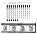
- a preferred kit contains a support with capture molecules specifically binding proteins as exemplified in figures 3, 4 and 5.
- the present invention is directed to a kit for an absolute quantification of the level of phosphorylation of multiple targets within a sample, comprising the steps of a) contacting a sample containing or suspected of containing multiple target proteins with two distinct arrays, said arrays comprising a plurality of capture molecules immobilized on a non-membrane solid support, said capture molecules being capable of specifically binding to both the phosphorylated and non-phosphorylated forms of the target proteins, and b) contacting the target proteins immobilized on the first array with detection molecules being capable of specifically binding to the phosphorylated target proteins but not to the non-phosphorylated target proteins, and contacting the target proteins immobilized on the second array with detection molecules being capable of specifically binding to both the phosphorylated and non phosphorylated forms of the target proteins, and c) assessing the level of phosphorylation of said immobilized target proteins by measurement the signal ratio between the phosphorylated versus the total target proteins present in the sample.
- the level of phosphorylation gives a quantiffer
- the present kit is preferably used in a high throughput format.
- the kit comprises a plurality of arrays of multiple capture molecules immobilized on the same or different solid supports, each of said arrays being identical or having capture molecules able of specifically binding to the same proteins or to the same group of structurally and/or functionally related target proteins.
- the present method involves the absolute quantification of the level of phosphorylation in multiple targets within a sample.
- Hybridization chambers were placed onto diaglass functionalized slides (EAT, Namur, Belgium) spotted with antibodies specific of the protein to be detected, in this case anti-Akt antibodies.
- the spotting was performed with a split pin or a pin and ring or a plain pin. Each spot contained between 0.5 and 10 nl depending on the type of pin used and its diameter.
- the diameters of the spots were between about 250 and 450 ⁇ m. For each antibody, 4 spots were present on the array, and positive and negative controls of spotting and a negative hybridization control for testing the non specific binding of the protein on non specific antibodies were included.
- the slides were washed 4 x 1 min in Washing Buffer (EAT, Namur, Belgium). The slides were then incubated for 1 h with 500 ⁇ l of a specific rabbit detection antibody against phospho-Akt diluted 500 x in Blocking Buffer (EAT, Namur, Belgium). The slides were washed 4 x 1 min in Washing Buffer. The slides were then incubated for 45 min at room temperature with a conjugated antibody (anti-rabbit Cy3) diluted 1000 x in Blocking Buffer. The slides were washed 4 x 1 min in Washing Buffer and then rinsed twice in water. The slides were scanned in a laser confocal scanner "ScanArray" (Packard, USA) at a resolution of 10 ⁇ m.
- ScanArray Packard, USA
- the assay is also specific due to the absence of any signal in the absence of phosphorylated Akt protein (negative control).
- the assay is very sensitive with a detection limit of phospho-Akt of 2 ng/50 ⁇ l which corresponds to 4 x 10 -14 mol/50 ⁇ l or 8 x 10 -10 M.
- the assay was performed as described in example 1, but two identical arrays were present on each slide.
- the capture antibodies spotted on the left and right arrays were the same, but the detection antibodies were either specific to the phospho-Akt (right arrays) or to the total Akt (left arrays).
- Four different mixes were prepared as follows: (1) 40 ng of purified Akt and 40 ng of purified phospho Akt; (2) 40 ng of purified Akt and 20 ng of purified phospho Akt; (3) 40 ng of purified Akt and 10 ng of purified phospho Akt; (4) 40 ng of purified Akt and 5 ng of purified phospho Akt. In this experiment, four slides were used.
- Both arrays (left and right) on each slide were incubated with one of the mixes described above (ex: slide 1 with mix 1, etc.).
- the detection step was done as follows: the left arrays on each slide were incubated with a biotinylated anti-total-Akt antibody diluted 100x.
- the right arrays were incubated with a biotinylated anti-phospho-Akt antibody diluted 100x.
- An anti-biotin conjugated Cy3 antibody was incubated with each array for the detection (dilution on the left array : 1000x; dilution on the right array : 100x).
- the intensity of the spots on each array was then quantified and the results clearly show decreasing signal intensities correlating with decreasing concentrations of phospho-Akt on the right arrays.
- the calculated ratios of phospho vs. total Akt directly relates to the known concentration used in the four different mixes.
- the objective of this experiment was to detect the quantity of total Akt in HepG2 cells incubated in both normoxia and hypoxia for 5 or 16 hours.
- the slides were processed as described in example 1.
- the quantity of proteins present in each cell lysate was measured by the Bradford technique. This value was used to normalize the signals obtained after quantification. 10 ⁇ l of cell lysates were diluted in 50 ⁇ l Blocking Buffer. Four conditions were tested: normoxia 5h, hypoxia 5h, normoxia 16h, hypoxia 16h. The slides were incubated for 2h at 22°C, on a thermomixer (1400 rpm). Following washing, the slides were incubated with an anti-Akt antibody. An anti-rabbit Cy3 is used for the revelation.
- Each slide contained 2 identical arrays.
- the two arrays were incubated with the same samples possibly containing the proteins to be detected.
- the left array was then incubated with a mixture of 7 detection antibodies directed against the 7 proteins to be detected.
- the right array was incubated with a mixture of 7 detection antibodies directed against the phospho-specific form of the 7 proteins to be detected.
- the assay was performed as in examples 1 and 2.
- An anti-biotin-Cy3 antibody was used for the revelation on both arrays.
- the samples consisted of each of the 7 proteins to be detected, in their non-phosphorylated or phosphorylated form, or combinations thereof.
- the experiment was performed on slides containing a double array as presented in example 4.
- Jurkat cells were treated with 8 different concentrations of anisomycin.
- Eight cell lysates were prepared and put into contact with the multiplex arrays.
- the protein chips allowed to simultaneously quantify the level of phosphorylation of p38 and JNK1/2 due to the effect of anisomycin on cells.
- JNK1/2 and p38 phosphorylation in Jurkat cells are dependent on the levels of stimulation.
- the levels of total JNK1/2 and p38 remain constant while the levels of the corresponding phospho forms increase with the anisomycin concentration.
- the ratios of Phospho vs. total forms are of 0.33 and 0.88 respectively for anisomycin concentrations of 0.01 and 1 ⁇ M, whereas for p38 the ratios are of 0.33 and 1.0 respectively for anisomycin concentrations of 1 and 100 ⁇ M.
- Hela cells were grown to 80% confluence in DMEM/high glucose medium supplemented with 10% FBS and antibiotics. Medium is replaced with serum-free medium and phorbol 12-myristate 13-acetate (PMA) is added for one hour at a final concentration of 100nM.
- PMA phorbol 12-myristate 13-acetate
- Cell extracts from non stimulated and PMA-stimulated cells are prepared with the lysis buffer from Biosource (Belgium) according to the manufacturer recommendations.
- the spots intensities were quantified and the comparison of the signals obtained on the two arrays from each slide allowed to get information about the activation state of the test samples. Moreover, comparing the information obtained separately for Hela and PMA-treated Hela allowed to get information about the influence of PMA treatment on different activation cascades in Hela cells.
- the spots intensities are quantified and the comparison of the signals obtained on the two arrays from each slide allows to get information about the activation state of each of the tested samples.
- the comparison of data obtained on the 4 different slides allowed to get information about the effect of hypoxia on the PI3K/Akt cascades in HepG2 cells.
- Extracts from Hela cells +/- treated with 3 ⁇ M nocodazole for 5 or 20 hours are prepared as in example 7. Aliquots of cell extracts from each test condition are contacted with two arrays from a same slide. Left arrays from each slide are then contacted with a mix of biotinylated pan antibodies, each being specific for one of the above-mentioned proteins, and right arrays with a mix of biotinylated phospho-specific antibodies, each recognizing the phosphorylated form of one of the above-mentioned proteins. These mixes contain one antibody specific for each of the above-mentioned proteins. An anti-biotin-Cy3 antibody is then added as in previous examples.
- the spots intensities are quantified and the comparison of the signals obtained on the two arrays from each slide allows to get information about the activation state of each of the tested samples.
- the comparison of data obtained for non treated and nocodazole treated Hela cells showed the effect of the drug on the apoptosis cascade. in these cells.
- Extracts from Hela cells +/- treated with 5mM hydroxyurea for 16 hours are prepared as in example 7. Aliquots of cell extracts from each test condition are contacted with two arrays from a same slide. Left arrays from each slide are then contacted with a mix of biotinylated pan antibodies, each being specific for one of the above-mentioned proteins, and right arrays with a mix of biotinylated phospho-specific antibodies, each recognizing the phosphorylated form of one of the above-mentioned proteins. These mixes contain one antibody specific for each of the above-mentioned proteins. An anti-biotin-Cy3 antibody is then added as in previous examples.
- the spots intensities are quantified and the comparison of the signals obtained on the two arrays from each slide allows to get information about the activation state of each of the test samples.
- the comparison of data obtained for non treated and hydroxyurea treated Hela cells showed the effect of the molecule on the cell cycle control in these cells.
- Each array is composed of triplicate spots of capture antibodies specific for the following proteins: Erk1/2, Akt, Hsp27 and p38, as well as positive and negative detection controls. Slides are then processes as in previous examples. Eight-well hybridization chambers (Grace Bio-Labs, Inc.) are then applied onto each slide to enable subsequent processing of each array individually.
- Extracts from Hela cells +/- treated with PMA are produced as in example 7, and aliquots from each cell extract are contacted with four of the eight arrays on each slide.
- the four top arrays correspond to non-stimulated Hela, while the four bottom arrays correspond to PMA-stimulated Hela.
- biotinylated pan antibodies each being specific for one of the above-mentioned proteins
- biotinylated phospho-specific antibodies each recognizing the phosphorylated form of one of the above-mentioned proteins.
- the antibodies are not added as a mix, but individually to two of the eight arrays, one top and one bottom array.
- An anti-biotin-Cy3 antibody is then added as in previous examples.
- the spots intensities are quantified and the comparison of the signals obtained on the corresponding top and bottom arrays allows to get information about the effect of PMA on the phosphorylated and non-phosphorylated forms of each protein individually in Hela cells.
- the comparison of the corresponding arrays from the two slides allowed to get information about the phosphorylation of each of the four studied proteins.
- the right arrays from each slide are contacted with phospho-specific antibodies that differ for every duplicate: the right arrays from each of the seven duplicate series are contacted with antibodies against phospho-tyrosine residues, phospho-serine residues, phospho-threonine residues, or combinations thereof. All combinations are assayed, leading to a total of seven different phospho-antibody pools.
- the spots intensities are quantified and the comparison of the signals obtained on the two arrays from each slide allows to get information about the activation state of each of the tested samples.
- the comparison of data obtained on the 2 slides in each duplicate series allows to get information about the effect of PMA on the phosphorylation of the five studied proteins in Jurkat cells.
- the comparison of the seven series of duplicate slides allowed to get information about the different residues that are phosphorylated upon PMA stimulation in Jurkat cells. Table 2. Examples of tyrosine kinases A.
- transcription factors activated by phosphorylation/dephosphorylation Kinases Transcription factors ERK 1/2 Elk-1, Stat 1/3, Ets-1, ER, c-Myc, SRF, CREB SAPK/JNK c-Jun, ATF-2, Elk-1, p53, DPC4 p38 MAPK ATF-2, MEF2C, Elk-1, Myc/Max, Stat1, CREB, CHOP ERK5/BMK MEF2C p90 rsk c-Fos, SRF, CREB MSK1 CREB JAKs STATs PKA CREB, SAF-1, GATA-4, SOX9, HNF-4, AR PKB/Akt forkhead, AFX GSK3 ⁇ AP1, ⁇ -catenin, C/EBP ⁇ , CREB, HSF-1, Myc, NFAT, NF ⁇ B ALKs SMADs CaMK Ets-1, CREB, STAT1
Landscapes
- Chemical & Material Sciences (AREA)
- Organic Chemistry (AREA)
- Life Sciences & Earth Sciences (AREA)
- Zoology (AREA)
- Wood Science & Technology (AREA)
- Proteomics, Peptides & Aminoacids (AREA)
- Health & Medical Sciences (AREA)
- Engineering & Computer Science (AREA)
- Microbiology (AREA)
- Biochemistry (AREA)
- Physics & Mathematics (AREA)
- Molecular Biology (AREA)
- Biotechnology (AREA)
- Biophysics (AREA)
- Analytical Chemistry (AREA)
- Immunology (AREA)
- Bioinformatics & Cheminformatics (AREA)
- General Engineering & Computer Science (AREA)
- General Health & Medical Sciences (AREA)
- Genetics & Genomics (AREA)
- Measuring Or Testing Involving Enzymes Or Micro-Organisms (AREA)
- Investigating Or Analysing Biological Materials (AREA)
Applications Claiming Priority (2)
| Application Number | Priority Date | Filing Date | Title |
|---|---|---|---|
| US606162 | 1996-02-23 | ||
| US10/606,162 US20040265938A1 (en) | 2003-06-25 | 2003-06-25 | Method for the determination of cell activation |
Publications (2)
| Publication Number | Publication Date |
|---|---|
| EP1491894A2 true EP1491894A2 (de) | 2004-12-29 |
| EP1491894A3 EP1491894A3 (de) | 2009-06-10 |
Family
ID=33418682
Family Applications (1)
| Application Number | Title | Priority Date | Filing Date |
|---|---|---|---|
| EP04014730A Withdrawn EP1491894A3 (de) | 2003-06-25 | 2004-06-23 | Verfahren zur Bestimmung der Zellaktivierung |
Country Status (2)
| Country | Link |
|---|---|
| US (2) | US20040265938A1 (de) |
| EP (1) | EP1491894A3 (de) |
Cited By (7)
| Publication number | Priority date | Publication date | Assignee | Title |
|---|---|---|---|---|
| EP1736780A1 (de) * | 2005-06-24 | 2006-12-27 | Eppendorf Array Technologies S.A. | Verfahren und Vorrichtungen zum Nachweis und/oder zur Quantifizierung hierarchischer, molekularer Veränderungen einer Zellantwort auf einen externen Stimulus |
| WO2007047754A3 (en) * | 2005-10-18 | 2007-06-07 | Lance A Liotta | Mtor pathway theranostic |
| EP2614372A4 (de) * | 2010-09-07 | 2014-05-07 | Stephen G Marx | Kit zur überwachung, erkennung und einstufung einer transplantat-gegen-wirt-reaktion |
| WO2022159520A3 (en) * | 2021-01-20 | 2022-08-25 | Nautilus Biotechnology, Inc. | Methods for biomolecule quantitation |
| CN115286701A (zh) * | 2022-03-07 | 2022-11-04 | 江苏靶标生物医药研究所有限公司 | 一种调节哺乳动物的肠道粘膜稳态或炎症性肠病的靶点及其应用 |
| US11768201B1 (en) | 2016-12-01 | 2023-09-26 | Nautilus Subsidiary, Inc. | Methods of assaying proteins |
| US12148509B2 (en) | 2017-12-29 | 2024-11-19 | Nautilus Subsidiary, Inc. | Decoding approaches for protein identification |
Families Citing this family (16)
| Publication number | Priority date | Publication date | Assignee | Title |
|---|---|---|---|---|
| WO2007127002A2 (en) * | 2006-03-31 | 2007-11-08 | Cell Signaling Technology, Inc. | Protein markers of responsiveness to type iii receptor tyrosine kinase inhibitors |
| BRPI0717416A2 (pt) | 2006-09-21 | 2013-11-12 | Prometheus Lab Inc | Método para realizar um imunoensaio complexo de alta produtividade, e, arranjo |
| US20080254481A1 (en) * | 2006-11-13 | 2008-10-16 | Invitrogen Corporation | Methods and kits for detecting prostate cancer biomarkers |
| EP2179291B1 (de) | 2007-07-13 | 2014-10-22 | Nestec S.A. | Arzneistoffauswahl für die lungenkrebstherapie unter verwendung von arrays auf antikörperbasis |
| AU2009219437B2 (en) | 2008-02-25 | 2014-10-16 | Nestec S.A. | Drug selection for breast cancer therapy using antibody-based arrays |
| WO2010044892A1 (en) * | 2008-10-17 | 2010-04-22 | President And Fellows Of Harvard College | Diagnostic method based on large scale identification of post-translational modification of proteins |
| WO2010127057A2 (en) * | 2009-05-01 | 2010-11-04 | Meso Scale Technologies, Llc. | Biomarkers of therapeutic responsiveness |
| JP5795311B2 (ja) * | 2009-07-15 | 2015-10-14 | ネステク ソシエテ アノニム | 抗体ベースのアレイを使用する胃癌療法のための薬物選択 |
| US9719995B2 (en) | 2011-02-03 | 2017-08-01 | Pierian Holdings, Inc. | Drug selection for colorectal cancer therapy using receptor tyrosine kinase profiling |
| EP2751562B1 (de) | 2011-09-02 | 2015-09-16 | Nestec S.A. | Profilierung von signalwegproteinen zur bestimmung der therapeutischen wirksamkeit |
| US9139829B2 (en) * | 2012-02-28 | 2015-09-22 | Medical Diagnostic Laboratories, Llc | SiRNA targeting ETS1 and ELK1 and method of using same in the inhibition of CIP2A gene in cancer treatment |
| CA2910034C (en) | 2013-04-26 | 2022-08-30 | Med-Logics, Inc. | Tissue removal device, systems, and methods using vacuum pulses and ultrasonic vibrations |
| US11971354B2 (en) | 2015-04-08 | 2024-04-30 | Molecular Devices, Llc | Methods and systems for fluorescence detection using infrared dyes |
| US10379046B2 (en) * | 2015-04-08 | 2019-08-13 | Molecular Devices, Llc | Method and system for multiplexed time-resolved fluorescence detection |
| US10571396B2 (en) * | 2015-04-08 | 2020-02-25 | Molecular Devices, Llc | Methods and systems for fluorescence detection |
| US10386276B2 (en) | 2016-09-20 | 2019-08-20 | International Business Machines Corporation | Phosphoprotein detection using a chip-based pillar array |
Family Cites Families (5)
| Publication number | Priority date | Publication date | Assignee | Title |
|---|---|---|---|---|
| US5413939A (en) * | 1993-06-29 | 1995-05-09 | First Medical, Inc. | Solid-phase binding assay system for interferometrically measuring analytes bound to an active receptor |
| WO2001027624A2 (en) * | 1999-10-08 | 2001-04-19 | Superarray, Inc. | Compositions and methods for detecting protein modification and enzymatic activity |
| US20020192654A1 (en) * | 2001-06-15 | 2002-12-19 | Tao-Kuang Chang | Bio-chip substrate for the embedding of DNA or protein and its fabrication method |
| AU2002341621A1 (en) * | 2001-09-10 | 2003-03-24 | Meso Scale Technologies, Llc | Methods, reagents, kits and apparatus for protein function analysis |
| CN1665790A (zh) * | 2002-05-03 | 2005-09-07 | 莫莱丘莱尔探针公司 | 用于检测和分离磷酸化分子的组合物及方法 |
-
2003
- 2003-06-25 US US10/606,162 patent/US20040265938A1/en not_active Abandoned
-
2004
- 2004-06-23 EP EP04014730A patent/EP1491894A3/de not_active Withdrawn
-
2008
- 2008-02-11 US US12/029,389 patent/US20080146457A1/en not_active Abandoned
Cited By (10)
| Publication number | Priority date | Publication date | Assignee | Title |
|---|---|---|---|---|
| EP1736780A1 (de) * | 2005-06-24 | 2006-12-27 | Eppendorf Array Technologies S.A. | Verfahren und Vorrichtungen zum Nachweis und/oder zur Quantifizierung hierarchischer, molekularer Veränderungen einer Zellantwort auf einen externen Stimulus |
| WO2006135992A1 (en) * | 2005-06-24 | 2006-12-28 | Eppendorf Array Technologies S.A. | Method and means for detecting and/or quantifying hierarchical molecular change of a cell in response to an external stimulus |
| WO2007047754A3 (en) * | 2005-10-18 | 2007-06-07 | Lance A Liotta | Mtor pathway theranostic |
| US8628931B2 (en) | 2005-10-18 | 2014-01-14 | George Mason Intellectual Properties, Inc. | mTOR pathway theranostic |
| EP2614372A4 (de) * | 2010-09-07 | 2014-05-07 | Stephen G Marx | Kit zur überwachung, erkennung und einstufung einer transplantat-gegen-wirt-reaktion |
| US11768201B1 (en) | 2016-12-01 | 2023-09-26 | Nautilus Subsidiary, Inc. | Methods of assaying proteins |
| US12148509B2 (en) | 2017-12-29 | 2024-11-19 | Nautilus Subsidiary, Inc. | Decoding approaches for protein identification |
| WO2022159520A3 (en) * | 2021-01-20 | 2022-08-25 | Nautilus Biotechnology, Inc. | Methods for biomolecule quantitation |
| US12092642B2 (en) | 2021-01-20 | 2024-09-17 | Nautilus Subsidiary, Inc. | Systems and methods for biomolecule quantitation |
| CN115286701A (zh) * | 2022-03-07 | 2022-11-04 | 江苏靶标生物医药研究所有限公司 | 一种调节哺乳动物的肠道粘膜稳态或炎症性肠病的靶点及其应用 |
Also Published As
| Publication number | Publication date |
|---|---|
| US20040265938A1 (en) | 2004-12-30 |
| US20080146457A1 (en) | 2008-06-19 |
| EP1491894A3 (de) | 2009-06-10 |
Similar Documents
| Publication | Publication Date | Title |
|---|---|---|
| EP1491894A2 (de) | Verfahren zur Bestimmung der Zellaktivierung | |
| CA2383945A1 (en) | Compositions and methods for detecting protein modification and enzymatic activity | |
| EP1451348B1 (de) | Verfahren, reagentien, kits und vorrichtungen zur proteinfunktionsanalyse | |
| US7771940B2 (en) | Methods of detecting a plurality of sequence specific DNA binding proteins with oligonucleotide detection duplexes | |
| Iliuk et al. | Is phosphoproteomics ready for clinical research? | |
| Thiele et al. | Deciphering enzyme function using peptide arrays | |
| US20100210478A1 (en) | Make and use of surface molecules of varied densities | |
| Wilson et al. | Monitoring proteins and protein networks using reverse phase protein arrays | |
| US20050147978A1 (en) | Method for quantitative determination of multi-drug resistance in tumors | |
| US8129113B2 (en) | Analysis chip with reference range, kits and methods of analysis | |
| US7332296B2 (en) | Screening assay for enzymes involved in pathophysiology using SELDI-TOF mass spectrometry | |
| EP2274439B1 (de) | Verfahren zur erstellung von profilen für arzneistoffverbindungen | |
| KR20100099138A (ko) | 키나아제 활성 측정용 단백질 칩 | |
| JP2002538836A (ja) | 遺伝子発現の変化の分析 | |
| Boyle et al. | Dissecting kinase signaling pathways | |
| KR100923048B1 (ko) | 미지의 생체분자와 단일가닥핵산의 결합 프로파일을생성하기 위한 핵산칩, 핵산칩의 제조방법, 및 핵산칩을이용한 미지의 생체분자 분석방법 | |
| US20060257861A1 (en) | Screening assay for inhibitors of severe acute respiratory syndrome (SARS) using SELDI-TOF Mass Spectrometry | |
| JP2011512132A (ja) | 骨髄毒性の予測方法 | |
| JP2008104356A (ja) | リン酸化−脱リン酸化反応検出用基質群およびそれを用いた検出方法 | |
| JP2008289374A (ja) | タンパク質キナーゼの新規基質ポリペプチド | |
| US20050153298A1 (en) | Protein micro-arrays and multi-layered affinity interaction detection | |
| RU2795795C1 (ru) | Способ выявления генетических факторов риска развития деменций альцгеймеровского типа на основе гидрогелевого матричного биочипа | |
| US20100317544A1 (en) | Methods, Particles, and Kits for Determining Activity of a Kinase | |
| US20120258481A1 (en) | Cross-linking atp analogs | |
| Zhou et al. | Photocleavable peptide-conjugated magnetic beads for protein kinase assays by MALDI-TOF MS |
Legal Events
| Date | Code | Title | Description |
|---|---|---|---|
| PUAI | Public reference made under article 153(3) epc to a published international application that has entered the european phase |
Free format text: ORIGINAL CODE: 0009012 |
|
| AK | Designated contracting states |
Kind code of ref document: A2 Designated state(s): AT BE BG CH CY CZ DE DK EE ES FI FR GB GR HU IE IT LI LU MC NL PL PT RO SE SI SK TR |
|
| AX | Request for extension of the european patent |
Extension state: AL HR LT LV MK |
|
| PUAL | Search report despatched |
Free format text: ORIGINAL CODE: 0009013 |
|
| AK | Designated contracting states |
Kind code of ref document: A3 Designated state(s): AT BE BG CH CY CZ DE DK EE ES FI FR GB GR HU IE IT LI LU MC NL PL PT RO SE SI SK TR |
|
| AX | Request for extension of the european patent |
Extension state: AL HR LT LV MK |
|
| AKX | Designation fees paid | ||
| STAA | Information on the status of an ep patent application or granted ep patent |
Free format text: STATUS: THE APPLICATION IS DEEMED TO BE WITHDRAWN |
|
| 18D | Application deemed to be withdrawn |
Effective date: 20091211 |
|
| REG | Reference to a national code |
Ref country code: DE Ref legal event code: 8566 |