EP1498087A2 - Künstliche Hornhaut - Google Patents
Künstliche Hornhaut Download PDFInfo
- Publication number
- EP1498087A2 EP1498087A2 EP04254317A EP04254317A EP1498087A2 EP 1498087 A2 EP1498087 A2 EP 1498087A2 EP 04254317 A EP04254317 A EP 04254317A EP 04254317 A EP04254317 A EP 04254317A EP 1498087 A2 EP1498087 A2 EP 1498087A2
- Authority
- EP
- European Patent Office
- Prior art keywords
- skirt
- keratoprosthesis
- lens
- corneal
- coating
- Prior art date
- Legal status (The legal status is an assumption and is not a legal conclusion. Google has not performed a legal analysis and makes no representation as to the accuracy of the status listed.)
- Withdrawn
Links
- 210000004087 cornea Anatomy 0.000 title claims abstract description 45
- 238000000576 coating method Methods 0.000 claims description 27
- 239000011248 coating agent Substances 0.000 claims description 23
- 229920000642 polymer Polymers 0.000 claims description 23
- 239000003607 modifier Substances 0.000 claims description 12
- 239000004480 active ingredient Substances 0.000 claims description 11
- 150000001875 compounds Chemical class 0.000 claims description 11
- 229920003229 poly(methyl methacrylate) Polymers 0.000 claims description 11
- 239000004926 polymethyl methacrylate Substances 0.000 claims description 11
- 239000003242 anti bacterial agent Substances 0.000 claims description 8
- 229940088710 antibiotic agent Drugs 0.000 claims description 7
- 239000000463 material Substances 0.000 claims description 7
- HTTJABKRGRZYRN-UHFFFAOYSA-N Heparin Chemical compound OC1C(NC(=O)C)C(O)OC(COS(O)(=O)=O)C1OC1C(OS(O)(=O)=O)C(O)C(OC2C(C(OS(O)(=O)=O)C(OC3C(C(O)C(O)C(O3)C(O)=O)OS(O)(=O)=O)C(CO)O2)NS(O)(=O)=O)C(C(O)=O)O1 HTTJABKRGRZYRN-UHFFFAOYSA-N 0.000 claims description 6
- 239000003814 drug Substances 0.000 claims description 6
- 229940079593 drug Drugs 0.000 claims description 6
- 229960002897 heparin Drugs 0.000 claims description 6
- 229920000669 heparin Polymers 0.000 claims description 6
- 239000010410 layer Substances 0.000 claims description 6
- 239000002260 anti-inflammatory agent Substances 0.000 claims description 5
- 229940124599 anti-inflammatory drug Drugs 0.000 claims description 5
- 229920001296 polysiloxane Polymers 0.000 claims description 5
- 150000003431 steroids Chemical class 0.000 claims description 5
- 239000003146 anticoagulant agent Substances 0.000 claims description 4
- 229940127219 anticoagulant drug Drugs 0.000 claims description 4
- 239000000227 bioadhesive Substances 0.000 claims description 4
- 238000010276 construction Methods 0.000 claims description 4
- 102100026735 Coagulation factor VIII Human genes 0.000 claims description 3
- 101000911390 Homo sapiens Coagulation factor VIII Proteins 0.000 claims description 3
- WOBHKFSMXKNTIM-UHFFFAOYSA-N Hydroxyethyl methacrylate Chemical compound CC(=C)C(=O)OCCO WOBHKFSMXKNTIM-UHFFFAOYSA-N 0.000 claims description 3
- 239000011247 coating layer Substances 0.000 claims description 3
- 230000035876 healing Effects 0.000 claims description 3
- 239000003120 macrolide antibiotic agent Substances 0.000 claims description 3
- 229920002338 polyhydroxyethylmethacrylate Polymers 0.000 claims description 3
- LISFMEBWQUVKPJ-UHFFFAOYSA-N quinolin-2-ol Chemical compound C1=CC=C2NC(=O)C=CC2=C1 LISFMEBWQUVKPJ-UHFFFAOYSA-N 0.000 claims description 3
- 229920002379 silicone rubber Polymers 0.000 claims description 3
- 102000016359 Fibronectins Human genes 0.000 claims description 2
- 108010067306 Fibronectins Proteins 0.000 claims description 2
- 238000004873 anchoring Methods 0.000 claims description 2
- 239000000560 biocompatible material Substances 0.000 claims description 2
- KPHWPUGNDIVLNH-UHFFFAOYSA-M diclofenac sodium Chemical compound [Na+].[O-]C(=O)CC1=CC=CC=C1NC1=C(Cl)C=CC=C1Cl KPHWPUGNDIVLNH-UHFFFAOYSA-M 0.000 claims description 2
- 239000004205 dimethyl polysiloxane Substances 0.000 claims description 2
- 239000007788 liquid Substances 0.000 claims description 2
- 239000000203 mixture Substances 0.000 claims description 2
- 229940021182 non-steroidal anti-inflammatory drug Drugs 0.000 claims description 2
- 229920000435 poly(dimethylsiloxane) Polymers 0.000 claims description 2
- -1 prednisolone Chemical class 0.000 claims description 2
- OIGNJSKKLXVSLS-VWUMJDOOSA-N prednisolone Chemical compound O=C1C=C[C@]2(C)[C@H]3[C@@H](O)C[C@](C)([C@@](CC4)(O)C(=O)CO)[C@@H]4[C@@H]3CCC2=C1 OIGNJSKKLXVSLS-VWUMJDOOSA-N 0.000 claims description 2
- 229960005205 prednisolone Drugs 0.000 claims description 2
- 230000003115 biocidal effect Effects 0.000 claims 2
- 208000035965 Postoperative Complications Diseases 0.000 claims 1
- 239000003795 chemical substances by application Substances 0.000 claims 1
- 235000013870 dimethyl polysiloxane Nutrition 0.000 claims 1
- 239000000041 non-steroidal anti-inflammatory agent Substances 0.000 claims 1
- 239000003106 tissue adhesive Substances 0.000 claims 1
- 208000015181 infectious disease Diseases 0.000 abstract description 11
- 238000001356 surgical procedure Methods 0.000 abstract description 6
- 210000001519 tissue Anatomy 0.000 description 20
- 210000002159 anterior chamber Anatomy 0.000 description 12
- 201000004569 Blindness Diseases 0.000 description 11
- 238000000034 method Methods 0.000 description 9
- 239000012528 membrane Substances 0.000 description 8
- 230000002093 peripheral effect Effects 0.000 description 8
- 230000002265 prevention Effects 0.000 description 8
- 238000011161 development Methods 0.000 description 7
- 230000003287 optical effect Effects 0.000 description 7
- 230000000007 visual effect Effects 0.000 description 7
- 206010052428 Wound Diseases 0.000 description 6
- 208000027418 Wounds and injury Diseases 0.000 description 6
- 230000028709 inflammatory response Effects 0.000 description 6
- 238000001125 extrusion Methods 0.000 description 5
- 206010031264 Osteonecrosis Diseases 0.000 description 4
- 230000000694 effects Effects 0.000 description 4
- 239000007943 implant Substances 0.000 description 4
- 208000014674 injury Diseases 0.000 description 4
- 230000003449 preventive effect Effects 0.000 description 4
- 239000003894 surgical glue Substances 0.000 description 4
- 230000008733 trauma Effects 0.000 description 4
- 206010061788 Corneal infection Diseases 0.000 description 3
- 208000001860 Eye Infections Diseases 0.000 description 3
- 102000009123 Fibrin Human genes 0.000 description 3
- 108010073385 Fibrin Proteins 0.000 description 3
- BWGVNKXGVNDBDI-UHFFFAOYSA-N Fibrin monomer Chemical compound CNC(=O)CNC(=O)CN BWGVNKXGVNDBDI-UHFFFAOYSA-N 0.000 description 3
- 230000002411 adverse Effects 0.000 description 3
- 230000001580 bacterial effect Effects 0.000 description 3
- 230000008901 benefit Effects 0.000 description 3
- 238000004132 cross linking Methods 0.000 description 3
- 230000006378 damage Effects 0.000 description 3
- 238000013461 design Methods 0.000 description 3
- 229950003499 fibrin Drugs 0.000 description 3
- 238000004519 manufacturing process Methods 0.000 description 3
- 238000000465 moulding Methods 0.000 description 3
- 244000052769 pathogen Species 0.000 description 3
- 230000004044 response Effects 0.000 description 3
- 230000037390 scarring Effects 0.000 description 3
- 210000003786 sclera Anatomy 0.000 description 3
- 230000002459 sustained effect Effects 0.000 description 3
- 241000894006 Bacteria Species 0.000 description 2
- 241000446313 Lamella Species 0.000 description 2
- 241000243985 Onchocerca volvulus Species 0.000 description 2
- 241000700584 Simplexvirus Species 0.000 description 2
- 230000003110 anti-inflammatory effect Effects 0.000 description 2
- 239000002775 capsule Substances 0.000 description 2
- 238000013270 controlled release Methods 0.000 description 2
- 208000021921 corneal disease Diseases 0.000 description 2
- 238000005520 cutting process Methods 0.000 description 2
- 238000007598 dipping method Methods 0.000 description 2
- 230000003628 erosive effect Effects 0.000 description 2
- 239000012530 fluid Substances 0.000 description 2
- 229920001477 hydrophilic polymer Polymers 0.000 description 2
- 230000004410 intraocular pressure Effects 0.000 description 2
- 206010023332 keratitis Diseases 0.000 description 2
- 239000002184 metal Substances 0.000 description 2
- 229910052751 metal Inorganic materials 0.000 description 2
- 230000000149 penetrating effect Effects 0.000 description 2
- 239000005020 polyethylene terephthalate Substances 0.000 description 2
- 102000004169 proteins and genes Human genes 0.000 description 2
- 108090000623 proteins and genes Proteins 0.000 description 2
- 230000000717 retained effect Effects 0.000 description 2
- 238000005507 spraying Methods 0.000 description 2
- 238000010561 standard procedure Methods 0.000 description 2
- 229920001059 synthetic polymer Polymers 0.000 description 2
- 238000002054 transplantation Methods 0.000 description 2
- 230000000472 traumatic effect Effects 0.000 description 2
- 208000009299 Benign Mucous Membrane Pemphigoid Diseases 0.000 description 1
- 208000002177 Cataract Diseases 0.000 description 1
- 229920002160 Celluloid Polymers 0.000 description 1
- 102000008186 Collagen Human genes 0.000 description 1
- 108010035532 Collagen Proteins 0.000 description 1
- 206010051625 Conjunctival hyperaemia Diseases 0.000 description 1
- 206010010985 Corneal abscess Diseases 0.000 description 1
- 229920004934 Dacron® Polymers 0.000 description 1
- 208000003556 Dry Eye Syndromes Diseases 0.000 description 1
- 206010013774 Dry eye Diseases 0.000 description 1
- 208000010412 Glaucoma Diseases 0.000 description 1
- 208000003923 Hereditary Corneal Dystrophies Diseases 0.000 description 1
- 241000282412 Homo Species 0.000 description 1
- 206010061218 Inflammation Diseases 0.000 description 1
- 201000005505 Measles Diseases 0.000 description 1
- 241001465754 Metazoa Species 0.000 description 1
- 208000012192 Mucous membrane pemphigoid Diseases 0.000 description 1
- 206010034277 Pemphigoid Diseases 0.000 description 1
- 208000037111 Retinal Hemorrhage Diseases 0.000 description 1
- 206010038848 Retinal detachment Diseases 0.000 description 1
- 241001303601 Rosacea Species 0.000 description 1
- 206010042033 Stevens-Johnson syndrome Diseases 0.000 description 1
- 231100000168 Stevens-Johnson syndrome Toxicity 0.000 description 1
- 208000002847 Surgical Wound Diseases 0.000 description 1
- 229920004933 Terylene® Polymers 0.000 description 1
- 206010064996 Ulcerative keratitis Diseases 0.000 description 1
- 230000010100 anticoagulation Effects 0.000 description 1
- 230000009286 beneficial effect Effects 0.000 description 1
- 230000002146 bilateral effect Effects 0.000 description 1
- 239000011230 binding agent Substances 0.000 description 1
- 230000005540 biological transmission Effects 0.000 description 1
- 210000000845 cartilage Anatomy 0.000 description 1
- 210000004027 cell Anatomy 0.000 description 1
- 230000010261 cell growth Effects 0.000 description 1
- 239000000919 ceramic Substances 0.000 description 1
- 229910052729 chemical element Inorganic materials 0.000 description 1
- 201000010002 cicatricial pemphigoid Diseases 0.000 description 1
- 229920001436 collagen Polymers 0.000 description 1
- 210000000795 conjunctiva Anatomy 0.000 description 1
- 230000008602 contraction Effects 0.000 description 1
- 206010011005 corneal dystrophy Diseases 0.000 description 1
- 210000003683 corneal stroma Anatomy 0.000 description 1
- 201000007717 corneal ulcer Diseases 0.000 description 1
- 239000013078 crystal Substances 0.000 description 1
- 230000008021 deposition Effects 0.000 description 1
- 238000001514 detection method Methods 0.000 description 1
- 230000003292 diminished effect Effects 0.000 description 1
- 201000010099 disease Diseases 0.000 description 1
- 208000037265 diseases, disorders, signs and symptoms Diseases 0.000 description 1
- 238000012377 drug delivery Methods 0.000 description 1
- 206010014801 endophthalmitis Diseases 0.000 description 1
- 210000002919 epithelial cell Anatomy 0.000 description 1
- 239000003889 eye drop Substances 0.000 description 1
- 229940012356 eye drops Drugs 0.000 description 1
- 239000003885 eye ointment Substances 0.000 description 1
- 230000006870 function Effects 0.000 description 1
- 230000002538 fungal effect Effects 0.000 description 1
- 230000004927 fusion Effects 0.000 description 1
- 239000011521 glass Substances 0.000 description 1
- 230000036541 health Effects 0.000 description 1
- 230000002209 hydrophobic effect Effects 0.000 description 1
- 238000002513 implantation Methods 0.000 description 1
- 238000001727 in vivo Methods 0.000 description 1
- 201000001371 inclusion conjunctivitis Diseases 0.000 description 1
- 238000010348 incorporation Methods 0.000 description 1
- 239000012678 infectious agent Substances 0.000 description 1
- 210000004969 inflammatory cell Anatomy 0.000 description 1
- 230000002757 inflammatory effect Effects 0.000 description 1
- 230000004054 inflammatory process Effects 0.000 description 1
- 239000004615 ingredient Substances 0.000 description 1
- 238000003780 insertion Methods 0.000 description 1
- 230000037431 insertion Effects 0.000 description 1
- 238000009434 installation Methods 0.000 description 1
- 230000007774 longterm Effects 0.000 description 1
- 231100000053 low toxicity Toxicity 0.000 description 1
- 238000005259 measurement Methods 0.000 description 1
- 230000008018 melting Effects 0.000 description 1
- 238000002844 melting Methods 0.000 description 1
- 238000013508 migration Methods 0.000 description 1
- 230000005012 migration Effects 0.000 description 1
- 238000012986 modification Methods 0.000 description 1
- 230000004048 modification Effects 0.000 description 1
- 231100000252 nontoxic Toxicity 0.000 description 1
- 230000003000 nontoxic effect Effects 0.000 description 1
- 235000015097 nutrients Nutrition 0.000 description 1
- 208000003177 ocular onchocerciasis Diseases 0.000 description 1
- 208000002042 onchocerciasis Diseases 0.000 description 1
- 229940126701 oral medication Drugs 0.000 description 1
- 230000036961 partial effect Effects 0.000 description 1
- 239000002245 particle Substances 0.000 description 1
- 230000008447 perception Effects 0.000 description 1
- 229920003225 polyurethane elastomer Polymers 0.000 description 1
- 230000002980 postoperative effect Effects 0.000 description 1
- 230000008569 process Effects 0.000 description 1
- 210000001747 pupil Anatomy 0.000 description 1
- 230000004264 retinal detachment Effects 0.000 description 1
- 230000002207 retinal effect Effects 0.000 description 1
- 201000004700 rosacea Diseases 0.000 description 1
- 231100000241 scar Toxicity 0.000 description 1
- 230000009528 severe injury Effects 0.000 description 1
- 239000004575 stone Substances 0.000 description 1
- 239000000126 substance Substances 0.000 description 1
- 230000001225 therapeutic effect Effects 0.000 description 1
- 206010044325 trachoma Diseases 0.000 description 1
Images







Classifications
-
- A—HUMAN NECESSITIES
- A61—MEDICAL OR VETERINARY SCIENCE; HYGIENE
- A61F—FILTERS IMPLANTABLE INTO BLOOD VESSELS; PROSTHESES; DEVICES PROVIDING PATENCY TO, OR PREVENTING COLLAPSING OF, TUBULAR STRUCTURES OF THE BODY, e.g. STENTS; ORTHOPAEDIC, NURSING OR CONTRACEPTIVE DEVICES; FOMENTATION; TREATMENT OR PROTECTION OF EYES OR EARS; BANDAGES, DRESSINGS OR ABSORBENT PADS; FIRST-AID KITS
- A61F2/00—Filters implantable into blood vessels; Prostheses, i.e. artificial substitutes or replacements for parts of the body; Appliances for connecting them with the body; Devices providing patency to, or preventing collapsing of, tubular structures of the body, e.g. stents
- A61F2/02—Prostheses implantable into the body
- A61F2/14—Eye parts, e.g. lenses or corneal implants; Artificial eyes
-
- A—HUMAN NECESSITIES
- A61—MEDICAL OR VETERINARY SCIENCE; HYGIENE
- A61F—FILTERS IMPLANTABLE INTO BLOOD VESSELS; PROSTHESES; DEVICES PROVIDING PATENCY TO, OR PREVENTING COLLAPSING OF, TUBULAR STRUCTURES OF THE BODY, e.g. STENTS; ORTHOPAEDIC, NURSING OR CONTRACEPTIVE DEVICES; FOMENTATION; TREATMENT OR PROTECTION OF EYES OR EARS; BANDAGES, DRESSINGS OR ABSORBENT PADS; FIRST-AID KITS
- A61F2/00—Filters implantable into blood vessels; Prostheses, i.e. artificial substitutes or replacements for parts of the body; Appliances for connecting them with the body; Devices providing patency to, or preventing collapsing of, tubular structures of the body, e.g. stents
- A61F2/02—Prostheses implantable into the body
- A61F2/14—Eye parts, e.g. lenses or corneal implants; Artificial eyes
- A61F2/142—Cornea, e.g. artificial corneae, keratoprostheses or corneal implants for repair of defective corneal tissue
-
- A—HUMAN NECESSITIES
- A61—MEDICAL OR VETERINARY SCIENCE; HYGIENE
- A61L—METHODS OR APPARATUS FOR STERILISING MATERIALS OR OBJECTS IN GENERAL; DISINFECTION, STERILISATION OR DEODORISATION OF AIR; CHEMICAL ASPECTS OF BANDAGES, DRESSINGS, ABSORBENT PADS OR SURGICAL ARTICLES; MATERIALS FOR BANDAGES, DRESSINGS, ABSORBENT PADS OR SURGICAL ARTICLES
- A61L15/00—Chemical aspects of, or use of materials for, bandages, dressings or absorbent pads
- A61L15/16—Bandages, dressings or absorbent pads for physiological fluids such as urine or blood, e.g. sanitary towels, tampons
- A61L15/42—Use of materials characterised by their function or physical properties
- A61L15/425—Porous materials, e.g. foams or sponges
-
- A—HUMAN NECESSITIES
- A61—MEDICAL OR VETERINARY SCIENCE; HYGIENE
- A61F—FILTERS IMPLANTABLE INTO BLOOD VESSELS; PROSTHESES; DEVICES PROVIDING PATENCY TO, OR PREVENTING COLLAPSING OF, TUBULAR STRUCTURES OF THE BODY, e.g. STENTS; ORTHOPAEDIC, NURSING OR CONTRACEPTIVE DEVICES; FOMENTATION; TREATMENT OR PROTECTION OF EYES OR EARS; BANDAGES, DRESSINGS OR ABSORBENT PADS; FIRST-AID KITS
- A61F2/00—Filters implantable into blood vessels; Prostheses, i.e. artificial substitutes or replacements for parts of the body; Appliances for connecting them with the body; Devices providing patency to, or preventing collapsing of, tubular structures of the body, e.g. stents
- A61F2/02—Prostheses implantable into the body
- A61F2/14—Eye parts, e.g. lenses or corneal implants; Artificial eyes
- A61F2/15—Implant having one or more holes, e.g. for nutrient transport, for facilitating handling
-
- A—HUMAN NECESSITIES
- A61—MEDICAL OR VETERINARY SCIENCE; HYGIENE
- A61F—FILTERS IMPLANTABLE INTO BLOOD VESSELS; PROSTHESES; DEVICES PROVIDING PATENCY TO, OR PREVENTING COLLAPSING OF, TUBULAR STRUCTURES OF THE BODY, e.g. STENTS; ORTHOPAEDIC, NURSING OR CONTRACEPTIVE DEVICES; FOMENTATION; TREATMENT OR PROTECTION OF EYES OR EARS; BANDAGES, DRESSINGS OR ABSORBENT PADS; FIRST-AID KITS
- A61F2/00—Filters implantable into blood vessels; Prostheses, i.e. artificial substitutes or replacements for parts of the body; Appliances for connecting them with the body; Devices providing patency to, or preventing collapsing of, tubular structures of the body, e.g. stents
- A61F2/02—Prostheses implantable into the body
- A61F2/14—Eye parts, e.g. lenses or corneal implants; Artificial eyes
- A61F2/16—Intraocular lenses
-
- A—HUMAN NECESSITIES
- A61—MEDICAL OR VETERINARY SCIENCE; HYGIENE
- A61L—METHODS OR APPARATUS FOR STERILISING MATERIALS OR OBJECTS IN GENERAL; DISINFECTION, STERILISATION OR DEODORISATION OF AIR; CHEMICAL ASPECTS OF BANDAGES, DRESSINGS, ABSORBENT PADS OR SURGICAL ARTICLES; MATERIALS FOR BANDAGES, DRESSINGS, ABSORBENT PADS OR SURGICAL ARTICLES
- A61L27/00—Materials for grafts or prostheses or for coating grafts or prostheses
- A61L27/14—Macromolecular materials
- A61L27/22—Polypeptides or derivatives thereof, e.g. degradation products
- A61L27/24—Collagen
Definitions
- the present invention relates to a keratoprosthesis or artificial cornea, and a method for implanting a keratoprosthesis.
- Keratoprosthesis are referred to as an artificial cornea as the keratoprosthesis replaces the cornea of the patient, for example, when this is damaged by infection, trauma or otherwise.
- the visual part of the keratoprosthesis is referred to as a "lens”
- a keratoprosthesis should not be confused with an intra-ocular lens (or artificial lens), as there are no functional or physical similarities between the two devices.
- the optical part of each device will mirror the optical properties of a natural cornea/lens and so have totally different optical properties.
- the intro-ocular lens is designed to replace the opaque lens of a patient due to cataract and resides within the eye. This is distinct from the keratoprosthesis which resides in the front part of the eye.
- Corneal blindness is a major health, social and economic problem worldwide. It accounts for up to 5% of all blindness in the developed countries and for up to 50% in the developing countries. It is estimated that at least 10 million people suffer from bilateral corneal blindness and probably a larger number from unilateral corneal blindness.
- corneal blindness There are a variety of causes responsible for corneal blindness.
- the common causes are herpes simplex virus corneal infection, trauma, chemical and metal burn, cicatricial pemphigoid, Stevens-Johnson syndrome, Rosacea keratitis, corneal dystrophy, dry eye and failed keratoplasty.
- corneal infections such as trachoma, bacterial and fungal corneal ulcer and abscess, herpes simplex virus corneal infection, onchocerciasis (river blindness) and a post-measles keratitis are the main causes of corneal blindness.
- a number of these diseases cause severe corneal scarring and vascularization, uneven corneal thickness and damage to the anterior segment of the eye.
- keratoplasty has been successful in restoring vision in patients with uncomplicated corneal scarring, while, in patients with severe corneal scarring and vascularization and in those with damaged anterior segment, the results of keratoplasty have been rather poor. In the developing countries, keratoplasty is not frequently performed because of the scarcity of donor corneas.
- corneal blindness In developed countries, of an estimated 2 million blind people (0.2% of the population) 100,000 (5%) are blind because of corneal diseases. The incidence of corneal blindness is estimated at about 5000 per annum. In the USA over 50,000 corneal transplantations are being carried out annually. In developing countries it is estimated that 10 million people suffer from blindness because of corneal diseases. The incidence of corneal blindness is estimated at about 450,000 cases per annum.
- the supporting plate wasthen modified using various anchoring materials such as tooth, cartilage, terylene, dacron, ceramic and metal.
- various anchoring materials such as tooth, cartilage, terylene, dacron, ceramic and metal.
- the Caldwell-Py keratoprosthesis was made of polyurethane elastomers and consisted of a central transparent optical part and a peripheral white and porous skirt with extended arms attached to the skirt to fix the keratoprosthesis to the sclera.
- the keratoprosthesis was implanted in six eyes and results were followed for one year.
- Vision was improved from light perception to a level of 3/60 in two eyes, to counting fingers at two metres in two eyes, but deteriorated later and did not improve at all in the remaining two eyes, because of pre-operative retinal disfunction (unpublished report).
- the main adverse effects and complications were discolouration, dryness and erosion of the optical part and adhesion of cells and debris to its surface in all eyes; partial extrusion of keratoprosthesis in five eyes, because of poor binding of the skirt to surrounding tissues; moderate to severe intra-ocular infection in four eyes, closely associated with the extrusion of keratoprosthesis; and moderate to severe retroprosthetic membrane in four eyes, associated with moderate to severe damage to the anterior segment of the eye.
- the present invention seeks to provide a keratoprosthesis which is retained in the eye without causing major complications and adverse effects commonly associated with available keratoprosthesis.
- a keratoprosthesis comprising a lens surrounded by a skirt, where the keratoprosthesis is a one-part construction with the thickness of the lens being greater than the thickness of the skirt.
- the lens is generally circular with a diameter of between 5mm and 9mm, preferably about 7mm; and/or the skirt comprises a generally circular ring around the lens with the ring having a width of between 1mm and 3mm, preferably about 2mm.
- the overall construction is thus small and lightweight.
- the lens preferably has a thickness of between 0.5mm and 2mm, preferably about 1mm; and/or the skirt has a thickness of between 0.1mm and 0.4mm, preferably about 0.3mm.
- the overall thickness of the construction is thus small compared to the presently available artificial corneas.
- the keratoprosthesis skirt advantageously includes a plurality of large holes of about 1 mm diameter; and/or the skirt includes a plurality of small holes of about 0.3mm diameter.
- the large holes are particularly useful for suturing.
- the large and small holes allow the tissues of the eye to grow therebetween and fluids to cross the plane of the implant.
- the keratoprosthesis is advantageously formed from a biocompatible material selected from the group consisting of: silicon elastomers; PMMA including modified and/or crosslinked PMMA; HEMA; polyHEMA; mixtures of the aforementioned compounds.
- the skirt is preferably coated with a polymer which promotes the binding of the keratoprosthesis in the eye which improves retainability of the implant.
- a kit comprising the keratoprosthesis according to the first aspect of the invention and as defined in claims 7 and 8 and a suture assembly where only two types of suture is required to hold the keratoprosthesis in place when inserted in the eye of a patient.
- the special suture can advantageously be used to secure other devices.
- the keratoprosthesis is a one-piece convex disc resembling the human cornea with a refractive power of 45 to 48 dioptre or higher.
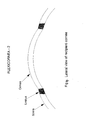
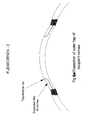
- the keratoprosthesis consists of a full thickness transparent central visual section with smooth surfaces (Figs 1 and 2) and a thinner net-type peripheral section or skirt with additional large holes.
- a specific corneal shield as defined in claim 9.
- the keratoprosthesis of the present invention may be used as a method of installing a keratoprosthesis comprising the steps of:
- the overall diameter of keratoprosthesis is 11 m (Fig. 1).
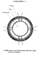
- the visual section has a diameter of 7mm and a thickness of 1 mm (Fig. 1, 2 and 3).
- the width of the skirt is 2mm with a thickness of 0.3mm approximately (Fig. 1, 2 and 3).
- the diameter of large holes in the skirt is 1 mm approximately (Fig. 2).
- the slots have a width of 1 mm approx and a length of about 6/7mm.
- the keratoprosthesis (also referred to as a flexicornea) will advantageously be made from a single polymer with the following features: highly transparent; semiflexible; highly tensile; non-erodible; non-binding; durable with no physical changes after long-term exposure to tears, aqueous, air and extreme climatic conditions; non-toxic and bio-compatible with human tissues. Silicon elastomers, PMMA with some modifications and cross-linking (to make it flexible), HEMA, PolyHEMA and other polymers used for making flexible intra-ocular lens may be suitable for the production of the keratoprosthesis.
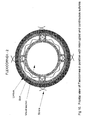
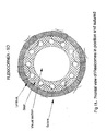
- the skirt will be coated with one or more layers of a durable and non-erodible polymer with strong affinity to binding tissues, proteins and fibrin. Ready made sutures will attach to the keratoprosthesis (Fig. 8A, 8B, 10 and 11).
- the operation to install the keratoprosthesis is simple, non-traumatic and similar to the keratoplasty operation.
- the main stages of the operation are as follows:
- the invention in its preferred embodiment has a number of preferred features which are:
- the coating concept is very well known and much used in microbiology, where microtitre plates are being coated with relevant polymers to either enhance or prohibit deposition of proteins and growth of cells on the microtitre plate.
- the coating is also used to deliver active ingredients, e.g. to prevent complications.
- Fig. 1 shows the particularly preferred design of the keratoprosthesis of the present invention. This has a lens section coated as hereinafter described and a slit also coated as hereinafter described.
- the skirt is in the form of two rings joined by four ribs thus defining four large slots. Each rib has a hole.
- the slots and holes shown in Fig. 1 or small and large holes in the skirt shown in Fig. 2 will allow the corneal and scar tissues to grow through the skirt and produce a strong fusion with the tissues of the opposite flap. This process will strengthen the attachment of the corneal flaps to the skirt, prevent extrusion of the skirt, leakage of aqueous and transmission of infectious agents into the anterior chamber.
- the net-type skirt shown in Fig. 2 or the rings and ribs in Fig. 1 with large openings will allow free movement of fluids and nutrients between the two flaps of the cornea, thus preventing development of aseptic necrosis and melting of the peripheral cornea resulting in keratoprosthesis extrusion.
- the large openings in the skirt will also be used as a base for suturing keratoprosthesis to the limbus.
- the keratoprosthesis operation is simple, practical and can be performed by most ophthalmic surgeons.
- the skirt of keratoprosthesis is made of polymethylmethacrylate or silicone is hydrophobic and non-binding. To improve and consolidate the binding between the keratoprosthesis and the host cornea the following novel systems are incorporated.
- Ocular infections associates with keratoprosthesis usually develop following introduction of one or more pathogens into the wound and anterior chamber of the eye.
- the source of infection may include: the eye of recipient (normal bacterial flora of the eye), hands of surgeon, contaminated surgical equipment and materials and internal sources (rare).
- Most gram positive and gram negative bacteria may cause post-operative ocular infection. These pathogens may reside and proliferate at the interface between the keratoprosthesis and the cornea or within damaged corneal tissues surrounding the keratoprosthesis and subsequently spread into the anterior chamber, causing severe endophthalmitis and destruction of the eye.
- the present invention involves the use of a novel and highly effective system for prevention of infection associated with keratoprosthesis.
- the system is based on our patented drug delivery insert devices for delivery of drugs at the target area continuously and at controlled level for several weeks, now modified for the prevention of infection for this type of surgical procedure. See for example WO-A-9501764.
- two antibiotics e.g. a combination of a macrolide and a quinolone, for providing maximum effect on both gram positive and gram negative bacteria
- release modifiers into the single coating or one of the two coating layers on the skirt.
- the antibiotics will be released at a zero or near zero order for several weeks into the space between the skirt and adjacent corneal tissues, where they can effectively neutralise the invading pathogens and preventing them from causing infection.
- one or both antibiotics can be incorporated into the base material of the skirt before moulding.
- Inflammatory responses generally occur in response to operation trauma and/or presence of the polymer (foreign body) or polymer particles shedding from the surface of the skirt.
- the inflammatory responses caused by the operation may be moderate or severe, but generally subside within a few weeks.
- the inflammatory responses related to the polymer may start as mild responses, but gradually become more severe and causing damage to tissues surrounding the prosthesis and aseptic necrosis. Both of these inflammatory responses respond well to treatment with steroids or non-steroid anti-inflammatory compounds.
- a system for sustained, continuous and controlled release of steroids e.g. prednisolone
- non-steroid anti-inflammatory drugs e.g. sodium diclofenac
- This is being achieved by: (a) incorporating a selected anti-inflammatory drug into the single or the two coating layers of the skirt; or (b) incorporating into the base material of the skirt before moulding. The compound will be released at zero or near zero order for a few weeks at the junction between the skirt and corneal tissues, which will be absorbed into the surrounding corneal tissues.
- Retroprosthetic membrane commonly develops following trauma by surgical procedures to already damaged tissues within the anterior chamber of the eye and/or as a response to the presence of a synthetic polymer. After implanting a keratoprosthesis, the traumatised tissues within the anterior chamber release fibrin into the anterior chamber. Subsequently, the fibrin coagulates forming a pseudomembrane. After a few days, inflammatory cells invade the pseudomembrane and new vessels develop in the membrane and converting the pseudomembrane into a thick and well established membrane within the anterior chamber, which can block the pupil. Development of retroprosthetic membrane within the anterior chamber of the eye will be prevented by sustained, continuous and controlled release of anti-coagulants including heparin or its derivatives. This will be achieved by releasing heparin into the anterior chamber of the eye by one or more of the following novel methods:
- a novel preventive corneal shield to enhance healing of the corneal wound and to assist in preventing development of complications is also adopted in the preferred embodiments.
- the novel shield consist of a soft contact lens made of PMMA, silicone or other suitable polymers which will be coated on both sides by thin layers of a liquid silicone containing release modifiers and active ingredients.
- the coating of the concave side may contain medroxyprogestron (a drug known to prevent epithelial in-growth) or compounds with similar function and an anti-inflammatory drug, to be released continuously and at controlled levels for several weeks.
- the coating of the convex side may contain antibiotics (e.g. a macrolide and a quinolone) and other active ingredients (if required) to be released at continuous and controlled levels for several weeks.
- the coating can be achieved by dipping, spraying or crosslinking.
- the application of the specifically designed shield may:
- Instruments required for implanting keratoprosthesis of the invention will be similar to those required for corneal transplantation with the addition of a trephine with a graduated guard and an angled corneal dissector. It should be noted that the diameter of the trephine should be 1 ⁇ 2 of a millimetre smaller than the diameter of the visual section of selected keratoprosthesis in order to compensate for contraction of the host cornea after trephination.
- the keratoprosthesis of the invention contains the following novel physical, polymeric, surgical and chemical elements which are particularly designed to remedy problems of rejection and complications commonly associated with keratoprosthesis.
- Keratoprosthesis is a flexible one-piece prosthesis similar to the human cornea. In our in vivo studies it is shown that the prosthesis appears as a normal cornea.
- the keratoprosthesis is made of a single polymer and as such it is technically simple to manufacture.
- skirt particularly its thinness and inclusion of holes and slots, coating with a hydrophilic polymer containing a binding agent and specific sutures will promote a strong binding between the skirt and the host cornea.
- Incorporation of antibiotics, anti-inflammatory, anti-coagulation and other active ingredients into the coatings of the skirt, to be released at sustained, continuous and controlled level for several weeks are designed to prevent infection, inflammation, aseptic necrosis, retroprosthetic membrane and epithelial in-growth, which are the common causes of failure and rejection of keratoprosthesis.
- the keratoprosthesis allows examination of anterior and posterior segments of the eye.
- the keratoprosthesis allows measurement of intra-ocular pressure and detection of glaucoma.
- the present invention relates to a keratoprosthesis or artificial cornea which is designed as a light weight disc resembling a human cornea.
- the keratoprosthesis is maintained in the eye after surgery and is preferably adapted to reduce complications after surgery such as infection.
Landscapes
- Health & Medical Sciences (AREA)
- Life Sciences & Earth Sciences (AREA)
- Ophthalmology & Optometry (AREA)
- Engineering & Computer Science (AREA)
- Veterinary Medicine (AREA)
- Transplantation (AREA)
- Public Health (AREA)
- General Health & Medical Sciences (AREA)
- Animal Behavior & Ethology (AREA)
- Oral & Maxillofacial Surgery (AREA)
- Biomedical Technology (AREA)
- Vascular Medicine (AREA)
- Heart & Thoracic Surgery (AREA)
- Cardiology (AREA)
- Chemical & Material Sciences (AREA)
- Epidemiology (AREA)
- Hematology (AREA)
- Materials Engineering (AREA)
- Dispersion Chemistry (AREA)
- Biophysics (AREA)
- Dermatology (AREA)
- Medicinal Chemistry (AREA)
- Prostheses (AREA)
- Materials For Medical Uses (AREA)
Applications Claiming Priority (2)
| Application Number | Priority Date | Filing Date | Title |
|---|---|---|---|
| GB0316745 | 2003-07-17 | ||
| GB0316745A GB2403908B (en) | 2003-07-17 | 2003-07-17 | Artificial cornea |
Publications (2)
| Publication Number | Publication Date |
|---|---|
| EP1498087A2 true EP1498087A2 (de) | 2005-01-19 |
| EP1498087A3 EP1498087A3 (de) | 2005-03-16 |
Family
ID=27763997
Family Applications (1)
| Application Number | Title | Priority Date | Filing Date |
|---|---|---|---|
| EP04254317A Withdrawn EP1498087A3 (de) | 2003-07-17 | 2004-07-19 | Künstliche Hornhaut |
Country Status (2)
| Country | Link |
|---|---|
| EP (1) | EP1498087A3 (de) |
| GB (1) | GB2403908B (de) |
Cited By (9)
| Publication number | Priority date | Publication date | Assignee | Title |
|---|---|---|---|---|
| WO2008127653A3 (en) * | 2007-04-13 | 2008-12-04 | Gore Enterprise Holdings Inc | Artificial cornea |
| WO2009135068A1 (en) * | 2008-04-30 | 2009-11-05 | University Of Washington | Artificial cornea |
| CN102920532A (zh) * | 2012-07-23 | 2013-02-13 | 于好勇 | 人工角膜 |
| US20150223930A1 (en) * | 2010-09-30 | 2015-08-13 | KeraMed, Inc. | Corneal implants |
| EP3600469A4 (de) * | 2017-03-30 | 2020-11-25 | Marquette University | Synthetische prothese für chirurgische osteo-odonto-keratoprothese (ookp) |
| WO2021048853A1 (en) * | 2019-09-12 | 2021-03-18 | Corneat Vision Ltd. | Graft patch for the treatment of myopia and ophthalmic conditions |
| CN113521385A (zh) * | 2021-06-03 | 2021-10-22 | 中南大学湘雅二医院 | 一种人工角膜 |
| CN113747856A (zh) * | 2019-04-25 | 2021-12-03 | 科尔尼特视觉有限公司 | 角膜假体装置和套件及其使用的外科方法 |
| WO2022139749A3 (en) * | 2020-12-21 | 2022-08-18 | T.C. Marmara Üni̇versi̇tesi̇ | A method for thi̇el-behnke corneal dystrophy (tbcd) and an artificial tissue scaffold produced by said method |
Families Citing this family (2)
| Publication number | Priority date | Publication date | Assignee | Title |
|---|---|---|---|---|
| US9974646B2 (en) | 2012-09-05 | 2018-05-22 | University Of Miami | Keratoprosthesis, and system and method of corneal repair using same |
| IT201800006146A1 (it) * | 2018-06-08 | 2019-12-08 | Impianto corneale |
Family Cites Families (10)
| Publication number | Priority date | Publication date | Assignee | Title |
|---|---|---|---|---|
| DE2705234A1 (de) * | 1977-02-08 | 1978-08-17 | Hennig Gerhard | Keratoprothese |
| GB2124500B (en) * | 1982-07-22 | 1986-04-30 | Mazzocco Thomas R | Improved fixation system for intraocularers structures |
| EP0391452B1 (de) * | 1983-08-30 | 1994-01-05 | Ezekiel Nominees Pty.Ltd. | Intraokulares Linsenimplantat |
| US4842599A (en) * | 1986-10-28 | 1989-06-27 | Ann M. Bronstein | Prosthetic cornea and method of implantation therefor |
| US5489301A (en) * | 1993-09-03 | 1996-02-06 | Barber; John C. | Corneal prosthesis |
| FR2744013B1 (fr) * | 1996-01-30 | 1998-04-24 | Corneal Ind | Dispositif de keratoprothese |
| US5843185A (en) * | 1996-10-23 | 1998-12-01 | Leon Rolden; Carlos R. | Keratoprosthesis and method of corneal replacement |
| ID21031A (id) * | 1996-11-13 | 1999-04-08 | Menicon Co Ltd | Kornea buatan |
| US6814755B2 (en) * | 2000-06-16 | 2004-11-09 | Corneal Industrie | Synthetic cornea |
| WO2002043622A1 (de) * | 2000-11-30 | 2002-06-06 | Aachener Centrum Für Technologietransfer In Der Ophthalmologie E.V. | Hornhautprothese |
-
2003
- 2003-07-17 GB GB0316745A patent/GB2403908B/en not_active Expired - Fee Related
-
2004
- 2004-07-19 EP EP04254317A patent/EP1498087A3/de not_active Withdrawn
Cited By (15)
| Publication number | Priority date | Publication date | Assignee | Title |
|---|---|---|---|---|
| WO2008127653A3 (en) * | 2007-04-13 | 2008-12-04 | Gore Enterprise Holdings Inc | Artificial cornea |
| WO2009135068A1 (en) * | 2008-04-30 | 2009-11-05 | University Of Washington | Artificial cornea |
| US20200246133A1 (en) * | 2010-09-30 | 2020-08-06 | KeraMed, Inc. | Corneal Implants |
| US20150223930A1 (en) * | 2010-09-30 | 2015-08-13 | KeraMed, Inc. | Corneal implants |
| CN107530164A (zh) * | 2010-09-30 | 2018-01-02 | 克拉梅德有限公司 | 角膜植入物 |
| US10675145B2 (en) * | 2010-09-30 | 2020-06-09 | KeraMed, Inc. | Corneal implants |
| US11540914B2 (en) * | 2010-09-30 | 2023-01-03 | KeraMed, Inc. | Corneal implants |
| CN102920532A (zh) * | 2012-07-23 | 2013-02-13 | 于好勇 | 人工角膜 |
| EP3600469A4 (de) * | 2017-03-30 | 2020-11-25 | Marquette University | Synthetische prothese für chirurgische osteo-odonto-keratoprothese (ookp) |
| US11338067B2 (en) | 2017-03-30 | 2022-05-24 | Marquette University | Synthetic prosthesis for use in osteo-odonto-keratoprosthesis (OOKP) surgery |
| CN113747856A (zh) * | 2019-04-25 | 2021-12-03 | 科尔尼特视觉有限公司 | 角膜假体装置和套件及其使用的外科方法 |
| WO2021048853A1 (en) * | 2019-09-12 | 2021-03-18 | Corneat Vision Ltd. | Graft patch for the treatment of myopia and ophthalmic conditions |
| CN114423379A (zh) * | 2019-09-12 | 2022-04-29 | 科尔尼特视觉有限公司 | 用于治疗近视和眼科病症的移植补片 |
| WO2022139749A3 (en) * | 2020-12-21 | 2022-08-18 | T.C. Marmara Üni̇versi̇tesi̇ | A method for thi̇el-behnke corneal dystrophy (tbcd) and an artificial tissue scaffold produced by said method |
| CN113521385A (zh) * | 2021-06-03 | 2021-10-22 | 中南大学湘雅二医院 | 一种人工角膜 |
Also Published As
| Publication number | Publication date |
|---|---|
| EP1498087A3 (de) | 2005-03-16 |
| GB2403908A (en) | 2005-01-19 |
| GB0316745D0 (en) | 2003-08-20 |
| GB2403908B (en) | 2007-03-28 |
Similar Documents
| Publication | Publication Date | Title |
|---|---|---|
| US20070168025A1 (en) | Artificial cornea | |
| US5030230A (en) | Corneal implant | |
| JP7012770B2 (ja) | 可逆的に変形可能な人工角膜および植え込むための方法 | |
| US4772283A (en) | Corneal implant | |
| US4586929A (en) | Hydrogel keratoprosthesis | |
| CA1329310C (en) | Intraocular prosthesis | |
| Barber | Keratoprostheses: past and present | |
| US5269812A (en) | Methods and devices employed in replacement of the corneal endothelium | |
| Pintucci et al. | New Dacron tissue colonisable keratoprosthesis: clinical experience. | |
| Refojo | Current status of biomaterials in ophthalmology | |
| Werblin et al. | Epikeratophakia: the surgical correction of aphakia. II. Preliminary results in a non-human primate model | |
| Caldwell | The soft keratoprosthesis | |
| EP1498087A2 (de) | Künstliche Hornhaut | |
| Pintucci et al. | The Dacron felt colonizable keratoprosthesis: after 15 years | |
| Liu et al. | Keratoprosthesis surgery | |
| Maskati et al. | Asian experience with the Pintucci keratoprosthesis | |
| Kim et al. | Long-term outcome in ocular intractable surface disease with Seoul-type keratoprosthesis | |
| RU2348384C1 (ru) | Способ реконструкции поверхности роговицы у пациентов с монокулярными ожоговыми бельмами | |
| Wolter et al. | Reactive membrane on a foldable silicone lens implant in the posterior chamber of a human eye | |
| RU2691857C1 (ru) | Способ хирургического лечения заболеваний роговицы при дисфункции лимбальной ростковой зоны | |
| Kirkness et al. | Large corneal grafts can be successful | |
| RU2332971C1 (ru) | Способ сквозной кератопластики с биопокрытием трансплантата | |
| McClellan et al. | Suppurative keratitis: a late complication of radial keratotomy | |
| Slettebergy et al. | Keratoprosthesis I. Results obtained after implantation of 12 one‐piece prostheses A retrospective, follow‐up study | |
| RU2826851C1 (ru) | Способ интраоперационной коррекции индуцированного астигматизма во время кератопластики |
Legal Events
| Date | Code | Title | Description |
|---|---|---|---|
| PUAI | Public reference made under article 153(3) epc to a published international application that has entered the european phase |
Free format text: ORIGINAL CODE: 0009012 |
|
| AK | Designated contracting states |
Kind code of ref document: A2 Designated state(s): AT BE BG CH CY CZ DE DK EE ES FI FR GB GR HU IE IT LI LU MC NL PL PT RO SE SI SK TR |
|
| AX | Request for extension of the european patent |
Extension state: AL HR LT LV MK |
|
| PUAL | Search report despatched |
Free format text: ORIGINAL CODE: 0009013 |
|
| AK | Designated contracting states |
Kind code of ref document: A3 Designated state(s): AT BE BG CH CY CZ DE DK EE ES FI FR GB GR HU IE IT LI LU MC NL PL PT RO SE SI SK TR |
|
| AX | Request for extension of the european patent |
Extension state: AL HR LT LV MK |
|
| 17P | Request for examination filed |
Effective date: 20050913 |
|
| AKX | Designation fees paid |
Designated state(s): AT BE BG CH CY CZ DE DK EE ES FI FR GB GR HU IE IT LI LU MC NL PL PT RO SE SI SK TR |
|
| 17Q | First examination report despatched |
Effective date: 20071121 |
|
| STAA | Information on the status of an ep patent application or granted ep patent |
Free format text: STATUS: THE APPLICATION IS DEEMED TO BE WITHDRAWN |
|
| 18D | Application deemed to be withdrawn |
Effective date: 20090627 |