EP1536235A2 - Procedé de détection de pollution de l'environnement utilisant des protéines 14-3-3 comme biomarqueurs. - Google Patents
Procedé de détection de pollution de l'environnement utilisant des protéines 14-3-3 comme biomarqueurs. Download PDFInfo
- Publication number
- EP1536235A2 EP1536235A2 EP04029992A EP04029992A EP1536235A2 EP 1536235 A2 EP1536235 A2 EP 1536235A2 EP 04029992 A EP04029992 A EP 04029992A EP 04029992 A EP04029992 A EP 04029992A EP 1536235 A2 EP1536235 A2 EP 1536235A2
- Authority
- EP
- European Patent Office
- Prior art keywords
- protein
- detection
- peptide
- proteins
- elisa
- Prior art date
- Legal status (The legal status is an assumption and is not a legal conclusion. Google has not performed a legal analysis and makes no representation as to the accuracy of the status listed.)
- Withdrawn
Links
Images



Classifications
-
- G—PHYSICS
- G01—MEASURING; TESTING
- G01N—INVESTIGATING OR ANALYSING MATERIALS BY DETERMINING THEIR CHEMICAL OR PHYSICAL PROPERTIES
- G01N33/00—Investigating or analysing materials by specific methods not covered by groups G01N1/00 - G01N31/00
- G01N33/48—Biological material, e.g. blood, urine; Haemocytometers
- G01N33/50—Chemical analysis of biological material, e.g. blood, urine; Testing involving biospecific ligand binding methods; Immunological testing
- G01N33/5005—Chemical analysis of biological material, e.g. blood, urine; Testing involving biospecific ligand binding methods; Immunological testing involving human or animal cells
- G01N33/5008—Chemical analysis of biological material, e.g. blood, urine; Testing involving biospecific ligand binding methods; Immunological testing involving human or animal cells for testing or evaluating the effect of chemical or biological compounds, e.g. drugs, cosmetics
- G01N33/5082—Supracellular entities, e.g. tissue, organisms
- G01N33/5085—Supracellular entities, e.g. tissue, organisms of invertebrates
-
- G—PHYSICS
- G01—MEASURING; TESTING
- G01N—INVESTIGATING OR ANALYSING MATERIALS BY DETERMINING THEIR CHEMICAL OR PHYSICAL PROPERTIES
- G01N33/00—Investigating or analysing materials by specific methods not covered by groups G01N1/00 - G01N31/00
- G01N33/48—Biological material, e.g. blood, urine; Haemocytometers
- G01N33/50—Chemical analysis of biological material, e.g. blood, urine; Testing involving biospecific ligand binding methods; Immunological testing
- G01N33/5005—Chemical analysis of biological material, e.g. blood, urine; Testing involving biospecific ligand binding methods; Immunological testing involving human or animal cells
- G01N33/5008—Chemical analysis of biological material, e.g. blood, urine; Testing involving biospecific ligand binding methods; Immunological testing involving human or animal cells for testing or evaluating the effect of chemical or biological compounds, e.g. drugs, cosmetics
- G01N33/5082—Supracellular entities, e.g. tissue, organisms
- G01N33/5088—Supracellular entities, e.g. tissue, organisms of vertebrates
-
- G—PHYSICS
- G01—MEASURING; TESTING
- G01N—INVESTIGATING OR ANALYSING MATERIALS BY DETERMINING THEIR CHEMICAL OR PHYSICAL PROPERTIES
- G01N33/00—Investigating or analysing materials by specific methods not covered by groups G01N1/00 - G01N31/00
- G01N33/48—Biological material, e.g. blood, urine; Haemocytometers
- G01N33/50—Chemical analysis of biological material, e.g. blood, urine; Testing involving biospecific ligand binding methods; Immunological testing
- G01N33/68—Chemical analysis of biological material, e.g. blood, urine; Testing involving biospecific ligand binding methods; Immunological testing involving proteins, peptides or amino acids
- G01N33/6893—Chemical analysis of biological material, e.g. blood, urine; Testing involving biospecific ligand binding methods; Immunological testing involving proteins, peptides or amino acids related to diseases not provided for elsewhere
- G01N33/6896—Neurological disorders, e.g. Alzheimer's disease
Definitions
- the present invention relates to (1) the application of the novel biomarker 14-3-3 protein (s) for the detection of pollution of freshwater, sea water, drinking water or soil samples with natural or anthropogenic environmental chemicals, in particular polychlorinated Biphenyls (PCBs) and / or estrogens / xenoestrogens [(xeno) estrogens] and (2) the Development of a new method (an ELISA method) for rapid qualitative and quantitative measurement of 14-3-3 proteins.
- the ELISA method can also be used for detection the appearance of 14-3-3 protein in body fluids of humans and animals with the pathogen of prion diseases are infected.
- PCBs polychlorinated biphenyls
- the biomarker 14-3-3 was used as a bioindicator for the purpose of effect monitoring of PCBs and (xeno) estrogens with the freshwater mussel Corbicula fluminea, the North Sea dab, Limanda limanda and the Mediterranean sponge Suberites domuncula .
- the alkaline phosphatase can be used; as a substrate for detection then serves 5-bromo-4-chloro-3-indolyl phosphate / nitro blue tetrazolium.
- the method described can be used both in field studies and in laboratory investigations (Storage experiments) for biomonitoring (eg monitoring of communal and industrial sewage treatment plants and effluents, river waters, seawater) using appropriate Bioindicators are applied.
- the waters are studied in caging experiments after direct incubations. For potentially weak effects, eg. B. in river water, the waters to be examined can also be concentrated.
- the described ELISA method for the detection of 14-3-3 in cells eg. peripheral blood lymphocytes
- tissues eg, brain
- PCBs Polychlorinated biphenyls
- PCBs can be found in the air, in the soil, in sediments, in marine waters and especially in fresh waters; They are in the food chain - fish, meat from wild animals - and human adipose tissue (Voogt et al., Int J. Environ., Anal. Chem., 40, 1-46, 1990).
- CYP1A expression was enzymatic Ansatz (Dehnen et al., Anal. Biochem. 53, 373-383, 1973), on immunological detection methods (Williams and Buhler, Biochem Pharmacol 33, 3742-3753, 1984), and / or by quantification of the corresponding mRNAs (Heilman et al., DNA 7, 379-387, 1988) certainly. It is established that the level of CYP1A inducibility is relatively reliable Statement on PCB contamination in fish (Stegeman et al., In: Huggett et al. (eds), Biomarkers: biochemical, physiological and histological markers of anthropogenic stress. Boca Raton, FL: Lewis, pp. 235-335, 1992).
- PCBs become different due to their chemical structure and (chemical) properties Classes classified as non-ortho- and mono-ortho-substituted PCBs. Functional The PCBs can be divided into toxic and hormone-like substances (Toppari et al., Environ. Health Perspect. 104 [Suppl. 4], 741-803, 1996). Toxicity tests are natural for PCBs in vertebrates as well as in invertebrates. However, adequate test systems are lacking for detecting a subtoxic effect of PCBs in invertebrates in general and a (xeno) estrogenic (Xeno) estrogenic effect of PCBs in particular in invertebrates.
- Estrogens are xenobiotics that contain a (xeno) estrogen or an anti (xeno) estrogen Trigger effect on metazoans.
- the PCBs take an outstanding Position. It is known that low chlorinated PCBs, such as Aroclor 1221, 1232, 1242 or 1248 (xeno) estrogens / anti (xeno) show estrogenic effects while being more highly chlorinated PCBs are more toxic (Toppari et al., Environ. Health Perspect. 104 [Suppl. 4], 741-803, 1996). In vertebrates, vitellogenin provides a reliable biomarker.
- vitellogenin in vertebrates can be in serum and tissue of male animals (Heppell et al., Environ. Heath Persp. 103 [suppl. 7], 9-15, 1995).
- the heat shock protein HSP70 has been established as a biomarker for the indication of Environmental stress, especially of osmotic stress and exposure to heavy metals, is used (Koziol et al., Canad., Technical Report of Fisheries and Aquatic Sci., 2093, 104-109, 1996; Krasko et al., Aquatic Toxicol. 37, 157-168, 1997; Koziol et al., Marine Ecol .; Progr. Ser. 154, 261-268, 1997). HSP70 represents a molecular chaperone whose main function is to to control and maintain the functional folding of a protein (Becker and Craig, Eur. J. Biochem. 219, 11-23, 1994); ABB. 1.
- the protein binds 14-3-3 (Aitken et al., Trends Biochem Sci., 17, 498-501, 1992) to functional molecules such as (i) Receptors (eg the adrenodixone precursor) (Alam et al., J. Biochem 116, 416-425, 1994), (ii) to signal transduction proteins such as Raf-1 (Muslin et al., Cell 84, 889-897, 1996) or (iii) key molecules of apoptosis (such as the BAD molecule or A20) (Zha et al., Cell 87, 619-628, 1996) and prevents the transport of these molecules to their functional location; ABB. 1.
- Receptors eg the adrenodixone precursor
- Raf-1 Rost al., Cell 84, 889-897, 1996)
- key molecules of apoptosis such as the BAD molecule or A20
- 14-3-3 represents a novel chaperone whose function is to associate with other proteins bind and "fix” them in certain cell compartments. 14-3-3 controls essential Functions of the cell and "immobilized” their metabolism (Aitken et al., Trends Biochem. Sci. 17, 498-501, 1992). Initially discovered as a neuronal protein, 14-3-3 is considered today as ubiquitous protein, which is more crucial for the mentioned processes of all Metazoa Meaning is; the family of 14-3-3 proteins comprises seven isoforms (Aitken et al., Trends Biochem. Sci. 17, 498-501, 1992).
- the polyclonal Antibody (rabbit IgG, cat # PC70) against the conserved amino acid residues of the 14-3-3 protein family and the 14-3-3 peptides aa221 to aa242 were from Calbiochem / Oncogene (Cambridge, MA, USA).
- PCB 118 was developed by Dr. Ing. Ehrenstorfer GmbH (Augsburg, Germany).
- the extracts from the waters may be concentrated over Amberlite XAD-7.
- the fractions containing the organic halogens, PCBs, phenols, etc. become corresponding previously published methods (Müller et al., Environm., Toxicol Chem. 1203-1208, 1995).
- the quantitative evaluation of the signals can be carried out, for example, with the aid of the chemiluminescence method (Stanley and Kricka, Bioluminescence and chemiluminescence: current status. John Wiley & Sons, New York, 1990) in conjunction with a phospholug (eg GS-525 Molecular Imager made by Bio-Rad); Disodium 2-chloro-5- (4-methoxyspiro 1,2-dioxetane-3,2 '- (5'-chloro) -tricyclo [3.3.1.13,7] decan-4-yl) phenyl phosphate (CDP) is added applied as a substrate.
- chemiluminescence method Stanley and Kricka, Bioluminescence and chemiluminescence: current status. John Wiley & Sons, New York, 1990
- a phospholug eg GS-525 Molecular Imager made by Bio-Rad
- the detection of the expression of 14-3-3 at the protein level is carried out by means of antibodies. Either commercial (eg Calbiochem / Oncogene) or anti-recombinant 14-3-3 Geodia cydonium Invertebrate protein can be used.
- Western blots are prepared with extracts of the bioindicator. The signals can be quantified again using the chemiluminescence method. The tissue extracts may, for. By homogenization in a phosphate buffer containing 1 mM EDTA and 1 mM phenylmethylsulfonyl fluoride.
- the 14-3-3 cDNA of the marine sponge Geodia cydonium was used as a gene probe to detect expression at the level of the mRNA and the corresponding antibodies to determine the amount of protein; ABB. 3A and B.
- PCB 118 was used as the model PCB (FIG. 4).
- Example 2 Use of 14-3-3 as a biomarker for (xeno) estrogens: detection by Northern blotting
- 14-3-3 is a specific phosphoserine binding protein; the 14-3-3 binding motif is identified (Muslin et al., Cell 84, 889-897, 1996).
- LPKINRSApSEPSLHR phosphoserine
- pS phosphoserine
- Variant 1 Here, the chemically synthesized peptide CAALPKINRSApSEPSLHR was synthesized by reaction of the sulfhydryl group of the N-terminal cysteine with Reacti-Bind TM maleimide-activated microtiter plates (capacity: 100-150 pmol of SH-containing peptides per well; Pierce, Rockford, IL, USA ) covalently bound. The procedure was according to the instructions of the manufacturer. Excess maleimide groups on the plates were blocked by incubating the plates with cysteine solution (10 ⁇ g / ml) for one hour.
- the peptide solution with Ellman's reagent became the presence tested by SH groups. If appropriate (in the case of oxidation of the SH groups with formation of disulfide bonds), the peptide with the ReduceImm Kit of the Company Pierce (see above) treated.
- Variant 2 In this alternative method, the chemically synthesized peptide CAALPKINRSApSEPSLHR was synthesized by reaction of the sulfhydryl group of the N-terminal cysteine with the EZ-Link TM biotinylation reagent biotin-BMCC 1-biotinamido-4- [4 '- (maleimidomethyl) -cyclohexanecarboxyamido] butane covalently bound by the company Pierce (see above). The binding of the biotinylated proteins to the microtiter plates is in this case via streptavidin (use of streptavidin-coated microtiter plates, FIG. 6).
- Example 3 Quantification of 14-3-3 in dab after exposure to PCBs and cadmium: detection by the described ELISA method
- PCB 77, PCB 118 and PCB 153 given in Table 1 were, after dissolution in corn oil, injected into dab ( Limanda limanda ). Injection of the indicated amounts of cadmium (Table 2) was carried out after dissolution in PBS. After the indicated periods the animals were taken from the liver and immediately frozen. The content of 14-3-3 in the livers was determined after homogenization by mortaring the frozen tissue samples in three times the volume of phosphate buffer containing 1 mM EDTA and 1 mM phenylmethylsulfonyl fluoride. Injection [ip] of 0.03 mg PCB per kg limanda limanda (dabbed fish).
- the concentration of 14-3-3 was determined by the ELISA method Protein in various bovine serum samples (BSE-affected bovine and uninfected Controls) as well as in the cerebrospinal fluid (patients with Creutzfeldt Jakob disease) certainly.
- the detection limit is of 14-3-3 protein at about 1 ng (sample volume: 100 ⁇ l), ie at one concentration of about 10 ng / ml of 14-3-3 protein.
- the detection limit can be further reduced be, for example, by later stopping the color reaction to less than 1 ng / ml.
- Those in serums (FIGURE 9) and cerebrospinal fluid (not shown in FIGURE 9) of BSE-infected Bovine concentrations of 14-3-3 protein are within the measurement range of the ELISA method.
- the detection limit of the conventional Western Blotting method is about 40 ng / ml; So this method is much less sensitive than the one we developed ELISA method.
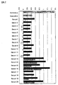
- FIG. 2 shows a comparison of the deduced 14-3-3 protein of G. cydonium [GEODIAge] with the following isoforms: ⁇ rat [RAT-gamma], ⁇ human [HOMO-eta], ⁇ sheep [SHEEP-zeta], Xenopus laevis 14-3-3-related polypeptide [XENLA-D2,214097], rat ⁇ [rat-beta], ⁇ rat [RATtheta], Drosophila melanogaster 14-3-3 protein [DROME-LP], the 14-3-3 protein of Caenorhabditis elegans [CAEL-cds4] and human ⁇ [HOMO-sigma].
- conserveed amino acids in all sequences are shown inverted; those that occur in more than five sequences are shaded. The sequence used for antibody production is labeled (*****).
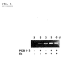
- FIG. 3 shows the effect of PCB 118 on the expression of mRNA from 14-3-3 after incubation of animals with 2 ⁇ g PCB 118 / g wet weight for 0.5 to 6 days.
- the investigations were probed by Northern blotting [14-3-3 probe] (A) and Western blotting [14-3-3 antibody] (B) performed.
- FIG. Figure 5 shows the effect of PCB 118 (2 ⁇ g / g) and 17ss-estradiol (20 ng / g) on the expression of the 14-3-3 gene.
- the two chemicals were added either alone or in combination to the experimental animals ( G. cydonium ).
- the incubation period was limited to 3 or 6 days.
- FIG. 6 shows a schematic representation of the principle of the developed ELISA method for the quantitative determination of 14-3-3 (variant 2).
- FIG. 7 shows the concentration of 14-3-3 protein in bovine serum samples (given is the extinction on the y-axis, which is proportional to the 14-3-3 protein concentration).
- Controls (gray bars): (a) without peptide and without blocking buffer with both antibodies; (b) with peptide and with BSA blocking and with both antibodies. The black ones Bars show the measured values, the white and striated bars the measured values after subtraction the control b. In each case duplicate determinations were carried out. The sera 13, 14, 15, 16, 19 and 20 show an increased 14-3-3 protein level (striated bars).
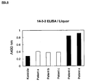
- FIG. 8 shows the concentration of 14-3-3 protein in 5 human CSF cerebrospinalis samples (indicated on the y-axis is the extinction, that of the 14-33 protein concentration is proportional).
- Samples d and e black bars
- the samples a-c white bars
- the gray bar is the negative control.
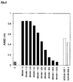
- FIG. 9 shows the measurement values for the concentration of 14-3-3 protein in a dilution series bovine brain extract (black bars; the extinction is indicated on the y-axis, which is proportional to the 14-3-3 protein concentration).
- the specified dilution levels correspond to the following amounts of 14-3-3 protein: 280 ng (dilution stage 1: 5), 140 ng (Dilution stage 1: 10), 70 ng (dilution stage 1: 20), 28 ng (dilution stage 1: 50), 14 ng (dilution stage 1: 100), 7 ng (dilution stage 1: 200), 3.5 ng (dilution stage 1: 400), 1.75 ng (dilution level 1: 800), 0.88 ng (dilution level 1: 1600) and 0.44 ng (dilution level 1: 3200).
- the values of the serum samples shown by two BSE-infected cattle (white bars). Negative control (gray bar).
Landscapes
- Health & Medical Sciences (AREA)
- Life Sciences & Earth Sciences (AREA)
- Engineering & Computer Science (AREA)
- Biomedical Technology (AREA)
- Immunology (AREA)
- Chemical & Material Sciences (AREA)
- Hematology (AREA)
- Urology & Nephrology (AREA)
- Molecular Biology (AREA)
- Cell Biology (AREA)
- Analytical Chemistry (AREA)
- Biochemistry (AREA)
- Biotechnology (AREA)
- Pathology (AREA)
- General Physics & Mathematics (AREA)
- Food Science & Technology (AREA)
- Medicinal Chemistry (AREA)
- Physics & Mathematics (AREA)
- General Health & Medical Sciences (AREA)
- Microbiology (AREA)
- Toxicology (AREA)
- Bioinformatics & Cheminformatics (AREA)
- Tropical Medicine & Parasitology (AREA)
- Neurology (AREA)
- Neurosurgery (AREA)
- Proteomics, Peptides & Aminoacids (AREA)
- Investigating Or Analysing Biological Materials (AREA)
- Measuring Or Testing Involving Enzymes Or Micro-Organisms (AREA)
- Peptides Or Proteins (AREA)
- Steroid Compounds (AREA)
Applications Claiming Priority (3)
| Application Number | Priority Date | Filing Date | Title |
|---|---|---|---|
| DE19810721A DE19810721A1 (de) | 1998-03-12 | 1998-03-12 | Anwendung der 14-3-3 Proteine sowie Verfahren zur schnellen Bestimmung der 14-3-3 Proteine als sensitive Biomarker für Pollution u. a. mit polychlorierten Biphenylen (PCBs) und (Xeno)Östrogenen |
| DE19810721 | 1998-03-12 | ||
| EP99908954A EP0983377B1 (fr) | 1998-03-12 | 1999-03-10 | Procedé de détection de pollution de l'environnement utilisant des proteines 14-3-3 comme biomarqueurs |
Related Parent Applications (2)
| Application Number | Title | Priority Date | Filing Date |
|---|---|---|---|
| EP99908954.3 Division | 1999-03-10 | ||
| EP99908954A Division EP0983377B1 (fr) | 1998-03-12 | 1999-03-10 | Procedé de détection de pollution de l'environnement utilisant des proteines 14-3-3 comme biomarqueurs |
Publications (2)
| Publication Number | Publication Date |
|---|---|
| EP1536235A2 true EP1536235A2 (fr) | 2005-06-01 |
| EP1536235A3 EP1536235A3 (fr) | 2005-09-07 |
Family
ID=7860626
Family Applications (2)
| Application Number | Title | Priority Date | Filing Date |
|---|---|---|---|
| EP99908954A Expired - Lifetime EP0983377B1 (fr) | 1998-03-12 | 1999-03-10 | Procedé de détection de pollution de l'environnement utilisant des proteines 14-3-3 comme biomarqueurs |
| EP04029992A Withdrawn EP1536235A3 (fr) | 1998-03-12 | 1999-03-10 | Procédé de détection de pollution de l'environnement utilisant des protéines 14-3-3 comme biomarqueurs. |
Family Applications Before (1)
| Application Number | Title | Priority Date | Filing Date |
|---|---|---|---|
| EP99908954A Expired - Lifetime EP0983377B1 (fr) | 1998-03-12 | 1999-03-10 | Procedé de détection de pollution de l'environnement utilisant des proteines 14-3-3 comme biomarqueurs |
Country Status (5)
| Country | Link |
|---|---|
| EP (2) | EP0983377B1 (fr) |
| AT (1) | ATE295898T1 (fr) |
| AU (1) | AU2836299A (fr) |
| DE (3) | DE19810721A1 (fr) |
| WO (1) | WO1999046401A2 (fr) |
Cited By (1)
| Publication number | Priority date | Publication date | Assignee | Title |
|---|---|---|---|---|
| KR101933692B1 (ko) | 2017-07-12 | 2018-12-28 | 동국대학교 산학협력단 | 폴리염화바이페닐77 노출 진단용 바이오 마커를 포함하는 진단 키트 |
Families Citing this family (6)
| Publication number | Priority date | Publication date | Assignee | Title |
|---|---|---|---|---|
| DE10131912A1 (de) * | 2001-06-06 | 2002-12-19 | Werner E G Mueller | Verwendung von 14-3-3 Proteinen und Verfahren zu deren Bestimmung |
| PL2021792T3 (pl) | 2006-05-09 | 2013-08-30 | Univ British Columbia | Rozpuszczone białkowe markery zapalenia stawów |
| JP5615181B2 (ja) | 2007-11-27 | 2014-10-29 | ザ・ユニバーシティ・オブ・ブリティッシュ・コロンビア | 関節炎の予防及び治療のための組成物及び方法 |
| PT2768851T (pt) | 2011-10-21 | 2017-09-08 | Augurex Life Sciences Corp | Antigénios derivados de 14-3-3 citrulinada e utilizações dos mesmos no diagnóstico de artrite reumatoide |
| RU2702852C1 (ru) * | 2018-07-20 | 2019-10-11 | федеральное государственное автономное образовательное учреждение высшего образования "Казанский (Приволжский) федеральный университет" (ФГАОУ ВО КФУ) | Способ использования маркерных белков сапробных групп индикаторных организмов для оценки экологического состояния окружающей среды |
| DE102022119709A1 (de) | 2022-08-05 | 2024-02-08 | Mann+Hummel Gmbh | Sekundärelement und filtersystem |
Family Cites Families (2)
| Publication number | Priority date | Publication date | Assignee | Title |
|---|---|---|---|---|
| US5948765A (en) * | 1996-03-15 | 1999-09-07 | Washington University | Inhibition of intracellular signal transduction by 14-3-3-binding peptides |
| US5998149A (en) * | 1996-04-05 | 1999-12-07 | The United States Of America As Represented By The Department Of Health And Human Services | Method of detecting transmissible spongiform encephalopathies |
-
1998
- 1998-03-12 DE DE19810721A patent/DE19810721A1/de not_active Withdrawn
-
1999
- 1999-03-10 AT AT99908954T patent/ATE295898T1/de not_active IP Right Cessation
- 1999-03-10 DE DE59912059T patent/DE59912059D1/de not_active Expired - Fee Related
- 1999-03-10 WO PCT/EP1999/001545 patent/WO1999046401A2/fr not_active Ceased
- 1999-03-10 EP EP99908954A patent/EP0983377B1/fr not_active Expired - Lifetime
- 1999-03-10 DE DE19980374T patent/DE19980374D2/de not_active Expired - Fee Related
- 1999-03-10 EP EP04029992A patent/EP1536235A3/fr not_active Withdrawn
- 1999-03-10 AU AU28362/99A patent/AU2836299A/en not_active Abandoned
Cited By (1)
| Publication number | Priority date | Publication date | Assignee | Title |
|---|---|---|---|---|
| KR101933692B1 (ko) | 2017-07-12 | 2018-12-28 | 동국대학교 산학협력단 | 폴리염화바이페닐77 노출 진단용 바이오 마커를 포함하는 진단 키트 |
Also Published As
| Publication number | Publication date |
|---|---|
| EP1536235A3 (fr) | 2005-09-07 |
| WO1999046401A2 (fr) | 1999-09-16 |
| WO1999046401A3 (fr) | 2000-03-09 |
| AU2836299A (en) | 1999-09-27 |
| EP0983377A1 (fr) | 2000-03-08 |
| EP0983377B1 (fr) | 2005-05-18 |
| DE59912059D1 (de) | 2005-06-23 |
| ATE295898T1 (de) | 2005-06-15 |
| DE19810721A1 (de) | 1999-10-14 |
| DE19980374D2 (de) | 2000-09-28 |
Similar Documents
| Publication | Publication Date | Title |
|---|---|---|
| Ryan et al. | Stress proteins as molecular biomarkers for environmental toxicology | |
| Babić et al. | Embryotoxic and genotoxic effects of sewage effluents in zebrafish embryo using multiple endpoint testing | |
| Kammann et al. | Genotoxic and teratogenic potential of marine sediment extracts investigated with comet assay and zebrafish test | |
| Clayton et al. | Different expression patterns of heat shock proteins hsp 60 and hsp 70 in zebra mussels (Dreissena polymorpha) exposed to copper and tributyltin | |
| Pesce et al. | Integrated survey on toxic effects of lindane on neotropical fish: Corydoras paleatus and Jenynsia multidentata | |
| Vincze et al. | Impact of wastewater on fish health: a case study at the Neckar River (Southern Germany) using biomarkers in caged brown trout as assessment tools | |
| Ben-Khedher et al. | Biochemical effects in crabs (Carcinus maenas) and contamination levels in the Bizerta Lagoon: an integrated approach in biomonitoring of marine complex pollution | |
| Lister et al. | Pollutant resilience in embryos of the Antarctic sea urchin Sterechinus neumayeri reflects maternal antioxidant status | |
| KR100637955B1 (ko) | 수용체의 생물학적 활성을 조절하는 화합물의 능력을예측하는 방법 | |
| Cooke et al. | Fate of prions in soil: detergent extraction of PrP from soils | |
| Wecker et al. | Exposure to the environmentally-persistent insecticide chlordecone induces detoxification genes and causes polyp bail-out in the coral P. damicornis | |
| Kerambrun et al. | A multibiomarker approach in juvenile turbot, Scophthalmus maximus, exposed to contaminated sediments | |
| Leita et al. | Interactions of prion proteins with soil | |
| EP1536235A2 (fr) | Procedé de détection de pollution de l'environnement utilisant des protéines 14-3-3 comme biomarqueurs. | |
| Ünal et al. | Gonadal histology and some biochemical characteristics of Chalcalburnus tarichi (Pallas, 1811) having abnormal gonads | |
| WO2002099434A2 (fr) | Utilisation de proteines 14-3-3 et procede permettant leur determination dans des liquides ou des tissus d'organismes | |
| Kowalska et al. | Interaction of pharmaceutical metabolites with blood proteins and membrane lipids in the view of bioconcentration: a preliminary study based on in vitro assessment | |
| DE60125783T2 (de) | Verfahren zum nachweis von acetylase oder deacetylase-aktivität in einem immunoassay | |
| Reid et al. | Lead tolerance in Aporrectodea rosea earthworms from a clay pigeon shooting site | |
| Pałecz et al. | Na+ K+-ATPase activity as a biomarker of toxaphene toxicity in Unio tumidus | |
| Massicotte et al. | Immunotoxicological response of the earthworm Lumbricus terrestris following exposure to cement kiln dusts | |
| DE69020733T2 (de) | Sublethaler biotest zur bestimmung der umweltqualität. | |
| da Silva Souza et al. | Histological and histochemical analysis of the fat body of Rhinocricus padbergi (Diplopoda) exposed to contaminated industrial soil | |
| DE602004013320T2 (de) | Verfahren zur demonstration eines molekularen ereignisses in einer zelle mittels fluoreszenzmarkerproteinen | |
| Maleri et al. | Photometric application of the MTT-and NRR-assays as biomarkers for the evaluation of cytotoxicity ex vivo in Eisenia andrei |
Legal Events
| Date | Code | Title | Description |
|---|---|---|---|
| PUAI | Public reference made under article 153(3) epc to a published international application that has entered the european phase |
Free format text: ORIGINAL CODE: 0009012 |
|
| 17P | Request for examination filed |
Effective date: 20041217 |
|
| AC | Divisional application: reference to earlier application |
Ref document number: 0983377 Country of ref document: EP Kind code of ref document: P |
|
| AK | Designated contracting states |
Kind code of ref document: A2 Designated state(s): AT BE CH DE DK ES FI FR GB GR IE IT LI NL PT SE |
|
| PUAL | Search report despatched |
Free format text: ORIGINAL CODE: 0009013 |
|
| AK | Designated contracting states |
Kind code of ref document: A3 Designated state(s): AT BE CH DE DK ES FI FR GB GR IE IT LI NL PT SE |
|
| AKX | Designation fees paid |
Designated state(s): AT BE CH DE DK ES FI FR GB GR IE IT LI NL PT SE |
|
| 17Q | First examination report despatched |
Effective date: 20060926 |
|
| STAA | Information on the status of an ep patent application or granted ep patent |
Free format text: STATUS: THE APPLICATION IS DEEMED TO BE WITHDRAWN |
|
| 18D | Application deemed to be withdrawn |
Effective date: 20070207 |