EP1554680B1 - Automatisierte histologische graduierung von tubuli - Google Patents
Automatisierte histologische graduierung von tubuli Download PDFInfo
- Publication number
- EP1554680B1 EP1554680B1 EP03758324A EP03758324A EP1554680B1 EP 1554680 B1 EP1554680 B1 EP 1554680B1 EP 03758324 A EP03758324 A EP 03758324A EP 03758324 A EP03758324 A EP 03758324A EP 1554680 B1 EP1554680 B1 EP 1554680B1
- Authority
- EP
- European Patent Office
- Prior art keywords
- image
- objects
- tubules
- parameter
- pixels
- Prior art date
- Legal status (The legal status is an assumption and is not a legal conclusion. Google has not performed a legal analysis and makes no representation as to the accuracy of the status listed.)
- Expired - Lifetime
Links
Images


Classifications
-
- G—PHYSICS
- G06—COMPUTING OR CALCULATING; COUNTING
- G06T—IMAGE DATA PROCESSING OR GENERATION, IN GENERAL
- G06T7/00—Image analysis
- G06T7/0002—Inspection of images, e.g. flaw detection
- G06T7/0012—Biomedical image inspection
-
- G—PHYSICS
- G06—COMPUTING OR CALCULATING; COUNTING
- G06T—IMAGE DATA PROCESSING OR GENERATION, IN GENERAL
- G06T7/00—Image analysis
- G06T7/60—Analysis of geometric attributes
- G06T7/62—Analysis of geometric attributes of area, perimeter, diameter or volume
-
- G—PHYSICS
- G06—COMPUTING OR CALCULATING; COUNTING
- G06V—IMAGE OR VIDEO RECOGNITION OR UNDERSTANDING
- G06V10/00—Arrangements for image or video recognition or understanding
- G06V10/20—Image preprocessing
- G06V10/28—Quantising the image, e.g. histogram thresholding for discrimination between background and foreground patterns
-
- G—PHYSICS
- G06—COMPUTING OR CALCULATING; COUNTING
- G06V—IMAGE OR VIDEO RECOGNITION OR UNDERSTANDING
- G06V20/00—Scenes; Scene-specific elements
- G06V20/60—Type of objects
- G06V20/69—Microscopic objects, e.g. biological cells or cellular parts
- G06V20/695—Preprocessing, e.g. image segmentation
-
- G—PHYSICS
- G06—COMPUTING OR CALCULATING; COUNTING
- G06T—IMAGE DATA PROCESSING OR GENERATION, IN GENERAL
- G06T2207/00—Indexing scheme for image analysis or image enhancement
- G06T2207/30—Subject of image; Context of image processing
- G06T2207/30004—Biomedical image processing
- G06T2207/30024—Cell structures in vitro; Tissue sections in vitro
Definitions
- This invention relates to a method, an apparatus and a computer program for grading tubules: it is particularly (although not exclusively) relevant to assessment of histological slides to provide clinical information on potentially cancerous tissue such as breast cancer tissue.
- Breast cancer is a common form of female cancer: Once a lesion indicative of breast cancer has been detected, tissue samples are taken and examined by a histopathologist to establish a diagnosis, prognosis and treatment plan.
- pathological analysis of tissue samples is a time consuming and inaccurate process. It entails interpretation of images by human eye, which is highly subjective: it is characterised by considerable inaccuracies in observations of the same samples by different observers and even by the same observer at different times. For example, two different observers assessing the same ten tissue samples may easily give different opinions for three of the slides - 30% error. The problem is exacerbated by heterogeneity, i.e. complexity of some tissue sample features.
- the invention provides the advantage that it is an objective method of grading tubules in a histological slide.
- the step of providing a second image may incorporate:
- the step of providing a third image may comprise thresholding the first image to provide a binary fourth image in which relatively lighter image pixels have a different binary value to that of relatively darker image pixels.
- the step of combining data from the second and third images comprises multiplying pixels in the second image by or ANDing them with respective corresponding pixels located in like positions the third image.
- the step of grading the first image's tubules employs parameter threshold values set to obtain a grading comparable with that obtainable by an appropriate medical expert.
- the present invention provides a method of grading tubules in a first image of a histological slide having the steps of:
- the invention provides the advantage that it is an objective method which grades tubules from a variety of parameters yielding multiple grading from which a median can be derived.
- the present invention provides a computer apparatus for grading tubules in a first image of a histological slide programmed to:
- the present invention provides a computer apparatus for grading tubules in a first image of a histological slide programmed to:
- the present invention provides a computer program for use in grading tubules in a first image of a histological slide containing instructions for controlling a computer apparatus to:
- the present invention provides a computer program for grading tubules in a first image of a histological slide containing instructions for controlling a computer apparatus to:
- the computer program and apparatus aspects of the invention may have preferred features corresponding to those of respective method aspects.
- FIG. 1 there is illustrated a procedure 10 of the invention for assessment of tubule activity in tissue samples presented as histopathological slides of potential carcinomas of the breast.
- the procedure 10 requires data from histological slides in a suitable form. Sections are taken (cut) from breast tissue samples (biopsies) and placed on respective slides. Slides are stained using the staining agent Haemotoxylin & Eosin (H&E), which is the standard stain for delineating tissue and cellular structure. Tissue specimens stained with H&E are used to assess tubule activity.
- image data were obtained by a pathologist using Zeiss Axioskop microscope with a Jenoptiks Progres 3012 digital camera. Image data from a slide is a set of digital images obtained at a linear magnification of 10 (i.e. 10X).
- a pathologist scans the microscope over a slide, and at 10X magnification selects two regions (referred to as tiles) of the slide which appear to be most promising in terms of an analysis to be performed. Both these regions are then photographed using the microscope and digital camera referred to above.
- the digital camera produces for each region a respective digitised image in three colours, i.e. red, green and blue (R, G & B) eight bit values for each pixel and therefore in the range 0 to 255: each image is an electronic equivalent of a tile.
- Three intensity values are obtained for each pixel in a pixel array to provide an image as a combination of R, G and B image planes.
- the image data from the two tiles are stored in a database 12 for later use.
- Tubule activity is determined using a tubule feature detection process 14: this provides a tubule score 18 for input to a diagnostic report at 20.
- the objective of the procedure 10 is to perform an extraction and a count of the tubules present in an image.
- a tubule is an image of a section through a mammary duct produced in the slide production process: it can appear round, oval, cylindrical or irregular depending on the angle of the section to the duct axis and the shape of the duct after sectioning. It appears as a white area surrounded by a dark epithelial layer (or boundary).
- a tubule score is 1, 2 or 3 according to whether the condition is least, moderately or most serious respectively. Tubules may be less in number or absent in images scored 2 or 3 because cancerous cells are invading them.
- the procedure 10 seeks to identify those white areas in an image that are surrounded by a dark epithelial layer: This should exclude fat, which also appears white but tends not to be surrounded by a darker epithelial layer.
- a clinician places a slide under a microscope and examines a region of it at magnification of x10 for indications of tubule activity.
- the prior art manual procedure for scoring tubule activity involves a pathologist subjectively estimating the amount of tubules present in a tissue sample, taking care to ignore those areas considered to be fat cells.
- the process described below in this example replaces the prior art manual procedure with an objective procedure.
- FIG. 2 illustrates various kinds of initial image feature, tubules 51 with pronounced dark boundaries containing one or more holes such as 52, large dark objects 53, fat cells 55 with only very narrow faint boundaries and small dark objects 56 of no interest.
- Step 32 is shown in more detail as a number of constituent steps a) to e) within chain lines 33.
- the objective is to select from the greyscale image 50 only relatively darker pixels and omit relatively lighter pixels.
- An image is obtained from a) which contains relatively darker objects of varying size: the larger of these objects are likely to contain tubules, but others smaller in size are unlikely to do so.
- To implement step a), initially all pixels in the greyscale image 50 are compared with one another to obtain their maximum (Maxg) and minimum (Ming) values: these values are used to compute a parameter P given by: P 12 Maxg - Ming / 100
- Each pixel in the greyscale image 50 is then divided by 255 so that it lies in the range 0 to 1.
- the value P is then used to transform the greyscale image 50 into an output image as shown in Table 1 below: Table 1 Input image pixel value Output image pixel value ⁇ 1/255 0 ⁇ 1/255 AND ⁇ (Maxg - P)/255 Mapped to the range [0,1] > (Maxg - P)/255 1
- step b) the image output from step a) is inverted so that epithelial layers now appear white and background and holes appear black.
- a morphological dilation is applied to the inverted image which thickens and closes small gaps in the white epithelial layers but leaving background and tubule holes unaffected.
- the objective of b) is to close boundaries likely to contain tubules that might have been left open by step a).
- Morphological operations are often used to fuse narrow gaps and eliminate small holes in individual groups of contiguous pixels appearing as blobs in an image. They can be thought of as removal of irregularities or spatial "noise", and they are standard image processing procedures published in Umbaugh S.C., 'Colour vision and image processing', Prentice Hall, 1998.
- the morphological dilation is represented by a disc-shaped structuring element with 5 rows and 5 columns and corresponding (by convention) to a radius R of two pixels: here the expression "disc-shaped” arises from the distribution of 1 s in the element which appear as a rudimentary disc.
- Morphological dilation is an expansion operation: for an original binary image (i.e. having pixel values 1 and 0 only), the expansion operation comprises locating each pixel with value 1 and setting pixels nearby to it also to 1.
- the structuring element defined by Equation (2) above illustrates this: the central 1 indicates a pixel under consideration and 1 s above, below, on either side of it and diagonally near to it indicate pixels in the binary image set to 1; 0s in the structuring element indicate relative positions of pixels that remain unchanged.
- a median filter is applied to the output of step b): the median filter operation selects each pixel in the output of step b) in succession and takes a 3 x 3 array of those pixels centred on the selected pixel; the 3 x 3 array of pixels is then sorted into ascending order of pixel value, i.e. 0s followed by 1s. The median pixel value (fifth of nine) is then taken as the filter output to replace the value of the selected pixel. The filter output will be 0 or 1 according to whether or not the 3 x 3 array has more 0s than 1 s. This is repeated across the image.
- Step c) is desirable (but not essential) - it has the effect of smoothing blobs in the median filtered binary image produced at b).
- the output image from step c) contains image regions or blobs which are white outlines around tubule structures, and the next process at d) is to fill these blobs to make them solid for further processing - the objective is to fill in holes which ideally lie within tubules.
- all pixels contained within each boundary of white pixels are set to the same value as the boundary.
- a hole is a set of background pixels that cannot be reached by filling in the background from the edge of the image: the technique of hole-filling is based on morphological reconstruction as published in Piere Soille, 'Morphological image analysis: Principals and Applications', Springer-Verlag, 1999, pp 173-174.
- Step e) removes small groups of dark pixels such as 56 that are not part of larger objects or boundaries around tubules.
- Steps a) to e) identify blobs in the greyscale image 50 (corresponding either to dark areas with holes inside or to wholly dark objects) that are more likely to be the boundaries of tubules.
- a binary image 60 is shown which is the output of step e): identified blobs are now represented as closed blobs 61 (i.e. containing no holes) of varying sizes of pixel value 1 in a background 62 of 0s.
- Step 34 is a procedure undertaken to extract white regions of the initial greyscale image 50, these regions appearing as relatively high value pixel groups. This step is shown in more detail at f), g) and h) within chain lines 35.
- each pixel value in the initial image 50 is divided by 255 so that it lies in the range 0 to 1, and then the pixel values x in the resulting image (input image in the table below) are remapped to an output image for the selection of light regions in accordance with the following table:
- mapping to the range [0,1] means that: a pixel value x which is BOTH ⁇ (maximum - Q)/255 AND ⁇ 1.0 becomes: x - maximun - Q / 255 / 1 - maximum - Q / 255 ) .
- the resulting output image values are thresholded at g) to produce a binary image: all output image pixel values less than a threshold value of 0.35 are set to zero, and all other pixel values are set to 1.
- the threshold of 0.35 was arrived at experimentally using trial images.
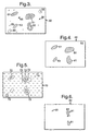
- An image 70 resulting from g) is shown in Figure 5: it is equivalent to the image 50 with "white island” areas 72 shown cross-hatched.
- the image 60 resulting from step e) contains black objects 61 among which there are dark tubule boundaries with respective filled (darkened) inner regions (originally white), all marked as a single object of pixel 1 values and in a background of 0s.
- the image 70 resulting from step g) contains regions 72 of pixel value 1 which corresponding to lighter regions 52/55 of the greyscales image 50.
- the image 70 also contains 0s in positions corresponding to darker regions 51/53 of the image 50.
- a logical AND operation is now carried out between each pixel in the image 60 with the respective corresponding pixel in the same position in the other image 70.
- the AND operation is equivalent in this instance to multiplication because a one-bit AND operation gives the product of two input bits.
- the output of the AND operation is 1 in each case where both ANDed pixels are 1, and is 0 otherwise. Also at h), the result of the AND operation is smoothed.
- Figure 6 shows a binary image 80 resulting from the AND operation between the images 60 and 70 and subsequent smoothing at h).
- the image 80 has 1 s only at locations 81 (shown cross-hatched) corresponding (see image 50) to central holes such as 52 in potential tubules 51. This has the effect of removing objects which correspond in the original greyscales image 50 to isolated white islands (usually fat cells) which are not surrounded by a dark boundary.
- CCL connected component labelling
- a tubule may contain one or more holes, and this is required to be detected to avoid an incorrect tubule count.
- each pixel in the h) image 80 is multiplied by the corresponding pixel in the same location in the CCL of image 60, which is a colour image when displayed on a colour monitor because CCL gives different colours to different objects.
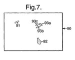
- Figure 7 shows an image 90 resulting from multiplication at 38: the image 90 does not have an image feature corresponding to the lower left hand blob in Figure 4, because it has been eliminated by multiplication by 0s in corresponding locations in the image 80.
- the image 90 retains features 91, 92 and 93a to 93c corresponding to tubule holes such as 52 in the initial image 50.
- the areas of the objects and holes labelled are also obtained using a Matlab function "find" in order to compute the number of pixels per coloured object or hole. This gives the total object area in each case for the four objects 61 in Figure 4 (of which the lower left object 61 is later discarded), and the hole area for each of the holes 91 to 93 in Figure 7.
- the parameters obtained above are used to grade tubules detected in an image, and to derive a score with a value of 1, 2 or 3: the score is derived with reference to prearranged thresholds obtained by analysing a set of images which a pathologist has graded.
- the thresholding operation is performed using independent tests: the results of these tests are subsequently combined to yield a composite tubule score. Since the tests are all independent, any one on its own or combination of two or more can be used to provide a tubule score, but use of all five tests to provide a composite score produces better results.
- the final tubule score for an image is taken as the median value of five scores obtained respectively from the above five tests.
- two results for each test are obtained: the mean of these two results is computed and used with corresponding mean values from the other tests to derive a median value over the test results.
- the invention was tested on a set of 206 images acquired at low magnification XI0: the results were 44 images at score 1, 24 images at score 2, 138 images at score 3 were gathered.
- the thresholds thereafter were set after a thorough observation of the image data obtained (grouping, clustering%) and reaching a compromise. It is important to note that those thresholds may not be appropriate to another set of data because of staining differences. Furthermore those thresholds might be biased due to the fact that the image dataset contained a disproportionately large number of score 3 images.
- Tubule gradings were calculated over 206 available images and compared with a pathologist's scores with the following results:
- the image dataset contained more than three times as many score 3 images (138) compared to score 1 images (44) and more than five times as many compared to score 2 images (24). Best results were obtained for images scored 3 by pathologists with an 73.2% correct classification: this might be attributable to the high number of images available enabling thresholds to be more optimised for score 3 images.
- a classification which is at least 70% correct was achieved for images of all three scores 1, 2 and 3. Since pathologists themselves are not necessarily more than 70% correct, this means that so far as it is possible to do so the invention is verified as regards all three scores.
- the average time to compute results for an image was estimated to be in the range 20 to 40 seconds.
Landscapes
- Engineering & Computer Science (AREA)
- Physics & Mathematics (AREA)
- General Physics & Mathematics (AREA)
- Theoretical Computer Science (AREA)
- Multimedia (AREA)
- Health & Medical Sciences (AREA)
- General Health & Medical Sciences (AREA)
- Computer Vision & Pattern Recognition (AREA)
- Quality & Reliability (AREA)
- Molecular Biology (AREA)
- Nuclear Medicine, Radiotherapy & Molecular Imaging (AREA)
- Medical Informatics (AREA)
- Geometry (AREA)
- Life Sciences & Earth Sciences (AREA)
- Biomedical Technology (AREA)
- Radiology & Medical Imaging (AREA)
- Investigating Or Analysing Biological Materials (AREA)
- Image Processing (AREA)
- Investigating Or Analysing Materials By Optical Means (AREA)
- Fats And Perfumes (AREA)
- Sampling And Sample Adjustment (AREA)
- Breeding Of Plants And Reproduction By Means Of Culturing (AREA)
Claims (27)
- Computerimplementiertes automatisiertes Verfahren zum Klassifizieren von Tubuli in einem ersten Bild (50) eines histologischen Objektträgers, das die folgenden Schritte umfasst:a) Bereitstellen eines zweiten Bildes (60) erster Objekte (61) in dem ersten Bild (50), die ausreichend groß sind und an den Grenzen geeignete Pixelwerteigenschaften besitzen, um möglicherweise als Tubuli zu gelten,b) Bereitstellen eines dritten Bildes (70) zweiter Objekte (72) in dem ersten Bild (50), die Pixelwerteigenschaften von Fett und von Löchern in Tubuli haben,c) Kombinieren von Daten des zweiten und des dritten Bildes (60, 70), um ausgewählte zweite Objekte (81) zu identifizieren, die sich in den ersten Objekten (61) befinden,d) Ausführen eines oder mehrerer der folgenden Schritte:i) Zählen erster Objekte (61) in dem ersten Bild (50), die möglicherweise als Tubuli gelten können, um einen Parameter NOB bereitzustellen,ii) Zählen der ersten Objekte (51), die in sich ausgewählte zweite Objekte (81) besitzen und wahrscheinlich Tubuli sind, um einen Parameter N bereitzustellen,iii) Bestimmen der relativen Flächen ausgewählter zweiter Objekte (81) als Anteile entsprechender erster Objekte (51), in denen sie sich befinden, um Parameter RATIO bereitzustellen,iv) Bestimmen der Gesamtfläche der ausgewählten zweiten Objekte (81) als einen Anteil der Gesamtfläche der ersten Objekte (51), in denen sie sich befinden, um einen Parameter SURF bereitzustellen,v) Bestimmen eines Parameters PERCENT = N/NOB, undvi) Zählen der Anzahl erster Objekte (51), die wenigstens mittelgroße Löcher (52) enthalten, um einen Parameter T bereitzustellen, unde) Klassifizieren der Tubuli (51) des ersten Bildes anhand des einen oder der mehreren Parameter wie oben angegeben in Bezug auf Parameter-Schwellenwerte.
- Verfahren nach Anspruch 1, dadurch gekennzeichnet, dass der Schritt des Bereitstellens des zweiten Bildes (60) das Füllen von Löchern (52) in den Objekten (51) des ersten Bildes umfasst, um erste Objekte (61) des zweiten Bildes ohne Löcher bereitzustellen.
- Verfahren nach Anspruch 1 oder 2, dadurch gekennzeichnet, dass ein Loch als eine Gruppe von Hintergrundpixeln definiert ist, die nicht durch Füllen des Hintergrundes ausgehend von einer Kante des ersten Bildes (51) erreicht werden können.
- Verfahren nach Anspruch 1, 2 oder 3, dadurch gekennzeichnet, dass es den Schritt des Verarbeitens des dritten Bildes (70) umfasst, um zwischen ersten Objekten (51), die einzelne ausgewählte zweite Objekte (92) enthalten, und ersten Objekten (51), die mehrere ausgewählte zweite Objekte (93a, 93b, 93c) enthalten, zu unterscheiden.
- Verfahren nach Anspruch 1, dadurch gekennzeichnet, dass der Schritt des Bereitstellens eines zweiten Bildes (60) umfasst:a) Anwenden von Schwellenwerten auf das erste Bild (50), um ein viertes Bild bereitzustellen, indem verhältnismäßig dunklere Bildpixel beibehalten werden und verhältnismäßig hellere Bildpixel verworfen werden,b) Verarbeiten des vierten Bildes durch aufeinander folgendes Anwenden der folgenden Schritte, wovon der erste Schritt auf das vierte Bild angewendet wird und jeder Schritt nach dem ersten Schritt auf das Ergebnis des jeweils direkt vorhergehenden Schrittes angewendet wird: Inversion, morphologische Dilatation, Medianfilterung, Lochbefüllung und morphologische Öffnung.
- Verfahren nach Anspruch 1, dadurch gekennzeichnet, dass der Schritt des Bereitstellens eines dritten Bildes (70) das Anwenden von Schwellenwerten auf das erste Bild (50) umfasst, um ein binäres viertes Bild bereitzustellen, in dem verhältnismäßig hellere Bildpixel einen anderen binären Wert als verhältnismäßig dunklere Bildpixel haben.
- Verfahren nach Anspruch 1, dadurch gekennzeichnet, dass der Schritt des Kombinierens von Daten aus dem zweiten und dem dritten Bild (60, 70) das Multiplizieren von Pixeln in dem zweiten Bild (60) mit entsprechenden Pixeln, die sich an ähnlichen Positionen im dritten Bild (70) befinden, oder die Anwendung einer UND-Verknüpfung auf diese umfasst.
- Verfahren nach Anspruch 1, dadurch gekennzeichnet, dass der Schritt des Klassifizierens der Tubuli (51) des ersten Bildes Parameter-Schwellenwerte verwendet, die so gesetzt sind, dass eine Klassifizierung erhalten wird, die mit jener vergleichbar ist, die durch einen geeigneten medizinischen Experten erhalten werden kann.
- Verfahren nach Anspruch 1, dadurch gekennzeichnet, dass im Schritt d) alle Parameter NOB, N, RATIO, SURF, PERCENT und T erhalten werden und im Schritt e) diese Parameter verwendet werden, um die Tubuli (51) des ersten Bildes zu klassifizieren.
- Computervorrichtung zum Klassifizieren von Tubuli in einem ersten Bild (50) eines histologischen Objektträgers, wobei die Computervorrichtung so programmiert ist, dass sie:a) ein zweites Bild (60) erster Objekte (61) in dem ersten Bild (50) berechnet, die ausreichend groß sind und an den Grenzen geeignete Pixelwerteigenschaften haben, um möglicherweise als Tubuli zu gelten,b) ein drittes Bild (70) zweiter Objekte (72) in dem ersten Bild (50) berechnet, das Pixelwerteigenschaften von Fett und von Löchern in Tubuli besitzt,c) Daten aus dem zweiten und dem dritten Bild (60, 70) kombiniert, um ausgewählte zweite Objekte (61), die sich in den ersten Objekten (61) befinden, zu identifizieren,d) einen oder mehrere der folgenden Schritte implementiert:i) Zählen erster Objekte (61) in dem ersten Bild (50), die möglicherweise als Tubuli gelten können, um einen Parameter NOB bereitzustellen,ii) Zählen der ersten Objekte (51), die in sich ausgewählte zweite Objekte (81) besitzen und wahrscheinlich Tubuli sind, um einen Parameter N bereitzustellen,iii) Bestimmen der relativen Flächen ausgewählter zweiter Objekte (81) als Anteile entsprechender erster Objekte (51), in denen sie sich befinden, um Parameter RATIO bereitzustellen,iv) Bestimmen der Gesamtfläche der ausgewählten zweiten Objekte (81) als einen Anteil der Gesamtfläche der ersten Objekte (51), in denen sie sich befinden, um einen Parameter SURF bereitzustellen,v) Bestimmen eines Parameters PERCENT = N/NOB undvi) Zählen der Anzahl erster Objekte (51), die wenigstens mittelgroße Löcher (52) enthalten, um einen Parameter T bereitzustellen, unde) die Tubuli (51) des ersten Bildes anhand des einen oder der mehreren Parameter wie oben genannt mit Bezug auf Parameter-Schwellenwerte klassifiziert.
- Computervorrichtung nach Anspruch 10, dadurch gekennzeichnet, dass sie so programmiert ist, dass sie das zweite Bild (60) durch Füllen von Löchern (52) in den ersten Bildobjekten (51) bereitstellt, um erste Objekte (61) des zweiten Bildes ohne Löcher bereitzustellen.
- Computervorrichtung nach Anspruch 10 oder 11, dadurch gekennzeichnet, dass sie so programmiert ist, dass sie ein Loch (52) als eine Menge von Hintergrundpixeln definiert, die nicht durch Füllen des Hintergrundes ausgehend von einer Kante des ersten Bildes (51) erreicht werden können.
- Computervorrichtung nach Anspruch 10, 11 oder 12, dadurch gekennzeichnet, dass sie so programmiert ist, dass sie das dritte Bild (70) verarbeitet, um zwischen ersten Objekten (51), die einzelne ausgewählte zweite Objekte (92) enthalten, und ersten Objekten (51), die mehrere ausgewählte zweite Objekte (93a, 93b, 93c) enthalten; zu unterscheiden.
- Computervorrichtung nach Anspruch 10, dadurch gekennzeichnet, dass sie so programmiert ist, dass sie ein zweites Bild (60) dadurch schafft, dass:a) auf das erste Bild (50) Schwellenwerte angewendet werden, um ein viertes Bild bereitzustellen, indem verhältnismäßig dunklere Bildpixel beibehalten werden und verhältnismäßig hellere Bildpixel verworfen werden,b) das vierte Bild durch aufeinander folgendes Anwenden der folgenden Schritte verarbeitet wird, wovon der erste Schritt auf das vierte Bild angewendet wird und jeder Schritt nach dem ersten Schritt auf das Ergebnis des jeweils unmittelbar vorhergehenden Schrittes angewendet wird: Inversion, morphologische Dilatation, Medianfilterung, Lochbefüllung und morphologische Öffnung.
- Computervorrichtung nach Anspruch 10, dadurch gekennzeichnet, dass sie so programmiert ist, dass sie ein drittes Bild (70) durch Anwenden von Schwellenwerten auf das erste Bild (50) bereitstellt, um ein binäres viertes Bild bereitzustellen, in dem verhältnismäßig hellere Bildpixel einen anderen binären Wert haben als verhältnismäßig dunklere Bildpixel.
- Computervorrichtung nach Anspruch 10, dadurch gekennzeichnet, dass sie so programmiert ist, dass sie die Daten aus dem zweiten und dem dritten Bild (60, 70) dadurch kombiniert, dass sie Pixel im zweiten Bild (60) mit entsprechenden Pixeln, die sich an ähnlichen Positionen im dritten Bild (70) befinden, multipliziert oder auf diese eine UND-Verknüpfung anwendet.
- Computervorrichtung nach Anspruch 10, dadurch gekennzeichnet, dass sie so programmiert ist, dass sie die Tubuli (51) des ersten Bildes mit Parameter-Schwellenwerten klassifiziert, die so gesetzt sind, dass eine Klassifizierung erhalten wird, die mit jener vergleichbar ist, die durch einen geeigneten medizinischen Experten erhalten werden kann.
- Computervorrichtung nach Anspruch 10, dadurch gekennzeichnet, dass sie so programmiert ist, dass alle Parameter NOB, N, RATIO, SURF, PERCENT und T erhalten werden, und dass diese Parameter verwendet werden, um die Tubuli (51) des ersten Bildes zu klassifizieren.
- Computerprogramm für die Klassifizierung von Tubuli in einem ersten Bild (50) eines histologischen Objektträgers, wobei das Computerprogramm Befehle enthält, um eine Computervorrichtung zu steuern, damit siea) ein zweites Bild (60) erster Objekte (61) in dem ersten Bild (50) berechnet, die ausreichend groß sind und an den Grenzen geeignete Pixelwerteigenschaften haben, damit sie möglicherweise als Tubuli gelten können,b) ein drittes Bild (70) zweiter Objekte (72) in dem ersten Bild (50) berechnet, das Pixelwerteigenschaften von Fett und von Löchern in Tubuli besitzt,c) Daten aus dem zweiten und dem dritten Bild (60, 70) kombiniert, um ausgewählte zweite Objekte (61), die sich in den ersten Objekten (61) befinden, zu identifizieren,d) einen oder mehrere der folgenden Schritte implementiert:i) Zählen erster Objekte (61) in dem ersten Bild (50), die möglicherweise als Tubuli gelten können, um einen Parameter NOB bereitzustellen,ii) Zählen der ersten Objekte (51), die in sich ausgewählte zweite Objekte (81) besitzen und wahrscheinlich Tubuli sind, um einen Parameter N bereitzustellen,iii) Bestimmen der relativen Flächen ausgewählter zweiter Objekte (81) als Anteile entsprechender erster Objekte (51), in denen sie sich befinden, um Parameter RATIO bereitzustellen,iv) Bestimmen der Gesamtfläche der ausgewählten zweiten Objekte (81) als einen Anteil der Gesamtfläche der ersten Objekte (51), in denen sie sich befinden, um einen Parameter SURF bereitzustellen,v) Bestimmen eines Parameters PERCENT = N/NOB undvi) Zählen der Anzahl erster Objekte (51), die wenigstens mittelgroße Löcher (52) enthalten, um einen Parameter T bereitzustellen, unde) die Tubuli (51) des ersten Bildes anhand des einen oder der mehreren Parameter wie oben genannt mit Bezug auf Parameter-Schwellenwerte klassifiziert.
- Computerprogramm nach Anspruch 19, dadurch gekennzeichnet, dass sie Befehle enthält, um das zweite Bild (60) durch Füllen von Löchern (52) in Objekten (51) des ersten Bildes bereitzustellen, um erste Objekte (61) des zweiten Bildes ohne Löcher bereitzustellen.
- Computerprogramm nach Anspruch 19 oder 20, dadurch gekennzeichnet, dass es Befehle enthält, um ein Loch (52) als eine Menge von Hintergrundpixeln zu definieren, die nicht durch Füllen des Hintergrundes ausgehend von einer Kante des ersten Bildes (51) erreicht werden können.
- Computerprogramm nach Anspruch 19, 20 oder 21, dadurch gekennzeichnet, dass es Befehle enthält, um das dritte Bild (70) zu verarbeiten, um zwischen ersten Objekten (51), die einzelne ausgewählte zweite Objekte (92) enthalten, und ersten Objekten (51), die mehrere ausgewählte zweite Objekte (93a, 93b, 93c) enthalten, zu unterscheiden.
- Computerprogramm nach Anspruch 19, dadurch gekennzeichnet, dass es Befehle enthält, um ein zweites Bild (60) dadurch bereitzustellen, dass:a) auf das erste Bild (50) Schwellenwerte angewendet werden, um ein viertes Bild bereitzustellen, indem verhältnismäßig dunklere Bildpixel beibehalten werden und verhältnismäßig hellere Bildpixel verworfen werden,b) das vierte Bild durch aufeinander folgendes Anwenden der folgenden Schritte verarbeitet wird, wovon der erste Schritt auf das vierte Bild angewendet wird und jeder Schritt nach dem ersten Schritt auf das Ergebnis des jeweils unmittelbar vorhergehenden Schrittes angewendet wird: Inversion, morphologische Dilatation, Medianfilterung, Lochbefüllung und morphologische Öffnung.
- Computerprogramm nach Anspruch 19, dadurch gekennzeichnet, dass es Befehle enthält, um ein drittes Bild (70) durch Anwenden von Schwellenwerten auf das erste Bild (50) bereitzustellen, um ein binäres viertes Bild bereitzustellen, in dem verhältnismäßig hellere Bildpixel einen anderen binären Wert als verhältnismäßig dunklere Bildpixel haben.
- Computerprogramm nach Anspruch 19, dadurch gekennzeichnet, dass es Befehle enthält, um Daten aus dem zweiten und dem dritten Bild (60, 70) dadurch zu kombinieren, dass Pixel in dem zweiten Bild (60) mit jeweils entsprechenden Pixeln, die sich an ähnlichen Positionen in dem dritten Bild (70) befinden, multipliziert werden oder auf diese eine UND-Verknüpfung angewendet wird.
- Computerprogramm nach Anspruch 19, dadurch gekennzeichnet, dass es Befehle enthält, um die Tubuli (51) des ersten Bildes mit Parameter-Schwellenwerten zu klassifizieren, die so gesetzt sind, dass eine Klassifizierung erhalten wird, die mit jener vergleichbar ist, die durch einen geeigneten medizinischen Experten erhalten werden kann.
- Computerprogramm nach Anspruch 19, dadurch gekennzeichnet, dass es Befehle enthält, um eine Computervorrichtung so zu steuern, dass alle Parameter NOB, N, RATIO, SURF, PERCENT und T erhalten werden, und dass diese Parameter verwendet werden, um die Tubuli (51) des ersten Bildes zu klassifizieren.
Applications Claiming Priority (3)
| Application Number | Priority Date | Filing Date | Title |
|---|---|---|---|
| GB0224626 | 2002-10-23 | ||
| GBGB0224626.2A GB0224626D0 (en) | 2002-10-23 | 2002-10-23 | Tubule grading |
| PCT/GB2003/004527 WO2004038633A1 (en) | 2002-10-23 | 2003-10-20 | Automated histological grading of tubules |
Publications (2)
| Publication Number | Publication Date |
|---|---|
| EP1554680A1 EP1554680A1 (de) | 2005-07-20 |
| EP1554680B1 true EP1554680B1 (de) | 2006-11-29 |
Family
ID=9946400
Family Applications (1)
| Application Number | Title | Priority Date | Filing Date |
|---|---|---|---|
| EP03758324A Expired - Lifetime EP1554680B1 (de) | 2002-10-23 | 2003-10-20 | Automatisierte histologische graduierung von tubuli |
Country Status (8)
| Country | Link |
|---|---|
| US (1) | US8229674B2 (de) |
| EP (1) | EP1554680B1 (de) |
| JP (1) | JP4452624B2 (de) |
| AT (1) | ATE347150T1 (de) |
| AU (1) | AU2003274337A1 (de) |
| DE (1) | DE60310120T2 (de) |
| GB (1) | GB0224626D0 (de) |
| WO (1) | WO2004038633A1 (de) |
Families Citing this family (7)
| Publication number | Priority date | Publication date | Assignee | Title |
|---|---|---|---|---|
| GB2430026A (en) | 2005-09-09 | 2007-03-14 | Qinetiq Ltd | Automated selection of image regions |
| JP5543310B2 (ja) * | 2010-09-29 | 2014-07-09 | 富士フイルム株式会社 | イムノクロマトグラフ検査方法および装置 |
| JP5543888B2 (ja) * | 2010-09-29 | 2014-07-09 | 富士フイルム株式会社 | イムノクロマトグラフ検査方法および装置 |
| JP5811923B2 (ja) * | 2012-03-28 | 2015-11-11 | 富士通株式会社 | 情報処理装置、画像処理方法およびプログラム |
| JP5834253B2 (ja) | 2013-03-27 | 2015-12-16 | パナソニックIpマネジメント株式会社 | 画像処理装置、画像処理方法、及び画像処理プログラム |
| JP5849206B2 (ja) * | 2013-03-27 | 2016-01-27 | パナソニックIpマネジメント株式会社 | 画像処理装置、画像処理方法、及び画像処理プログラム |
| US9298968B1 (en) * | 2014-09-12 | 2016-03-29 | Flagship Biosciences, Inc. | Digital image analysis of inflammatory cells and mediators of inflammation |
Family Cites Families (4)
| Publication number | Priority date | Publication date | Assignee | Title |
|---|---|---|---|---|
| US5544650A (en) * | 1988-04-08 | 1996-08-13 | Neuromedical Systems, Inc. | Automated specimen classification system and method |
| US5257182B1 (en) | 1991-01-29 | 1996-05-07 | Neuromedical Systems Inc | Morphological classification system and method |
| US6031930A (en) * | 1996-08-23 | 2000-02-29 | Bacus Research Laboratories, Inc. | Method and apparatus for testing a progression of neoplasia including cancer chemoprevention testing |
| GB2395263A (en) * | 2002-11-12 | 2004-05-19 | Qinetiq Ltd | Image analysis |
-
2002
- 2002-10-23 GB GBGB0224626.2A patent/GB0224626D0/en not_active Ceased
-
2003
- 2003-10-20 JP JP2004546146A patent/JP4452624B2/ja not_active Expired - Fee Related
- 2003-10-20 AT AT03758324T patent/ATE347150T1/de not_active IP Right Cessation
- 2003-10-20 WO PCT/GB2003/004527 patent/WO2004038633A1/en not_active Ceased
- 2003-10-20 DE DE60310120T patent/DE60310120T2/de not_active Expired - Lifetime
- 2003-10-20 AU AU2003274337A patent/AU2003274337A1/en not_active Abandoned
- 2003-10-20 US US10/529,509 patent/US8229674B2/en not_active Expired - Fee Related
- 2003-10-20 EP EP03758324A patent/EP1554680B1/de not_active Expired - Lifetime
Also Published As
| Publication number | Publication date |
|---|---|
| US8229674B2 (en) | 2012-07-24 |
| AU2003274337A1 (en) | 2004-05-13 |
| DE60310120D1 (de) | 2007-01-11 |
| ATE347150T1 (de) | 2006-12-15 |
| JP4452624B2 (ja) | 2010-04-21 |
| JP2006507486A (ja) | 2006-03-02 |
| EP1554680A1 (de) | 2005-07-20 |
| WO2004038633A1 (en) | 2004-05-06 |
| GB0224626D0 (en) | 2002-12-04 |
| US20060036369A1 (en) | 2006-02-16 |
| DE60310120T2 (de) | 2007-10-04 |
Similar Documents
| Publication | Publication Date | Title |
|---|---|---|
| CN118115466B (zh) | 一种眼底伪病灶检测方法 | |
| US7555161B2 (en) | Image analysis | |
| US7079675B2 (en) | Histological assessment | |
| US5939278A (en) | Automated histological specimen classification system and method | |
| JP4864857B2 (ja) | 有糸分裂活性の測定 | |
| Hazra et al. | Brain tumor detection based on segmentation using MATLAB | |
| Agarwal et al. | Automatic glaucoma detection using adaptive threshold based technique in fundus image | |
| AU2003207787A2 (en) | Image processing using measures of similarity | |
| EP1579366B1 (de) | Histologische bewertung des nuklearpleomorphismus | |
| WO2010131944A2 (en) | Apparatus for monitoring and grading diabetic retinopathy | |
| Subramanian et al. | Diabetic retinopathy-feature extraction and classification using adaptive super pixel algorithm | |
| EP1554680B1 (de) | Automatisierte histologische graduierung von tubuli | |
| CN120182231A (zh) | 基于卷积神经网络的血液细胞图像检测方法及系统 | |
| JP4981112B2 (ja) | 組織学的アセスメント | |
| Kinnard et al. | Automatic segmentation of mammographic masses using fuzzy shadow and maximum-likelihood analysis | |
| LU507803B1 (de) | Ein intelligentes vorhersageverfahren und -system für das risiko einer nsclc-lymphknotenmetastasierung | |
| Lakshmi et al. | MATLAB-BASED IMAGE PROCESSING SOLUTIONS FOR ENHANCED DIABETIC RETINOPATHY DETECTION | |
| MOROZ-DUBENCO | COMPARISON OF GRADIENT-BASED EDGE DETECTORS APPLIED ON MAMMOGRAMS. | |
| Popescu et al. | Computer aided diagnosis in mammography based on fractal analysis | |
| Milenovic et al. | Cell classification for diagnostic of reactive histocytic hyperplasia using neural networks | |
| ChiangHau | Algorithms for Tissue Image Analysis using Multifractal Techniques | |
| Ometto | Global tortuosity estimation of retinal vessel network in wide-field images taken from infants with retinopathy of prematurity |
Legal Events
| Date | Code | Title | Description |
|---|---|---|---|
| PUAI | Public reference made under article 153(3) epc to a published international application that has entered the european phase |
Free format text: ORIGINAL CODE: 0009012 |
|
| 17P | Request for examination filed |
Effective date: 20050329 |
|
| AK | Designated contracting states |
Kind code of ref document: A1 Designated state(s): AT BE BG CH CY CZ DE DK EE ES FI FR GB GR HU IE IT LI LU MC NL PT RO SE SI SK TR |
|
| AX | Request for extension of the european patent |
Extension state: AL LT LV MK |
|
| DAX | Request for extension of the european patent (deleted) | ||
| GRAP | Despatch of communication of intention to grant a patent |
Free format text: ORIGINAL CODE: EPIDOSNIGR1 |
|
| RIC1 | Information provided on ipc code assigned before grant |
Ipc: G06T 7/00 20060101AFI20060411BHEP |
|
| GRAS | Grant fee paid |
Free format text: ORIGINAL CODE: EPIDOSNIGR3 |
|
| GRAL | Information related to payment of fee for publishing/printing deleted |
Free format text: ORIGINAL CODE: EPIDOSDIGR3 |
|
| GRAS | Grant fee paid |
Free format text: ORIGINAL CODE: EPIDOSNIGR3 |
|
| GRAA | (expected) grant |
Free format text: ORIGINAL CODE: 0009210 |
|
| AK | Designated contracting states |
Kind code of ref document: B1 Designated state(s): AT BE BG CH CY CZ DE DK EE ES FI FR GB GR HU IE IT LI LU MC NL PT RO SE SI SK TR |
|
| PG25 | Lapsed in a contracting state [announced via postgrant information from national office to epo] |
Ref country code: IT Free format text: LAPSE BECAUSE OF FAILURE TO SUBMIT A TRANSLATION OF THE DESCRIPTION OR TO PAY THE FEE WITHIN THE PRESCRIBED TIME-LIMIT;WARNING: LAPSES OF ITALIAN PATENTS WITH EFFECTIVE DATE BEFORE 2007 MAY HAVE OCCURRED AT ANY TIME BEFORE 2007. THE CORRECT EFFECTIVE DATE MAY BE DIFFERENT FROM THE ONE RECORDED. Effective date: 20061129 Ref country code: CZ Free format text: LAPSE BECAUSE OF FAILURE TO SUBMIT A TRANSLATION OF THE DESCRIPTION OR TO PAY THE FEE WITHIN THE PRESCRIBED TIME-LIMIT Effective date: 20061129 Ref country code: SI Free format text: LAPSE BECAUSE OF FAILURE TO SUBMIT A TRANSLATION OF THE DESCRIPTION OR TO PAY THE FEE WITHIN THE PRESCRIBED TIME-LIMIT Effective date: 20061129 Ref country code: FI Free format text: LAPSE BECAUSE OF FAILURE TO SUBMIT A TRANSLATION OF THE DESCRIPTION OR TO PAY THE FEE WITHIN THE PRESCRIBED TIME-LIMIT Effective date: 20061129 Ref country code: AT Free format text: LAPSE BECAUSE OF FAILURE TO SUBMIT A TRANSLATION OF THE DESCRIPTION OR TO PAY THE FEE WITHIN THE PRESCRIBED TIME-LIMIT Effective date: 20061129 Ref country code: RO Free format text: LAPSE BECAUSE OF FAILURE TO SUBMIT A TRANSLATION OF THE DESCRIPTION OR TO PAY THE FEE WITHIN THE PRESCRIBED TIME-LIMIT Effective date: 20061129 Ref country code: SK Free format text: LAPSE BECAUSE OF FAILURE TO SUBMIT A TRANSLATION OF THE DESCRIPTION OR TO PAY THE FEE WITHIN THE PRESCRIBED TIME-LIMIT Effective date: 20061129 Ref country code: BE Free format text: LAPSE BECAUSE OF FAILURE TO SUBMIT A TRANSLATION OF THE DESCRIPTION OR TO PAY THE FEE WITHIN THE PRESCRIBED TIME-LIMIT Effective date: 20061129 |
|
| REG | Reference to a national code |
Ref country code: GB Ref legal event code: FG4D |
|
| REG | Reference to a national code |
Ref country code: CH Ref legal event code: EP |
|
| REG | Reference to a national code |
Ref country code: IE Ref legal event code: FG4D |
|
| REF | Corresponds to: |
Ref document number: 60310120 Country of ref document: DE Date of ref document: 20070111 Kind code of ref document: P |
|
| PG25 | Lapsed in a contracting state [announced via postgrant information from national office to epo] |
Ref country code: SE Free format text: LAPSE BECAUSE OF FAILURE TO SUBMIT A TRANSLATION OF THE DESCRIPTION OR TO PAY THE FEE WITHIN THE PRESCRIBED TIME-LIMIT Effective date: 20070228 Ref country code: DK Free format text: LAPSE BECAUSE OF FAILURE TO SUBMIT A TRANSLATION OF THE DESCRIPTION OR TO PAY THE FEE WITHIN THE PRESCRIBED TIME-LIMIT Effective date: 20070228 Ref country code: BG Free format text: LAPSE BECAUSE OF FAILURE TO SUBMIT A TRANSLATION OF THE DESCRIPTION OR TO PAY THE FEE WITHIN THE PRESCRIBED TIME-LIMIT Effective date: 20070228 |
|
| PG25 | Lapsed in a contracting state [announced via postgrant information from national office to epo] |
Ref country code: ES Free format text: LAPSE BECAUSE OF FAILURE TO SUBMIT A TRANSLATION OF THE DESCRIPTION OR TO PAY THE FEE WITHIN THE PRESCRIBED TIME-LIMIT Effective date: 20070312 |
|
| REG | Reference to a national code |
Ref country code: CH Ref legal event code: NV Representative=s name: E. BLUM & CO. AG PATENT- UND MARKENANWAELTE VSP |
|
| PG25 | Lapsed in a contracting state [announced via postgrant information from national office to epo] |
Ref country code: PT Free format text: LAPSE BECAUSE OF FAILURE TO SUBMIT A TRANSLATION OF THE DESCRIPTION OR TO PAY THE FEE WITHIN THE PRESCRIBED TIME-LIMIT Effective date: 20070430 |
|
| ET | Fr: translation filed | ||
| PLBE | No opposition filed within time limit |
Free format text: ORIGINAL CODE: 0009261 |
|
| STAA | Information on the status of an ep patent application or granted ep patent |
Free format text: STATUS: NO OPPOSITION FILED WITHIN TIME LIMIT |
|
| 26N | No opposition filed |
Effective date: 20070830 |
|
| PG25 | Lapsed in a contracting state [announced via postgrant information from national office to epo] |
Ref country code: GR Free format text: LAPSE BECAUSE OF FAILURE TO SUBMIT A TRANSLATION OF THE DESCRIPTION OR TO PAY THE FEE WITHIN THE PRESCRIBED TIME-LIMIT Effective date: 20070301 |
|
| PG25 | Lapsed in a contracting state [announced via postgrant information from national office to epo] |
Ref country code: MC Free format text: LAPSE BECAUSE OF NON-PAYMENT OF DUE FEES Effective date: 20071031 |
|
| PG25 | Lapsed in a contracting state [announced via postgrant information from national office to epo] |
Ref country code: EE Free format text: LAPSE BECAUSE OF FAILURE TO SUBMIT A TRANSLATION OF THE DESCRIPTION OR TO PAY THE FEE WITHIN THE PRESCRIBED TIME-LIMIT Effective date: 20061129 |
|
| PG25 | Lapsed in a contracting state [announced via postgrant information from national office to epo] |
Ref country code: CY Free format text: LAPSE BECAUSE OF FAILURE TO SUBMIT A TRANSLATION OF THE DESCRIPTION OR TO PAY THE FEE WITHIN THE PRESCRIBED TIME-LIMIT Effective date: 20061129 Ref country code: LU Free format text: LAPSE BECAUSE OF NON-PAYMENT OF DUE FEES Effective date: 20071020 |
|
| PG25 | Lapsed in a contracting state [announced via postgrant information from national office to epo] |
Ref country code: TR Free format text: LAPSE BECAUSE OF FAILURE TO SUBMIT A TRANSLATION OF THE DESCRIPTION OR TO PAY THE FEE WITHIN THE PRESCRIBED TIME-LIMIT Effective date: 20061129 Ref country code: HU Free format text: LAPSE BECAUSE OF FAILURE TO SUBMIT A TRANSLATION OF THE DESCRIPTION OR TO PAY THE FEE WITHIN THE PRESCRIBED TIME-LIMIT Effective date: 20070530 |
|
| PGFP | Annual fee paid to national office [announced via postgrant information from national office to epo] |
Ref country code: IE Payment date: 20091028 Year of fee payment: 7 |
|
| PGFP | Annual fee paid to national office [announced via postgrant information from national office to epo] |
Ref country code: NL Payment date: 20091016 Year of fee payment: 7 |
|
| REG | Reference to a national code |
Ref country code: NL Ref legal event code: V1 Effective date: 20110501 |
|
| PG25 | Lapsed in a contracting state [announced via postgrant information from national office to epo] |
Ref country code: NL Free format text: LAPSE BECAUSE OF NON-PAYMENT OF DUE FEES Effective date: 20110501 |
|
| PG25 | Lapsed in a contracting state [announced via postgrant information from national office to epo] |
Ref country code: IE Free format text: LAPSE BECAUSE OF NON-PAYMENT OF DUE FEES Effective date: 20101020 |
|
| PGFP | Annual fee paid to national office [announced via postgrant information from national office to epo] |
Ref country code: FR Payment date: 20121031 Year of fee payment: 10 Ref country code: CH Payment date: 20121023 Year of fee payment: 10 Ref country code: DE Payment date: 20121023 Year of fee payment: 10 |
|
| PGFP | Annual fee paid to national office [announced via postgrant information from national office to epo] |
Ref country code: GB Payment date: 20121019 Year of fee payment: 10 |
|
| REG | Reference to a national code |
Ref country code: CH Ref legal event code: PL |
|
| GBPC | Gb: european patent ceased through non-payment of renewal fee |
Effective date: 20131020 |
|
| PG25 | Lapsed in a contracting state [announced via postgrant information from national office to epo] |
Ref country code: LI Free format text: LAPSE BECAUSE OF NON-PAYMENT OF DUE FEES Effective date: 20131031 Ref country code: CH Free format text: LAPSE BECAUSE OF NON-PAYMENT OF DUE FEES Effective date: 20131031 Ref country code: GB Free format text: LAPSE BECAUSE OF NON-PAYMENT OF DUE FEES Effective date: 20131020 |
|
| REG | Reference to a national code |
Ref country code: DE Ref legal event code: R119 Ref document number: 60310120 Country of ref document: DE Effective date: 20140501 |
|
| REG | Reference to a national code |
Ref country code: FR Ref legal event code: ST Effective date: 20140630 |
|
| PG25 | Lapsed in a contracting state [announced via postgrant information from national office to epo] |
Ref country code: FR Free format text: LAPSE BECAUSE OF NON-PAYMENT OF DUE FEES Effective date: 20131031 Ref country code: DE Free format text: LAPSE BECAUSE OF NON-PAYMENT OF DUE FEES Effective date: 20140501 |