EP1556071B1 - Verfahren zur prävention von tumorrezidiven durch blockade von tgf-beta - Google Patents
Verfahren zur prävention von tumorrezidiven durch blockade von tgf-beta Download PDFInfo
- Publication number
- EP1556071B1 EP1556071B1 EP03779310A EP03779310A EP1556071B1 EP 1556071 B1 EP1556071 B1 EP 1556071B1 EP 03779310 A EP03779310 A EP 03779310A EP 03779310 A EP03779310 A EP 03779310A EP 1556071 B1 EP1556071 B1 EP 1556071B1
- Authority
- EP
- European Patent Office
- Prior art keywords
- tgf
- tumor
- cells
- antibody
- cell
- Prior art date
- Legal status (The legal status is an assumption and is not a legal conclusion. Google has not performed a legal analysis and makes no representation as to the accuracy of the status listed.)
- Expired - Lifetime
Links
Images



Classifications
-
- G—PHYSICS
- G01—MEASURING; TESTING
- G01N—INVESTIGATING OR ANALYSING MATERIALS BY DETERMINING THEIR CHEMICAL OR PHYSICAL PROPERTIES
- G01N33/00—Investigating or analysing materials by specific methods not covered by groups G01N1/00 - G01N31/00
- G01N33/48—Biological material, e.g. blood, urine; Haemocytometers
- G01N33/50—Chemical analysis of biological material, e.g. blood, urine; Testing involving biospecific ligand binding methods; Immunological testing
- G01N33/74—Chemical analysis of biological material, e.g. blood, urine; Testing involving biospecific ligand binding methods; Immunological testing involving hormones or other non-cytokine intercellular protein regulatory factors such as growth factors, including receptors to hormones and growth factors
-
- A—HUMAN NECESSITIES
- A61—MEDICAL OR VETERINARY SCIENCE; HYGIENE
- A61P—SPECIFIC THERAPEUTIC ACTIVITY OF CHEMICAL COMPOUNDS OR MEDICINAL PREPARATIONS
- A61P35/00—Antineoplastic agents
-
- A—HUMAN NECESSITIES
- A61—MEDICAL OR VETERINARY SCIENCE; HYGIENE
- A61P—SPECIFIC THERAPEUTIC ACTIVITY OF CHEMICAL COMPOUNDS OR MEDICINAL PREPARATIONS
- A61P35/00—Antineoplastic agents
- A61P35/02—Antineoplastic agents specific for leukemia
-
- A—HUMAN NECESSITIES
- A61—MEDICAL OR VETERINARY SCIENCE; HYGIENE
- A61P—SPECIFIC THERAPEUTIC ACTIVITY OF CHEMICAL COMPOUNDS OR MEDICINAL PREPARATIONS
- A61P35/00—Antineoplastic agents
- A61P35/04—Antineoplastic agents specific for metastasis
-
- A—HUMAN NECESSITIES
- A61—MEDICAL OR VETERINARY SCIENCE; HYGIENE
- A61P—SPECIFIC THERAPEUTIC ACTIVITY OF CHEMICAL COMPOUNDS OR MEDICINAL PREPARATIONS
- A61P37/00—Drugs for immunological or allergic disorders
-
- A—HUMAN NECESSITIES
- A61—MEDICAL OR VETERINARY SCIENCE; HYGIENE
- A61P—SPECIFIC THERAPEUTIC ACTIVITY OF CHEMICAL COMPOUNDS OR MEDICINAL PREPARATIONS
- A61P37/00—Drugs for immunological or allergic disorders
- A61P37/02—Immunomodulators
-
- A—HUMAN NECESSITIES
- A61—MEDICAL OR VETERINARY SCIENCE; HYGIENE
- A61P—SPECIFIC THERAPEUTIC ACTIVITY OF CHEMICAL COMPOUNDS OR MEDICINAL PREPARATIONS
- A61P37/00—Drugs for immunological or allergic disorders
- A61P37/02—Immunomodulators
- A61P37/04—Immunostimulants
-
- A—HUMAN NECESSITIES
- A61—MEDICAL OR VETERINARY SCIENCE; HYGIENE
- A61P—SPECIFIC THERAPEUTIC ACTIVITY OF CHEMICAL COMPOUNDS OR MEDICINAL PREPARATIONS
- A61P43/00—Drugs for specific purposes, not provided for in groups A61P1/00-A61P41/00
-
- C—CHEMISTRY; METALLURGY
- C07—ORGANIC CHEMISTRY
- C07K—PEPTIDES
- C07K16/00—Immunoglobulins [IG], e.g. monoclonal or polyclonal antibodies
- C07K16/18—Immunoglobulins [IG], e.g. monoclonal or polyclonal antibodies against material from animals or humans
- C07K16/22—Immunoglobulins [IG], e.g. monoclonal or polyclonal antibodies against material from animals or humans against growth factors ; against growth regulators
-
- G—PHYSICS
- G01—MEASURING; TESTING
- G01N—INVESTIGATING OR ANALYSING MATERIALS BY DETERMINING THEIR CHEMICAL OR PHYSICAL PROPERTIES
- G01N33/00—Investigating or analysing materials by specific methods not covered by groups G01N1/00 - G01N31/00
- G01N33/48—Biological material, e.g. blood, urine; Haemocytometers
- G01N33/50—Chemical analysis of biological material, e.g. blood, urine; Testing involving biospecific ligand binding methods; Immunological testing
- G01N33/5005—Chemical analysis of biological material, e.g. blood, urine; Testing involving biospecific ligand binding methods; Immunological testing involving human or animal cells
- G01N33/5008—Chemical analysis of biological material, e.g. blood, urine; Testing involving biospecific ligand binding methods; Immunological testing involving human or animal cells for testing or evaluating the effect of chemical or biological compounds, e.g. drugs, cosmetics
- G01N33/5011—Chemical analysis of biological material, e.g. blood, urine; Testing involving biospecific ligand binding methods; Immunological testing involving human or animal cells for testing or evaluating the effect of chemical or biological compounds, e.g. drugs, cosmetics for testing antineoplastic activity
-
- G—PHYSICS
- G01—MEASURING; TESTING
- G01N—INVESTIGATING OR ANALYSING MATERIALS BY DETERMINING THEIR CHEMICAL OR PHYSICAL PROPERTIES
- G01N33/00—Investigating or analysing materials by specific methods not covered by groups G01N1/00 - G01N31/00
- G01N33/48—Biological material, e.g. blood, urine; Haemocytometers
- G01N33/50—Chemical analysis of biological material, e.g. blood, urine; Testing involving biospecific ligand binding methods; Immunological testing
- G01N33/5005—Chemical analysis of biological material, e.g. blood, urine; Testing involving biospecific ligand binding methods; Immunological testing involving human or animal cells
- G01N33/5008—Chemical analysis of biological material, e.g. blood, urine; Testing involving biospecific ligand binding methods; Immunological testing involving human or animal cells for testing or evaluating the effect of chemical or biological compounds, e.g. drugs, cosmetics
- G01N33/502—Chemical analysis of biological material, e.g. blood, urine; Testing involving biospecific ligand binding methods; Immunological testing involving human or animal cells for testing or evaluating the effect of chemical or biological compounds, e.g. drugs, cosmetics for testing non-proliferative effects
- G01N33/5041—Chemical analysis of biological material, e.g. blood, urine; Testing involving biospecific ligand binding methods; Immunological testing involving human or animal cells for testing or evaluating the effect of chemical or biological compounds, e.g. drugs, cosmetics for testing non-proliferative effects involving analysis of members of signalling pathways
-
- G—PHYSICS
- G01—MEASURING; TESTING
- G01N—INVESTIGATING OR ANALYSING MATERIALS BY DETERMINING THEIR CHEMICAL OR PHYSICAL PROPERTIES
- G01N33/00—Investigating or analysing materials by specific methods not covered by groups G01N1/00 - G01N31/00
- G01N33/48—Biological material, e.g. blood, urine; Haemocytometers
- G01N33/50—Chemical analysis of biological material, e.g. blood, urine; Testing involving biospecific ligand binding methods; Immunological testing
- G01N33/5005—Chemical analysis of biological material, e.g. blood, urine; Testing involving biospecific ligand binding methods; Immunological testing involving human or animal cells
- G01N33/5008—Chemical analysis of biological material, e.g. blood, urine; Testing involving biospecific ligand binding methods; Immunological testing involving human or animal cells for testing or evaluating the effect of chemical or biological compounds, e.g. drugs, cosmetics
- G01N33/5044—Chemical analysis of biological material, e.g. blood, urine; Testing involving biospecific ligand binding methods; Immunological testing involving human or animal cells for testing or evaluating the effect of chemical or biological compounds, e.g. drugs, cosmetics involving specific cell types
- G01N33/5047—Cells of the immune system
-
- A—HUMAN NECESSITIES
- A61—MEDICAL OR VETERINARY SCIENCE; HYGIENE
- A61K—PREPARATIONS FOR MEDICAL, DENTAL OR TOILETRY PURPOSES
- A61K39/00—Medicinal preparations containing antigens or antibodies
- A61K2039/505—Medicinal preparations containing antigens or antibodies comprising antibodies
-
- C—CHEMISTRY; METALLURGY
- C07—ORGANIC CHEMISTRY
- C07K—PEPTIDES
- C07K2317/00—Immunoglobulins specific features
- C07K2317/70—Immunoglobulins specific features characterized by effect upon binding to a cell or to an antigen
- C07K2317/76—Antagonist effect on antigen, e.g. neutralization or inhibition of binding
-
- G—PHYSICS
- G01—MEASURING; TESTING
- G01N—INVESTIGATING OR ANALYSING MATERIALS BY DETERMINING THEIR CHEMICAL OR PHYSICAL PROPERTIES
- G01N2333/00—Assays involving biological materials from specific organisms or of a specific nature
- G01N2333/435—Assays involving biological materials from specific organisms or of a specific nature from animals; from humans
- G01N2333/475—Assays involving growth factors
- G01N2333/495—Transforming growth factor [TGF]
Definitions
- the present disclosure is related to methods of preventing tumor recurrence. More specifically, the disclosure relates to methods of blocking transforming growth factor (TGF)- ⁇ signaling in order to inhibit the immunosuppressive effects of TGF- ⁇ thereby preventing the recurrence of a tumor.
- TGF transforming growth factor
- the transforming growth factor (TGF)- ⁇ superfamily comprises a large group of multifunctional polypeptide growth and differentiation factors whose family members are grouped into three classes based on a variety of structural and functional criteria ( U.S. Patent No. 6,046,165 ).
- the three classes include: 1) TGF- ⁇ isoforms; 2) activins; and 3) bone morphogenic proteins. More distantly related members of this protein family include murine nodal gene products, Drosophila decapentaplegic complex gene products, and Vg1 from Xenopus .
- TGF- ⁇ is a homodimeric protein of which there are at least five isoforms.
- TGF- ⁇ family proteins are homodimers, wherein each functional protein complex includes two identical, associated monomer subunits.
- the crystal structure of the TGF- ⁇ 1 homodimer is known ( Hinck et al., Biochem., 35:8517, 1996 ; Qian et al., J. Biol. Chem, 271:30656, 1996 ).
- TOP- ⁇ is a very compact protein, having four intramolecular disulfide bridges within each subunit, as well as one intermolecular disulfide bridge.
- Each monomer of the protein is synthesized as a large ( ⁇ 55 kDa) precursor molecule with a long (about 278 residue) N-terminal pro-region and a much shorter (112 residue, 12.5 kDa) C-terminal active domain (the mature region).
- the pro-region is important for proper folding of and proper association between the two active domain monomers.
- the pro-region of each monomer is proteolytically cleaved from the associated active domain; in most instances however, the pro-region remains associated with the mature TGF- ⁇ fragment.
- the members of the TGF- ⁇ family act by stimulating the formation of specific heteromeric complexes containing type I and type II transmembrane serine/threonine kinase receptors.
- the type II receptor binds ligand and phosphorylates the type I receptor in its glycine- and serine-rich domain. This series of molecular events transmits a signal to downstream molecules, the Smads, ultimately influencing protein expression and cell function (see, for example Moustakas et al., J Cell Science 114:4359, 2001 ; Massague and Wotton, EMBO J. 19:1745, 2000 ).
- TGF- ⁇ s and their receptors are expressed in essentially all tissues, and have been found to be important in many cellular processes since TGF- ⁇ has been shown to play a role in cell growth and differentiation, immunosupression, inflammation, and the expression of extracellular matrix proteins.
- TGF- ⁇ inhibits the growth of many cell types, including epithelial cells, but it also has been shown to stimulate the proliferation of various types of mesenchymal cells.
- TGF- ⁇ has been shown to attenuate the symptoms associated with various diseases and disorders, including rheumatoid arthritis, multiple sclerosis, wound healing, bronchial asthma, and inflammatory bowel disease, and has been used in the clinical setting to enhance wound healing.
- TGF- ⁇ also has many immunoregulatory functions, including modulation of T cell proliferation, apoptosis, activation and differentiation.
- TGF- ⁇ s are expressed in large amounts in many tumors and are believed to have two important roles in cancer as discussed, for instance, in U.S. Patent No. 6,046,165 .
- TGF- ⁇ is generally growth inhibitory.
- TGF- ⁇ is also highly immunosuppressive, thus it has been proposed that TGF- ⁇ is involved in tumor "escape.”
- Tumor cells that are no longer responsive to the growth inhibitory effects of TGF- ⁇ up-regulate the expression of the cytokine to protect themselves from the immune system and thereby escape immunosurveillance ( Mule et al., Cancer Immunol Immunother 26:95, 1988 ; Gorelik and Flavell, Nature Medicine 7:1118, 2001 ).
- the disclosure provides antibodies for use in methods of inhibiting tumor recurrence in a subject by administering a therapeutically effective amount of the antibody to the subject.
- the antibody neutralizes the activity of TGF- ⁇ , which blocks the immunosuppressive effects of TGF- ⁇ and prevents recurrence of the tumor.
- the antibody binds TGF- ⁇ . .
- Described herein are methods for enhancing an immune response in a subject to inhibit tumor recurrence. Examples of such methods include administering to a subject a therapeutically effective amount of an agent which blocks the TGF- ⁇ signaling pathway in an immune cell that expresses a TGF- ⁇ receptor. Methods for enhancing the activity of an immune cell to inhibit tumor recurrence are also described. Examples of such methods include contacting an immune cell that expresses a TGF- ⁇ receptor with an agent which blocks a TGF- ⁇ signaling pathway. Also disclosed are methods of screening for an agent that inhibits or measurably reduces the recurrence of a tumor, or that enhances an immune response.
- Activity of a TGF- ⁇ receptor expressing immune cell A biological activity of a cell that expresses a TGF- ⁇ receptor.
- the biological activity of such a cell can include target cell lysis, cell proliferation, cytokine production, inhibition of tumor growth, or inhibition of tumor recurrence.
- a change in activity of a cell that expresses a TGF- ⁇ receptor, such as a reduction in target cell lysis, cytokine production, or inhibition of tumor recurrence, can result from a blockade of TGF- ⁇ signaling.
- the ability to measure the activity of a cell can be performed by any method know to one of skill in the art.
- the ability to lyse a target cell can be measured by a chromium (Cr) release assay, as disclosed herein.
- the ability to produce cytokines can be measured by western blot or northern analysis.
- the ability to inhibit tumor recurrence can be measured by the number of mice with tumors following treatment (for example, following administration of an anti-TGF- ⁇ antibody) versus control mice.
- Agent Any substance, including, but not limited to, an antibody, chemical compound, small molecule, peptide mimetic, peptide or protein.
- the agent can be produced by a subject's body.
- an agent blocks the immunosuppressive effects of TGF- ⁇ .
- the agent inhibits or reduces a recurrence of a tumor.
- an agent prevents a recurrence of a tumor.
- the agent neutralizes an activity of a TGF- ⁇ .
- Animal Living multi-cellular vertebrate organisms, a category that includes, for example, mammals and birds.
- mammal includes both human and non-human mammals.
- subject includes both human and veterinary subjects.
- Immunoglobulin (Ig) molecules and immunologically active portions of Ig molecules for instance, molecules that contain an antigen binding site which specifically binds (immunoreacts with) an antigen.
- the antigen is TGF- ⁇ .
- the TGF- ⁇ receptor and TGF- ⁇ downstream signaling molecules for example, Smad2, Smad3, Smad4, Smad complex DNA-binding co-factors are also described
- Monoclonal, polyclonal, and humanized immunoglobulins are encompassed by the disclosure.
- the disclosure also includes synthetic and genetically engineered variants of these immunoglobulins.
- a naturally occurring antibody for example, IgG
- IgG immunoglobulfide-binding protein
- H heavy chain
- L light chain inter-connected by disulfide bonds
- antigen-binding function of an antibody can be performed by fragments of a naturally occurring antibody.
- these antigen-binding fragments are also intended to be designated by the term "antibody”.
- binding fragments encompassed within the term antibody include (i) an Fab fragment consisting of the VL, VH, CL and CH1 domains; (ii) an Fd fragment consisting of the VH and CH1 domains; (iii) an Fv fragment consisting of the VL and VH domains of a single arm of an antibody, (iv) a dAb fragment ( Ward et al., Nature 341:544, 1989 ) which consists of a VH domain; and (v) an F(ab') 2 fragment, a bivalent fragment comprising two Fab fragments linked by a disulfide bridge at the hinge region.
- a synthetic linker can be made that enables them to be made as a single protein chain (known as single chain Fv (scFv); Bird et al. Science 242:423, 1988 ; and Huston et al. Proc. Natal. Acad. Sci. 85:5879, 1988 ) by recombinant methods.
- single chain Fv single chain Fv
- dsFv disulfide stabilized Fv
- dimeric Fvs diabodies
- antibody fragments for use in this disclosure are those which are capable of cross-linking their target antigen, for example, bivalent fragments such as F(ab') 2 fragments.
- an antibody fragment which does not itself cross-link its target antigen for example, a Fab fragment
- a secondary antibody which serves to cross-link the antibody fragment, thereby cross-linking the target antigen.
- Antibodies can be fragmented using conventional techniques and the fragments screened for utility in the same manner as described for whole antibodies.
- An antibody is further intended to include humanized monoclonal molecules that specifically bind the target antigen.
- Specifically binds refers to the ability of individual antibodies to specifically immunoreact with an antigen. This binding is a non-random binding reaction between an antibody molecule and the antigen.
- the antigen is a TGF- ⁇ . Binding specificity is typically determined from the reference point of the ability of the antibody to differentially bind the antigen of interest and an unrelated antigen, and therefore distinguish between two different antigens, particularly where the two antigens have unique epitopes.
- An antibody that specifically binds to a particular epitope is referred to as a "specific antibody”.
- the monoclonal antibody is obtained from hybridoma 1D11.16 (ATCC Accession No. HB 9849) binds TGF- ⁇ and therefore is specific.
- Antigen Any molecule that is specifically bound by an antibody.
- An antigen is also a substance that antagonizes or stimulates the immune system to produce antibodies, for example an antigen on the surface of an antigen-presenting cell.
- Antigens are often substances, such as allergens, bacteria or viruses, that invade the body.
- the antigen is a TGF- ⁇ .
- the TGF- ⁇ receptor or a TGF- ⁇ downstream signaling molecules for example, Smad2, Smad3, Smad4, Smad complex DNA-binding co-factors, are also described.
- Cytokines Proteins, made by cells, that mediate inflammatory and immune reactions.
- a cytokine is a chemokine, a molecule that affects cell movement
- Cytokines include, but are not limited to, interleukins (for example, IL-4, IL-8, IL-10, IL-13), granulocyte-macrophage colony stimulating-factor (GM-CSF), neurokinin, tumor necrosis factors (TNFs) (for example, TNF- ⁇ , TNF- ⁇ ), interferons (IFNs) (for example, IFN- ⁇ , IFN- ⁇ , IFN- ⁇ ) and TGF- ⁇ s (for example, TGP- ⁇ -1, TGF- ⁇ -2).
- interleukins for example, IL-4, IL-8, IL-10, IL-13
- GM-CSF granulocyte-macrophage colony stimulating-factor
- TNFs tumor necrosis factors
- IFNs interferons
- Cytotoxic T lymphocyte A lymphocyte that is able to kill either self cells presenting foreign antigens, or abnormal self cells, including tumor cells, marked for destruction by the cellular immune system.
- CTLs can destroy cells infected with viruses, fungi, parasites, or certain bacteria.
- CTLs usually express the CD8 cell surface marker and recognize microbial peptides displayed by class I major histocompatibility complex (MHC) molecules.
- MHC major histocompatibility complex
- CTLs kill virus-infected cells, whereas antibodies generally target free-floating viruses in the blood.
- CTL killing of infected cells involves the release of cytoplasmic granules whose contents include membrane pore-forming proteins and enzyme.
- CTLs perform an immune surveillance function by recognizing and killing potentially malignant cells that express peptides that are derived from mutant cellular proteins or oncogenic viral proteins and are presented in association with class I MHC molecules.
- CTL-mediated tumor immunosurveillance is down-regulated by TGF- ⁇ as disclosed herein.
- CTL assay Activated CTLs generally kill any cells that display the specific peptide:MHC class I complex they recognize.
- CTL activity can be determined by using an assay that measures the ability of a CTL to kill a target cell (a cell expressing a specific peptide:MHC class I complex).
- the classical assay for CTL activity is the chromium release assay.
- Target cells expressing an antigen on their surface are labeled with a radioactive isotope of chromium ( 51 Cr).
- CTLs of a subject are then mixed with the target cell and incubated for several hours. Lysis of antigen-expressing cells by CTLs releases 51 Cr into the medium which can be detected and quantified.
- the ability of CTLs to cause antigen-specific lysis is calculated by comparing lysis (correlated with chromium release) of target cells expressing the antigen or control antigens in the presence or absence of effector cells, and is usually expressed as the percent antigen-specific lysis.
- Epitope tags Short peptides or sequences of amino acids to which an antibody can be generated.
- peptide tags allow one to specifically identify and track a protein of interest, for example a tagged protein that has been added to a living organism or to cultured cells. Detection of the tagged protein can be achieved using a number of different techniques. Examples of such techniques include: immunohistochemistry, immunoprecipitation, flow cytometry, immunofluorescence microscopy, ELISA, immunoblotting ("western”), and affinity chromatography. Examples of useful epitope tags include FLAG, T7, HA (hemagglutinin) and myc.
- Immune cell Any cell involved in a host defense mechanism. These include, for example, T cells, B cells, natural killer (NK) cells, NKT cells, neutrophils, mast cells, macrophages, antigen-presenting cells, basophils, eosinophils, and neutrophils.
- Immune response A collective and coordinated response to the introduction of a foreign (for example, non-self) substance in a subject, which response is mediated by the cells and molecules of the immune system.
- a foreign substance for example, non-self
- an immune response is CTL-mediated tumor immunosurveillance.
- Another example of an immune response is one that is specific for a particular antigen (an "antigen-specific response"), such as a tumor-specific antigen.
- an immune response is one that is stimulated by the presence of a cytokine.
- Immunosuppression Inhibition of one or more components of the adaptive or innate immune system as a result of an underlying disease, or intentionally induced by drugs for the purpose of preventing or treating graft rejection or autoimmune disease (in Cellular and Molecular Immunology, fourth edition, WB Saunders Co., 2000 ).
- Immunosuppressive agent An agent that has an inhibitory effect on at least one function of the immune response and that causes immunosuppression.
- An immunosuppressive agent is TGF- ⁇ .
- An immunosuppressive agent can prevent the immune system from reacting to foreign (non-self) substances and fighting disease, such as a tumor or other abnormal growth.
- TGF- ⁇ is highly immunosuppressive as illustrated by the fact that CD8 + CTL-meditated tumor immunosurveillance is down-regulated by TGF- ⁇ . It has been proposed that TGF- ⁇ is involved in tumor "escape.” Tumor cells that are no longer responsive to the growth-inhibitory effects of TGF- ⁇ up-regulate the expression of TGF- ⁇ to protect themselves from the immune system and thereby escape immunosurveillance ( Mule et al., Cancer Immunol Immunother 26:95, 1988 ; Gorelik and Flavell, Nature Medicine 7:1118, 2001 ).
- TGF- ⁇ tumor immunosurveillance and immunosuppression by TGF- ⁇
- TGF- ⁇ tumor immunosurveillance and immunosuppression by TGF- ⁇
- Immunosurveillance Function of the immune system to recognize and destroy cells that express a foreign antigen (for example, tumor or microbial antigens).
- immunosurveillance is the function of T lymphocytes to recognize and destroy transformed cells before they grow into tumors, and to kill tumors after they are formed.
- a foreign antigen for example, tumor or microbial antigens.
- immunosurveillance is CD8 + CTL-mediated tumor immunosurveillance.
- Isolated An "isolated" biological component (such as a nucleic acid molecule, protein or organelle) has been substantially separated or purified away from other biological components in the cell of the organism in which the component naturally occurs, for instance, other chromosomal and extra-chromosomal DNA and RNA, proteins and organelles.
- Nucleic acids and proteins that have been “isolated” include nucleic acids and proteins purified by standard purification methods. The term also embraces nucleic acids and proteins prepared by recombinant expression in a host cell, as well as chemically synthesized biopolymers.
- isolated does not require absolute isolation.
- substantially separated does not require absolute separation.
- Lymphocytes A type of white blood cell that is involved in the immune response of the body. There are two main classes of lymphocytes: B-cells and T-cells. A third class of lymphocytes is Natural Killer (NK) cells. Cytotoxic T lymphocytes (CTL) and NKT cells are types of T cells.
- NK Natural Killer
- Mammal This term includes both human and non-human mammals. Similarly, the term “subject” includes both human and veterinary subjects.
- Metastasis The spread of a tumor from one part of the body to another. Tumors formed from cells that have spread are called "secondary tumors" and contain cells that are like those in the original (primary) tumor. Metastasis is caused by at least a single tumor cell that is derived from an original tumor and that circulates or migrates to a different site from the original tumor. Metastasis requires the establishment of a new blood supply at the new tumor site.
- NK cells A type of lymphocyte (neither a T cell nor a B cell) that does not express the CD3 cell surface marker and does not use a conventional T cell receptor or B cell receptor to recognize its target. NK cells have activating or inhibitory receptors that detect the presence or absence ofMHC molecules on target cells but, unlike T cell receptors, these are not antigen specific or MHC restricted.
- NK cells provide part of the innate immune defense against virus-infected cells and cancer cells that is nonspecific. They do not have memory and are not induced by immunization with specific antigen. NK cells can mediate antibody-dependent cellular cytotoxicity (ADCC) through their Fc receptors. In the mouse, they have been identified by a surface marker called NK1.1,but are negative for the T cell markers CD3, CD4, and CD8. Subjects with immunodeficiencies, such as those caused by HIV infection, often have a decrease in "natural" killer cell activity.
- ADCC antibody-dependent cellular cytotoxicity
- Neutralize Descriptive of an agent that can inhibit the activity of a molecule.
- a neutralizable molecule include TGF- ⁇ ,the TGF- ⁇ receptor, or a TGF- ⁇ downstream signaling molecule.
- neutralizing TGF- ⁇ inhibits the TGF- ⁇ signaling pathway, thereby inhibiting the immunosuppressive effects of TGF- ⁇ .
- Agents are disclosed herein to neutralize an activity of a molecule, for instance by any measure amount
- neutralize does not require absolute neutralization.
- the term “inhibits” does not require absolute inhibition.
- an agent can neutralize a molecule by specifically binding it, thereby preventing the molecule from performing its function or one of its functions.
- the neutralizing agent prevents a molecule from interacting with other molecules, for example by preventing TGF- ⁇ from interacting with the TGF- ⁇ receptor, thereby neutralizing an activity of TGF- ⁇ .
- a neutralizing agent is the 1D11.16 anti-TGF- ⁇ monoclonal antibody.
- NKT cells T cells that express the CD3 cell surface marker and have a conventional type of alpha-beta T cell receptor, but the repertoire of the alpha-beta T cell receptor is limited, so that most NKT cells recognize a glycolipid antigen presented by the non-classical class I MHC molecule CD1d.
- CD1d molecules are MHC (major histocompatibility complex) class I-like molecules that present glycolipids, rather than peptides, to T lymphocytes.
- NKT cells use a limited repertoire ofT cell receptors, especially the V-alpha 14/ V-beta 8 pair in the mouse and the V-alpha 24 in the human. They have the ability to kill target cells, but one of their major functions is to secrete cytokines very early in an immune response.
- Nucleotide This term includes, but is not necessarily limited to, a monomer that includes a base linked to a sugar, such as a pyrimidine, purine or synthetic analogs thereof, or a base linked to an amino acid, as in a peptide nucleic acid (PNA).
- a nucleotide is one monomer in a polynucleotide. The term also includes other art-obvious modifications of such molecules that can form part of a polynucleotide.
- a nucleotide sequence refers to the sequence of bases in a polynucleotide.
- parenteral Administered outside of the intestine, for example, not via the alimentary tract.
- parenteral formulations are those that will be administered through any possible mode except ingestion. This term especially refers to injections, whether administered intravenously, intrathecally, intramuscularly, intraperitoneally, or subcutaneously, and various surface applications including intranasal, intradermal, and topical application, for instance.
- Peptide Any compound containing two or more amino-acid residues joined by amide bonds, formed from the carboxyl group of one residue and the amino group of the next.
- the broad term "peptide” includes oligopeptides, polypeptides, and proteins.
- compositions and formulations suitable for pharmaceutical delivery of the fusion proteins herein disclosed are conventional. Remington's Pharmaceutical Sciences, by E. W. Martin, Mack Publishing Co., Easton, PA, 15th Edition (1975 ), describes compositions and formulations suitable for pharmaceutical delivery of the fusion proteins herein disclosed.
- parenteral formulations usually comprise injectable fluids that include pharmaceutically and physiologically acceptable fluids such as water, physiological saline, balanced salt solutions, aqueous dextrose, glycerol or the like as a vehicle.
- pharmaceutically and physiologically acceptable fluids such as water, physiological saline, balanced salt solutions, aqueous dextrose, glycerol or the like as a vehicle.
- physiologically acceptable fluids such as water, physiological saline, balanced salt solutions, aqueous dextrose, glycerol or the like
- solid compositions for example, powder, pill, tablet, or capsule forms
- conventional non-toxic solid carriers can include, for example, pharmaceutical grades of mannitol, lactose, starch, or magnesium stearate.
- compositions to be administered can contain minor amounts of non-toxic auxiliary substances, such as wetting or emulsifying agents, preservatives, and pH buffering agents and the like, for example sodium acetate or sorbitan monolaurate.
- non-toxic auxiliary substances such as wetting or emulsifying agents, preservatives, and pH buffering agents and the like, for example sodium acetate or sorbitan monolaurate.
- Protein A biological molecule expressed by an encoding nucleic acid molecule (for example, a gene) and comprised of amino acids. Proteins are a subset of the broader molecular class "peptide.”
- a purified protein preparation is one in which the protein is more enriched than the protein is in its generative environment, for instance within a cell or in a biochemical reaction chamber.
- a preparation of protein is purified such that the protein represents at least 50%, at least 60%, at least 70%, at least 75%, at least 80%, at least 85%, at least 90%, at least 95%, or at least 99% of the total protein content of the preparation.
- Recombinant nucleotide A recombinant nucleotide is one that has a sequence that is not naturally occurring or has a sequence that is made by an artificial combination of two otherwise separated segments of nucleotide sequence. This artificial combination can be accomplished by chemical synthesis or, more commonly, by the artificial manipulation of isolated segments of nucleic acids, for example, by genetic engineering techniques. Similarly, a recombinant protein is one encoded for by a recombinant nucleotide.
- Sequence identity The similarity between two nucleic acid sequences, or two amino acid sequences, is expressed in terms of the similarity between the sequences, otherwise referred to as sequence identity. Sequence identity is frequently measured in terms of percentage identity (or similarity or homology); the higher the percentage, the more similar the two sequences are.
- the alignment tools ALIGN Myers and Miller, CABIOS 4:11-17, 1989
- LFASTA Pulson and Lipman, 1988
- ALIGN compares entire sequences against one another
- LFASTA compares regions of local similarity.
- These alignment tools and their respective tutorials are available on the Internet at the NCSA Website.
- the "Blast 2 sequences" function can be employed using the default BLOSUM62 matrix set to default parameters, (gap existence cost of 11, and a per residue gap cost of 1).
- the alignment should be performed using the "Blast 2 sequences" function, employing the PAM30 matrix set to default parameters (open gap 9, extension gap 1 penalties).
- the BLAST sequence comparison system is available, for instance, from the NCBI web site; see also Altschul et al., J. Mol. Biol. 215:403-410, 1990 ; Gish. & States, Nature Genet. 3:266-272, 1993 ; Madden et al. Meth. Enzymol. 266:131-141,1996 ; Altschul et al., Nucleic Acids Res. 25:3389-3402, 1997 ; and Zhang & Madden, Genome Res. 7:649-656, 1997 .
- Orthologs (equivalent to proteins of other species) of proteins are in some instances characterized by possession of greater than 75% sequence identity counted over the full-length alignment with the amino acid sequence of specific protein using ALIGN set to default parameters. Proteins with even greater similarity to a reference sequence will show increasing percentage identities when assessed by this method, such as at least 80%, at least 85%, at least 90%, at least 92%, at least 95%, or at least 98% sequence identity.
- sequence identity can be compared over the full length of one or both binding domains of the disclosed fusion proteins.
- homologous sequences When significantly less than the entire sequence is being compared for sequence identity, homologous sequences will typically possess at least 80% sequence identity over short windows of 10-20, and may possess sequence identities of at least 85%, at least 90%, at least 95%, or at least 99% depending on their similarity to the reference sequence. Sequence identity over such short windows can be determined using LFASTA; methods are described at the NCSA Website. One of skill in the art will appreciate that these sequence identity ranges are provided for guidance only; it is entirely possible that strongly significant homologs could be obtained that fall outside of the ranges provided. Similar homology concepts apply for nucleic acids as are described for protein.
- Stringent conditions are sequence-dependent and are different under different environmental parameters. Generally, stringent conditions are selected to be about 5°C to 20°C lower than the thermal melting point (T m ) for the specific sequence at a defined ionic strength and pH. The T m is the temperature (under defined ionic strength and pH) at which 50% of the target sequence hybridizes to a perfectly matched probe.
- T m is the temperature (under defined ionic strength and pH) at which 50% of the target sequence hybridizes to a perfectly matched probe.
- nucleic acid sequences that do not show a high degree of identity may nevertheless encode similar amino acid sequences, due to the degeneracy of the genetic code. It is understood that changes in nucleic acid sequence can be made using this degeneracy to produce multiple nucleic acid sequences that each encode substantially the same protein.
- Specific binding agent An agent that binds substantially only to a defined target.
- a peptide-specific binding agent binds substantially only the defined peptide, or a peptide region within a protein, such as a fusion protein.
- [X] specific binding agent includes anti-[X] antibodies (and functional fragments thereof) and other agents (such as soluble receptors) that bind substantially only to [X]. It is contemplated that [X] can be a family of closely-related proteins (for instance, closely-related TGF- ⁇ s) that are recognized by one specific binding agent.
- An antibody is one example of a specific binding agent
- Subject Living multi-cellular vertebrate organisms, a category that includes both human and non-human mammals.
- TGF- ⁇ family of proteins A family of secreted signaling molecules involved in a number of cellular and developmental processes in eukaryotic cells, including inflammation, immune surveillance, and neoplasia.
- Members of the TGF- ⁇ family of proteins include, but are not limited to: TGF- ⁇ 2, TGF- ⁇ 3, TGF- ⁇ 1, TGF- ⁇ 4, (chicken), TGF- ⁇ 5 ( Xenopus ) , GDF-9 (mouse/human), BMP-16/nodal (mouse), Fugacin ( Xenopus ) , BMP3, Sumitomo-BIP/GDF-10 (mouse), ADMP (Xenopus), BMP-9, Dorsalin-1 (Chicken), BMP-10, BMP-13/GDF-6 (mouse), Radar.
- TGF- ⁇ is used generally herein to mean any isoform of TGF- ⁇ , provided the isoform has immunosuppressive activity
- TGF- ⁇ family protein function includes all functions that are associated with a TGF- ⁇ family protein, including for instance secondary folding of each TGF- ⁇ monomer, tertiary association between the members of the multimeric (for example, homodimeric) TGF- ⁇ complex, maturation by cleavage and/or removal of the pro-region (LAP), secretion of the protein from the cell in which it was translated, specific receptor binding, and down-stream activities that result from the binding of a TGF- ⁇ family ligand protein with its cognate receptor(s).
- LAP pro-region
- Such downstream activities include (depending on the TGF- ⁇ family member examined and the system used), for instance, regulation of cell growth (proliferation), stimulation of cell growth or proliferation, stimulation of cell differentiation, inhibition of cell growth or proliferation, regulation of cytokine production, induction of cellular differentiation, cell cycle inhibition, control of adhesion molecule expression, stimulation of angiogenesis, induction of leukocyte chemotaxis, induction of apoptosis, suppression of lymphocyte activation, suppression of inflammation, enhancement of wound healing by mechanisms including, stimulation of synthesis of matrix proteins, regulation of immunoglobulin production, including isotype switch recombination, and suppression of tumorigenesis.
- TGF- ⁇ family proteins are known to one of ordinary skill in the art. See, for instance, Doetschman, Lab.Anim.Sci. 49:137-143, 1999 ; Letterio and Roberts, Annu. Rev. Immunol. 16:137-61:137-161, 1998 ; Wahl.J. Exp. Med. 180:1587-1590, 1994 ; Letterio and Roberts, J. Leukoc. Biol. 59:769-774, 1996 ; Piek et al., FASEB J. 13:2105-2124, 1999 ; Heldin et al., Nature 390:465-471, 1979 ; and De Caestecker et al., J. Nat'l Cancer Inst., 92:1388-1402, 2000 .
- TGF- ⁇ mutants including fragments of TGF- ⁇ and TGF- ⁇ peptides, that retain the ability to bind a TGF- ⁇ receptor but cannot induce a TGF- ⁇ signaling pathway are encompassed by the disclosure. Also encompassed by the disclosure are TGF- ⁇ point mutants that retain the ability to bind a TGF- ⁇ receptor but cannot induce the TGF- ⁇ signaling pathway. Certain TGF- ⁇ mutants, such as those disclosed herein, are "neutralizing" molecules.
- TGF- ⁇ signaling pathway TGF- ⁇ transmits a signal across a cell membrane by stimulating the formation of specific heteromeric complexes of type I and type II serine/threonine kinase receptors (for example, a TGF- ⁇ receptor).
- type II receptors bind ligand (for example, a TGF- ⁇ ), and phosphorylate and activate the type I receptors, whereas the type I receptors are responsible for the specificity of downstream signaling.
- the downstream intracellular molecules, or effectors, of the phosphorylated type I receptor are known as Smads.
- Smads the only substrates for type I receptor kinases known to have a signaling function, have two conserved domains, the N-terminal Mad homology 1 and the C-terminal Mad homology 2 domains. Smads are ubiquitously expressed throughout development and in all adult tissues. Functionally, Smads fall into three subfamilies: receptor-activated Smads (R-Smads; Smad1, Smad2, Smad3, Smad5, Smad8), which become activated by type I receptors; common mediator Smads (Co-Smads; Smad4), which oligomerize with activated R-Smads; and inhibitory Smads (I-Smads; Smad 6 and Smad7), which are induced by TGF- ⁇ family members.
- R-Smads receptor-activated Smads
- Smad1, Smad2, Smad3, Smad5, Smad8 which become activated by type I receptors
- common mediator Smads Co-Smads; Smad4
- I-Smads inhibitory Smads
- TGF- ⁇ receptors phosphorylate Smad2 and Smad3.
- Phosphorylation of the C-terminal serine residues in R-Smads by type I receptor kinases is a crucial step in TGF- ⁇ signaling.
- the two most C-terminal serine residues become phosphorylated and, together with a third non-phosphorylated serine residue, form an evolutionarily conserved SSXS motif in all R-Smads.
- Unphosphorylated Smad proteins exist primarily as monomers, and upon phosphorylation, R-Smads form homo-oligomers, which quickly convert to hetero-oligomers containing the Co-Smad, Smad4.
- All R-Smads, mammalian Smad4, and Xenopus Smad4 ⁇ reside in the cytoplasm. However, heteromeric R-Smad/Co-Smad complexes are found in the nucleus, thus the Smads must translocate to the nucleus.
- the NH1 domains of all eight Smads each contain a lysine-rich motif that, in the case of Smad1 and Smad3, has been shown to function as a nuclear localization signal.
- Smad3 and Smad4 bind directly, but with low affinity to Smad binding elements (SBEs), through a conserved ⁇ -hairpin loop in the MH1 domain. Additional MH1 sequences, such as ⁇ -helix 2, contribute to SBE DNA-binding by Smad3. Because of the low affinity to SBEs, DNA-binding co-factors must be involved in providing a tight and highly specific recognition of the regulatory elements in target genes. The choice of target gene by an activated Smad complex is made by the association of this complex with specific DNA-binding co-factors.
- Smad complex binds DNA it may control the transcription of target genes, for example by altering nucleosome structure ( Massague and Chen, Genes and Development 14:627-644, 2000 ; Moustakas et al., J. Cell Sci. 114:4359-4369, 2001 ).
- TGF- ⁇ binds TGF- ⁇ , the TGF- ⁇ receptor, or any of the TGF- ⁇ receptor's downstream signaling partners can block the TGF- ⁇ signaling pathway (a blockade of TGF- ⁇ signaling).
- the agent is a neutralizing agent that results in an inhibition of the activity of the molecule to which it binds.
- TGF- ⁇ mutants including fragments of TGF- ⁇ and TGF- ⁇ peptides, which retain the ability to bind a TGF- ⁇ receptor but cannot induce the TGF- ⁇ signaling pathway are encompassed by the disclosure.
- TGF- ⁇ point mutants that retain the ability to bind a TGF- ⁇ receptor but cannot induce the TGF- ⁇ signaling pathway.
- a blockade of TGF- ⁇ signaling can prevent, for example, the phosphorylation of a type I receptor, the phosphorylation of a Smad, the binding of a Smad to a Smad binding element, or the transcription of a target gene.
- Therapeutically effective amount of an agent A quantity of an agent sufficient to achieve a desired effect in a subject being treated. For instance, when referring to a neutralizing agent such as an anti-TGF- ⁇ antibody, this can be the amount necessary to induce a dose-dependent effect. Examples of dose-dependent effects of a neutralizing anti-TGF- ⁇ antibody include:
- neutralizing agents for example, chemical compounds, small molecules, or peptides
- Other neutralizing agents are clinically relevant and can be administered in a therapeutically effective dose in order to achieve a desired effect in a subject being treated.
- an effective amount of an agent may be administered in a single dose, or in several doses, for example daily, during a course of treatment.
- the effective amount of agent will be dependent on the agent applied, the subject being treated, the severity and type of the affliction, and the manner of administration of the agent.
- a therapeutically effective amount of the neutralizing anti-TGF- ⁇ antibody 1D11.16 can vary from about 0.01 mg/kg body weight to about 1. g/kg body weight. In one specific, non-limiting example, a therapeutically effective amount is about 3-4 mg/kg body weight.
- the agent disclosed in the present invention have equal application in medical and veterinary settings. Therefore, the general term "subject being treated” is understood to include all animals (for example, humans, apes, dogs, cats, horses, and cows).
- Treatment Refers to both prophylactic inhibition of disease (such as tumor recurrence) and therapeutic interventions to alter the natural course of an untreated disease process, such as a tumor growth.
- Treatment of a tumor include, for instance, the surgical removal of the tumor.
- Treatment of a tumor can also include chemotherapy, immunotherapy, or radiation therapy.
- Two or more methods of treating a tumor can be provided to a subject in combination.
- Treatment of a subject includes inhibiting or measurably reducing the recurrence of a tumor.
- Tumor recurrence The return of a tumor, at the same site as the original (primary) tumor, after the tumor has been removed surgically, by drug or other treatment, or has otherwise disappeared. Tumor recurrence often occurs even though a tumor appears to be completely eradicated (by any method) or has disappeared. However, the eradication is often not complete and, as there exists an established blood supply, a tumor can recur. A subject that has had a tumor removed by any method (for example, surgical removal, drug or other treatment) or that has had a tumor disappear, is at risk for recurrence of a tumor.
- Methods are disclosed herein to inhibit a recurrence of a tumor, for instance by any measure amount.
- the term "inhibits” does not require absolute inhibition.
- the term prevents" does not require absolute prevention.
- Reducing a recurrence of a tumor includes reducing the recurrence of a tumor by measurable amounts, for example at least 5%, at least 10%, at least 20%, at least 30%, at least 40%, at least 50%, at least 60%, at least 70%, at least 80%, at least 85%, at least 90%,at least 95%, at least 99%, or 100%.
- a nucleic acid molecule as introduced into a host cell, thereby producing a transformed host cell.
- a vector may include nucleic acid sequences that permit it to replicate in a host cell, such as an origin of replication.
- a vector may also include one or more selectable marker genes and other genetic elements known to those of ordinary skill in the art.
- This disclosure provides antibodies as defined in the claims for use in a method of inhibiting or reducing a recurrence of a tumor in a subject (for instance, a human subject), which method involves administering a therapeutically effective amount of the antibody to the subject in order to block the immunosuppressive effect of TGF- ⁇ , wherein the subject is at risk for recurrence of the tumor and wherein the antibody neutralizes an activity of transforming growth factor (TGF)- ⁇ , thereby inhibiting or reducing the recurrence of the tumor.
- TGF transforming growth factor
- the agent is a monoclonal antibody and the monoclonal antibody is specific for a TGF- ⁇ such as the monoclonal antibody obtained from hybridoma 1 D 11.16 (ATCC Accession No. HB 9849).
- the anti-TGF- ⁇ antibody in some examples inhibits TGF- ⁇ from binding a TGF- ⁇ receptor.
- the tumor referred to in the methods provided herein may be benign or malignant.
- the tumor can include a carcinoma, a sarcoma, a leukemia, a lymphoma, or a tumor of the nervous system.
- the tumor includes a breast tumor, a liver tumor, a pancreatic tumor, a gastrointestinal tumor, a colon tumor a uterine tumor, a ovarian tumor, a cervical tumor, a testicular tumor, a brain tumor, a skin tumor, a melanoma, a retinal tumor, a lung tumor, a kidney tumor, a bone tumor, a prostate tumor, a nasopharygeal tumor, a thryoid tumor, a leukemia, or a lymphoma.
- Agents used in methods described can be administered, for instance, intravenously, subcutaneously, intradermally, or intramuscularly.
- lymphocytes for instance, T cells or B cells, or a mixture of both
- the lymphocytes in some methods include T cells, and the T cells are cytotoxic T lymphocytes (CTL), CD8 + cells, CD4 + cells, CD4 + CD1d-restricted T cells, natural killer cells, or a combination of two or more thereof.
- CTL cytotoxic T lymphocytes
- Increased immunosurveillance can be measured as an increased biological activity of the lymphocyte, for instance measured by a CTL assay (for example, a chromium release assay).
- a CTL assay for example, a chromium release assay
- Also described are methods of inhibiting or reducing a recurrence of a tumor in a subject which methods involve contacting a TGF- ⁇ receptor with the agent, thereby neutralizing the activity of the TGF- ⁇ .
- the agent in such methods can be an antagonist, an antibody, a small molecule, a chemical compound, a peptide mimetic, a peptide or a protein, or combinations thereof
- the agent inhibits TGF- ⁇ receptor signaling.
- administering the agent comprises contacting a downstream signaling molecule of the TGF- ⁇ receptor with the agent.
- the agent in such methods can be an antagonist,an antibody, a small molecule, a chemical compound, a peptide mimetic, a peptide or a protein, or combinations thereof.
- the downstream signaling molecule includes a Smad protein or a Smad complex DNA-binding co-factor.
- Also described is a method of enhancing an activity of an immune cell to inhibit recurrence of a tumor involves contacting a TGF- ⁇ receptor-expressing immune cell with an agent (for example, an antagonist, an antibody, a small molecule, a chemical compound, a peptide mimetic, a peptide or a protein) that blocks a TGF- ⁇ signaling pathway, wherein blocking the TGF- ⁇ signaling pathway results in increased tumor immunosurveillance by the TGF- ⁇ receptor-expressing immune cell, thereby enhancing the activity of the immune cell to inhibit tumor recurrence.
- an agent for example, an antagonist, an antibody, a small molecule, a chemical compound, a peptide mimetic, a peptide or a protein
- the TGF- ⁇ receptor-expressing immune cell in this method can be a T cell or a B cell
- the TGF- ⁇ receptor-expressing immune cell is a lymphocyte.
- it is a T cell, which T cell is a CTL, a CD8 + cell, a CD4 + cell, a CD4 + CD1d-restricted T cell, or a natural killer cell.
- contacting a TGF- ⁇ receptor-expressing immune cell with an agent involves contacting a TGF- ⁇ or a TGF- ⁇ receptor, or contacting both.
- contracting TGF- ⁇ can include contacting with an anti- TGF- ⁇ monoclonal antibody, for instance one that is obtained from hybridoma 1D11.16 having ATCC Accession No. HB 9849.
- an immune response for instance, a T cell response
- a subject such as a human
- a method of enhancing an immune response involves administering to the subject a therapeutically effective amount of an agent that blocks a TGF- ⁇ signaling pathway, wherein blocking the TGF- ⁇ signaling pathway results in increased tumor immunosurveillance in the subject, thereby enhancing the immune response in the subject to inhibit recurrence of a tumor.
- the immune response can be a T cell response, which T cell response includes a CTL response, a CD8 + CTL response, a CD4 + T cell response, a CD4 + CD1d-restricted T cell response or a natural killer cell response.
- the cytotoxic T cell response is CTL-mediated immumosurveillance.
- Agents for use in each of these methods can be an antagonist, an antibody, a small molecule, a chemical compound, a peptide mimetic, a peptide or a protein.
- the agent contacts a TGF- ⁇ or a TGF- ⁇ receptor.
- the agent includes an anti-TGF- ⁇ monoclonal antibody that is obtained from hybridoma 1D 11.16 (ATCC Accession No. HB 9849).
- Described herein is a method for screening for an agent that inhibits or reduces tumor recurrence, which method involves contacting a TGF- ⁇ receptor-expressing immune cell with TGF- ⁇ ;contacting the TGF- ⁇ receptor-expressing immune cell with an agent; and assaying for a decrease in activity of TGF- ⁇ signaling in the TGF- ⁇ receptor-expressing immune cell, as compared to a TGF- ⁇ receptor-expressing control immune cell, wherein the control immune cell is not contacted with the agent, thereby screening for an agent that inhibits or reduces tumor recurrence.
- the method further involves assaying for an increase in activity of the TGF- ⁇ receptor-expressing immune cell, for instance a CTL.
- Assaying the activity of a CTL can included, for instance, measurement using a CTL assay such as those described herein.
- the decrease in activity of TGF- ⁇ signaling includes decreased phosphorylation of a Smad protein, decreased nuclear translocation of a Smad protein, or decreased DNA binding of a Smad complex.
- the increase in activity of the TGF- ⁇ receptor-expressing immune cell comprises increased immunosurveillance.
- increased immunosurveillance can include in some instances increased CTL activity.
- Methods are disclosed herein of inhibiting a recurrence of a tumor in a subject by blocking the TGF- ⁇ signaling pathway in order to block TGF- ⁇ 's immunosuppressive effects.
- Methods of enhancing the activity of an immune cell by blocking the TGF- ⁇ signaling pathway are also disclosed.
- Immune cells that are susceptible to a block in the TGF- ⁇ signaling pathway are those cells that express the TGF- ⁇ receptor.
- the disclosure also provides methods of enhancing an immune response in a subject by blocking the TGF- ⁇ signaling pathway. Any of the agents disclosed herein can be used to block the TGF- ⁇ signaling pathway.
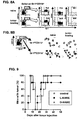
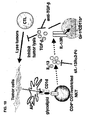
- IL-13 made by CD4 + CD1d-restricted NKT cells induces CD1 1b + Gr-1 + non-lymphoid cells of myeloid origin to produce TGF- ⁇ . It is also disclosed herein that TGF- ⁇ causes the down-regulation of CD8 + CTL-mediated tumor immunosurveillance ( FIG. 10 ).
- the method includes administering to a subject a therapeutically effective amount of an agent which, for example, blocks TGF- ⁇ binding to the TGF- ⁇ receptor, thereby blocking the TGF- ⁇ signaling pathway.
- Administration of an agent which blocks the TGF- ⁇ signaling pathway is particularly effective against tumors that have escaped CTL immunosurveillance as a result of the immunosuppressive effects of TGF- ⁇ .
- the disclosure provides methods of enhancing the activity of an immune cell by blocking the TGF- ⁇ signaling pathway.
- Immune cells that are susceptible to a block in the TGF- ⁇ signaling pathway are those cells that express the TGF- ⁇ receptor. Any of the agents disclosed herein can be used to block the TGF- ⁇ signaling pathway.
- Immune cells include leukocytes (for instance, neutrophils, eosinophils, monocytes, basophils, macrophages, B cells, T cells, dendritic cells, and mast cells), as well as other types of cells involved in an immune response.
- the method includes contacting an immune cell that expresses a TGF- ⁇ receptor with an agent which blocks the TGF- ⁇ signaling pathway.
- the immune cell is a lymphocyte, such as a T cell or a B cell.
- the immune cell is a CTL, a CD8 + CTL, a CD4 + T cell, a CD4 + CD1d-restricted T cell, an NK cell, or an NKT cell.
- the immune cell is a granulocyte.
- the immune cell can be either in vivo or in vitro.
- the agent can either bind TGF- ⁇ , a TGF- ⁇ receptor or a TGF- ⁇ receptor downstream signaling molecule.
- an immune cell is enhanced in a subject, following the administration of an agent as defined in the claims which bocks the TGF- ⁇ signaling pathway.
- Immune cells having an enhanced activity, for example increased tumor immunosurveillance, following the administration of the agent include cells that express a TGF- ⁇ receptor, such as a CTL.
- the immune cell with the enhanced activity is in a subject suffering from a tumor that has escaped CTL immunosurveillance.
- an enhanced activity of an immune cell such as enhanced CTL immunosurveillance, inhibits the recurrence of a tumor.
- the disclosure provides methods of enhancing an immune response in a subject by blocking the TGF- ⁇ signaling pathway.
- an enhanced immune response for example increased tumor immunosurveillance, inhibits the recurrence of a tumor.
- Any of the agents disclosed herein can be used to block the TGF- ⁇ signaling pathway, thereby enhance an immune response.
- a method for enhancing an immune response in a subject includes administering to the subject a therapeutically effective amount of an agent, which blocks the TGF- ⁇ signaling pathway, to enhance the immune response.
- the immune response is a T cell response.
- the immune response involves a TGF- ⁇ receptor-expressing cell.
- the cell expressing a TGP- ⁇ receptor can be, but is not limited to, a CTL a CD8 + CTL, a CD4 + T cell, a CD4 + CD1d-restricted T cell, an NK cell, or an NKT cell.
- the immune response is CTL-mediated immunosurveillance.
- a subject with an enhanced immune response is suffering from a tumor that has escaped CTL immunosurveillance.
- an enhanced immune response inhibits the recurrence of a tumor in a subject
- a method for enhancing a T cell-mediated immune response includes administering to the subject a therapeutically effective amount of an agent, which blocks the TGF- ⁇ signaling pathway, to improve a T cell-mediated immune response.
- the T cell-mediated immune response is CTL-mediated immunosurveillance.
- the T cell-mediated immune response is an NKT cell response.
- T cell-mediated immune response is a CD4 + CD1d-restricted T cell response.
- agents that block the TGF- ⁇ signaling pathway are disclosed herein. Also disclosed herein are agents that block the immunosuppressive effects of TGF- ⁇ , thus these agents enhance an activity of an immune cell, such as CTL immunosurveillance, or an immune response in a subject. In one embodiment, an agent inhibits the recurrence of a tumor that has escaped CTL immunosurveillance.
- the agent can be any substance, including, but not limited to, an antagonist, an antibody, a chemical compound, a small molecule, a peptide mimetic, a peptide or a protein.
- the agent is preferably a non-toxic agent.
- An agent can be, for example, an enzyme (for example, a kinase or a phosphorylase) or another catalytic molecule that selectively binds and alters the function of a protein and/or the activity of a molecular pathway, such as the TGF- ⁇ signaling pathway.
- proteins can be functional when phosphorylated and nonfunctional when de-phosphorylated.
- a functional, phosphorylated, protein can become nonfunctional when exposed to a de-phosphorylating agent such as a phosphorylase.
- a cell that is active as the result of expressing a functional protein can become inactivated when it is in contact with an agent that inhibits (neutralizes) the function of the protein.
- an agent that inhibits (neutralizes) the function of the protein can become activated when it is in contact with an agent that inhibits (neutralizes) the function of the protein.
- the agent is a neutralizing agent as defined in the claims.
- An agent can neutralize (inhibit an activity of) a molecule by specifically binding it, thereby preventing the molecule from performing at least one function.
- a neutralizing agent can prevent a molecule from interacting with other molecules.
- a neutralizing agent prevents TGF- ⁇ from specifically binding the TGF- ⁇ receptor. TGF- ⁇ activity can be neutralized with any of the agents disclosed herein.
- the agent is an antagonist.
- An antagonist is any substance that tends to nullify, or neutralize, the action of another, for example a drug that binds to a receptor, such as a TGP- ⁇ receptor, without eliciting a biological response.
- the antagonist is a chemical compound that neutralizes TGF- ⁇ directly.
- a chemical compound that neutralizes the TGF- ⁇ receptor- or at least one of its downstream signaling molecules for example, Smad 2, Smad3, or Smad 4
- Smad complex DNA-binding co-factor for example, Smad 2, Smad3, or Smad 4
- the agent interacts (for example, specifically binds) with the TGF- ⁇ molecule directly.
- the agent is an anti-TGF- ⁇ antibody.
- the anti-TGF- ⁇ antibody is a monoclonal antibody obtained from the hybridoma 1D11.16 (ATCC Accession No. HB 9849) binds TGF- ⁇ .
- Agents, such as the 1D11.16 antibody can bind TGF- ⁇ and neutralize its activity by preventing it from binding the anti-TGF- ⁇ receptor.
- an agent can form a complex with a ligand, such as TGF- ⁇ so that it is still capable of binding a receptor, such as a TOF- ⁇ receptor, but the ligand:agent complex is incapable of activating the receptor and transmitting a signal.
- a ligand such as TGF- ⁇
- a receptor such as a TOF- ⁇ receptor
- An agent can specifically bind a receptor, such as the TGF- ⁇ receptor, and prevent the receptor from transmitting a signal across the cell membrane into the cell. More specifically, an agent can specifically blind a receptor, such as the TGF- ⁇ receptor, at its ligand-binding site thereby preventing a ligand, such as TGF- ⁇ from binding to the receptor.
- TGF- ⁇ is used generally herein to mean any isoform of TGF- ⁇ , provided the isoform has immunosuppressive activity.
- the agent is an anti- TGF- ⁇ receptor antibody.
- the agent is a TGF- ⁇ mutant.
- TGF- ⁇ mutants include fragments of TGF- ⁇ and TGF- ⁇ peptides that retain the ability to bind a TGF- ⁇ receptor but cannot induce the TGF- ⁇ signaling pathway.
- TGF- ⁇ mutants also include TGF- ⁇ point mutants that retain the ability to bind a TGF- ⁇ receptor but cannot induce the TGF- ⁇ signaling pathway, or induce it only at a low level compared to the wildtype TGF- ⁇ .
- An agent can also specifically bind one or more of the receptor's downstream signaling molecules.
- some agents neutralizes TGF- ⁇ activity by specifically binding a downstream signaling molecule and preventing the transmission of an intracellular TGF- ⁇ signal.
- TGF- ⁇ downstream signaling molecules include, but are not limited to, Smad2, Smad3, Smad4, or Smad complex DNA-binding co-factors.
- a neutralizing agent may be soluble TGF- ⁇ receptor.
- the soluble TGF- ⁇ receptor specifically binds TGF- ⁇ and competes with the TGF- ⁇ cell surface receptor for any available TGF- ⁇ . Preventing TGF- ⁇ from binding its endogenous receptor neutralizes the activity of TGF- ⁇ . provided that sufficient soluble TGF- ⁇ receptor is present in order to bind all of the available TGF- ⁇ ligand.
- the TGF- ⁇ receptor can be expressed in a lymphocyte, such as a T lymphocyte. More specifically, the TGF- ⁇ receptor can be expressed in a CTL.
- the method of using an agent to neutralize the activity of TGF- ⁇ prevents TGF- ⁇ signaling in a TGF- ⁇ receptor -expressing CTL.
- the method of administering an agent that blocks the TGF- ⁇ signaling pathway results in a change in CTL activity.
- the change in CTL activity can be measured in any number of ways known to one of skill in the art.
- CTL activity can be monitored by measuring production of some chemokines, for example RANGES, or the production of IFN- ⁇ by ELISA, ELISPOT (enzyme-linked immunospot) or RT-PCR.
- Other methods include intracellular cytokine staining in combination with detection of the MHC class I-tetramer and a CTL assay.
- a CTL assay is the chromium release assay.
- target cells expressing an antigen for example the P18-IIIB peptide, on their surface are labeled with a radioactive isotope of chromium ( 51 Cr).
- Cell of interest such as CD8 + T cells, are then mixed with the target cell, either in the presence or absence of a cytokine, and incubated for several hours.
- cytokines include IL-4, IL-13, and TGF- ⁇ . Lysis of antigen-expressing cells releases 51 Cr into the medium.
- Antigen-specific lysis is calculated by comparing lysis of target cells expressing the antigen or control antigens in the presence or absence of effector cells, and is usually expressed as the percent antigen-specific lysis.
- the change in activity can be an increase or a decrease in CTL activity.
- the change in CTL activity can be measured as at least about a two-fold, at least about a five-fold, at least about a ten-fold, at least about a twenty-fold, at least about a fifty-fold, at least about a one hundred-fold, or at least about a two-hundred fold increase or decrease in CTL activity.
- the agents disclosed herein can be administered to a subject thought to be a candidate for tumor recurrence.
- the agent is administered to a subject prior to treatment of a tumor.
- Treatment of the tumor includes, but is not limited to, surgical removal of the tumor, chemotherapy, immunotherapy or radiation therapy.
- the agent is administered to the subject simultaneously with treatment of the tumor, or following treatment of the tumor.
- the agent is administered to the subject in combination with at least one additional agent either prior to, simultaneously with, or following treatment of the tumor.
- the additional agent can also be another agent described herein that inhibits or reduces tumor recurrence.
- the additional agent can be an agent that improves the subject's ability to inhibit a tumor recurrence or an agent (such as an antibiotic) that helps the subject fight infection during the course of treatment for the tumor.
- the additional agent can be an agent that stimulates the immune system, such as a cytokine.
- the agent is administered to a subject in an amount sufficient to inhibit the recurrence of a tumor at the original site of the primary tumor growth.
- the agent is administered to a subject in an amount sufficient to inhibit an immunsuppressive effect of TGF- ⁇ .
- the amount administered to a subject can vary from about 0.01 mg/kg body weight to about 1 g/kg body weight. In one specific, non-limiting example, the amount administered is about 3-4 mg/kg body weight.
- the method of inhibiting tumor recurrence can be applied to a subject suffering from any type of tumor.
- the tumor is susceptible to an immune response.
- the tumor is susceptible to a T cell-mediated immune response.
- the tumor is susceptible to a CTL-mediated immune response.
- the tumor is susceptible to immunosurveillance, such as a melanoma, a renal tumor, a breast tumor, a colon tumor or an osteosarcoma.
- the tumor can be a benign tumor or a malignant tumor.
- the tumor can include a carcinoma, a sarcoma, a leukemia, a lymphoma, or a tumor of the nervous system.
- the tumor includes a breast tumor, a liver tumor, a pancreatic tumor, a gastrointestinal tumor, a colon tumor, a uterine tumor, a ovarian tumor, a cervical tumor, a testicular tumor, a prostate tumor, a brain tumor, a skin tumor, a melanoma, a retinal tumor, a lung tumor, a kidney tumor, a bone tumor, an osteosarcoma, a nasopharygeal tumor, a thryoid tumor, a leukemia, or a lymphoma.
- the subject can be a mammal such as a human, a monkey, a horse, a cow, a pig, a dog, a cat, a mouse or a rat.
- Methods are descrided for screening agents that inhibits tumor recurrence.
- an immune cell expressing the TGF- ⁇ receptor is contacted with an agent (a compound) of interest, and the effect of the agent on the immune cell or on TGF- ⁇ signaling is then assayed.
- a decrease in TGF- ⁇ signaling for example a decrease in phosphorylation of downstream signaling molecule, or a decrease in Smad complex DNA binding indicates that the compound is an agent that blocks the TGF- ⁇ signaling pathway.
- an agent is identified as one that blocks the TGF- ⁇ signaling pathway if an immune cell, whose activity is normally inhibited by TGF- ⁇ , demonstrates an increase in activity or a prolonged period of activation following contact with the agent.
- a method for screening for an agent that blocks the TGF- ⁇ signaling pathway, using a cell of the immune system includes, in other examples, contacting a TGF- ⁇ receptor-expressing immune cell, such as a CTL, with an agent that binds the TGF- ⁇ receptor, and assaying the activity of the TGF- ⁇ receptor-expressing immune cell as compared to the activity of a TGF- ⁇ receptor-expressing control immune cell that has not been contracted with the agent.
- Examples of activity of a TGF- ⁇ receptor-expressing immune cell include cell lysis, cell proliferation, cytokine production, inhibition of tumor growth or inhibition of tumor recurrence.
- the method also includes contacting a TGF- ⁇ receptor-expressing immune cell, such as a CTL, with an agent that binds the TGF- ⁇ receptor, and assaying the activity of the TGF- ⁇ receptor signaling pathway in the cell and comparing it to the TGF- ⁇ receptor signaling pathway in a control cell that has not been contacted with the agent.
- the screening method can be performed in single vessels, or in arrays of vessels, such as in a high-throughput assay.
- the method may include contacting a TGF- ⁇ receptor-expressing immune cell, such as a CTL, with the combination of TGF- ⁇ and an agent that binds TGF- ⁇ , and assaying the activity of the TGF- ⁇ receptor-expressing immune cell as compared to a TGF- ⁇ receptor-expressing control immune cell that has not been contracted with the agent
- a TGF- ⁇ receptor-expressing immune cell examples include cell lysis, cell proliferation, cytokine production, inhibition of tumor growth or inhibition of tumor recurrence.
- Measuring a blockade of TGF- ⁇ signaling, and thus a change in activity of a cell expressing a TGF- ⁇ receptor can be assessed by any means known to one of skill in the art.
- western blot analysis is used to measure the level of a protein in a cell incubated in the presence or absence of an agent, or the level of protein phosphorylation in a cell incubated in the presence or absence of an agent.
- a luciferase assay is used to measure the effectiveness of the TGF- ⁇ signaling pathway to activate gene expression in a cell that is incubated in the presence or absence of an agent.
- TGF- ⁇ signaling also can be measured using cell proliferation assays of TGF- ⁇ -dependent cell lines.
- measuring a blockade of TGF- ⁇ signaling is assessed by measuring CTL function.
- CTL activity is increased in the presence or absence of a TGF- ⁇ blocking agent.
- CTL activity can be measured using any method known to one of skill in the art, for instance a CTL assay, such as a chromium release assay.
- the effects of a blockade of TGF- ⁇ signaling also can be measured, for instance by examining the formation or recurrence of tumors, or the relative rate of growth of a recurring tumor in a subject following the administration of an agent to a subject, such as a mouse.
- the recurrence of a tumor can be examined in the presence or absence of an agent prior to, during, or following the treatment of the primary tumor.
- Treatment of the primary tumor includes surgical removal of the tumor, chemotherapy, immunotherapy, or radiation therapy.
- functional assays of agents that block the TGF- ⁇ signaling pathway permit optimization of the dosage amounts of each agent effective in therapeutic uses, such as inhibiting a tumor recurrence.
- assays also can be used to test known agents, as well as newly identifies agents putative agents, or combinations of these agents for their ability to block the immunosuppressive effects of TGF- ⁇ .
- Candidate agents can initially be screened for subsequent selection and testing in one or more of the assays described herein.
- compositions that include an active agent can be formulated with an appropriate solid or liquid carrier, depending on the particular mode of administration chosen.
- the pharmaceutically acceptable carriers and excipients useful in this disclosure are conventional.
- parenteral formulations usually comprise injectable fluids that are pharmaceutically and physiologically acceptable fluid vehicles such as water, physiological saline, other balanced salt solutions, aqueous dextrose, glycerol or the like.
- Excipients that can be included are, for instance, other proteins, such as human serum albumin or plasma preparations.
- the pharmaceutical composition to be administered can also contain minor amounts of non-toxic auxiliary substances, such as wetting or emulsifying agents, preservatives, and pH buffering agents and the like, for example sodium acetate or sorbitan monolaurate.
- the dosage form of the pharmaceutical composition will be determined by the mode of administration chosen.
- topical and oral formulations can be employed.
- Topical preparations can include eye drops, ointments, sprays and the like.
- Oral formulations can be liquid (for example, syrups, solutions or suspensions), or solid (for example, powders, pills, tablets, or capsules).
- solid compositions conventional non-toxic solid carriers can include pharmaceutical grades of mannitol, lactose, starch, or magnesium stearate. Actual methods of preparing such dosage forms are known, or will be apparent, to those skilled in the art.
- the agents of this disclosure can be administered to humans or other animals on whose cells they are effective in various manners such as topically, orally, intravenously, intramuscularly, intraperitoneally, intranasally, intradermally, intrathecally, and subcutaneously.
- the particular mode of administration and the dosage regimen will be selected by the attending clinician, taking into account the particulars of the case (for example, the subject, the disease, the disease state involved, and whether the treatment is prophylactic). Treatment can involve daily or multi-daily doses of compound(s) over a period of a few days to months, or even years.
- compositions that comprise an agent, such as the 1D11.16 anti-TGF- ⁇ neutralizing monoclonal antibody and other agents effective at blocking the TGF- ⁇ signaling pathway will be formulated in unit dosage form, suitable for individual administration of precise dosages.
- a therapeutically effective amount of the 1D11.16 anti-TGF- ⁇ neutralizing monoclonal antibody can vary from about 0.1 mg/Kg body weight to about 1 g/Kg body weight.
- An effective amount of an agent, such as the 1D11.16 anti-TGF- ⁇ neutralizing monoclonal antibody can be administered in a single dose, or in several doses, for example daily, during a course of treatment.
- the amount of active compound(s) administered will be dependent on the agent being used, the subject being treated, the severity of the affliction, and the manner of administration, and is best left to the judgment of the prescribing clinician.
- An effective amount of an agent can be administered prior to, simultaneously with, or following treatment of a tumor.
- the formulation to be administered will contain a quantity of the active components) in amounts effective to achieve the desired effect in the subject being treated, for instance to measurably reduce the recurrence of a tumor.
- a therapeutically effective amount of an agent can be the amount of agent necessary to inhibit the recurrence of a tumor or the amount necessary to measurably reduce the recurrence of a tumor.
- a tumor suppressive amount of an agent is an amount sufficient to inhibit or reduce the recurrence of a tumor (for instance, any of the tumor suppressive amounts discussed herein) without causing a substantial cytotoxic effect (for example, without killing more than 1%, 2%, 3%, 5%, or 10% of normal cells in a sample).
- Site-specific administration of the disclosed compounds can be used, for instance by applying an agent, such as the 1D11.16 anti-TGF- ⁇ neutralizing monoclonal antibody, to a region of tissue from which a tumor has been removed or near a region of tissue from which a tumor has been removed.
- sustained intra-tumoral (or near-tumoral) release of the pharmaceutical preparation that comprises a therapeutically effective amount of an agent, such as the 1D11.16 anti- TGF- ⁇ neutralizing monoclonal antibody may be beneficial.
- Slow-release formulations are known to those of ordinary skill in the art.
- polymers such as bis(p-carboxyphenoxy)propane-sebacic-acid or lecithin suspensions may be used to provide sustained intra-tumoral release.
- delivery is via an injected and/or implanted drug depot, for instance comprising multi-vesicular liposomes such as in DepoFoam (SkyePharma, Inc, San Diego, CA) (see, for instance, Chamberlain et al., Arch. Neuro. 50:261-264, 1993 ; Katri et al., J. Pharm. Sci. 87:1341-1346, 1998 ; Ye et al., J. Control Release 64:155-166, 2000 ; and Howell, Cancer J. 7:219-227, 2001 ).
- DepoFoam Stema, Inc, San Diego, CA
- CTM complete T cell medium
- IL-2 20 U/ml
- IL-13 50 ng/ml, 5 ng/ml, 0.5 ng/ml
- the P18 peptide is the immunodominant epitope peptide of the V3 loop of HIV gp160 responsible for much of the CTL activity against the 15-12RM tumor ( Matsui et al., J. Immunol. 163:184, 1999 ).
- Recombinant mouse IL-2 and IL-13 were obtained from R&D systems, Minneapolis, MA.
- CD8 + T cells against several target cells such as 18 Neo fibroblasts
- Cytotoxic activity of CD8 + T cells against several target cells was measured by a four hour- 51 Cr-release assay.
- the 18Neo fibroblast is a BALB/c 3T3-derived cell line transfected with a null vector expressing only the Neo r gene as a control.
- CTM which consisted of RPMI1640 with 10% FCS, L-glutamine, sodium pyruvate, nonessential amino acids, penicillin, streptomycin and 5 ⁇ 10 -5 M 2-ME, containing 200 ⁇ g/ml of geneticin (Sigma, St. Louis, MO).
- the percentage of specific 51 Cr release in the CTL assay was calculated as follows: 100 ⁇ (experimental release - spontaneous release)/(maximum release - spontaneous release). Maximum release was determined from supernatants of cells that were lysed by addition of 5 % Triton X-100. Spontaneous release was determined from target cells incubated without added effector cells.
- the CTL stimulated in the presence of IL-13 did not show any enhancement or reduction of CTL activity against P18-pulsed target cells ( FIG. 1A ).
- IL-13 also had no effect on the cytolytic activity when it was added during the CTL assay.
- mice Female BALB/c mice were purchased from Charles River Breeding Laboratories (Frederick, MD). RAG2 knockout (KO) mice on a BALB/c background were obtained from Taconic (Germantown, NY). Mice deficient for both RAG2, IL-4R ⁇ (RAG2IL-4R ⁇ double KO) were bred under pathogen-free conditions. All the mice were maintained in a pathogen-free animal facility and were used at 6-10 weeks of age.
- RAG2 knockout mice on a BALB/c background were obtained from Taconic (Germantown, NY). Mice deficient for both RAG2, IL-4R ⁇ (RAG2IL-4R ⁇ double KO) were bred under pathogen-free conditions. All the mice were maintained in a pathogen-free animal facility and were used at 6-10 weeks of age.
- T cells require IL-4R ⁇ to as part of the receptor to respond to IL-13.
- T cells were negatively isolated from spleen cells of wild-type (RAG2 KO) or IL-4R ⁇ KO BALB/c mice (obtained from Dr. Nancy Noben-Trauth, George Washigton University).
- Single-cell suspensions of spleen cells were purified by negative selection for cells expressing MHC class II, CD11b, CD11c and DX5 by using magnetic beads (Mylteni Biotech). The latter cell surface marker is expressed on NK but not NKT cells ( Kronenberg and Gapin, Nat. Rev. Immuno/., 2:557-568, 2002 ).
- the purified T cells contained 2-3% CD1d tetramer positive cells (NKT cells), compared to 1-1.5% in spleen cells.
- 15-12RM is a fibrosarcoma derived from a BALB/c 3T3 fibroblast line transfected with the HIV envelope gp160 gene, mutant k-ras and c-myc ( Matsui et al., J. Immunol. 163:184, 1999 ).
- the 15-12RM cells were maintained in complete T cell medium (CTM) which consisted of RPMI1640 with 10% FCS, L-glutamine, sodium pyruvate, nonessential amino acids, penicillin, streptomycin and 5 ⁇ 10 -5 M 2-ME, containing 200 ⁇ g/ml of geneticin (Sigma, St. Louis, MO).
- CTM complete T cell medium
- mice were inoculated intraperitonealy. with 0.5 mg of anti-CD4 monoclonal antibody (GK1.5; Frederick Cancer Research and Development Center, NCI, Frederick, MD) or control rat IgG (Sigma, St. Louis, MO) three days in a row from day 0 and twice a week thereafter.
- anti-CD4 monoclonal antibody GK1.5; Frederick Cancer Research and Development Center, NCI, Frederick, MD
- control rat IgG Sigma, St. Louis, MO
- mice were treated on day 0, 1 and 3 with 0.2 mg of a fusion protein of murine IL-13R ⁇ 2 and human IgG1 (sIL-13R ⁇ 2-Fc), made by Genetics Institute, as described previously ( Donaldson et al., J. Immunol.
- mice 161:2317, 1998 ).
- mice were treated every other day for 10 days with 0.1 mg of either anti-TGF- ⁇ monoclonal antibody 1D11.16, specific for TGF- ⁇ 1, - ⁇ 2, - ⁇ 3 ( Dasch et al., J. Immunol. 142:1536, 1989 ), or the isotype matched control monoclonal (13C4), by intraperitoneal injection ( Strockbine et al., Infect. Immun. 50:695, 1985 ). 1D11.16 and 13C4 were made by Genzyme (Cambridge, MA).
- RAG2 KO or RAG2/IL-4R ⁇ double KO mice did not reject tumors at all because they lack T and B cells.
- RAG2 KO mice receiving transferred T cells regardless of the expression of IL-4R ⁇ by the T cells, behaved like wild-type BALB/c mice, implying that both CD8 + effector cells and CD1d-restricted T regulatory cells were successfully reconstituted.
- T cells from IL-4R ⁇ KO mice reconstitute as well as wild-type T cells implies that neither the effector nor the regulatory T cells themselves need to be able to respond to IL-13.
- TGF- ⁇ 1 Suppresses CTL Function
- TGF- ⁇ 1 is known for its ability to suppress lytic activity of CD8 + T cell function ( Mule et al., Cancer Immunol. Immunother. 26:95, 1988 ).
- TGF- ⁇ 1 To examine the ability of TGF- ⁇ 1 to suppress CTL function, spleen cells of vPE16-immunized mice were stimulated in vitro for a week with P18 peptide in the presence of various doses of TGF- ⁇ (100 ng/ml, 10 ng/ml, 1 ng/ml) and then tested these in a CTL assay (see Example 1, above).
- Recombinant human TGF- ⁇ 1 was purchased from Peprotech (Rocky Hill, NJ).
- non-T-non-NK cells were prepared from spleen cells from both na ⁇ ve and 15-12RM-injected BALB/c mice by depleting CD4 + , CD8 + , and DX5 + cells by magnetic beads conjugated with anti-CD4, anti-CD8, and anti-DX5 antibodies (Miltenyi Biotec; Auburn, CA).
- the cells After being pulsed with 1 ⁇ M P18-IIIB peptide, the cells were admixed in CTM supplemented with with IL-2 (20 U/ml), either in the presence or absence of anti-TGF- ⁇ antibody (50 ⁇ g/ml, 1D11), with T cells from vPE16-injected mouse spleen cells that were purified with magnetic beads (Miltenyi Biotec; Auburn, CA) as indicated in FIG. 3 . After a week of culture, viable cells were harvested and used as effector cells for the CTL assay, as described above.
- TGF- ⁇ had no effect in the lytic assay itself.
- TGF- ⁇ 1 Production is Up-Regulated in Tumor-Bearing Mice In Vivo
- TGF- ⁇ 1 is the cytokine induced by IL-13 in tumor-bearing mice to down-regulate tumor immunosurveillance
- TGF- ⁇ 1 production should be increased in non-T and non-B cells of tumor bearing mice, since expression of IL-4R ⁇ , which is a component of IL-13R, was not necessary in T or B cells to down-regulate immunosurveillance.
- non-lymphoid cells that were negative for Thy 1.2, anti-CD4, anti-CD8, B220, and DX5 were purified from spleen cells of both naive and 15-12RM-injected mice at day 3 after 15-12RM injection, and TGF- ⁇ 1 production ex vivo was examined without in vitro stimulation.
- Non-T non-B non-NK cells referred to as non-lymphoid cells
- Spleen cells were depleted of T cells, B cells, and NK cells using magnetic beads coated with anti-Thy 1.2, anti-CD4, anti-CD8, anti-B220, and anti-DX5 (Miltenyi Biotec; Auburn, CA).
- the cells were cultured at a density of 2 x 10 5 /well of a 96 well plate in 200 ⁇ l of X Vivo 20 medium (BioWhittaker, Inc., Walkersville, MD). At the indicated times, 100 ⁇ l of culture medium was harvested from each well and stored at -80°C until cytokine measurement.
- the concentration of total TGF- ⁇ 1 in the culture supernatant was determined by ELISA kit (R&D) according to the manufacturer's instructions. All samples were assayed in triplicate and each value is the average ⁇ standard deviation (SD) of the triplicate assay. p ⁇ 0.005 at 6 hours and p ⁇ 0.01 at 12 hours with Student's t-test between naive and tumor bearing groups.
- concentration of culture supernatant pg/ml
- concentration of culture supernatant pg/ml
- concentration of culture supernatant pg/ml
- concentration of culture supernatant pg/ml
- x 0.2 (ml)/(2 x 10 5 /1 x 10 6 ) pg produced per million cells.
- the data were analyzed for statistical significance using a Log-Rank test or Student's t-test. The data were considered significant at p ⁇ 0.05.
- Latent TGF- ⁇ 1 although not detected at 3 hours of culture, was already detected in the culture supernatants of the non-lymphoid cells from 15-12RM-injected mice after only 6 hours of culture, but was undetected in cells from naive mice at that time point (p ⁇ 0.005 by Student's t-test) ( FIG. 4A ). This earliest time point probably reflects the in vivo activity of these cells at the time they were removed from the mouse, without any in vitro stimulation. By 12 hours in tissue culture plastic, even the cells from non-tumor-bearing mice were making some TGF- ⁇ , suggesting a non-specific stimulation by the tissue culture plates or medium.
- the cells from 15-12RM-injected mice still produced more TGF- ⁇ 1 than the cells from naive mice (p ⁇ 0.01 by Student's t-test).
- the kinetics of TGF- ⁇ 1 production by non-lymphoid cells was also examined ( FIG. 4B ). The TGF- ⁇ 1 production by non-lymphoid cells was increased until at least day 10 after tumor injection.
- TGF- ⁇ 1 is Necessary for Tumor Recurrence In Vivo
- an anti-CD4 antibody a neutralizing anti-TGF- ⁇ monoclonal antibody (1D11.16; Genzyme, Cambridge, MA) or an isotype matched monoclonal antibody (13C4; Genzyme, Cambridge, MA) were injected intraperitoneally into mice that had received tumor cells.
- mice were treated with anti-CD4, anti-TGF- ⁇ , or the isotype matched monoclonal antibody for the first 10 days after tumor injection.
- the anti-CD4 monoclonal antibody (0.5 mg) was inoculated on day 0, 1, 2, 6, and 10 after tumor injection. 100 ⁇ g of anti-TGF- ⁇ monoclonal antibody or isotype matched control monoclonal antibody were injected every other day from day 0 to day 10.
- mice after subcutaneous inoculation of 15-12RM cells, the mice (5/group) were treated with 100 ⁇ g of anti-TGF- ⁇ antibody from day 0 of day 5, or control isotype-matched antibody every other day for ten days. Size of primary tumors in 15-12RM-injected BALB/c mice treated with 100 ⁇ g of anti-TGF- ⁇ antibody intraperitonealy every other day for ten days, or without any antibody treatment, was measured in two perpendicular dimensions with calipers every other day.
- mice treated with anti-TGF- ⁇ monoclonal antibody were protected from the recurrence of tumor similarly to anti-CD4-treated ( FIG. 4C ) or IL-13 inhibitor-treated mice.
- the effect of anti-TGF- ⁇ was seen only in the recurrent phase when the difference in tumor size was enormous (>400 mm 2 in the controls versus none in the anti-TGF- ⁇ -treated mice). In contrast, there was little difference in the primary tumors. Although the sizes of the primary tumors during the initial growth period in anti-TGF- ⁇ -treated mice were slightly smaller than those of tumors in control mice in all the experiments, the difference was not significant ( FIG. 4E ). These results demonstrate that TGF- ⁇ 1 is necessary for the tumor recurrence.
- TGF- ⁇ 1 produced early after tumor injection plays a critical role in down-regulation of anti-tumor immunosurveillance.
- blockade of TGF- ⁇ for the first 10 days is sufficient suggests that it is critical only during the priming of CTL, consistent with the observation that TGF- ⁇ inhibits the induction of CTL ( FIG. 1B ) but not their effector function.
- TGF- ⁇ 1 might be induced by IL-13 and act downstream to down-regulate CTL mediated tumor immunosurveillance, the possibility remained that both IL-13 and TGF- ⁇ 1 were necessary, and acting in parallel, to inhibit tumor immunosurveillance, albeit on different cell populations. To distinguish between these possibilities, the effect of an IL-13 inhibitor, which protects mice from tumor recurrence, on TGF- ⁇ 1 production in tumor-bearing mice three days after tumor inoculation was tested.
- splenic non-lymphoid cells were prepared from naive and 15-12RM-injected mice with or without IL-13 inhibitor treatment intraperitonealy on days 0, 1, 2 following tumor injection. Two hundred thousand cells were then cultured in vitro without any stimulation, as in FIG 4A , above.
- CD1-restricted NKT cells were responsible for IL-13 production induced in tumor-bearing mice and CD1 KO mice that lack NKT cells were resistant against tumor recurrence.
- TGF- ⁇ 1 production in two hundred thousand splenic non-lymphoid cells from wild-type BALB/c mice (na ⁇ ve), 15-12RM tumor-injected BALB/c mice, or tumor-injected CD1 KO mice at day 3 post-injection was also examined.
- CD1 KO mice with BALB/c background were bred, and maintained, under pathogen-free conditions and were used at 6-10 weeks of age.
- the cells from 15-12RM-injected mice produced more TGF- ⁇ 1 than the cells from naive mice, consistent with the results shown in FIG. 4A (Example 4, above).
- the IL-13 inhibitor treatment reduced the TGF- ⁇ 1 level to almost the same level seen in naive mice.
- TGF- ⁇ 1 is up-regulated by CD11b + Gr-1 + cells from tumor-bearing mice.
- Non-lymphoid spleen cells were obtained from naive and 15-12RM-injected mice (day 3) by depleteing CD4 + , CD8 + , B220 + , and DX5 + cells with magnetic beads.
- the cells were stained with anti-Gr-1, anti-CD11c, anti-F4/80 and anti-CD11b antibodies for 30 minutes after blocking CD16/CD32 (anti-FcR 2.4G2, Pharmingen, San Diego, CA) for 15 minutes.
- the cells were washed once and fixed with Cytofix/Cytoperm (Pharmingen), and washed with Perm/Wash buffer. After washing, the cells were analyzed by FACScan or FACS Caliber by using CELLQuest software (BD Biosciences).
- non-lymphoid spleen cells were purified from naive BALB/c (white bar) and 15-12RM tumor-injected BALB/c mice (black bars).
- CD11b + and CD11c + cells were depleted during the purification of the splenic non-lymphoid cells.
- mice were injected intraperitonealy with 200, 100, 10, or 1 ⁇ g of anti-Gr-1 antibody (Cederlane Laboratories Ltd., Ontario, Canada) on days 5, 6, 10,15, 20 after tumor injection. Two hundred thousand purified cells were cultured in vitro without particular stimulation. The culture supernatant was collected after 6 or 12 hours in culture and the concentration of TGF- ⁇ 1 was determined by ELISA. Each value is the average ⁇ SD of triplicate assays.
- Intracellular cytokine staining was also performed to confirm TGF- ⁇ 1 production by CD11b + Gr-1 + cells. Freshly isolated spleen cells from na ⁇ ve and 15-12RM-injected mice were stained with anti-CD11b and anti-Gr-1. Then cells were fixed, permeabilized and stained with anti-TGF- ⁇ 1 antibody.
- the splenic non-lymphoid spleen cells include CD11b + (45.33 ⁇ 10.23 % in naive mice, 33.72 ⁇ 8.61 % in tumor-bearing mice), CD11c + (3.78 ⁇ 1.21 % in naive mice, 3.47 ⁇ 1.76 % in tumor-bearing mice), F4/80 + (23.75 ⁇ 9.80 % in naive mice, 17.83 ⁇ 7.21 % in tumor-bearing mice) and Gr-1 + cells (45.09 ⁇ 7.59 % in na ⁇ ve mice, 35.92 ⁇ 11.88 % in tumor-bearing mice).
- CD11b + cells almost 80 % (37.52 ⁇ 7.72 % and 28.16 ⁇ 10.68 % of non-lymphoid spleen cells in naive and tumor-bearing mice) were positive for Gr-1 and more than 50 % (23.75 ⁇ 9.8 % and 17.83 ⁇ 7.21 % % of non-lymphoid spleen cells in naive and tumor-bearing mice) were positive for F4/80. Most of the CD11c + cells were myeloid dendritic cells that also expressed CD11b ( FIG. 6 ).
- CD11c + cells may play some role, among the cells expressing CD11b, CD11b + Gr-1 + double positive cells were the major source of TGF- ⁇ 1 production by non-lymphoid cells from 15-12RM injected mice.
- Anti-CD11b or anti-CD11c magnetic beads (Mylteni Biotech) were used at the same time with anti-Thy1.2, anit-B220, and anti-DX5 antibodies.
- Gr-1 + cells were not only necessary. for ex vivo TGF- ⁇ 1 production, but in fact necessary for down-regulation of immunosurveillance in vivo.
- Non-lymphoid cells prepared from BALB/c spleen cells were stained with FITC-conjugated anti-Gr-1 and APC-conjugated anti-CD11b antibodies for 30 minutes after blocking CD16/CD32 (Pharmingen) for 15 minutes. After washing, the cells were gated on Gr-1 hi CD11b + and Gr-1 int CD11b + cell populations and sorted by FACSVantage (BD Bioscience). Sorted cells were collected by cytospin onto glass slide, and dried completely. The cells were stained with Wright-Giemsa stain using a Diff-Quik stain set (Dade Behring Inc., Deerfield, IL).
- F4/80 was expressed on around 75% of the Gr-1 + CD11b + cells from both na ⁇ ve and 15-12RM-injected mice.
- CD31 was expressed relatively higher on the cells from tumor-bearing mice compared with the cells from na ⁇ ve mice (averages among several experiments were 24% and 19 % in tumor-bearing and na ⁇ ve mice respectively), although the difference was not significant IL-4R ⁇ , a component of the IL-13 receptor, was also expressed on the Gr-1 + CD11b + cells almost at the same level on na ⁇ ve and tumor-bearing mice-derived cells (15-20%). Expression of IL-4R ⁇ on the Gr-1 + CD11b + cells suggested that the Gr-1 + CD11b + cells express IL-13 receptor. This result is consistent with the result in FIG. 5B that in vivo blockade of IL-13 reduced ex vivo TGF- ⁇ production by these cells.
- Gr-1 hi CD11b + cells from both na ⁇ ve and 15-12RM-injected mice were primarily mature and some immature neutrophils, and no difference was observed between the cells from na ⁇ ve and 15-12RM-injected mice ( FIG. 8B , upper two panels).
- Gr-1 int CD11b + cells ( FIG. 8B , lower two panels) represented primarily immature myeloid cells, so-called "bands" (indicated by arrow heads), with some immature monocytes (indicated by arrows).
- the population from the 15-12RM-injected mice contained relatively fewer monocytes than the cells from the naive mice.
- Gr-1 + CD11b + myeloid cells induced by tumors may suppress T cell responses via nitric oxide.
- a possible role for NO tumor suppression in vivo was investigated in this Example.
- L-NAME N -nitro-L-arginine-methyl ester
- iNOS inducible nitric oxide synthase
- D-NAME N -nitro-D-arginine-methyl ester
- Mouse colon carcinoma line CT26 was maintained in CTM. Twenty thousand CT26 cells were injected via the tail vein into BALB/c mice. The mice were inoculated intraperitonealy with 0.1 mg of either anti-TGF- ⁇ monoclonal antibody (1D11.16; Genzyme, Cambridge, MA) or control monoclonal antibody (13C4; Genzyme, Cambridge, MA) every other day for two weeks. At the time when control mice had developed a sufficient number of tumor metastases (around 3 weeks after tumor inoculation), all the mice were sacrificed and the lungs were perfused with a 15% solution of India ink. After fixation by Fekete's solution, lungs were evaluated for pulmonary metastases by macroscopically counting nodules in the lungs.
- the maximum number of the nodules counted per lung was 250.
- Anti-TGF- ⁇ treatment significantly reduced the number of lung metastases. This result indicates that the role of TGF- ⁇ in down-regulation of CTL-mediated tumor immunosurveillance is not unique to the recurrence of a 15-12RM fibrosarcoma.
Landscapes
- Health & Medical Sciences (AREA)
- Life Sciences & Earth Sciences (AREA)
- Engineering & Computer Science (AREA)
- Immunology (AREA)
- Chemical & Material Sciences (AREA)
- Biomedical Technology (AREA)
- Molecular Biology (AREA)
- Hematology (AREA)
- General Health & Medical Sciences (AREA)
- Medicinal Chemistry (AREA)
- Urology & Nephrology (AREA)
- Organic Chemistry (AREA)
- Cell Biology (AREA)
- Biochemistry (AREA)
- Bioinformatics & Cheminformatics (AREA)
- Pathology (AREA)
- Biotechnology (AREA)
- Physics & Mathematics (AREA)
- Food Science & Technology (AREA)
- General Physics & Mathematics (AREA)
- Microbiology (AREA)
- Analytical Chemistry (AREA)
- Veterinary Medicine (AREA)
- Public Health (AREA)
- Animal Behavior & Ethology (AREA)
- Pharmacology & Pharmacy (AREA)
- Nuclear Medicine, Radiotherapy & Molecular Imaging (AREA)
- General Chemical & Material Sciences (AREA)
- Chemical Kinetics & Catalysis (AREA)
- Tropical Medicine & Parasitology (AREA)
- Toxicology (AREA)
- Oncology (AREA)
- Genetics & Genomics (AREA)
- Proteomics, Peptides & Aminoacids (AREA)
- Biophysics (AREA)
- Endocrinology (AREA)
- Medicines That Contain Protein Lipid Enzymes And Other Medicines (AREA)
- Medicines Containing Antibodies Or Antigens For Use As Internal Diagnostic Agents (AREA)
- Investigating Or Analysing Biological Materials (AREA)
- Peptides Or Proteins (AREA)
Claims (20)
- Antikörper, der ein von Hybridom 1 D11.16 (ATCC-Zugangsnummer HB 9849) erhaltener monoklonaler Antikörper oder eine humanisierte Version davon ist, zur Verwendung in einem Verfahren zur Hemmung des Wiederauftretens eines Tumors in einem Individuum, wobei der monoklonale Antikörper für TGF-β spezifisch ist und eine Aktivität von TGF-β neutralisiert, wodurch das Wiederauftreten des Tumors in dem Individuum gehemmt wird.
- Verwendung eines Antikörpers, der ein von Hybridom 1D11.16 (ATCC-Zugangsnummer HB 9849) erhaltener monoklonaler Antikörper oder eine humanisierte Version davon ist, zur Herstellung eines Medikaments zur Hemmung des Wiederauftretens eines Tumors in einem Individuum, wobei der monoklonale Antikörper für TGF-β spezifisch ist und eine Aktivität von TGF-β neutralisiert, wodurch das Wiederauftreten des Tumors im Individuum gehemmt wird.
- Antikörper zur Verwendung nach Anspruch 1 oder Verwendung nach Anspruch 2, wobei der monoklonale Antikörper eine immunsuppressive Wirkung eines transformierenden Wachstumsfaktors β (TGF-β) im Individuum blockiert.
- Antikörper zur Verwendung oder Verwendung nach Anspruch 3, wobei der monoklonale Antikörper TGF-β daran hindert, einen TGF-β-Rezeptor zu binden.
- Antikörper zur Verwendung nach Anspruch 1 oder Verwendung nach Anspruch 2, wobei das Individuum ein Mensch ist.
- Antikörper zur Verwendung nach Anspruch 1 oder Verwendung nach Anspruch 2, wobei der Tumor gutartig oder bösartig ist.
- Antikörper zur Verwendung nach Anspruch 1 oder Verwendung nach Anspruch 2, wobei der Tumor ein Karzinom, ein Sarkom, Leukämie, ein Lymphom oder einen Tumor des Nervensystems umfasst.
- Antikörper zur Verwendung nach Anspruch 1 oder Verwendung nach Anspruch 2, wobei der Tumor einen Brusttumor, einen Lebertumor, einen Pankreastumor, einen gastrointestinalen Tumor, einen Kolontumor, einen Uterustumor, einen Ovarialtumor, einen Zervixtumor, einen Hodentumor, einen Hirntumor, einen Hauttumor, ein Melanom, einen Retinatumor, einen Lungentumor, einen Nierentumor, einen Knochentumor, einen Prostatatumor, einen Nasopharynxtumor, einen Schilddrüsentumor, Leukämie oder ein Lymphom umfasst.
- Antikörper zur Verwendung nach Anspruch 1 oder Verwendung nach Anspruch 2, wobei der Antikörper zur intravenösen, subkutanen, intradermalen oder intramuskulären Verabreichung dient.
- Antikörper zur Verwendung nach Anspruch 1 oder Verwendung nach Anspruch 2, wobei der monoklonale Antikörper in vivo oder in vitro zu fehlendem Tumorwachstum führt.
- Antikörper zur Verwendung nach Anspruch 1 oder Verwendung nach Anspruch 2, wobei das Verfahren zum Blockieren der immunsuppressiven Wirkung des TGF-β zu einer erhöhten Immunüberwachung durch Lymphozyten des Individuums führt.
- Antikörper zur Verwendung oder Verwendung nach Anspruch 11, wobei die Lymphozyten T-Zellen oder B-Zellen umfassen.
- Antikörper zur Verwendung oder Verwendung nach Anspruch 11, wobei die Lymphozyten T-Zellen umfassen und die T-Zellen einen zytotoxischen T-Lymphozyten (CTL), einen CD8+-CTL, eine CD4+-Zelle, eine CD4+-CD1d-restringierte T-Zelle, eine NKT-Zelle oder eine Kombination davon umfassen.
- Antikörper zur Verwendung oder Verwendung nach Anspruch 11, wobei eine erhöhte Immunüberwachung mittels einer erhöhten biologischen Aktivität des Lymphozyten gemessen wird.
- Antikörper zur Verwendung oder Verwendung nach Anspruch 14, wobei die erhöhte Aktivität des Lymphozyten mithilfe eines CTL-Tests gemessen wird.
- Antikörper zur Verwendung oder Verwendung nach Anspruch 15, wobei der CTL-Test einen Chromfreisetzungstest umfasst.
- Antikörper zur Verwendung nach Anspruch 1 oder Verwendung nach Anspruch 2, wobei der monoklonale Antikörper TGF-β-Rezeptor-Signalübertragung hemmt.
- Antikörper zur Verwendung nach Anspruch 1 oder Verwendung nach Anspruch 2, wobei der monoklonale Antikörper bei Kontakt mit einem TGF-β-Rezeptoren exprimierenden Immunozyten einen TGF-β-Signalübertragungsweg blockiert und wobei das Blockieren des TGF-β-Signalübertragungswegs zu erhöhter Tumor-Immunüberwachung durch den TGF-β-Rezeptoren exprimierenden Immunozyten führt, wodurch die Aktivität des Immunozyten zur Hemmung des Wiederauftretens des Tumors erhöht wird.
- Antikörper zur Verwendung oder Verwendung nach Anspruch 18, wobei der TGF-β-Rezeptoren exprimierende Immunozyt eine T-Zelle oder eine B-Zelle ist.
- Antikörper zur Verwendung oder Verwendung nach Anspruch 18, wobei der TGF-β-Rezeptoren exprimierende Immunozyt T-Zellen umfasst und die T-Zellen einen CTL, einen CD8+-CTL, eine CD4+-Zelle, eine CD4+-CD1d-restringierte T-Zelle oder eine NKT-Zelle umfassen.
Applications Claiming Priority (3)
| Application Number | Priority Date | Filing Date | Title |
|---|---|---|---|
| US42128602P | 2002-10-25 | 2002-10-25 | |
| US421286P | 2002-10-25 | ||
| PCT/US2003/034023 WO2004037209A2 (en) | 2002-10-25 | 2003-10-24 | METHODS TO PREVENT TUMOR RECURRENCE BY BLOCKADE OF TGF-β |
Publications (3)
| Publication Number | Publication Date |
|---|---|
| EP1556071A2 EP1556071A2 (de) | 2005-07-27 |
| EP1556071A4 EP1556071A4 (de) | 2009-07-15 |
| EP1556071B1 true EP1556071B1 (de) | 2011-01-19 |
Family
ID=32176694
Family Applications (1)
| Application Number | Title | Priority Date | Filing Date |
|---|---|---|---|
| EP03779310A Expired - Lifetime EP1556071B1 (de) | 2002-10-25 | 2003-10-24 | Verfahren zur prävention von tumorrezidiven durch blockade von tgf-beta |
Country Status (8)
| Country | Link |
|---|---|
| US (1) | US20060057145A1 (de) |
| EP (1) | EP1556071B1 (de) |
| JP (1) | JP4989850B2 (de) |
| AT (1) | ATE496066T1 (de) |
| AU (1) | AU2003284994B2 (de) |
| CA (1) | CA2502663C (de) |
| DE (1) | DE60335817D1 (de) |
| WO (1) | WO2004037209A2 (de) |
Families Citing this family (5)
| Publication number | Priority date | Publication date | Assignee | Title |
|---|---|---|---|---|
| AU2006214052A1 (en) * | 2005-02-17 | 2006-08-24 | The Government Of The United States Of America As Represented By The Secretary Of The Department Of Health And Human Services | Synergistic effect of TGF-beta blockade and immunogenic agents on tumors |
| EP2058388A1 (de) * | 2007-10-05 | 2009-05-13 | INSERM (Institut National de la Santé et de la Recherche Médicale) | Nicht-konventionelle NKT-Zellen zur Verwendung in der Krebstherapie |
| BRPI0915255A2 (pt) * | 2008-11-13 | 2018-02-06 | Inst De Medicina Molecular | célula t assassina natural foxp3+; população de células; e método para produção e aumento do número de células t assassinas naturais foxp3+; método para administrar uma célula t assassinanatural ao fígado ou ao tecido da mucosa em um indivíduo, e para suprimir uma resposta imunológica em órgão de um indivíduo, e composição farmacêutica |
| WO2010124276A2 (en) * | 2009-04-24 | 2010-10-28 | Vanderbilt Universtiy | Anti-tgf-beta induction of bone cell function and bone growth |
| CA2988504A1 (en) * | 2014-07-02 | 2016-01-07 | Institut National De La Sante Et De La Recherche Medicale (Inserm) | Methods for diagnosing and treating cd1d restricted gamma/delta t cell lymphomas |
Family Cites Families (13)
| Publication number | Priority date | Publication date | Assignee | Title |
|---|---|---|---|---|
| AU582800B2 (en) * | 1985-10-03 | 1989-04-13 | Sumitomo Rubber Industries, Ltd. | Ionomer resin |
| US5571714A (en) * | 1988-12-22 | 1996-11-05 | Celtrix Pharmaceuticals, Inc. | Monoclonal antibodies which bind both transforming growth factors β1 and β2 and methods of use |
| IE62496B1 (en) * | 1990-04-19 | 1995-02-08 | Res Dev Foundation | Antibody conjugates for treatment of neoplastic disease |
| US6297041B1 (en) * | 1992-03-11 | 2001-10-02 | Institute Of Virology, Slovak Academy Of Sciences | MN gene and protein |
| US5470952A (en) * | 1993-10-20 | 1995-11-28 | Regeneron Pharmaceuticals, Inc. | CNTF and IL-6 antagonists |
| US5772995A (en) * | 1994-07-18 | 1998-06-30 | Sidney Kimmel Cancer Center | Compositions and methods for enhanced tumor cell immunity in vivo |
| DE19613691A1 (de) * | 1996-04-05 | 1997-10-09 | Boehringer Ingelheim Int | Arzneimittel für die Behandlung von Tumorerkrankungen |
| US6046165A (en) * | 1997-06-23 | 2000-04-04 | Ophidian Pharmaceuticals, Inc. | Compositions and methods for identifying and testing TGF-β pathway agonists and antagonists |
| JP4027485B2 (ja) * | 1998-01-14 | 2007-12-26 | 中外製薬株式会社 | 医薬品のスクリーニング方法ならびに医薬品の分子設計方法 |
| US6413535B1 (en) * | 1998-05-07 | 2002-07-02 | The University Of Tennessee Research Corporation | Method for chemoprevention of prostate cancer |
| US6224866B1 (en) * | 1998-10-07 | 2001-05-01 | Biocrystal Ltd. | Immunotherapy of B cell involvement in progression of solid, nonlymphoid tumors |
| US20040197333A1 (en) * | 2000-02-10 | 2004-10-07 | Cornell Research Foundation, Inc. | Use of TGF-beta antagonists to inhibit tumor cell formation or progression |
| US20030125251A1 (en) * | 2001-06-21 | 2003-07-03 | Wakefield Lalage M. | Transforming growth factor beta (TGF-beta) antagonist selectively neutralizes "pathological" TGF-beta |
-
2003
- 2003-10-24 EP EP03779310A patent/EP1556071B1/de not_active Expired - Lifetime
- 2003-10-24 DE DE60335817T patent/DE60335817D1/de not_active Expired - Lifetime
- 2003-10-24 AU AU2003284994A patent/AU2003284994B2/en not_active Ceased
- 2003-10-24 AT AT03779310T patent/ATE496066T1/de not_active IP Right Cessation
- 2003-10-24 US US10/532,374 patent/US20060057145A1/en not_active Abandoned
- 2003-10-24 JP JP2004547201A patent/JP4989850B2/ja not_active Expired - Fee Related
- 2003-10-24 CA CA2502663A patent/CA2502663C/en not_active Expired - Fee Related
- 2003-10-24 WO PCT/US2003/034023 patent/WO2004037209A2/en not_active Ceased
Also Published As
| Publication number | Publication date |
|---|---|
| ATE496066T1 (de) | 2011-02-15 |
| WO2004037209A2 (en) | 2004-05-06 |
| AU2003284994B2 (en) | 2009-11-12 |
| US20060057145A1 (en) | 2006-03-16 |
| AU2003284994A1 (en) | 2004-05-13 |
| CA2502663C (en) | 2015-10-06 |
| DE60335817D1 (de) | 2011-03-03 |
| WO2004037209A3 (en) | 2004-11-25 |
| JP4989850B2 (ja) | 2012-08-01 |
| EP1556071A2 (de) | 2005-07-27 |
| CA2502663A1 (en) | 2004-05-06 |
| EP1556071A4 (de) | 2009-07-15 |
| JP2006503899A (ja) | 2006-02-02 |
Similar Documents
| Publication | Publication Date | Title |
|---|---|---|
| EP3645568B1 (de) | Chimäre anti-b-zellreifungsantigenrezeptoren mit humanen domänen | |
| RU2698975C2 (ru) | Слитые иммуномодулирующие белки и способы их получения | |
| US11229645B2 (en) | Dipeptidylpeptidase 4 inhibition enhances lymphocyte trafficking, improving both naturally occurring tumor immunity and immunotherapy | |
| US8822405B2 (en) | Uses of mammalian cytokines and agonists; related reagents | |
| KR100620334B1 (ko) | 헤르페스 심플렉스 바이러스 유입 매개체에 대한 리간드및 사용 방법 | |
| US11951131B2 (en) | Anti-SLAMF7 chimeric antigen receptors | |
| KR20200128026A (ko) | 항-muc1 항체 및 il-15를 포함하는 융합 단백질 작제물 | |
| JP7097465B2 (ja) | キメラ抗原受容体細胞療法と併用するil-10剤の組成およびその使用方法 | |
| TW201307385A (zh) | 胃腸炎症和銀屑病以及哮喘的治療 | |
| KR20230126713A (ko) | Cea6 결합 분자 및 이의 사용 | |
| JP2023520572A (ja) | オルソゴナルな受容体を発現するようにゲノムが改変されたヒト免疫細胞 | |
| KR20230126714A (ko) | 메소텔린 결합 분자 및 이의 사용 | |
| EP1556071B1 (de) | Verfahren zur prävention von tumorrezidiven durch blockade von tgf-beta | |
| CN114616247B (zh) | Ox40/pd-l1双特异性抗体 | |
| JP2024542879A (ja) | Pd-1およびtgf-brii結合ドメインを含む多重特異性結合部位 | |
| WO2025048860A1 (en) | Ox40 agonist therapy | |
| KR20230125808A (ko) | EpCAM 결합 분자 및 이의 사용 | |
| KR20230125809A (ko) | Fap 결합 분자 및 이의 사용 | |
| NZ768752A (en) | Anti-human 4-1 bb antibodies and use thereof | |
| US20080267964A1 (en) | Synergistic Effect of Tgf-Beta Blockade and Immunogenic Agents on Tumors | |
| NZ753036A (en) | Anti-human 4-1 bb antibodies and use thereof | |
| NZ753036B2 (en) | Anti-human 4-1 bb antibodies and use thereof | |
| HK1026913B (en) | Ligand for herpes simplex virus entry mediator and methods of use |
Legal Events
| Date | Code | Title | Description |
|---|---|---|---|
| PUAI | Public reference made under article 153(3) epc to a published international application that has entered the european phase |
Free format text: ORIGINAL CODE: 0009012 |
|
| 17P | Request for examination filed |
Effective date: 20050511 |
|
| AK | Designated contracting states |
Kind code of ref document: A2 Designated state(s): AT BE BG CH CY CZ DE DK EE ES FI FR GB GR HU IE IT LI LU MC NL PT RO SE SI SK TR |
|
| AX | Request for extension of the european patent |
Extension state: AL LT LV MK |
|
| DAX | Request for extension of the european patent (deleted) | ||
| REG | Reference to a national code |
Ref country code: HK Ref legal event code: DE Ref document number: 1078486 Country of ref document: HK |
|
| RIC1 | Information provided on ipc code assigned before grant |
Ipc: A61K 38/00 20060101ALI20090528BHEP Ipc: C07K 16/22 20060101AFI20090528BHEP Ipc: A61K 39/395 20060101ALI20090528BHEP Ipc: G01N 33/50 20060101ALI20090528BHEP |
|
| A4 | Supplementary search report drawn up and despatched |
Effective date: 20090612 |
|
| 17Q | First examination report despatched |
Effective date: 20090928 |
|
| GRAP | Despatch of communication of intention to grant a patent |
Free format text: ORIGINAL CODE: EPIDOSNIGR1 |
|
| GRAS | Grant fee paid |
Free format text: ORIGINAL CODE: EPIDOSNIGR3 |
|
| GRAA | (expected) grant |
Free format text: ORIGINAL CODE: 0009210 |
|
| AK | Designated contracting states |
Kind code of ref document: B1 Designated state(s): AT BE BG CH CY CZ DE DK EE ES FI FR GB GR HU IE IT LI LU MC NL PT RO SE SI SK TR |
|
| REG | Reference to a national code |
Ref country code: GB Ref legal event code: FG4D |
|
| REG | Reference to a national code |
Ref country code: CH Ref legal event code: EP |
|
| REG | Reference to a national code |
Ref country code: IE Ref legal event code: FG4D |
|
| REF | Corresponds to: |
Ref document number: 60335817 Country of ref document: DE Date of ref document: 20110303 Kind code of ref document: P |
|
| REG | Reference to a national code |
Ref country code: DE Ref legal event code: R096 Ref document number: 60335817 Country of ref document: DE Effective date: 20110303 |
|
| REG | Reference to a national code |
Ref country code: NL Ref legal event code: VDEP Effective date: 20110119 |
|
| PG25 | Lapsed in a contracting state [announced via postgrant information from national office to epo] |
Ref country code: ES Free format text: LAPSE BECAUSE OF FAILURE TO SUBMIT A TRANSLATION OF THE DESCRIPTION OR TO PAY THE FEE WITHIN THE PRESCRIBED TIME-LIMIT Effective date: 20110430 Ref country code: GR Free format text: LAPSE BECAUSE OF FAILURE TO SUBMIT A TRANSLATION OF THE DESCRIPTION OR TO PAY THE FEE WITHIN THE PRESCRIBED TIME-LIMIT Effective date: 20110420 Ref country code: PT Free format text: LAPSE BECAUSE OF FAILURE TO SUBMIT A TRANSLATION OF THE DESCRIPTION OR TO PAY THE FEE WITHIN THE PRESCRIBED TIME-LIMIT Effective date: 20110519 Ref country code: SE Free format text: LAPSE BECAUSE OF FAILURE TO SUBMIT A TRANSLATION OF THE DESCRIPTION OR TO PAY THE FEE WITHIN THE PRESCRIBED TIME-LIMIT Effective date: 20110119 |
|
| PG25 | Lapsed in a contracting state [announced via postgrant information from national office to epo] |
Ref country code: CY Free format text: LAPSE BECAUSE OF FAILURE TO SUBMIT A TRANSLATION OF THE DESCRIPTION OR TO PAY THE FEE WITHIN THE PRESCRIBED TIME-LIMIT Effective date: 20110119 Ref country code: BG Free format text: LAPSE BECAUSE OF FAILURE TO SUBMIT A TRANSLATION OF THE DESCRIPTION OR TO PAY THE FEE WITHIN THE PRESCRIBED TIME-LIMIT Effective date: 20110419 Ref country code: SI Free format text: LAPSE BECAUSE OF FAILURE TO SUBMIT A TRANSLATION OF THE DESCRIPTION OR TO PAY THE FEE WITHIN THE PRESCRIBED TIME-LIMIT Effective date: 20110119 Ref country code: NL Free format text: LAPSE BECAUSE OF FAILURE TO SUBMIT A TRANSLATION OF THE DESCRIPTION OR TO PAY THE FEE WITHIN THE PRESCRIBED TIME-LIMIT Effective date: 20110119 Ref country code: BE Free format text: LAPSE BECAUSE OF FAILURE TO SUBMIT A TRANSLATION OF THE DESCRIPTION OR TO PAY THE FEE WITHIN THE PRESCRIBED TIME-LIMIT Effective date: 20110119 Ref country code: FI Free format text: LAPSE BECAUSE OF FAILURE TO SUBMIT A TRANSLATION OF THE DESCRIPTION OR TO PAY THE FEE WITHIN THE PRESCRIBED TIME-LIMIT Effective date: 20110119 Ref country code: AT Free format text: LAPSE BECAUSE OF FAILURE TO SUBMIT A TRANSLATION OF THE DESCRIPTION OR TO PAY THE FEE WITHIN THE PRESCRIBED TIME-LIMIT Effective date: 20110119 |
|
| PG25 | Lapsed in a contracting state [announced via postgrant information from national office to epo] |
Ref country code: DK Free format text: LAPSE BECAUSE OF FAILURE TO SUBMIT A TRANSLATION OF THE DESCRIPTION OR TO PAY THE FEE WITHIN THE PRESCRIBED TIME-LIMIT Effective date: 20110119 Ref country code: EE Free format text: LAPSE BECAUSE OF FAILURE TO SUBMIT A TRANSLATION OF THE DESCRIPTION OR TO PAY THE FEE WITHIN THE PRESCRIBED TIME-LIMIT Effective date: 20110119 |
|
| PLBE | No opposition filed within time limit |
Free format text: ORIGINAL CODE: 0009261 |
|
| STAA | Information on the status of an ep patent application or granted ep patent |
Free format text: STATUS: NO OPPOSITION FILED WITHIN TIME LIMIT |
|
| PG25 | Lapsed in a contracting state [announced via postgrant information from national office to epo] |
Ref country code: SK Free format text: LAPSE BECAUSE OF FAILURE TO SUBMIT A TRANSLATION OF THE DESCRIPTION OR TO PAY THE FEE WITHIN THE PRESCRIBED TIME-LIMIT Effective date: 20110119 Ref country code: CZ Free format text: LAPSE BECAUSE OF FAILURE TO SUBMIT A TRANSLATION OF THE DESCRIPTION OR TO PAY THE FEE WITHIN THE PRESCRIBED TIME-LIMIT Effective date: 20110119 Ref country code: RO Free format text: LAPSE BECAUSE OF FAILURE TO SUBMIT A TRANSLATION OF THE DESCRIPTION OR TO PAY THE FEE WITHIN THE PRESCRIBED TIME-LIMIT Effective date: 20110119 |
|
| 26N | No opposition filed |
Effective date: 20111020 |
|
| PG25 | Lapsed in a contracting state [announced via postgrant information from national office to epo] |
Ref country code: IT Free format text: LAPSE BECAUSE OF FAILURE TO SUBMIT A TRANSLATION OF THE DESCRIPTION OR TO PAY THE FEE WITHIN THE PRESCRIBED TIME-LIMIT Effective date: 20110119 |
|
| REG | Reference to a national code |
Ref country code: DE Ref legal event code: R097 Ref document number: 60335817 Country of ref document: DE Effective date: 20111020 |
|
| PG25 | Lapsed in a contracting state [announced via postgrant information from national office to epo] |
Ref country code: MC Free format text: LAPSE BECAUSE OF NON-PAYMENT OF DUE FEES Effective date: 20111031 |
|
| REG | Reference to a national code |
Ref country code: CH Ref legal event code: PL |
|
| REG | Reference to a national code |
Ref country code: HK Ref legal event code: WD Ref document number: 1078486 Country of ref document: HK |
|
| PG25 | Lapsed in a contracting state [announced via postgrant information from national office to epo] |
Ref country code: CH Free format text: LAPSE BECAUSE OF NON-PAYMENT OF DUE FEES Effective date: 20111031 Ref country code: LI Free format text: LAPSE BECAUSE OF NON-PAYMENT OF DUE FEES Effective date: 20111031 |
|
| REG | Reference to a national code |
Ref country code: IE Ref legal event code: MM4A |
|
| REG | Reference to a national code |
Ref country code: AT Ref legal event code: MK05 Ref document number: 496066 Country of ref document: AT Kind code of ref document: T Effective date: 20110119 |
|
| PG25 | Lapsed in a contracting state [announced via postgrant information from national office to epo] |
Ref country code: IE Free format text: LAPSE BECAUSE OF NON-PAYMENT OF DUE FEES Effective date: 20111024 |
|
| PG25 | Lapsed in a contracting state [announced via postgrant information from national office to epo] |
Ref country code: LU Free format text: LAPSE BECAUSE OF NON-PAYMENT OF DUE FEES Effective date: 20111024 |
|
| PG25 | Lapsed in a contracting state [announced via postgrant information from national office to epo] |
Ref country code: TR Free format text: LAPSE BECAUSE OF FAILURE TO SUBMIT A TRANSLATION OF THE DESCRIPTION OR TO PAY THE FEE WITHIN THE PRESCRIBED TIME-LIMIT Effective date: 20110119 |
|
| PG25 | Lapsed in a contracting state [announced via postgrant information from national office to epo] |
Ref country code: HU Free format text: LAPSE BECAUSE OF FAILURE TO SUBMIT A TRANSLATION OF THE DESCRIPTION OR TO PAY THE FEE WITHIN THE PRESCRIBED TIME-LIMIT Effective date: 20110119 |
|
| REG | Reference to a national code |
Ref country code: FR Ref legal event code: PLFP Year of fee payment: 13 |
|
| REG | Reference to a national code |
Ref country code: FR Ref legal event code: PLFP Year of fee payment: 14 |
|
| PGFP | Annual fee paid to national office [announced via postgrant information from national office to epo] |
Ref country code: FR Payment date: 20161025 Year of fee payment: 14 Ref country code: GB Payment date: 20161027 Year of fee payment: 14 Ref country code: DE Payment date: 20161027 Year of fee payment: 14 |
|
| REG | Reference to a national code |
Ref country code: DE Ref legal event code: R119 Ref document number: 60335817 Country of ref document: DE |
|
| GBPC | Gb: european patent ceased through non-payment of renewal fee |
Effective date: 20171024 |
|
| REG | Reference to a national code |
Ref country code: FR Ref legal event code: ST Effective date: 20180629 |
|
| PG25 | Lapsed in a contracting state [announced via postgrant information from national office to epo] |
Ref country code: DE Free format text: LAPSE BECAUSE OF NON-PAYMENT OF DUE FEES Effective date: 20180501 Ref country code: GB Free format text: LAPSE BECAUSE OF NON-PAYMENT OF DUE FEES Effective date: 20171024 |
|
| PG25 | Lapsed in a contracting state [announced via postgrant information from national office to epo] |
Ref country code: FR Free format text: LAPSE BECAUSE OF NON-PAYMENT OF DUE FEES Effective date: 20171031 |