EP1556701B1 - Verfahren zur verbesserten diagnose von dysplasien - Google Patents
Verfahren zur verbesserten diagnose von dysplasien Download PDFInfo
- Publication number
- EP1556701B1 EP1556701B1 EP03809343A EP03809343A EP1556701B1 EP 1556701 B1 EP1556701 B1 EP 1556701B1 EP 03809343 A EP03809343 A EP 03809343A EP 03809343 A EP03809343 A EP 03809343A EP 1556701 B1 EP1556701 B1 EP 1556701B1
- Authority
- EP
- European Patent Office
- Prior art keywords
- marker
- ink4a
- gene
- cells
- cell proliferation
- Prior art date
- Legal status (The legal status is an assumption and is not a legal conclusion. Google has not performed a legal analysis and makes no representation as to the accuracy of the status listed.)
- Expired - Lifetime
Links
Images





Classifications
-
- C—CHEMISTRY; METALLURGY
- C12—BIOCHEMISTRY; BEER; SPIRITS; WINE; VINEGAR; MICROBIOLOGY; ENZYMOLOGY; MUTATION OR GENETIC ENGINEERING
- C12Q—MEASURING OR TESTING PROCESSES INVOLVING ENZYMES, NUCLEIC ACIDS OR MICROORGANISMS; COMPOSITIONS OR TEST PAPERS THEREFOR; PROCESSES OF PREPARING SUCH COMPOSITIONS; CONDITION-RESPONSIVE CONTROL IN MICROBIOLOGICAL OR ENZYMOLOGICAL PROCESSES
- C12Q1/00—Measuring or testing processes involving enzymes, nucleic acids or microorganisms; Compositions therefor; Processes of preparing such compositions
- C12Q1/68—Measuring or testing processes involving enzymes, nucleic acids or microorganisms; Compositions therefor; Processes of preparing such compositions involving nucleic acids
- C12Q1/6876—Nucleic acid products used in the analysis of nucleic acids, e.g. primers or probes
- C12Q1/6883—Nucleic acid products used in the analysis of nucleic acids, e.g. primers or probes for diseases caused by alterations of genetic material
- C12Q1/6886—Nucleic acid products used in the analysis of nucleic acids, e.g. primers or probes for diseases caused by alterations of genetic material for cancer
-
- G—PHYSICS
- G01—MEASURING; TESTING
- G01N—INVESTIGATING OR ANALYSING MATERIALS BY DETERMINING THEIR CHEMICAL OR PHYSICAL PROPERTIES
- G01N33/00—Investigating or analysing materials by specific methods not covered by groups G01N1/00 - G01N31/00
- G01N33/48—Biological material, e.g. blood, urine; Haemocytometers
- G01N33/50—Chemical analysis of biological material, e.g. blood, urine; Testing involving biospecific ligand binding methods; Immunological testing
- G01N33/53—Immunoassay; Biospecific binding assay; Materials therefor
- G01N33/575—Immunoassay; Biospecific binding assay; Materials therefor for cancer
-
- G—PHYSICS
- G01—MEASURING; TESTING
- G01N—INVESTIGATING OR ANALYSING MATERIALS BY DETERMINING THEIR CHEMICAL OR PHYSICAL PROPERTIES
- G01N33/00—Investigating or analysing materials by specific methods not covered by groups G01N1/00 - G01N31/00
- G01N33/48—Biological material, e.g. blood, urine; Haemocytometers
- G01N33/50—Chemical analysis of biological material, e.g. blood, urine; Testing involving biospecific ligand binding methods; Immunological testing
- G01N33/53—Immunoassay; Biospecific binding assay; Materials therefor
- G01N33/575—Immunoassay; Biospecific binding assay; Materials therefor for cancer
- G01N33/5755—Immunoassay; Biospecific binding assay; Materials therefor for cancer of the uterine cervix, uterine corpus or endometrium
-
- G—PHYSICS
- G01—MEASURING; TESTING
- G01N—INVESTIGATING OR ANALYSING MATERIALS BY DETERMINING THEIR CHEMICAL OR PHYSICAL PROPERTIES
- G01N33/00—Investigating or analysing materials by specific methods not covered by groups G01N1/00 - G01N31/00
- G01N33/48—Biological material, e.g. blood, urine; Haemocytometers
- G01N33/50—Chemical analysis of biological material, e.g. blood, urine; Testing involving biospecific ligand binding methods; Immunological testing
- G01N33/53—Immunoassay; Biospecific binding assay; Materials therefor
- G01N33/575—Immunoassay; Biospecific binding assay; Materials therefor for cancer
- G01N33/5758—Immunoassay; Biospecific binding assay; Materials therefor for cancer involving compounds serving as markers for tumours, cancers or neoplasias, e.g. cellular determinants, receptors, heat shock/stress proteins, A-protein, oligosaccharides or metabolites
Definitions
- the present invention relates to a method for improved diagnosis of dysplasias based on simultaneous detection of INK4a gene products and at least one marker for cell proliferation.
- the present invention provides a method for discriminating dysplastic cells over-expressing INK4a gene products from cells over-expressing INK4a gene products without being dysplastic by detection of a marker suitable for characterising the proliferation properties of the respective cell.
- the characterisation of the proliferation properties may comprise the detection of a marker or a set of markers characteristic for active cell proliferation and/or a marker or a set of markers characteristic for retarded or ceased cell proliferation.
- the method presented herein thus enables for a specific diagnosis of dysplasias in histological and cytological specimens.
- the detection of the over-expression of p16 INK4a in biological samples has proven as a useful marker in the detection of anogenital lesions, such as carcinoma of the uterine cervix (see WO00/01845 ; Klaes et al., Int. J. Cancer: 92, 276-284 (2001 )).
- the method based on p16 INK4a -specific immuno-chemical staining allows for a sensitive and specific identification of dysplastic cells in tissue section and in cytological samples.
- neoplastic cells can be stained using a p16 INK4a specific antibody mediated staining procedure.
- the histological diagnosis of neoplastic lesions can thus be supported by a staining procedure based on a molecular marker characteristic for transformation of cells in anogenital lesions.
- the diagnosis, whether or not cells are neoplastic, in these procedures is not solely based on the p16 INK4a specific staining, but does also rely on the histological information.
- metaplastic cells show some immunoreactivity with p16 INK4a specific antibodies, and thus are stained in the course of the procedures. Yet the staining pattern yielded from these metaplastic cells differs from the pattern, which renders from neoplastic lesions. Metaplastic cells give rise to a patchy or focal staining pattern, whereas neoplastic lesions give rise to diffuse staining pattern. Moreover the staining intensities of metaplastic cells are predominantly less than that of neoplastic cells.
- the percentage of cells showing over-expression of p16 INK4a increases in the course of emergence of dysplasias. So in neoplastic or pre-neoplastic stages, when only a restricted population of neoplastic or pre-neoplastic cells is present in samples the immuno-reactivity of p16 INK4a may be weak. This weak immuno-reactivity may be of about the level as the level caused by metaplastic cells. In later stages of dysplasias the overall immuno-reactivity of p16 INK4a is stronger and so neoplastic lesions are easily discernible from metaplasias even in a cytological testing format. This might lead to cases, where the presence of metaplastic cells expressing p16 INK4a might be confused with the presence of neoplastic cells, and thus produces a false positive result.
- a method for discriminating the metaplasias from neoplastic and dysplastic lesions would be desirable.
- a method for the discrimination of metaplasias from neoplastic and pre-neoplastic lesions is provided within the embodiments claimed according to the present invention.
- a marker molecule For supporting the discrimination of metaplasias from neoplastic lesions in testing procedures based on the over-expression of p16 INK4a a marker molecule would be desirable, that is expressed in neoplastic and/or pre-neoplastic cells and tissues and, which is not expressed simultaneously with INK4a gene products in one single metaplastic cells.
- a solution to the problem present in the art is provided by the methods claimed according to this invention.
- the inventors have found, that a combination of detection of the presence or absence and/or the level of p16 INK4a in combination with at least one marker for cell proliferation such as e.g. Ki67, Ki-S2, mcm5 or mcm2 may solve the problem present in the art.
- EP1217377 a method for automated detection of cervical malignancies is disclosed mediated by detection of more than one marker molecules.
- Some defined marker combinations are named within the document. There is no disclosure in this document relating to the choice of suitable markers for a combination. The purpose of the combination in this application is improved fidelity of the automated analysis of staining patterns in biological cytological specimens. This document mentions combination of p16 INK4a with other tumor markers.
- WO02059616 discloses a method for detection of cell-cycle disturbances for improved diagnosis of cervical malignancies.
- the document discloses, that dysplastic cells exhibit disturbances in cell cycle control and may thus be identified by means of detection of cyclin E type proteins together with post G1 substances in cells.
- p16 INK4a over-expressed in various dysplasias, may also be detected in some other non-dysplastic cells.
- the discrimination between non-dysplastic p16 INK4a positive cells and dysplastic cells over-expressing p16 INK4a may be based on the proliferation characteristics of the respective cells. In normally controlled cells p16 INK4a inhibits cdk4 and thus inhibits proliferation. In contrast in dysplastic cells this regulation is impaired. Thus p16 INK4a does despite it's uncommonly high expression level not lead to an inhibition of the cell proliferation.
- dysplastic cells may be discriminated from cells exhibiting controlled cell proliferation by the simultaneous detection of p16 INK4a with a marker characteristic for cell proliferation. Due to the fact, that in normal cells elevated levels of p16 INK4a inhibit cell proliferation, cells over-expressing p16 INK4a may be classified as being dysplastic provided they are exhibiting the characteristics of active cell proliferation.
- the present invention relates to a method for discrimination of neoplastic, pre-neoplastic and/or dysplastic lesions from non-dysplastic cells showing elevated levels of p16 INK4a , in biological samples in a histological or cytological testing procedure based on the detection of the presence or absence of cells expressing p16 INK4a gene simultaneously with cell proliferation markers in said biological samples.
- Suitable markers for cell proliferation may be e.g. Ki67, Ki-S2, Ki-S5, mcm5 or mcm2.
- protein or mRNA of the markers for cell proliferation may serve as a marker for discrimination of metaplasias from early dysplastic or pre-neoplastic lesions in samples.
- It is one aspect of the present invention to provide a method for discriminating dysplastic cells over-expressing INK4a gene products from other cells expressing INK4a gene products at a detectable level in biological samples comprising determining in a cytological or histological testing procedure the co-expression of at least two marker molecules in at least one single cell, wherein at least one marker molecule is an expression product encoded by the INK4a gene and at least one further marker molecule is a cell proliferation marker, wherein the over-expression of at least one INK4a gene product and expression of at least one marker for active cell proliferation at an immunochemically detectable level within said single cell is indicative of the dysplastic state of the cell and wherein the over-expression of at least one INK4a gene product and expression of at least one marker for senescence, terminal differentiation of cells, apoptosis or cell cycle arrest at a detectable level within said single cell is indicative of the non-dysplastic state of the cell.
- a second aspect of the present invention relates to a test kit for determination of dysplasias in samples according to the method disclosed herein, as embodied in the kit claims.
- non-dysplastic cells may show immuno-reactivity for p16 INK4a .
- the inventors found that these cells exhibiting ordered control of cell proliferation in response to the elevated levels of the INK4a gene products are subject to growth arrest. Thus these individual cells do not show immunoreactivity for markers of cell proliferation.
- transformed cells over-expressing p16 INK4a exhibit dysregulated control of cell proliferation and do not respond to the elevated level of p16 INK4a by cessation of proliferation.
- these dysplastic cell show simultaneous expression of p16 INK4a with markers for cell proliferation.
- a simultaneous detection of p16 INK4a with markers for cell proliferation may serve to discriminate dysplastic cells from arrested cells over-expressing p16 INK4a such as e.g. metaplastic cells.
- the method disclosed herein is based on the fact that dysplastic cells may be discriminated from cells exhibiting normally controlled cell proliferation by the simultaneous detection of a INK4a gene product such as e.g. p16 INK4a with a marker characteristic for cell proliferation.
- a INK4a gene product such as e.g. p16 INK4a with a marker characteristic for cell proliferation.
- marker as well as marker molecules in general shall be used herein to pertain to proliferation marker gene expression products as well as to INK4a gene expression products.
- the denominations given throughout this text for genes may in part relate to the genes or proteins as they have been discovered from any organism.
- this denomination shall confer to the respective homologue of the named markers in the organism which is particularly in question for a method as disclosed herein.
- this organism is a mammal and in one embodiment may be a human being.
- the named markers shall be the human homologues of the respective denominated ones.
- (cell) proliferation marker or “marker for cell proliferation” in the various grammatical forms is used to denominate proteins as well as nucleic acid markers.
- protein name of a marker such as e.g. "replication protein” is used herein, this use shall be understood to be metonymically and pertain as well to the protein as to the nucleic acid marker molecules encoding the particular protein
- a marker useful according to the present invention may be any molecule transcribed from a gene or any molecule translated from such a transcript.
- gene product as used in the context of the present invention may comprise polynucleotides such as e.g. DNA or RNA and polypeptides such as proteins, proteoglycans, peptides etc.
- Expression product(s) as used in the context of the present invention shall comprise any transcript of a gene locus in forward or reverse direction including any reading frames, splicing variants. Expression products as used herein shall thus comprise any alternative products encoded by the nucleic acids of a particular gene locus.
- INK4a encoded gene-products as used in the context of the present invention shall be any mRNA transcribed from the INK4a gene locus or any polypeptide translated from such an mRNA.
- the expression products encoded by the INK4a gene may exhibit molecular weights of about 5 to 40 kDa or any value in between and preferably of about 10 to 20 kDa or any value in between and most preferably of about 14 to about 19 kDa or any value in between respectively.
- INK4a gene-products suitable for the method according to the present invention may comprise e.g. gene-products such as e.g. p16 INK4a and p14ARF.
- (cell) proliferation marker or "marker for cell proliferation” as used in the context of the present invention shall comprise any marker molecule known in the art to be characteristic for the proliferation status of cells.
- the proliferation status may e.g. be a status of actively proliferating cells, of retarded cell proliferation, of arrested cell proliferation, of senescent cells, of terminally differentiated cells, of apoptosis etc.
- the cell proliferation marker is a marker molecule characteristic for active cell proliferation.
- the proliferation marker molecule may be a molecule characteristic for arrested, terminally differentiated, senescent or apoptotic cells.
- proliferation markers for use in the context of the present invention may comprise genes engaged in the DNA replication such as e.g. proteins of the pre-initiation complex or of the replication fork.
- Such molecules may e.g. comprise helicases, such as eucaryotic helicase or MCM proteins (MCM2, MCM3, MCM4, MCM5, MCM6, MCM7), protein TP as disclosed in W00050451 and WO0217947 (also denominated HELAD1, Pomfil2, Unc-53), kinases or phosphatases engaged in the replication process such as e.g. CDC6, CDC7 protein kinase, Dbf4, CDC14 protein phosphatase, CDC45 and MCM10.
- proliferation markers may comprise proteins engaged in the processive replication fork such as e.g. PCNA or DNA polymerase delta, replication protein A (RPA), replication factor C (RFC), FEN1.
- the proliferation markers may comprise molecules necessary for the maintenance of cell proliferation such as Ki67.
- proteins may be e.g. present throughout the whole cell cycle. They are useful for performing a method according to the present invention provided they are characteristic for active cell proliferation and are not significantly expressed in arrested, terminally differentiated, apoptotic or senescent states of cells.
- Ki67, Ki-S2 and ki-S5 as used herein shall denominate the protein marker molecules detected by the respective antibodies as well as the nucleic acids encoding these antigens.
- the cell proliferation markers for use in a method according to the present invention may be a marker molecule characteristic for retarded or ceased cell proliferation such as e.g. a senescence marker, a cell cycle arrest marker, a marker characteristic for terminally differentiated cells or an apoptosis marker.
- a marker molecule characteristic for retarded or ceased cell proliferation such as e.g. a senescence marker, a cell cycle arrest marker, a marker characteristic for terminally differentiated cells or an apoptosis marker.
- Such molecules comprise e.g. p21, p27, Caspases, BAD, CD95, fas-ligand, parp-proteins etc.
- Discrimination as used in the context of the present invention shall comprise an assessment whether a sample is to be classified in one or another way.
- the discrimination pertains to the assessment of a tissue or components thereof being dysplastic or being not dysplastic.
- the discrimination as used herein is a judgement about the growth properties of cells in a biological sample.
- the discrimination comprises the detection of expression of an INK4a gene product simultaneously with the detection of expression of a marker characteristic for active cell proliferation.
- cells co-expressing both marker molecules are to be classified as being dysplastic.
- the discrimination comprises the detection of an INK4a gene product simultaneously with the detection of expression of a marker characteristic for arrested, ceased or retarded cell proliferation.
- cells co-expressing both marker molecules are to be classified as being non-dysplastic.
- one INK4a gene expression product will be detected in concert with two or more markers for cell proliferation. This may be useful to enhance the identification of proliferation properties of the INK4a gene product expressing cells in samples.
- Some proliferation markers are restricted to specific phases of the cell cycle or are present in low abundance in cells. Due to this fact in some cases the detection of proliferating cells expressing INK4a gene products may be improved by detection of two or more proliferation markers. In such cases e.g. one proliferation being expressed during the whole proliferative cell cycle may be detected simultaneously to markers being characteristic for specific cells cycle phases. E.g.
- Ki67, Ki-S5 or Ki-S2 may be detected together with mcm5, mcm2, PCNA, rpA, rfC etc..
- proteins engaged in the DNA replication may be detected together with Ki67, Ki-S5 or Ki-S2.
- Ki67 may be detected together with Ki-S2. It must be understood, that these examples are intended to exemplify combinatorial possibilities and shall not be comprehensive, so that various other combinations of proliferation markers are likewise useful and suitable in the procedure of the method according to the present invention.
- combination of two or more cell proliferation marker molecules may be applied for a method as disclosed herein.
- two or more marker molecules being detectable over larger stretches of the cell cycle or even over the whole cell cycle or actively proliferating cell may be detected in concert in a method as disclosed herein.
- a combination of more than one cell proliferation marker molecules may generally be useful for improving the sensitivity of the detection of the proliferation characteristics of cells.
- a combination may furthermore comprise other marker molecules such as senescence marker molecules, markers for arrested cells, markers for terminally differentiated cells, markers for apoptotic cells, markers for viral infection or for viral activity in cells or cell cycle regulatory proteins as markers.
- a detection of HPV associated marker molecules or markers for viral activity may be of use for a detection of a dysplasia.
- the methods useful for detection of detection of HPV infection in samples are known to those of skill in the art. These methods may comprise methods employing probes specific for HPV agents or may employ nucleic acid amplification reactions. The detection of a viral infection may be carried out simultaneously or subsequently to the detection of the INK4a and proliferation marker molecules.
- Dysplastic as used in the context of the present invention shall refer to dysplasias from mild to severe dysplasias and their precursory stages, as well as carcinoma such as carcinomas in situ or invasive carcinomas and disseminated tumor cells. Thus dysplastic as used herein shall also comprise early and precursory stages of dysplasias and carcinomas.
- Non-dysplastic as used herein
- Cells over-expressing INK4a gene products without being dysplastic may e.g. comprise metaplastic cells, senescent cells, terminally differentiated cells or cells, that in certain stages of the cell cycle exhibit elevated levels of the INK4a gene products.
- elevated levels of INK4a gene products may even be effected as a response to external signals such as hormones, transmitters etc.
- the non-dysplastic cells over-expressing INK4a gene products comprise e.g. metaplastic cells, endometrial cells etc.
- the method for detection of the expression level of the INK4a encoded gene-products and/or the proliferation marker gene products according to the present invention is any method, which may (but need not) be e.g. suited to detect even very small amounts of specific biological molecules in biological samples.
- the detection reaction according to the present invention is a detection either on the level of nucleic acids or on the level of polypeptides.
- a marker molecule is said to be detectable as used in the context of the present invention, provided the marker may be detected in the course of suitable detection procedure such as e.g. in-situ-hybridization, immuno-chemical staining, hybrid capture assay etc..
- the level of expression of a marker molecule may be made detectable using suitable reporter reactions such as e.g. a chromogenic or fluorescence based immuno-chemical staining or in-situ-hybridization procedure for microscopic or automated analysis.
- suitable methods for enhancing the reporter signal known to those of skill in the art may be applied in the course of a method according to the present invention.
- the marker is said to be detectable in a case where the staining supersedes the respective background staining inherently obtained in the immuno-chemical staining procedure so as to produce significant staining results.
- the marker molecules may be detected using reagents that specifically recognise these molecules.
- the detection reaction for the INK4a gene-products and/or the proliferation marker gene products may comprise one or more reactions with detecting agents either recognising the initial marker molecules or recognising the prior molecules used to recognise other molecules.
- two or more probes may be used for the detection of one single marker molecule.
- two or more different binding agents e.g. antibodies
- oligonucleotide probes directed against one single marker molecule as the case may be against different epitopes or different sequences
- two or more different binding agents e.g. antibodies
- oligonucleotide probes directed against one single marker molecule as the case may be against different epitopes or different sequences
- the detection of the different gene products may be performed in one reaction vessel or containment or in different containments simultaneously or subsequently in time.
- the different gene products may be detected simultaneously in one cell co-expressing both products.
- cells co-expressing the gene products may be used for separated detection reaction (separated in space or in time) to detect each a single marker in the cells.
- the detection of the marker molecules in the different cells may as well be performed simultaneously or separately in time and/or space.
- the detection reaction further may comprise a reporter reaction indicating the presence or absence and/or the level of the marker molecule gene-products.
- the reporter reaction may be for example a reaction producing a coloured compound, a bioluminescence reaction, a fluorescence reaction, generally a radiation emitting reaction etc..
- different marker molecules may be recognised by agents, that produce different reporter signals, so that the signals referring to marker molecules could be distinguished.
- the detection of the expression of the two or more INK4a gene-products and/or proliferation marker gene products is carried out simultaneously.
- the reporter reaction may for example employ different fluorescent labels for the different molecules detected.
- the question is whether any proliferation marker and/or INK4a gene product is expressed.
- a procedure may be chosen, that gives the same fluorescence signal as indication of the presence of a proliferation marker.
- This procedure is suitable to improve sensitivity of the detection of the cell proliferation characteristics (different markers characteristic for active cell proliferation).
- the procedure may be applied as to render one detectable signal for three, four or even more marker molecules being characteristic for cell proliferation. Analogous the same may under certain circumstances be true for the INK4a gene expression products. It must be understood, that as the case may be different staining signals for different proliferation marker molecules may be desirable.
- the procedures may be applied to the necessities of the respective experiment.
- a combination of one or more (e.g. two different) INK4a gene products may be detected with a combination of one or more e.g. a set of two, a set of three, a set of four, a set of five or a set of even more markers for cells proliferation.
- the detection of the marker molecules for cell proliferation may render only one reporter signal.
- each single marker for cell proliferation may render a specific reporter signal or groups of marker molecules may render specific reporter signals.
- Signals for the indication of the presence of immuno-reactivity may be chromogenic signals produced by various methods known in the art Alternatively or even in combination fluorescent signals may be used. Suitable reporter signals comprise fluorescent labels such as fluorescein, rhodamin etc.
- Applicable formats for the detection reaction according to the present invention may be, blotting techniques, such as Western-Blot, Southern-blot, Northern-blot, immuno-cytochemical or immuno-histochemical procedures.
- the blotting techniques are known to those of ordinary skill in the art and may be performed for example as electro-blots, semidry-blots, vacuum-blots or dot-blots.
- Immunocyto/histochemical staining procedures are known to those of skill in the art and may comprise binding agent mediated detection of polypeptides as well as in situ hybridisation techniques. Both different techniques may even be applied simultaneously.
- hybrid capture of nucleic acids may be used for the detection.
- Amplification reaction may also be applicable for the detection of e.g. nucleic acid molecules.
- the detection of the level of INK4a and/or proliferation marker gene-products is carried out by detection of the respective nucleic acids (e.g. mRNA) or fragments thereof present in the sample.
- the means for detection of nucleic acid molecules are known to those skilled in the art.
- the procedure for the detection of nucleic acids can for example be carried out by a binding reaction of the molecule to be detected to complementary nucleic acid probes, proteins with binding specificity for the nucleic acids or any other entities specifically recognising and binding to said nucleic acids.
- in situ hybridisation of oligonucleotide probes to nucleic acids in a sample may be used for the detection of expression products or markers.
- Probes as used in the context of the present invention may be any agent binding specifically to a molecule.
- a probe may be an oligonucleotide hybridising to a particular sequence.
- the probe may be e.g. a primer.
- the probe as used herein may be e.g. a binding agent such as an antibody.
- the probes may be detectably labelled.
- the label may be selected from the group comprising a radioisotope, a bioluminescent compound, a chemiluminescent compound, a fluorescent compound, a metal chelate, or an enzyme.
- Probes may be applied in any detection procedure known in the art e.g. in the course of an in situ hybridisation procedure, in the course of hybrid capture assays, in the course of immuno-chemical staining reaction, in the course of blotting techniques etc..
- This method may be performed as well in vitro as directly in situ for example in the course of a detecting staining reaction.
- Another way of detecting the marker mRNAs in a sample performed in the method according to the present invention is an amplification reaction of nucleic acids, which can be carried out in a quantitative manner such as for example the polymerase chain reaction.
- real time RT PCR may be used to quantify the level of marker mRNAs in samples of dysplasias or tumors (cells or tissue samples).
- the detection of the level of INK4a and/or proliferation marker gene-products is carried out by determining the level of expression of a protein or fragments thereof.
- the determination of the marker gene-product on the protein level can for example be carried out in a reaction comprising a binding agent specific for the detection of the particular marker polypeptide.
- binding agents can be used in many different detection techniques for example in western-blot, ELISA or immuno-precipitation.
- polypeptide binding agent based detection can be carried out as well in vitro as directly in situ for example in the course of an immuno-chemical staining reaction. Any other method for determining the amount of particular polypeptides in biological samples can be used according to the present invention.
- the immuno-cytochemical imaging procedures for use in the context of the present invention may comprise e.g. the staining of cytological or histological preparations with chromogenic or fluorescent dyes.
- the staining may e.g. comprise binding of the molecules to be detected by a first binding agent, which itself is detected by a secondary binding agent, which may be labelled.
- the first binding agent may in certain embodiments be a nucleic acid or a protein binding agent (e.g. an antibody) and the secondary binding agent may be e.g. a secondary antibody recognizing the first binding agent
- Binding agents as used in the context of the present invention for the detection of the level of INK4a polypeptides such as p16 INK4a or p14ARF polypeptides and proliferation marker polypeptides such as e.g. mcm5, mcm2, Kl67, Ki-S5, PCNA or Ki-S2 polypeptides may comprise antibodies and antigen-binding fragments, bi-functional hybrid antibodies, peptidomimetics containing minimal antigen-binding epitopes, anti-cullines (anti-calineTM) etc.
- an antibody or antigen-binding agent is said to react specifically, if it reacts at a detectable level with a protein disclosed herein, and does not significantly react with other proteins.
- the antibodies that may be used in the context of the present invention may be monoclonal or polyclonal antibodies. As used herein, the term antibody or monoclonal antibody is meant to include intact molecules as well as antibody fragments. Moreover, antibodies that may be used in the context of the present invention include chimeric, single chain, and humanised antibodies.
- binding agents may be used isolated or in combination. By means of combination it is possible to achieve a higher degree of sensitivity.
- the term antibody preferably, relates to antibodies which consist essentially of pooled monoclonal antibodies with different epitopic specificities, as well as distinct monoclonal antibody preparations.
- Monoclonal antibodies are made from antigen containing fragments of the polypeptide of the invention using any of a variety of techniques known to those of ordinary skill in the art; see, e.g., Harlow and Lane, Antibodies: A Laboratory Manual, Cold Spring Harbor Laboratory, 1988 .
- an immunogen comprising the antigenic polypeptide or a synthetic part thereof is initially injected into any of a wide variety of mammals (e.g., mice, rats, rabbits, sheep and goats).
- the polypeptides of this invention may serve as the immunogen without modification.
- a superior immune response may be elicited if the polypeptide is joined to a carrier protein, such as bovine serum albumin or keyhole limpet hemocyanin.
- a carrier protein such as bovine serum albumin or keyhole limpet hemocyanin.
- the immunogen is injected into the animal host, preferably according to a predetermined schedule incorporating one or more booster immunizations, and the animals are bled periodically.
- Polyclonal antibodies specific for the polypeptide may then be purified from such antisera by, for example, affinity chromatography using the polypeptide coupled to a suitable solid support.
- main question may be whether any proliferation marker is expressed.
- a procedure was chosen, that gives the same fluorescence signal as indication of the presence of a proliferation marker.
- This procedure is suitable to improve sensitivity of the detection of the cell proliferation characteristics.
- the procedure may be applied as to render one detectable signal for three, four or even more marker molecules being characteristic for cell proliferation.
- the same may under certain circumstances be true for the INK4a gene expression products.
- the procedures may be applied to the necessities of the respective experiment.
- the INK4a gene-products and/or proliferation marker gene products may according to the present invention be detected simultaneously.
- simultaneously according to the present invention shall mean either literally at the same instant or within the same testing procedure, whereby the single detection steps are temporarily consecutive.
- the detection procedure according to the present invention may furthermore comprise a cytochemical staining procedure rendering a chromogenic or fluorescent staining of cells or cell compartments.
- cytochemical staining procedures are known to those of skill in the art and may for example comprise e.g. staining for acidophilic or basophilic structures, of subcellular regions (e.g. the nucleus, the mitochondria, the golgi, the cytoplasm etc.), of specific molecules (the chormosomes, of lipids, of glycoproteins, of polysaccharids etc.) in the cytological specimens.
- Fluorescence dyes such as DAPI, Quinacrin, Chromomycin, etc. may be employed.
- chromogenic dyes such as Azan, Acridin-orange, Hematoxylin, Eosin, Sudan-red, Thiazin-stains (Toluidin-blue, Thionin) may be applied.
- staining procedures such as Pap-staining, Giemsa- staining, Hematoxylin-Eosin staining, van-Gieson staining, Schiff-staining (using Schiff reagent), staining procedures employing precipitation of metals (such as e.g. of silver in staining procedures employing Silver Nitrate) or insoluble stains such as e.g. of Turnbulls-blue (or other insoluble metal cyanides), etc. may be used in the course of a method as disclosed herein. It must be understood, that the named dyes and staining methods shall be examples for the applicable methods and that any other method known in the art may be applied to a method as disclosed herein.
- the staining procedures may produce chromogenic stains for light microscopic inspection or fluorescent stains for inspection under fluorescence microscopic conditions.
- radiation emitting procedures procedures employing substances impairing the transmission of radiation or other contrast media for imaging of the cytological conditions in a sample (e.g. the generation of optical impression by means such as (micro-)autoradiographic or (micro-)radiographic picture generation) may be of use for a method according to the present invention.
- All the staining and imaging procedures may be used for analysis not only in microscopic procedures but also in automated analysis procedures such flow cytometry, automated microscopic (computerized or computer aided) analysis or any other method for analysis of stained cytological specimens.
- the analysis of the staining or imaging results of the different procedures may be performed in a single analysis step or in different subsequent steps.
- the light microscopic inspection of a specimen may be performed before or after fluorescence microsopic inspection of the specimen.
- Fluorescence microscopy the analysis of different stains with different excitation wavelengths may be analyses simultaneous or subsequently.
- Other imaging methods may be employed simultaneously or subsequently to the named procedures.
- a sample according to the method of the present invention may comprise any sample comprising cells.
- Samples may comprise e.g. secretions, smears, body fluids, and cell- or tissue-samples.
- samples comprise cells of the anogenital tract, of the respiratory tract or of the skin and its appendages.
- the cells may be cells of the uterine cervix, the vagina, the vulva, the penis, the anus, the rectum, the bronchic tree, the lung, the peritoneum, the peritoneal space, the naso-pharyngeal space, the oral cavity or the skin.
- the sample may be a histological sample, a biopsy, or a cytological sample such as e.g.
- the samples may comprise cells infected by papilloma virus.
- the samples may in certain embodiments comprise cervical smears, bronchioalveolar lavages etc.
- the sample may be prepared as a monolayer or thin layer preparation of a cytological specimen.
- the respective methods for preparation of monolayer or thin-layer preparation in cytology are known to those of skill in the art.
- the preparation may e.g. comprise the ThinPrep technology.
- Other methods comprise conventional smears, or method employing suspensions of cells for preparation of the cytological specimens.
- Preparation of a sample may comprise e.g. obtaining a sample of a tissue, of a body fluid, of cells from a patient.
- preparation of the sample may also comprise several steps of further preparations of the sample, such as preparation of dissections, preparation of cell suspensions, spreading or applying the cells to be examined onto microscopic slides, preparation of tissue arrays, isolation of polypeptides or nucleic acids, preparation of solid phase fixed peptides or nucleic acids or preparation of beads, membranes or slides to which the molecules to be determined are coupled covalently or non-covalently.
- the method may be performed in an automated manner.
- the automation of the method may be achieved by automated staining and analysis of histological or cytological specimens on a solid surface by microscopic means.
- the automation may comprise a flow-cytometric analysis of the staining of cells in solution.
- the dysplastic lesions to which the method according to the present invention may be applied comprise any dysplastic lesions characterized by over-expression of INK4a gene products such as e.g. p16 INK4a or p14ARF.
- those lesions are dysplasias associated with infections by papilloma viruses such as e.g. HPV.
- the HPV may be a high risk HPV subtype such as HPV16, HPV18, HPV31, HPV 33, HPV35, HPV 39, HPV 45, HPV 51, HPV 52, HPV56, HPV 58, HPV 59, HPV 66, HPV 68 etc.
- the dysplastic lesions that may be detected according to the present invention comprise anogenital lesions, lesions of the respiratory tract, lesions of the head and the neck or lesions of the skin and its appendages.
- Such lesions may e.g. comprise dysplasias of the anus or rectum, of the vulva, the vagina, the cervix or the penis, of the bronchic tree, the lung, the oral cavity, or the nasopharyngeal space.
- kits for performing the method according to the present invention.
- the kit may be for example a diagnostic kit or a research kit.
- kits according to the present invention comprises at least an agent suitable for detecting the INK4a gene-products.
- a kit according to present invention comprises at least one or more probes for the detection of the presence or absence and/or the level of the over-expression of at least one INK4a gene-product and at least one cell proliferation marker gene-product in biological samples, wherein the kit components facilitate simultaneous detection of at least one INK4a gene-product and at least one cell proliferation marker gene-product in one single cell within the biological samples and wherein at least two distinguishable detectable signals are generated, at least one detectable signal being indicative for the level of expression of at least one INK4a gene product and at least one other detectable signal being indicative for the level of expression of at least one cell proliferation marker gene, and wherein the probes are a) one or more antibodies or antibody fragments directed against INK4a encoded polypeptides or fragments thereof and one or more antibodies or antibody fragments directed against cell proliferation marker encoded polypeptides or fragments thereof; or b) one or more nucleic acids specifically
- the reagents for the detection of the marker gene-products may include any agent capable of binding to the marker gene-products.
- Such reagents may include proteins, polypeptides, nucleic acids, peptide nucleic acids, glycoproteins, proteoglycans, polysaccharids or lipids.
- the INK4a gene-product and proliferation marker gene product samples for carrying out a positive control may comprise for example nucleic acids in applicable form, such as solution or salt, peptides in applicable form, tissue section samples or positive cells.
- the detection of the marker gene-products is carried out on the level of polypeptides.
- the binding agents may be for example antibodies specific for the marker gene-products or fragments thereof.
- the detection of the marker gene-products is carried out on the nucleic acid level.
- the reagent for the detection may be for example a nucleic acid probe or a primer reverse-complementary to said marker nucleic acids.
- the problem to be solved was to provide a method for discrimination between dysplastic cells and other cells lacking malignant growth potential.
- the method may be applied to any stage of dysplasias and may be especially useful in early stages, when cytological diagnostic methods based on the p 1 6 INK4a over-expression needs a further information for the identification of metaplastic cells.
- the present invention provides a kit for performing the method according to the present invention.
- Example 1 Immuno-fluorescent detection of the over-expression of p16 INK4a and Ki67in samples of the uterine cervix ( double staining )
- Sections of formalin fixed, paraffin embedded tissue samples of the cervix uteri were immuno-fluorescent stained using antibodies specific for p16 INK4a and Ki67.
- tissue sections were rehydrated through incubation in xylene and graded ethanol, and transferred to Aqua bidest Antigen Retrieval was carried out with 10mM citrate buffer (pH 6.0) for p16 INK4a and Ki67. Therefore the slides were heated in a waterbath for 40 min at 95 °C - 98°C. The slides were cooled down to RT for 20 minutes, transferred to washing buffer (50mM Tris-HCl, 150mM NaCl, 0.05% Tween 20 / DakoCytomation: code no.: S3006).
- the specimens were incubated with 10% goat serum for 30min at RT.
- the slides were then incubated with the primary antibodies, mouse anti-human p16 INK4a antibody (3.48 ⁇ g/ml) and rabbit anti Ki67 (1:25) for 30 min at RT , the slides were then rinsed with washing buffer and placed in a fresh buffer bath for 5 min. Excess buffer was tapped off and each specimen was covered with 200 ⁇ l of the secondary reagent , containing goat anti-mouse antibody , AlexaFluor®488 conjugated and goat anti rabbit antibody , Alexa Fluor®546 conjugated and then incubated for 30min at RT. Then slides were washed two times as before and directly mounted with a special mounting medium for fluorescence.
- the microscopic examination of the slides reveals, that cells immuno-reactive with p16 INK4a and being also immuno-reactive for Ki67 may only be found in samples, that may microscopically be identified as samples of dysplastic lesions.
- Cells stained by the p16 INK4a specific reaction that are originating from metaplasias, are not stained by the reaction specific for Ki67.
- the microscopic inspection of the cell proliferation marker staining shows, that metaplastic cells over-expressing p16 INK4a are not immuno-reactive with the antibodies directed against Ki67.
- Samples containing dysplastic tissue areas in contrast comprise cells, that are immuno-reactive with KI67 and with antibodies directed against p16 INK4a . So in contrast to dysplasias in metaplasias no cells may be double stained using the Ki67 and p16 INK4a specific antibodies.
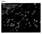
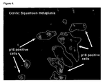
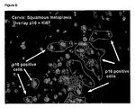
- Figures 1-6 show the staining results for a severe dysplasia of the cervix uteri and for a squamous metaplasia using antibodies directed against Ki67 and those directed against p16 INK4a .
- Immunoreactivity for Ki67 renders red fluorescence
- immunoreactivity for p16 INK4a renders green fluorescence and overlay of red and green fluorescence renders yellow fluorescence.
- the dysplastic specimen Fig. 1-3
- many cells of the dysplasia show nuclear positivity for Ki67 as well as positivity for p16INK4a and give thus rise to yellow fluorescence (see Figure 3 )
- the metaplastic specimen Fig.
- Example 2 Detection of cells co-expressing p14ARF and mcm2 in samples of the uterine cervix by in situ hybridization
- Smears of the uterine cervix may be semi-quantitatively analysed for the mRNA level of p16 INK4a and mcm2 in an in-situ staining reaction.
- the staining reaction is performed as follows:
- the spray-fixed smears are incubated in fresh 50% EtOH on a rocking device.
- the PEG film produced by the fixation procedure is removed by intensive rinsing.
- Following the smears are rinsed in aqua bidest
- the smears are incubated with porteinase K (10 ⁇ g/ml in PBS) for 10 min at 37°C.
- the slides are transferred to washing buffer (PBS / 0.1% Tween20) and finally the area containing the cells was surrounded with a lipid-pencil.
- the hybridization mixture is prepared by mixing 50 ⁇ l of ready to use hybridization buffer (DAKO A/S, Glostrup, Danmark) with about 5 - 10 pmol of the probes.
- the probes are biotin-and Digoxygenin-labelled oligonucleotides of sequences complememtary to the respective mRNAs.
- the hybridization mixture is heated to 95°C and afterwards equilibrated to 37°C. After the boiling procedure the smears are incubated with each 50 ⁇ l of the hybridization mixture for 4 hours at 42°C. The samples are washed in excess volumes of the wash buffers two times in 2 x SSC at 37°C for 15 min and once in 1 x SSC at 37 °C for 15 min Then the smears are rinsed two times at room temperature in 2 x SSC. Following this washing procedure the dissections are incubated for 30 min with blocking buffer (NEN, Blockingbuffer) at room temperature.
- blocking buffer NNN, Blockingbuffer
- Excess substrate is washed 3 times with 1x PBS/0,1% Triton X-100 for 10 min at room temperature.
- the section is incubated with Tyramides-Alexa-Fluor 594 for 10 sec to 7 min.
- Excess substrate is washed 3 times with 1x PBS/0,1% Triton X-100 for 10 min at room temperature..
- the smears are dipped in H 2 O dest and embedded with Fluorescence mounting medium (DakoCytomation). Then the stained dissections can be analysed by fluorescence microscopy.
- the microscopic examination of the slides reveals, that cells positive for expression of p16 INK4a and also expressing mcm2 may only be found in samples, that may microscopically be identified as samples of dysplastic lesions.
- Cells stained by the p16 INK4a specific reaction that are identifiable as metaplasias, are not stained by the reaction specific for mcm2.
- the microscopic inspection of the mRNA hybridisation shows, that metaplastic cells over-expressing p16 INK4a do not significantly express mRNA of mcm2.
- Dysplastic cells in contrast may be stained by in situ hybridisation with probes specific for mcm2 and with probes directed against p16 INK4a . So in contrast to dysplastic cells in metaplastic cells no double staining using the Ki67 and p16 INK4a specific probes may be observed.
- Example 3 Immuno-fluorescent detection of the over-expression of p16 INK4a and Ki-S2 in samples of the uterine cervix ( double staining )
- the specimens were incubated with 10% goat serum for 30min at RT.
- the slides were then incubated with the primary antibodies, mouse anti-human p16 INK4a antibody (clone E6H4) (3.48 ⁇ g/ml) and rabbit anti Ki-S2 (1:25) for 30 min at RT , the slides were then rinsed with washing buffer and placed in a fresh buffer bath for 5 min. Excess buffer was tapped off and the specimen was covered with 200 ⁇ l of the secondary reagent , containing goat anti-mouse antibody , AlexaFluor®488 conjugated and goat anti rabbit antibody , Alexa Fluor®546 conjugated and then incubated for 30min at RT. Then slides were washed two times as before and directly mounted with a special mounting medium for fluorescence.
- the microscopic examination of the slides reveals, that cells immuno-reactive with p16 INK4a and being also immuno-reactive for Ki-S2 may microscopically be identified as dysplastic cells.
- Cells stained by the p16 INK4a specific reaction which are not stained by the reaction specific for Ki-S2, may be classified by an experienced pathologist as either being metaplastic or being of endometrial origin.
- the microscopic inspection of the cell proliferation marker staining shows, that metaplastic cells over-expressing p16 INK4a are not immuno-reactive with the antibodies directed against Ki-S2.
- Dysplastic cells in contrast are immuno-reactive with KI-S2 and with antibodies directed against p16 INK4a . So in contrast to dysplastic cells, in metaplasias no cells may be double stained using the Ki-S2 and p16 INK4a specific antibodies.
- Example 4 Immuno-fluorescent detection of the over-expression of p16 INK4a , Ki67 and PCNA in bronchioalveolar-lavage samples of individuals with diagnosed small cell lung cancer (double staining)
- tissue sections were rehydrated through incubation in xylene and graded ethanol, and transferred to Aqua bidest.
- Conventional smears and liquid based cytological samples ( ThinPreps®) were rehydrated in ethanol (50%) for 10min and transferred in Aqua bidest.
- Antigen Retrieval was carried out with 10mM citrate buffer (pH 6.0) for p16 INK4a , Ki67and PCNA. Therefore the slides were heated in a waterbath for 40 min at 95 °C - 98°C.
- the slides were cooled down to RT for 20 minutes, transferred to washing buffer (50mM Tris-HCl, 150mM NaCl, 0.05% Tween 20 / DakoCytomation: code no.: S3006) and finally the samples were surrounded with a lipid-pencil.
- washing buffer 50mM Tris-HCl, 150mM NaCl, 0.05% Tween 20 / DakoCytomation: code no.: S3006
- the specimens were incubated with 10% goat serum for 30min at RT.
- the slides were then incubated with the primary antibodies, mouse anti-human p16 INK4a antibody (3.48 ⁇ g/ml), rabbit anti Ki67 and rabbit anti PCNA (each 1:25 ) for 30 min at RT , the slides were then rinsed with washing buffer and placed in a fresh buffer bath for 5 min. Excess buffer was tapped off and the specimen was covered with 200 ⁇ l of the secondary reagent, containing goat anti-mouse antibody , AlexaFluor®488 conjugated and goat anti rabbit antibody , Alexa Fluor®546 conjugated and then incubated for 30min at RT . Then slides were washed two times as before and directly mounted with a special mounting medium for fluorescence.
- the microscopic examination of the slides reveals, that cells immuno-reactive with p16 INK4a and being also immuno-reactive for Ki67 or PCNA may microscopically be identified as cells of small cell lung cancer.
- Cells stained by the p16 INK4a specific reaction that are originating from metaplasias, are not stained by the reaction specific for Ki67 and PCNA.
- the microscopic inspection of the cell proliferation marker staining shows, that metaplastic cells over-expressing p16 INK4a are not immuno-reactive with the antibodies directed against Ki67 and PCNA.
- Samples containing dysplastic cells in contrast comprise cells, that are immuno-reactive with Ki67 / PCNA and with antibodies directed against p16 INK4a . So in contrast to dysplasias in metaplasias no cells may be triple stained using the Ki67 and PCNA and p16 INK4a specific antibodies.
- Example 5 Flow cytometric detection of dysplastic cells by simultaneous detection of
- Cytological samples (Cell suspensions in PBS, pH 7,4) of the cervix uteri were fluorescent stained using antibodies specific for p14ARF and Ki-67 and oligoprobes for mcm-5 and evaluated by three-color fluorescent FACS analysis.
- Cells were centrifuged, the supernatant decanted and fixed and permeabilized with 100 ml Permeafix (Ortho Diagnostic, Raitan, NJ, USA) 1 hour at room temperature. Cells were washed in sterile PBS, pH 7,4, pelleted and resuspended in 100 ml Permeafix 1 hour at room temperature. Cells were washed in sterile PBS, pH 7,4, pelleted and resuspended in sterile PBS. They were incubated with PE-conjugated anti-p14ARF antibody and PE-Cy5-conjugated anti-Ki67 antibody for 1 h at +4°C.
- Cells were washed in sterile PBS, pH 7,4, pelleted and resuspended in 100 ml Permeafix 30 min at room temperature. Cells were washed in sterile PBS, pelleted by centrifugation, and then washed again in 2x standard saline citrate (SSC). After centrifugation , the cell pellet was resuspended in hybridisation solution (2x SSC, 30% formamide, sonicated salmon sperm, yeast transfer DNA) containing 500 ng of 5-carboxy-fluorescein double end labelled, mcm5-specific oligonucleotides probes.
- SSC standard saline citrate
- the intercellular hybridisation was performed at 42°C for 1 hour, followed by successive washes in 2x SSC, 0,5 % Triton X-100, and 1x SSC, 0,5 % Triton X-100 at 42°C.
- the cells were resuspended for analysis in PBS, pH 8,3 and analysed on a flow cytometer (FACScan, Becton Dickinson, IS). For each analysis 30.000-100.000 gated events were collected. Data analysis was performed using CellQuest (Becton Dickinson, IS)
- the flow cytometer analysis reveals, that cells immuno-reactive with p14ARF and being also immuno-reactive for Ki-67 and/or reactive for the oligoprobes of mcm5 could be only identified in samples of patients with dysplastic lesions of the cervics. Samples from women with no dysplastic lesions showed no concomitant staining of p14ARF with Ki67 or mcm5. The results show, that the double or triple staining of cells with reagents specific for Ki67 and/or mcm5 allows to discriminate p14ARF over-expressing non-dysplastic cells from p14ARF over-expressing dysplastic cells.
- Example 6 Immunoenzymatic detection of the over-expression of p16INK4a and Ki67in histological samples of the uterine cervix ( sequential double staining )
- Sections of formalin fixed, paraffin embedded tissue samples of the cervix uteri were immunoenzymatic double stained using antibodies specific for p16INK4a and Ki67.
- tissue sections were rehydrated through incubation in xylene and graded ethanol, and transferred to Aqua bidest.
- Antigen Retrieval was carried out with 10mM citrate buffer (pH 6.0) for p16INK4a and Ki67. Therefore the slides were heated in a waterbath for 40 min at 95 °C - 98°C. The slides were cooled down to RT for 20 minutes and transferred to washing buffer ( DakoCytomation).
- mice anti-human p16INK4a antibody MTM
- Excess buffer was tapped off and each specimen was covered with 200 ⁇ l of the secondary reagent (EnVision goat anti mouse - Peroxidase/ DakoCytomation) and incubated for 30min at RT .
- the secondary reagent EnVision goat anti mouse - Peroxidase/ DakoCytomation
- DAB Deionized water
- the microscopic examination of the slides reveals, that cells immuno-reactive with p16INK4a and being also immuno-reactive for Ki67 may only be found in samples, that may microscopically be identified as samples of dysplastic lesions.
- Cells stained by the p16INK4a specific reaction that are originating from metaplasias, are not stained by the reaction specific for Ki67.
- the microscopic inspection of the cell proliferation marker staining shows, that metaplastic cells over-expressing p16INK4a are not immuno-reactive with the antibodies directed against Ki67.
- Samples containing dysplastic tissue areas in contrast comprise cells, that are immuno-reactive with Ki67 and with antibodies directed against p16INK4a. So in contrast to dysplasias in metaplasias no cells may be double stained using the Ki67 and p16INK4a specific antibodies.
- Example 7 Immunoenzymatic detection of the over-expression of p16 INK4a and Ki67in cytological samples of the uterine cervix ( sequential double staining )
- mice anti-human p16 INK4a antibody After washing the slides they were incubated with the first primary antibody, mouse anti-human p16 INK4a antibody for 30 min at RT and were then rinsed with washing buffer and placed in a fresh buffer bath for 5 min. Excess buffer was tapped off and each specimen was covered with 200 ⁇ l of the secondary reagent ( EnVision goat anti mouse - Peroxidase) and incubated for 30min at RT . Then slides were washed three times as before. For the chromogenic visualization DAB ( DakoCytomation) was used by incubating the slides with the substrate chromogen complex for 10min at RT. The reaction was stopped in deionized water and slides placed in wash buffer.
- DAB DakoCytomation
- the microscopic examination of the slides reveals, that cells immuno-reactive with p16 INK4a and being also immuno-reactive for Ki67 may be identified as dysplastic cells on the basis of their morphology.
- Cells stained by the p16 INK4a specific reaction that are originating from metaplasias, are not stained by the reaction specific for Ki67.
- the microscopic inspection of the cell proliferation marker staining shows, that metaplastic cells over-expressing p16 INK4a are not immuno-reactive with the antibodies directed against Ki67.
- Dysplastic cells in contrast are immuno-reactive with K167 and with antibodies directed against p16 INK4a . So in contrast to metaplastic cells in dysplastic cells double-staining of single cells may be produced using the Ki67 and p16 INK4a specific antibodies.
Landscapes
- Health & Medical Sciences (AREA)
- Life Sciences & Earth Sciences (AREA)
- Immunology (AREA)
- Engineering & Computer Science (AREA)
- Chemical & Material Sciences (AREA)
- Molecular Biology (AREA)
- Biomedical Technology (AREA)
- Urology & Nephrology (AREA)
- Hematology (AREA)
- Pathology (AREA)
- Analytical Chemistry (AREA)
- Physics & Mathematics (AREA)
- General Health & Medical Sciences (AREA)
- Biotechnology (AREA)
- Microbiology (AREA)
- Biochemistry (AREA)
- Cell Biology (AREA)
- General Physics & Mathematics (AREA)
- Medicinal Chemistry (AREA)
- Food Science & Technology (AREA)
- Organic Chemistry (AREA)
- Proteomics, Peptides & Aminoacids (AREA)
- Wood Science & Technology (AREA)
- Zoology (AREA)
- Genetics & Genomics (AREA)
- Hospice & Palliative Care (AREA)
- Oncology (AREA)
- Biophysics (AREA)
- General Engineering & Computer Science (AREA)
- Bioinformatics & Cheminformatics (AREA)
- Measuring Or Testing Involving Enzymes Or Micro-Organisms (AREA)
- Investigating Or Analysing Biological Materials (AREA)
Claims (36)
- Verfahren zur Unterscheidung dysplastischer Zellen, die INK4a Genprodukte überexprimieren, von anderen Zellen, die INK4a Genprodukte in einem nachweisbaren Level in biologischen Proben exprimieren ausgewählt aus einer Gruppe bestehend ausa. ein Verfahren umfassend die Bestimmung in einem zytologischen oder histologischen Testverfahren der Co-Expression von mindestens zwei Markermolekülen in mindestens einer einzelnen Zelle, wobei mindestens ein Markermolekül ein Expressionsprodukt ist, das durch INK4a Gene kodiert wird, und mindestens ein weiteres Markermolekül ein Zellproliferationsmarker für aktive Zellproliferation ist, wobei die Überexpression von mindestens einem INK4a Genprodukt und die Expression von mindestens einem Marker für aktive Zellproliferation in einem nachweisbaren Level innerhalb besagter einzelner Zelle hinweisend für den dysplastischen Zustand der Zelle ist; undb. ein Verfahren umfassend die Bestimmung in einem zytologischen oder histologischen Testverfahren der Co-Expression von mindestens zwei Markermolekülen in mindestens einer einzelnen Zelle, wobei mindestens ein Markermolekül ein Expressionsprodukt ist, das durch INK4a Gene kodiert wird und wobei mindestens ein weiteres Markermolekül ein Proliferationsmarker ist, der charakteristisch für verlangsamte oder ausbleibende Zellproliferation ist, wobei die Überexpression von mindestens einem INK4a Genprodukt und die Expression von mindestens einem Marker, der charakteristisch für verlangsamte oder ausbleibende Zellproliferation in einem nachweisbaren Level innerhalb besagter einzelner Zellen ist, hinweisend für den nicht-dysplastischen Zustand der Zelle ist.
- Verfahren gemäß Anspruch 1, wobei ein Set von zwei oder mehreren Zellproliferationsmarkern nachgewiesen wird.
- Verfahren gemäß Anspruch 1 oder 2, wobei mindestens ein INK4a Genprodukt ein molekulares Gewicht zwischen 13 und 19 kDa hat.
- Verfahren gemäß der Ansprüche 1 bis 3, wobei mindestens ein INK4a Genprodukt aus einer Gruppe umfassend p16INK4a und p14ARF ausgewählt ist.
- Verfahren gemäß einem beliebigen der vorgehenden Ansprüche, wobei der Zellproliferationsmarker für aktive Zellproliferation ausgewählt ist aus einer Gruppe umfassend Proliferationsmarker, die für die Aufrechterhaltung der Zellproliferation erforderlich sind, Proliferationsmarker, die an der DNA Replikation beteiligt sind und Proliferationsmarker, die ein Teil der prozessiven Replikationsgabel sind oder einen solchen kodieren.
- Verfahren gemäß Anspruch 5, wobei das Genprodukt, das für die Aufrechterhaltung der Zellproliferation erforderlich ist, ein Molekül ist, das ausgewählt ist aus einer Gruppe umfassend Ki67 Moleküle, Ki-S5 Moleküle und Ki-S2 Moleküle.
- Verfahren gemäß Anspruch 6, wobei das Genprodukt, das an der DNA Replikation beteiligt ist, ausgewählt ist aus einer Gruppe umfassend Helikasen oder Untereinheiten davon, Zellteilungszyklusmoleküle (cdc), Phosphatasemoleküle und Kinasemoleküle.
- Verfahren gemäß Anspruch 7, wobei die Helikasen oder Untereinheiten davon ausgewählt sind aus einer Gruppe enthaltend MCM2, MCM3; MCM4, MCM5, MCM6, MCM7 und HELAD1.
- Verfahren gemäß Anspruch 7, wobei die cdc Moleküle, Kinasen und Phosphatasen ausgewählt sind aus einer Gruppe enthaltend CDC6, CDC7 Protein Kinase, Dbf4, CDC14 Proteinphosphatase, CDC45 und MCM10.
- Verfahren gemäß Anspruch 5, wobei die Moleküle, die ein Teil der prozessiven Replikationsgabel sind , aus einer Gruppe umfassend PCNA und POLD ausgewählt sind.
- Verfahren gemäß Anspruch 1, wobei die Marker, die charakteristisch für verlangsamte oder ausbleibende Zellproliferation sind, ausgewählt sind aus einer Gruppe umfassend Seneszenzmarker, Zellzyklusarrestmarker, Apoptosemarker, p21, p27, Caspasen, BAD, CD95, Fas-Ligand und Parp-Proteine.
- Verfahren gemäß einem beliebigen der Ansprüche 1-11, wobei das Genprodukt ein Polypeptid oder ein Nukleinsäuremolekül ist.
- Verfahren gemäß einem beliebigen der vorgehenden Ansprüche, wobei zusätzlich mindestens ein weiteres Markermolekül zur Verbesserung der Diagnose- oder Prognosestellung nachgewiesen wird.
- Verfahren gemäß Anspruch 13, wobei das weitere Markermolekül ausgewählt ist aus einer Gruppe umfassend Seneszenzmarker, Apoptosemarker, Zellzyklusarrestmarker, Marker für terminale Zelldifferenzierung, Marker für virale Infektion, Marker für virale Aktivität, zellzyklusregulierende Proteine, für die Aufrechterhaltung von Zellproliferation erforderliche Genprodukte, an der DNA Replikation beteiligte Genprodukte, Genprodukte, die ein Teil der prozessiven Replikationsgabel sind.
- Verfahren gemäß Anspruch 14, wobei der Marker für eine virale Infektion ein Marker für eine Hochrisiko-HPV Infektion ist, ausgewählt aus einer Gruppe umfassend HPV16, HPV18, HPV31, HPV33, HPV35, HPV39, HPV45, HPV51, HPV52, HPV56, HPV58, HPV59, HPV66 und HPV68.
- Verfahren gemäß einem beliebigen der vorgehenden Ansprüche, wobei zusätzlich ein zytologisches Färbeverfahren unter Einsatz mindestens eines Farbstoffs angewendet wird, der ausgewählt ist aus einer Gruppe umfassend DAPI, Quinacrin, Chromomyzin, Azan, Acridin-Orange, Hematoxylin, Eosin, Sudan-Rot, Toluidin-Blau, und Thionin, oder eine Färbemethode ausgewählt aus einer Gruppe umfassend Pap-Färbung, Giemsa-Färbung, Hematoxylin-Eosin-Färbung, van-Gieson-Färbung, Schiff-Färbung, Färbung durch Metallpräzipitate, Turnbulls-Blau-Färbung und Färbung durch Metallzyanide.
- Verfahren gemäß einem beliebigen der vorgehenden Ansprüche, wobei die dysplastischen Zellen Zellen einer kanzerösen oder präkanzerösen Läsion sind.
- Verfahren gemäß Anspruch 17, wobei die Läsion aus einer Gruppe umfassend Läsionen des Anogenitaltraktes, Läsionen des Respirationstraktes und Läsionen der Haut und deren Anhangsgebilde ausgewählt ist.
- Verfahren gemäß Anspruch 18, wobei die Läsion aus einer Gruppe umfassend die Läsionen des Gebärmutterhalses, Läsionen der Vagina, Läsionen der Vulva, Läsionen des Penis, Läsionen des Anus, Läsionen des Rektum, Läsionen des Bronchialbaums, Läsionen der Lunge, Läsionen des Peritonealraums, Läsionen des Hals-Nasenraums, Läsionen der Mundhöhle oder Läsionen der Haut ausgewählt wird.
- Verfahren gemäß einem beliebigen vorgehenden Anspruch, wobei die biologische Probe eine Probe ist, die Zellen enthält, insbesondere eine zytologische oder histologische Präparation, welche vom Anogenitaltrakt, vom Respirationstrakt oder von der Haut und deren Anhangsgebilden stammt.
- Verfahren gemäß Anspruch 20, wobei die Zellen Zellen sind, die vom Gebärmutterhals, von der Vagina, der Vulva, dem Penis, dem Anus, dem Rektum, dem Bronchialbaum, der Lunge, dem Hals-Nasenraum, der Mundhöhle oder der Haut stammen.
- Verfahren gemäß einem beliebigen der vorgehenden Ansprüche, wobei der Nachweis der INK4a Genprodukte und/oder der Zellproliferationsmarkermoleküle mit mindestens einer Sonde durchgeführt wird, die mindestens eines der jeweiligen nachzuweisenden Moleküle erkennt.
- Verfahren gemäß Anspruch 22, wobei mindestens eine Sonde eine nachweisbare Markierung trägt.
- Verfahren gemäß Anspruch 23, wobei mindestens eine Markierung aus der Gruppe ausgewählt ist, die aus Radioisotopen, biolumineszenten Verbindungen, chemilumineszenten Verbindungen, fluoreszenten Verbindungen, Metallchelaten, Enzymen, Biotin oder Digoxigenin besteht.
- Verfahren gemäß einem beliebigen der Ansprüche 22 bis 24, wobei mindestens eine Sonde ein Protein und/oder eine Nukleinsäure ist.
- Verfahren gemäß Anspruch 25, wobei mindestens eine Sonde ein Antikörper oder ein Antikörperfragment ist, der gegen ein INK4a kodiertes Genprodukt oder ein Zellproliferationsmarker-Genprodukt gerichtet ist.
- Verfahren gemäß Anspruch 26, welches ein immunozytochemisches Färbeverfahren umfasst.
- Verfahren gemäß der Ansprüche 23 oder 24, wobei mindestens eine Sonde eine Nukleinsäure ist, die spezifisch an ein INK4a-Genprodukt oder ein Zellproliferationsmarker-Genprodukt hybridisiert.
- Verfahren gemäß Anspruch 28, welches eine Nachweisreaktion umfasst, ausgewählt aus einer Gruppe umfassend a) eine In-Situ Hybridisierungsreaktion und b) eine Nukleinsäurenamplifikationsreaktion insbesondere PCR, NASBA oder LCR.
- Verfahren gemäß einem beliebigen der vorgehenden Ansprüche, wobei Nachweisreaktionen mit Nukleinsäuresonden und Polypeptidsonden gleichzeitig durchgeführt werden.
- Ein Kit zur Durchführung eines Verfahrens gemäß einem beliebigen der vorgehenden Ansprüche, welches ein diagnostisches Kit oder ein Forschungskit ist, umfassend mindestens eine oder mehrere Sonden zum Nachweis der Anwesenheit oder Abwesenheit und/oder des Levels der Überexpression von mindestens einem INK4a Genprodukt und mindestens einem Zellproliferationsmarker-Genprodukt in biologischen Proben, wobei die Kitkomponenten den gleichzeitigen Nachweis von mindestens einem INK4a Genprodukt und mindestens einem Zellproliferationsmarker-Genprodukt in einer einzigen Zelle innerhalb der biologischen Proben ermöglichen, und wobei mindestens zwei unterscheidbare nachweisbare Signale erzeugt werden, mindestens ein nachweisbares Signal, dashinweisend für den Level der Expression von mindestens einem INK4a Genprodukt ist, und mindestens ein anderes nachweisbares Signal, das hinweisend für den Level der Expression von mindestens einem Zellproliferationsmarkergen ist und wobei die Sonden a) ein oder mehrere Antikörper oder Antikörperfragmente sind die gegen INK4a kodierte Polypeptide oder Polypeptidfragmente gerichtet sind und ein oder mehrere Antikörper oder Antikörperfragmente die gegen Zellproliferationsmarker kodierte Polypeptide oder Polypeptidfragmente gerichtet sind, oder b) eine oder mehrere Nukleinsäuren sind, die spezifisch an INK4a-Genprodukte hybridisieren und eine oder mehrere Nukleinsäuren, die spezifisch an Zellproliferationsmarker-Genprodukte hybridisieren.
- Das Kit gemäß Anspruch 31, wobei die Antikörper oder Antikörperfragmente, die gegen INK4a kodierte Polypeptide oder Polypeptidfragmente gerichtet sind, eine nachweisbare Markierung tragen, die unterschiedlich und unterscheidbar von der nachweisbaren Markierung ist, welche die Antikörper oder Antikörperfragmente, die gegen Proliferationsmarker-Genprodukte gerichtet sind tragen.
- Das Kit gemäß Anspruch 31, wobei die Nukleinsäuresonden, die spezifisch an INK4a Genprodukte hybridisieren eine nachweisbare Markierung tragen, die unterschiedlich und unterscheidbar von der nachweisbaren Markierung ist, welche die Nukleinsäuresonden tragen, die spezifisch an die Proliferationsmarker-Genprodukte hybridisieren.
- Ein Kit gemäß einem beliebigen der Ansprüche 31 bis 33, wobei die INK4a Genprodukte aus einer Gruppe umfassend p16INK4a und p14ARF ausgewählt sind.
- Ein Kit gemäß einem beliebigen der Ansprüche 31 bis 34, wobei die Zellproliferationsmarker-Genprodukte ausgewählt sind aus einer Gruppe enthaltend CDC6, MCM3, MCM3, MCM4, MCM5, MCM6, MCM7, CDC7 Proteinkinase, Dbf4, CDC14 Proteinphosphatase, CDC45 und MCM10, Ki67, Ki-S2, PCNA oder POLD.
- Das Kit gemäß einem beliebigen der Ansprüche 31 bis 35 weiterhin umfassend mindestens eins der Folgenden:a. eine p16INK4a Probe zur Durchführung einer positiven Kontrollreaktionb. eine p14ARF Probe zur Durchführung einer positiven Kontrollreaktionc. eine Ki67 Probe zur Durchführung einer positiven Kontrollreaktiond. eine Ki-S2 Probe zur Durchführung einer positiven Kontrollreaktione. eine MCM5 Probe zur Durchführung einer positiven Kontrollreaktionf. eine MCM2 Probe zur Durchführung einer positiven Kontrollreaktiong. eine PCNA Probe zur Durchführung einer positiven Kontrollreaktionh. Reagenzien zum Nachweis der Anwesenheit oder Abwesenheit und/oder des Levels von p161NK4ai. Reagenzien zum Nachweis der Anwesenheit oder Abwesenheit und/oder des Levels von p14ARFj. Reagenzien zum Nachweis der Anwesenheit oder Abwesenheit und/oder des Levels von Ki67k. Reagenzien zum Nachweis der Anwesenheit oder Abwesenheit und/oder des Levels von Ki-S2l. Reagenzien zum Nachweis der Anwesenheit oder Abwesenheit und/oder des Levels von MCM5m. Reagenzien zum Nachweis der Anwesenheit oder Abwesenheit und/oder des Levels von MCM2n. Reagenzien zum Nachweis der Anwesenheit oder Abwesenheit und/oder des Levels von PCNAo. eine oder mehrere Proben von INK4a Genprodukten zur Durchführung positiver Kontrollreaktionenp. eine oder mehrere Proben von Zellproliferationsmarker-Genprodukten zur Durchführung positiver Kontrollreaktionenq. eine oder mehrere Reagenzien zum Nachweis der Anwesenheit oder Abwesenheit und/oder des Levels anderer INK4a Genprodukter. und eine oder mehrere Reagenzien zum Nachweis der Anwesenheit oder Abwesenheit und/oder des Levels anderer Zellproliferationsmarker-Genprodukte.
Priority Applications (1)
| Application Number | Priority Date | Filing Date | Title |
|---|---|---|---|
| EP03809343A EP1556701B1 (de) | 2002-10-28 | 2003-10-21 | Verfahren zur verbesserten diagnose von dysplasien |
Applications Claiming Priority (6)
| Application Number | Priority Date | Filing Date | Title |
|---|---|---|---|
| EP02024030A EP1416278B1 (de) | 2002-10-28 | 2002-10-28 | Verfahren zur verbesserten Diagnose von Dysplasien |
| EP02024030 | 2002-10-28 | ||
| EP03100584A EP1422526A1 (de) | 2002-10-28 | 2003-03-07 | Verfahren zur verbesserten Diagnose von Dysplasien |
| EP03100584 | 2003-03-07 | ||
| PCT/EP2003/050738 WO2004038418A1 (en) | 2002-10-28 | 2003-10-21 | Method for improved diagnosis of dysplasias |
| EP03809343A EP1556701B1 (de) | 2002-10-28 | 2003-10-21 | Verfahren zur verbesserten diagnose von dysplasien |
Publications (2)
| Publication Number | Publication Date |
|---|---|
| EP1556701A1 EP1556701A1 (de) | 2005-07-27 |
| EP1556701B1 true EP1556701B1 (de) | 2012-08-15 |
Family
ID=32178671
Family Applications (2)
| Application Number | Title | Priority Date | Filing Date |
|---|---|---|---|
| EP03100584A Withdrawn EP1422526A1 (de) | 2002-10-28 | 2003-03-07 | Verfahren zur verbesserten Diagnose von Dysplasien |
| EP03809343A Expired - Lifetime EP1556701B1 (de) | 2002-10-28 | 2003-10-21 | Verfahren zur verbesserten diagnose von dysplasien |
Family Applications Before (1)
| Application Number | Title | Priority Date | Filing Date |
|---|---|---|---|
| EP03100584A Withdrawn EP1422526A1 (de) | 2002-10-28 | 2003-03-07 | Verfahren zur verbesserten Diagnose von Dysplasien |
Country Status (9)
| Country | Link |
|---|---|
| US (8) | US20070128599A1 (de) |
| EP (2) | EP1422526A1 (de) |
| JP (3) | JP2006503589A (de) |
| AU (1) | AU2003301564A1 (de) |
| CA (1) | CA2496465C (de) |
| DK (1) | DK1556701T3 (de) |
| ES (1) | ES2391893T3 (de) |
| PL (1) | PL214860B1 (de) |
| WO (1) | WO2004038418A1 (de) |
Families Citing this family (32)
| Publication number | Priority date | Publication date | Assignee | Title |
|---|---|---|---|---|
| EP1369694A1 (de) | 2002-04-09 | 2003-12-10 | MTM Laboratories AG | Verfahren zur Unterscheidung zwischen Metaplasien und neoplastischen oder präneoplastischen Läsionen |
| DE60200248T2 (de) | 2002-08-01 | 2005-01-27 | Mtm Laboratories Ag | Verfahren für Lösung-basierte Diagnose |
| WO2006052822A2 (en) | 2004-11-05 | 2006-05-18 | Cytolution, Inc. | Methods and devices for screening cervical cancer |
| JP4734636B2 (ja) * | 2005-11-02 | 2011-07-27 | 国立大学法人 鹿児島大学 | 免疫染色法、及び当該免疫染色法を利用した細胞の評価方法 |
| US8131476B2 (en) | 2006-08-07 | 2012-03-06 | General Electric Company | System and method for co-registering multi-channel images of a tissue micro array |
| US8060348B2 (en) | 2006-08-07 | 2011-11-15 | General Electric Company | Systems for analyzing tissue samples |
| GB0720113D0 (en) | 2007-10-15 | 2007-11-28 | Cambridge Cancer Diagnostics L | Diagnostic, prognostic and predictive testing for cancer |
| EP2356460B1 (de) * | 2008-09-04 | 2018-01-24 | Ventana Medical Systems, Inc. | Verfahren zur vorhersage der gefahr des fortschreitens von tumoren |
| US20120041274A1 (en) | 2010-01-07 | 2012-02-16 | Myriad Genetics, Incorporated | Cancer biomarkers |
| WO2011098262A2 (en) * | 2010-02-09 | 2011-08-18 | Universität Bremen | P19arf, hmga2 and mdm2 for use in the diagnosis and treatment of aberrant cell growth |
| JP5553661B2 (ja) * | 2010-03-30 | 2014-07-16 | シスメックス株式会社 | 癌の再発リスク判定方法 |
| EP3812469A1 (de) * | 2010-07-07 | 2021-04-28 | Myriad Genetics, Inc. | Gensignaturen für krebsprognosen |
| US9605319B2 (en) | 2010-08-30 | 2017-03-28 | Myriad Genetics, Inc. | Gene signatures for cancer diagnosis and prognosis |
| WO2012065004A2 (en) * | 2010-11-12 | 2012-05-18 | Incelldx, Inc. | Methods and systems for predicting whether a subject has a cervical intraepithelial neoplasia (cin) lesion from a suspension sample of cervical cells |
| CA2891653A1 (en) | 2012-11-16 | 2014-05-22 | Myriad Genetics, Inc. | Gene signatures for cancer prognosis |
| CA2947624A1 (en) | 2014-05-13 | 2015-11-19 | Myriad Genetics, Inc. | Gene signatures for cancer prognosis |
| CN104807788B (zh) * | 2014-12-18 | 2018-09-14 | 中山大学 | 一种用于监测血管重构现象的方法与试剂盒 |
| JP6854246B2 (ja) | 2015-06-08 | 2021-04-07 | アーケア ダイアグノスティクス リミテッド | 尿サンプルの分析方法 |
| EP3304084B1 (de) | 2015-06-08 | 2022-03-23 | Arquer Diagnostics Limited | Verfahren und kits |
| WO2016207414A1 (en) | 2015-06-25 | 2016-12-29 | Universitaet Des Saarlandes | Improved diagnostic method for hpv-induced cancer and precancerous lesion screening and diagnosis |
| CN105241698A (zh) * | 2015-09-25 | 2016-01-13 | 四川农业大学 | 一种黄石爬鮡皮肤石蜡切片的制作方法 |
| US10300153B2 (en) * | 2015-11-30 | 2019-05-28 | Asthra, Llc | Immunological detection of altered cells |
| EP3396375B1 (de) * | 2015-12-24 | 2021-08-04 | Konica Minolta, Inc. | Bildverarbeitungsvorrichtung und -programm |
| US20190128899A1 (en) * | 2016-06-13 | 2019-05-02 | Koninklijke Philips N.V. | Method for inferring activity of a transcription factor of a signal transduction pathway in a subject |
| US12522862B2 (en) | 2017-10-23 | 2026-01-13 | The University Of Western Australia | Cell analysis |
| WO2019234176A1 (en) | 2018-06-07 | 2019-12-12 | Universitaet Des Saarlandes | Novel biomarkers for carcinoma diagnosis |
| CN110553889A (zh) * | 2019-09-18 | 2019-12-10 | 宋建华 | 自动染色机He染色工艺 |
| EP3816934B1 (de) | 2019-11-04 | 2025-01-01 | UMMON HealthTech | Verfahren und computerisiertes system zur markierung eines bildes von zellen eines patienten |
| CN111077302A (zh) * | 2019-12-26 | 2020-04-28 | 江苏美克医学技术有限公司 | 一种基于葡聚糖信号放大ki67和p16INK4a检测试剂盒 |
| WO2022064422A1 (en) * | 2020-09-24 | 2022-03-31 | Cytosystems Limited | Diagnosis of cancer by imaging flow cytometry |
| CN113552362B (zh) * | 2021-07-23 | 2024-08-09 | 湖北百奥斯生物科技有限公司 | 一种新型信号放大的免疫荧光试剂盒 |
| US20230377140A1 (en) * | 2022-02-01 | 2023-11-23 | 4D Path Inc. | Systems and Methods for Image-Based Disease Characterization |
Family Cites Families (23)
| Publication number | Priority date | Publication date | Assignee | Title |
|---|---|---|---|---|
| US4281061A (en) * | 1979-07-27 | 1981-07-28 | Syva Company | Double antibody for enhanced sensitivity in immunoassay |
| US6316208B1 (en) * | 1994-01-07 | 2001-11-13 | Memorial Sloan-Kettering Cancer Center | Methods for determining isolated p27 protein levels and uses thereof |
| JPH087229A (ja) * | 1994-06-13 | 1996-01-12 | Sony Corp | 磁気抵抗効果型磁気ヘッド |
| US6033847A (en) * | 1995-02-06 | 2000-03-07 | St. Jude Children's Research Hospital | InK4c-p18 and InK4d-p19, inhibitors of cyclin-dependent kinases CDK4 and CDK6, and uses thereof |
| US5723313A (en) | 1995-09-27 | 1998-03-03 | St. Jude Children's Research Hospital | ARF-p19, a novel regulator of the mammalian cell cycle |
| US6407062B1 (en) * | 1995-09-27 | 2002-06-18 | St. Jude Children's Research Hospital | ARF-P19, a novel regulator of the mammalian cell cycle |
| US5858683A (en) * | 1996-08-30 | 1999-01-12 | Matritech, Inc. | Methods and compositions for the detection of cervical cancer |
| GB2332515B (en) | 1997-10-21 | 1999-12-15 | Cancer Res Campaign Tech | Determination of cellular growth abnormality |
| US6303323B1 (en) * | 1997-10-21 | 2001-10-16 | Cancer Research Campaign Technology Limited | Detection of dysplastic or neoplastic cells using anti-MCM5 antibodies |
| DE19829473C2 (de) | 1998-07-01 | 2000-08-10 | Magnus Von Knebel Doeberitz Ch | Verfahren zur frühen Diagnose von Carcinomen |
| AUPP765398A0 (en) * | 1998-12-11 | 1999-01-14 | University Of Queensland, The | Treatment of papillomavirus infections |
| DE19957689A1 (de) | 1999-11-30 | 2001-06-13 | Deutsches Krebsforsch | Proteaseresistenter Tumorsuppressor p14(ARF) |
| GB0018140D0 (en) | 2000-07-24 | 2000-09-13 | Medical Res Council | Screening for abnormalities |
| DE10063179A1 (de) | 2000-12-18 | 2002-06-20 | Bayer Ag | Verfahren zur spezifischen Detektion von Tumorzellen und ihren Vorstufen in Gebärmutterhalsabstrichen durch simultane Messung von mindestens zwei verschiedenen molekularen Markern |
| DE10063112A1 (de) | 2000-12-18 | 2002-06-20 | Bayer Ag | Verfahren zur Erhöhung der klinischen Spezifität bei der Detektion von Tumoren und ihren Vorstufen durch simultane Messung von mindestens zwei verschiedenen molekularen Markern |
| KR20030094281A (ko) * | 2001-03-12 | 2003-12-11 | 모노젠, 인크. | 세포에 기초한 질병상태의 검출 및 질병상태의 구별 |
| EP1369694A1 (de) | 2002-04-09 | 2003-12-10 | MTM Laboratories AG | Verfahren zur Unterscheidung zwischen Metaplasien und neoplastischen oder präneoplastischen Läsionen |
| WO2004013631A2 (en) | 2002-07-29 | 2004-02-12 | Mtm Laboratories Ag | Compositions and methods for diagnosis and therapy of cancer |
| EP1387173A1 (de) * | 2002-07-29 | 2004-02-04 | MTM Laboratories AG | Verfahren zur verbesserten Diagnose von Zervixläsionen basierend auf dem Nachweis von INK4a Genprodukten |
| DE60200248T2 (de) | 2002-08-01 | 2005-01-27 | Mtm Laboratories Ag | Verfahren für Lösung-basierte Diagnose |
| US7361460B2 (en) | 2003-04-11 | 2008-04-22 | Digene Corporation | Approach to molecular diagnosis of human papillomavirus-related diseases |
| EP2356460B1 (de) | 2008-09-04 | 2018-01-24 | Ventana Medical Systems, Inc. | Verfahren zur vorhersage der gefahr des fortschreitens von tumoren |
| US8277190B2 (en) | 2009-03-27 | 2012-10-02 | General Electric Company | Turbomachine rotor assembly and method |
-
2003
- 2003-03-07 EP EP03100584A patent/EP1422526A1/de not_active Withdrawn
- 2003-10-21 PL PL374898A patent/PL214860B1/pl unknown
- 2003-10-21 ES ES03809343T patent/ES2391893T3/es not_active Expired - Lifetime
- 2003-10-21 JP JP2005501536A patent/JP2006503589A/ja active Pending
- 2003-10-21 AU AU2003301564A patent/AU2003301564A1/en not_active Abandoned
- 2003-10-21 US US10/533,384 patent/US20070128599A1/en not_active Abandoned
- 2003-10-21 EP EP03809343A patent/EP1556701B1/de not_active Expired - Lifetime
- 2003-10-21 WO PCT/EP2003/050738 patent/WO2004038418A1/en not_active Ceased
- 2003-10-21 CA CA2496465A patent/CA2496465C/en not_active Expired - Lifetime
- 2003-10-21 DK DK03809343.1T patent/DK1556701T3/da active
-
2008
- 2008-10-29 US US12/289,487 patent/US8975036B2/en not_active Expired - Lifetime
-
2009
- 2009-03-06 US US12/381,098 patent/US8367353B2/en not_active Expired - Lifetime
- 2009-03-23 JP JP2009071043A patent/JP2009142294A/ja not_active Withdrawn
-
2013
- 2013-02-27 JP JP2013038040A patent/JP5442884B2/ja not_active Expired - Lifetime
- 2013-03-13 US US13/799,093 patent/US20130189686A1/en not_active Abandoned
- 2013-03-13 US US13/798,998 patent/US9528159B2/en not_active Expired - Fee Related
-
2019
- 2019-09-23 US US16/579,495 patent/US20200017923A1/en not_active Abandoned
-
2020
- 2020-11-02 US US17/086,852 patent/US20210071267A1/en not_active Abandoned
- 2020-11-02 US US17/086,817 patent/US20210047697A1/en not_active Abandoned
Also Published As
| Publication number | Publication date |
|---|---|
| PL214860B1 (pl) | 2013-09-30 |
| US8975036B2 (en) | 2015-03-10 |
| JP2013150616A (ja) | 2013-08-08 |
| US20130189685A1 (en) | 2013-07-25 |
| US20130189686A1 (en) | 2013-07-25 |
| PL374898A1 (pl) | 2005-11-14 |
| ES2391893T3 (es) | 2012-11-30 |
| US20090176204A1 (en) | 2009-07-09 |
| US8367353B2 (en) | 2013-02-05 |
| JP2006503589A (ja) | 2006-02-02 |
| AU2003301564A1 (en) | 2004-05-13 |
| US20200017923A1 (en) | 2020-01-16 |
| JP5442884B2 (ja) | 2014-03-12 |
| AU2003301564A8 (en) | 2004-05-13 |
| EP1556701A1 (de) | 2005-07-27 |
| US20210047697A1 (en) | 2021-02-18 |
| CA2496465A1 (en) | 2004-05-06 |
| US20070128599A1 (en) | 2007-06-07 |
| EP1422526A1 (de) | 2004-05-26 |
| JP2009142294A (ja) | 2009-07-02 |
| US20090068675A1 (en) | 2009-03-12 |
| DK1556701T3 (da) | 2012-11-19 |
| US20210071267A1 (en) | 2021-03-11 |
| CA2496465C (en) | 2010-08-17 |
| WO2004038418A1 (en) | 2004-05-06 |
| US9528159B2 (en) | 2016-12-27 |
Similar Documents
| Publication | Publication Date | Title |
|---|---|---|
| US20210047697A1 (en) | Method for improved diagnosis of dysplasias | |
| US11155878B2 (en) | Method for discrimination of metaplasias from neoplastic or preneoplastic lesions | |
| US20230133528A1 (en) | Method for prediction of the progression risk of tumors | |
| JP2013150616A5 (de) | ||
| EP1416278B1 (de) | Verfahren zur verbesserten Diagnose von Dysplasien | |
| EP1387173A1 (de) | Verfahren zur verbesserten Diagnose von Zervixläsionen basierend auf dem Nachweis von INK4a Genprodukten |
Legal Events
| Date | Code | Title | Description |
|---|---|---|---|
| PUAI | Public reference made under article 153(3) epc to a published international application that has entered the european phase |
Free format text: ORIGINAL CODE: 0009012 |
|
| 17P | Request for examination filed |
Effective date: 20050429 |
|
| AK | Designated contracting states |
Kind code of ref document: A1 Designated state(s): AT BE BG CH CY CZ DE DK EE ES FI FR GB GR HU IE IT LI LU MC NL PT RO SE SI SK TR |
|
| AX | Request for extension of the european patent |
Extension state: AL LT LV MK |
|
| RIN1 | Information on inventor provided before grant (corrected) |
Inventor name: RIDDER, RUEDIGER Inventor name: TRUNK-GEHMACHER, MARCUS Inventor name: REICHERT, ANJA Inventor name: BATRLA, RICHARD |
|
| DAX | Request for extension of the european patent (deleted) | ||
| RAP1 | Party data changed (applicant data changed or rights of an application transferred) |
Owner name: MTM LABORATORIES AG |
|
| 17Q | First examination report despatched |
Effective date: 20100128 |
|
| GRAP | Despatch of communication of intention to grant a patent |
Free format text: ORIGINAL CODE: EPIDOSNIGR1 |
|
| GRAS | Grant fee paid |
Free format text: ORIGINAL CODE: EPIDOSNIGR3 |
|
| GRAA | (expected) grant |
Free format text: ORIGINAL CODE: 0009210 |
|
| AK | Designated contracting states |
Kind code of ref document: B1 Designated state(s): AT BE BG CH CY CZ DE DK EE ES FI FR GB GR HU IE IT LI LU MC NL PT RO SE SI SK TR |
|
| REG | Reference to a national code |
Ref country code: AT Ref legal event code: REF Ref document number: 571094 Country of ref document: AT Kind code of ref document: T Effective date: 20120815 Ref country code: GB Ref legal event code: FG4D Ref country code: CH Ref legal event code: EP |
|
| REG | Reference to a national code |
Ref country code: IE Ref legal event code: FG4D |
|
| REG | Reference to a national code |
Ref country code: DE Ref legal event code: R096 Ref document number: 60341864 Country of ref document: DE Effective date: 20121011 |
|
| REG | Reference to a national code |
Ref country code: SE Ref legal event code: TRGR |
|
| REG | Reference to a national code |
Ref country code: DK Ref legal event code: T3 |
|
| REG | Reference to a national code |
Ref country code: ES Ref legal event code: FG2A Ref document number: 2391893 Country of ref document: ES Kind code of ref document: T3 Effective date: 20121130 |
|
| REG | Reference to a national code |
Ref country code: NL Ref legal event code: T3 |
|
| REG | Reference to a national code |
Ref country code: CH Ref legal event code: NV Representative=s name: VOSSIUS AND PARTNER, CH |
|
| PG25 | Lapsed in a contracting state [announced via postgrant information from national office to epo] |
Ref country code: CY Free format text: LAPSE BECAUSE OF FAILURE TO SUBMIT A TRANSLATION OF THE DESCRIPTION OR TO PAY THE FEE WITHIN THE PRESCRIBED TIME-LIMIT Effective date: 20120815 |
|
| PG25 | Lapsed in a contracting state [announced via postgrant information from national office to epo] |
Ref country code: PT Free format text: LAPSE BECAUSE OF FAILURE TO SUBMIT A TRANSLATION OF THE DESCRIPTION OR TO PAY THE FEE WITHIN THE PRESCRIBED TIME-LIMIT Effective date: 20121217 Ref country code: GR Free format text: LAPSE BECAUSE OF FAILURE TO SUBMIT A TRANSLATION OF THE DESCRIPTION OR TO PAY THE FEE WITHIN THE PRESCRIBED TIME-LIMIT Effective date: 20121116 Ref country code: SI Free format text: LAPSE BECAUSE OF FAILURE TO SUBMIT A TRANSLATION OF THE DESCRIPTION OR TO PAY THE FEE WITHIN THE PRESCRIBED TIME-LIMIT Effective date: 20120815 |
|
| PG25 | Lapsed in a contracting state [announced via postgrant information from national office to epo] |
Ref country code: CZ Free format text: LAPSE BECAUSE OF FAILURE TO SUBMIT A TRANSLATION OF THE DESCRIPTION OR TO PAY THE FEE WITHIN THE PRESCRIBED TIME-LIMIT Effective date: 20120815 Ref country code: RO Free format text: LAPSE BECAUSE OF FAILURE TO SUBMIT A TRANSLATION OF THE DESCRIPTION OR TO PAY THE FEE WITHIN THE PRESCRIBED TIME-LIMIT Effective date: 20120815 Ref country code: EE Free format text: LAPSE BECAUSE OF FAILURE TO SUBMIT A TRANSLATION OF THE DESCRIPTION OR TO PAY THE FEE WITHIN THE PRESCRIBED TIME-LIMIT Effective date: 20120815 |
|
| PG25 | Lapsed in a contracting state [announced via postgrant information from national office to epo] |
Ref country code: MC Free format text: LAPSE BECAUSE OF NON-PAYMENT OF DUE FEES Effective date: 20121031 Ref country code: SK Free format text: LAPSE BECAUSE OF FAILURE TO SUBMIT A TRANSLATION OF THE DESCRIPTION OR TO PAY THE FEE WITHIN THE PRESCRIBED TIME-LIMIT Effective date: 20120815 |
|
| REG | Reference to a national code |
Ref country code: CH Ref legal event code: PCOW Free format text: NEW ADDRESS: SANDHOFER STRASSE 116, 68305 MANNHEIM (DE) |
|
| PLBE | No opposition filed within time limit |
Free format text: ORIGINAL CODE: 0009261 |
|
| STAA | Information on the status of an ep patent application or granted ep patent |
Free format text: STATUS: NO OPPOSITION FILED WITHIN TIME LIMIT |
|
| REG | Reference to a national code |
Ref country code: DE Ref legal event code: R082 Ref document number: 60341864 Country of ref document: DE Representative=s name: VOSSIUS & PARTNER, DE |
|
| REG | Reference to a national code |
Ref country code: DK Ref legal event code: EBP |
|
| 26N | No opposition filed |
Effective date: 20130516 |
|
| REG | Reference to a national code |
Ref country code: FR Ref legal event code: CA Effective date: 20130625 |
|
| PG25 | Lapsed in a contracting state [announced via postgrant information from national office to epo] |
Ref country code: BG Free format text: LAPSE BECAUSE OF FAILURE TO SUBMIT A TRANSLATION OF THE DESCRIPTION OR TO PAY THE FEE WITHIN THE PRESCRIBED TIME-LIMIT Effective date: 20121115 |
|
| REG | Reference to a national code |
Ref country code: DE Ref legal event code: R081 Ref document number: 60341864 Country of ref document: DE Owner name: ROCHE MTM LABORATORIES AG, DE Free format text: FORMER OWNER: MTM LABORATORIES AG, DAKO-CYTOMATION DENMARK A/S, , DK Effective date: 20120816 Ref country code: DE Ref legal event code: R081 Ref document number: 60341864 Country of ref document: DE Owner name: ROCHE MTM LABORATORIES AG, DE Free format text: FORMER OWNER: ROCHE MTM LABORATORIES AG, 69120 HEIDELBERG, DE Effective date: 20130701 Ref country code: DE Ref legal event code: R082 Ref document number: 60341864 Country of ref document: DE Representative=s name: VOSSIUS & PARTNER, DE Effective date: 20130701 Ref country code: DE Ref legal event code: R082 Ref document number: 60341864 Country of ref document: DE Representative=s name: VOSSIUS & PARTNER PATENTANWAELTE RECHTSANWAELT, DE Effective date: 20130701 Ref country code: DE Ref legal event code: R081 Ref document number: 60341864 Country of ref document: DE Owner name: VENTANA MEDICAL SYSTEMS, INC., TUCSON, US Free format text: FORMER OWNER: ROCHE MTM LABORATORIES AG, 69120 HEIDELBERG, DE Effective date: 20130701 Ref country code: DE Ref legal event code: R081 Ref document number: 60341864 Country of ref document: DE Owner name: VENTANA MEDICAL SYSTEMS, INC., TUCSON, US Free format text: FORMER OWNER: MTM LABORATORIES AG, DAKO-CYTOMATION DENMARK A/S, , DK Effective date: 20120816 Ref country code: DE Ref legal event code: R081 Ref document number: 60341864 Country of ref document: DE Owner name: VENTANA MEDICAL SYSTEMS, INC., TUCSON, US Free format text: FORMER OWNERS: MTM LABORATORIES AG, 69120 HEIDELBERG, DE; DAKO-CYTOMATION DENMARK A/S, GLOSTRUP, DK Effective date: 20120816 |
|
| REG | Reference to a national code |
Ref country code: DE Ref legal event code: R097 Ref document number: 60341864 Country of ref document: DE Effective date: 20130516 |
|
| REG | Reference to a national code |
Ref country code: DK Ref legal event code: EGE Effective date: 20131219 |
|
| PG25 | Lapsed in a contracting state [announced via postgrant information from national office to epo] |
Ref country code: TR Free format text: LAPSE BECAUSE OF FAILURE TO SUBMIT A TRANSLATION OF THE DESCRIPTION OR TO PAY THE FEE WITHIN THE PRESCRIBED TIME-LIMIT Effective date: 20120815 |
|
| PG25 | Lapsed in a contracting state [announced via postgrant information from national office to epo] |
Ref country code: LU Free format text: LAPSE BECAUSE OF NON-PAYMENT OF DUE FEES Effective date: 20121021 |
|
| PG25 | Lapsed in a contracting state [announced via postgrant information from national office to epo] |
Ref country code: HU Free format text: LAPSE BECAUSE OF FAILURE TO SUBMIT A TRANSLATION OF THE DESCRIPTION OR TO PAY THE FEE WITHIN THE PRESCRIBED TIME-LIMIT Effective date: 20031021 |
|
| REG | Reference to a national code |
Ref country code: DE Ref legal event code: R082 Ref document number: 60341864 Country of ref document: DE Representative=s name: VOSSIUS & PARTNER PATENTANWAELTE RECHTSANWAELT, DE |
|
| REG | Reference to a national code |
Ref country code: DE Ref legal event code: R082 Ref document number: 60341864 Country of ref document: DE Representative=s name: VOSSIUS & PARTNER PATENTANWAELTE RECHTSANWAELT, DE Effective date: 20150327 Ref country code: DE Ref legal event code: R081 Ref document number: 60341864 Country of ref document: DE Owner name: VENTANA MEDICAL SYSTEMS, INC., TUCSON, US Free format text: FORMER OWNER: ROCHE MTM LABORATORIES AG, 68305 MANNHEIM, DE Effective date: 20150327 |
|
| REG | Reference to a national code |
Ref country code: GB Ref legal event code: 732E Free format text: REGISTERED BETWEEN 20150416 AND 20150422 |
|
| REG | Reference to a national code |
Ref country code: ES Ref legal event code: PC2A Owner name: VENTANA MEDICAL SYSTEMS, INC. Effective date: 20150806 |
|
| REG | Reference to a national code |
Ref country code: FR Ref legal event code: PLFP Year of fee payment: 13 |
|
| REG | Reference to a national code |
Ref country code: CH Ref legal event code: PUE Owner name: VENTANA MEDICAL SYSTEMS, INC., US Free format text: FORMER OWNER: ROCHE MTM LABORATORIES AG, DE |
|
| REG | Reference to a national code |
Ref country code: FR Ref legal event code: TP Owner name: VENTANA MEDICAL SYSTEMS, INC., US Effective date: 20160217 |
|
| REG | Reference to a national code |
Ref country code: CH Ref legal event code: PFA Owner name: VENTANA MEDICAL SYSTEMS, INC., US Free format text: FORMER OWNER: VENTANA MEDICAL SYSTEMS, INC., US |
|
| REG | Reference to a national code |
Ref country code: FR Ref legal event code: PLFP Year of fee payment: 14 |
|
| REG | Reference to a national code |
Ref country code: FR Ref legal event code: PLFP Year of fee payment: 15 |
|
| REG | Reference to a national code |
Ref country code: FR Ref legal event code: PLFP Year of fee payment: 16 |
|
| PGFP | Annual fee paid to national office [announced via postgrant information from national office to epo] |
Ref country code: NL Payment date: 20220916 Year of fee payment: 20 Ref country code: IE Payment date: 20220926 Year of fee payment: 20 Ref country code: GB Payment date: 20220914 Year of fee payment: 20 Ref country code: FI Payment date: 20220926 Year of fee payment: 20 Ref country code: DK Payment date: 20220927 Year of fee payment: 20 |
|
| PGFP | Annual fee paid to national office [announced via postgrant information from national office to epo] |
Ref country code: FR Payment date: 20220916 Year of fee payment: 20 Ref country code: BE Payment date: 20220915 Year of fee payment: 20 |
|
| PGFP | Annual fee paid to national office [announced via postgrant information from national office to epo] |
Ref country code: SE Payment date: 20221011 Year of fee payment: 20 Ref country code: IT Payment date: 20221014 Year of fee payment: 20 Ref country code: ES Payment date: 20221101 Year of fee payment: 20 Ref country code: DE Payment date: 20220914 Year of fee payment: 20 Ref country code: AT Payment date: 20220926 Year of fee payment: 20 |
|
| PGFP | Annual fee paid to national office [announced via postgrant information from national office to epo] |
Ref country code: CH Payment date: 20221101 Year of fee payment: 20 |
|
| REG | Reference to a national code |
Ref country code: DE Ref legal event code: R071 Ref document number: 60341864 Country of ref document: DE |
|
| REG | Reference to a national code |
Ref country code: DK Ref legal event code: EUP Expiry date: 20231021 |
|
| REG | Reference to a national code |
Ref country code: NL Ref legal event code: MK Effective date: 20231020 |
|
| REG | Reference to a national code |
Ref country code: ES Ref legal event code: FD2A Effective date: 20231027 |
|
| REG | Reference to a national code |
Ref country code: CH Ref legal event code: PL |
|
| REG | Reference to a national code |
Ref country code: GB Ref legal event code: PE20 Expiry date: 20231020 |
|
| REG | Reference to a national code |
Ref country code: BE Ref legal event code: MK Effective date: 20231021 |
|
| REG | Reference to a national code |
Ref country code: SE Ref legal event code: EUG |
|
| REG | Reference to a national code |
Ref country code: IE Ref legal event code: MK9A |
|
| REG | Reference to a national code |
Ref country code: AT Ref legal event code: MK07 Ref document number: 571094 Country of ref document: AT Kind code of ref document: T Effective date: 20231021 |
|
| PG25 | Lapsed in a contracting state [announced via postgrant information from national office to epo] |
Ref country code: GB Free format text: LAPSE BECAUSE OF EXPIRATION OF PROTECTION Effective date: 20231020 |
|
| PG25 | Lapsed in a contracting state [announced via postgrant information from national office to epo] |
Ref country code: ES Free format text: LAPSE BECAUSE OF EXPIRATION OF PROTECTION Effective date: 20231022 |
|
| PG25 | Lapsed in a contracting state [announced via postgrant information from national office to epo] |
Ref country code: IE Free format text: LAPSE BECAUSE OF EXPIRATION OF PROTECTION Effective date: 20231021 Ref country code: GB Free format text: LAPSE BECAUSE OF EXPIRATION OF PROTECTION Effective date: 20231020 Ref country code: ES Free format text: LAPSE BECAUSE OF EXPIRATION OF PROTECTION Effective date: 20231022 |