EP1586931A2 - Konfokales Spaltmikroskop und Verfahren - Google Patents
Konfokales Spaltmikroskop und Verfahren Download PDFInfo
- Publication number
- EP1586931A2 EP1586931A2 EP05251461A EP05251461A EP1586931A2 EP 1586931 A2 EP1586931 A2 EP 1586931A2 EP 05251461 A EP05251461 A EP 05251461A EP 05251461 A EP05251461 A EP 05251461A EP 1586931 A2 EP1586931 A2 EP 1586931A2
- Authority
- EP
- European Patent Office
- Prior art keywords
- sample
- light
- detectors
- pixels
- focusing
- Prior art date
- Legal status (The legal status is an assumption and is not a legal conclusion. Google has not performed a legal analysis and makes no representation as to the accuracy of the status listed.)
- Withdrawn
Links
- 238000000034 method Methods 0.000 title claims description 16
- 238000001514 detection method Methods 0.000 claims abstract description 19
- 239000003086 colorant Substances 0.000 claims description 12
- 230000033001 locomotion Effects 0.000 claims description 4
- 238000003491 array Methods 0.000 claims description 2
- 238000005286 illumination Methods 0.000 description 10
- 238000003384 imaging method Methods 0.000 description 7
- 230000010354 integration Effects 0.000 description 5
- 230000000694 effects Effects 0.000 description 4
- 230000003287 optical effect Effects 0.000 description 4
- 238000004624 confocal microscopy Methods 0.000 description 3
- 230000008933 bodily movement Effects 0.000 description 1
- 238000010226 confocal imaging Methods 0.000 description 1
- 238000001816 cooling Methods 0.000 description 1
- 230000007774 longterm Effects 0.000 description 1
- 238000001000 micrograph Methods 0.000 description 1
- 239000000203 mixture Substances 0.000 description 1
- 230000035945 sensitivity Effects 0.000 description 1
- 230000000007 visual effect Effects 0.000 description 1
- 238000012800 visualization Methods 0.000 description 1
Images
Classifications
-
- G—PHYSICS
- G02—OPTICS
- G02B—OPTICAL ELEMENTS, SYSTEMS OR APPARATUS
- G02B21/00—Microscopes
- G02B21/0004—Microscopes specially adapted for specific applications
- G02B21/002—Scanning microscopes
- G02B21/0024—Confocal scanning microscopes (CSOMs) or confocal "macroscopes"; Accessories which are not restricted to use with CSOMs, e.g. sample holders
Definitions
- the invention relates to a slit confocal microscope and method of operating such a microscope.
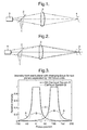
- a point source 1 generates a light beam which impinges upon a beam splitter 2 from which it is reflected onto a focussing lens 3.
- the lens 3 focuses the light onto an object 4 and then light reflected or emitted by the object passes back to the lens 3 where it is focussed through the beam splitter 2 onto a detector 5 such as a photomultiplier tube located behind a pinhole (not shown) acting as a field stop.
- the illumination source 1 is the same size as the pinhole.
- FIG. 2 An out-of-focus situation is shown in Figure 2.
- the detector 5 and illumination area is normally moved across the object 4 in a raster scanning mechanism, usually with moving mirrors. If three-dimensional images are required then these can be produced by producing the two-dimensional images at different focal planes. This three-dimensional image can then be visualised using a three-dimensional imaging device or using a three-dimensional visualisation package. Alternatively, a two-dimensional image can be produced by merging the two-dimensional images of the different focus planes and thus produce an image with greater depth of focus than that which can be produced with a multiphoton microscope with the same optical resolution.
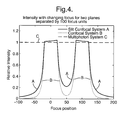
- the light level drop is proportional to the fourth power of the defocus. This is because the light level illuminating the object drops proportional to the square of the defocus and the light level detected from the object drops proportional to the square of the defocus.
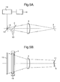
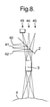
- the detector 13 is connected to a processor 14 which in turn is connected to a data store 15.
- the slit opening 12 or detector 13 is maintained stationary and a mirror system used to cause light from the light source 1 to impinge on different lines of pixels on the object surface 4.
- Suitable systems are shown in Figures 2 and 3 of "Handbook of Biological Confocal Miscroscopy ", 2 nd Edition, James B Pawley, Plenum Press, chapter 25.
- a mirror system is provided to cause successive lines of pixels on the object to be illuminated by the line of light from the source 1 and the detector 13 is moved across the confocal plane defined by the lens 3 so as to record light from each illuminated line of pixels.
- the slit in front of the detector is omitted and the detector array is effectively moved in phase with the "slit".
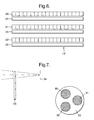
- the filter wheel 33 is then rotated to bring the green filter 31 into alignment with the white light and the process repeated.
- the process is repeated once more with the filter 32 in alignment with the white light source.
- the three sets of colour information for each pixel in the same line on the object 4 are then processed as described above.
Landscapes
- Physics & Mathematics (AREA)
- Chemical & Material Sciences (AREA)
- Analytical Chemistry (AREA)
- General Physics & Mathematics (AREA)
- Optics & Photonics (AREA)
- Microscoopes, Condenser (AREA)
Applications Claiming Priority (2)
| Application Number | Priority Date | Filing Date | Title |
|---|---|---|---|
| US818585 | 1997-03-14 | ||
| US10/818,585 US20050225849A1 (en) | 2004-04-05 | 2004-04-05 | Slit confocal microscope and method |
Publications (2)
| Publication Number | Publication Date |
|---|---|
| EP1586931A2 true EP1586931A2 (de) | 2005-10-19 |
| EP1586931A3 EP1586931A3 (de) | 2006-10-11 |
Family
ID=34940562
Family Applications (1)
| Application Number | Title | Priority Date | Filing Date |
|---|---|---|---|
| EP05251461A Withdrawn EP1586931A3 (de) | 2004-04-05 | 2005-03-10 | Konfokales Spaltmikroskop und Verfahren |
Country Status (3)
| Country | Link |
|---|---|
| US (1) | US20050225849A1 (de) |
| EP (1) | EP1586931A3 (de) |
| JP (1) | JP2005292839A (de) |
Cited By (5)
| Publication number | Priority date | Publication date | Assignee | Title |
|---|---|---|---|---|
| WO2007062039A2 (en) | 2005-11-23 | 2007-05-31 | Illumina, Inc. | Confocal imaging methods and apparatus |
| CN101872064B (zh) * | 2009-04-24 | 2012-07-04 | 陈亮嘉 | 线型多波长共焦显微镜模块以及其共焦显微方法与系统 |
| EP3355038A1 (de) * | 2017-01-25 | 2018-08-01 | Specim, Spectral Imaging Oy Ltd | Bildgebungsvorrichtung und betriebsverfahren |
| US10317656B2 (en) | 2013-12-20 | 2019-06-11 | Centre National De La Recherche Scientifique | Optical coherence tomography apparatus and method using line confocal filtering |
| US10642013B2 (en) | 2014-04-30 | 2020-05-05 | Olympus Corporation | Specimen observation apparatus |
Families Citing this family (10)
| Publication number | Priority date | Publication date | Assignee | Title |
|---|---|---|---|---|
| DE602005004332T2 (de) | 2004-06-17 | 2009-01-08 | Cadent Ltd. | Verfahren zum Bereitstellen von Daten im Zusammenhang mit der Mundhöhle |
| US7684048B2 (en) | 2005-11-15 | 2010-03-23 | Applied Materials Israel, Ltd. | Scanning microscopy |
| US8509565B2 (en) * | 2008-12-15 | 2013-08-13 | National Tsing Hua University | Optimal multi-resolution blending of confocal microscope images |
| DE102011078817A1 (de) * | 2011-06-17 | 2012-12-20 | Siemens Aktiengesellschaft | Verfahren zur dreidimensionalen Vermessung eines Körpers und Vorrichtung |
| DE102011114500B4 (de) * | 2011-09-29 | 2022-05-05 | Fei Company | Mikroskopvorrichtung |
| US20150192461A1 (en) * | 2012-07-05 | 2015-07-09 | National University Of Singapore | Light microscope and method of controlling the same |
| JP6519578B2 (ja) * | 2016-12-27 | 2019-05-29 | カシオ計算機株式会社 | 姿勢検出装置、及び姿勢検出方法 |
| WO2018190125A1 (ja) * | 2017-04-13 | 2018-10-18 | 横河電機株式会社 | 共焦点スキャナ、顕微鏡システム、及び共焦点顕微鏡 |
| JP6677238B2 (ja) | 2017-04-13 | 2020-04-08 | 横河電機株式会社 | 共焦点スキャナ、及び共焦点顕微鏡 |
| US12061329B2 (en) | 2020-03-27 | 2024-08-13 | Sony Group Corporation | Microscope system, imaging method, and imaging device |
Citations (2)
| Publication number | Priority date | Publication date | Assignee | Title |
|---|---|---|---|---|
| US5936764A (en) * | 1993-04-15 | 1999-08-10 | Kowa Company Ltd. | Laser scanning optical microscope |
| US6111690A (en) | 1997-01-23 | 2000-08-29 | Yokogawa Electric Corporation | Confocal microscopic equipment |
Family Cites Families (6)
| Publication number | Priority date | Publication date | Assignee | Title |
|---|---|---|---|---|
| US3013467A (en) * | 1957-11-07 | 1961-12-19 | Minsky Marvin | Microscopy apparatus |
| US5963676A (en) * | 1997-02-07 | 1999-10-05 | Siemens Corporate Research, Inc. | Multiscale adaptive system for enhancement of an image in X-ray angiography |
| US6134002A (en) * | 1999-01-14 | 2000-10-17 | Duke University | Apparatus and method for the rapid spectral resolution of confocal images |
| JP2000275027A (ja) * | 1999-03-23 | 2000-10-06 | Takaoka Electric Mfg Co Ltd | スリット共焦点顕微鏡とそれを用いた表面形状計測装置 |
| DE10029680B4 (de) * | 2000-06-23 | 2016-06-16 | Leica Microsystems Cms Gmbh | Mikroskop-Aufbau |
| KR20020084786A (ko) * | 2001-05-04 | 2002-11-11 | 이재웅 | 선형 선 스캐닝을 이용하는 공초점 영상 형성 장치 및 방법 |
-
2004
- 2004-04-05 US US10/818,585 patent/US20050225849A1/en not_active Abandoned
-
2005
- 2005-03-10 EP EP05251461A patent/EP1586931A3/de not_active Withdrawn
- 2005-04-04 JP JP2005107427A patent/JP2005292839A/ja active Pending
Patent Citations (2)
| Publication number | Priority date | Publication date | Assignee | Title |
|---|---|---|---|---|
| US5936764A (en) * | 1993-04-15 | 1999-08-10 | Kowa Company Ltd. | Laser scanning optical microscope |
| US6111690A (en) | 1997-01-23 | 2000-08-29 | Yokogawa Electric Corporation | Confocal microscopic equipment |
Cited By (14)
| Publication number | Priority date | Publication date | Assignee | Title |
|---|---|---|---|---|
| US8884211B2 (en) | 2005-11-23 | 2014-11-11 | Illumina, Inc. | Confocal imaging methods and apparatus |
| US9816929B2 (en) | 2005-11-23 | 2017-11-14 | Illumina, Inc. | Confocal imaging methods and apparatus |
| US7329860B2 (en) | 2005-11-23 | 2008-02-12 | Illumina, Inc. | Confocal imaging methods and apparatus |
| US7589315B2 (en) | 2005-11-23 | 2009-09-15 | Illumina, Inc. | Confocal imaging methods and apparatus |
| US7960685B2 (en) | 2005-11-23 | 2011-06-14 | Illumina, Inc. | Confocal imaging methods and apparatus |
| US8158926B2 (en) | 2005-11-23 | 2012-04-17 | Illumina, Inc. | Confocal imaging methods and apparatus |
| EP2594981A3 (de) * | 2005-11-23 | 2013-06-19 | Illumina Inc. | Konfokale Abbildungsverfahren und Vorrichtung |
| CN101361015B (zh) * | 2005-11-23 | 2012-11-21 | 伊鲁米那股份有限公司 | 共焦成像的方法和装置 |
| WO2007062039A3 (en) * | 2005-11-23 | 2007-07-19 | Illumina Inc | Confocal imaging methods and apparatus |
| WO2007062039A2 (en) | 2005-11-23 | 2007-05-31 | Illumina, Inc. | Confocal imaging methods and apparatus |
| CN101872064B (zh) * | 2009-04-24 | 2012-07-04 | 陈亮嘉 | 线型多波长共焦显微镜模块以及其共焦显微方法与系统 |
| US10317656B2 (en) | 2013-12-20 | 2019-06-11 | Centre National De La Recherche Scientifique | Optical coherence tomography apparatus and method using line confocal filtering |
| US10642013B2 (en) | 2014-04-30 | 2020-05-05 | Olympus Corporation | Specimen observation apparatus |
| EP3355038A1 (de) * | 2017-01-25 | 2018-08-01 | Specim, Spectral Imaging Oy Ltd | Bildgebungsvorrichtung und betriebsverfahren |
Also Published As
| Publication number | Publication date |
|---|---|
| JP2005292839A (ja) | 2005-10-20 |
| EP1586931A3 (de) | 2006-10-11 |
| US20050225849A1 (en) | 2005-10-13 |
Similar Documents
| Publication | Publication Date | Title |
|---|---|---|
| US5751417A (en) | Arrangement for confocal fluorescence microscopy | |
| US7335898B2 (en) | Method and apparatus for fluorescent confocal microscopy | |
| US9864182B2 (en) | High-resolution scanning microscopy | |
| US11372223B2 (en) | High-resolution scanning microscopy | |
| US9234846B2 (en) | High-resolution microscope and method for determining the two- or three-dimensional positions of objects | |
| EP2864741B1 (de) | Objektträgerscanner für fluoreszenz- und hellfeldbildgebung in der pathologie und verfahren zum betrieb | |
| CN110023811B (zh) | 针对用于显微镜的探测光的光学组件、用于显微镜检查的方法和显微镜 | |
| EP1586931A2 (de) | Konfokales Spaltmikroskop und Verfahren | |
| GB2321517A (en) | Confocal microscopic equipment for measuring solid shapes | |
| EP3198324B1 (de) | Auflösungserhöhung für systeme und verfahren zur zeilenabtastenden anregungsmikroskopie | |
| US10514533B2 (en) | Method for creating a microscope image, microscopy device, and deflecting device | |
| US11106026B2 (en) | Scanning microscope for 3D imaging using MSIA | |
| CA3035876A1 (en) | Line scan photon reassignment microscopy systems | |
| US11686928B2 (en) | Light microscope | |
| CA3065917A1 (en) | Super-resolution line scanning confocal microscopy with pupil filtering | |
| US7312920B2 (en) | Confocal microscope | |
| CN120359447A (zh) | 显微镜 | |
| JP2025502743A (ja) | 顕微鏡及び顕微鏡法のための方法 | |
| CN120178490A (zh) | 显微镜以及借助于扫描显微镜进行扫描图像获取的方法 | |
| JP2007506146A (ja) | 共焦点レーザ走査顕微鏡 | |
| US20140293037A1 (en) | Optical microscope and method for examining a microscopic sample | |
| US20050017197A1 (en) | Scanning microscope and method for scanning microscopy | |
| KR102058780B1 (ko) | 라인 스캐닝 방식의 공초점 현미경에서의 자동초점조절 방법 및 장치 | |
| US20230236408A1 (en) | A method for obtaining an optically-sectioned image of a sample, and a device suitable for use in such a method | |
| JP2004029373A (ja) | カラー顕微鏡 |
Legal Events
| Date | Code | Title | Description |
|---|---|---|---|
| PUAI | Public reference made under article 153(3) epc to a published international application that has entered the european phase |
Free format text: ORIGINAL CODE: 0009012 |
|
| AK | Designated contracting states |
Kind code of ref document: A2 Designated state(s): AT BE BG CH CY CZ DE DK EE ES FI FR GB GR HU IE IS IT LI LT LU MC NL PL PT RO SE SI SK TR |
|
| AX | Request for extension of the european patent |
Extension state: AL BA HR LV MK YU |
|
| PUAL | Search report despatched |
Free format text: ORIGINAL CODE: 0009013 |
|
| AK | Designated contracting states |
Kind code of ref document: A3 Designated state(s): AT BE BG CH CY CZ DE DK EE ES FI FR GB GR HU IE IS IT LI LT LU MC NL PL PT RO SE SI SK TR |
|
| AX | Request for extension of the european patent |
Extension state: AL BA HR LV MK YU |
|
| 17P | Request for examination filed |
Effective date: 20070328 |
|
| AKX | Designation fees paid |
Designated state(s): DE GB |
|
| 17Q | First examination report despatched |
Effective date: 20080429 |
|
| RAP1 | Party data changed (applicant data changed or rights of an application transferred) |
Owner name: FUJIFILM ELECTRONIC IMAGING LIMITED |
|
| RAP1 | Party data changed (applicant data changed or rights of an application transferred) |
Owner name: FFEI LIMITED |
|
| RAP1 | Party data changed (applicant data changed or rights of an application transferred) |
Owner name: FFEI LIMITED |
|
| RAP3 | Party data changed (applicant data changed or rights of an application transferred) |
Owner name: FFEI LIMITED |
|
| STAA | Information on the status of an ep patent application or granted ep patent |
Free format text: STATUS: THE APPLICATION IS DEEMED TO BE WITHDRAWN |
|
| 18D | Application deemed to be withdrawn |
Effective date: 20170927 |
|
| RIC1 | Information provided on ipc code assigned before grant |
Ipc: G02B 21/00 20060101AFI20050827BHEP |