EP1593736B1 - Vorrichtung und Verfahren zum Nachweis von Mikroorganismen - Google Patents
Vorrichtung und Verfahren zum Nachweis von Mikroorganismen Download PDFInfo
- Publication number
- EP1593736B1 EP1593736B1 EP05009824.3A EP05009824A EP1593736B1 EP 1593736 B1 EP1593736 B1 EP 1593736B1 EP 05009824 A EP05009824 A EP 05009824A EP 1593736 B1 EP1593736 B1 EP 1593736B1
- Authority
- EP
- European Patent Office
- Prior art keywords
- microorganisms
- detecting
- section
- sample
- reagent
- Prior art date
- Legal status (The legal status is an assumption and is not a legal conclusion. Google has not performed a legal analysis and makes no representation as to the accuracy of the status listed.)
- Expired - Lifetime
Links
Images



Classifications
-
- C—CHEMISTRY; METALLURGY
- C12—BIOCHEMISTRY; BEER; SPIRITS; WINE; VINEGAR; MICROBIOLOGY; ENZYMOLOGY; MUTATION OR GENETIC ENGINEERING
- C12Q—MEASURING OR TESTING PROCESSES INVOLVING ENZYMES, NUCLEIC ACIDS OR MICROORGANISMS; COMPOSITIONS OR TEST PAPERS THEREFOR; PROCESSES OF PREPARING SUCH COMPOSITIONS; CONDITION-RESPONSIVE CONTROL IN MICROBIOLOGICAL OR ENZYMOLOGICAL PROCESSES
- C12Q1/00—Measuring or testing processes involving enzymes, nucleic acids or microorganisms; Compositions therefor; Processes of preparing such compositions
- C12Q1/02—Measuring or testing processes involving enzymes, nucleic acids or microorganisms; Compositions therefor; Processes of preparing such compositions involving viable microorganisms
- C12Q1/04—Determining presence or kind of microorganism; Use of selective media for testing antibiotics or bacteriocides; Compositions containing a chemical indicator therefor
Definitions
- the present invention relates to a microorganisms detecting device and a microorganisms detecting method for detecting microorganisms in a sample, and in more detail to a microorganisms detecting device and a microorganisms detecting method to be utilized in diagnosis of infection, tests for microorganisms in food products, detection of microorganisms in circumstances, quality control of cleaning water for semiconductors and the like.
- microorganisms detecting methods have also been developed with the advancement of the tissue engineering.
- a microorganisms detecting method using precursor phenolic oxidase also is one of such microorganisms detecting methods (refer to, for example, Japanese Patent Application Laid-Open No. 98,798/1997 ).
- a microorganism detecting device according to the preamble of independent claim 1 is shown from US 3,756,920 A and WO 02/068580 A2 .
- the present invention provides a microorganisms detecting device and a microorganisms detecting method, which are simple and able to shorten the time for detecting microorganisms and further intend to improve the sensitivity of detection for microorganisms, particularly gram negative bacteria.
- the microorganisms detecting device is a device for detecting microorganisms, comprising: a sampling section adapted for taking a sample from a detection target, a sample concentrating section adapted for concentrating the microorganisms existing in the sample taken in the sampling section, said sample concentrating section including filtering means for collecting the microorganisms, a reagent adding section adapted for adding a reagent having a component which changes the reagent color tone by existence of the microorganisms to the sample concentrated in the sample concentrating section, a microorganisms detecting section adapted for detecting the change of the color tone of the reagent added to the sample in the reagent adding section by means of an optical system, a detected result outputting section adapted for outputting the detected result detected in the microorganisms detecting section, and a controller adapted for controlling the sampling section, the sample concentrating section, the reagent adding section, the microorganisms detecting section, and the detected result outputting section
- the microorganism detecting device is characterized in that the sample concentrating section includes detecting sensitivity improving means for improving detecting sensitivity for particular microorganisms in the microorganisms detecting section.
- the microorganism detecting device is characterized in that the detecting sensitivity improving means is breakout of the microorganisms' membrane structures by heating or drying.
- the microorganisms detecting device is characterized in that the sample concentrating section comprises temperature maintaining means for maintaining the sample at a predetermined temperature.
- the microorganisms detecting device is characterized in that the optical system comprises a light source outputting light that has wave lengths of the visible light range.
- the microorganisms detecting method is a method for detecting microorganisms using the device of the invention, comprising: a sampling step for taking a sample from a detection target, a sample concentrating step for concentrating the microorganism existing in the sample taken in the sampling step by means of a filtering means for collecting the microorganisms, a reagent adding step for adding a reagent having a component which changes the reagent color tone by existence of the microorganisms to the sample concentrated in the sample concentrating step, a microorganisms detecting step for detecting the change of the color tone of the reagent added in the reagent adding step, a detected result outputting step for outputting the detected result detected in the microorganisms detecting step, and controlling the sampling step, the sample concentrating step, the reagent adding step, the microorganisms detecting step, and the detected result outputting step by control processing, characterized in that the sample concentration step further includes supplying a culture medium to the
- the microorganism detecting method characterized in that the sample concentrating step includes detecting sensitivity improving treatment for improving detecting sensitivity for particular microorganisms in the microorganisms detecting step.
- the microorganism detecting method is characterized in that the detecting sensitivity improving treatment is breakout of the microorganisms' membrane structures by heating or drying.
- the invention it is possible to detect the existence of microorganisms securely with the simple detecting device or method, to shorten the time for detection and to improve the sensitivity of detection, particularly sensitivity of detection for microorganisms such gram negative bacteria and the like.
- FIG. 1 is a system organization chart of the microorganisms detecting system 100 according to the invention.
- the microorganisms detecting system 100 comprises a sampling section 10, a sample concentrating section 20, a reagent adding section 30, and a microorganisms detecting section 40, and the sample concentrating section 20, the reagent adding section 30 and the microorganisms detecting section 40 are arranged in a constant temperature bath 50.
- a controller 60 controls operations of these sections and setting of environmental conditions, and receives detected results from the microorganisms detecting section 40, which are transmitted to a detected result outputting section 70 to output the microorganisms detected results.
- a culture medium for culturing cell which may be discarded was added to test tubes.
- substantially all the amount of culture medium for culturing cells is employed for detection so that more accurate detected results can be obtained.
- the sampling section 10 in the microorganisms detecting system 100 is most in risk of invasion of microorganisms from exterior, it is desired to arrange the microorganisms detecting system 100 in its entirety in a biological clean chamber. Further, it is desired to arrange the sampling section 10 in a biological clean chamber and to employ a structure capable of automatically replacing the culture medium for culturing the cells from the flask for culturing cells into a microorganisms detecting test tube.
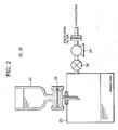
- a drain tank 23 under a microorganisms detecting test tube 22 is evacuated by a vacuum pump 24 provided on the drain tank 23 to collect the microorganisms in the culture medium by means of a filter 26 at the bottom of the microorganisms detecting test tube 22.
- the filter 26 has pore size of the order of 0.1 to 10 ⁇ m, preferably 0.2 ⁇ m in diameter. In this system, a filter of "Ultra Free MC" manufactured by Millipore Co., Ltd. is used.
- the culture medium from which microorganisms have been collected is discharged from the microorganisms detecting test tube 22 by the vacuum pump 24, and a discharging valve 28 is closed. Thereafter, a culture medium for culturing the microorganisms is added into the microorganisms detecting test tube 22 from its upper portion.
- Glucose-pepton liquid medium (GP liquid medium) is used as a culture medium for culturing microorganisms
- other media such as Soybean-casein-digest liquid medium (SCD liquid medium), Thioglycolic acid medium (TG medium), Sabouraud-glucose medium, Brainheart infusion, Mullerhinton, usual bouillon, or Glucose peptone and the like.
- SCD liquid medium Soybean-casein-digest liquid medium
- TG medium Thioglycolic acid medium
- Sabouraud-glucose medium Brainheart infusion, Mullerhinton, usual bouillon, or Glucose peptone and the like.
- a plurality of microorganisms detecting test tubes 22 may be previously prepared, and after microorganisms have been collected in the respective test tubes, culture media for culturing plural kinds of microorganisms, which are commensurate with microorganisms expected to be detected, may be added into the microorganisms detecting test tubes, respectively.
- the microorganisms After the culture medium for culturing the microorganisms is added into the microorganisms detecting test tube 22, the microorganisms are cultured at a constant temperature of 30 to 40°C for 3 to 12 hours, preferably 5 to 8 hours. As shown in FIG. 3 , the microorganisms have a property increasing twice in number for about 30 minutes although depending on their kinds. Therefore, even if only one microorganism before cultivation, it will breed more than 1000 for five hours.
- Microorganisms of fungi such as Candida Albicanis, Candida Lusitaniae, Candida Krusei, Candida Glabrata, Cryptococcus Neoformans, Aspergillus Fumigatus, Pneumocistis Carinii and the like actively grow within a temperature range of about 30°C
- microorganisms of bacteria such as Bacillus Subtilis, Streptococcus Pyogenes, Salmonella Typhimurium, Salmonella Bongori, Salmonella Enteritidis, Escherichia Coli, Staphylococcus Epidermidis, Staphylococcus Aureus, Pseudomonasu Aeruginosa and the like actively grow within a temperature range of about 37°C.
- a plurality of microorganisms detecting test tubes 22 may be previously prepared, and after microorganisms have been collected in the microorganisms detecting test tubes, respectively, the microorganisms may be cultured within temperature ranges of plural kinds commensurate with the microorganisms expected to be detected.
- the detecting sensitivity of this reagent for gram negative bacteria is lower in comparison with other fungi and bacteria. Therefore, after microorganisms have been cultured in the microorganisms detecting test tube 22, the microorganisms may be broken by the use of drying, heating, ultrasonic wave or surface-active agent. As shown in FIG. 4 , the gram negative bacteria 90 have peptide glycan 92 reacting to the reagent A inside an external membrane 94 so that the detecting sensitivity with the reagent A is lower.
- the external membrane 94 or cytoplasmic membrane 96 is broken as described above to cause the peptide glycan 92 which has been between the external membrane 94 and the cytoplasmic membrane 96 of the gram negative bacteria 90 to be exposed outside so that it becomes likely to react to the reagent A.
- the microorganisms are cultured to improve the detecting sensitivity for the microorganisms in the manner described above, and the culture medium for culturing the microorganisms is discharged and the treatment for further improving the sensitivity such as breaking the microorganisms is carried out. Thereafter, a reagent is added into the microorganisms detecting test tube 22 in the reagent adding section 30.
- reagent A is used here, Limulus reagent (reagent B) manufactured by Wako pure Industries and the like may be used.
- microorganisms detecting test tubes 22 may be previously prepared, and after microorganisms have been collected in the respective test tubes, reagents of plural kinds may be added into the microorganisms detecting test tubes commensurate with expected microorganisms to be detected, respectively.
- the microorganisms detecting section 40 comprises a locating stage 42 for the microorganisms detecting test tube 22, a light source (630 nm) 44, a half mirror 46, mirrors 47a and 47b, and light receiving elements 48a and 48b, and these parts are arranged in a light shielding chamber 49.
- the reagent A is added into the microorganisms detecting test tube 22, it is required to wait for the progress of reaction for approximately 120 minutes until the color tone in the microorganisms detecting test tube 22 is stabilized.
- the microorganisms detecting test tube 22 is located on the locating stage 42 for the microorganisms detecting test tube in the microorganisms detecting section 40.
- the temperature of the locating stage 42 is set at a temperature suitable for reaction of the reagent, for example, at 30°C with the reagent A or at 37°C with the reagent B.
- the quantity of the transmitted light in the light receiving element 48b that is, the degree of light absorption of the reagent in the microorganisms detecting test tube 22 is calculated with the aid of the signal from the light receiving element 48a as reference signal.
- These measured results and calculated results are transmitted from the controller 60 to the detected result outputting section 70 from which these results are output as data.
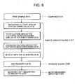
- Example 1 A method for detecting microorganisms in Example 1 with reference to FIG. 6 .
- Step 1 In the sampling section 20, a sample is taken by replacing a culture medium used for culturing cells from a flask for culturing the cells to a microorganisms detecting test tube 22 (S 11).
- Step 2 The discharging valve 28 is opened and the vacuum pump 24 is started so that the culture medium for culturing the cells is discharged through the filter 26 from the bottom of the microorganisms detecting test tube 22 in which the sample has been taken, thereby causing microorganisms to remain on the filter 26 if there have been microorganisms in the culture medium for culturing the cells (S 12)
- Step 3 After the discharge of the culture medium for culturing the cells has been completed, the vacuum pump 24 is stopped and the discharging valve 28 is closed and then a culture medium for culturing the microorganisms is poured into the microorganisms detecting test tube 22 from its upper portion. After the pouring of the culture medium for culturing the microorganisms has been completed, the microorganisms detecting test tube 22 is laid to rest in the constant temperature bath 50 at 37°C for 5 hours to culture the microorganisms (S 13).
- Step 4 Again the discharging valve 28 is opened and the vacuum pump 24 is started so that the culture medium for culturing the microorganisms is discharged through the filter 26 from the bottom of the microorganisms detecting test tube 22 (S 14).
- Step 5 After the discharge of the culture medium for culturing the microorganisms has been completed, the vacuum pump 24 is stopped and the discharging valve 28 is closed.
- the temperature of the constant temperature bath 50 is set at 100°C and the microorganisms detecting test tube 22 is heated in the bath for 20 minutes, thereby breaking the cell membranes of the microorganisms (S 15).
- Step 6 After the heating treatment in the constant temperature bath 50 has been completed, the reagent A is added into the microorganisms detecting test tube 22 from its upper portion (S 16).
- Step 7 The microorganisms detecting test tube 22 is arranged on the locating stage 42 in the microorganisms detecting section 40 to measure degree of light absorption or absorbance of the reagent A in the microorganisms detecting test tube 22. In this manner, it is possible to detect whether there have been microorganisms in the culture medium for culturing the cells (S 17).
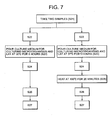
- a method for detecting microorganisms in the Example 2 will then be explained with reference to FIG. 7 .
- Step 1 Two microorganisms detecting test tubes 22 are prepared, and a culture medium used for culturing cells is replaced from the flask for culturing cells to the microorganisms detecting test tubes 22 in the sampling section 20 to prepare two samples (S 21).
- Step 2 For each of the two microorganisms detecting test tubes 22, the discharging valve 28 is opened and the vacuum pump 24 is started so that the culture medium for culturing the cells is discharged through the filter 26 from the bottom of the microorganisms detecting test tube accommodating the sample (S 22).
- Step 3 After the discharge of the culture medium for culturing the cells has been completed, the vacuum pump 24 is stopped and the discharging valve 28 is closed and then a culture medium for culturing the microorganisms is poured into each of the microorganisms detecting test tubes 22 from its upper portion. After the pouring of the culture medium for culturing the microorganisms has been completed, one microorganisms detecting test tube 22 is laid to rest in the constant temperature bath 50 being set at 30°C for 5 hours to culture the microorganisms. On the other hand, the other microorganisms detecting test tube 22 is also laid to rest in the constant temperature bath 50 being set at 37°C for 5 hours to culture the microorganisms (S 23).
- Step 4 For each of the microorganisms detecting test tubes 22, again the discharging valve 28 is opened and the vacuum pump 24 is started so that the culture medium for culturing the microorganisms is discharged through the filter 26 from the bottom of the microorganisms detecting test tube 22 (S 24).
- Step 5 After the discharge of the culture medium for culturing the microorganisms has been completed, the vacuum pump 24 is stopped and the discharging valve 28 is closed. The other microorganisms detecting test tube 22 is heated in the constant temperature bath 50 being set at 100°C for 20 minutes. This heating treatment for the one microorganisms detecting test tube 22 is not carried out since it is not necessary for this tube (S 25).
- Step 6 After the heating treatment for the other microorganisms detecting tube 22 has been finished (after the step 4 for the one microorganisms detecting test tube 22 has been finished), the reagent A as a reagent is added into the two microorganisms detecting test tubes 22 from their upper portions, respectively (S 26).
- Step 7 The two microorganisms detecting test tubes 22 are each arranged on the locating stage 42 of the microorganisms detecting section 40 to measure light absorption or absorbance of the reagent A in the microorganisms detecting test tube 22. In this manner, it is possible to detect whether there have been microorganisms in the culture medium for culturing the cells and further whether there have been fungi or bacteria in the culture medium (S 27).
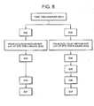
- a method for detecting microorganisms in the Example 3 will then be explained with reference to FIG. 8 .
- Step 1 Two microorganisms detecting test tubes are prepared, and a culture medium used for culturing cells is replaced from the flask for culturing cells to the microorganisms detecting test tubes 22 in the sampling section 20 to prepare two samples (S 31).
- Step 2 For each of the two microorganisms detecting test tubes 22, the discharging valve 28 is opened and the vacuum pump 24 is started so that the culture medium for culturing the cells is discharged through the filter 26 from the bottom of the microorganisms detecting test tube accommodating the sample (S 32).
- Step 3 After the discharge of the culture medium for culturing the cells has been completed, the vacuum pump 24 is stopped and the discharging valve 28 is closed and then GP liquid medium as a culture medium for culturing the microorganisms is poured into one microorganisms detecting test tube 22 from its upper portion, while SCD liquid medium as a culture medium for culture the microorganisms is poured into the other microorganisms detecting test tube 22 from its upper portion.
- both the microorganisms detecting test tubes 22 may be laid in the constant temperature bath whose temperature is set at 35°C to culture microorganisms.
- the one microorganisms detecting test tube 22 may be laid in the constant temperature bath being set at 30°C and the other test tube 22 may be laid in the constant temperature bath set at 37°C, respectively for 5 hours to culture the microorganisms, thereby enabling the measurement with higher sensitivity (S 33).
- Step 4 For each of the microorganisms detecting test tubes 22, again the discharging valve 28 is opened and the vacuum pump 24 is started so that the culture medium for culturing the microorganisms is discharged through the filter 26 from the bottom of the microorganisms detecting test tube 22 (S 34).
- Step 5 After the discharge of the culture medium for culturing the microorganisms has been completed, the vacuum pump 24 is stopped and the discharging valve 28 is closed. The other microorganisms detecting test tube 22 is heated in the constant temperature bath 50 being set at 100°C for 20 minutes. This heating treatment for the one microorganisms detecting test tube 22 is not carried out since it is not necessary for this tube (S 35).
- Step 6 After the heating treatment for the other microorganisms detecting tube 22 has been finished (after the step 4 for the one microorganisms detecting test tube 22 has been finished), the reagent A as a reagent is added into the two microorganisms detecting test tubes 22 from their upper portions, respectively (S 36).
- Step 7 The two microorganisms detecting test tubes 22 are each arranged on the locating stage 42 of the microorganisms detecting section 40 to measure light absorption or absorbance of the reagent A in the microorganisms detecting test tube 22. In this manner, it is possible to detect whether there have been microorganisms in the culture medium for culturing the cells and further whether there have been fungi or bacteria in the culture medium (S 37).
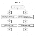
- Step 1 Two microorganisms detecting test tubes are prepared, and a culture medium used for culturing cells is replaced from the flask for culturing cells to the microorganisms detecting test tubes 22 in the sampling section 20 to prepare two samples (S 41).
- Step 2 For each of the two microorganisms detecting test tubes 22, the discharging valve 28 is opened and the vacuum pump 24 is started so that the culture medium for culturing the cells is discharged through the filter 26 from the bottom of the microorganisms detecting test tube accommodating the sample (S 42).
- Step 3 After the discharge of the culture medium for culturing the cells has been completed, the vacuum pump 24 is stopped and the discharging valve 28 is closed and then a culture medium for culturing the microorganisms is poured into each of the microorganisms detecting test tubes 22 from its upper portion. After the pouring of the culture medium for culturing the microorganisms has been completed, both the microorganisms detecting test tubes 22 are laid to rest in the constant temperature bath 50 being set at 35°C for 5 hours to culture the microorganisms (S 43).
- Step 4 For each of the microorganisms detecting test tubes 22, again the discharging valve 28 is opened and the vacuum pump 24 is started so that the culture medium for culturing the microorganisms is discharged through the filter 26 from the bottom of the microorganisms detecting test tube 22 (S 44).
- Step 5 After the discharge of the culture medium for culturing the microorganisms has been completed, the vacuum pump 24 is stopped and the discharging valve 28 is closed. The reagent A is added into the one microorganisms detecting test tube 22 from its upper portion, and the reagent B is added into the other microorganisms detecting test tube 22 from its upper portion (S 46).
- Step 6 The two microorganisms detecting test tubes 22 are each arranged on the locating stage 42 of the microorganisms detecting section 40 to measure light absorption or absorbance of the respective reagent in the microorganisms detecting test tube 22. In this manner, it is possible to detect whether there have been microorganisms in the culture medium for culturing the cells without treatment for breaking the cell membranes as by heating (S 47).
- the experiment was executed by the use of Escherichia Coli (IF03301) as a sample of the gram negative bacteria, Bacillus Subtilis (IF03035) as a sample of the gram positive bacteria and Aspergillus Nigger (IF06341) as a sample of the fungi.
- the samples were prepared by making serial 1:10 dilution of the respective microorganism, and the samples were each separately attached with 50 ⁇ l to three micro plates (only one micro plate for the fungi). Then, these samples were divided into three groups, that is, the first group in that without being particularly treated, the samples are mixed with the (untreated) reagent A, the second group in that after being heat treated, the samples are mixed with the reagent A, and the third group in that after being treated by drying, the samples are mixed with the reagent A.
- the number of bacteria (viable bacteria) of each dilute group was determined with the culture on the plates of the respective dilute group and analytical curves obtained therefrom.
- the reagent A was added to them.
- the treated group by vacuum drying after the samples were heated at 40°C for 2 hours and added with water for adjustment, the regent A was added to them.
- Table 1 Number of bacteria Gram negative bacteria Gram positive bacteria Fungi Untreated Heated Dried Untreated Heated Dried Untreated Heated Dried Untreated 10 5 /ml ⁇ ⁇ ⁇ ⁇ ⁇ ⁇ ⁇ 10 5 /ml ⁇ ⁇ ⁇ ⁇ ⁇ ⁇ 10 5 /ml ⁇ ⁇ ⁇ ⁇ ⁇ ⁇ 10 5 /ml ⁇ ⁇ ⁇ ⁇ ⁇ ⁇ 10 5 /ml ⁇ ⁇ ⁇ ⁇ ⁇ ⁇ 10 5 /ml ⁇ ⁇ ⁇ ⁇ ⁇ ⁇ ⁇ ⁇ ⁇ ⁇ ⁇ ⁇
- the detection target containing microorganisms are not limited to the culture medium for culturing cells and may include washing water to be used in producing steps for foods, and liquid foods and beverages such as juice, wine and the like, air in the sanitarily managed circumstances (filters through which the air flows) or cleaning water for semiconductors.
- a flat membrane may be used for collecting microorganisms.
- porous electrodes made of carbon By attaching porous electrodes made of carbon to the flat membrane and causing electric current to flow through the electrodes, more effective collection of microorganisms becomes possible.
- microorganisms can be collected by centrifugal separation, adsorption using beads, and the like.
- the GP liquid medium is used as a culture medium for culturing microorganisms
- SCD liquid medium, TG medium, Sabouraud glucose medium, Brainheart infusion, Mullerhinton, usual bouillon, or glucose peptone may be used.
- the culture medium such as GP liquid medium, SCD liquid medium, TG medium, Mullerhinton and the like
- purified water is poured into a microorganisms detecting test tube so that the step of cleaning the microorganisms detecting test tube is inserted before adding a reagent, whereby more accurate detection becomes possible.
- fungi and bacteria are cited in the above description. However, detection is sufficiently possible using other than these such as fungi belonging to the genus Candida, genus Hansenula, genus Saccharomyces, genus Trichosporon, genus Cryptococcus, genus Aspergillus, genus Penicillium, genus Blastomyces, genus Coccidioides, genus Pneumocystis, genus Malassezia, genus Debaryomyces and the like, and bacteria belonging to the genus Bacillus, genus Streptococcus, genus Pseudomonas, genus Escherichia, genus Staphylococcus, genus Klebsiella, genus Serratia, genus Shigella, genus Vibrio, genus Campylobacter, genus Clostridium, genus Yersinia and like.
- the microorganisms detecting section is the device for measuring the absorbance of the reagents using the light source of 630 nm in the above explanation.
- the wave length is not limited to 630 nm insofar as the wave length is in a marginal zone capable of measuring variances in color tone of reagents.
- the absorbance is measured in the above explanation, fluorescent intensity or reflected light may be measured.
- the present invention relates to a general purpose microorganisms detecting device and method capable not only of detecting microorganisms contained in a culture medium for culturing cells but also detecting microorganisms in a short period of time with higher sensitivity from all kinds of fluids (liquids and gases) from which detection of microorganisms is desired, such as washing water to be used in producing steps for foods, and liquid food and beverages such as juice, wine and the like, air in the sanitarily managed circumstances (filters through which the air flows) or cleaning water for semiconductors.
- fluids liquids and gases
Landscapes
- Chemical & Material Sciences (AREA)
- Life Sciences & Earth Sciences (AREA)
- Organic Chemistry (AREA)
- Health & Medical Sciences (AREA)
- Engineering & Computer Science (AREA)
- Zoology (AREA)
- Wood Science & Technology (AREA)
- Proteomics, Peptides & Aminoacids (AREA)
- Molecular Biology (AREA)
- Toxicology (AREA)
- Immunology (AREA)
- Microbiology (AREA)
- Biophysics (AREA)
- Analytical Chemistry (AREA)
- Physics & Mathematics (AREA)
- Biotechnology (AREA)
- Biochemistry (AREA)
- Bioinformatics & Cheminformatics (AREA)
- General Engineering & Computer Science (AREA)
- General Health & Medical Sciences (AREA)
- Genetics & Genomics (AREA)
- Measuring Or Testing Involving Enzymes Or Micro-Organisms (AREA)
- Investigating Or Analysing Biological Materials (AREA)
- Apparatus Associated With Microorganisms And Enzymes (AREA)
Claims (8)
- Mikroorganismus-Detektorvorrichtung mit:einem Probenabschnitt (10), der zum Nehmen einer Probe von einem Detektionsziel ausgebildet ist,einem Probenkonzentrations-Abschnitt (20), der zum Konzentrieren der Mikroorganismen, die in der in dem Probenabschnitt genommenen Probe enthalten sind, ausgebildet ist, wobei der Probenkonzentrations-Abschnitt Filtermittel (26) zum Sammeln der Mikroorganismen aufweist,einem Reagensadditions-Abschnitt (30), der zum Zufügen eines Reagens mit einer Komponente ausgebildet ist, die den Reagens-Farbton durch die Existenz der Mikroorganismen in der Probe, die in dem Probenkonzentration-Abschnitt konzentriert wurde, änderteinem Mikroorganismus-Detektionsabschnitt (40), der zur Erfassung der Änderung des Farbtons des Reagens geeignet ist, das der Probe in dem Reagens-Additions-Abschnitt zugefügt wurde, mittels eines optischen Systems,einem Detektorergebnis-Ausgabeabschnitt (70), der zum Ausgeben des erfassten Ergebnisses, das in dem Mikroorganismen-Detektionsabschnitten detektiert wurde, ausgebildet ist, undeinem Controller (70), der zum Steuern des Probenabschnittes, des Probenkonzentration-Abschnittes, des Reagensadditions-Abschnittes, des Mikroorganismen-Detektionsabschnittes und des Detektionsergebnis-Ausgabeabschnittes ausgebildet ist,dadurch gekennzeichnet, dassder Probenkonzentrations-Abschnitt (20) und der Reagensadditions-Abschnitt (30) ein Mikroorganismendetektions-Testrohr (22) aufweist, wobei die Filtermittel (26) am Boden des Mikroorganismendetektions-Testrohrs (22) vorgesehen sind und wobei ein Ablauftank (23) unter dem Mikroorganismendetektions-Testrohr (22) mit einem Ablassventil (28) und einer Vakuumpumpe (24) verbunden ist,wobei der Probenkonzentrations-Abschnitt (20) ferner Kultivierungsmittel durch Zuführen eines Kulturmediums an die Filtermittel (26) über das Mikroorganismendetektions-Testrohr (22) aufweist, undwobei der Reagensadditions-Abschnitt (30) das Reagens an die Filtermittel (26) über das Mikroorganismendetektions-Testrohr (22) zuführt.
- Mikroorganismus-Detektionsvorrichtung nach Anspruch 1, dadurch gekennzeichnet, dass der Probenkonzentrations-Abschnitt Detektionsempfindlichkeits-Verbesserungsmittel aufweist zum Verbessern der Detektionsempfindlichkeit für bestimmte Mikroorganismen in dem Mikroorganismendetektions-Abschnitt.
- Mikroorganismen-Detektionsvorrichtung nach Anspruch 2, dadurch gekennzeichnet, dass die Detektionsempfindlichkeits-Verbesserungsmittel ein Aufbrechen der Membranstrukturen der Mikroorganismen durch Aufheizen oder Trocknen sind.
- Mikroorganismus-Detektionsvorrichtung nach einem der Ansprüche 1-3, dadurch gekennzeichnet, dass der Probenkonzentrations-Abschnitt ein Temperatur-Haltemittel zum Halten der Probe bei einer vorgegebenen Temperatur aufweist.
- Mikroorganismus-Detektionsvorrichtung nach einem der Ansprüche 1-4, dadurch gekennzeichnet, dass das optische System eine Lichtquelle aufweist, die Licht mit der Wellenlänge des sichtbaren Lichtbereichs ausgibt.
- Mikroorganismus-Detektionsverfahren unter Verwendung der Vorrichtung nach Anspruch 1 mit:einem Probenschritt zum Nehmen einer Probe von einem Detektionsziel,einem Probenkonzentrations-Schritt zum Konzentrieren der Mikroorganismen, die in der Probe enthalten sind, die in dem Probenschritt genommen wurden, mittels einer Filtereinrichtung zum Sammeln der Mikroorganismen,einem Reagensadditions-Schritt zum Zufügen eines Reagens mit einer Komponente, die den Reagens-Farbton durch Existenz der Mikroorganismen der Probe, die in dem Probenkonzentration-Schritt konzentriert wurde, ändert,einem Mikroorganismen-Detektionsschritt zum Erfassen der Änderung des Farbtons des Reagens, das in dem Reagensadditions-Schritt zugefügt wurde,einem Detektorergebnis-Ausgabe-Schritt zum Ausgaben des Detektorergebnisses, das in dem Mikroorganismus-Detektionsschritt detektiert wurde, undSteuern des Probenschrittes, des Probenkonzentrations-Schrittes, des Reagensadditions-Schrittes, des Mikroorganismen-Detektorschrittes und des Detektionsergebnis-Ausgabeschrittes durch Steuerverarbeitung,dadurch gekennzeichnet, dassder Probenkonzentrations-Schritt ferner die Zufuhr eines Kulturmediums an die Filtermittel aufweist, wobei der Reagensadditions-Schritt das Reagens an die Filtermittel zuführt.
- Mikroorganismus-Detektionsverfahren nach Anspruch 6, dadurch gekennzeichnet, dass der Proben-Konzentrationsschritt eine Detektionsempfindlichkeits-Verbesserungsbehandlung umfasst zum Verbessern der Detektionsempfindlichkeit für bestimmte Mikroorganismen in dem Mikroorganismen-Detektionsschritt.
- Mikroorganismus-Detektionsverfahren nach Anspruch 7, dadurch gekennzeichnet, dass die Detektionsempfindlichkeits-Verbesserungsbehandlung ein Aufbrechen der Membranstruktur der Mikroorganismen durch Aufheizen oder Trocknen ist.
Applications Claiming Priority (2)
| Application Number | Priority Date | Filing Date | Title |
|---|---|---|---|
| JP2004137319 | 2004-05-06 | ||
| JP2004137319A JP4429069B2 (ja) | 2004-05-06 | 2004-05-06 | 微生物の検出装置および方法 |
Publications (2)
| Publication Number | Publication Date |
|---|---|
| EP1593736A1 EP1593736A1 (de) | 2005-11-09 |
| EP1593736B1 true EP1593736B1 (de) | 2015-07-08 |
Family
ID=34936169
Family Applications (1)
| Application Number | Title | Priority Date | Filing Date |
|---|---|---|---|
| EP05009824.3A Expired - Lifetime EP1593736B1 (de) | 2004-05-06 | 2005-05-04 | Vorrichtung und Verfahren zum Nachweis von Mikroorganismen |
Country Status (3)
| Country | Link |
|---|---|
| US (1) | US20050250176A1 (de) |
| EP (1) | EP1593736B1 (de) |
| JP (1) | JP4429069B2 (de) |
Families Citing this family (7)
| Publication number | Priority date | Publication date | Assignee | Title |
|---|---|---|---|---|
| JP4646716B2 (ja) | 2005-02-03 | 2011-03-09 | 三洋電機株式会社 | 微生物検出装置及び微生物検出用カセット |
| JP5320915B2 (ja) * | 2008-09-08 | 2013-10-23 | 株式会社Ihi | 微生物汚染検出用キット、微生物汚染検出方法、及び汚染源判別方法 |
| US8481302B2 (en) * | 2008-11-03 | 2013-07-09 | General Electric Company | Total bacteria monitoring system |
| JP5706088B2 (ja) * | 2010-01-06 | 2015-04-22 | 大阪瓦斯株式会社 | インフルエンザウイルスの検知方法およびインフルエンザウイルスの検知装置 |
| EP3357997A1 (de) * | 2015-10-01 | 2018-08-08 | Consejo Superior De Investigaciones Científicas (CSIC) | Integrierter filterhalter und mikroorganismenkonzentrations- und detektionsverfahren |
| KR101847130B1 (ko) | 2017-01-04 | 2018-05-28 | 국민대학교산학협력단 | 식품 내 미생물 분리용 조성물 및 이를 이용한 미생물 분리 방법 |
| CN112094735B (zh) * | 2020-09-02 | 2022-03-29 | 山东省食品药品检验研究院 | 一种用于检验药品中微生物的模拟消化系统装置及方法 |
Family Cites Families (9)
| Publication number | Priority date | Publication date | Assignee | Title |
|---|---|---|---|---|
| US3756920A (en) * | 1971-04-30 | 1973-09-04 | Nasa | In biological samples my measuring light reactions automatic instrument for chemical processing to dedect microorganisms |
| US5420017A (en) * | 1991-01-24 | 1995-05-30 | Orion Corporation Ltd. | Method and kit for the detection of microorganisms |
| US5550032A (en) * | 1994-05-27 | 1996-08-27 | George Mason University | Biological assay for microbial contamination |
| US6599715B1 (en) * | 1999-05-12 | 2003-07-29 | The Regents Of The University Of California | Real time viability detection of bacterial spores |
| US6372895B1 (en) * | 2000-07-07 | 2002-04-16 | 3M Innovative Properties Company | Fluorogenic compounds |
| US7803608B2 (en) * | 2001-02-28 | 2010-09-28 | Biomerieux, Inc. | Integrated filtration and detection device |
| JP2003144193A (ja) * | 2001-11-09 | 2003-05-20 | Mitsubishi Heavy Ind Ltd | 微生物の検出方法及び装置 |
| EP1546367A4 (de) * | 2002-07-24 | 2006-08-16 | Univ Texas | Einfang und nachweis von mikroben mit membranverfahren |
| CA2515075C (en) * | 2003-02-05 | 2012-10-02 | Iquum, Inc. | Sample processing |
-
2004
- 2004-05-06 JP JP2004137319A patent/JP4429069B2/ja not_active Expired - Fee Related
-
2005
- 2005-04-27 US US11/115,294 patent/US20050250176A1/en not_active Abandoned
- 2005-05-04 EP EP05009824.3A patent/EP1593736B1/de not_active Expired - Lifetime
Also Published As
| Publication number | Publication date |
|---|---|
| JP2005318807A (ja) | 2005-11-17 |
| US20050250176A1 (en) | 2005-11-10 |
| EP1593736A1 (de) | 2005-11-09 |
| JP4429069B2 (ja) | 2010-03-10 |
Similar Documents
| Publication | Publication Date | Title |
|---|---|---|
| EP2307879B1 (de) | Einweg-bioreaktor mit darin eingebautem einweg-sensor | |
| US8647592B2 (en) | Method for transport of suspensions containing mechanically sensitive material | |
| US9404072B2 (en) | Near-infrared optical interfaces for disposable bioprocessing vessels | |
| RU2125267C1 (ru) | Устройство и способ анализа текучей среды | |
| US7169599B2 (en) | Fluid interface for bioprocessor systems | |
| JP3025015B2 (ja) | 流体媒体分析装置 | |
| JP2001509885A (ja) | 改良された加熱反応カップアッセンブリを備える自動化学分析装置 | |
| EP1593736B1 (de) | Vorrichtung und Verfahren zum Nachweis von Mikroorganismen | |
| CN102460177A (zh) | 用于对培养试样容器进行自动地通风和取样的系统和方法 | |
| JPH0559380B2 (de) | ||
| KR20160056876A (ko) | 생물공정 분석을 위한 시스템 및 방법 | |
| Mandenius et al. | Evaluation of a dialysis probe for continuous sampling in fermentors and in complex media | |
| EP0775014A1 (de) | Membran-filtereinheit | |
| EP3875944A1 (de) | Atomfluoreszenzspektroskopieverfahren und vorrichtung mit wasser als trägerflüssigkeit | |
| JP2001524670A (ja) | 電気化学的分析システム | |
| Mattiasson et al. | Sampling and sample handling—crucial steps in process monitoring and control | |
| US20060024710A1 (en) | Method and device for identifying germs | |
| CN113588739A (zh) | 一种连续动脉血液检测系统 | |
| CN107058074B (zh) | 一种可移动式微生物分类取样、培养及检测一体化装置 | |
| CN112505122B (zh) | 一种双指标酶电极检测装置对底物和产物的测试方法 | |
| Hilmer et al. | A new version of an in situ sampling system for bioprocess analysis | |
| Kyröläinen et al. | On‐line calibration of a computerized biosensor system for continuous measurements of glucose and lactate | |
| EP0328749A2 (de) | Verfahren und Vorrichtung zur quantitativen Bestimmung von Mikroorganismen oder Pyrogenen | |
| JP3697766B2 (ja) | 濃度測定装置 | |
| JP3337193B2 (ja) | 濃度測定方法 |
Legal Events
| Date | Code | Title | Description |
|---|---|---|---|
| PUAI | Public reference made under article 153(3) epc to a published international application that has entered the european phase |
Free format text: ORIGINAL CODE: 0009012 |
|
| AK | Designated contracting states |
Kind code of ref document: A1 Designated state(s): AT BE BG CH CY CZ DE DK EE ES FI FR GB GR HU IE IS IT LI LT LU MC NL PL PT RO SE SI SK TR |
|
| AX | Request for extension of the european patent |
Extension state: AL BA HR LV MK YU |
|
| 17P | Request for examination filed |
Effective date: 20060427 |
|
| AKX | Designation fees paid |
Designated state(s): CH DE FR GB LI |
|
| 17Q | First examination report despatched |
Effective date: 20070117 |
|
| RAP1 | Party data changed (applicant data changed or rights of an application transferred) |
Owner name: PANASONIC HEALTHCARE CO., LTD. |
|
| GRAP | Despatch of communication of intention to grant a patent |
Free format text: ORIGINAL CODE: EPIDOSNIGR1 |
|
| INTG | Intention to grant announced |
Effective date: 20150123 |
|
| GRAS | Grant fee paid |
Free format text: ORIGINAL CODE: EPIDOSNIGR3 |
|
| GRAA | (expected) grant |
Free format text: ORIGINAL CODE: 0009210 |
|
| RAP1 | Party data changed (applicant data changed or rights of an application transferred) |
Owner name: PANASONIC HEALTHCARE HOLDINGS CO., LTD. |
|
| AK | Designated contracting states |
Kind code of ref document: B1 Designated state(s): CH DE FR GB LI |
|
| REG | Reference to a national code |
Ref country code: GB Ref legal event code: FG4D |
|
| REG | Reference to a national code |
Ref country code: CH Ref legal event code: EP |
|
| REG | Reference to a national code |
Ref country code: DE Ref legal event code: R082 Ref document number: 602005046887 Country of ref document: DE |
|
| REG | Reference to a national code |
Ref country code: DE Ref legal event code: R096 Ref document number: 602005046887 Country of ref document: DE |
|
| REG | Reference to a national code |
Ref country code: DE Ref legal event code: R097 Ref document number: 602005046887 Country of ref document: DE |
|
| PLBE | No opposition filed within time limit |
Free format text: ORIGINAL CODE: 0009261 |
|
| STAA | Information on the status of an ep patent application or granted ep patent |
Free format text: STATUS: NO OPPOSITION FILED WITHIN TIME LIMIT |
|
| 26N | No opposition filed |
Effective date: 20160411 |
|
| REG | Reference to a national code |
Ref country code: DE Ref legal event code: R119 Ref document number: 602005046887 Country of ref document: DE |
|
| REG | Reference to a national code |
Ref country code: CH Ref legal event code: PL |
|
| PG25 | Lapsed in a contracting state [announced via postgrant information from national office to epo] |
Ref country code: CH Free format text: LAPSE BECAUSE OF NON-PAYMENT OF DUE FEES Effective date: 20160531 Ref country code: LI Free format text: LAPSE BECAUSE OF NON-PAYMENT OF DUE FEES Effective date: 20160531 |
|
| REG | Reference to a national code |
Ref country code: FR Ref legal event code: ST Effective date: 20170131 |
|
| PG25 | Lapsed in a contracting state [announced via postgrant information from national office to epo] |
Ref country code: FR Free format text: LAPSE BECAUSE OF NON-PAYMENT OF DUE FEES Effective date: 20160531 Ref country code: DE Free format text: LAPSE BECAUSE OF NON-PAYMENT OF DUE FEES Effective date: 20161201 |
|
| PGFP | Annual fee paid to national office [announced via postgrant information from national office to epo] |
Ref country code: GB Payment date: 20200422 Year of fee payment: 16 |
|
| GBPC | Gb: european patent ceased through non-payment of renewal fee |
Effective date: 20210504 |
|
| PG25 | Lapsed in a contracting state [announced via postgrant information from national office to epo] |
Ref country code: GB Free format text: LAPSE BECAUSE OF NON-PAYMENT OF DUE FEES Effective date: 20210504 |