EP1594890B1 - Von vca-p18 kapsidantigen des epstein-barr-virus abgeleitete peptide und ihre verwendung - Google Patents
Von vca-p18 kapsidantigen des epstein-barr-virus abgeleitete peptide und ihre verwendung Download PDFInfo
- Publication number
- EP1594890B1 EP1594890B1 EP04708760A EP04708760A EP1594890B1 EP 1594890 B1 EP1594890 B1 EP 1594890B1 EP 04708760 A EP04708760 A EP 04708760A EP 04708760 A EP04708760 A EP 04708760A EP 1594890 B1 EP1594890 B1 EP 1594890B1
- Authority
- EP
- European Patent Office
- Prior art keywords
- ebv
- peptide
- antigen
- infection
- epstein
- Prior art date
- Legal status (The legal status is an assumption and is not a legal conclusion. Google has not performed a legal analysis and makes no representation as to the accuracy of the status listed.)
- Expired - Lifetime
Links
- 108090000765 processed proteins & peptides Proteins 0.000 title claims abstract description 98
- 241000701044 Human gammaherpesvirus 4 Species 0.000 title claims abstract description 68
- 239000000427 antigen Substances 0.000 title claims description 70
- 102000036639 antigens Human genes 0.000 title claims description 70
- 108091007433 antigens Proteins 0.000 title claims description 70
- 206010015108 Epstein-Barr virus infection Diseases 0.000 claims abstract description 61
- 238000000034 method Methods 0.000 claims abstract description 29
- 230000001154 acute effect Effects 0.000 claims abstract description 23
- 238000001514 detection method Methods 0.000 claims abstract description 13
- 241000700605 Viruses Species 0.000 claims abstract description 7
- 150000001413 amino acids Chemical class 0.000 claims description 44
- 238000012360 testing method Methods 0.000 claims description 40
- 230000014509 gene expression Effects 0.000 claims description 20
- 230000004927 fusion Effects 0.000 claims description 18
- 230000003612 virological effect Effects 0.000 claims description 18
- 125000003275 alpha amino acid group Chemical group 0.000 claims description 13
- 239000003153 chemical reaction reagent Substances 0.000 claims description 12
- 239000013598 vector Substances 0.000 claims description 12
- 210000000234 capsid Anatomy 0.000 claims description 11
- 239000013612 plasmid Substances 0.000 claims description 8
- 108020004707 nucleic acids Proteins 0.000 claims description 5
- 102000039446 nucleic acids Human genes 0.000 claims description 5
- 150000007523 nucleic acids Chemical class 0.000 claims description 5
- 102000004196 processed proteins & peptides Human genes 0.000 abstract description 21
- 238000003745 diagnosis Methods 0.000 abstract description 9
- 208000015181 infectious disease Diseases 0.000 description 52
- 235000001014 amino acid Nutrition 0.000 description 43
- 230000009257 reactivity Effects 0.000 description 22
- 238000002965 ELISA Methods 0.000 description 20
- 210000002966 serum Anatomy 0.000 description 13
- 238000006243 chemical reaction Methods 0.000 description 11
- 239000013615 primer Substances 0.000 description 11
- 239000007790 solid phase Substances 0.000 description 11
- 239000000020 Nitrocellulose Substances 0.000 description 10
- 238000003556 assay Methods 0.000 description 10
- 229920001220 nitrocellulos Polymers 0.000 description 10
- 239000000523 sample Substances 0.000 description 10
- 230000000405 serological effect Effects 0.000 description 10
- 239000003550 marker Substances 0.000 description 8
- 108020004414 DNA Proteins 0.000 description 7
- 210000004027 cell Anatomy 0.000 description 7
- 239000012528 membrane Substances 0.000 description 7
- 239000000047 product Substances 0.000 description 7
- 235000018102 proteins Nutrition 0.000 description 7
- 102000004169 proteins and genes Human genes 0.000 description 7
- 108090000623 proteins and genes Proteins 0.000 description 7
- 230000004044 response Effects 0.000 description 7
- 238000010186 staining Methods 0.000 description 7
- 230000015572 biosynthetic process Effects 0.000 description 6
- 238000011156 evaluation Methods 0.000 description 6
- 238000003786 synthesis reaction Methods 0.000 description 6
- 239000000126 substance Substances 0.000 description 5
- 238000005406 washing Methods 0.000 description 5
- 241000588724 Escherichia coli Species 0.000 description 4
- 230000005875 antibody response Effects 0.000 description 4
- 239000012634 fragment Substances 0.000 description 4
- 238000001502 gel electrophoresis Methods 0.000 description 4
- 238000011534 incubation Methods 0.000 description 4
- 239000006166 lysate Substances 0.000 description 4
- 238000004519 manufacturing process Methods 0.000 description 4
- AEMBWNDIEFEPTH-UHFFFAOYSA-N n-tert-butyl-n-ethylnitrous amide Chemical compound CCN(N=O)C(C)(C)C AEMBWNDIEFEPTH-UHFFFAOYSA-N 0.000 description 4
- 239000008188 pellet Substances 0.000 description 4
- 238000001262 western blot Methods 0.000 description 4
- 108010001336 Horseradish Peroxidase Proteins 0.000 description 3
- 239000000872 buffer Substances 0.000 description 3
- 230000001413 cellular effect Effects 0.000 description 3
- 239000013613 expression plasmid Substances 0.000 description 3
- 238000003119 immunoblot Methods 0.000 description 3
- 238000010166 immunofluorescence Methods 0.000 description 3
- 239000000243 solution Substances 0.000 description 3
- 230000001629 suppression Effects 0.000 description 3
- 230000002123 temporal effect Effects 0.000 description 3
- BFSVOASYOCHEOV-UHFFFAOYSA-N 2-diethylaminoethanol Chemical compound CCN(CC)CCO BFSVOASYOCHEOV-UHFFFAOYSA-N 0.000 description 2
- 241000894006 Bacteria Species 0.000 description 2
- 102000004190 Enzymes Human genes 0.000 description 2
- 108090000790 Enzymes Proteins 0.000 description 2
- 108010026292 Epstein-Barr viral capsid antigen Proteins 0.000 description 2
- 101100476885 Epstein-Barr virus (strain B95-8) SCP gene Proteins 0.000 description 2
- 108700007698 Genetic Terminator Regions Proteins 0.000 description 2
- 206010062016 Immunosuppression Diseases 0.000 description 2
- 102000003992 Peroxidases Human genes 0.000 description 2
- 239000012614 Q-Sepharose Substances 0.000 description 2
- 229920002684 Sepharose Polymers 0.000 description 2
- 230000000890 antigenic effect Effects 0.000 description 2
- 238000013459 approach Methods 0.000 description 2
- 210000001124 body fluid Anatomy 0.000 description 2
- 239000010839 body fluid Substances 0.000 description 2
- 239000003795 chemical substances by application Substances 0.000 description 2
- 238000007796 conventional method Methods 0.000 description 2
- 238000012217 deletion Methods 0.000 description 2
- 230000037430 deletion Effects 0.000 description 2
- 238000011161 development Methods 0.000 description 2
- 230000018109 developmental process Effects 0.000 description 2
- 238000010586 diagram Methods 0.000 description 2
- 238000010790 dilution Methods 0.000 description 2
- 239000012895 dilution Substances 0.000 description 2
- 230000007717 exclusion Effects 0.000 description 2
- 125000000487 histidyl group Chemical group [H]N([H])C(C(=O)O*)C([H])([H])C1=C([H])N([H])C([H])=N1 0.000 description 2
- 210000000987 immune system Anatomy 0.000 description 2
- 238000003018 immunoassay Methods 0.000 description 2
- 230000001506 immunosuppresive effect Effects 0.000 description 2
- 230000006872 improvement Effects 0.000 description 2
- 210000003000 inclusion body Anatomy 0.000 description 2
- 201000006747 infectious mononucleosis Diseases 0.000 description 2
- 239000000203 mixture Substances 0.000 description 2
- 230000004048 modification Effects 0.000 description 2
- 238000012986 modification Methods 0.000 description 2
- 238000010369 molecular cloning Methods 0.000 description 2
- 108040007629 peroxidase activity proteins Proteins 0.000 description 2
- 238000001742 protein purification Methods 0.000 description 2
- 230000002285 radioactive effect Effects 0.000 description 2
- 238000009589 serological test Methods 0.000 description 2
- 238000010532 solid phase synthesis reaction Methods 0.000 description 2
- 238000010561 standard procedure Methods 0.000 description 2
- 238000006467 substitution reaction Methods 0.000 description 2
- 239000000758 substrate Substances 0.000 description 2
- 208000024891 symptom Diseases 0.000 description 2
- 238000013519 translation Methods 0.000 description 2
- 230000009385 viral infection Effects 0.000 description 2
- 108091032973 (ribonucleotides)n+m Proteins 0.000 description 1
- UAIUNKRWKOVEES-UHFFFAOYSA-N 3,3',5,5'-tetramethylbenzidine Chemical compound CC1=C(N)C(C)=CC(C=2C=C(C)C(N)=C(C)C=2)=C1 UAIUNKRWKOVEES-UHFFFAOYSA-N 0.000 description 1
- QFVHZQCOUORWEI-UHFFFAOYSA-N 4-[(4-anilino-5-sulfonaphthalen-1-yl)diazenyl]-5-hydroxynaphthalene-2,7-disulfonic acid Chemical compound C=12C(O)=CC(S(O)(=O)=O)=CC2=CC(S(O)(=O)=O)=CC=1N=NC(C1=CC=CC(=C11)S(O)(=O)=O)=CC=C1NC1=CC=CC=C1 QFVHZQCOUORWEI-UHFFFAOYSA-N 0.000 description 1
- FWMNVWWHGCHHJJ-SKKKGAJSSA-N 4-amino-1-[(2r)-6-amino-2-[[(2r)-2-[[(2r)-2-[[(2r)-2-amino-3-phenylpropanoyl]amino]-3-phenylpropanoyl]amino]-4-methylpentanoyl]amino]hexanoyl]piperidine-4-carboxylic acid Chemical compound C([C@H](C(=O)N[C@H](CC(C)C)C(=O)N[C@H](CCCCN)C(=O)N1CCC(N)(CC1)C(O)=O)NC(=O)[C@H](N)CC=1C=CC=CC=1)C1=CC=CC=C1 FWMNVWWHGCHHJJ-SKKKGAJSSA-N 0.000 description 1
- 206010000807 Acute HIV infection Diseases 0.000 description 1
- 241001136792 Alle Species 0.000 description 1
- 101710145634 Antigen 1 Proteins 0.000 description 1
- 206010011831 Cytomegalovirus infection Diseases 0.000 description 1
- 239000003155 DNA primer Substances 0.000 description 1
- 241000206602 Eukaryota Species 0.000 description 1
- 108060003951 Immunoglobulin Proteins 0.000 description 1
- 125000000570 L-alpha-aspartyl group Chemical group [H]OC(=O)C([H])([H])[C@]([H])(N([H])[H])C(*)=O 0.000 description 1
- 125000000010 L-asparaginyl group Chemical group O=C([*])[C@](N([H])[H])([H])C([H])([H])C(=O)N([H])[H] 0.000 description 1
- 125000002061 L-isoleucyl group Chemical group [H]N([H])[C@]([H])(C(=O)[*])[C@](C([H])([H])[H])([H])C(C([H])([H])[H])([H])[H] 0.000 description 1
- 125000001176 L-lysyl group Chemical group [H]N([H])[C@]([H])(C(=O)[*])C([H])([H])C([H])([H])C([H])([H])C(N([H])[H])([H])[H] 0.000 description 1
- 125000002435 L-phenylalanyl group Chemical group O=C([*])[C@](N([H])[H])([H])C([H])([H])C1=C([H])C([H])=C([H])C([H])=C1[H] 0.000 description 1
- 125000002842 L-seryl group Chemical group O=C([*])[C@](N([H])[H])([H])C([H])([H])O[H] 0.000 description 1
- 125000003580 L-valyl group Chemical group [H]N([H])[C@]([H])(C(=O)[*])C(C([H])([H])[H])(C([H])([H])[H])[H] 0.000 description 1
- 206010024238 Leptospirosis Diseases 0.000 description 1
- 206010025323 Lymphomas Diseases 0.000 description 1
- 125000001429 N-terminal alpha-amino-acid group Chemical group 0.000 description 1
- 102000019040 Nuclear Antigens Human genes 0.000 description 1
- 108010051791 Nuclear Antigens Proteins 0.000 description 1
- 108010038807 Oligopeptides Proteins 0.000 description 1
- 102000015636 Oligopeptides Human genes 0.000 description 1
- 108020004511 Recombinant DNA Proteins 0.000 description 1
- 241000710799 Rubella virus Species 0.000 description 1
- 201000005485 Toxoplasmosis Diseases 0.000 description 1
- XSQUKJJJFZCRTK-UHFFFAOYSA-N Urea Chemical compound NC(N)=O XSQUKJJJFZCRTK-UHFFFAOYSA-N 0.000 description 1
- 238000010521 absorption reaction Methods 0.000 description 1
- 238000001261 affinity purification Methods 0.000 description 1
- 230000004520 agglutination Effects 0.000 description 1
- 230000009435 amidation Effects 0.000 description 1
- 238000007112 amidation reaction Methods 0.000 description 1
- 150000001408 amides Chemical class 0.000 description 1
- AVKUERGKIZMTKX-NJBDSQKTSA-N ampicillin Chemical compound C1([C@@H](N)C(=O)N[C@H]2[C@H]3SC([C@@H](N3C2=O)C(O)=O)(C)C)=CC=CC=C1 AVKUERGKIZMTKX-NJBDSQKTSA-N 0.000 description 1
- 229960000723 ampicillin Drugs 0.000 description 1
- 238000004458 analytical method Methods 0.000 description 1
- 230000003171 anti-complementary effect Effects 0.000 description 1
- 238000002820 assay format Methods 0.000 description 1
- 239000011324 bead Substances 0.000 description 1
- 239000004202 carbamide Substances 0.000 description 1
- 230000021523 carboxylation Effects 0.000 description 1
- 238000006473 carboxylation reaction Methods 0.000 description 1
- 238000005119 centrifugation Methods 0.000 description 1
- 239000013522 chelant Substances 0.000 description 1
- 125000003636 chemical group Chemical group 0.000 description 1
- 238000004140 cleaning Methods 0.000 description 1
- 238000003776 cleavage reaction Methods 0.000 description 1
- 238000010367 cloning Methods 0.000 description 1
- 230000000052 comparative effect Effects 0.000 description 1
- 150000001875 compounds Chemical class 0.000 description 1
- 230000037029 cross reaction Effects 0.000 description 1
- 238000012258 culturing Methods 0.000 description 1
- 238000005520 cutting process Methods 0.000 description 1
- 125000000151 cysteine group Chemical group N[C@@H](CS)C(=O)* 0.000 description 1
- 230000009089 cytolysis Effects 0.000 description 1
- 238000004925 denaturation Methods 0.000 description 1
- 230000036425 denaturation Effects 0.000 description 1
- 239000003599 detergent Substances 0.000 description 1
- 230000004069 differentiation Effects 0.000 description 1
- 230000008034 disappearance Effects 0.000 description 1
- 238000009826 distribution Methods 0.000 description 1
- 239000003814 drug Substances 0.000 description 1
- 239000000975 dye Substances 0.000 description 1
- 230000002255 enzymatic effect Effects 0.000 description 1
- 238000006911 enzymatic reaction Methods 0.000 description 1
- 150000002148 esters Chemical class 0.000 description 1
- 230000003203 everyday effect Effects 0.000 description 1
- 238000001400 expression cloning Methods 0.000 description 1
- 239000013604 expression vector Substances 0.000 description 1
- 238000010353 genetic engineering Methods 0.000 description 1
- 230000013595 glycosylation Effects 0.000 description 1
- 238000006206 glycosylation reaction Methods 0.000 description 1
- 208000006454 hepatitis Diseases 0.000 description 1
- 231100000283 hepatitis Toxicity 0.000 description 1
- HNDVDQJCIGZPNO-UHFFFAOYSA-N histidine Natural products OC(=O)C(N)CC1=CN=CN1 HNDVDQJCIGZPNO-UHFFFAOYSA-N 0.000 description 1
- 230000001900 immune effect Effects 0.000 description 1
- 230000028993 immune response Effects 0.000 description 1
- 230000000984 immunochemical effect Effects 0.000 description 1
- 102000018358 immunoglobulin Human genes 0.000 description 1
- 229940072221 immunoglobulins Drugs 0.000 description 1
- 230000006698 induction Effects 0.000 description 1
- 230000005764 inhibitory process Effects 0.000 description 1
- 238000011835 investigation Methods 0.000 description 1
- 238000001696 ion exchange chromatographie Methods 0.000 description 1
- 238000002372 labelling Methods 0.000 description 1
- 208000032839 leukemia Diseases 0.000 description 1
- 239000007788 liquid Substances 0.000 description 1
- 239000007791 liquid phase Substances 0.000 description 1
- 238000012423 maintenance Methods 0.000 description 1
- 230000003211 malignant effect Effects 0.000 description 1
- 210000004962 mammalian cell Anatomy 0.000 description 1
- 239000000463 material Substances 0.000 description 1
- MYWUZJCMWCOHBA-VIFPVBQESA-N methamphetamine Chemical compound CN[C@@H](C)CC1=CC=CC=C1 MYWUZJCMWCOHBA-VIFPVBQESA-N 0.000 description 1
- 108091005601 modified peptides Proteins 0.000 description 1
- 210000004897 n-terminal region Anatomy 0.000 description 1
- 230000002276 neurotropic effect Effects 0.000 description 1
- 239000002245 particle Substances 0.000 description 1
- 244000052769 pathogen Species 0.000 description 1
- 239000013610 patient sample Substances 0.000 description 1
- 238000010647 peptide synthesis reaction Methods 0.000 description 1
- 230000026731 phosphorylation Effects 0.000 description 1
- 238000006366 phosphorylation reaction Methods 0.000 description 1
- 229920002401 polyacrylamide Polymers 0.000 description 1
- 229920001184 polypeptide Polymers 0.000 description 1
- 230000035935 pregnancy Effects 0.000 description 1
- 230000009465 prokaryotic expression Effects 0.000 description 1
- 238000000746 purification Methods 0.000 description 1
- 239000012264 purified product Substances 0.000 description 1
- 238000003259 recombinant expression Methods 0.000 description 1
- 230000010076 replication Effects 0.000 description 1
- 108091008146 restriction endonucleases Proteins 0.000 description 1
- 150000003839 salts Chemical class 0.000 description 1
- 230000007017 scission Effects 0.000 description 1
- 230000035945 sensitivity Effects 0.000 description 1
- 238000000926 separation method Methods 0.000 description 1
- 238000002415 sodium dodecyl sulfate polyacrylamide gel electrophoresis Methods 0.000 description 1
- 239000007787 solid Substances 0.000 description 1
- 230000009466 transformation Effects 0.000 description 1
- 238000002054 transplantation Methods 0.000 description 1
- 241001529453 unidentified herpesvirus Species 0.000 description 1
- XLYOFNOQVPJJNP-UHFFFAOYSA-N water Substances O XLYOFNOQVPJJNP-UHFFFAOYSA-N 0.000 description 1
Images


Classifications
-
- G—PHYSICS
- G01—MEASURING; TESTING
- G01N—INVESTIGATING OR ANALYSING MATERIALS BY DETERMINING THEIR CHEMICAL OR PHYSICAL PROPERTIES
- G01N33/00—Investigating or analysing materials by specific methods not covered by groups G01N1/00 - G01N31/00
- G01N33/48—Biological material, e.g. blood, urine; Haemocytometers
- G01N33/50—Chemical analysis of biological material, e.g. blood, urine; Testing involving biospecific ligand binding methods; Immunological testing
- G01N33/53—Immunoassay; Biospecific binding assay; Materials therefor
- G01N33/569—Immunoassay; Biospecific binding assay; Materials therefor for microorganisms, e.g. protozoa, bacteria, viruses
- G01N33/56983—Viruses
- G01N33/56994—Herpetoviridae, e.g. cytomegalovirus, Epstein-Barr virus
Definitions
- the present invention relates to the field of viral diagnostics, in particular Epstein-Barr virus (EBV) diagnostics. Improved methods for detecting EBV infections and appropriate means are provided.
- EBV Epstein-Barr virus
- Epstein-Barr virus is a human herpesvirus that causes infectious mononucleosis. Infection with EBV is often subclinical, sometimes with mild symptoms. In immunosuppressed individuals, however, EBV infection can lead to severe malignant manifestations.
- EBV diagnosis is of great differential diagnostic importance, as the symptoms of acute EBV infection may overlap with many clinical images caused by other agents or causes.
- acute EBV infection with primary HIV infection, rubella virus infection, cytomegalovirus infection, classical hepatitis virus infection, toxoplasmosis, leptospirosis, infection with different neurotropic pathogens and be confused with a leukemia or lymphoma.
- a clear EBV serology thus represents a great obligation to the patient.
- EBV serology is based on the detection of the IgG and IgM antibody response against viral capsid antigens (VCA) and against EBV-specific core antigens (EBNA), especially EBNA-1.
- VCA viral capsid antigens
- EBNA EBV-specific core antigens
- EBV antigen classes are based on the biological cycle of virus multiplication with early (EA) and late (VCA) required proteins as well as antigens for the maintenance of latency (EBNA). These antigen classes were detectable and distinguishable in the first serological test systems (immunofluorescence with EBV-infected cells). All subsequent developments in serological test systems maintained this scheme, thus allowing discrimination of antibody specificities against EBNA, VCA, and EA.
- the markers VCA and EBNA were initially defined in the immunofluorescence technique. In recent tests, the purified, mostly recombinantly produced components p18 and p23 of the VCA complex and p72 corresponding to the EBNA-1 are used.
- the EBNA-1 antigen is formed late after a primary infection with the establishment of latently infected cells and is responsible for maintaining this status. This means that anti-EBNA-1 is formed only with a delay of up to six months after initial infection. At the same time, however, it also means that in the presence of this marker a safe exclusion of a fresh infection can be diagnosed and thus an important marker for a temporal classification of the infection time is given.
- EBV serology shows a high degree of variability, which often leads to ambiguous or false findings, if only the classical markers VCA-IgG, VCA-IgM and anti-EBNA-1 are determined. For example, not all acute EBV infections show a detectable VCA-IgM response, so that test strategies based solely on the determination of VCA-IgG and VCA-IgM lead to a false conclusion. On the other hand, in rare cases, a VCA-IgM response may persist for months, simulating a fresh infection.
- Positive anti-EBNA-1 proves an expired EBV infection and, in combination with positive VCA IgG, represents the only clear serological constellation.
- Negative anti-EBNA-1 (with concomitant positive VCA IgG) may indicate either acute EBV infection or be caused by secondary anti-EBNA-1 loss upon immunosuppression. In addition, around 5% of those infected never develop detectable anti-EBNA-1, thus fooling the serological status of an acute EBV infection throughout their lifetime.
- Cases 2) and 3) are z. B. more frequently found in the examination material of large hospitals than the real fresh EBV infection.
- van Grunsven et al. (Journal of Infectious Diseases, 1994, 13-19) describe immunodominant epitopes at the C-terminus of the VCA p18 capsid protein.
- IgG antibodies to a recombinantly produced, purified complete p18 antigen are detectable even with fresh EBV infection and are therefore not suitable for discriminating between acute and expired infections.
- N-terminally deleted p18 recognizes antibodies that are formed only weeks after infection.
- the present invention therefore relates to a peptide derived from the viral p18 antigen of the Epstein-Barr virus and characterized in that it reacts with antibodies from individuals with more recent Epstein-Barr virus infection, but not or weakly with Antibodies from individuals with acute or recent Epstein-Barr virus infection, characterized in that it is compared to the viral p18 antigen N-terminal shortened by 10-70 amino acids.
- peptide includes oligopeptides and polypeptides.
- the length of the peptides is not limited unless indicated.
- the peptide of the invention is a modified p18 antigen, i. it is not identical to the viral p18, the "wild type protein".
- the modifications to viral p18 are not particularly limited as long as the modified peptide reacts with antibodies from individuals with more recent EBV infection, but not or weakly with antibodies from individuals with acute or recent EBV infection, and the antigen is characterized in that it is shortened by 10 to 70 amino acids compared to the viral p18 antigen N-terminally.
- That the peptide of the invention is derived from p18 means that it comprises at least 1 fragment having at least 1 epitope of the complete viral p18 antigen. Such an epitope is present when a positive reaction is obtained in a line assay (see Example 3) when incubated with sera from individuals with more recent EBV infection.
- the amino acid sequence of viral p18 is shown in Figure 7 (SEQ ID NO: 1).
- the peptide according to the invention has an amino acid sequence differing from SEQ ID NO: 1. This is preferably one or more deletions and / or substitutions.
- the peptide according to the invention has one or more deletions with respect to the amino acid sequence shown in SEQ ID NO: 1, such that 10-70 amino acids are deleted at the N-terminus of p18.
- the peptide according to the invention preferably comprises or consists of at least 125 consecutive amino acids of the amino acid sequence SEQ ID NO: 1.
- At least one epitope of the complete p18-VCA, against which antibodies are formed after infection is absent.
- Additional amino acids or chemical groups may be added at the N- or C-terminus to provide a "left" link that can be used to advantageously couple the peptide to a support.
- a "left” usually has 1 to 60 amino acids, but usually 1 to 10 amino acids.
- binding of the peptide to a carrier or solid phase need not be covalent. Cysteine residues can also be added to couple with other peptides.
- the peptides of the invention may be further modified, for example by glycosylation, amidation, carboxylation or phosphorylation.
- Functional derivatives such as salts, amides, esters, C-amidated or N-acylated derivatives are also included in the invention.
- a particular p18 derivative is suitable for distinguishing between acute and past infection.
- a given p18 derivative for example, only in a line assay as shown in Example 3, to be tested.
- the peptides according to the invention can be prepared in various ways known to the person skilled in the art. Usually, they are prepared either by chemical synthesis or by expression of recombinant DNA in suitable host cells.
- Solid phase synthesis As a method of chemical synthesis of peptides, solid-phase synthesis or liquid-phase synthesis can be used. Solid phase synthesis is preferred. Methods for the chemical synthesis of peptides are described in Bodanszky and Ondetti: Peptide Synthesis, Interscience Publishers, New York (1966); The Peptides, Analysis, Synthesis, Biology Volume 1-3 (Ed. Gross and Maienhofer) 1979, 1980, 1981 (Academic Press, Inc.).
- nucleic acid which codes for the amino acid sequence of the peptide is provided.
- the nucleic acid may be DNA or RNA, DNA is preferred.
- the coding nucleic acid is usually inserted into a vector or plasmid containing suitable sequences that allow expression of the encoded peptide.
- suitable sequences include, for example, a promoter, a terminator sequence, and sequences enabling replication of the plasmid. They may be expression plasmids for expression in prokaryotes or in eukaryotes. The skilled worker is aware that different promoter sequences are used. Prokaryotic expression plasmids are preferred.
- the invention also relates to a vector or a plasmid which contains a nucleic acid which codes for a peptide according to the invention.
- a vector or a plasmid which contains a nucleic acid which codes for a peptide according to the invention.
- suitable host cells for example mammalian cells Bacteria introduced, which is then cultured under suitable conditions, so that expression of the desired peptide takes place.
- the invention also relates to a cell comprising a vector according to the invention or a plasmid according to the invention.
- Another aspect of the invention is a method for producing a peptide of the invention which comprises culturing cells of the invention under suitable conditions to express the desired peptide and optionally recovering the peptide.
- the peptide can be obtained by methods known to those skilled in the art. Suitable methods for recombinant expression of peptides are described in Molecular Cloning: A Laboratory Manual by Sambrook et al. (see above).
- the peptide is in isolated form, that is, it is substantially free of other peptides.
- the purity of the peptide of the invention is preferably greater than 90%, more preferably greater than 95%, most preferably greater than 99%, determined by SDS-PAGE followed by Coomassie staining.
- the peptide is usually purified after expression.
- a wide variety of cleaning methods known to those skilled in the art can be used. Methods for purifying proteins and peptides are described in Methods in Enzymology, Vol. 182, Guide to Protein Purification, Academic Press, New York 1990 or Scopes R.K., Protein Purification, Springer-Verlag, Heidelberg 1994.
- the peptide of the invention is characterized by reacting with antibodies from individuals with more recent Epstein-Barr virus infection, but not or weakly with antibodies from individuals with acute or recent Epstein-Barr virus infection.
- the peptide does not react with antibodies from individuals with recent EBV infection.
- the information relates to IgG antibodies.
- the peptide according to the invention reacts with antibodies from sera which have these serological properties.
- an infection of about 1.5 years ago may be referred to as a "past infection”.
- the peptide according to the invention does not react or react weakly with antibodies from sera which have these serological properties.
- a peptide "reacts" with antibodies in the context of the present application means that the peptide with one or more antibodies forms antigen-antibody immune complexes after peptide and antibody have been brought into contact with each other. There is no or little response of the peptide to antibodies if no or only weak antigen-antibody complexes are formed after the peptide and antibody have been contacted. Whether a peptide reacts with antibodies can be determined by a "line assay" as shown in Example 3.
- the invention further relates to a fusion peptide which comprises a peptide according to the invention described above and one or more further amino acids which do not correspond to the amino acid sequence of the viral p18 antigen.
- These can be a variety of foreign sequences. It is preferred, however, that only a few Foreign amino acids are included in the fusion peptide to avoid cross-reaction of the foreign amino acids with antibodies.
- foreign sequences are amino acid sequences that facilitate the affinity purification of the expressed peptide, eg an oligohistidine stretch (6 to 8 consecutive histidine residues) at the N- or C-terminus.
- a diagnostic reagent for the detection of antibodies against Epstein-Barr virus comprising a peptide according to the invention and / or a fusion peptide according to the invention.
- a "diagnostic reagent" within the meaning of the present invention comprises a solid phase or a detection substance.
- Solid phases may be, for example, microtest plates, cuvettes, vessels, membranes, filters, test strips or particles such as beads. Methods for binding peptides to solid surfaces or solid phases are known in the art.
- Detecting substances may be, for example, radioactive isotopes, fluorescent compounds, enzymes, dyes, etc.
- the peptides or fusion peptides of the invention may be labeled or unlabeled, depending on the intended use.
- the mark can be of any kind, e.g. As enzymatic, chemical, fluorescent, luminescent or radioactive.
- Antibodies to Epstein-Barr virus can be detected, for example, by contacting a diagnostic reagent according to the present invention with serum derived from an individual and subsequently detecting the formed antigen-antibody complexes.
- the invention therefore also relates to a method of detecting antibodies to Epstein-Barr virus in a sample, comprising contacting a peptide or fusion peptide or diagnostic reagent of the invention with the sample, followed by the antigenic antibodies produced Complexes.
- the sample is usually a body fluid derived from an individual.
- the sample is serum derived from an individual.
- the serum can be diluted with a suitable liquid.
- a diagnostic reagent described above is used in the method according to the invention.
- the immunochemical reaction is a so-called sandwich reaction, agglutination reaction, competition or inhibition.
- the peptide or fusion peptide of the invention is immobilized on a solid phase.
- the solid phase is then contacted with the sample to be tested, for example, serum, whereby antigen-antibody complexes can form.
- the solid phase is then washed one or more times to remove unbound antibody.
- the immobilized antigen-antibody complexes are detected. This can be done for example by so-called anti-antibodies, which are coupled to a labeling substance.
- the solid phase may be contacted with an anti-IgG antibody coupled to horseradish peroxidase.
- This anti-antibody binds to the immobilized immune complexes.
- the bound secondary antibody can be visualized by the enzyme reaction of horseradish peroxidase in a known manner.
- the secondary antibody preferably recognizes specific human IgG.
- the method uses membranes or test strips on which one or more antigens are immobilized.
- an ELISA test is performed.
- the implementation of such methods is known per se to the person skilled in the art. An overview of suitable detection methods can be found in "Laboratory and Diagnosis”; L. Thomas; The medical publishing company, Marburg; ISBN 3-921320-21-6.
- a peptide or a mixture of several peptides is bound to a solid phase, e.g. B. to a microtiter plate.
- a suitable dilution of the body fluid (serum) to be tested is contacted with the solid phase to which the peptide or peptides are bound.
- the subsequent incubation is carried out for a time sufficient to form antigen-antibody complexes.
- unbound components are washed off.
- the detection of the immune complexes is generally carried out via antibodies which bind specifically to human immunoglobulins and which are labeled (suitable markers see above).
- a product is formed which can easily be detected visually, photometrically, spectrometrically, luminometrically or electrochemically.
- Another variant is the competition assay, which is also known to the person skilled in the art.
- another antigen of the Epstein-Barr virus is brought into contact with the sample and then the formed Detected antigen-antibody complexes.
- the further antigen of EBV is preferably an antigen characteristic of a fresh infection.
- the further antigen is selected from the group consisting of p18, p23, p138 and p54.
- the late nuclear antigen EBNA-1 (p72) is used in addition to the peptide or fusion peptide according to the invention.
- the antigens p72, p18, p23, p138 and / or p54 are used in addition to the peptide or fusion peptide according to the invention.
- test kit for the detection of antibodies to Epstein-Barr virus comprising a peptide or fusion peptide of the invention and / or a diagnostic reagent as described above.
- the test kit may contain further agents suitable for carrying out the method according to the invention, for example a composition containing a secondary antibody.
- the test kit contains a solid phase.
- Western blot test strips on which at least one peptide or fusion peptide according to the invention is immobilized are particularly preferred. Additional EBV antigens may be immobilized, e.g. p18, p23, p72, p54 and / or p138. These may be nitrocellulose strips.
- the test kit may further comprise washing solutions, optionally in concentrated form.
- Another aspect of the invention is the use of a modified Epstein-Barr virus virus capsid antigen to determine whether there is an acute / recent or a recent EBV infection in a sample.
- the preferred embodiments of the use according to the invention correspond to the preferred embodiments of the peptide, the fusion peptide, the diagnostic reagent, the test kit and / or the method described above.
- the invention further relates to the use of a modified Epstein-Barr virus virus capsid antigen for the serological discrimination of fresh / recent and past infections in a sample.
- the invention further relates to the use of a modified virus capsid antigen of the Epstein-Barr virus for the detection of antibodies against EBV. It can be distinguished in a sample preferably between a fresh and an expired infection.
- the invention further relates to the use of a modified Epstein-Barr virus virus capsid antigen for detecting an expired EBV infection.
- the invention relates to the use of a modified Epstein-Barr virus virus capsid antigen for distinguishing between an elapsed and a fresh EBV infection in a sample.
- the modified virus capsid antigen according to Uses of the invention preferably corresponds to the peptide or fusion peptide according to the invention. All the preferred variants of the invention described above apply mutatis mutandis to the uses of the invention.
- the present invention provides a second, later marker besides EBNA-1 which, moreover, does not have the problem of non-formation of antibodies. Unlike EBNA-1, antibodies to p18 are not lost after cellular immunosuppression. The detection of IgG against a p18 modified according to the invention therefore represents a safe exclusion of an acute EBV infection.
- modified p18 in suitable measuring systems, such as the immunoblot, therefore, fresh and expired EBV infections can be detected even if the anti-EBNA response shows an atypical course.
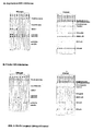
- FIG. 1 shows the improvement in the informative value of the EBV diagnosis by using a modified p18 antigen.
- the figure shows schematically in the upper half of a conventional immunoblot for the determination of antibodies against EBV and in the lower half of a test according to the invention, which additionally includes N-terminally truncated p18 antigen (referred to the strip as "p18").
- the markers p23 (VCA), p54 (early antigen) and p138 (early antigen) represent seropositivity markers that can not differentiate between fresh and expired infections.
- the antibody response to these markers is variable so that different patterns of immune response can be discernible. Positivity of the antibody response against at least one of these markers allows the diagnosis "EBV-infected" without further differentiation between "fresh infection” and "expired infection”.
- a positive IgG response to p72 allows for a reliable diagnosis of "expired infection".
- the classic EBV infection (A1) due to the positive p72 IgG is correctly recognized, while an EBV infection without a p72 IgG formation (A2) or an EBV infection with a subsequent secondary p72 IgG has disappeared Loss (A3) does not differ from a true fresh EBV infection (B) with p72 IgG response not yet established to let. Cases A2 and A3 are usually misdiagnosed in the conventional test. In everyday practice, these cases are more common than the real fresh EBV infections!
- the size differences of the total p18 antigen are due to different N-terminal amino acids; the pQE vector has an additional histidine strand.
- the N-terminally truncated p18 shows, as expected, an approximately 4 kDa lower molecular weight.
- Figure 4 shows a reactivity comparison of the N-terminally truncated p18 with a full-length p18 VCA in a commercially available streak-based EBV antibody assay (Viramed, Planegg-ViraStripe EBV).
- p18 denotes the N-terminal truncated p18.
- p18 denotes the full-length viral p18.
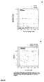
- Figure 5 shows a reactivity comparison of a test according to the invention with a commercially available ELISA system.
- the ODs of both tests are plotted against each other; x-axis - intensity of the band with the N-terminal truncated p18; Y-axis - ELISA values with total p18.
- the OD values of the ELISA were determined by means of a conventional ELISA reader and are between 0 and 3.0; the nitrocellulose strips with the N-terminally truncated p18 were scanned in and the intensity of the band was measured by means of a Software program detected; the scale here covers values from 0 to 200.
- the two additional lines at represent the respective cut-off limits.
- Figure 6 shows the correlation of p18 reactivity in various assay formats.
- the results of testing sera with fresh and expired infections from Example 4 ( Figure 5) were additionally examined in strip tests with conventional VCA-p18 (Viramed) and the results with the VCA ELISA values and the reactivity with the N-terminal truncated p18 correlates.
- the graphical evaluation was similar to that described in the previous example: x-axis with the ELISA OD values (from 0-3.0); y-axis with p18 reactivities of the two stripe tests (open circles - Viramed with total p18; dots - N-terminally truncated p18 antigen).
- Example 1 Recombinant representation of total p18 and N-terminally truncated p18 antigen
- an antigen As an example of an N-terminally truncated p18, the expression cloning of an antigen is described which lacks 30 amino acids N-terminally compared to total viral p18.
- the region coding for amino acids 31-176 of the p18 antigen was amplified by PCR and corresponding oligonucleotide primers from isolated EBV DNA (strain B95-8). (Reading frame BFRF3; sequence data after Genbank entry).
- Primer for total p18 5 'primer: GAG GGA TCC ATC ATG AAA CGC CGG CTG CCC AAG CCC ACC (SEQ ID NO: 3) 3 'primer: CGC CTG CAG TTA CTG TTT CTT ACG TGC CCC GCG (SEQ ID NO: 4)
- Primer for shortened p18 5 'primer: GAG GGA TCC CTG AAC CAG AAT AAT CTC CCC (SEQ ID NO: 5) 3 'primer: CGC CTG CAG TTA CTG TTT CTT ACG TGC CCC GCG (SEQ ID NO: 6)
- Bold bolds correspond to p18 coding sequences; Italics in italics represent restriction enzyme cleavage sites used for further cloning steps (GGATCC-BamHI; CTGCAG-Pst I).
- the PCR was carried out in 100 ⁇ l batches according to standard methods and with standard buffers (94 ° C. denaturation 1 min, 55 ° C. primer binding 1 min, 72 ° C. synthesis 1 min, a total of 40 cycles).
- pDS1 is a vector with ampicillin resistance and an optimized lac promoter and subsequent translation start with the BamHI and PstI sites, which mediate correct translation of the PCR products in the correct reading frame.
- Other expression systems are also suitable.
- the total p18-encoding fragment was inserted into the commercial vector pQE30 (QIAGEN).
- E. coli clones with expression plasmid and correct inserts were induced by standard methods and the lysates analyzed by SDS gel electrophoresis followed by Coomassie staining.
- the total p18 has a molecular weight of about 22 kDa, the N-terminal truncated by 30 amino acids protein size of about 18 kDa with a slightly reduced expression yield.
- the pQE30 expression of total p18 is in a similar range in yield, due to the addition of histidine residues, the product is slightly larger ( Figure 2).
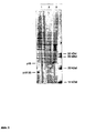
- Example 2 Comparison of the reactivity of total p18 with N-terminally shortened antigen.
- the lysates of the total and shortened p18 clones were again separated by gel electrophoresis, transferred to nitrocellulose and then assayed for reactivity with various EBV-positive sera.
- the nitrocellulose membranes were incubated with 1: 100 diluted sera and visualized specific antibodies with a second peroxidase-conjugated antibody and subsequent staining reaction. Buffers, dilutions and incubation times were carried out analogously to the EBV western blot "recomBlot EBV" developed by Mikrogen. Other Western blotting procedures or staining procedures may also be used.
- the protein can thus be obtained in a purity> 99%.
- the purified product was tested in a "line assay" for its reactivity with antibodies from EBV-infected individuals.
- the product together with other recombinant EBV antigens (EBNA-1, p23-VCA, p138-EA, p54-EA) on nitrocellulose membranes by means of a so-called needle-ruler (isoflow, Fa. Imagene Inc.) in narrow lines applied and then further processed to test strips (saturation unbound sites on the membrane, applying markings, cutting into narrow strips).
- the test principle of the "line assay” corresponds to the commercially available immunoassay "recomLine EBV IgG” from Mikrogen (Article No. 4572).
- the strips were tested with sera from EBV fresh infections and expired infections. For this purpose, the strips were incubated with 1: 100 diluted serum. After washing three times, incubation with anti-human IgG (horseradish peroxidase conjugate) followed. After repeated washing (triplicate), incubation was carried out with TMB substrate solution. Once the cut off control band was visible, the substrate solution was removed and the strips washed with water. A reaction is considered positive when the intensity of a band is equal to or higher than that of the cut-off control band. Thus, a peptide will react with an antibody if a band intensity equal to the "cut-off" value or above the "cut-off” value is obtained.
- No or a weak reaction means no band or band with an intensity below the "cut off” value.
- a cut-off value in this test is defined by using the weak staining intensity of a so-called control band on the strip ("cut-off" band) as the limit for assessing the reactivity of the antigenic bands (see, eg, the above-mentioned immunoassay "recomLine EBV IgG "from Mikrogen).
- the weak staining of the "cut off” band is always the same with the use of certain reagents in all runs and patient samples.
- the ODs of both tests are plotted against each other.
- the x-axis corresponds to the intensity of the band with the shortened p18.
- the y-axis corresponds to the ELISA values with total p18.
- the OD values of the ELISA were determined by means of a conventional ELISA reader and are between 0 and 3.0; the nitrocellulose strips with the shortened p18 were scanned in and the intensity of the band was determined by means of a software program; the scale here includes values from 0 to 200.
- the two additional lines at represent the respective cut-off limits.
- the ELISA OD values are plotted (from 0-3.0); the y axis corresponds to the p18 reactivities of the two stripe assays (open circles - total 18 (Viramed), points - truncated p18 antigen).
Landscapes
- Health & Medical Sciences (AREA)
- Life Sciences & Earth Sciences (AREA)
- Engineering & Computer Science (AREA)
- Virology (AREA)
- Immunology (AREA)
- Urology & Nephrology (AREA)
- Molecular Biology (AREA)
- Biomedical Technology (AREA)
- Chemical & Material Sciences (AREA)
- Hematology (AREA)
- Food Science & Technology (AREA)
- Analytical Chemistry (AREA)
- Tropical Medicine & Parasitology (AREA)
- Cell Biology (AREA)
- Biotechnology (AREA)
- Medicinal Chemistry (AREA)
- Physics & Mathematics (AREA)
- Microbiology (AREA)
- Biochemistry (AREA)
- General Health & Medical Sciences (AREA)
- General Physics & Mathematics (AREA)
- Pathology (AREA)
- Peptides Or Proteins (AREA)
- Micro-Organisms Or Cultivation Processes Thereof (AREA)
- Preparation Of Compounds By Using Micro-Organisms (AREA)
Description
- Die vorliegende Erfindung betrifft das Gebiet der Virusdiagnostik, insbesondere der Epstein-Barr-Virus (EBV)-Diagnostik. Es werden verbesserte Verfahren zum Nachweis von EBV-Infektionen und dazu geeignete Mittel bereitgestellt.
- Das Epstein-Barr-Virus ist ein humanes Herpesvirus, das als Auslöser der infektiösen Mononukleose gilt. Die Infektion mit EBV verläuft oft subklinisch, gelegentlich mit leichten Symptomen. Bei immunsupprimierten Personen kann eine EBV-Infektion aber zu schweren malignen Manifestationen führen.
- Die EBV-Diagnostik besitzt eine große differentialdiagnostische Bedeutung, da die Symptomatik der akuten EBV-Infektion mit vielen klinischen Bildern überlappen kann, die durch andere Erreger oder Ursachen hervorgerufen werden. So kann eine akute EBV-Infektion mit einer HIV-Primärinfektion, Rötelnvirusinfektion, Zytomegalievirusinfektion, Infektion mit klassischen Hepatitisviren, Toxoplasmose, Leptospirose, Infektion mit verschiedenen neurotropen Erregern und mit einer Leukämie oder einem Lymphom verwechselt werden. Eine eindeutige EBV-Serologie stellt damit eine große Verpflichtung gegenüber dem Patienten dar.
- Die klassische EBV-Serologie beruht auf dem Nachweis der IgG- und IgM-Antikörperantwort gegen virale Kapsidantigene (VCA) und gegen EBV-spezifische Kernantigene (EBNA), besonders EBNA-1.
- Die Einordnung verschiedener EBV-Antigen-Klassen beruht auf dem biologischen Zyklus der Virusvermehrung mit früh (EA) und spät (VCA) benötigten Proteinen sowie Antigenen zur Aufrechterhaltung der Latenz (EBNA). Diese Antigenklassen waren in den ersten serologischen Testsystemen (Immunfluoreszenz mit EBV-infizierten Zellen) nachweisbar und unterscheidbar. Alle weiteren nachfolgenden Entwicklungen an serologischen Testsystemen behielten dieses Schema bei und erlauben somit die Unterscheidung von Antikörperspezifitäten gegen EBNA, VCA und EA.
- Die Marker VCA und EBNA wurden zunächst in der lmmunfluoreszenztechnik definiert. In neueren Tests werden die gereinigten, meist rekombinant hergestellten Komponenten p18 und p23 des VCA-Komplexes und p72, das dem EBNA-1 entspricht, verwendet. Das EBNA-1 Antigen wird spät nach einer Erstinfektion mit der Etablierung latent infizierter Zellen gebildet und ist für die Aufrechterhaltung dieses Status mit verantwortlich. Das bedeutet, daß anti-EBNA-1 erst mit einer Verzögerung von bis zu sechs Monaten nach Erstinfektion gebildet wird. Gleichzeitig bedeutet es aber auch, daß bei Vorhandensein dieses Markers ein sicherer Ausschluß einer frischen Infektion diagnostiziert werden kann und damit ein wichtiger Marker für eine zeitliche Einordnung des Infektionszeitpunktes gegeben ist.
- EBV-Serologie weist ein hohes Maß an Variabilität auf, diese führt häufig zu uneindeutigen oder falschen Befunden, wenn nur die klassischen Marker VCA-IgG, VCA-IgM und Anti-EBNA-1 bestimmt werden. So zeigen beispielsweise nicht alle akuten EBV-Infektionen eine nachweisbare VCA-IgM-Antwort, so daß hier Teststrategien, die nur auf der Bestimmung von VCA-IgG und VCA-IgM beruhen, zu einer falschen Schlußfolgerung führen. Andererseits kann eine VCA-IgM-Antwort in seltenen Fällen auch monatelang persistieren und dadurch eine frische Infektion vortäuschen.
- Positives Anti-EBNA-1 beweist eine abgelaufene EBV-Infektion und stellt in Kombination mit positivem VCA-IgG die einzige eindeutige serologische Konstellation dar. Negatives Anti-EBNA-1 (bei gleichzeitig positivem VCA-IgG) kann entweder eine akute EBV-Infektion anzeigen oder durch sekundären Anti-EBNA-1-Verlust bei Immunsuppression verursacht sein. Außerdem entwickeln rund 5 % der Infizierten niemals nachweisbares Anti-EBNA-1 und täuschen damit lebenslang die serologische Situation einer akuten EBV-Infektion vor.
- So wird durch gleichzeitig positives VCA-IgG und Anti-EBNA-1 eine akute EBV-Infektion sicher ausgeschlossen. Positives VCA-IgG bei negativem Anti-EBNA-1 stellt jedoch eine uneindeutige Konstellation dar, die in drei Situationen auftritt:
- 1) bei frischer EBV-Infektion;
- 2) bei abgelaufener Infektion und Anti-EBNA-1-Verlust durch Suppression des zellulären Immunsystems;
- 3) bei abgelaufener Infektion und gleichzeitig fehlender Anti-EBNA-1-Bildung (bei rund 5% der gesunden Bevölkerung).
- Die Fälle 2) und 3) sind z. B. im Untersuchungsgut großer Kliniken häufiger zu finden als die echte frische EBV-Infektion.
- Dieses zentrale Problem der EBV-Serologie läßt sich bisher nur durch aufwendige, zusätzliche Tests oder durch Wiederholungen im zeitlichen Verlauf im Zusammenhang mit klinischen Daten verläßlich lösen.
- Das Fehlen von anti-EBNA-1 wird deshalb in allgemeinen als Indikator einer Frischinfektion verwendet werden. Dies kann - wie oben angeführt - zu drastischen Fehlinterpretationen im Falle von EBV-infizierten Personen führen; die anti-EBNA-1-negativ bleiben oder aufgrund Suppression des zellulären Immunsystems Antikörper dagegen verlieren. Die derzeitigen, in der Routine-Diagnostik verwendeten Verfahren mit anti-EBNA-1 erlauben nicht mit Sicherheit die Diagnose einer frischen EBV-Infektion.
- Die Abgrenzung primärer Anti-EBNA-1-Negativität bei akuter EBV-Infektion einerseits, von Anti-EBNA-1-Verlust und fehlender Anti-EBNA-1-Bildung andererseits, stellt somit das größte Problem der EBV-Serologie dar. Diese besondere Problematik wird bei den meisten publizierten Evaluationen serologischer Tests nicht berücksichtigt. Es muß angenommen werden, daß bis zu 20 Prozent der Untersuchungen auf EBV-Infektion uneindeutig sind und zu Fehlbefunden führen. Diese sind besonders in der Onkologie, der Transplantationsmedizin und bei der Betreuung in der Schwangerschaft von gravierender Bedeutung.
- Hinderer W. et al., Journal of Clinical Microbiology, United States, Oct. 1999, Bd. 37, Nr. 10, Oktober 1999 (1999-10), Seiten 3239-3244, offenbaren ein GST-p18 EBV Fusionskonstrukt, welches p18 105-176 enthält. Dieses Konstrukt erkennt bezüglich der IgG Antikörper besser zurückliegende Infektionen als Primärinfektionen und reagiert auch weniger gut mit Antikörpern aus Primärinfektionen im Vergleich zu zurückliegenden Infektionen.
- Bauer (Clin Lab 2001, 47, 223-230) beschreibt in einem Übersichtsartikel Immunoblots mit rekombinanten Antigenen bei der Epstein-Barr-Virus-Serologie.
- Tranchand-Bunel et al., Journal of Clinical Microbiology 1999, 2399-2368, erläutern IgM ELISA-Tests, bei denen Peptide des VCA p18 Kapsid-Antigens eingesetzt werden.
- van Grunsven et al. (Journal of Infectious Diseases, 1994, 13-19) beschreiben immundominante Epitope am C-Terminus des VCA p18 Kapsid-Proteins.
- Eine Aufgabe der vorliegenden Erfindung ist es, Mittel zum Nachweis einer EBV-Infektion bereitzustellen, die eine sicherere Unterscheidung von akuter und abgelaufener Infektion ermöglichen.
- Überraschenderweise wurde gefunden, daß durch Modifikation des viralen EBV-Kapsidantigens p18 ein Peptid erhalten werden kann, das mit Antikörpern reagiert, die mit einer ähnlichen Sicherheit wie anti-EBNA-1 erst spät nach Infektion gebildet werden und die Problematik der Nicht-Bildung oder das Verschwinden bei Immunsupprimierten nicht im selben Umfang zeigen.
- Es handelt sich dabei um Peptide, die von dem EBV-p18 Antigen abgeleitet sind, welches vom Leserahmen BFRF3 codiert wird. Dieses Antigen wurde schon 1988 als VCA-Marker beschrieben (Middeldorp und Herbrink, J. Virol. Meth., 21 (1988) 133 - 146).
- Die molekulare Zuordnung von p18 und die Darstellung des Proteins mittels gentechnologischer Methoden sowie die Verwendung bestimmter immunologisch aktiver Fragmente daraus sind in EP 574 075 A2 beschrieben. Allerdings wurde bei der Beschreibung der Eigenschaften der rekombinanten Antigene bzw. der chemisch synthetisierten Fragmente immer auf eine optimale Reaktivität abgestellt.
- lgG-Antikörper gegen ein rekombinant hergestelltes, gereinigtes komplettes p18-Antigen sind schon bei frischer EBV-Infektion nachweisbar und eignen sich daher nicht zur Diskriminierung zwischen akuten und abgelaufenen Infektionen.
- Überraschenderweise hat sich im Verlauf von Untersuchungen gezeigt, daß beispielsweise durch das Weglassen des N-terminalen Bereichs des p18-Antigens eine zeitliche Diskriminierung von Antikörper-Antworten erreicht wird: N-terminal deletiertes p18 erkennt Antikörper, die erst Wochen nach Infektion gebildet werden.
- Die vorliegende Erfindung betrifft daher ein Peptid, das vom viralen p18-Antigen des Epstein-Barr-Virus abgeleitet ist und dadurch gekennzeichnet ist, daß es mit Antikörpern aus Individuen mit länger zurückliegender Epstein-Barr-Virus-Infektion reagiert, aber nicht oder schwach mit Antikörpern aus Individuen mit akuter oder kurz zurückliegender Epstein-Barr-Virus-Infektion, dadurch gekennzeichnet, dass es gegenüber dem viralen p18 Antigen N-terminal um 10 - 70 Aminosäuren verkürzt ist.
- Der Ausdruck "Peptid" umfaßt Oligopeptide und Polypeptide. Die Länge der Peptide ist nicht beschränkt, soweit es nicht angegeben ist.
- Das erfindungsgemäße Peptid ist ein modifiziertes p18-Antigen, d.h. es ist nicht identisch mit dem viralen p18, dem "Wildtyp-Protein". Die Modifikationen gegenüber dem viralen p18 sind nicht besonders eingeschränkt, solange das modifizierte Peptid mit Antikörpern aus Individuen mit länger zurückliegender EBV-Infektion reagiert, aber nicht oder schwach mit Antikörpern aus Individuen mit akuter oder kurz zurückliegender EBV-Infektion und, das Antigen ist dadurch gekennzeichnet, dass es gegenüber dem viralen p18 Antigen N-terminal um 10 - 70 Aminosäuren verkürzt ist.
- Daß das erfindungsgemäße Peptid von p18 abgeleitet ist, bedeutet, daß es wenigstens 1 Fragment mit wenigstens 1 Epitop des vollständigen viralen p18-Antigens umfaßt. Ein solches Epitop ist vorhanden, wenn in einem line assay (siehe Beispiel 3) bei Inkubation mit Seren aus Individuen mit länger zurückliegender EBV-Infektion eine positive Reaktion erhalten wird.
- Die Aminosäuresequenz des viralen p18 ist in Figur 7 dargestellt (SEQ ID NO:1). In der Regel hat das erfindungsgemäße Peptid eine von SEQ ID NO:1 abweichende Aminosäuresequenz. Dabei handelt es sich vorzugsweise um eine oder mehrere Deletionen und/oder Substitutionen. Das erfindungsgemäße Peptid weist eine oder mehrere Deletionen gegenüber der in SEQ ID NO:1 dargestellten Aminosäuresequenz auf, so dass am N-Terminus von p18 deletiert 10-70 Aminosäuren sind.
- Das erfindungsgemäße Peptid umfaßt oder besteht vorzugsweise aus wenigstens 125 aufeinanderfolgenden Aminosäuren der Aminosäuresequenz SEQ ID NO:1.
- Es ist bevorzugt, daß in dem erfindungsgemäßen Peptid wenigstens ein Epitop des vollständigen p18-VCA, gegen das nach Infektion Antikörper gebildet werden, nicht vorhanden ist.
- Das erfindungsgemäße Peptid ist gegenüber dem viralen p18-Antigen N-terminal um 10 bis 70 Aminosäuren, bevorzugt um 15 bis 50 Aminosäuren, bevorzugter um 20 bis 40 Aminosäuren, und noch bevorzugter um ungefähr 30 Aminosäuren, verkürzt. In einer besonderen Ausführungsform besteht das erfindungsgemäße Peptid aus der Aminosäuresequenz SEQ ID NO:2. Diese Aminosäuresequenz entspricht den Aminosäuren 31 bis 176 von SEQ ID NO:1. In weiteren bevorzugten Ausführungsformen hat das erfindungsgemäße Peptid im wesentlichen eine Aminosäuresequenz, die ausgewählt ist aus der Gruppe bestehend aus
- Aminosäuren 21 bis 176 von SEQ ID NO:1,
- Aminosäuren 22 bis 176 von SEQ ID NO:1,
- Aminosäuren 23 bis 176 von SEQ ID NO:1,
- Aminosäuren 24 bis 176 von SEQ ID NO:1,
- Aminosäuren 25 bis 176 von SEQ ID NO:1,
- Aminosäuren 26 bis 176 von SEQ ID NO:1,
- Aminosäuren 27 bis 176 von SEQ ID NO:1,
- Aminosäuren 28 bis 176 von SEQ ID NO:1,
- Aminosäuren 29 bis 176 von SEQ ID NO:1,
- Aminosäuren 30 bis 176 von SEQ ID NO:1,
- Aminosäuren 31 bis 176 von SEQ ID NO:1,
- Aminosäuren 32 bis 176 von SEQ ID NO:1,
- Aminosäuren 33 bis 176 von SEQ ID NO:1,
- Aminosäuren 34 bis 176 von SEQ ID NO:1,
- Aminosäuren 35 bis 176 von SEQ ID NO:1,
- Aminosäuren 36 bis 176 von SEQ ID NO:1,
- Aminosäuren 37 bis 176 von SEQ ID NO:1,
- Aminosäuren 38 bis 176 von SEQ ID NO:1,
- Aminosäuren 39 bis 176 von SEQ ID NO:1,
- Aminosäuren 40 bis 176 von SEQ ID NO:1 und
- Aminosäuren 41 bis 176 von SEQ ID NO:1.
- Dem Fachmann ist auch bewußt, daß die immunologischen Eigenschaften der angegebenen Sequenzen oft nur unwesentlich verändert werden, wenn Aminosäuren inseriert, substituiert oder deletiert sind. Substitutionen, die oft als konservativ angesehen werden, sind solche, bei denen die chemische Natur der substituierenden Aminosäure ähnlich der der substituierten Aminosäure ist. Aminosäurekombinationen, die als konservativ angesehen werden können, sind beispielsweise Gly/Ala, Asp/Glu, Asn/Gln, Val/Ile/Leu, Ser/Thr, Lys/Arg und Phe/Tyr.
- Es können zusätzliche Aminosäuren oder chemische Gruppen am N- oder C-Terminus hinzugefügt werden, um eine Art "linker" zu schaffen, durch den das Peptid vorteilhaft an einen Träger gekoppelt werden kann. Ein solcher "linker" hat in der Regel 1 bis 60 Aminosäuren, meistens jedoch 1 bis 10 Aminosäuren. Die Bindung des Peptids an einen Träger oder eine feste Phase muß aber nicht kovalent sein. Es können ebenfalls Cysteinreste angefügt werden, um eine Kopplung mit anderen Peptiden zu erreichen.
- Die erfindungsgemäßen Peptide können weiter modifiziert sein, beispielsweise durch Glycosylierung, Amidierung, Carboxylierung oder Phosphorylierung. Funktionelle Derivate wie z.B. Salze, Amide, Ester, C-amidierte oder N-acylierte Derivate sind von der Erfindung ebenfalls umfaßt.
- Der Fachmann kann aufgrund der Informationen in dieser Anmeldung auf einfache Weise ermitteln, ob ein bestimmtes p18-Derivat geeignet ist, um zwischen akuter und abgelaufener Infektion zu unterscheiden. Dazu muß ein gegebenes p18-Derivat beispielsweise lediglich in einem line assay wie er in Beispiel 3 dargestellt ist, getestet werden.
- Die erfindungsgemäßen Peptide können auf verschiedene, dem Fachmann bekannte Weisen hergestellt werden. In der Regel werden sie entweder durch chemische Synthese oder durch Expression rekombinanter DNA in geeigneten Wirtszellen hergestellt.
- Als Verfahren zur chemischen Synthese von Peptiden kann Festphasensynthese oder Flüssigphasensynthese verwendet werden. Festphasensynthese ist bevorzugt. Verfahren zur chemischen Synthese von Peptiden sind beschrieben in Bodanszky und Ondetti: Peptide Synthesis, Interscience Publishers, New York (1966); The Peptides, Analysis, Synthesis, Biology Volume 1-3 (Ed. Gross und Maienhofer) 1979, 1980, 1981 (Academic Press, Inc.).
- Zur rekombinanten Herstellung des erfindungsgemäßen Peptids wird zunächst eine Nukleinsäure, die für die Aminosäuresequenz des Peptides codiert, bereitgestellt. Die Nukleinsäure kann DNA oder RNA sein, DNA ist bevorzugt. Die codierende Nukleinsäure wird üblicherweise in einen Vektor oder ein Plasmid eingefügt, das geeignete Sequenzen enthält, die die Expression des codierten Peptids erlauben. Derartige geeignete Sequenzen sind beispielsweise ein Promotor, eine Terminatorsequenz und Sequenzen, die die Replikation des Plasmids ermöglichen. Es kann sich um Expressionsplasmide zur Expression in Prokaryonten oder in Eukaryonten handeln. Dem Fachmann ist bewußt, daß dabei unterschiedliche Promotorsequenzen verwendet werden. Prokaryontische Expressionsplasmide sind bevorzugt.
- Die Erfindung betrifft auch einen Vektor oder ein Plasmid, das eine Nukleinsäure enthält, die für ein erfindungsgemäßes Peptid codiert. Übliche Verfahren zur Herstellung von Vektoren und Plasmiden sind beschrieben in Sambrook et al., Molecular Cloning: A Laboratory Manual, 2001, Cold Spring Harbour Laboratory Press. Weiterhin wird der Vektor oder das Plasmid in geeignete Wirtszellen, z.B. Säugerzellen oder bevorzugt Bakterien, eingebracht, der dann unter geeigneten Bedingungen kultiviert wird, so daß eine Expression des gewünschten Peptids erfolgt. Die Erfindung betrifft auch eine Zelle enthaltend einen erfindungsgemäßen Vektor oder ein erfindungsgemäßes Plasmid. Ein weiterer Aspekt der Erfindung ist ein Verfahren zur Herstellung eines erfindungsgemäßen Peptids, das umfaßt, daß man erfindungsgemäße Zellen unter geeigneten Bedingungen kultiviert, so daß des gewünschte Peptid exprimiert wird, und gegebenenfalls das Peptid gewinnt. Nach erfolgter Expression kann das Peptid durch dem Fachmann an sich bekannte Methoden gewonnen werden. Geeignete Verfahren zur rekombinanten Expression von Peptiden sind in Molecular Cloning: A Laboratory Manual von Sambrook et al. (siehe oben) beschrieben.
- Vorzugsweise liegt das Peptid in isolierter Form vor, das heißt es ist im wesentlichen frei von anderen Peptiden. Die Reinheit des erfindungsgemäßen Peptids ist vorzugsweise größer 90%, bevorzugter größer 95%, am bevorzugtesten größer 99%, bestimmt anhand von SDS-PAGE mit anschließender Coomassiefärbung.
- Daher wird das Peptid nach erfolgter Expression in der Regel gereinigt. Dabei können verschiedenste dem Fachmann bekannte Reinigungsverfahren eingesetzt werden. Verfahren zur Reinigung von Proteinen und Peptiden sind beschrieben in Methods in Enzymology, Vol. 182, Guide to Protein Purification, Academic Press, New York 1990 oder Scopes R.K., Protein Purification, Springer-Verlag, Heidelberg 1994.
- Das erfindungsgemäße Peptid zeichnet sich dadurch aus, daß es mit Antikörpern aus Individuen mit länger zurückliegender Epstein-Barr Virusinfektion reagiert, aber nicht oder schwach mit Antikörpern aus Individuen mit akuter oder kurz zurückliegender Epstein-Barr Virusinfektion. Vorzugsweise reagiert das Peptid nicht mit Antikörpern aus Individuen mit kurz zurückliegender EBV-Infektion. Vorzugsweise beziehen sich die Angaben auf IgG-Antikörper.
- Seren aus einem Individuum mit länger zurückliegender Infektion können folgende serologische Eigenschaften aufweisen:
- anti-EBNA-1-lgG: positiv;
- anti-p23-lgG: positiv;
- anti-p54-lgG: negativ;
- anti-p138-lgG: negativ; und
- anti-p23-IgM: negativ.
- Das erfindungsgemäße Peptid reagiert mit Antikörper aus Seren, die diese serologischen Eigenschaften aufweisen.
- In der Regel kann eine ungefähr 1,5 Jahre zurückliegende Infektion als "länger zurückliegende Infektion" bezeichnet werden.
- Die Infektion eines Individuums mit EBV gilt als "akut", wenn das klinische Bild einer infektiösen Mononukleose vorliegt. Seren aus einem Individuum mit kurz zurückliegender EBV-Infektion können folgende serologische Eigenschaften aufweisen:
- anti-EBNA-1-lgG: negativ;
- anti-p54-IgG und/oder anti-p138-lgG (EA): positiv;
- anti-p23-lgG (VCA): positiv; und
- anti-p54- und/oder -p138- und/oder -p54-lgM: positiv.
- Das erfindungsgemäße Peptid reagiert nicht oder schwach mit Antikörpern aus Seren, die diese serologischen Eigenschaften aufweisen.
- In der Regel kann eine ungefähr 4 Wochen zurückliegende Infektion als "kurz zurückliegende Infektion" bezeichnet werden.
- Der Ausdruck, daß ein Peptid mit Antikörpern "reagiert", bedeutet im Sinne der vorliegenden Anmeldung, daß das Peptid mit einem oder mehreren Antikörpern Antigen-Antikörper-Immunkomplexe bildet, nachdem Peptid und Antikörper miteinander in Kontakt gebracht worden sind. Keine oder eine schwache Reaktion des Peptids mit Antikörpern liegt vor, wenn keine oder nur schwache Antigen-Antikörper-Komplexe gebildet werden, nachdem Peptid und Antikörper miteinander in Kontakt gebracht worden sind. Ob ein Peptid mit Antikörpern reagiert, kann durch einen "line-assay" festgestellt werden, wie er in Beispiel 3 dargestellt ist.
- Die Erfindung betrifft weiterhin ein Fusionspeptid, das ein oben beschriebenes erfindungsgemäßes Peptid umfaßt und eine oder mehrere weitere Aminosäuren, die nicht der Aminosäuresequenz des viralen p18-Antigens entsprechen. Dabei kann es sich um verschiedenste Fremdsequenzen handeln. Bevorzugt ist es aber, daß nur wenige Fremdaminosäuren in dem Fusionspeptid enthalten sind, um eine Kreuzreaktion der Fremdaminosäuren mit Antikörpern zu vermeiden. Beispiele für Fremdsequenzen sind Aminosäuresequenzen, die die Affinitätsreinigung des exprimierten Peptids erleichtern, z.B. ein Oligohistidin-stretch (6 bis 8 aufeinanderfolgende Histidinreste) am N- oder C-Terminus.
- Ein weiterer Aspekt der Erfindung ist ein diagnostisches Reagenz zum Nachweis von Antikörpern gegen Epstein-Barr Virus, umfassend ein erfindungsgemäßes Peptid und/oder ein erfindungsgemäßes Fusionspeptid. Ein "diagnostisches Reagenz" im Sinne der vorliegenden Erfindung umfaßt eine feste Phase oder eine Nachweissubstanz. Feste Phasen können beispielsweise Mikrotestplatten, Küvetten, Gefäße, Membranen, Filter, Teststreifen oder Partikel wie Kügelchen sein. Verfahren zum Binden von Peptiden an feste Oberflächen oder feste Phasen sind dem Fachmann an sich bekannt.
- Nachweissubstanzen können beispielsweise radioaktive Isotopen, fluoreszierende Verbindungen, Enzyme, Farbstoffe, etc. sein. Die erfindungsgemäßen Peptide oder Fusionspeptide können markiert oder unmarkiert sein, je nach der beabsichtigten Verwendung. Die Markierung kann jeder Art sein, z. B. enzymatisch, chemisch, fluoreszierend, lumineszierend oder radioaktiv.
- Antikörper gegen Epstein-Barr Virus können beispielsweise nachgewiesen werden, indem ein diagnostisches Reagenz gemäß der vorliegenden Erfindung mit Serum, das von einem Individuum stammt, in Kontakt gebracht wird und anschließend die gebildeten Antigen-Antikörper-Komplexe nachgewiesen werden. Die Erfindung betrifft daher ebenfalls ein Verfahren zum Nachweis von Antikörpern gegen Epstein-Barr Virus in einer Probe, das umfaßt, daß man ein erfindungsgemäßes Peptid oder Fusionspeptid oder ein diagnostisches Reagenz gemäß der Erfindung mit der Probe in Kontakt bringt und anschließend die gebildeten Antigen-Antikörper-Komplexe nachweist.
- Die Probe ist in der Regel eine Körperflüssigkeit, die von einem Individuum stammt. Vorzugsweise ist die Probe Serum, das von einem Individuum stammt. Das Serum kann mit einer geeigneten Flüssigkeit verdünnt werden.
- Üblicherweise wird im erfindungsgemäßen Verfahren ein oben beschriebenes diagnostisches Reagenz eingesetzt. Abhängig von der Natur und weiteren Merkmalen des diagnostischen Reagenz ist die immunchemische Reaktion eine sogenannte Sandwich-Reaktion, eine Agglutinierungsreaktion, eine Kompetition oder eine Inhibition.
- In einem bevorzugten Format des Verfahrens ist das erfindungsgemäße Peptid oder Fusionspeptid auf einer festen Phase immobilisiert. Die feste Phase wird dann mit der zu untersuchenden Probe, beispielsweise Serum in Kontakt gebracht, wodurch sich Antigen-Antikörperkomplexe bilden können. Die feste Phase wird anschließend ein oder mehrere Male gewaschen, um ungebundene Antikörper zu entfernen. Anschließend werden die immobilisierten Antigen-Antikörperkomplexe nachgewiesen. Dies kann beispielsweise durch sogenannte anti-Antikörper erfolgen, die an eine Markierungssubstanz gekoppelt sind. So kann nach dem Waschen die feste Phase mit einem anti-IgG-Antikörper, der an Meerrettichperoxidase gekoppelt ist, in Kontakt gebracht werden. Dieser anti-Antikörper bindet an die immobilisierten Immunkomplexe. Der gebundene sekundäre Antikörper kann durch die Enzymreaktion der Meerrettichperoxidase in bekannter Weise sichtbar gemacht werden. Der sekundäre Antikörper erkennt vorzugsweise spezifisch humanes IgG.
- Vorzugsweise werden in dem Verfahren Membranen oder Teststreifen eingesetzt, auf denen ein oder mehrere Antigene immobilisiert sind. In einer anderen Ausführungsform des Verfahrens wird ein ELISA-Test durchgeführt. Die Durchführung derartiger Verfahren ist dem Fachmann an sich bekannt. Eine Übersicht über geeignete Nachweisverfahren findet sich in "Labor und Diagnose"; L. Thomas; Die medizinische Verlagsgesellschaft, Marburg; ISBN 3-921320-21-6.
- Zur Durchführung eines ELISA wird üblicherweise ein Peptid oder eine Mischung mehrerer Peptide an eine feste Phase gebunden, z. B. an eine Mikrotiterplatte. Eine geeignete Verdünnung der zu testenden Körperflüssigkeit (Serum) wird mit der festen Phase, an die das oder die Peptide gebunden sind, in Kontakt gebracht. Die anschließende Inkubation wird für einen Zeitraum durchgeführt, der ausreichend ist zur Bildung von Antigen-Antikörper-Komplexen. Anschließend werden ungebundene Komponenten abgewaschen. Die Detektion der Immunkomplexe erfolgt in der Regel über Antikörper, die spezifisch an humane Immunglobuline binden und die markiert sind (geeignete Marker s.o.). Oft wird dann nach Zugabe eines Substrates ein Produkt gebildet, das leicht visuell, photometrisch, spektrometrisch, luminometrisch oder elektrochemisch nachgewiesen werden kann.
- Eine andere Variante ist der Kompetitionsassay, der dem Fachmann auch bekannt ist.
- In einer besonderen Ausführungsform des Verfahrens wird ein weiteres Antigen des Epstein-Barr Virus mit der Probe in Kontakt gebracht und anschließend die gebildeten Antigen-Antikörper-Komplexe nachgewiesen. Bei dem weiteren Antigen des EBV handelt es sich vorzugsweise um ein Antigen, das charakteristisch für eine frische Infektion ist. Vorzugsweise ist das weitere Antigen ausgewählt aus der Gruppe bestehend aus p18, p23, p138 und p54. In einer weiteren Ausführungsform des Verfahrens wird neben dem erfindungsgemäßen Peptid oder Fusionspeptid das späte nukleäre Antigen EBNA-1 (p72) eingesetzt. In einer weiteren Ausführungsform werden neben dem erfindungsgemäßen Peptid oder Fusionspeptid die Antigene p72, p18, p23, p138 und/oder p54 eingesetzt.
- Ein weiterer Aspekt der Erfindung ist ein Testkit zum Nachweis von Antikörpern gegen Epstein-Barr Virus, der ein erfindungsgemäßes Peptid oder Fusionspeptid und/oder ein diagnostisches Reagenz, wie es oben beschrieben wurde, umfaßt. Der Testkit kann weitere, zur Durchführung des erfindungsgemäßen Verfahrens geeignete Mittel enthalten, beispielsweise eine Zusammensetzung, die einen sekundären Antikörper enthält. Vorzugsweise enthält der Testkit eine feste Phase. Besonders bevorzugt sind dabei Western Blot-Teststreifen, auf denen wenigstens ein erfindungsgemäßes Peptid oder Fusionspeptid immobilisiert ist. Es können weitere EBV-Antigene immobilisiert sein, z.B. p18, p23, p72, p54 und/oder p138. Dabei kann es sich um Nitrocellulosestreifen handeln. Der Testkit kann weiterhin Waschlösungen, gegebenenfalls in konzentrierter Form, umfassen.
- Ein weiterer Aspekt der Erfindung ist die Verwendung eines modifizierten Viruskapsid-Antigens des Epstein-Barr Virus zur Bestimmung, ob eine akute/kurz zurückliegende oder eine länger zurückliegende/abgelaufene EBV-Infektion in einer Probe vorliegt. Die bevorzugten Ausführungsformen der erfindungsgemäßen Verwendung entsprechen den oben beschriebenen bevorzugten Ausführungsformen des Peptids, des Fusionspeptids, des diagnostischen Reagenz, des Testkits und/oder des Verfahrens. Die Erfindung betrifft weiterhin die Verwendung eines modifizierten Viruskapsid-Antigens des Epstein-Barr Virus zur serologischen Diskriminierung von frischen/kurz zurückliegenden und abgelaufenen Infektionen in einer Probe. Die Erfindung betrifft weiterhin die Verwendung eines modifizierten Viruskapsid-Antigens des Epstein-Barr Virus zum Nachweis von Antikörpern gegen EBV. Dabei kann in einer Probe vorzugsweise zwischen einer frischen und einer abgelaufenen Infektion unterschieden werden. Die Erfindung betrifft weiterhin die Verwendung eines modifizierten Viruskapsid-Antigens des Epstein-Barr Virus zum Nachweis einer abgelaufenen EBV-Infektion. Schließlich betrifft die Erfindung die Verwendung eines modifizierten Viruskapsid-Antigens des Epstein-Barr Virus zur Unterscheidung zwischen einer abgelaufenen und einer frischen EBV-Infektion in einer Probe. Das modifizierte Viruskapsid-Antigen gemäß den Verwendungen der Erfindung entspricht vorzugsweise dem erfindungsgemäßen Peptid oder Fusionspeptid. Alle weiter oben beschriebenen bevorzugten Varianten der Erfindung gelten sinngemäß für die Verwendungen der Erfindung.
- Alle in der vorliegenden Anmeldung beschrieben Aspekte und bevorzugten Ausführungsformen der Erfindung können in verschiedenster Weise miteinander kombiniert werden. Derartige Kombinationen sind ebenfalls von der Erfindung erfaßt.
- Durch die vorliegende Erfindung wird ein zweiter später Marker neben dem EBNA-1 bereitgestellt, der zudem nicht die Problematik der Nichtbildung von Antikörpern aufweist. Im Gegensatz zu EBNA-1 werden Antikörper gegen p18 nach zellulärer Immunsuppression nicht verloren. Der Nachweis von IgG gegen ein erfindungsgemäß modifiziertes p18 stellt demnach einen sicheren Ausschluß einer akuten EBV-Infektion dar.
- Durch die Verwendung von modifiziertem p18 in geeigneten Meßsystemen, wie zum Beispiel dem lmmunblot, lassen sich also frische und abgelaufene EBV-Infektionen auch dann nachweisen, wenn die Anti-EBNA-Antwort einen untypischen Verlauf zeigt.
- Figur 1 zeigt die Verbesserung der Aussagekraft der EBV-Diagnostik durch Verwendung eines modifizierten p18-Antigens.
- Die Abbildung zeigt schematisch in der oberen Hälfte einen konventionellen Immunblot zur Bestimmung von Antikörpern gegen EBV und in der unteren Hälfte einen erfindungsgemäßen Test, der zusätzlich N-terminal verkürztes p18-Antigen beinhaltet (auf den Streifen als "p18" bezeichnet). Die Marker p23 (VCA), p54 (frühes Antigen) und p138 (frühes Antigen) stellen Seropositivitätsmarker dar, die nicht zwischen frischen und abgelaufenen Infektionen differenzieren können. Die Antikörperantwort gegen diese Marker ist variabel, so daß unterschiedliche Muster der Immunantwort erkennbar sein können. Positivität der Antikörperantwort gegen mindestens einen dieser Marker erlaubt die Diagnose "EBV-infiziert", ohne weitere Differenzierung zwischen "frischer Infektion" und "abgelaufener Infektion". Eine positive IgG-Antwort gegen p72 (= EBNA-1) erlaubt die sichere Diagnose "abgelaufene Infektion". Im konventionellen Test wird die klassische abgelaufene EBV-Infektion (A1) aufgrund des positiven p72-lgG richtig erkannt, während sich eine abgelaufene EBV-Infektion ohne p72-IgG-Bildung (A2) oder eine abgelaufene EBV-Infektion mit nachfolgendem sekundärem p72-lgG-Verlust (A3) nicht von einer echten frischen EBV-Infektion (B) mit noch nicht ausgebildeter p72-IgG-Antwort unterscheiden lassen. Die Fälle A2 und A3 werden also im konventionellen Test in der Regel falsch diagnostiziert. In der täglichen Praxis sind diese Fälle aber häufiger als die echten frischen EBV-Infektionen!
- Bei Verwendung des modifizierten Tests (untere Hälfte), der z.B. N-terminal verkürztes p18 (in Figur 1 als "p18" bezeichnet) enthält, wird die frische EBV-Infektion mit fehlendem p18-IgG (B) klar von den abgelaufenen EBV-Infektionen (A1-A3) unterschieden, selbst dann, wenn p72-IgG fehlt.
- Figur 2 zeigt die Expression verschiedener Peptide. Es ist ein Coomassie-blau gefärbtes SDS-Polyacrylamid-Gel von Lysaten der exprimierenden E.coli-Klone nach Induktion dargestellt (siehe Beispiel 1). Die Spuren entsprechen folgenden Ansätzen:
- Spur 1:
- Expression des N-terminal verkürzten p18 (p18-30)
- Spur 2:
- Expression des gesamt-p18 im pDS-Vektor
- Spur 3:
- Expression des gesamt-p18 im pQE-Vektor.
- Spur 4:
- Molekulargewichtsmarker
- Die Größenunterschiede des gesamt-p18-Antigens beruhen auf unterschiedlichen N-terminalen Aminosäuren; der pQE-Vektor besitzt einen zusätzlichen Histidin-Strang.
Das N-terminal verkürzte p18 zeigt, wie erwartet ein ca. 4 kDa geringeres Molekulargewicht. - Figur 3 zeigt die Reaktivität der Peptide im Immunblot. Lysate von induzierten E.coli-Klonen mit den beiden p18-Varianten wurden Gel-elektrophoretisch aufgetrennt und anschließend auf Nitrozelluose-Membranen transferiert (Western Blot).
- Spur 1:
- Gesamt p18 ohne His-tag
- Spur 2:
- p18-30 (verkürzte p18-Version)
- Die Nitrozellulose-Membranen wurden mit Seren EBV-positiver Personen inkubiert; spezifisch gebundene Antikörper wurden anschließend mit einem Peroxidase-konjugierten zweiten anti-human-IgG Antikörper and TMB-Färbung sichtbar gemacht.
- A; Serum eines Patienten mit frischer EBV-Infektion
- B; Serum mit lang zurückliegender EBV-Infektion
- Figur 4 zeigt einen Reaktivitätsvergleich des N-terminal verkürzten p18 mit einem Volllängen-p18-VCA in einem kommerziell erhältlichen EBV-Antikörper-Test auf Streifen-Basis (Fa. Viramed, Planegg - ViraStripe EBV).
- Jeweils 10 Seren von Personen mit lang zurückliegender EBV-Infektion, aber problematischer Serologie (schwache Antikörper-Titer, fehlendes anti-EBNA-1; A;) und 10 Proben von Patienten mit frischer EBV-Infektion (B;) wurden mit zwei Versionen von EBV-Antigen-Streifen getestet:
- "Mikrogen"-Streifen enthalten neben dem N-terminal verkürzten p18 die zusätzlichen EBV-Antigene EBNA-1, p23 (VCA) sowie p54 und p138 (EAs).
- Viramed-Streifen enthalten neben einem vollständigen p18 Antigen noch EBV-Proteine EBNA-1, EA-p54 sowie gp125 (VCA).
- In der linken Hälfte (Mikrogen-Test) bezeichnet "p18" das N-terminal verkürzte p18. In der rechten Hälfte (Viramed-Test) bezeichnet "p18" das virale Volllängen-p18.
- Bei den zurückliegenden Infektionen (A) ist bei beiden Tests immer eine mehr oder weniger ausgeprägte anti-p18 Reaktion zu finden.
Seren von Patienten nach Frischinfektion (B) zeigen in dem Test mit N-terminal verkürztem p18 jedoch in keinem Fall eine Reaktion, mit dem Virastripe (Gesamt-p18) jedoch in allen Proben. - Figur 5 zeigt einen Reaktivitätsvergleich eines erfindungsgemäßen Tests mit einem kommerziell erhältlichen ELISA-System.
- 32 Proben von Patienten mit frischer EBV-Infektion ("acute") und 25 Serumproben von Personen mit abgelaufener (alter) EBV-Infektion ("past") wurden jeweils mit N-terminal verkürztem p18 (gesprüht auf Nitrozellulose-Streifen, Mikrogen, (vergleiche Antigenstreifen aus Abb. 4) und einem kommerziellen ELISA (Fa. Sorin) mit konventionellem p18-VCA-Antigen getestet.
- In den beiden Diagrammen sind jeweils die OD's beider Teste gegeneinander aufgetragen; x-Achse - Intensität der Bande mit dem N-terminal verkürzten p18; Y-Achse - ELISA-Werte mit Gesamt-p18. Die OD-Werte des ELISA wurden durch mittels eines gängigen ELISA-Readers ermittelt und liegen zwischen 0 und 3,0; die Nitrozellulose-Streifen mit dem N-terminal verkürzten p18 wurden eingescannt und die Intensität der Bande mittels eines Software-Programms ermittelt; die Skala umfaßt hier Werte von 0 bis 200. Die beiden zusätzlichen Linien bei stellen die jeweiligen cut-off Grenzen dar.
- Figur 6 zeigt die Korrelation der p18-Reaktivität in verschiedenen Testformaten. Die Ergebnisse der Testung von Seren mit frischen und abgelaufenen Infektionen aus Beispiel 4 (Figur 5) wurden zusätzlich in Streifen-Tests mit konventionellem VCA-p18 (Fa. Viramed) untersucht und die Ergebnisse mit den VCA-ELISA-Werten und der Reaktivität mit dem N-terminal verkürzten p18 korreliert.
- Die graphische Auswertung erfolgte ähnlich wie im vorigen Beispiel beschrieben: x-Achse mit den ELISA OD-Werten (von 0 - 3,0); y-Achse mit p18-Reaktivitäten der beiden Streifen-Teste (offene Kreise - Fa. Viramed mit Gesamt-p18; Punkte - N-terminal verkürztes p18 - Antigen).
- Die nachfolgenden Beispiele erläutern die Erfindung näher.
- Als Beispiel für ein N-terminal verkürztes p18 wird die Expressionsklonierung eines Antigens beschrieben, dem gegenüber viralem Gesamt-p18 30 Aminosäuren N-terminal fehlen.
- Der für die Aminosäuren 31-176 des p18-Antigens kodierende Bereich wurden mittels PCR und entsprechenden Oligonukleotid-Primern aus isolierter EBV-DNA (Stamm B95-8) amplifiziert.
(Leserahmen BFRF3; Sequenzdaten nach Genbank-Eintrag). - Primer für gesamt-p18:
5'-Primer:
GAG GGA TCC ATC ATG AAA CGC CGG CTG CCC AAG CCC ACC (SEQ ID NO:3)
3'-Primer:
CGC CTG CAG TTA CTG TTT CTT ACG TGC CCC GCG (SEQ ID NO:4) - Primer für verkürztes p18:
5'-Primer:
GAG GGA TCC CTG AAC CAG AAT AAT CTC CCC (SEQ ID NO:5)
3'-Primer:
CGC CTG CAG TTA CTG TTT CTT ACG TGC CCC GCG (SEQ ID NO:6) - Nukleotide in Fettschrift entsprechen p18-kodierenden Sequenzen; Nukleotide in Kursivschrift stellen Restriktionsenzymschnittstellen dar, die für weitere Klonierungsschritte verwendet wurden (GGATCC - BamHI; CTGCAG - Pst I).
- Die PCR wurde in 100µl Ansätzen nach Standardmethoden und mit Standardpuffern durchgeführt (94°C Denaturierung 1 min; 55°C Primerbindung 1 min; 72°C Synthese 1 min; insgesamt 40 Zyklen).
- Nach Überprüfung der PCR-Produkte auf richtige Größe in der Gel-Elektrophorese (ca. 530 nt für gesamt-p18 und ca. 440nt für verkürztes p18) wurden beide mittels konventioneller Methoden gereinigt und entsalzt, anschließend mit BamHl und Pstl geschnitten und schließlich in die entsprechenden Stellen des Expressionsvektors pDS1 eingesetzt.
pDS1 ist ein Vektor mit Ampicillin-Resistenz und einem optimierten lac-Promotor und anschließendem Translationsstart mit den BamHI und Pst I Stellen, die eine korrekte Translation der PCR-Produkte im richtigen Leserahmen vermitteln.
Andere Expressionssysteme sind auch geeignet.
Zu Vergleichszwecken wurde schließlich noch das gesamt-p18-kodierende Fragment in den kommerziellen Vektor pQE30 (QIAGEN) inseriert. - Nach Transformation wurden E.coli-Klone mit Expressionsplasmid und korrekten Inserts nach Standardmethoden induziert und die Lysate durch SDS-Gel-Elektrophorese und anschließende Coomassie-Färbung analysiert.
- Beide Ansätze zeigen eine deutliche Expression von rekombinantem p18 bzw. p18-Derivat.
Das gesamt-p18 besitzt ein Molekulargewicht von ca. 22 kDa, das N-terminal um 30 Aminosäuren verkürzte Protein eine Größe von etwa 18 kDa mit einer etwas verringerten Expressionsausbeute.
Die pQE30-Expression des gesamt-p18 liegt in der Ausbeute in einem ähnlichen Bereich, aufgrund der zusätzlich vorhandenen Histidin-Reste ist das Produkt etwas größer (Fig. 2). - Die Lysate der Klone mit gesamt- sowie verkürztem p18 wurden wiederum Gel-elektrophoretisch aufgetrennt, auf Nitrozellulose transferiert und anschließend auf ihre Reaktivität mit diversen EBV-positiven Seren untersucht.
Hierzu wurden die Nitrozellulose-Membranen mit 1:100 verdünnten Seren inkubiert und spezifische Antikörper mit einem zweiten Peroxidase-konjugierten Antikörper und anschließender Färbereaktion sichtbar gemacht.
Puffer, Verdünnungen und Inkubationszeiten wurden analog zu dem bei Mikrogen entwickelten EBV-Western Blot "recomBlot EBV" durchgeführt.
Andere Western Blot-Prozeduren oder Färbeverfahren können ebenfalls angewendet werden. - Gestestet wurde je ein Serum nach Frischinfektion (ca. 3 Wochen zurückliegend) sowie mit lange zurückliegender Infektion (ca. 1,5 Jahre zurückliegend) (Figur 3).
Überraschenderweise zeigt sich hier, daß das gesamt-p18-Antigen im Vergleich zu der verkürzten Version deutliche Reaktivitätsunterschiede aufweist:
Bei lange zurückliegender Infektion reagieren beide Antigenvarianten in etwa gleich stark; bei Seren von Frischinfektionen zeigt jedoch nur das gesamt-p18-Antigen eine deutliche Reaktion. - Damit ergibt sich ein zweiter Marker (neben EBNA-1), der für die zeitliche Einordnung einer EBV-Infektion verwendet werden kann.
- Die verkürzte Variante des p18-Antigens wurde in größeren Mengen exprimiert und mittels konventioneller Methoden aufgereinigt; die Schritte waren hierbei im Wesentlichen:
- Lyse der induzierten E.coli-Zellen mit dem Expressionsprodukt;
- Abtrennung von p18 als unlösliche "inclusion bodies" durch Zentrifugation;
- Waschen des unlöslichen Pellets mit diversen nicht-ionischen Detergenzien;
- Lösen des restlichen Pellets mit dem p18 in 8M Hamstoffpuffer;
- Verschiedene lonenaustausch-Chromatographien: DEAE, Q-Sepharose, S-Sepharose (alle Pharmacia).
- Das Protein kann so in einer Reinheit >99% gewonnen werden.
- Alternativ können auch andere Reinigungsverfahren oder Reihenfolgen von Säulenschritten verwendet werden. Bei Verwendung des pQE30-p18 Produkts kann insbesondere auch die selektive Anbindung an Ni-Chelat-Säulen ausgenutzt werden.
- Das gereinigte Produkt wurde in einem "line assay" auf seine Reaktivität mit Antikörpern aus EBV-infizierten Individuen hin untersucht. Dazu wurde das Produkt zusammen mit anderen rekombinanten EBV-Antigenen (EBNA-1, p23-VCA, p138-EA, p54-EA) auf Nitrozellulose-Membranen mittels eines sogenannten Nadel-Linierers (isoflow, Fa. Imagene Inc.) in schmalen Linien aufgebracht und anschließend zu Teststreifen weiter prozessiert (Absättigung ungebundener Stellen auf der Membran, Aufbringen von Markierungen, Schneiden in schmale Streifen). Das Testprinzip des "line assay" entspricht dem kommerziell erhältlichen Immunassay "recomLine EBV IgG" der Firma Mikrogen (Artikel-Nr. 4572).
- Die Streifen wurden mit Seren von EBV-Frischinfektionen und abgelaufenen Infektionen getestet. Dazu wurden die Streifen mit 1:100 verdünntem Serum inkubiert. Nach dreimaligem Waschen folgte eine Inkubation mit anti-human-IgG (Meerrettich-Peroxidase-Konjugat). Nach nochmaligem Waschen (dreifach) wurde mit TMB-Substratlösung inkubiert. Sobald die cut off-Kontrollbande zu sehen war, wurde die Substratlösung entfernt und die Streifen mit Wasser gewaschen. Eine Reaktion gilt als positiv, wenn die Intensität einer Bande gleich oder höher ist als die der cut off-Kontrollbande. Somit reagiert ein Peptid mit einem Antikörper, wenn eine Bandenintensität gleich dem "cut-off"-Wert oder oberhalb des "cut-off"-Werts erhalten wird. Keine oder eine schwache Reaktion bedeutet keine Bande bzw. eine Bande mit einer Intensität, die unterhalb des "cut off"-Werts liegt. Ein cut off-Wert in diesem Test ist dadurch definiert, daß die schwache Färbungsintensität einer sogenannten Kontrollbande auf dem Streifen ("cut off"-Bande) als Grenzwert für die Beurteilung der Reaktivität der Antigenbanden verwendet wird (siehe z.B. der oben genannte Immunassay "recomLine EBV IgG" der Firma Mikrogen). Die schwache Färbung der "cut off"-Bande ist durch die Verwendung bestimmter Reagenzien bei allen Durchführungen und Patientenproben immer gleich.
- Als Vergleich wurde ein kommerzieller Streifentest der Fa. Viramed, Planegg (EBV-ViraStripe) durchgeführt, der sehr ähnlich den oben beschriebenen Streifen ist, aber zum Teil andere EBV-Antigene aufweist, darunter auch ein Gesamt-p18.
- Wie aus der Figur 4 mit den immungefärbten Streifen deutlich hervorgeht, wird das Gesamt-p18 (rechts) bzw. das N-terminal verkürzte p18 (links) in beiden Tests von Seren mit abgelaufener EBV-Infektion gleich gut erkannt, die Reaktivitäten ähneln sich sehr. Deutlich wird der Unterschied hingegen bei den Seren frisch EBV-Infizierter:
Bei dem Vergleichstest mit herkömmlichem p18 wird dieses in allen Fällen mehr oder weniger stark erkannt; auf den Streifen mit dem verkürzten p18-Antigen ist in keinem der frischen Fälle eine Reaktion zu finden. - Dies unterstreicht wiederum die Wertigkeit und Zuverlässigkeit des veränderten p18 für eine Interpretation des EBV-Status.
- 32 Proben von Patienten mit frischer EBV-Infektion ("acute") und 25 Serumproben von Personen mit abgelaufener (alter) EBV-Infektion ("past") wurden jeweils mit verkürztem p18 aus Beispiel 3 (gesprüht auf Nitrozellulose-Streifen, line assay p18) und einem kommerziellen ELISA (Epstein-Barr-Capsid-Antigen ETI-VCA-G; Fa. DiaSorin) mit konventionellem VCA-Antigen getestet (Figur 5).
- In den beiden Diagrammen sind jeweils die OD's beider Tests gegeneinander aufgetragen. Die x-Achse entspricht der Intensität der Bande mit dem verkürzten p18. Die y-Achse entspricht den ELISA-Werten mit Gesamt-p18.
Die OD-Werte des ELISA wurden durch mittels eines gängigen ELISA-Readers ermittelt und liegen zwischen 0 und 3,0; die Nitrozellulose-Streifen mit dem verkürzten p18 wurden eingescannt und die Intensität der Bande mittels eines Software-Programms ermittelt; die Skala umfaßt hier Werte von 0 bis 200.
Die beiden zusätzlichen Linien bei stellen die jeweiligen cut-off Grenzen dar. - Bei den zurückliegenden Infektionen ähnelt sich die Reaktivitätsverteilung in hohem Maß - hoch positive Seren in dem einen Test werden vom anderen meist auch als stark positiv erkannt - d.h. bei zurückliegenden Infektionen reagieren das ELISA-Antigen und das verkürzte p18 sehr ähnlich.
- Drastisch anders ist das Bild bei den Frischinfektionen: Bei den ELISA-Werten kommen - wie bei den zurückliegenden Infektionen auch - alle Werte zwischen cut-off-Grenze und
hoch positiv (OD 3,0) vor; es ist kein Unterschied zum Bild bei abgelaufenen Infektionen zu erkennen.
Die OD-Werte des verkürzten p18 liegen jedoch größtenteils unter der cut-off-Grenze und nur in zwei Fällen im schwach positiven Bereich. Dies demonstriert sehr eindrucksvoll die unterschiedliche Reaktivität des verkürzten p18 bei frischen und abgelaufenen Infektionen. - In einer weiteren Studie wurden die Seren von frischen und abgelaufenen Infektionen aus Beispiel 4 zusätzlich in Streifen-Testen mit konventionellen VCA-p18 (Fa. Viramed) untersucht und die Ergebnisse mit den VCA-ELISA-Werten und der Reaktivität mit dem verkürzten p18 aus Beispiel 3 korreliert (Figur 6).
- Auf der x-Achse sind die ELISA OD-Werte aufgetragen (von 0 - 3,0); die y-Achse entspricht den p18-Reaktivitäten der beiden Streifen-Tests (offene Kreise - Gesamt-18 (Fa. Viramed), Punkte - verkürztes p18 -Antigen).
- Bei dem Bild der Seren mit abgelaufener Infektionen sind - wie zu erwarten - alle Seren ELISA-positiv und gleichzeitig auch in beiden Streifen-Testen reaktiv; d.h. das Testformat hat keinen Einfluß.
Bei den Seren von frischen Infektionen zeigt sich jedoch wiederum die völlig andere Reaktivität des verkürzten p18: Die ELISA-Werte streuen - wie zu erwarten im Bereich des cut-off bis zu stark reaktiv; dasselbe Muster besitzen die Seren im Streifen-Test mit Gesamt-p18; das verkürzte p18-Antien hingegen ist im negativen oder - im Falle von zwei Proben - im schwach-positiven Bereich. - Diese Auswertung belegt nochmals sehr eindeutig die Unfähigkeit bisher verfügbarer EBV-Testsysteme zur Differenzierung von abgelaufener und frischer Infektion mit einem VCA-Marker. Durch die beschriebenen Maßnahmen kann ein verändertes (N-terminal verkürztes) p18-Antigen diese Unterscheidung leisten.
- Damit ist bei Verwendung des verkürzten p18 in den allermeisten Fällen ein weiterer, sicherer Marker für die Diagnose einer Frischinfektion gefunden worden, der in Kombination mit den bekannten EBV-Markem eine wesentliche Verbesserung und Sicherheit bei der Interpretation es serologischen EBV-Status leisten kann.
-
- G. Bauer. Die Aussagemöglichkeiten der Epstein-Barr-Virus-Diagnostik Der Internist 33: 586-592 (1992)
- G. Bauer. Epstein-Barr-Virus: Bedeutung und Möglichkeiten der Labordiagnostik Therapeutische Umschau 51: 558-562 (1994),
- G. Bauer. Diagnostik der Herpesviren. Teil I: Epstein-Barr-Virus (EBV) und Zytomegalievirus (CMV). mta 10, 502-508 (1995).
- G. Bauer. Diagnostik der Herpesviren. Teil II: Herpes simplex-Virus (HSV), Varizella-Zoster-Virus (VZV) und Humanes Herpesvirus-6 (HHV-6). mta 10, 587-593 (1995)
- G. Bauer. Rationale und rationelle EBV-Diagnostik. Clin. Lab. 41, 623 - 634 (1995).
- G. Bauer. IgM - ein variabler und mehrdeutiger diagnostischer Marker. Der Mikrobiologe 6: 44-51 (1996)
- T. Wolter, C. Gassmann, V. Vetter and G. Bauer. Avidity determination: utilization of a basic immunological mechanism allows to improve serological diagnosis of infections. Clin. Laboratory 43: 125-135, 1997.
- M. Kampmann, K. Henninger and G. Bauer. Determination of antibodies directed specifically against Epstein-Barr virus nuclear antigen-1 (EBNA-1) by anticomplementary immunofluorescence (ACIF). Med. Microbiol. Letters 2: 1-8 (1993)
- M. Schillinger, M. Kampmann, K. Henninger, G. Murray, I. Hanselmann, and G. Bauer. Variability of humoral immune response to acute Epstein-Barr virus (EBV) infection: evaluation of the significance of serological markers. Med. Microbiol. Letters 2: 296-303 (1993)
- H.-J. Sohn and G. Bauer. Absorption of IgG increases the sensitivity of detection of the Epstein-Barr virus capsid antigen-specific lgM test. Med. Microbiol. Letters 2: 371-378 (1993)
- V. Vetter, L. Kreutzer and G. Bauer. Differentiation of primary from secondary anti-EBNA-1-negative cases by determination of avidity of VCA-1gG. Clinical and Diagnostic Virology, 2: 29-39. (1994)
- A. Andersson, V. Vetter, L. Kreutzer and G. Bauer. The avidities of IgG directed against viral capsid antigen (VCA) or early antigen (EA): useful markers for a more significant Epstein-Barr virus serology. J. Med. Virol., 43: 238-244 (1994)
- G. Sigel, M. Schillinger, K. Henninger and G. Bauer. IgA directed against early antigen of Epstein-Barr virus is no specific marker for the diagnosis of nasopharyngeal carcinoma. J. Med. Virol., 43: 222-227 (1994)
- W. Karner and G. Bauer. Activation of a varicella zoster virus-specific 1gA response during acute Epstein-Barr virus infection. J. Med. Virol., 44: 258-262 (1994).
- C. Rapp, H. Berthold and G. Bauer: Loss of Anti-EBNA-1 during active hepatitis B virus infection: A diagnostic problem and indication for HBV-induced transient cellular immunosuppression. Med. Microbiol. Letters, 3: 235-243 (1994).
- C. Alpers, C. Scheid and G. Bauer. Acute EBV infection results in IgM responses against unrelated viruses. Med. Microbiol. Letters 3: 306-314 (1994).
-
- <110> MIKROGEN molekularbiologische Entwicklungs-GmbH
- <120> Von Kapsidantigenen des Epstein-Barr-Virus abgeleitete Peptide und ihre Verwendung
- <130> modifiziertes VCA
- <160> 6
- <170> PatentIn version 3.1
- <210> 1
<211> 176
<212> PRT
<213> Human herpesvirus 4 - <400> 1

- <210> 2
<211> 146
<212> PRT
<213> Künstliche Sequenz - <220>
<223> N-terminal verkürztes p18 - <400> 2

- <210> 3
<211> 39
<212> DNA
<213> Künstliche Sequenz - <220>
<223> Primer - <400> 3
gagggatcca tcatgaaacg ccggctgccc aagcccacc 39 - <210> 4
<211> 33
<212> DNA
<213> Künstliche Sequenz - <220>
<223> Primer - <400> 4
cgcctgcagt tactgtttct tacgtgcccc gcg 33 - <210> 5
<211> 30
<212> DNA
<213> Künstliche Sequenz - <220>
<223> Primer - <400> 5
gagggatccc tgaaccagaa taatctcccc 30 - <210> 6
<211> 33
<212> DNA
<213> Künstliche Sequenz - <220>
<223> Primer - <400> 6
cgcctgcagt tactgtttct tacgtgcccc gcg 33
Claims (12)
- Peptid, das vom viralen p18-Antigen des Epstein-Barr-Virus abgeleitet ist, und mit Antikörpern aus Individuen mit länger zurückliegender Epstein-Barr-Virus-Infektion reagiert, aber nicht oder schwach mit Antikörpern aus Individuen mit akuter oder kurz zurückliegender Epstein-Barr-Virus-Infektion, dadurch gekennzeichnet, dass es gegenüber dem viralen p18-Antigen N-terminal um 10 bis 70 Aminosäuren verkürzt ist.
- Peptid nach Anspruch 1, dadurch gekennzeichnet, dass es die Aminosäuresequenz SEQ ID NO:2 aufweist.
- Fusionspeptid umfassend ein Peptid nach einem der Ansprüche 1 oder 2 und eine oder mehrere weitere Aminosäuren, die nicht der Aminosäuresequenz des viralen p18-Antigens entsprechen.
- Diagnostisches Reagens zum Nachweis von Antikörpern gegen Epstein-Barr-Virus umfassend ein Peptid nach einem der Ansprüche 1 oder 2 und/oder ein Fusionspeptid nach Anspruch 3.
- Testkit zum Nachweis von Antikörpern gegen Epstein-Barr-Virus umfassend ein Peptid nach einem der Ansprüche 1 oder 2 und/oder ein Fusionspeptid nach Anspruch 3 und/oder ein diagnostisches Reagens nach Anspruch 4.
- Verfahren zum Nachweis von Antikörpern gegen Epstein-Barr-Virus in einer Probe, das umfasst, dass man ein Peptid nach einem der Ansprüche 1 oder 2 oder ein Fusionspeptid nach Anspruch 3 oder ein diagnostisches Reagens nach Anspruch 4 mit der Probe in Kontakt bringt und anschließend die gebildeten Antigen-IgG-Antikörper-Komplexe nachweist.
- Verfahren nach Anspruch 6, dadurch gekennzeichnet, dass wenigstens ein weiteres Antigen des EBV mit der Probe in Kontakt gebracht wird und anschließend die gebildeten Antigen-Antikörper-Komplexe nachgewiesen werden.
- Verfahren nach Anspruch 7, dadurch gekennzeichnet, dass das weitere EBV-Antigen mit Antikörpern aus Individuen mit akuter oder kurz zurückliegender EBV-Infektion reagiert.
- Verfahren nach Anspruch 8, dadurch gekennzeichnet, dass das weitere EBV-Antigen ausgewählt ist aus der Gruppe bestehend aus den viralen Antigenen des EBV p18, p23, p72, p138 und p54.
- Verwendung eines modifizierten Viruskapsid-Antigens des Epstein-Barr-Virus zur Bestimmung in einer zu untersuchenden Probe, oba) eine akute oder kurz zurückliegende oderb) eine abgelaufene oder länger zurückliegende EBV-Infektion vorliegt,dadurch gekennzeichnet, dass das modifizierte Viruskapsid-Antigen ein Peptid nach einem der Ansprüche 1 oder 2 ist.
- Vektor oder Plasmid enthaltend eine Nucleinsäure, die für ein Peptid nach einem der Ansprüche 1 oder 2 oder für ein Fusionspeptid nach Anspruch 3 codiert, dadurch gekennzeichnet, dass durch Expression in einer geeigneten Wirtszelle ein Peptid nach einem der Ansprüche 1 oder 2 oder ein Fusionspeptid nach Anspruch 3 erhältlich ist.
- Zelle enthaltend einen Vektor oder ein Plasmid nach Anspruch 11.
Applications Claiming Priority (3)
| Application Number | Priority Date | Filing Date | Title |
|---|---|---|---|
| DE10307517 | 2003-02-21 | ||
| DE10307517A DE10307517A1 (de) | 2003-02-21 | 2003-02-21 | Von Kapsidantigenen des Epstein-Barr-Virus abgeleitete Peptide und ihre Verwendung |
| PCT/EP2004/001127 WO2004074317A1 (de) | 2003-02-21 | 2004-02-06 | Von vca-p18 kapsidantigen des epstein-barr-virus abgeleitete peptide und ihre verwendung |
Publications (2)
| Publication Number | Publication Date |
|---|---|
| EP1594890A1 EP1594890A1 (de) | 2005-11-16 |
| EP1594890B1 true EP1594890B1 (de) | 2007-04-11 |
Family
ID=32797632
Family Applications (1)
| Application Number | Title | Priority Date | Filing Date |
|---|---|---|---|
| EP04708760A Expired - Lifetime EP1594890B1 (de) | 2003-02-21 | 2004-02-06 | Von vca-p18 kapsidantigen des epstein-barr-virus abgeleitete peptide und ihre verwendung |
Country Status (8)
| Country | Link |
|---|---|
| US (1) | US7476498B2 (de) |
| EP (1) | EP1594890B1 (de) |
| JP (1) | JP2007523603A (de) |
| CN (1) | CN100467483C (de) |
| AT (1) | ATE359293T1 (de) |
| DE (2) | DE10307517A1 (de) |
| ES (1) | ES2282847T3 (de) |
| WO (1) | WO2004074317A1 (de) |
Families Citing this family (5)
| Publication number | Priority date | Publication date | Assignee | Title |
|---|---|---|---|---|
| DE102013001699A1 (de) * | 2013-02-01 | 2014-08-07 | Euroimmun Medizinische Labordiagnostika Ag | Verfahren und Vorrichtung zur Diagnose eines Nasopharynx-Karzinoms |
| EP3389707B1 (de) * | 2015-12-15 | 2025-08-20 | Institut National de la Santé et de la Recherche Médicale (INSERM) | Immunogenes konstrukt enthaltend ein ebv b-zell antigen und eine zielsuchende einheit sowie deren anwendungen |
| CN109929040B (zh) * | 2019-04-17 | 2021-03-23 | 北京贝思泰生物科技有限公司 | 一种eb病毒bfrf3-bzlf1融合蛋白、基因、包含其的载体、宿主细胞、试纸条及其生产方法和应用 |
| CN111548411B (zh) * | 2020-04-30 | 2022-03-29 | 中山大学肿瘤防治中心(中山大学附属肿瘤医院、中山大学肿瘤研究所) | 一种中和eb病毒的单克隆抗体及其应用 |
| CN111574621B (zh) * | 2020-04-30 | 2022-03-29 | 中山大学肿瘤防治中心(中山大学附属肿瘤医院、中山大学肿瘤研究所) | 一种中和eb病毒的单克隆抗体及其应用 |
Family Cites Families (3)
| Publication number | Priority date | Publication date | Assignee | Title |
|---|---|---|---|---|
| US6008327A (en) * | 1992-03-13 | 1999-12-28 | Akzo Nobel, N.V. | Peptides and nucleic acid sequences related to the Epstein Barr virus |
| EP0574048B1 (de) * | 1992-03-13 | 2002-08-14 | Organon Teknika B.V. | Epstein-Barr-Virus verwandte Peptide und Nukleinsäuresegmenten |
| EP0649904B1 (de) * | 1993-09-14 | 2003-01-22 | bioMérieux BV | Diagnostische Reagenzien zum Nachweis von Antikörper gegen EBV |
-
2003
- 2003-02-21 DE DE10307517A patent/DE10307517A1/de not_active Withdrawn
-
2004
- 2004-02-06 WO PCT/EP2004/001127 patent/WO2004074317A1/de not_active Ceased
- 2004-02-06 EP EP04708760A patent/EP1594890B1/de not_active Expired - Lifetime
- 2004-02-06 AT AT04708760T patent/ATE359293T1/de active
- 2004-02-06 CN CN200480004654.6A patent/CN100467483C/zh not_active Expired - Fee Related
- 2004-02-06 DE DE502004003467T patent/DE502004003467D1/de not_active Expired - Lifetime
- 2004-02-06 ES ES04708760T patent/ES2282847T3/es not_active Expired - Lifetime
- 2004-02-06 JP JP2006501763A patent/JP2007523603A/ja active Pending
-
2005
- 2005-07-29 US US11/192,070 patent/US7476498B2/en not_active Expired - Lifetime
Also Published As
| Publication number | Publication date |
|---|---|
| DE10307517A1 (de) | 2004-09-02 |
| US20060057563A1 (en) | 2006-03-16 |
| EP1594890A1 (de) | 2005-11-16 |
| CN100467483C (zh) | 2009-03-11 |
| US7476498B2 (en) | 2009-01-13 |
| DE502004003467D1 (de) | 2007-05-24 |
| JP2007523603A (ja) | 2007-08-23 |
| ES2282847T3 (es) | 2007-10-16 |
| ATE359293T1 (de) | 2007-05-15 |
| CN1751063A (zh) | 2006-03-22 |
| WO2004074317A1 (de) | 2004-09-02 |
Similar Documents
| Publication | Publication Date | Title |
|---|---|---|
| DE69332197T2 (de) | Epstein-Barr-Virus verwandte Peptide und Nukleinsäuresegmenten | |
| DE69533442T2 (de) | Rekombinante Mono- und Polyantigene zum Nachweis von Cytomegalievierus-spezifischen IgM mittels Enzymimmunotests | |
| Landini et al. | Recombinant mono-and polyantigens to detect cytomegalovirus-specific immunoglobulin M in human sera by enzyme immunoassay | |
| DE69716305T2 (de) | Peptidereagenz zum nachweis von humanem cytomegalovirus (cmv) | |
| DE69432043T2 (de) | Diagnostische Reagenzien zum Nachweis von Antikörper gegen EBV | |
| DE69614393T2 (de) | Herpes simplex viren diagnostik | |
| DE69618080T2 (de) | Epstein-Barr Viruspeptide, und Antikörper gegen diese Peptide | |
| EP1594890B1 (de) | Von vca-p18 kapsidantigen des epstein-barr-virus abgeleitete peptide und ihre verwendung | |
| Rothe et al. | An antigen fragment encompassing the AD2 domains of glycoprotein B from two different strains is sufficient for differentiation of primary vs. recurrent human cytomegalovirus infection by ELISA | |
| DE69620996T2 (de) | Peptidisches reagenz und verfahren zum nachweis einer primärinfektion durch den epstein-barr-virus mittels test der entsprechenden antikörper | |
| Ohlin et al. | Human antibody reactivity against the lower matrix protein (pp65) produced by cytomegalovirus | |
| DE69333511T2 (de) | Epstein-barr virus peptide, und antikörper gegen diese peptide | |
| Vornhagen et al. | The DNA-binding protein pUL57 of human cytomegalovirus is a major target antigen for the immunoglobulin M antibody response during acute infection | |
| EP0702083A2 (de) | Polypeptide und Fusionsproteine bestehend aus dem UL 57 Leserahmen bzw. dem C-terminalen Bereich des Tegumentproteins pp150 aus HCMV, entsprechende Oligonucleotide und Nachweis reagentien | |
| DE4426453C1 (de) | Rekombinant hergestellte Fusionsproteine des Cytomegalievirus und diese enthaltende diagnostische Testkits | |
| DE19756214C1 (de) | Polypeptide mit Aminosäuresequenzen aus dem N-terminalen Bereich von gp116 und deren Verwendung bei der Diagnostik, Prophylaxe und Therapie | |
| DE69809576T2 (de) | Immuntest zum nachweis von menschlichen cytomegalovirus spezifischen igm | |
| DE19526384C2 (de) | Rekombinante autologe Fusionsproteine des Epstein-Barr-Virus, sowie diese enthaltende Testkits zum Nachweis von Epstein-Barr-Virus-spezifischen Antikörpern | |
| DE69837562T2 (de) | Peptidstrukturen und deren anwendung in der diagnostik von herpes simplex virus typ 2 | |
| DE69925911T2 (de) | Immunodiagnostischer wirkstoff zum nachweiss von maedi-visna virus und caev infektionen | |
| Kakkanas et al. | Escherichia coli expressed herpes simplex virus gG1 and gG2 proteins in ELISA and immunoblotting assays | |
| DE4435789C1 (de) | Peptide und Fusionsproteine umfassend eine aus dem Leserahmen UL 57 des Cytomegalievirus stammende Aminosäuresequenz und diese enthaltende diagnostische Testkits | |
| Mansueto et al. | Laboratory diagnosis of boutonneuse fever by enzyme-linked immunosorbent assay | |
| DE69720470T2 (de) | Western blot test zur identifizierung von hcmv spezifische antiköpern | |
| DE19919121B4 (de) | Multimeres CMV-Antigen |
Legal Events
| Date | Code | Title | Description |
|---|---|---|---|
| PUAI | Public reference made under article 153(3) epc to a published international application that has entered the european phase |
Free format text: ORIGINAL CODE: 0009012 |
|
| 17P | Request for examination filed |
Effective date: 20050615 |
|
| AK | Designated contracting states |
Kind code of ref document: A1 Designated state(s): AT BE BG CH CY CZ DE DK EE ES FI FR GB GR HU IE IT LI LU MC NL PT RO SE SI SK TR |
|
| AX | Request for extension of the european patent |
Extension state: AL LT LV MK |
|
| DAX | Request for extension of the european patent (deleted) | ||
| GRAP | Despatch of communication of intention to grant a patent |
Free format text: ORIGINAL CODE: EPIDOSNIGR1 |
|
| GRAS | Grant fee paid |
Free format text: ORIGINAL CODE: EPIDOSNIGR3 |
|
| GRAA | (expected) grant |
Free format text: ORIGINAL CODE: 0009210 |
|
| AK | Designated contracting states |
Kind code of ref document: B1 Designated state(s): AT BE BG CH CY CZ DE DK EE ES FI FR GB GR HU IE IT LI LU MC NL PT RO SE SI SK TR |
|
| PG25 | Lapsed in a contracting state [announced via postgrant information from national office to epo] |
Ref country code: SI Free format text: LAPSE BECAUSE OF FAILURE TO SUBMIT A TRANSLATION OF THE DESCRIPTION OR TO PAY THE FEE WITHIN THE PRESCRIBED TIME-LIMIT Effective date: 20070411 Ref country code: FI Free format text: LAPSE BECAUSE OF FAILURE TO SUBMIT A TRANSLATION OF THE DESCRIPTION OR TO PAY THE FEE WITHIN THE PRESCRIBED TIME-LIMIT Effective date: 20070411 |
|
| REG | Reference to a national code |
Ref country code: GB Ref legal event code: FG4D Free format text: NOT ENGLISH |
|
| REG | Reference to a national code |
Ref country code: CH Ref legal event code: EP |
|
| REG | Reference to a national code |
Ref country code: CH Ref legal event code: NV Representative=s name: PATENTANWAELTE SCHAAD, BALASS, MENZL & PARTNER AG |
|
| REG | Reference to a national code |
Ref country code: IE Ref legal event code: FG4D Free format text: LANGUAGE OF EP DOCUMENT: GERMAN |
|
| REF | Corresponds to: |
Ref document number: 502004003467 Country of ref document: DE Date of ref document: 20070524 Kind code of ref document: P |
|
| GBT | Gb: translation of ep patent filed (gb section 77(6)(a)/1977) |
Effective date: 20070516 |
|
| PG25 | Lapsed in a contracting state [announced via postgrant information from national office to epo] |
Ref country code: SE Free format text: LAPSE BECAUSE OF FAILURE TO SUBMIT A TRANSLATION OF THE DESCRIPTION OR TO PAY THE FEE WITHIN THE PRESCRIBED TIME-LIMIT Effective date: 20070711 |
|
| ET | Fr: translation filed | ||
| PG25 | Lapsed in a contracting state [announced via postgrant information from national office to epo] |
Ref country code: PT Free format text: LAPSE BECAUSE OF FAILURE TO SUBMIT A TRANSLATION OF THE DESCRIPTION OR TO PAY THE FEE WITHIN THE PRESCRIBED TIME-LIMIT Effective date: 20070911 |
|
| NLV1 | Nl: lapsed or annulled due to failure to fulfill the requirements of art. 29p and 29m of the patents act | ||
| REG | Reference to a national code |
Ref country code: ES Ref legal event code: FG2A Ref document number: 2282847 Country of ref document: ES Kind code of ref document: T3 |
|
| REG | Reference to a national code |
Ref country code: IE Ref legal event code: FD4D |
|
| PG25 | Lapsed in a contracting state [announced via postgrant information from national office to epo] |
Ref country code: BG Free format text: LAPSE BECAUSE OF FAILURE TO SUBMIT A TRANSLATION OF THE DESCRIPTION OR TO PAY THE FEE WITHIN THE PRESCRIBED TIME-LIMIT Effective date: 20070711 Ref country code: NL Free format text: LAPSE BECAUSE OF FAILURE TO SUBMIT A TRANSLATION OF THE DESCRIPTION OR TO PAY THE FEE WITHIN THE PRESCRIBED TIME-LIMIT Effective date: 20070411 Ref country code: CZ Free format text: LAPSE BECAUSE OF FAILURE TO SUBMIT A TRANSLATION OF THE DESCRIPTION OR TO PAY THE FEE WITHIN THE PRESCRIBED TIME-LIMIT Effective date: 20070411 Ref country code: IE Free format text: LAPSE BECAUSE OF FAILURE TO SUBMIT A TRANSLATION OF THE DESCRIPTION OR TO PAY THE FEE WITHIN THE PRESCRIBED TIME-LIMIT Effective date: 20070411 Ref country code: DK Free format text: LAPSE BECAUSE OF FAILURE TO SUBMIT A TRANSLATION OF THE DESCRIPTION OR TO PAY THE FEE WITHIN THE PRESCRIBED TIME-LIMIT Effective date: 20070411 |
|
| PLBE | No opposition filed within time limit |
Free format text: ORIGINAL CODE: 0009261 |
|
| STAA | Information on the status of an ep patent application or granted ep patent |
Free format text: STATUS: NO OPPOSITION FILED WITHIN TIME LIMIT |
|
| PG25 | Lapsed in a contracting state [announced via postgrant information from national office to epo] |
Ref country code: SK Free format text: LAPSE BECAUSE OF FAILURE TO SUBMIT A TRANSLATION OF THE DESCRIPTION OR TO PAY THE FEE WITHIN THE PRESCRIBED TIME-LIMIT Effective date: 20070411 |
|
| 26N | No opposition filed |
Effective date: 20080114 |
|
| PG25 | Lapsed in a contracting state [announced via postgrant information from national office to epo] |
Ref country code: GR Free format text: LAPSE BECAUSE OF FAILURE TO SUBMIT A TRANSLATION OF THE DESCRIPTION OR TO PAY THE FEE WITHIN THE PRESCRIBED TIME-LIMIT Effective date: 20070712 |
|
| PG25 | Lapsed in a contracting state [announced via postgrant information from national office to epo] |
Ref country code: RO Free format text: LAPSE BECAUSE OF FAILURE TO SUBMIT A TRANSLATION OF THE DESCRIPTION OR TO PAY THE FEE WITHIN THE PRESCRIBED TIME-LIMIT Effective date: 20070411 |
|
| BERE | Be: lapsed |
Owner name: MIKROGEN MOLEKULARBIOLOGISCHE ENTWICKLUNGS-GMBH Effective date: 20080228 |
|
| PG25 | Lapsed in a contracting state [announced via postgrant information from national office to epo] |
Ref country code: MC Free format text: LAPSE BECAUSE OF NON-PAYMENT OF DUE FEES Effective date: 20080228 |
|
| PG25 | Lapsed in a contracting state [announced via postgrant information from national office to epo] |
Ref country code: EE Free format text: LAPSE BECAUSE OF FAILURE TO SUBMIT A TRANSLATION OF THE DESCRIPTION OR TO PAY THE FEE WITHIN THE PRESCRIBED TIME-LIMIT Effective date: 20070411 |
|
| PG25 | Lapsed in a contracting state [announced via postgrant information from national office to epo] |
Ref country code: BE Free format text: LAPSE BECAUSE OF NON-PAYMENT OF DUE FEES Effective date: 20080228 |
|
| PG25 | Lapsed in a contracting state [announced via postgrant information from national office to epo] |
Ref country code: CY Free format text: LAPSE BECAUSE OF FAILURE TO SUBMIT A TRANSLATION OF THE DESCRIPTION OR TO PAY THE FEE WITHIN THE PRESCRIBED TIME-LIMIT Effective date: 20070411 |
|
| PG25 | Lapsed in a contracting state [announced via postgrant information from national office to epo] |
Ref country code: HU Free format text: LAPSE BECAUSE OF FAILURE TO SUBMIT A TRANSLATION OF THE DESCRIPTION OR TO PAY THE FEE WITHIN THE PRESCRIBED TIME-LIMIT Effective date: 20071012 Ref country code: LU Free format text: LAPSE BECAUSE OF NON-PAYMENT OF DUE FEES Effective date: 20080206 |
|
| PG25 | Lapsed in a contracting state [announced via postgrant information from national office to epo] |
Ref country code: TR Free format text: LAPSE BECAUSE OF FAILURE TO SUBMIT A TRANSLATION OF THE DESCRIPTION OR TO PAY THE FEE WITHIN THE PRESCRIBED TIME-LIMIT Effective date: 20070411 |
|
| REG | Reference to a national code |
Ref country code: FR Ref legal event code: PLFP Year of fee payment: 13 |
|
| REG | Reference to a national code |
Ref country code: FR Ref legal event code: PLFP Year of fee payment: 14 |
|
| REG | Reference to a national code |
Ref country code: FR Ref legal event code: PLFP Year of fee payment: 15 |
|
| PGFP | Annual fee paid to national office [announced via postgrant information from national office to epo] |
Ref country code: DE Payment date: 20190123 Year of fee payment: 16 Ref country code: GB Payment date: 20190221 Year of fee payment: 16 Ref country code: IT Payment date: 20190222 Year of fee payment: 16 Ref country code: ES Payment date: 20190319 Year of fee payment: 16 Ref country code: CH Payment date: 20190221 Year of fee payment: 16 |
|
| PGFP | Annual fee paid to national office [announced via postgrant information from national office to epo] |
Ref country code: FR Payment date: 20190221 Year of fee payment: 16 Ref country code: AT Payment date: 20190215 Year of fee payment: 16 |
|
| REG | Reference to a national code |
Ref country code: DE Ref legal event code: R119 Ref document number: 502004003467 Country of ref document: DE |
|
| REG | Reference to a national code |
Ref country code: CH Ref legal event code: PL |
|
| REG | Reference to a national code |
Ref country code: AT Ref legal event code: MM01 Ref document number: 359293 Country of ref document: AT Kind code of ref document: T Effective date: 20200206 |
|
| GBPC | Gb: european patent ceased through non-payment of renewal fee |
Effective date: 20200206 |
|
| PG25 | Lapsed in a contracting state [announced via postgrant information from national office to epo] |
Ref country code: CH Free format text: LAPSE BECAUSE OF NON-PAYMENT OF DUE FEES Effective date: 20200229 Ref country code: LI Free format text: LAPSE BECAUSE OF NON-PAYMENT OF DUE FEES Effective date: 20200229 Ref country code: AT Free format text: LAPSE BECAUSE OF NON-PAYMENT OF DUE FEES Effective date: 20200206 |
|
| PG25 | Lapsed in a contracting state [announced via postgrant information from national office to epo] |
Ref country code: FR Free format text: LAPSE BECAUSE OF NON-PAYMENT OF DUE FEES Effective date: 20200229 Ref country code: GB Free format text: LAPSE BECAUSE OF NON-PAYMENT OF DUE FEES Effective date: 20200206 Ref country code: DE Free format text: LAPSE BECAUSE OF NON-PAYMENT OF DUE FEES Effective date: 20200901 |
|
| PG25 | Lapsed in a contracting state [announced via postgrant information from national office to epo] |
Ref country code: ES Free format text: LAPSE BECAUSE OF NON-PAYMENT OF DUE FEES Effective date: 20200207 |
|
| PG25 | Lapsed in a contracting state [announced via postgrant information from national office to epo] |
Ref country code: IT Free format text: LAPSE BECAUSE OF NON-PAYMENT OF DUE FEES Effective date: 20200206 |