EP1600511A1 - Procede de traitement de maladie ischemique - Google Patents
Procede de traitement de maladie ischemique Download PDFInfo
- Publication number
- EP1600511A1 EP1600511A1 EP04706870A EP04706870A EP1600511A1 EP 1600511 A1 EP1600511 A1 EP 1600511A1 EP 04706870 A EP04706870 A EP 04706870A EP 04706870 A EP04706870 A EP 04706870A EP 1600511 A1 EP1600511 A1 EP 1600511A1
- Authority
- EP
- European Patent Office
- Prior art keywords
- ang1
- vector
- cells
- gene
- angiopoietin
- Prior art date
- Legal status (The legal status is an assumption and is not a legal conclusion. Google has not performed a legal analysis and makes no representation as to the accuracy of the status listed.)
- Withdrawn
Links
- 238000000034 method Methods 0.000 title claims abstract description 101
- 208000023589 ischemic disease Diseases 0.000 title claims abstract description 35
- 239000013598 vector Substances 0.000 claims abstract description 272
- 108010019530 Vascular Endothelial Growth Factors Proteins 0.000 claims abstract description 113
- 210000004165 myocardium Anatomy 0.000 claims abstract description 67
- 108010048154 Angiopoietin-1 Proteins 0.000 claims abstract description 45
- 208000028867 ischemia Diseases 0.000 claims abstract description 43
- 208000031225 myocardial ischemia Diseases 0.000 claims abstract description 31
- 102000005789 Vascular Endothelial Growth Factors Human genes 0.000 claims abstract description 5
- 108090000623 proteins and genes Proteins 0.000 claims description 158
- 239000013603 viral vector Substances 0.000 claims description 119
- 108010073929 Vascular Endothelial Growth Factor A Proteins 0.000 claims description 112
- 230000014509 gene expression Effects 0.000 claims description 97
- 102000009088 Angiopoietin-1 Human genes 0.000 claims description 44
- 230000000302 ischemic effect Effects 0.000 claims description 37
- 238000002347 injection Methods 0.000 claims description 29
- 239000007924 injection Substances 0.000 claims description 29
- 230000001225 therapeutic effect Effects 0.000 claims description 27
- 239000000203 mixture Substances 0.000 claims description 20
- 108091061960 Naked DNA Proteins 0.000 claims description 19
- 230000002103 transcriptional effect Effects 0.000 claims description 9
- 239000003937 drug carrier Substances 0.000 claims description 6
- 238000004519 manufacturing process Methods 0.000 claims description 6
- 101000924552 Homo sapiens Angiopoietin-1 Proteins 0.000 abstract description 199
- 102100034594 Angiopoietin-1 Human genes 0.000 abstract description 170
- 208000010125 myocardial infarction Diseases 0.000 abstract description 80
- 241000700159 Rattus Species 0.000 abstract description 72
- 230000000694 effects Effects 0.000 abstract description 61
- 206010061216 Infarction Diseases 0.000 abstract description 48
- 238000001415 gene therapy Methods 0.000 abstract description 33
- 210000004204 blood vessel Anatomy 0.000 abstract description 23
- 201000002818 limb ischemia Diseases 0.000 abstract description 22
- 238000011282 treatment Methods 0.000 abstract description 20
- 230000004217 heart function Effects 0.000 abstract description 19
- 230000001154 acute effect Effects 0.000 abstract description 16
- 238000002560 therapeutic procedure Methods 0.000 abstract description 15
- 230000006872 improvement Effects 0.000 abstract description 12
- 230000009467 reduction Effects 0.000 abstract description 7
- 239000013604 expression vector Substances 0.000 abstract description 6
- 238000010171 animal model Methods 0.000 abstract description 3
- 230000002491 angiogenic effect Effects 0.000 abstract 1
- 210000004027 cell Anatomy 0.000 description 281
- 210000002216 heart Anatomy 0.000 description 113
- 102000009524 Vascular Endothelial Growth Factor A Human genes 0.000 description 102
- 241000711408 Murine respirovirus Species 0.000 description 66
- 210000002901 mesenchymal stem cell Anatomy 0.000 description 49
- 108091032973 (ribonucleotides)n+m Proteins 0.000 description 48
- 241000282414 Homo sapiens Species 0.000 description 47
- 239000000243 solution Substances 0.000 description 45
- 241000700605 Viruses Species 0.000 description 42
- 210000001519 tissue Anatomy 0.000 description 42
- 230000007574 infarction Effects 0.000 description 38
- 230000017531 blood circulation Effects 0.000 description 34
- 230000033115 angiogenesis Effects 0.000 description 33
- 230000002107 myocardial effect Effects 0.000 description 31
- 108010005774 beta-Galactosidase Proteins 0.000 description 29
- 210000003141 lower extremity Anatomy 0.000 description 29
- 239000013612 plasmid Substances 0.000 description 29
- 241000701161 unidentified adenovirus Species 0.000 description 29
- 230000002861 ventricular Effects 0.000 description 29
- 210000003414 extremity Anatomy 0.000 description 27
- 210000005240 left ventricle Anatomy 0.000 description 27
- 230000001965 increasing effect Effects 0.000 description 26
- 230000002792 vascular Effects 0.000 description 26
- 210000004700 fetal blood Anatomy 0.000 description 21
- 230000001404 mediated effect Effects 0.000 description 20
- 210000003205 muscle Anatomy 0.000 description 20
- 235000018102 proteins Nutrition 0.000 description 20
- 102000004169 proteins and genes Human genes 0.000 description 20
- 102100026189 Beta-galactosidase Human genes 0.000 description 19
- 150000001413 amino acids Chemical group 0.000 description 19
- 210000002798 bone marrow cell Anatomy 0.000 description 19
- 239000002504 physiological saline solution Substances 0.000 description 19
- 210000002027 skeletal muscle Anatomy 0.000 description 19
- 108020004414 DNA Proteins 0.000 description 18
- 241000701022 Cytomegalovirus Species 0.000 description 17
- 210000000056 organ Anatomy 0.000 description 17
- 210000000130 stem cell Anatomy 0.000 description 17
- 108020004707 nucleic acids Proteins 0.000 description 16
- 102000039446 nucleic acids Human genes 0.000 description 16
- 150000007523 nucleic acids Chemical class 0.000 description 16
- 230000004087 circulation Effects 0.000 description 15
- 230000005484 gravity Effects 0.000 description 15
- 208000015181 infectious disease Diseases 0.000 description 15
- 238000002360 preparation method Methods 0.000 description 15
- OPIFSICVWOWJMJ-AEOCFKNESA-N 5-bromo-4-chloro-3-indolyl beta-D-galactoside Chemical compound O[C@@H]1[C@@H](O)[C@@H](O)[C@@H](CO)O[C@H]1OC1=CNC2=CC=C(Br)C(Cl)=C12 OPIFSICVWOWJMJ-AEOCFKNESA-N 0.000 description 14
- RTZKZFJDLAIYFH-UHFFFAOYSA-N Diethyl ether Chemical compound CCOCC RTZKZFJDLAIYFH-UHFFFAOYSA-N 0.000 description 14
- 210000001185 bone marrow Anatomy 0.000 description 13
- 239000003814 drug Substances 0.000 description 13
- 210000004072 lung Anatomy 0.000 description 13
- 108091003079 Bovine Serum Albumin Proteins 0.000 description 12
- 238000000432 density-gradient centrifugation Methods 0.000 description 12
- 210000005087 mononuclear cell Anatomy 0.000 description 12
- 230000003612 virological effect Effects 0.000 description 12
- 206010002383 Angina Pectoris Diseases 0.000 description 11
- 239000006144 Dulbecco’s modified Eagle's medium Substances 0.000 description 11
- 206010000891 acute myocardial infarction Diseases 0.000 description 11
- 235000001014 amino acid Nutrition 0.000 description 11
- 239000012091 fetal bovine serum Substances 0.000 description 11
- 210000002540 macrophage Anatomy 0.000 description 11
- 238000011552 rat model Methods 0.000 description 11
- 239000006228 supernatant Substances 0.000 description 11
- 229940124597 therapeutic agent Drugs 0.000 description 11
- 206010028980 Neoplasm Diseases 0.000 description 10
- 108700008625 Reporter Genes Proteins 0.000 description 10
- 241000251539 Vertebrata <Metazoa> Species 0.000 description 10
- 239000002870 angiogenesis inducing agent Substances 0.000 description 10
- 230000000747 cardiac effect Effects 0.000 description 10
- 210000004443 dendritic cell Anatomy 0.000 description 10
- LOKCTEFSRHRXRJ-UHFFFAOYSA-I dipotassium trisodium dihydrogen phosphate hydrogen phosphate dichloride Chemical compound P(=O)(O)(O)[O-].[K+].P(=O)(O)([O-])[O-].[Na+].[Na+].[Cl-].[K+].[Cl-].[Na+] LOKCTEFSRHRXRJ-UHFFFAOYSA-I 0.000 description 10
- 241001493065 dsRNA viruses Species 0.000 description 10
- 230000001605 fetal effect Effects 0.000 description 10
- 230000001976 improved effect Effects 0.000 description 10
- 239000002773 nucleotide Substances 0.000 description 10
- 125000003729 nucleotide group Chemical group 0.000 description 10
- 239000002953 phosphate buffered saline Substances 0.000 description 10
- 239000000126 substance Substances 0.000 description 10
- 101000834253 Gallus gallus Actin, cytoplasmic 1 Proteins 0.000 description 9
- 101000808011 Homo sapiens Vascular endothelial growth factor A Proteins 0.000 description 9
- 210000004351 coronary vessel Anatomy 0.000 description 9
- 238000011694 lewis rat Methods 0.000 description 9
- 230000010412 perfusion Effects 0.000 description 9
- 239000000523 sample Substances 0.000 description 9
- 238000010186 staining Methods 0.000 description 9
- 101100481408 Danio rerio tie2 gene Proteins 0.000 description 8
- WSFSSNUMVMOOMR-UHFFFAOYSA-N Formaldehyde Chemical compound O=C WSFSSNUMVMOOMR-UHFFFAOYSA-N 0.000 description 8
- 101100481410 Mus musculus Tek gene Proteins 0.000 description 8
- 230000002411 adverse Effects 0.000 description 8
- 229940024606 amino acid Drugs 0.000 description 8
- 210000000748 cardiovascular system Anatomy 0.000 description 8
- 230000006378 damage Effects 0.000 description 8
- 210000001105 femoral artery Anatomy 0.000 description 8
- 230000006870 function Effects 0.000 description 8
- 238000001727 in vivo Methods 0.000 description 8
- 208000014674 injury Diseases 0.000 description 8
- 239000002245 particle Substances 0.000 description 8
- 210000004976 peripheral blood cell Anatomy 0.000 description 8
- 230000008728 vascular permeability Effects 0.000 description 8
- 229920001917 Ficoll Polymers 0.000 description 7
- 241000726041 Human respirovirus 1 Species 0.000 description 7
- 206010028851 Necrosis Diseases 0.000 description 7
- 241000283973 Oryctolagus cuniculus Species 0.000 description 7
- 230000015572 biosynthetic process Effects 0.000 description 7
- 210000004369 blood Anatomy 0.000 description 7
- 239000008280 blood Substances 0.000 description 7
- 238000009826 distribution Methods 0.000 description 7
- 238000002592 echocardiography Methods 0.000 description 7
- 239000003623 enhancer Substances 0.000 description 7
- 102000058223 human VEGFA Human genes 0.000 description 7
- 238000000338 in vitro Methods 0.000 description 7
- BPICBUSOMSTKRF-UHFFFAOYSA-N xylazine Chemical compound CC1=CC=CC(C)=C1NC1=NCCCS1 BPICBUSOMSTKRF-UHFFFAOYSA-N 0.000 description 7
- 229960001600 xylazine Drugs 0.000 description 7
- PEDCQBHIVMGVHV-UHFFFAOYSA-N Glycerine Chemical compound OCC(O)CO PEDCQBHIVMGVHV-UHFFFAOYSA-N 0.000 description 6
- 241000712003 Human respirovirus 3 Species 0.000 description 6
- TWRXJAOTZQYOKJ-UHFFFAOYSA-L Magnesium chloride Chemical compound [Mg+2].[Cl-].[Cl-] TWRXJAOTZQYOKJ-UHFFFAOYSA-L 0.000 description 6
- 241000712079 Measles morbillivirus Species 0.000 description 6
- 241000699666 Mus <mouse, genus> Species 0.000 description 6
- 241000711897 Rinderpest morbillivirus Species 0.000 description 6
- 208000027418 Wounds and injury Diseases 0.000 description 6
- WQZGKKKJIJFFOK-FPRJBGLDSA-N beta-D-galactose Chemical compound OC[C@H]1O[C@@H](O)[C@H](O)[C@@H](O)[C@H]1O WQZGKKKJIJFFOK-FPRJBGLDSA-N 0.000 description 6
- 201000011510 cancer Diseases 0.000 description 6
- 210000000038 chest Anatomy 0.000 description 6
- KRKNYBCHXYNGOX-UHFFFAOYSA-N citric acid Chemical compound OC(=O)CC(O)(C(O)=O)CC(O)=O KRKNYBCHXYNGOX-UHFFFAOYSA-N 0.000 description 6
- 239000002299 complementary DNA Substances 0.000 description 6
- 229920001577 copolymer Polymers 0.000 description 6
- 210000003494 hepatocyte Anatomy 0.000 description 6
- 238000010255 intramuscular injection Methods 0.000 description 6
- 210000004185 liver Anatomy 0.000 description 6
- 239000002609 medium Substances 0.000 description 6
- 238000002156 mixing Methods 0.000 description 6
- 230000017074 necrotic cell death Effects 0.000 description 6
- 239000011886 peripheral blood Substances 0.000 description 6
- 210000005259 peripheral blood Anatomy 0.000 description 6
- YBYRMVIVWMBXKQ-UHFFFAOYSA-N phenylmethanesulfonyl fluoride Chemical compound FS(=O)(=O)CC1=CC=CC=C1 YBYRMVIVWMBXKQ-UHFFFAOYSA-N 0.000 description 6
- 230000035755 proliferation Effects 0.000 description 6
- 230000000250 revascularization Effects 0.000 description 6
- LWIHDJKSTIGBAC-UHFFFAOYSA-K tripotassium phosphate Chemical compound [K+].[K+].[K+].[O-]P([O-])([O-])=O LWIHDJKSTIGBAC-UHFFFAOYSA-K 0.000 description 6
- YQEZLKZALYSWHR-UHFFFAOYSA-N Ketamine Chemical compound C=1C=CC=C(Cl)C=1C1(NC)CCCCC1=O YQEZLKZALYSWHR-UHFFFAOYSA-N 0.000 description 5
- 241000699670 Mus sp. Species 0.000 description 5
- 206010037423 Pulmonary oedema Diseases 0.000 description 5
- 208000025865 Ulcer Diseases 0.000 description 5
- 210000001367 artery Anatomy 0.000 description 5
- 210000000988 bone and bone Anatomy 0.000 description 5
- 230000003247 decreasing effect Effects 0.000 description 5
- 230000001419 dependent effect Effects 0.000 description 5
- 208000037265 diseases, disorders, signs and symptoms Diseases 0.000 description 5
- 238000011156 evaluation Methods 0.000 description 5
- 238000009396 hybridization Methods 0.000 description 5
- 238000012744 immunostaining Methods 0.000 description 5
- 230000001771 impaired effect Effects 0.000 description 5
- 210000002570 interstitial cell Anatomy 0.000 description 5
- 239000007927 intramuscular injection Substances 0.000 description 5
- 238000001990 intravenous administration Methods 0.000 description 5
- 210000003734 kidney Anatomy 0.000 description 5
- 239000003446 ligand Substances 0.000 description 5
- 238000005259 measurement Methods 0.000 description 5
- 108020004999 messenger RNA Proteins 0.000 description 5
- 239000012188 paraffin wax Substances 0.000 description 5
- 239000013600 plasmid vector Substances 0.000 description 5
- 208000005333 pulmonary edema Diseases 0.000 description 5
- 210000000952 spleen Anatomy 0.000 description 5
- 238000006467 substitution reaction Methods 0.000 description 5
- 230000004083 survival effect Effects 0.000 description 5
- 231100000397 ulcer Toxicity 0.000 description 5
- XLYOFNOQVPJJNP-UHFFFAOYSA-N water Substances O XLYOFNOQVPJJNP-UHFFFAOYSA-N 0.000 description 5
- CIWBSHSKHKDKBQ-JLAZNSOCSA-N Ascorbic acid Chemical compound OC[C@H](O)[C@H]1OC(=O)C(O)=C1O CIWBSHSKHKDKBQ-JLAZNSOCSA-N 0.000 description 4
- 241000283690 Bos taurus Species 0.000 description 4
- 102100032912 CD44 antigen Human genes 0.000 description 4
- 241000712083 Canine morbillivirus Species 0.000 description 4
- 241001502545 Dolphin morbillivirus Species 0.000 description 4
- 241000588724 Escherichia coli Species 0.000 description 4
- 206010019280 Heart failures Diseases 0.000 description 4
- 102000003745 Hepatocyte Growth Factor Human genes 0.000 description 4
- 108090000100 Hepatocyte Growth Factor Proteins 0.000 description 4
- 101000868273 Homo sapiens CD44 antigen Proteins 0.000 description 4
- 101000738771 Homo sapiens Receptor-type tyrosine-protein phosphatase C Proteins 0.000 description 4
- 241001465754 Metazoa Species 0.000 description 4
- 241000711504 Paramyxoviridae Species 0.000 description 4
- 208000002606 Paramyxoviridae Infections Diseases 0.000 description 4
- 241000711899 Phocine morbillivirus Species 0.000 description 4
- NBIIXXVUZAFLBC-UHFFFAOYSA-N Phosphoric acid Chemical compound OP(O)(O)=O NBIIXXVUZAFLBC-UHFFFAOYSA-N 0.000 description 4
- 102100037422 Receptor-type tyrosine-protein phosphatase C Human genes 0.000 description 4
- 241001428894 Small ruminant morbillivirus Species 0.000 description 4
- 241000711975 Vesicular stomatitis virus Species 0.000 description 4
- 238000002266 amputation Methods 0.000 description 4
- -1 aromatic amino acids Chemical class 0.000 description 4
- AIYUHDOJVYHVIT-UHFFFAOYSA-M caesium chloride Chemical compound [Cl-].[Cs+] AIYUHDOJVYHVIT-UHFFFAOYSA-M 0.000 description 4
- 238000002659 cell therapy Methods 0.000 description 4
- 238000005119 centrifugation Methods 0.000 description 4
- 210000000349 chromosome Anatomy 0.000 description 4
- 238000011161 development Methods 0.000 description 4
- 230000018109 developmental process Effects 0.000 description 4
- 201000010099 disease Diseases 0.000 description 4
- 230000003511 endothelial effect Effects 0.000 description 4
- 230000002255 enzymatic effect Effects 0.000 description 4
- 239000012530 fluid Substances 0.000 description 4
- 238000005194 fractionation Methods 0.000 description 4
- 239000012634 fragment Substances 0.000 description 4
- 102000006602 glyceraldehyde-3-phosphate dehydrogenase Human genes 0.000 description 4
- 108020004445 glyceraldehyde-3-phosphate dehydrogenase Proteins 0.000 description 4
- 210000005003 heart tissue Anatomy 0.000 description 4
- 230000006698 induction Effects 0.000 description 4
- 230000001939 inductive effect Effects 0.000 description 4
- 229960003299 ketamine Drugs 0.000 description 4
- 239000012139 lysis buffer Substances 0.000 description 4
- 239000003550 marker Substances 0.000 description 4
- 229920000642 polymer Polymers 0.000 description 4
- 239000000047 product Substances 0.000 description 4
- 238000000746 purification Methods 0.000 description 4
- 230000002829 reductive effect Effects 0.000 description 4
- 210000002966 serum Anatomy 0.000 description 4
- 238000004904 shortening Methods 0.000 description 4
- 206010041823 squamous cell carcinoma Diseases 0.000 description 4
- 210000001694 thigh bone Anatomy 0.000 description 4
- 238000012546 transfer Methods 0.000 description 4
- 210000003556 vascular endothelial cell Anatomy 0.000 description 4
- 102000040650 (ribonucleotides)n+m Human genes 0.000 description 3
- 206010002091 Anaesthesia Diseases 0.000 description 3
- 241000711404 Avian avulavirus 1 Species 0.000 description 3
- 102000006734 Beta-Globulins Human genes 0.000 description 3
- 108010087504 Beta-Globulins Proteins 0.000 description 3
- 208000024172 Cardiovascular disease Diseases 0.000 description 3
- 102000008186 Collagen Human genes 0.000 description 3
- 108010035532 Collagen Proteins 0.000 description 3
- 102100035426 DnaJ homolog subfamily B member 7 Human genes 0.000 description 3
- 102100031573 Hematopoietic progenitor cell antigen CD34 Human genes 0.000 description 3
- 101000804114 Homo sapiens DnaJ homolog subfamily B member 7 Proteins 0.000 description 3
- 101000777663 Homo sapiens Hematopoietic progenitor cell antigen CD34 Proteins 0.000 description 3
- WHUUTDBJXJRKMK-VKHMYHEASA-N L-glutamic acid Chemical compound OC(=O)[C@@H](N)CCC(O)=O WHUUTDBJXJRKMK-VKHMYHEASA-N 0.000 description 3
- ZDXPYRJPNDTMRX-VKHMYHEASA-N L-glutamine Chemical compound OC(=O)[C@@H](N)CCC(N)=O ZDXPYRJPNDTMRX-VKHMYHEASA-N 0.000 description 3
- GDBQQVLCIARPGH-UHFFFAOYSA-N Leupeptin Natural products CC(C)CC(NC(C)=O)C(=O)NC(CC(C)C)C(=O)NC(C=O)CCCN=C(N)N GDBQQVLCIARPGH-UHFFFAOYSA-N 0.000 description 3
- 241001559185 Mammalian rubulavirus 5 Species 0.000 description 3
- 239000004677 Nylon Substances 0.000 description 3
- 206010030113 Oedema Diseases 0.000 description 3
- 239000002202 Polyethylene glycol Substances 0.000 description 3
- 241001113283 Respirovirus Species 0.000 description 3
- 102000012753 TIE-2 Receptor Human genes 0.000 description 3
- 108010090091 TIE-2 Receptor Proteins 0.000 description 3
- 229920004890 Triton X-100 Polymers 0.000 description 3
- 239000013504 Triton X-100 Substances 0.000 description 3
- 230000004075 alteration Effects 0.000 description 3
- 230000037005 anaesthesia Effects 0.000 description 3
- 230000002424 anti-apoptotic effect Effects 0.000 description 3
- 239000007864 aqueous solution Substances 0.000 description 3
- 238000003149 assay kit Methods 0.000 description 3
- 229920001222 biopolymer Polymers 0.000 description 3
- 239000000872 buffer Substances 0.000 description 3
- 239000000969 carrier Substances 0.000 description 3
- 239000003795 chemical substances by application Substances 0.000 description 3
- 229920001436 collagen Polymers 0.000 description 3
- 238000007887 coronary angioplasty Methods 0.000 description 3
- 238000012258 culturing Methods 0.000 description 3
- 238000012217 deletion Methods 0.000 description 3
- 230000037430 deletion Effects 0.000 description 3
- VHJLVAABSRFDPM-QWWZWVQMSA-N dithiothreitol Chemical compound SC[C@@H](O)[C@H](O)CS VHJLVAABSRFDPM-QWWZWVQMSA-N 0.000 description 3
- 239000002552 dosage form Substances 0.000 description 3
- 238000002474 experimental method Methods 0.000 description 3
- 238000009472 formulation Methods 0.000 description 3
- 239000003102 growth factor Substances 0.000 description 3
- 239000001963 growth medium Substances 0.000 description 3
- 210000003958 hematopoietic stem cell Anatomy 0.000 description 3
- 229910052588 hydroxylapatite Inorganic materials 0.000 description 3
- 238000003384 imaging method Methods 0.000 description 3
- 230000002458 infectious effect Effects 0.000 description 3
- GDBQQVLCIARPGH-ULQDDVLXSA-N leupeptin Chemical compound CC(C)C[C@H](NC(C)=O)C(=O)N[C@@H](CC(C)C)C(=O)N[C@H](C=O)CCCN=C(N)N GDBQQVLCIARPGH-ULQDDVLXSA-N 0.000 description 3
- 108010052968 leupeptin Proteins 0.000 description 3
- 229910001629 magnesium chloride Inorganic materials 0.000 description 3
- 239000011159 matrix material Substances 0.000 description 3
- 238000010172 mouse model Methods 0.000 description 3
- 229920001778 nylon Polymers 0.000 description 3
- 230000003287 optical effect Effects 0.000 description 3
- XYJRXVWERLGGKC-UHFFFAOYSA-D pentacalcium;hydroxide;triphosphate Chemical compound [OH-].[Ca+2].[Ca+2].[Ca+2].[Ca+2].[Ca+2].[O-]P([O-])([O-])=O.[O-]P([O-])([O-])=O.[O-]P([O-])([O-])=O XYJRXVWERLGGKC-UHFFFAOYSA-D 0.000 description 3
- 229920000747 poly(lactic acid) Polymers 0.000 description 3
- 229920001223 polyethylene glycol Polymers 0.000 description 3
- 229910000160 potassium phosphate Inorganic materials 0.000 description 3
- 235000011009 potassium phosphates Nutrition 0.000 description 3
- 210000003314 quadriceps muscle Anatomy 0.000 description 3
- 238000011084 recovery Methods 0.000 description 3
- 230000001105 regulatory effect Effects 0.000 description 3
- 238000003571 reporter gene assay Methods 0.000 description 3
- 108091008146 restriction endonucleases Proteins 0.000 description 3
- 210000002186 septum of brain Anatomy 0.000 description 3
- 230000019491 signal transduction Effects 0.000 description 3
- 239000003381 stabilizer Substances 0.000 description 3
- 238000007619 statistical method Methods 0.000 description 3
- 238000007892 surgical revascularization Methods 0.000 description 3
- 238000012360 testing method Methods 0.000 description 3
- 238000002054 transplantation Methods 0.000 description 3
- 230000008736 traumatic injury Effects 0.000 description 3
- 230000006711 vascular endothelial growth factor production Effects 0.000 description 3
- 238000005406 washing Methods 0.000 description 3
- CXENHBSYCFFKJS-OXYODPPFSA-N (Z,E)-alpha-farnesene Chemical compound CC(C)=CCC\C(C)=C\C\C=C(\C)C=C CXENHBSYCFFKJS-OXYODPPFSA-N 0.000 description 2
- JKMHFZQWWAIEOD-UHFFFAOYSA-N 2-[4-(2-hydroxyethyl)piperazin-1-yl]ethanesulfonic acid Chemical compound OCC[NH+]1CCN(CCS([O-])(=O)=O)CC1 JKMHFZQWWAIEOD-UHFFFAOYSA-N 0.000 description 2
- UPXRTVAIJMUAQR-UHFFFAOYSA-N 4-(9h-fluoren-9-ylmethoxycarbonylamino)-1-[(2-methylpropan-2-yl)oxycarbonyl]pyrrolidine-2-carboxylic acid Chemical compound C1C(C(O)=O)N(C(=O)OC(C)(C)C)CC1NC(=O)OCC1C2=CC=CC=C2C2=CC=CC=C21 UPXRTVAIJMUAQR-UHFFFAOYSA-N 0.000 description 2
- 241000712005 Bovine respirovirus 3 Species 0.000 description 2
- 101710155857 C-C motif chemokine 2 Proteins 0.000 description 2
- 241000282693 Cercopithecidae Species 0.000 description 2
- 102000000018 Chemokine CCL2 Human genes 0.000 description 2
- 108091026890 Coding region Proteins 0.000 description 2
- 108060005980 Collagenase Proteins 0.000 description 2
- 102000029816 Collagenase Human genes 0.000 description 2
- 102000004127 Cytokines Human genes 0.000 description 2
- 108090000695 Cytokines Proteins 0.000 description 2
- 102000018233 Fibroblast Growth Factor Human genes 0.000 description 2
- 108050007372 Fibroblast Growth Factor Proteins 0.000 description 2
- 102100037362 Fibronectin Human genes 0.000 description 2
- 108010067306 Fibronectins Proteins 0.000 description 2
- 241000711950 Filoviridae Species 0.000 description 2
- 241000287828 Gallus gallus Species 0.000 description 2
- WQZGKKKJIJFFOK-GASJEMHNSA-N Glucose Natural products OC[C@H]1OC(O)[C@H](O)[C@@H](O)[C@@H]1O WQZGKKKJIJFFOK-GASJEMHNSA-N 0.000 description 2
- DHMQDGOQFOQNFH-UHFFFAOYSA-N Glycine Chemical compound NCC(O)=O DHMQDGOQFOQNFH-UHFFFAOYSA-N 0.000 description 2
- 239000007995 HEPES buffer Substances 0.000 description 2
- 241000893570 Hendra henipavirus Species 0.000 description 2
- 101000935043 Homo sapiens Integrin beta-1 Proteins 0.000 description 2
- 101000946889 Homo sapiens Monocyte differentiation antigen CD14 Proteins 0.000 description 2
- 101000800116 Homo sapiens Thy-1 membrane glycoprotein Proteins 0.000 description 2
- 101000835093 Homo sapiens Transferrin receptor protein 1 Proteins 0.000 description 2
- 241001135569 Human adenovirus 5 Species 0.000 description 2
- 241000712041 Human parainfluenza virus 4a Species 0.000 description 2
- 241000712036 Human parainfluenza virus 4b Species 0.000 description 2
- 241001559187 Human rubulavirus 2 Species 0.000 description 2
- 102100025304 Integrin beta-1 Human genes 0.000 description 2
- 229930182816 L-glutamine Natural products 0.000 description 2
- HNDVDQJCIGZPNO-YFKPBYRVSA-N L-histidine Chemical compound OC(=O)[C@@H](N)CC1=CN=CN1 HNDVDQJCIGZPNO-YFKPBYRVSA-N 0.000 description 2
- AGPKZVBTJJNPAG-WHFBIAKZSA-N L-isoleucine Chemical compound CC[C@H](C)[C@H](N)C(O)=O AGPKZVBTJJNPAG-WHFBIAKZSA-N 0.000 description 2
- COLNVLDHVKWLRT-QMMMGPOBSA-N L-phenylalanine Chemical compound OC(=O)[C@@H](N)CC1=CC=CC=C1 COLNVLDHVKWLRT-QMMMGPOBSA-N 0.000 description 2
- AYFVYJQAPQTCCC-GBXIJSLDSA-N L-threonine Chemical compound C[C@@H](O)[C@H](N)C(O)=O AYFVYJQAPQTCCC-GBXIJSLDSA-N 0.000 description 2
- QIVBCDIJIAJPQS-VIFPVBQESA-N L-tryptophane Chemical compound C1=CC=C2C(C[C@H](N)C(O)=O)=CNC2=C1 QIVBCDIJIAJPQS-VIFPVBQESA-N 0.000 description 2
- OUYCCCASQSFEME-QMMMGPOBSA-N L-tyrosine Chemical compound OC(=O)[C@@H](N)CC1=CC=C(O)C=C1 OUYCCCASQSFEME-QMMMGPOBSA-N 0.000 description 2
- KZSNJWFQEVHDMF-BYPYZUCNSA-N L-valine Chemical compound CC(C)[C@H](N)C(O)=O KZSNJWFQEVHDMF-BYPYZUCNSA-N 0.000 description 2
- 101150014058 MMP1 gene Proteins 0.000 description 2
- 241000124008 Mammalia Species 0.000 description 2
- 108010000684 Matrix Metalloproteinases Proteins 0.000 description 2
- 102000002274 Matrix Metalloproteinases Human genes 0.000 description 2
- 102100035877 Monocyte differentiation antigen CD14 Human genes 0.000 description 2
- 241000712045 Morbillivirus Species 0.000 description 2
- 241000711386 Mumps virus Species 0.000 description 2
- 108090000028 Neprilysin Proteins 0.000 description 2
- 102000003729 Neprilysin Human genes 0.000 description 2
- 241000526636 Nipah henipavirus Species 0.000 description 2
- 241000713112 Orthobunyavirus Species 0.000 description 2
- 241000712464 Orthomyxoviridae Species 0.000 description 2
- 241000009328 Perro Species 0.000 description 2
- 244000203593 Piper nigrum Species 0.000 description 2
- 101710132082 Pyrimidine/purine nucleoside phosphorylase Proteins 0.000 description 2
- 241000711798 Rabies lyssavirus Species 0.000 description 2
- 101000885869 Rattus norvegicus Glyceraldehyde-3-phosphate dehydrogenase Proteins 0.000 description 2
- 241000711931 Rhabdoviridae Species 0.000 description 2
- 241001533467 Rubulavirus Species 0.000 description 2
- FAPWRFPIFSIZLT-UHFFFAOYSA-M Sodium chloride Chemical compound [Na+].[Cl-] FAPWRFPIFSIZLT-UHFFFAOYSA-M 0.000 description 2
- 208000000102 Squamous Cell Carcinoma of Head and Neck Diseases 0.000 description 2
- 108091081024 Start codon Proteins 0.000 description 2
- 208000002847 Surgical Wound Diseases 0.000 description 2
- 241000282898 Sus scrofa Species 0.000 description 2
- AYFVYJQAPQTCCC-UHFFFAOYSA-N Threonine Natural products CC(O)C(N)C(O)=O AYFVYJQAPQTCCC-UHFFFAOYSA-N 0.000 description 2
- 239000004473 Threonine Substances 0.000 description 2
- 102100033523 Thy-1 membrane glycoprotein Human genes 0.000 description 2
- 102100031372 Thymidine phosphorylase Human genes 0.000 description 2
- 102000005876 Tissue Inhibitor of Metalloproteinases Human genes 0.000 description 2
- 108010005246 Tissue Inhibitor of Metalloproteinases Proteins 0.000 description 2
- 102100026144 Transferrin receptor protein 1 Human genes 0.000 description 2
- QIVBCDIJIAJPQS-UHFFFAOYSA-N Tryptophan Natural products C1=CC=C2C(CC(N)C(O)=O)=CNC2=C1 QIVBCDIJIAJPQS-UHFFFAOYSA-N 0.000 description 2
- KZSNJWFQEVHDMF-UHFFFAOYSA-N Valine Natural products CC(C)C(N)C(O)=O KZSNJWFQEVHDMF-UHFFFAOYSA-N 0.000 description 2
- 230000003187 abdominal effect Effects 0.000 description 2
- 230000005856 abnormality Effects 0.000 description 2
- 239000002253 acid Substances 0.000 description 2
- 238000007792 addition Methods 0.000 description 2
- 239000000654 additive Substances 0.000 description 2
- 230000000996 additive effect Effects 0.000 description 2
- 229910000147 aluminium phosphate Inorganic materials 0.000 description 2
- 238000000540 analysis of variance Methods 0.000 description 2
- 230000000692 anti-sense effect Effects 0.000 description 2
- 239000002543 antimycotic Substances 0.000 description 2
- 239000003963 antioxidant agent Substances 0.000 description 2
- 230000003078 antioxidant effect Effects 0.000 description 2
- 235000006708 antioxidants Nutrition 0.000 description 2
- 230000006907 apoptotic process Effects 0.000 description 2
- 230000027746 artery morphogenesis Effects 0.000 description 2
- 229960005070 ascorbic acid Drugs 0.000 description 2
- 235000010323 ascorbic acid Nutrition 0.000 description 2
- 239000011668 ascorbic acid Substances 0.000 description 2
- 230000008901 benefit Effects 0.000 description 2
- 230000023555 blood coagulation Effects 0.000 description 2
- 230000036770 blood supply Effects 0.000 description 2
- 125000002091 cationic group Chemical group 0.000 description 2
- 239000006285 cell suspension Substances 0.000 description 2
- 230000001684 chronic effect Effects 0.000 description 2
- 229960002424 collagenase Drugs 0.000 description 2
- 108700004333 collagenase 1 Proteins 0.000 description 2
- 239000000306 component Substances 0.000 description 2
- 238000011109 contamination Methods 0.000 description 2
- 210000000805 cytoplasm Anatomy 0.000 description 2
- 230000007812 deficiency Effects 0.000 description 2
- 239000003599 detergent Substances 0.000 description 2
- 239000012153 distilled water Substances 0.000 description 2
- 230000002708 enhancing effect Effects 0.000 description 2
- 210000003743 erythrocyte Anatomy 0.000 description 2
- OVBPIULPVIDEAO-LBPRGKRZSA-N folic acid Chemical compound C=1N=C2NC(N)=NC(=O)C2=NC=1CNC1=CC=C(C(=O)N[C@@H](CCC(O)=O)C(O)=O)C=C1 OVBPIULPVIDEAO-LBPRGKRZSA-N 0.000 description 2
- 238000010353 genetic engineering Methods 0.000 description 2
- 239000008103 glucose Substances 0.000 description 2
- 229960002989 glutamic acid Drugs 0.000 description 2
- 229940093915 gynecological organic acid Drugs 0.000 description 2
- BCQZXOMGPXTTIC-UHFFFAOYSA-N halothane Chemical compound FC(F)(F)C(Cl)Br BCQZXOMGPXTTIC-UHFFFAOYSA-N 0.000 description 2
- 229960003132 halothane Drugs 0.000 description 2
- 208000019622 heart disease Diseases 0.000 description 2
- 201000011066 hemangioma Diseases 0.000 description 2
- HNDVDQJCIGZPNO-UHFFFAOYSA-N histidine Natural products OC(=O)C(N)CC1=CN=CN1 HNDVDQJCIGZPNO-UHFFFAOYSA-N 0.000 description 2
- JYGXADMDTFJGBT-VWUMJDOOSA-N hydrocortisone Chemical compound O=C1CC[C@]2(C)[C@H]3[C@@H](O)C[C@](C)([C@@](CC4)(O)C(=O)CO)[C@@H]4[C@@H]3CCC2=C1 JYGXADMDTFJGBT-VWUMJDOOSA-N 0.000 description 2
- 210000003090 iliac artery Anatomy 0.000 description 2
- 238000011532 immunohistochemical staining Methods 0.000 description 2
- 230000002757 inflammatory effect Effects 0.000 description 2
- 230000003601 intercostal effect Effects 0.000 description 2
- 239000007928 intraperitoneal injection Substances 0.000 description 2
- 238000010253 intravenous injection Methods 0.000 description 2
- AGPKZVBTJJNPAG-UHFFFAOYSA-N isoleucine Natural products CCC(C)C(N)C(O)=O AGPKZVBTJJNPAG-UHFFFAOYSA-N 0.000 description 2
- 229960000310 isoleucine Drugs 0.000 description 2
- 229960004184 ketamine hydrochloride Drugs 0.000 description 2
- 210000005248 left atrial appendage Anatomy 0.000 description 2
- 210000000265 leukocyte Anatomy 0.000 description 2
- 239000002502 liposome Substances 0.000 description 2
- 230000035800 maturation Effects 0.000 description 2
- 210000003098 myoblast Anatomy 0.000 description 2
- 239000013642 negative control Substances 0.000 description 2
- 210000002569 neuron Anatomy 0.000 description 2
- 201000002740 oral squamous cell carcinoma Diseases 0.000 description 2
- 150000007524 organic acids Chemical class 0.000 description 2
- 235000005985 organic acids Nutrition 0.000 description 2
- 230000002018 overexpression Effects 0.000 description 2
- 230000001717 pathogenic effect Effects 0.000 description 2
- 210000005164 penile vein Anatomy 0.000 description 2
- 230000002093 peripheral effect Effects 0.000 description 2
- 102000013415 peroxidase activity proteins Human genes 0.000 description 2
- 108040007629 peroxidase activity proteins Proteins 0.000 description 2
- COLNVLDHVKWLRT-UHFFFAOYSA-N phenylalanine Natural products OC(=O)C(N)CC1=CC=CC=C1 COLNVLDHVKWLRT-UHFFFAOYSA-N 0.000 description 2
- 229920001282 polysaccharide Polymers 0.000 description 2
- 239000005017 polysaccharide Substances 0.000 description 2
- 239000013641 positive control Substances 0.000 description 2
- 239000002243 precursor Substances 0.000 description 2
- 239000003755 preservative agent Substances 0.000 description 2
- 230000002335 preservative effect Effects 0.000 description 2
- 108090000765 processed proteins & peptides Proteins 0.000 description 2
- 102000005962 receptors Human genes 0.000 description 2
- 108020003175 receptors Proteins 0.000 description 2
- 230000008929 regeneration Effects 0.000 description 2
- 238000011069 regeneration method Methods 0.000 description 2
- 230000010076 replication Effects 0.000 description 2
- 230000036387 respiratory rate Effects 0.000 description 2
- 230000001177 retroviral effect Effects 0.000 description 2
- 238000003757 reverse transcription PCR Methods 0.000 description 2
- 230000002441 reversible effect Effects 0.000 description 2
- 150000003839 salts Chemical class 0.000 description 2
- 238000000926 separation method Methods 0.000 description 2
- 238000001179 sorption measurement Methods 0.000 description 2
- UCSJYZPVAKXKNQ-HZYVHMACSA-N streptomycin Chemical compound CN[C@H]1[C@H](O)[C@@H](O)[C@H](CO)O[C@H]1O[C@@H]1[C@](C=O)(O)[C@H](C)O[C@H]1O[C@@H]1[C@@H](NC(N)=N)[C@H](O)[C@@H](NC(N)=N)[C@H](O)[C@H]1O UCSJYZPVAKXKNQ-HZYVHMACSA-N 0.000 description 2
- 229960005322 streptomycin Drugs 0.000 description 2
- 239000000375 suspending agent Substances 0.000 description 2
- 208000024891 symptom Diseases 0.000 description 2
- XOAAWQZATWQOTB-UHFFFAOYSA-N taurine Chemical compound NCCS(O)(=O)=O XOAAWQZATWQOTB-UHFFFAOYSA-N 0.000 description 2
- 238000013518 transcription Methods 0.000 description 2
- 230000035897 transcription Effects 0.000 description 2
- OUYCCCASQSFEME-UHFFFAOYSA-N tyrosine Natural products OC(=O)C(N)CC1=CC=C(O)C=C1 OUYCCCASQSFEME-UHFFFAOYSA-N 0.000 description 2
- 238000005199 ultracentrifugation Methods 0.000 description 2
- 241000712461 unidentified influenza virus Species 0.000 description 2
- 239000004474 valine Substances 0.000 description 2
- 238000009423 ventilation Methods 0.000 description 2
- DGVVWUTYPXICAM-UHFFFAOYSA-N β‐Mercaptoethanol Chemical compound OCCS DGVVWUTYPXICAM-UHFFFAOYSA-N 0.000 description 2
- MTCFGRXMJLQNBG-REOHCLBHSA-N (2S)-2-Amino-3-hydroxypropansäure Chemical compound OC[C@H](N)C(O)=O MTCFGRXMJLQNBG-REOHCLBHSA-N 0.000 description 1
- 238000011265 2D-echocardiography Methods 0.000 description 1
- FWMNVWWHGCHHJJ-SKKKGAJSSA-N 4-amino-1-[(2r)-6-amino-2-[[(2r)-2-[[(2r)-2-[[(2r)-2-amino-3-phenylpropanoyl]amino]-3-phenylpropanoyl]amino]-4-methylpentanoyl]amino]hexanoyl]piperidine-4-carboxylic acid Chemical compound C([C@H](C(=O)N[C@H](CC(C)C)C(=O)N[C@H](CCCCN)C(=O)N1CCC(N)(CC1)C(O)=O)NC(=O)[C@H](N)CC=1C=CC=CC=1)C1=CC=CC=C1 FWMNVWWHGCHHJJ-SKKKGAJSSA-N 0.000 description 1
- 101150096316 5 gene Proteins 0.000 description 1
- 229930183010 Amphotericin Natural products 0.000 description 1
- QGGFZZLFKABGNL-UHFFFAOYSA-N Amphotericin A Natural products OC1C(N)C(O)C(C)OC1OC1C=CC=CC=CC=CCCC=CC=CC(C)C(O)C(C)C(C)OC(=O)CC(O)CC(O)CCC(O)C(O)CC(O)CC(O)(CC(O)C2C(O)=O)OC2C1 QGGFZZLFKABGNL-UHFFFAOYSA-N 0.000 description 1
- 241001535291 Analges Species 0.000 description 1
- 102000009840 Angiopoietins Human genes 0.000 description 1
- 108010009906 Angiopoietins Proteins 0.000 description 1
- 241000712892 Arenaviridae Species 0.000 description 1
- 241000712891 Arenavirus Species 0.000 description 1
- 239000004475 Arginine Substances 0.000 description 1
- 208000031104 Arterial Occlusive disease Diseases 0.000 description 1
- 206010003210 Arteriosclerosis Diseases 0.000 description 1
- DCXYFEDJOCDNAF-UHFFFAOYSA-N Asparagine Natural products OC(=O)C(N)CC(N)=O DCXYFEDJOCDNAF-UHFFFAOYSA-N 0.000 description 1
- 241000972773 Aulopiformes Species 0.000 description 1
- 235000011960 Brassica ruvo Nutrition 0.000 description 1
- 101000583086 Bunodosoma granuliferum Delta-actitoxin-Bgr2b Proteins 0.000 description 1
- UXVMQQNJUSDDNG-UHFFFAOYSA-L Calcium chloride Chemical compound [Cl-].[Cl-].[Ca+2] UXVMQQNJUSDDNG-UHFFFAOYSA-L 0.000 description 1
- 108090000565 Capsid Proteins Proteins 0.000 description 1
- 208000031229 Cardiomyopathies Diseases 0.000 description 1
- 102100023321 Ceruloplasmin Human genes 0.000 description 1
- 206010008469 Chest discomfort Diseases 0.000 description 1
- 206010008805 Chromosomal abnormalities Diseases 0.000 description 1
- 208000031404 Chromosome Aberrations Diseases 0.000 description 1
- 108020004635 Complementary DNA Proteins 0.000 description 1
- 108091035707 Consensus sequence Proteins 0.000 description 1
- 241000557626 Corvus corax Species 0.000 description 1
- SHZGCJCMOBCMKK-UHFFFAOYSA-N D-mannomethylose Natural products CC1OC(O)C(O)C(O)C1O SHZGCJCMOBCMKK-UHFFFAOYSA-N 0.000 description 1
- 102000012410 DNA Ligases Human genes 0.000 description 1
- 108010061982 DNA Ligases Proteins 0.000 description 1
- 101710187001 DNA terminal protein Proteins 0.000 description 1
- 102000016928 DNA-directed DNA polymerase Human genes 0.000 description 1
- 108010014303 DNA-directed DNA polymerase Proteins 0.000 description 1
- 102000016911 Deoxyribonucleases Human genes 0.000 description 1
- 108010053770 Deoxyribonucleases Proteins 0.000 description 1
- 208000000655 Distemper Diseases 0.000 description 1
- 102100027723 Endogenous retrovirus group K member 6 Rec protein Human genes 0.000 description 1
- 101710091045 Envelope protein Proteins 0.000 description 1
- 102000004190 Enzymes Human genes 0.000 description 1
- 108090000790 Enzymes Proteins 0.000 description 1
- 241001455610 Ephemerovirus Species 0.000 description 1
- 101900264058 Escherichia coli Beta-galactosidase Proteins 0.000 description 1
- 101150034814 F gene Proteins 0.000 description 1
- PNNNRSAQSRJVSB-SLPGGIOYSA-N Fucose Natural products C[C@H](O)[C@@H](O)[C@H](O)[C@H](O)C=O PNNNRSAQSRJVSB-SLPGGIOYSA-N 0.000 description 1
- 108091006027 G proteins Proteins 0.000 description 1
- 102000030782 GTP binding Human genes 0.000 description 1
- 108091000058 GTP-Binding Proteins 0.000 description 1
- 108700028146 Genetic Enhancer Elements Proteins 0.000 description 1
- 208000031448 Genomic Instability Diseases 0.000 description 1
- 208000032612 Glial tumor Diseases 0.000 description 1
- 206010018338 Glioma Diseases 0.000 description 1
- WHUUTDBJXJRKMK-UHFFFAOYSA-N Glutamic acid Natural products OC(=O)C(N)CCC(O)=O WHUUTDBJXJRKMK-UHFFFAOYSA-N 0.000 description 1
- 239000004471 Glycine Substances 0.000 description 1
- 101150008820 HN gene Proteins 0.000 description 1
- 101710154606 Hemagglutinin Proteins 0.000 description 1
- 102100021519 Hemoglobin subunit beta Human genes 0.000 description 1
- 108091005904 Hemoglobin subunit beta Proteins 0.000 description 1
- 208000032843 Hemorrhage Diseases 0.000 description 1
- 102100032742 Histone-lysine N-methyltransferase SETD2 Human genes 0.000 description 1
- 101000654725 Homo sapiens Histone-lysine N-methyltransferase SETD2 Proteins 0.000 description 1
- 101000611183 Homo sapiens Tumor necrosis factor Proteins 0.000 description 1
- 206010020880 Hypertrophy Diseases 0.000 description 1
- 102100022875 Hypoxia-inducible factor 1-alpha Human genes 0.000 description 1
- 108050009527 Hypoxia-inducible factor-1 alpha Proteins 0.000 description 1
- 206010061218 Inflammation Diseases 0.000 description 1
- 108010002352 Interleukin-1 Proteins 0.000 description 1
- 206010048858 Ischaemic cardiomyopathy Diseases 0.000 description 1
- 239000007836 KH2PO4 Substances 0.000 description 1
- XUJNEKJLAYXESH-REOHCLBHSA-N L-Cysteine Chemical compound SC[C@H](N)C(O)=O XUJNEKJLAYXESH-REOHCLBHSA-N 0.000 description 1
- ONIBWKKTOPOVIA-BYPYZUCNSA-N L-Proline Chemical compound OC(=O)[C@@H]1CCCN1 ONIBWKKTOPOVIA-BYPYZUCNSA-N 0.000 description 1
- QNAYBMKLOCPYGJ-REOHCLBHSA-N L-alanine Chemical compound C[C@H](N)C(O)=O QNAYBMKLOCPYGJ-REOHCLBHSA-N 0.000 description 1
- ODKSFYDXXFIFQN-BYPYZUCNSA-P L-argininium(2+) Chemical compound NC(=[NH2+])NCCC[C@H]([NH3+])C(O)=O ODKSFYDXXFIFQN-BYPYZUCNSA-P 0.000 description 1
- DCXYFEDJOCDNAF-REOHCLBHSA-N L-asparagine Chemical compound OC(=O)[C@@H](N)CC(N)=O DCXYFEDJOCDNAF-REOHCLBHSA-N 0.000 description 1
- CKLJMWTZIZZHCS-REOHCLBHSA-N L-aspartic acid Chemical compound OC(=O)[C@@H](N)CC(O)=O CKLJMWTZIZZHCS-REOHCLBHSA-N 0.000 description 1
- SHZGCJCMOBCMKK-DHVFOXMCSA-N L-fucopyranose Chemical compound C[C@@H]1OC(O)[C@@H](O)[C@H](O)[C@@H]1O SHZGCJCMOBCMKK-DHVFOXMCSA-N 0.000 description 1
- ROHFNLRQFUQHCH-YFKPBYRVSA-N L-leucine Chemical compound CC(C)C[C@H](N)C(O)=O ROHFNLRQFUQHCH-YFKPBYRVSA-N 0.000 description 1
- KDXKERNSBIXSRK-YFKPBYRVSA-N L-lysine Chemical compound NCCCC[C@H](N)C(O)=O KDXKERNSBIXSRK-YFKPBYRVSA-N 0.000 description 1
- FFEARJCKVFRZRR-BYPYZUCNSA-N L-methionine Chemical compound CSCC[C@H](N)C(O)=O FFEARJCKVFRZRR-BYPYZUCNSA-N 0.000 description 1
- 101710128836 Large T antigen Proteins 0.000 description 1
- ROHFNLRQFUQHCH-UHFFFAOYSA-N Leucine Natural products CC(C)CC(N)C(O)=O ROHFNLRQFUQHCH-UHFFFAOYSA-N 0.000 description 1
- 241000890403 Ligustrum virus A Species 0.000 description 1
- KDXKERNSBIXSRK-UHFFFAOYSA-N Lysine Natural products NCCCCC(N)C(O)=O KDXKERNSBIXSRK-UHFFFAOYSA-N 0.000 description 1
- 239000004472 Lysine Substances 0.000 description 1
- 241000711828 Lyssavirus Species 0.000 description 1
- 208000005647 Mumps Diseases 0.000 description 1
- 241001529936 Murinae Species 0.000 description 1
- 241000699660 Mus musculus Species 0.000 description 1
- 206010028289 Muscle atrophy Diseases 0.000 description 1
- OVBPIULPVIDEAO-UHFFFAOYSA-N N-Pteroyl-L-glutaminsaeure Natural products C=1N=C2NC(N)=NC(=O)C2=NC=1CNC1=CC=C(C(=O)NC(CCC(O)=O)C(O)=O)C=C1 OVBPIULPVIDEAO-UHFFFAOYSA-N 0.000 description 1
- 108700019961 Neoplasm Genes Proteins 0.000 description 1
- 102000048850 Neoplasm Genes Human genes 0.000 description 1
- 108700020796 Oncogene Proteins 0.000 description 1
- 241000150452 Orthohantavirus Species 0.000 description 1
- 241000150218 Orthonairovirus Species 0.000 description 1
- 101710093908 Outer capsid protein VP4 Proteins 0.000 description 1
- 101710135467 Outer capsid protein sigma-1 Proteins 0.000 description 1
- 229930040373 Paraformaldehyde Natural products 0.000 description 1
- 241001494479 Pecora Species 0.000 description 1
- 241000150350 Peribunyaviridae Species 0.000 description 1
- 241000713137 Phlebovirus Species 0.000 description 1
- 102000003993 Phosphatidylinositol 3-kinases Human genes 0.000 description 1
- 108090000430 Phosphatidylinositol 3-kinases Proteins 0.000 description 1
- 208000002151 Pleural effusion Diseases 0.000 description 1
- 206010035664 Pneumonia Diseases 0.000 description 1
- 241000711902 Pneumovirus Species 0.000 description 1
- ONIBWKKTOPOVIA-UHFFFAOYSA-N Proline Natural products OC(=O)C1CCCN1 ONIBWKKTOPOVIA-UHFFFAOYSA-N 0.000 description 1
- 101710176177 Protein A56 Proteins 0.000 description 1
- 101710188315 Protein X Proteins 0.000 description 1
- 239000013614 RNA sample Substances 0.000 description 1
- 101100228038 Rattus norvegicus Gapdh gene Proteins 0.000 description 1
- 108090000873 Receptor Protein-Tyrosine Kinases Proteins 0.000 description 1
- 102000004278 Receptor Protein-Tyrosine Kinases Human genes 0.000 description 1
- 108020005091 Replication Origin Proteins 0.000 description 1
- 241000725643 Respiratory syncytial virus Species 0.000 description 1
- 241000283984 Rodentia Species 0.000 description 1
- 238000010165 Scheffé test Methods 0.000 description 1
- 108091081021 Sense strand Proteins 0.000 description 1
- MTCFGRXMJLQNBG-UHFFFAOYSA-N Serine Natural products OCC(N)C(O)=O MTCFGRXMJLQNBG-UHFFFAOYSA-N 0.000 description 1
- 241000700584 Simplexvirus Species 0.000 description 1
- QAOWNCQODCNURD-UHFFFAOYSA-L Sulfate Chemical compound [O-]S([O-])(=O)=O QAOWNCQODCNURD-UHFFFAOYSA-L 0.000 description 1
- 240000001068 Thogoto virus Species 0.000 description 1
- 108700009124 Transcription Initiation Site Proteins 0.000 description 1
- 102000004142 Trypsin Human genes 0.000 description 1
- 108090000631 Trypsin Proteins 0.000 description 1
- 108091008605 VEGF receptors Proteins 0.000 description 1
- 241000700618 Vaccinia virus Species 0.000 description 1
- 102000009484 Vascular Endothelial Growth Factor Receptors Human genes 0.000 description 1
- 241000711970 Vesiculovirus Species 0.000 description 1
- 108010003533 Viral Envelope Proteins Proteins 0.000 description 1
- 206010052428 Wound Diseases 0.000 description 1
- 230000001133 acceleration Effects 0.000 description 1
- 230000002378 acidificating effect Effects 0.000 description 1
- 150000007513 acids Chemical class 0.000 description 1
- 230000009692 acute damage Effects 0.000 description 1
- 230000001464 adherent effect Effects 0.000 description 1
- 210000001789 adipocyte Anatomy 0.000 description 1
- 210000000577 adipose tissue Anatomy 0.000 description 1
- 239000011543 agarose gel Substances 0.000 description 1
- 230000032683 aging Effects 0.000 description 1
- 235000004279 alanine Nutrition 0.000 description 1
- 229940009444 amphotericin Drugs 0.000 description 1
- APKFDSVGJQXUKY-INPOYWNPSA-N amphotericin B Chemical compound O[C@H]1[C@@H](N)[C@H](O)[C@@H](C)O[C@H]1O[C@H]1/C=C/C=C/C=C/C=C/C=C/C=C/C=C/[C@H](C)[C@@H](O)[C@@H](C)[C@H](C)OC(=O)C[C@H](O)C[C@H](O)CC[C@@H](O)[C@H](O)C[C@H](O)C[C@](O)(C[C@H](O)[C@H]2C(O)=O)O[C@H]2C1 APKFDSVGJQXUKY-INPOYWNPSA-N 0.000 description 1
- 230000003110 anti-inflammatory effect Effects 0.000 description 1
- 230000002253 anti-ischaemic effect Effects 0.000 description 1
- 238000013459 approach Methods 0.000 description 1
- ODKSFYDXXFIFQN-UHFFFAOYSA-N arginine Natural products OC(=O)C(N)CCCNC(N)=N ODKSFYDXXFIFQN-UHFFFAOYSA-N 0.000 description 1
- 208000021328 arterial occlusion Diseases 0.000 description 1
- 208000011775 arteriosclerosis disease Diseases 0.000 description 1
- 235000009582 asparagine Nutrition 0.000 description 1
- 229960001230 asparagine Drugs 0.000 description 1
- 235000003704 aspartic acid Nutrition 0.000 description 1
- 238000003556 assay Methods 0.000 description 1
- QVGXLLKOCUKJST-UHFFFAOYSA-N atomic oxygen Chemical compound [O] QVGXLLKOCUKJST-UHFFFAOYSA-N 0.000 description 1
- 238000011888 autopsy Methods 0.000 description 1
- 239000011324 bead Substances 0.000 description 1
- OQFSQFPPLPISGP-UHFFFAOYSA-N beta-carboxyaspartic acid Natural products OC(=O)C(N)C(C(O)=O)C(O)=O OQFSQFPPLPISGP-UHFFFAOYSA-N 0.000 description 1
- 210000000601 blood cell Anatomy 0.000 description 1
- 239000012503 blood component Substances 0.000 description 1
- 230000008081 blood perfusion Effects 0.000 description 1
- 210000001772 blood platelet Anatomy 0.000 description 1
- 210000001124 body fluid Anatomy 0.000 description 1
- 239000010839 body fluid Substances 0.000 description 1
- 230000037396 body weight Effects 0.000 description 1
- 210000002449 bone cell Anatomy 0.000 description 1
- 229940098773 bovine serum albumin Drugs 0.000 description 1
- 239000011575 calcium Substances 0.000 description 1
- 239000001110 calcium chloride Substances 0.000 description 1
- 229910001628 calcium chloride Inorganic materials 0.000 description 1
- 239000001506 calcium phosphate Substances 0.000 description 1
- 229910000389 calcium phosphate Inorganic materials 0.000 description 1
- 235000011010 calcium phosphates Nutrition 0.000 description 1
- 238000004364 calculation method Methods 0.000 description 1
- 230000004856 capillary permeability Effects 0.000 description 1
- 210000000845 cartilage Anatomy 0.000 description 1
- 210000003321 cartilage cell Anatomy 0.000 description 1
- 238000004113 cell culture Methods 0.000 description 1
- 230000024245 cell differentiation Effects 0.000 description 1
- 230000012292 cell migration Effects 0.000 description 1
- 210000003855 cell nucleus Anatomy 0.000 description 1
- 239000002458 cell surface marker Substances 0.000 description 1
- 239000001913 cellulose Substances 0.000 description 1
- 229920002678 cellulose Polymers 0.000 description 1
- 230000008859 change Effects 0.000 description 1
- 239000003153 chemical reaction reagent Substances 0.000 description 1
- 231100000244 chromosomal damage Toxicity 0.000 description 1
- 208000035850 clinical syndrome Diseases 0.000 description 1
- 238000010367 cloning Methods 0.000 description 1
- 238000011260 co-administration Methods 0.000 description 1
- 238000000975 co-precipitation Methods 0.000 description 1
- 230000002301 combined effect Effects 0.000 description 1
- 230000001447 compensatory effect Effects 0.000 description 1
- 230000000295 complement effect Effects 0.000 description 1
- 150000001875 compounds Chemical class 0.000 description 1
- 238000007796 conventional method Methods 0.000 description 1
- 238000011443 conventional therapy Methods 0.000 description 1
- 208000029078 coronary artery disease Diseases 0.000 description 1
- 210000004748 cultured cell Anatomy 0.000 description 1
- XUJNEKJLAYXESH-UHFFFAOYSA-N cysteine Natural products SCC(N)C(O)=O XUJNEKJLAYXESH-UHFFFAOYSA-N 0.000 description 1
- 235000018417 cysteine Nutrition 0.000 description 1
- 230000034994 death Effects 0.000 description 1
- 230000002950 deficient Effects 0.000 description 1
- 230000007850 degeneration Effects 0.000 description 1
- 239000007857 degradation product Substances 0.000 description 1
- 238000001514 detection method Methods 0.000 description 1
- 230000003205 diastolic effect Effects 0.000 description 1
- 230000004069 differentiation Effects 0.000 description 1
- 208000035475 disorder Diseases 0.000 description 1
- 231100000673 dose–response relationship Toxicity 0.000 description 1
- 229940079593 drug Drugs 0.000 description 1
- 230000003503 early effect Effects 0.000 description 1
- 230000002526 effect on cardiovascular system Effects 0.000 description 1
- 230000001819 effect on gene Effects 0.000 description 1
- 235000013601 eggs Nutrition 0.000 description 1
- 210000004696 endometrium Anatomy 0.000 description 1
- 210000002889 endothelial cell Anatomy 0.000 description 1
- 238000005516 engineering process Methods 0.000 description 1
- 229940088598 enzyme Drugs 0.000 description 1
- 238000001976 enzyme digestion Methods 0.000 description 1
- YQGOJNYOYNNSMM-UHFFFAOYSA-N eosin Chemical compound [Na+].OC(=O)C1=CC=CC=C1C1=C2C=C(Br)C(=O)C(Br)=C2OC2=C(Br)C(O)=C(Br)C=C21 YQGOJNYOYNNSMM-UHFFFAOYSA-N 0.000 description 1
- 108060002566 ephrin Proteins 0.000 description 1
- 102000012803 ephrin Human genes 0.000 description 1
- 230000008472 epithelial growth Effects 0.000 description 1
- DEFVIWRASFVYLL-UHFFFAOYSA-N ethylene glycol bis(2-aminoethyl)tetraacetic acid Chemical compound OC(=O)CN(CC(O)=O)CCOCCOCCN(CC(O)=O)CC(O)=O DEFVIWRASFVYLL-UHFFFAOYSA-N 0.000 description 1
- 239000000284 extract Substances 0.000 description 1
- 239000000835 fiber Substances 0.000 description 1
- 238000001914 filtration Methods 0.000 description 1
- 238000007667 floating Methods 0.000 description 1
- 238000005206 flow analysis Methods 0.000 description 1
- 238000001943 fluorescence-activated cell sorting Methods 0.000 description 1
- 238000011010 flushing procedure Methods 0.000 description 1
- 229960000304 folic acid Drugs 0.000 description 1
- 235000019152 folic acid Nutrition 0.000 description 1
- 239000011724 folic acid Substances 0.000 description 1
- 238000012239 gene modification Methods 0.000 description 1
- 230000005017 genetic modification Effects 0.000 description 1
- 235000013617 genetically modified food Nutrition 0.000 description 1
- 235000013922 glutamic acid Nutrition 0.000 description 1
- 239000004220 glutamic acid Substances 0.000 description 1
- ZDXPYRJPNDTMRX-UHFFFAOYSA-N glutamine Natural products OC(=O)C(N)CCC(N)=O ZDXPYRJPNDTMRX-UHFFFAOYSA-N 0.000 description 1
- 150000004676 glycans Chemical class 0.000 description 1
- 230000003394 haemopoietic effect Effects 0.000 description 1
- 210000004209 hair Anatomy 0.000 description 1
- 239000000185 hemagglutinin Substances 0.000 description 1
- 230000001744 histochemical effect Effects 0.000 description 1
- 102000044072 human ANGPT1 Human genes 0.000 description 1
- 229960000890 hydrocortisone Drugs 0.000 description 1
- 210000001621 ilium bone Anatomy 0.000 description 1
- 238000007654 immersion Methods 0.000 description 1
- 210000000987 immune system Anatomy 0.000 description 1
- 208000026278 immune system disease Diseases 0.000 description 1
- 230000005847 immunogenicity Effects 0.000 description 1
- 238000011534 incubation Methods 0.000 description 1
- 230000004054 inflammatory process Effects 0.000 description 1
- 230000000977 initiatory effect Effects 0.000 description 1
- CDAISMWEOUEBRE-GPIVLXJGSA-N inositol Chemical compound O[C@H]1[C@H](O)[C@@H](O)[C@H](O)[C@H](O)[C@@H]1O CDAISMWEOUEBRE-GPIVLXJGSA-N 0.000 description 1
- 102000006495 integrins Human genes 0.000 description 1
- 108010044426 integrins Proteins 0.000 description 1
- 238000007912 intraperitoneal administration Methods 0.000 description 1
- 229940089027 kcl-40 Drugs 0.000 description 1
- 229940039412 ketalar Drugs 0.000 description 1
- 238000011813 knockout mouse model Methods 0.000 description 1
- 101150066555 lacZ gene Proteins 0.000 description 1
- 230000003902 lesion Effects 0.000 description 1
- 238000007798 limiting dilution analysis Methods 0.000 description 1
- 210000005229 liver cell Anatomy 0.000 description 1
- 210000005228 liver tissue Anatomy 0.000 description 1
- 230000007774 longterm Effects 0.000 description 1
- 238000004020 luminiscence type Methods 0.000 description 1
- 210000004962 mammalian cell Anatomy 0.000 description 1
- 239000000463 material Substances 0.000 description 1
- 230000007246 mechanism Effects 0.000 description 1
- 210000004779 membrane envelope Anatomy 0.000 description 1
- 210000003716 mesoderm Anatomy 0.000 description 1
- MYWUZJCMWCOHBA-VIFPVBQESA-N methamphetamine Chemical compound CN[C@@H](C)CC1=CC=CC=C1 MYWUZJCMWCOHBA-VIFPVBQESA-N 0.000 description 1
- 229930182817 methionine Natural products 0.000 description 1
- 210000004088 microvessel Anatomy 0.000 description 1
- 230000005012 migration Effects 0.000 description 1
- 238000013508 migration Methods 0.000 description 1
- 108091005573 modified proteins Proteins 0.000 description 1
- 102000035118 modified proteins Human genes 0.000 description 1
- 238000009126 molecular therapy Methods 0.000 description 1
- 229910000402 monopotassium phosphate Inorganic materials 0.000 description 1
- 208000010805 mumps infectious disease Diseases 0.000 description 1
- 210000000663 muscle cell Anatomy 0.000 description 1
- 206010028320 muscle necrosis Diseases 0.000 description 1
- 230000035772 mutation Effects 0.000 description 1
- 231100001079 no serious adverse effect Toxicity 0.000 description 1
- 230000000414 obstructive effect Effects 0.000 description 1
- 230000001151 other effect Effects 0.000 description 1
- 229910052760 oxygen Inorganic materials 0.000 description 1
- 239000001301 oxygen Substances 0.000 description 1
- 238000004806 packaging method and process Methods 0.000 description 1
- 229920002866 paraformaldehyde Polymers 0.000 description 1
- 230000036961 partial effect Effects 0.000 description 1
- 210000004197 pelvis Anatomy 0.000 description 1
- 210000003668 pericyte Anatomy 0.000 description 1
- 238000001050 pharmacotherapy Methods 0.000 description 1
- 230000035790 physiological processes and functions Effects 0.000 description 1
- 210000002826 placenta Anatomy 0.000 description 1
- 210000002381 plasma Anatomy 0.000 description 1
- 229920003023 plastic Polymers 0.000 description 1
- 239000004033 plastic Substances 0.000 description 1
- 239000004626 polylactic acid Substances 0.000 description 1
- 102000054765 polymorphisms of proteins Human genes 0.000 description 1
- 239000001267 polyvinylpyrrolidone Substances 0.000 description 1
- 229920000036 polyvinylpyrrolidone Polymers 0.000 description 1
- 235000013855 polyvinylpyrrolidone Nutrition 0.000 description 1
- 210000003137 popliteal artery Anatomy 0.000 description 1
- GNSKLFRGEWLPPA-UHFFFAOYSA-M potassium dihydrogen phosphate Chemical compound [K+].OP(O)([O-])=O GNSKLFRGEWLPPA-UHFFFAOYSA-M 0.000 description 1
- 230000003389 potentiating effect Effects 0.000 description 1
- 230000002028 premature Effects 0.000 description 1
- 230000037452 priming Effects 0.000 description 1
- 102000004196 processed proteins & peptides Human genes 0.000 description 1
- 230000001172 regenerating effect Effects 0.000 description 1
- 238000007634 remodeling Methods 0.000 description 1
- 210000005241 right ventricle Anatomy 0.000 description 1
- 235000019515 salmon Nutrition 0.000 description 1
- 229920006395 saturated elastomer Polymers 0.000 description 1
- CDAISMWEOUEBRE-UHFFFAOYSA-N scyllo-inosotol Natural products OC1C(O)C(O)C(O)C(O)C1O CDAISMWEOUEBRE-UHFFFAOYSA-N 0.000 description 1
- 239000004065 semiconductor Substances 0.000 description 1
- 208000018655 severe necrosis Diseases 0.000 description 1
- 210000003491 skin Anatomy 0.000 description 1
- 239000011780 sodium chloride Substances 0.000 description 1
- 239000001509 sodium citrate Substances 0.000 description 1
- NLJMYIDDQXHKNR-UHFFFAOYSA-K sodium citrate Chemical compound O.O.[Na+].[Na+].[Na+].[O-]C(=O)CC(O)(CC([O-])=O)C([O-])=O NLJMYIDDQXHKNR-UHFFFAOYSA-K 0.000 description 1
- AJPJDKMHJJGVTQ-UHFFFAOYSA-M sodium dihydrogen phosphate Chemical compound [Na+].OP(O)([O-])=O AJPJDKMHJJGVTQ-UHFFFAOYSA-M 0.000 description 1
- 229910000162 sodium phosphate Inorganic materials 0.000 description 1
- 239000007787 solid Substances 0.000 description 1
- 241000894007 species Species 0.000 description 1
- 230000002269 spontaneous effect Effects 0.000 description 1
- 230000006641 stabilisation Effects 0.000 description 1
- 238000011105 stabilization Methods 0.000 description 1
- 210000001562 sternum Anatomy 0.000 description 1
- 230000004936 stimulating effect Effects 0.000 description 1
- 238000001356 surgical procedure Methods 0.000 description 1
- 239000000725 suspension Substances 0.000 description 1
- 230000008685 targeting Effects 0.000 description 1
- 229960003080 taurine Drugs 0.000 description 1
- 231100000419 toxicity Toxicity 0.000 description 1
- 230000001988 toxicity Effects 0.000 description 1
- 238000001890 transfection Methods 0.000 description 1
- 239000012096 transfection reagent Substances 0.000 description 1
- 238000011830 transgenic mouse model Methods 0.000 description 1
- 230000001052 transient effect Effects 0.000 description 1
- QORWJWZARLRLPR-UHFFFAOYSA-H tricalcium bis(phosphate) Chemical compound [Ca+2].[Ca+2].[Ca+2].[O-]P([O-])([O-])=O.[O-]P([O-])([O-])=O QORWJWZARLRLPR-UHFFFAOYSA-H 0.000 description 1
- 239000012588 trypsin Substances 0.000 description 1
- 241001430294 unidentified retrovirus Species 0.000 description 1
- 210000001364 upper extremity Anatomy 0.000 description 1
- 229960005486 vaccine Drugs 0.000 description 1
- 210000000605 viral structure Anatomy 0.000 description 1
- 210000001835 viscera Anatomy 0.000 description 1
Images








Classifications
-
- C—CHEMISTRY; METALLURGY
- C12—BIOCHEMISTRY; BEER; SPIRITS; WINE; VINEGAR; MICROBIOLOGY; ENZYMOLOGY; MUTATION OR GENETIC ENGINEERING
- C12N—MICROORGANISMS OR ENZYMES; COMPOSITIONS THEREOF; PROPAGATING, PRESERVING, OR MAINTAINING MICROORGANISMS; MUTATION OR GENETIC ENGINEERING; CULTURE MEDIA
- C12N15/00—Mutation or genetic engineering; DNA or RNA concerning genetic engineering, vectors, e.g. plasmids, or their isolation, preparation or purification; Use of hosts therefor
- C12N15/09—Recombinant DNA-technology
- C12N15/63—Introduction of foreign genetic material using vectors; Vectors; Use of hosts therefor; Regulation of expression
- C12N15/79—Vectors or expression systems specially adapted for eukaryotic hosts
- C12N15/85—Vectors or expression systems specially adapted for eukaryotic hosts for animal cells
- C12N15/86—Viral vectors
-
- C—CHEMISTRY; METALLURGY
- C07—ORGANIC CHEMISTRY
- C07K—PEPTIDES
- C07K14/00—Peptides having more than 20 amino acids; Gastrins; Somatostatins; Melanotropins; Derivatives thereof
- C07K14/435—Peptides having more than 20 amino acids; Gastrins; Somatostatins; Melanotropins; Derivatives thereof from animals; from humans
- C07K14/475—Growth factors; Growth regulators
- C07K14/515—Angiogenesic factors; Angiogenin
-
- A—HUMAN NECESSITIES
- A61—MEDICAL OR VETERINARY SCIENCE; HYGIENE
- A61P—SPECIFIC THERAPEUTIC ACTIVITY OF CHEMICAL COMPOUNDS OR MEDICINAL PREPARATIONS
- A61P9/00—Drugs for disorders of the cardiovascular system
- A61P9/10—Drugs for disorders of the cardiovascular system for treating ischaemic or atherosclerotic diseases, e.g. antianginal drugs, coronary vasodilators, drugs for myocardial infarction, retinopathy, cerebrovascula insufficiency, renal arteriosclerosis
-
- C—CHEMISTRY; METALLURGY
- C12—BIOCHEMISTRY; BEER; SPIRITS; WINE; VINEGAR; MICROBIOLOGY; ENZYMOLOGY; MUTATION OR GENETIC ENGINEERING
- C12N—MICROORGANISMS OR ENZYMES; COMPOSITIONS THEREOF; PROPAGATING, PRESERVING, OR MAINTAINING MICROORGANISMS; MUTATION OR GENETIC ENGINEERING; CULTURE MEDIA
- C12N15/00—Mutation or genetic engineering; DNA or RNA concerning genetic engineering, vectors, e.g. plasmids, or their isolation, preparation or purification; Use of hosts therefor
- C12N15/09—Recombinant DNA-technology
- C12N15/63—Introduction of foreign genetic material using vectors; Vectors; Use of hosts therefor; Regulation of expression
- C12N15/79—Vectors or expression systems specially adapted for eukaryotic hosts
- C12N15/85—Vectors or expression systems specially adapted for eukaryotic hosts for animal cells
- C12N15/86—Viral vectors
- C12N15/861—Adenoviral vectors
-
- A—HUMAN NECESSITIES
- A01—AGRICULTURE; FORESTRY; ANIMAL HUSBANDRY; HUNTING; TRAPPING; FISHING
- A01K—ANIMAL HUSBANDRY; AVICULTURE; APICULTURE; PISCICULTURE; FISHING; REARING OR BREEDING ANIMALS, NOT OTHERWISE PROVIDED FOR; NEW BREEDS OF ANIMALS
- A01K2267/00—Animals characterised by purpose
- A01K2267/03—Animal model, e.g. for test or diseases
-
- A—HUMAN NECESSITIES
- A61—MEDICAL OR VETERINARY SCIENCE; HYGIENE
- A61K—PREPARATIONS FOR MEDICAL, DENTAL OR TOILETRY PURPOSES
- A61K38/00—Medicinal preparations containing peptides
-
- A—HUMAN NECESSITIES
- A61—MEDICAL OR VETERINARY SCIENCE; HYGIENE
- A61K—PREPARATIONS FOR MEDICAL, DENTAL OR TOILETRY PURPOSES
- A61K48/00—Medicinal preparations containing genetic material which is inserted into cells of the living body to treat genetic diseases; Gene therapy
-
- C—CHEMISTRY; METALLURGY
- C12—BIOCHEMISTRY; BEER; SPIRITS; WINE; VINEGAR; MICROBIOLOGY; ENZYMOLOGY; MUTATION OR GENETIC ENGINEERING
- C12N—MICROORGANISMS OR ENZYMES; COMPOSITIONS THEREOF; PROPAGATING, PRESERVING, OR MAINTAINING MICROORGANISMS; MUTATION OR GENETIC ENGINEERING; CULTURE MEDIA
- C12N2799/00—Uses of viruses
- C12N2799/02—Uses of viruses as vector
- C12N2799/021—Uses of viruses as vector for the expression of a heterologous nucleic acid
-
- C—CHEMISTRY; METALLURGY
- C12—BIOCHEMISTRY; BEER; SPIRITS; WINE; VINEGAR; MICROBIOLOGY; ENZYMOLOGY; MUTATION OR GENETIC ENGINEERING
- C12N—MICROORGANISMS OR ENZYMES; COMPOSITIONS THEREOF; PROPAGATING, PRESERVING, OR MAINTAINING MICROORGANISMS; MUTATION OR GENETIC ENGINEERING; CULTURE MEDIA
- C12N2799/00—Uses of viruses
- C12N2799/02—Uses of viruses as vector
- C12N2799/021—Uses of viruses as vector for the expression of a heterologous nucleic acid
- C12N2799/022—Uses of viruses as vector for the expression of a heterologous nucleic acid where the vector is derived from an adenovirus
Definitions
- the present invention relates to methods of treating ischemic diseases with the use of angiopoietin-1 (Ang1) or vectors encoding Ang1.
- the present invention also relates to ischemic disease treatment kits comprising Ang1 or an Ang1-encoding vector.
- Ischemia caused by acute injury or arterial occlusion sometimes results in loss of fingers, functional disorders, or serious diseases that lead to death.
- ischemic heart diseases such as acute myocardial infarction and severe angina pectoris, in particular have increased rapidly, and now account for the majority of lifestyle-related diseases.
- Surgical revascularization procedures such as percutaneous transluminal coronary angioplasty (PTCA) and coronary artery bypass graft (CABG) are used mainly to treat acute myocardial infarction.
- PTCA percutaneous transluminal coronary angioplasty
- CABG coronary artery bypass graft
- VEGF vascular endothelial growth factor
- VEGF production is enhanced in local cardiac muscle, peripheral blood leukocytes, mononuclear cells, and macrophages shortly after infarction, resulting in an exceedingly high level of circulating VEGF (Xu, X., et al. (2001) J Thorac Cardiovasc Surg. 121: 735-742; Li, J., et al. (1996) Am J Physiol. 270: H1803-1811; Ladoux, A. and C. Frelin. (1993) Biochem Biophys Res Commun. 195: 1005-1010; Seko Y, et al.
- the present invention provides methods for treating ischemic diseases using Ang1 or Ang1-encoding vectors.
- the present invention also provides ischemic disease treatment kits comprising Ang1 or an Ang1-encoding vector.
- VEGF165 having a strong angiogenesis-inducing activity, was expressed by an adenoviral vector in local cardiac muscle in the acute phase of myocardial infarction. The angiogenesis-inducing activity was then confirmed in infarcted hearts of the surviving rats. However, an increased mortality rate in the acute phase, i.e., four to five days after the infarction, was confirmed. Autopsies of the dead rats revealed marked pleural effusion (4 ml to 5 ml) (data not shown). In view that VEGF enhances capillary permeability, the effect of VEGF 165 administration on the vascular permeability in lungs after myocardial infarction was studied.
- vascular permeability was markedly increased (data not shown).
- Matsuno et al. have reported induction of high-level VEGF in ⁇ 1-antiplasmin knockout mice after myocardial infarction, and a consequential increase in the mortality rate of pulmonary edema.
- VEGF165 over-expression enhanced the vascular permeability in lungs and induced pulmonary edema, thus increasing the mortality rate.
- Ang1 angiopoietin-1
- Ang1 a ligand for the Tie-2 receptor and an important angiogenesis factor, acts synergistically with VEGF and is involved in angiogenesis, and vascular maturation and stabilization (Davis, S., et al. (1996) Cell. 87: 1161-1169; Sato, T. N., et al. (1995) Nature. 376: 70-74).
- Co-administration of Ang1 and VEGF has been reported to enhance revascularization in animal models of ischemia (Jones, M. K., et al. (2001) Gastroenterology. 121: 1040-1047; Chae, J. K., et al. (2000) Arterioscler Thromb Vasc Biol. 20: 2573-2578).
- the present inventors have also reported that in an obstructive arteriosclerosis model, gene therapy using a combination of the Ang1 and VEGF genes enhances the angiogenesis activity of VEGF while reducing such adverse effects as edema resulted from the increased vascular permeability by VEGF (Ito Y, et al., Molecular Therapy, 5(5), S162, 2002; WO02/100441).
- the present inventors examined the therapeutic effect of administering Ang1 alone into ischemic hearts. It is generally believed that Ang1 alone does not stimulate vascular endothelial proliferation, and the truth is in Ang1 transgenic mice, the vascular diameters are increased but not the vascular densities (Thurston, G, J. Anat.
- angiogenesis effect can be obtained through the sole administration of Ang1, in view of the extremely high level of endogenous VEGF in acute ischemia such as myocardial infarction.
- the inventors conceived that a strategy using Ang1 in the acute phase of myocardial infarction to enhance angiogenesis, in synergy with the actively produced VEGF in the body, and to promote revascularization while reducing the toxicity accompanied by the enhanced VEGF production would be possible.
- Ang1 antagonist ically suppresses the enhancement of vascular permeability and blood coagulation induced by inflammatory cytokines, such as VEGF, IL-1, and TNF, which are involved in the aggravation of myocardial infarction (Thurston, G. (2002) J Anat. 200: 575-580; Thurston, G, et al. (2000) Nat Med. 6: 460-463; Thurston, G., et al. (1999) Science. 286: 2511-2514).
- the present inventors predicted that the Ang1 administration could also prevent the elevation of vascular permeability and the acceleration of blood coagulation induced by inflammatory cytokines, which have been actively produced in the acute phase of myocardial infarction.
- the present inventors prepared an adenoviral vector expressing the Ang1 gene, and administered the vector intramyocardially to a rat myocardial infarction model.
- the inventors studied the angiogenesis effect, effect of reducing the infarct size, improvement of cardiac function, and decrease in mortality rate.
- the arterial ligation-induced myocardial infarction markedly decreased the vascular density in the infarcted site and its surrounding area. Furthermore, after the myocardial infarction, a decrease in vascular density in the septal myocardium distant from the gene administration site was also revealed. The reason remains unknown, but it can be assumed to reflect the post-myocardial infarction heart failure.
- Model rats were administered intramyocardially with the adenoviral vector in the heart at the surrounding area of the site to be infarcted. The expression level of the administered gene was examined five days after the surgical operation, and was shown to be comparable in the infarcted hearts (approximately 80%) and the normal hearts to which the vector had also been administered.
- minus-strand RNA viral vectors are highly effective in Ang1 gene therapy for ischemic diseases.
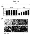
- a study, where the gene was introduced into myocardial cells demonstrated that the efficiency of gene introduction into myocardial cells was significantly higher with a minus-strand RNA viral vector than with an adenoviral vector.
- the minus-strand RNA viral vector carrying the Ang1 gene also exhibited an outstanding therapeutic effect on myocardial infarction and limb ischemia.
- adenoviral vectors are most commonly used because they ensures efficient gene transfer and high-level gene expression in nondividing cells including myocardial cells.
- the adenoviral vector may induce inflammation due to its high immunogenicity and produce adverse effects as a result of its exceedingly high affinity for the liver.
- AAV adeno-associated viral vector
- the adeno-associated viral vector (AAV) have been previously tested, and were found to exhibit long-term gene expression in the heart.
- retroviral vectors and DNA viral vectors interact with the chromosome in the host cell nucleus and become integrated into the host chromosome.
- the minus-strand RNA viral vector can strongly express the gene that it carries in the cytoplasm without being integrated into the host cell chromosome, and thus conferring no risk of chromosome damage.
- the use of minus-strand RNA viral vectors may allow a more effective and safer Ang1 gene therapy for ischemic diseases.
- Ang1 was found to increase vascular density in infarcted hearts. When Ang1 is used clinically, it is most important to assess whether the increase in the vascular density indeed contributes to reduction of infarcted region and improvement of cardiac functions.
- the infarcted region was measured four weeks after myocardial infarction. It was then found that the infarcted region was reduced and the infarcted wall was thickened in the Ang1 gene-administered group.
- the cardiac functions particularly fractional shortening (FS) of left ventricular short-axis diameter, left ventricular area at systole (LVAs), and left ventricular ejection fraction (EF), were found to be improved.
- FS fractional shortening
- LVAs left ventricular area at systole
- EF left ventricular ejection fraction
- HGF hepatocyte growth factor
- HIF-1 ⁇ hypoxia inducible factor-1 ⁇
- VEGF hepatocyte growth factor
- Circulation 104 I218-I222 (2001); Suzuki, K., Circulation 104: I207-212 (2001); Orlic, D., Proc. Natl. Acad. Sci. USA 98: 10344-10349 (2001)).
- the present invention demonstrated for the first time that administration of Ang1 alone can improve cardiac functions of an infarcted heart.
- the administration of Ang1 in the acute phase of myocardial infarction produces marked effects, such as decreasing the post-infarction mortality rate, increasing the number of blood vessels in cardiac muscle, reducing the infarct size, and improving cardiac functions.
- Ang1 gene therapy can be a new effective therapy for acute myocardial infarction.
- the present inventors also performed gene therapy where Ang1 gene alone was administered into an animal model of severe limb ischemia, using an adenoviral vector and a minus-strand RNA viral vector which highly express Ang1. Naked DNA was expressed with an exceedingly high efficiency in cardiac muscle. In contrast, the expression level of the introduced gene by the naked DNA vector was lower in skeletal muscles (Example 8). Therefore, it appeared that direct administration of the Ang1 plasmid to ischemic limbs produced a less-than-sufficient effect on limb salvage (WO02/100441).
- Ang1 gene alone using an Ang1-encoding viral vector produces a therapeutic effect, which would have been impossible with a plasmid vector, not only in ischemic heart diseases but also in general ischemic diseases including extremity ischemia, injuries associated with impaired circulation, and traumatic injury such as amputation, and fractures.
- Conventional therapy used in combination with VEGF is risky in that excess VEGF enhances vascular permeability and then aggravates pulmonary edema or such.
- ischemia can be treated effectively while such adverse effects are avoided.
- the present invention relates to methods for treating ischemic diseases using Ang1 or an Ang1-encoding vector, and ischemic disease treatment kits comprising Ang1 or an Ang1-encoding vector, and more specifically relates to the invention described in each claim.
- the present invention also relates to inventions comprising a desired combination of one or more (or all) of the inventions described in the respective claims, in particular, to inventions comprising a desired combination of one or more (or all) of the inventions described in claims (dependent claims) which cite identical independent claims (claims each relating to an invention which is not encompassed in the inventions described in any other claims).
- the invention described in each independent claim comprises inventions comprising an arbitrary combination of its dependent claims.
- the present invention provides:
- the present invention relates to methods for treating ischemic heart diseases, which comprises the step of administering Ang1 or an Ang1-encoding vector.
- Ang1 alone does not have the activity to stimulate vascular endothelial proliferation. It was unclear as to whether administration of Ang1 gene alone had produced any therapeutic effect on ischemic heart diseases. However, in the present invention, it was demonstrated that an administration of Ang1 alone produced a marked therapeutic effect in myocardial infarction.
- VEGF increases in the sera of patients with acute myocardial infarction two to three days after infarction, and the local VEGF expression level in the heart of a myocardial infarction model also increases one to three days after myocardial infarction, and the high-level expression continues for one week or longer.
- the local and serum levels of VEGF were confirmed to increase in a rat myocardial infarction model produced by the present inventors (data not shown). Accordingly, the therapeutic effect brought upon the administration of Ang1 alone may be a combined effect with endogenous VEGF. Excess VEGF enhances lung vascular permeability and causes pulmonary edema, thereby increasing mortality rate.
- the present invention provides methods for treating ischemic heart diseases, comprising the step of administering Ang1 or an Ang1-encoding vector without administration of vascular endothelial growth factor (VEGF) or its gene.
- VEGF vascular endothelial growth factor
- the vascular density was clearly increased in the infarcted site and its surrounding area when Ang1 is administered alone; the vascular density-increasing effect was comparable to that produced by introducing the same amount of VEGF 165 gene alone via an adenoviral vector.
- ischemic tissues can be protected in a safer and more effective manner by administering Ang-1 its gene without administration of VEGF.
- the methods of the present invention for treating ischemic diseases and ischemic heart diseases are useful methods for protection of ischemic tissues, regeneration of rejected tissues, and revascularization in rejected tissues.
- angiopoietin-1 refers to a ligand that binds to the Tie-2 receptor, and through the receptor activates signal transduction and enhances angiogenesis.
- Tie-2 is a tyrosine kinase receptor and is expressed in endothelial cell lines (Ac. No. NM_000459, protein ID. Q02763, NP_000450)(Ziegler, S. F. et al., Oncogene 8 (3), 663-670 (1993); Boon, L. M. et al., Hum. Mol. Genet. 3 (9), 1583-1587 (1994); Dumont, D. J.
- Tie-2 has been isolated not only from human but also from non-human mammals including cow and mouse (Sato, T. N. et al., Proc. Natl. Acad. Sci. U.S.A. 90 (20), 9355-9358 (1993); Iwama, A. et al., Biochem. Biophys. Res. Commun. 195 (1), 301-309 (1993)).
- the nucleotide sequence of a wild-type human Tie-2-encoding DNA and its amino acid sequence are shown in SEQ ID NOs: 1 and 2, respectively.
- Ligands for the human Tie-2 shown in SEQ ID NO: 2 and the above-described mammalian homologues which enhance angiogenesis can be used preferably in the present invention.
- the Ang1 of the present invention comprises not only the naturally-occurring protein, but also modified proteins and partial peptides thereof that have the function of a Tie-2 ligand similarly to naturally-occurring Ang1. Furthermore, it can be a fragment of an anti-Tie-2 antibody which binds to the extracellular domain of Tie-2, or a non-peptide compound which functions as a Tie-2 ligand.
- Mammalian Ang1 proteins have been isolated from various mammalian species, including human, mouse, rat, pig, and cow (Davis, S. et al., Cell 87 (7), 1161-1169 (1996); Valenzuela, D. M. et al., Proc. Natl. Acad. Sci. U.S.A. 96 (5), 1904-1909 (1999); Suri, C. et al., Cell 87 (7), 1171-1180 (1996); Valenzuela, D. M. et al., Proc. Natl. Acad. Sci. U.S.A. 96 (5), 1904-1909 (1999); Kim, I., et al., Cardiovasc. Res.
- the Ang1 of the present invention also comprises: a protein comprising an amino acid sequence with one or more amino acid substitutions, deletions, and/or additions in the human or other mammalian Ang1amino acid sequence; a protein comprising an amino acid sequence which has 70% or higher, preferably 75% or higher, more preferably 80% or higher, more preferably 85% or higher, still more preferably 90% or higher, yet more preferably 95% or higher identity to the human or other mammalian Ang1 amino acid sequence; and a protein encoded by a nucleic acid hybridizing under stringent conditions to a nucleic acid comprising the entire coding region or a portion of the human or other mammalian Ang1 gene, wherein the protein binds to a mammalian Tie-2 receptor and activates signal transduction via the receptor, thereby enhancing angiogenesis.
- Such proteins may comprise polymorphic and splicing variants of Ang1.
- the number of amino acids changed by amino acid substitution, deletion, and/or addition is typically 15 residues or less, preferably 11 residues or less, more preferably 9 residues or less, more preferably 7 residues or less, still more preferably 5 residues or less. Particularly when amino acids have been substituted conservatively, proteins tend to retain their original activities.
- Conservative substitutions include amino acid substitutions within each group of: basic amino acids (for example, lysine, arginine, and histidine); acidic amino acids (for example, aspartic acid and glutamic acid); non-charged polar amino acids (for example, glycine, asparagine, glutamine, serine, threonine, tyrosine, and cysteine); non-polar amino acids (for example, alanine, valine, leucine, isoleucine, proline, phenylalanine, methionine, and tryptophan), ⁇ -branched amino acids (for example, threonine, valine, and isoleucine), and aromatic amino acids (for example, tyrosine, phenylalanine, tryptophan, and histidine).
- basic amino acids for example, lysine, arginine, and histidine
- acidic amino acids for example, aspartic acid and glutamic acid
- non-charged polar amino acids
- Amino acid sequence identities can be determined, for example, using the BLASTP program (Altschul, S. F. et al., 1990, J. Mol. Biol. 215: 403-410). Specifically, the blastp program can be used for this purpose. For example, when the search is carried out using BLAST in the website of National Center for Biotechnology Information (NCBI), the filters comprising low complexity are all switched off and default parameters are used (Altschul, S. F. et al. (1993) Nature Genet. 3:266-272; Madden, T. L. et al. (1996) Meth. Enzymol. 266:131-141; Altschul, S. F. et al. (1997) Nucleic Acids Res.
- NCBI National Center for Biotechnology Information
- sequence identity can be determined by preparing an alignment of two sequences using the blast2sequences program for comparison of two sequences (Tatiana A et al. (1999) FEMS Microbiol Lett. 174:247-250). Gaps are treated similarly to mismatches. For example, identities to the entire amino acid sequence of mammalian wild-type Ang1 protein are computed. Alternatively, identities can be determined by hybridization, in which a probe is prepared either from a nucleic acid comprising the Ang1 protein coding sequence derived from human or other animals, or from a target nucleic acid of hybridization.
- Stringent hybridization conditions comprise hybridizing in a solution comprising 5x SSC, 7% (W/V) SDS, 100 ⁇ g/ml denatured salmon sperm DNA, and 5x Denhardt's solution (1x Denhardt's solution comprises 0.2% polyvinyl pyrrolidone, 0.2% bovine serum albumin, and 0.2% Ficoll) at 48°C, preferably at 50°C, more preferably at 52°C, followed by 2 hours of washing while shaking at the same temperature used in the hybridization, more preferably at 60°C, still more preferably at 65°C, most preferably at 68°C in 2x SSC, preferably in 1x SSC, more preferably in 0.5x SSC, and more preferably in 0.1x SSC.
- an “Ang1-encoding vector” refers to a vector comprising an Ang1 protein-encoding nucleic acid.
- protein-encoding means that a nucleic acid contains an ORF encoding an amino acid sequence of the protein in a sense or antisense strand (in a certain viral vector or such), so that the nucleic acid can express the protein under appropriate conditions.
- the nucleic acid may be a single- or double-stranded nucleic acid.
- the nucleic acid may be DNA or RNA.
- the vector includes plasmid vectors, other naked DNAs, and viral vectors.
- naked DNA refers to a DNA that does not bind to reagents for introducing nucleic acids into cells, such as viral envelope, liposome, and cationic lipids (Wolff et al., 1990, Science 247, 1465-1468). Naked DNA can be used after being dissolved in a physiologically acceptable solution, for example, sterilized water, physiological saline, or buffer.
- a physiologically acceptable solution for example, sterilized water, physiological saline, or buffer.
- the injection of naked DNA such as plasmid is the safest and simplest gene transfer method, and is used as the major procedure in previously approved clinical protocols of gene therapy for cardiovascular diseases (Lee, Y. et al., Biochem. Biophys. Res. Commun. 2000; 272: 230-235).
- CMV cytomegalovirus
- the present inventors found that when a naked plasmid is introduced into the cardiac muscle by direct injection, the expression level in the cardiac muscle is approximately an order of magnitude greater than that in the skeletal muscle.
- OOU optical units
- the gene therapy for ischemic diseases according to the present invention can be performed using a plasmid comprising the CA promoter or a promoter with transcriptional activity comparable to or higher than that of the CA promoter.
- the CA promoter is a chimeric promoter comprising the CMV immediately early enhancer and the chicken ⁇ -actin promoter (Niwa, H. et al. (1991) Gene. 108: 193-199). Safer gene therapy using a naked DNA can be achieved by using the CA promoter or a promoter with transcriptional activity comparable to or higher than that of the CA promoter.
- the CMV immediately early enhancer to be used may be an immediately early gene enhancer derived from a desired CMV strain.
- the enhancer includes the nucleotide sequence of positions 1 to 367 of SEQ ID NO: 5.
- the chicken ⁇ -actin promoter to be used includes a DNA fragment which comprises the transcription initiation site derived from the chicken ⁇ -actin genomic DNA and has a promoter activity. Since the first intron of the chicken ⁇ -actin gene has transcription-enhancing activity, it is preferred that a genomic DNA fragment comprising at least a portion of this intron is used. Specifically, such a chicken ⁇ -actin promoter includes the nucleotide sequence from positions 368 to 1615 of SEQ ID NO: 5.
- a sequence from another gene for example, the intron-acceptor sequence of rabbit ⁇ -globin, may be appropriately used as the intron acceptor sequence.
- a preferred CA promoter of the present invention is a DNA, in which the chicken ⁇ -actin promoter comprising a portion of the intron is ligated to the downstream of the CMV immediately early enhancer sequence, and then a desired intron-acceptor sequence is placed downstream thereof.
- An example of such DNA is shown in SEQ ID NO: 5.
- the Ang1 protein-encoding sequence may be attached to the last ATG of the above-described sequence which serves as the initiation codon.
- sequence polymorphisms in the CMV enhancer and the chicken ⁇ -actin gene may exist among isolated strains or isolated individuals.
- the SV40ori sequence is preferably deleted.
- the SV40 large T antigen is involved in some types of human cancers, and there is a risk that the SV40ori-comprising vector might be amplified in patients with SV40-associated cancer (Martini, F. et al., Cancer 2002; 94: 1037-1048; Malkin, D. Lancet 2002; 359: 812-813).
- the present inventors verified that the deletion of SV40ori from the vector had no effect on the expression level of the introduced gene in the heart as well as in the skeletal muscle (Example 8).
- the SV40ori-free vector which expresses Ang1 under the control of the CA promoter, is one of the safest and the most useful vectors in clinical applications of cardiac muscle gene therapy.
- the pCA1 vector in which SV40ori has been deleted is considered suitable for cardiac muscle gene therapy.
- a DNA can be appropriately administered in combination with a transfection reagent.
- the transfection efficiency can be improved by combining the DNA with liposomes or desired cationic lipids.
- Ang1 can be expressed at sufficiently high levels not only in the cardiac muscle, but also in other tissues such as the skeletal muscle by using a viral vector.
- the viral vector includes, but is not limited to, an adenoviral vector, an adeno-associated viral vector, a retroviral vector, a lentiviral vector, a herpes simplex virus vector, and a vaccinia virus vector.
- a preferred viral vector is an adenoviral vector.
- the adenoviral vector can introduce a gene into myocardial cells at high efficiencies and express the introduced gene at high levels.
- the Ang1-expressing adenoviral vector produces significant therapeutic effects on ischemic hearts and limbs.
- the adenoviral vector can be suitably used in the present invention.
- conventional adenoviral vectors can be appropriately used.
- genes of the wild-type virus may be modified, for example, to improve the expression level of a foreign gene or to attenuate their antigenicity.
- Adenoviral vectors can be prepared, for example, using the COS-TPC method developed by Saito et al. (Miyake, S., Proc. Natl. Acad. Sci. USA 93: 1320-1324 (1996)).
- the sequence around the initiation codon of Ang1 is preferably made into a Kozak's consensus sequence [for example, CC(G/A)CCATG] to increase the efficiency of the Ang1 gene expression (Kozak, M., Nucleic Acids Res 9(20), 5233 (1981); Kozak, M., Cell 44, 283 (1986); Kozak, M. Nucleic Acids Res.15:8125 (1987); Kozak, M., J. Mol. Biol. 196, 947 (1987); Kozak, M., J. Cell Biol. 108, 229 (1989); Kozak, M., Nucl. Acids Res. 18, 2828 (1990)).
- a Kozak's consensus sequence for example, CC(G/A)CCATG
- RNA viral vector Another viral vector preferably used in the present invention is a minus-strand RNA viral vector. As shown in the Examples, the minus-strand RNA viral vector could achieve higher expressions of an introduced gene with a lower titer than those of the adenovirus.
- the Ang1-encoding minus-strand RNA viral vector is one of the most preferably used vectors in the present invention.
- a "minus-strand RNA virus” refers to a virus comprising a minus-strand (an antisense strand complementary to a viral protein-encoding sense strand) RNA as the genome.
- the minus-strand RNA is also called a "negative strand RNA".
- the minus-strand RNA virus to be used in the present invention includes single-stranded minus-strand RNA viruses (also called “non-segmented minus-strand RNA viruses").
- a "single-stranded negative strand RNA virus” refers to a virus comprising a single-stranded negative strand (i.e., minus strand) RNA as the genome.
- Such viruses include: paramyxovirus (Paramyxoviridae such as the genus Paramyxovirus, the genus Morbillivirus, the genus Rubulavirus, and the genus Pneumovirus); rhabdovirus (Rhabdoviridae such as the genus Vesiculovirus, the genus Lyssavirus, and the genus Ephemerovirus); filovirus (Filoviridae); orthomyxovirus (Orthomyxoviridae such as Influenza viruses A, B, and C, and Thogoto-like virus); bunyavirus (Bunyaviridae such as the genus Bunyavirus, the genus Hantavirus, the genus Nairovirus, and the genus Phlebovirus); and arenavirus (Arenaviridae).
- paramyxovirus Paramyxoviridae such as the genus Paramyxovirus, the genus Morbillivirus, the
- the minus-strand RNA viral vector to be used in the present invention may be a vector having transmissibility, or a deficient vector having no transmissibility.
- the phrase "having transmissibility" means that after a viral vector infects host cells, the virus was replicated and infectious virus particles are produced in the cells.
- the minus-strand RNA virus that can be used preferably in the present invention includes Sendai virus, Newcastle disease virus, mumps virus, measles virus, respiratory syncytial virus (RS virus), rinderpest virus, distemper virus, simian parainfluenza virus (SV5), human parainfluenza viruses type 1, type 2, and type 3, which belong to Paramyxoviridae; influenza virus which belongs to Orthomyxoviridae; and vesicular stomatitis virus and rabies virus which belong to Rhabdoviridae.
- Sendai virus Newcastle disease virus, mumps virus, measles virus, respiratory syncytial virus (RS virus), rinderpest virus, distemper virus, simian parainfluenza virus (SV5), human parainfluenza viruses type 1, type 2, and type 3, which belong to Paramyxoviridae; influenza virus which belongs to Orthomyxoviridae; and vesicular stomatitis virus and
- the virus that can be used in the present invention further includes Sendai virus (SeV), human parainfluenza virus-1 (HPIV-1), human parainfluenza virus-3 (HPIV-3), phocine distemper virus (PDV), canine distemper virus (CDV), dolphin morbillivirus (DMV), peste-des-petits-ruminants virus (PDPR), measles virus (MV), rinderpest virus (RPV), Hendra virus (Hendra), Nipah virus (Nipah), human parainfluenza virus-2 (HPIV-2), simian parainfluenza virus 5 (SV5), human parainfluenza virus-4a (HPIV-4a), human parainfluenza virus-4b (HPIV-4b), mumps virus (Mumps), and Newcastle disease virus (NDV).
- Sendai virus SeV
- human parainfluenza virus-1 HPIV-1
- human parainfluenza virus-3 HPIV-3
- phocine distemper virus canine distemper virus (
- the virus includes that selected from the group consisting of Sendai virus (SeV), human parainfluenza virus-1 (HPIV-1), human parainfluenza virus-3 (HPIV-3), phocine distemper virus (PDV), canine distemper virus (CDV), dolphin morbillivirus (DMV), peste-des-petits-ruminants virus (PDPR), measles virus (MV), rinderpest virus (RPV), Hendra virus (Hendra), and Nipah virus (Nipah).
- Sendai virus SeV
- HIV-1 human parainfluenza virus-1
- HPIV-3 human parainfluenza virus-3
- PDV phocine distemper virus
- CDV canine distemper virus
- DMV dolphin morbillivirus
- PDPR measles virus
- RPV rinderpest virus
- Hendra virus Hendra
- Nipah virus Nipah virus
- the virus belongs to the subfamily Paramyxoviridae (including the genus Respirovirus, the genus Rubulavirus, and the genus Morbillivirus) or is a derivative thereof. Still more preferably, the virus belongs to the genus Respirovirus (also called the genus Paramyxovirus) or a derivative thereof. Such derivatives include viruses genetically modified or chemically modified without impairing the virus's ability to introduce genes.
- viruses of the genus Respirovirus to which the present invention can be applied include human parainfluenza virus type 1 (HPIV-1), human parainfluenza virus type 3 (HPIV-3), bovine parainfluenza virus type 3 (BPIV-3), Sendai virus (also called murine parainfluenza virus type 1), and simian parainfluenza virus type 10 (SPIV-10).
- the paramyxovirus of the present invention is most preferably the Sendai virus.
- These viruses may be derived from natural strains, wild-type strains, mutant strains, laboratory-passaged strains, artificially constructed strains, etc.
- Reconstitution of the recombinant minus-strand RNA viral vector can be achieved by using known methods (WO 97/16539; WO 97/16538; WO 00/70055; WO 00/70070; WO 03/025570; Durbin, A. P. et al., Virology 235, 323-332, 1997; Whelan, S. P. et al., Proc. Natl. Acad. Sci. USA 92, 8388-8392, 1995; Schnell. M. J. et al., EMBO J. 13, 4195-4203, 1994; Radecke, F. et al., EMBO J. 14, 5773-5784, 1995; Lawson, N. D.
- the minus-strand RNA virus including parainfluenza, vesicular stomatitis virus, rabies virus, measles virus, rinderpest virus, and Sendai virus, can be reconstituted from its DNA using such methods.
- the viruses of the present invention can be reconstituted according to these methods.
- the DNA that constitutes the viral genome when genes encoding the envelope-constituting proteins, such as the F gene, HN gene, and/or M gene, have been deleted from the viral genome, infectious viral particles are not formed automatically.
- infectious viral particles can be formed when the deleted genes and/or envelope proteins from another virus (for example, the gene encoding vesicular stomatitis virus (VSV) G protein (VSV-G) (J. Virology 39: 519-528 (1981)) are separately introduced into the host cells and expressed in the cells (Hirata, T. et al., 2002, J. Virol. Methods, 104:125-133; Inoue, M. et al., 2003, J. Virol. 77:6419-6429).
- VSV vesicular stomatitis virus
- VSV-G vesicular stomatitis virus
- minus-strand RNA virus takes place only in the host cell cytoplasm and since the virus has no DNA phase, the virus is not integrated into the chromosome (Lamb, R.A. and Kolakofsky, D., Paramyxoviridae: The viruses and their replication. In: Fields BN, Knipe DM, Howley PM, (eds). Fields Virology, 3rd Edition, Vol. 2. Lippincott - Raven Publishers: Philadelphia, 1996, pp. 1177-1204). Therefore, the vector does not cause safety problems, such as canceration and immortalization due to chromosomal abnormalities. This characteristic of minus-strand RNA viruses largely contributes to its safety when used as a vector.
- the minus-strand RNA viral vector is highly useful as a therapeutic vector in gene therapy for human ischemic diseases.
- the collected viral vector can be purified to substantial homogeneity.
- the purification can be achieved by using conventional purification/separation methods such as filtration, centrifugation, adsorption, and column purification, or any combination thereof.
- the phrase "substantially pure" means that the viral components constitute a large share of the solution that contains the viral vector.
- a viral vector composition can be verified to be substantially pure if the proteins contained as the viral vector components constitute 10%(W/W) or more, preferably 20% or more, more preferably 50% or more, preferably 70% or more, more preferably 80% or more, still more preferably 90% or more of the total proteins (excluding the proteins that have been added as carriers or stabilizers) in a solution.
- specific purification methods include, but are not limited to, the method using cellulose sulfate ester or crosslinked polysaccharide sulfate ester (Japanese Patent Application Kokoku Publication No. (JP-B) S62-30752 (examined, approved Japanese patent application published for opposition); JP-B S62-33879; and JP-B S62-30753), and the method based on the adsorption by fucose sulfate-containing polysaccharide and/or degradation products thereof (WO 97/32010).
- ischemic diseases refers to functional abnormality, or tissue degeneration or necrosis, caused by the reduction or disruption of blood supply to tissues, and specifically comprises ischemic heart diseases such as myocardial infarction and angina pectoris, and extremity ischemia, injuries associated with impaired circulation, traumatic injuries such as amputation, and fractures.
- the ischemic diseases of the present invention include not only ischemic diseases but also ischemic states as a result of injury or damage.
- Administration of Ang1 or an Ang1-encoding vector suppresses necrosis of tissues surrounding the ischemic site and improves their functions through induction of angiogenesis, and other effects such as anti-apoptotic effect and anti-inflammatory effect.
- VEGF is not included in the Ang1 administration of the present invention.
- Significant therapeutic effects can be produced by administering Ang1 alone without administering VEGF.
- the phrase "without administering VEGF" specifically means that VEGF or a vector encoding VEGF is not administered within at least 12 hours, preferably within 24 hours, more preferably within 14 days before or after the administration of Ang1 or an Ang1-encoding vector.
- VEGF is considered not to be administered.
- VEGF Vascular Endothelial Growth Factor
- VPF Vascular Permeability Factor
- VEGF A is further divided into six subtypes. Among them, soluble VEGF 121 and 165, in particular, have a strong vascular proliferation ability and are currently used in clinic.
- VEGF of the present invention includes particularly VEGF165 and VEGF121, and preferably various VEGF members comprising VEGF165 and VEGF121.
- the therapeutic methods of the present invention are highly effective for ischemic diseases which are accompanied by an elevation of the endogenous VEGF level.
- endogenous VEGF level is elevated means that the endogenous VEGF level in blood or in local tissues is higher than in healthy individuals.
- the endogenous VEGF level is elevated in the above-described myocardial infarction, angina pectoris, acute extremity ischemia, injuries associated with impaired circulation, amputation, fractures, or such.
- ischemic heart diseases refers to functional abnormality of the heart, or cardiac muscle degeneration or necrosis caused by the reduction or disruption of blood supply to cardiac muscle, and includes specifically angina pectoris, myocardial infarction, and some types of cardiomyopathy.
- Angiogenesis is enhanced and cardiac functions are improved by administering Ang1 or an Ang1-encoding vector into ischemic hearts.
- the methods of the present invention are highly effective for ischemic heart diseases which are accompanied by elevation of the endogenous VEGF level, and are appropriately used to treat, for example, angina pectoris, myocardial infarction, and ischemic cardiomyopathy (Xu, X., et al. (2001) J Thorac Cardiovasc Surg.
- the ischemic heart disease to which the methods of the present invention are most effective is myocardial infarction.
- Angina pectoris refers to a clinical syndrome with the main symptom as chest discomfort caused by transient ischemia, namely lack of oxygen, in the cardiac muscle (Ogawa H., "pharmacotherapy for angina pectoris", supplementary volume, Journal of Clinical and Experimental Medicine (Igaku no Ayumi); circulatory diseases: 352-355,1996, Eds. Yazaki Y. et al. Ishiyaku Publisher Inc.).
- Acute myocardial infarction is an ischemic heart disease, where necrosis of the cardiac muscle is caused by obstruction of blood flow in the coronary arteries (Abu M., and Takano T., Acute myocardial infarction: The latest therapy for circulatory diseases 2002-2003 II, coronary artery diseases: 37-42, 2002, eds., Shinoyama, S. and Yazaki Y., Nankodo Co. Ltd.).
- angiogenesis factor refers to a factor that is directly or indirectly involved in the development, migration, proliferation, or maturation of cells which participate in vascularization.
- vascular endothelial growth factors VEGFs
- FGFs fibroblast growth factors
- EGF epithelial growth factor
- HGF hepatocyte growth factor
- PDGF monocyte chemoattractant protein-1
- MCP-1 monocyte chemoattractant protein-1
- TP thymidine phosphorylase
- angiopoietin ephrin(Eph)
- MMP matrix metalloproteinase
- TMP tissue inhibitor of metalloproteinase
- TMP tissue inhibitor of metalloproteinase
- Ang1 or an Ang1-encoding vector is administered systemically or locally to ischemic tissues. Ang1 does not produce marked adverse effects even when administered at high doses. Therefore, ischemia can be treated by administering Ang1 systemically.
- Ang1 or an Ang1-encoding vector may be introduced directly or via carriers.
- the carrier should be physiologically acceptable, and includes organic substances such as biopolymer and inorganic substances such as hydroxyapatite; specifically, collagen matrix, polylactic acid polymer or copolymer, polyethylene glycol polymer or copolymer, and chemical derivatives thereof. Furthermore, the carrier may be mixed compositions of the physiologically acceptable materials described above.
- the vector to be used is not limited as long as it is a physiologically acceptable vector, and desired vectors including viral vectors and non-viral vectors can be used.
- the vector can be administered in the form of a vector-treated cell derived from the patient him/herself.
- the vector or cells into which the vector has been introduced can be administered via intramuscular injection (to cardiac muscle or skeletal muscle) or intravenous injection (in vivo and ex vivo administration). Cells which have been administered systemically (via intramuscular or intravenous injection) can transfer to lesion sites and enhance the survival of ischemic tissues.
- MSCs mesenchymal stem cells
- MSCs mesenchymal stem cells
- Ang1 or an Ang1-encoding vector can be injected into the cardiac muscle.
- cells into which an Ang1-encoding vector has been introduced may be transplanted into the cardiac muscle (ex vivo administration). The injection can be achieved using manufactured products such as standard medical injectors, and external and indwelling continuous infusers.
- the dosage can be administered, for example, at one or more sites (for example, two to ten sites) in the surviving muscle (skeletal muscle, cardiac muscle, or such) surrounding the ischemic site.
- the dosage preferably ranges from, for example, 10 10 to 10 13 pfu/body, and more preferably 10 11 to 10 13 pfu/body.
- the dosage of a minus-strand RNA virus preferably ranges from, for example, 2x 10 5 to 5x 10 11 CIU.
- a naked DNA can be administered at one or more sites (for example, two to ten sites) in the surviving muscle surrounding the ischemic site.
- the injection dosage per site preferably ranges from, for example, 10 ⁇ g to 10 mg, and more preferably 100 ⁇ g to 1 mg.
- the vector is introduced into the target cells (for example, in a test tube or dish) ex vivo, for example, at a multiplicity of infection (MOI) of 1 to 500.
- MOI multiplicity of infection
- minus-strand RNA viral vectors have been found to introduce foreign genes into mesenchymal cells with exceedingly high efficiency. Accordingly, when mesenchymal cells are used in an ex vivo administration, it is preferable to use a minus-strand RNA viral vector to introduce genes into the mesenchymal cells.
- Ang-1 gene-introduced cells for example, 10 5 to 10 9 cells, and preferably 10 6 to 10 8 cells can be transplanted to ischemic tissues.
- a protein preparation When a protein preparation is used, it may be administered at one or more sites (for example, two to ten sites) in the surviving muscle surrounding the ischemic site. The dosage preferably ranges from, for example, 1 ⁇ g/kg to 10 mg/kg, and more preferably 10 ⁇ g/kg to 1 mg/kg.
- the vector or the protein preparation may be administered, for example, several times (one to ten times) to the artery that leads to the ischemic tissue (for example, the coronary artery of an ischemic heart).
- the dosage per site preferably ranges from, for example, 1 ⁇ g/kg to 10 mg/kg, and more preferably 10 ⁇ g/kg to 1 mg/kg.
- a vector or protein preparation may be administered intravenously several times (one to ten times) or it may be administered continuously.
- the total dosage preferably ranges from, for example, 1 ⁇ g/kg to 10 mg/kg, and more preferably 10 ⁇ g/kg to 1 mg/kg.
- a vector When a vector is used, it may be administered at the same dosage as described above for the intramuscular injection. See, Freedman SB et al. Ann Intern Med 136:54-71 (2002), for dosage.
- the vector dosage may vary depending on patient's weight, age, sex, and symptoms; dosage form of the composition to be administered; method of administering the vector; and so on. Those skilled in the art can appropriately adjust the dosage.
- the frequency of administration may range from one to several times within clinically acceptable limits of adverse effects.
- the per kg dosage for non-human animals may be the same as that for human, or can be alternatively converted from the above-described dosage, for example, based on the volume ratio (for example, average value) between the ischemic organs (such as heart) of the subject animal and human.
- Animals subjected to the treatments of the present invention include human and other desired mammals, specifically, human, monkey, mouse, rat, rabbit, sheep, cow, and dog.
- the therapeutic methods of the present invention can be conducted singly or in combination with other standard or advanced methods.
- the methods of the present invention for treating ischemic heart diseases can be used preferably in combination with surgical revascularization, such as percutaneous transluminal coronary angioplasty (PTCA) or coronary artery bypass graft (CABG).
- PTCA percutaneous transluminal coronary angioplasty
- CABG coronary artery bypass graft
- Combined use with the therapeutic methods of the present invention can actively improve cardiac functions and reduce the period confined to bed.
- Treatments using Ang1 of the present invention are also expected to be more effective when used in combination with therapeutic methods that enhance remodeling in the infarcted region, for example, regeneration of infarcted myocardium.
- Ang1 gene therapy increases the infarct thickness, it has a relatively weak effect on improving the diastolic parameters such as LVAd and Edd, whose improvements are seen at the same time that a deficiency of the absolute mass of cardiac muscle is improved through combined use of cell therapy or such.
- the improvement by Ang1 of systolic volume, ejection fraction, and so on, is assumed to prevent the hypofunction of peri-infarct muscle by increasing the vascular density in the surviving cardiac muscle such as peri-infarct muscle, and then to also improve functions of the cardiac muscle by enhancing the compensatory hypertrophy of the surviving cardiac muscle.
- a cell therapy which compensates for the deficiency of the absolute mass of cardiac muscle, when considering transplantation of fetal cardiac muscle, ES cells, myoblasts, mesenchymal cells, or such, or induction of cell migration to infarcted sites. It is also useful to enhance the therapeutic effect on ischemic tissues by ex vivo introduction of Ang-1 gene into these cells.
- the present invention also provides therapeutic agents comprising Ang1 or an Ang1-encoding vector for ischemic heart diseases.
- the present invention also provides uses of Ang1 or an Ang1-encoding vector in administering to ischemic hearts for the treatment of ischemic heart diseases.
- the present invention also provides uses of Ang1 or an Ang1-encoding vector in producing therapeutic agents for ischemic heart diseases, which are used to administer Ang1 or an Ang1-encoding vector to ischemic hearts.
- the present invention provides therapeutic agents comprising Ang1 or an Ang1-encoding vector for ischemic heart diseases, to administer Ang1 or an Ang1-encoding vector to ischemic hearts without the administration of VEGF or a VEGF vector.
- the present invention also provides uses of Ang1 or an Ang1-encoding vector for the treatment of ischemic heart diseases, in which Ang1 or an Ang1-encoding vector is administered to ischemic hearts without the administration of VEGF or a VEGF vector.
- the present invention also provides uses of Ang1 or an Ang1-encoding vector in producing therapeutic agents for ischemic heart diseases, which are used to administer Ang1 or an Ang1-encoding vector to ischemic hearts without the administration of VEGF or a VEGF vector.
- Ang1 or an Ang1-encoding vector is preferably formulated for local administration to ischemic hearts.
- a formulation is preferably administered by injection into the cardiac muscle.
- the Ang1-encoding vector is preferably a viral vector or a naked DNA that encodes Ang1.
- the viral vector is not particularly limited, but adenoviral vectors and minus-strand RNA viral vectors are particularly preferable.
- the naked DNA includes plasmids, which may be circular or linear.
- the plasmid does not contain SV40ori.
- the vector promoter which drives the transcription of Ang1 preferably has strong transcriptional activity.
- a CA promoter can be used suitably.
- kits for treating ischemic heart diseases which comprise: (a) Ang1 or an Ang1-encoding vector, and (b) a recording medium containing a description of instruction that VEGF or a VEGF vector should not be administered when Ang1 or an Ang1-encoding vector is administered, or a link to the description.
- the kits are to be used for treating at least one of ischemic heart diseases including myocardial infarction and angina pectoris.
- the kits of the present invention are preferably used to treat angina pectoris and/or acute myocardial infarction.
- the kits comprise the Ang1 or the Ang1-encoding vector described above.
- the Ang1-encoding vector is preferably a naked DNA or a viral vector that encodes Ang1.
- the viral vector is not particularly limited, but adenoviral vectors and minus-strand RNA viral vectors, in particular, are preferred.
- the Ang1 or Ang1-encoding vector in the kits may be a composition that comprises in addition to Ang1, a desired pharmaceutically acceptable carrier and/or additive.
- the composition may comprise sterilized water, physiological saline, a standard buffer (such as phosphoric acid, citric acid, and other organic acids), a stabilizer, salt, an antioxidant (such as ascorbic acid), a detergent, a suspending agent, an isotonizing agent, or a preservative.
- the Ang1 or Ang1-encoding vector is preferably combined with an organic substance such as biopolymer, an inorganic substance such as hydroxyapatite, specifically collagen matrix, polymer or copolymer of polylactic acid, polymer or copolymer of polyethylene glycol, and derivatives thereof.
- the Ang1 or Ang1-encoding vector is prepared in a dosage form suitable for injection.
- the Ang1 or Ang1-encoding vector is preferably dissolved in a pharmaceutically acceptable aqueous solution, or is preferably a soluble freeze-dry formulation or such.
- the kits of the present invention may further comprise a desired pharmaceutically acceptable carrier that can be used to dissolve or dilute the Ang1 or Ang1-encoding vector.
- a carrier includes, for example, distilled water and physiological saline.
- the present invention also provides therapeutic agents for ischemic diseases, comprising an Ang1-encoding viral vector.
- the present invention also provides uses of an Ang1-encoding viral vector for the treatment of ischemic diseases.
- the present invention provides uses of an Ang1-encoding viral vector for the production of therapeutic agents for ischemic diseases, wherein the therapeutic agents comprises the Ang1-encoding viral vector.
- the present invention provides therapeutic agents for ischemic diseases, comprising an Ang1-encoding viral vector and which are used to administer the Ang1 viral vector to an individual with ischemia without the administration of VEGF or a VEGF vector.
- the present invention provides uses of an Ang1-encoding viral vector for the treatment of ischemic diseases, in administering the Ang1 viral vector to an individual with ischemia without the administration of VEGF or a VEGF vector.
- the present invention provides uses of the Ang1 viral vector in producing therapeutic agents for ischemic diseases, which are used to administer the Ang1-encoding viral vector to an individual with ischemia without the administration of VEGF or a VEGF vector.
- neither VEGF nor any other angiogenesis factors or vectors encoding these factors are preferably administered.
- the Ang1-encoding viral vector is preferably formulated for local administration to ischemic tissues.
- the preferable viral vectors that can be used are adenoviral vectors and minus-strand RNA viral vectors.
- kits for treating ischemic diseases which comprise (a) an Ang1-encoding viral vector, and (b) a recording medium containing a description of instruction that VEGF or a VEGF vector should not be administered when the Ang1-encoding viral vector is administered, or a link to the description.
- the kits are kits for treating at least one of ischemic heart diseases such as myocardial infarction and angina pectoris, and ischemic diseases such as extremity ischemia, injuries associated with impaired circulation, traumatic injuries including amputation, and fractures.
- the kits comprise the Ang1-encoding viral vector described above.
- the viral vector is not particularly limited, but adenoviral vectors and minus-strand RNA viral vectors are particularly preferred.
- the Ang1-encoding viral vector in the kits may be a composition that comprises in addition to the vector, a desired pharmaceutically acceptable carrier and/or additive.
- the composition may comprise sterilized water, physiological saline, a standard buffer (such as phosphoric acid, citric acid, and other organic acids), a stabilizer, salt, an antioxidant (such as ascorbic acid), a detergent, a suspending agent, an isotonizing agent, or a preservative.
- the vector is preferably combined with an organic substance such as biopolymer, an inorganic substance such as hydroxyapatite, and specifically collagen matrix, polylactic acid polymer or copolymer, polyethylene glycol polymer or copolymer, and derivatives thereof.
- the Ang1-encoding viral vector is prepared in a dosage form suitable for injection.
- the Ang1-encoding viral vector is preferably dissolved in pharmaceutically acceptable aqueous solution, or is preferably a soluble freeze-dry formulation or such.
- the kits of the present invention may further comprise a desired pharmaceutically acceptable carrier that can be used to dissolve or dilute the Ang1-encoding viral vector.
- Such carriers include, for example, distilled water and physiological saline.
- kits of the present invention comprise a recording medium containing a description of instruction that VEGF or a VEGF vector should not be administered when Ang1 or an Ang1-encoding vector is administered, or a link to the description.
- "Instruction that VEGF or a VEGF vector should not be administered” refers to, for example, a description of instruction or recommendation that the administration of VEGF or a VEGF should be contraindicated or avoided.
- the description instructs that VEGF or a vector encoding VEGF should not be administered within at least 12 hours before or after administering Ang1 or an Ang1-encoding vector.
- the description instructs that VEGF or a vector encoding VEGF should not be administered within 24 hours, more preferably within 14 days before and after the administration of Ang1 or an Ang1-encoding vector.
- VEGF includes VEGF165 and VEGF 121, in particular, and preferably various members of VEGF including VEGF 165 and VEGF 121.
- the kits of the present invention preferably include a description of a therapeutically effective amount of Ang1 or an Ang1-encoding vector to be administered to affected individuals, or a link to the description.
- the recording medium includes desired recording media, for example, print media such as paper and plastic, and computer-readable recording media such as flexible disc (FD), compact disc (CD), digital video disc (DVD), and semiconductor memory.
- a typical example of the recording medium is an instruction attached to the kits.
- “Link” means that the description, which instructs that VEGF121 should not be administered when Ang1 or an Ang1-encoding vector is administered, is linked via a mark or the like in the kits so that the mark provides a shortcut to the description, rather than being directly enclosed in the kits.
- the instruction may give directions or suggestions to refer to an attached sheet or URL when the description is contained in the attached sheet or URL.
- the mesenchymal cells of the present invention refer to preferably bone marrow cells (the mononuclear cell fraction component of bone marrow cells), cord blood cells or peripheral blood cells, mesenchymal stem cells, cells derived from these cells or such.
- the mesenchymal cells of the present invention also include, for example, cells associated with mesenchyme, and mesodermal stem cells. Even if a cell that is described herein as a "mesenchymal cell" is classified into a non-mesenchymal cell in future, the cell can be suitably used in the present invention.
- the bone marrow contains two stem cell types: hematopoietic stem cell and "mesenchymal stem cell (MSC)".
- stem cell typically refers to an undifferentiated cell that has both the abilities of self-reproduction and differentiation into cells with particular functions in the physiological process of proliferation and differentiation of cells constituting the living body.
- Hematopoietic stem cell refers to a stem cell that differentiates into an erythrocyte, leukocyte, or platelet.
- a mesenchymal stem cell may differentiate into neuron, cardiovascular system, internal organ, bone, cartilage, fat, or muscle.
- mesenchymal stem cells are mainly used; however hematopoietic stem cells and other stem cells (precursor cells) in the body may also be used.
- the mesenchymal stem cells can be obtained by separation from bone marrow cells collected from the bone marrow.
- bone marrow cells containing the mesenchymal stem cells although less effective, can also be used in the therapy.
- mesenchymal stem cell-like cells can be prepared from the peripheral blood.
- cells that have functions equivalent to those of the mesenchymal stem cells can be prepared by culturing cells in the peripheral blood and used in the present invention.
- mesodermal stem cell refers to a cell that constitutes tissues which are classified as mesoderm embryologically including a blood cell. Furthermore, “mesodermal stem cell” refers to a cell that can make (divide or proliferate into) copies of cells with the same ability as itself, and which can differentiate into all types of cells that constitute mesodermal tissues.
- the mesodermal stem cell comprises, but is not limited to, those cells with markers such as SH2(+), SH3(+), SH4(+), CD29(+), CD44(+), CD14(-), CD34(-), and CD45(-). Furthermore, the so-called “mesenchyme-associated stem cells” are also included in the mesodermal stem cells of the present invention.
- mesenchyme-associated cells refer to mesenchymal stem cells, mesenchymal cells, precursor cells of mesenchymal cells, and cells derived from mesenchymal cells.
- Mesenchymal stem cells refer to stem cells that can be obtained, for example, from the bone marrow, peripheral blood, skin, hair root, muscle tissue, uterine endometrium, blood, cord blood, and primary cultures of various tissues. Cells having functions equivalent to the mesenchymal stem cells, which can be obtained by culturing cells in the peripheral blood, are also included in the mesenchymal stem cells of the present invention.
- the preferred mesenchymal cells of the present invention include bone marrow cells and bone marrow stem cells (mesenchymal stem cells).
- examples of the preferred cells of the present invention are cord blood cells, peripheral blood cells, and fetal hepatocytes.
- bone marrow cells, cord blood cells, peripheral blood cells, and fetal hepatocytes include cell fractions separated from the bone marrow, cord blood, peripheral blood, or fetal liver, and cell fractions that can differentiate into cells of the cardiovascular system or myocardial cells.
- the cell fraction contains mesodermal liver cells with SH2(+), SH3(+), SH4(+), CD29(+), CD44(+), CD14(-), CD34(-), and CD45(-).
- the cell fractions of the present invention include a cell fraction that contains interstitial cells comprising the following cell markers: Lin(-), Sca-1(+), CD10(+), CD11D(+), CD44(+), CD45(+), CD71(+), CD90(+), CD105(+), CDw123(+), CD127(+), CD164(+), fibronectin(+), ALPH(+), and collagenase-1(+); and a cell fraction containing cells with the AC 13 3 (+).
- the bone marrow cells, cord blood cells, or peripheral blood cells (cell fractions) of the present invention are typically derived from vertebrates.
- the cells are preferably derived from mammals (for example, mouse, rat, rabbit, pig, dog, monkey, and human), but are not limited thereto.
- mesenchymal cells into which the Ang1 gene had been introduced were found to produce a stronger therapeutic effect on ischemia as compared with non-genetically modified mesenchymal cells.
- useful cells for treating ischemia can be prepared by introducing an exogenous Ang1 gene into mesenchymal cells.
- Angiogenesis and revascularization in ischemic tissues can be enhanced by administering these cells to the ischemic tissues.
- Ang1 gene can be introduced into mesenchymal cells using the plasmid vector described above, other naked DNAs, or viral vectors.
- the vector promoter is preferably a high efficiency promoter so that Ang1 is expressed at high levels in tissues to which the vector has been administered.
- the CA promoter described above is used preferably.
- Ang1 gene is introduced into mesenchymal cells using a viral vector.
- a particularly preferred viral vector includes an adenoviral vector and a minus-strand RNA viral vector.
- the minus-strand RNA viral vector is most preferably used.
- Ang1 gene can be expressed at exceedingly high levels in the mesenchymal cells by using a minus-strand RNA viral vector.
- mesenchymal cells are contacted with the viral vector carrying the gene to be introduced.
- the vector can be contacted with mesenchymal cells in vivo or in vitro, for example, in a desired physiological aqueous solution such as culture solution, physiological saline, blood, plasma, serum, and body fluids.
- the multiplicity of infection is preferably adjusted within the range of 1 to 500, more preferably 1 to 300, even more preferably 1 to 200, still more preferably 1 to 100, and yet still more preferably 1 to 70.
- the infection can be conducted by combining the viral vector with a cell fraction containing mesenchymal cells.
- a minus-strand RNA viral vector When a minus-strand RNA viral vector is used, it is sufficient to contact the mesenchymal cells with the only vector in a brief period for infection.
- the contact time can bel minute or longer, preferably 3 minutes or longer, 5 minutes or longer, 10 minutes or longer, or 20 minutes or longer, for example, about 1 to 60 minutes, and more specifically about 5 to 30 minutes. Needless to say, the contact may be continued for a longer time, for example, 24 hours, or several days or longer.
- the contact can be achieved in vivo or ex vivo.
- gene introduction methods which utilize a minus-strand RNA viral vector and thereby require a short contact between the vector and mesenchymal cells are preferably used.
- Mesenchymal cells into which a gene associated with angiogenesis has been introduced by the methods of the present invention are useful in gene therapy for cardiac ischemia, extremity ischemia, and so on.
- the mesenchymal cells can be prepared, for example, according to the method described in Tsuda, H. et al. Mol Ther 7(3): 354-65 (2003).
- As for the culturing of human mesenchymal cells see the description in Kobune M, et al., Hamada H, Telomerized human multipotent mesenchymal cells can differentiate into hematopoietic and cobblestone area-supporting cells Exp. Hematol Exp Hematol. 31(8):715-22, 2003.
- the prepared cells may be used for therapy immediately, or after being cultured in vitro to about 10 to 40 population doublings (PD).
- a cell fraction containing mesenchymal cells can be prepared by, for example, fractionation of bone marrow cells or cord blood cells collected from vertebrate by performing density gradient centrifugation at 2,000 rpm for long enough to separate the cells based on their specific gravity in the solution, and then collecting the cell fractions with a certain specific density in the range of 1.07 g/ml to 1.1 g/ml.
- the phrase "long enough to separate the cells based on their specific gravity” refers to a sufficient period of time which allows the cells to settle in a position that corresponds to their own specific gravity in the solution used for the density gradient centrifugation. The period of time typically ranges from about 10 minutes to 30 minutes.
- the specific gravity of the cell fraction to be collected preferably ranges from 1.07 g/ml to 1.08 g/ml (for example, 1.077 g/ml).
- Solutions to be used for the density gradient centrifugation include, but are not limited to, Ficoll solution and the Percoll solution.
- cord blood cells collected from vertebrate by the same procedure described above may be used as a cell fraction.
- a specific example involves first mixing a solution (2 ml L-15 + 3 ml Ficoll) with a bone marrow fluid (5 ⁇ l to 10 ⁇ l) collected from vertebrate, and centrifuging the resulting mixture at 2,000 rpm for 15 minutes to separate a mononuclear cell fraction (about 1 ml). The cells were then washed by mixing the mononuclear cell fraction with a culture solution (2 ml of DMEM), and centrifuging the mixture at 2,000 rpm for 15 minutes for the second time. The supernatant is then discarded, and the precipitated cells are collected.
- the cell fractions of the present invention have sources including the sternum, and the iliac bone which constitutes the pelvis.
- the cell fractions can be obtained not only from these bones but also from other large bones.
- the cell fractions can be also collected from bone marrow fluids or cord blood stored in bone-marrow banks. When cord blood cells are used, the cell fraction can be collected from cord blood stored in bone-marrow banks.
- the cell fraction is a mononuclear cell fraction isolated and purified from bone marrow cells, cord blood cells, or peripheral blood cells, comprising mesodermal stem cells (mesenchymal stem cells) that can differentiate into cells of the cardiovascular system.
- Cell fractions containing mesodermal stem cells can be obtained by selecting cells with cell surface markers, such as the above SH2, from the above-mentioned cell fractions, for example, from bone marrow cells, cord blood cells, or peripheral blood cells.
- a cell fraction containing mesodermal stem cells (mesenchymal stem cell) that can differentiate into cells of the cardiovascular system can be prepared by fractionation of bone marrow cells and cord blood cells collected from vertebrate, by performing density gradient centrifugation at 900 g for long enough to separate the cells based on their specific gravity in the solution, followed by collecting cell fractions with a particular specific density in the range of 1.07 g/ml to 1.1 g/ml.
- the phrase "long enough to separate the cells based on their specific gravity” refers to a sufficient period of time that allows the cells to settle in a position corresponding to their own specific gravity in the solution used for density gradient centrifugation. The time typically ranges from about 10 minutes to about 30 minutes.
- the specific gravity of the cell fraction to be collected can vary depending on the type of animal species (for example, human, rat, and mouse) from which the cells are derived.
- Solutions for the density gradient centrifugation include Ficoll solution and Percoll solution, but are not limited thereto.
- a specific example involves first mixing the bone marrow fluid (25 ml) or cord blood collected from vertebrate with an equal volume of PBS, centrifuging the resulting mixture at 900g for 10 minutes, and mixing the precipitated cells with PBS (the cell density is about 4x 10 7 cells /ml) to remove the blood component. A 5-ml aliquot of the cells is then mixed with a Percoll solution (1.073 g/ml), followed by centrifugation of the mixture at 900 g for 30 minutes to separate the mononuclear cell fraction. The mononuclear cell fraction is mixed with a culture medium (DMEM containing 10% FBS and 1% antibiotic-antimycotic solution) for cell washing. The mixture is centrifuged at 2,000 rpm for 15 minutes and the supernatant is discarded. The precipitated cells are collected and cultured (at 37°C with 5% CO 2 in air).
- DMEM containing 10% FBS and 1% antibiotic-antimycotic solution
- the cell fraction is a mononuclear cell fraction separated from bone marrow cells or cord blood cells, containing interstitial cells that can differentiate into cells of the cardiovascular system.
- the interstitial cells are, for example, cells with the markers: Lin(-), Sca-1(+), CD10(+), CD11D(+), CD44(+), CD45(+), CD71(+), CD90(+), CD105(+), CDW123(+), CD127(+), CD164(+), fibronectin(+), ALPH(+), and collagenase-1(+).
- the cell fraction containing interstitial cells can be obtained, for example, by selecting cells with cell surface markers, such as Lin, from the above-described cell fractions obtained from bone marrow cells or cord blood cells by centrifugation.
- the cell fraction can be prepared by fractionation of bone marrow cells or cord blood cells collected from vertebrate, by performing density gradient centrifugation at 800 g for long enough to separate the cells based on their specific gravity in the solution, followed by collecting cell fractions with a particular specific density in the range of 1.07 g/ml to 1.1 g/ml.
- the phrase "long enough to separate the cells based on their specific gravity” refers to a sufficient period of time that allows the cells to settle in a position corresponding to their own specific gravity in the solution used in the density gradient centrifugation. The period of time typically ranges from about 10 minutes to about 30 minutes.
- the specific gravity of the cell fraction to be collected preferably ranges from 1.07 g/ml to 1.08 g/ml (for example, 1.077 g/ml).
- Solutions used in the density gradient centrifugation include, but are not limited to, Ficoll solution and Percoll solution.
- a specific example involves first mixing a bone marrow fluid or cord blood collected from vertebrate with an equal volume of the solution (PBS containing 2% BSA, 0.6% sodium citrate, and 1% penicillin-streptomycin), and mixing a 5-ml aliquot of the cell sample with a Ficoll/Paque solution (1.077 g/ml). The resulting mixture is then centrifuged at 800 g for 20 minutes to separate a mononuclear cell fraction.
- PBS containing 2% BSA, 0.6% sodium citrate, and 1% penicillin-streptomycin
- the mononuclear cell fraction is then mixed with a culture solution (Alfa MEM containing 12.5% FBS, 12.5% horse serum, 0.2% i-inositol, 20 mM folic acid, 0.1 mM 2-mercaptoethanol, 2 mM L-glutamine, 1 ⁇ M hydrocortisone, and 1% antibiotic-antimycotic solution).
- a culture solution Alfa MEM containing 12.5% FBS, 12.5% horse serum, 0.2% i-inositol, 20 mM folic acid, 0.1 mM 2-mercaptoethanol, 2 mM L-glutamine, 1 ⁇ M hydrocortisone, and 1% antibiotic-antimycotic solution.
- the resulting mixture is then centrifuged at 2,000 rpm for 15 minutes, and the supernatant is discarded.
- the precipitated cells are collected and cultured (at 37°C with 5% CO 2 in air).
- the cell fraction is a mononuclear cell fraction separated from bone marrow cells, cord blood cells, peripheral blood cells, or fetal hepatocytes, and that contains cells which comprise the AC133(+) marker and can differentiate into cells of the cardiovascular system.
- the cell fraction can be obtained, for example, by selecting cells with the above cell surface marker AC133(+) from the above-described cell fractions, which are obtained from bone marrow cells, cord blood cells, or peripheral blood cells by centrifugation.
- the cell fraction can be prepared by fractionation of fetal hepatocytes collected from vertebrate, by performing density gradient centrifugation at 2,000 rpm for long enough to separate the cells based on their specific gravity in the solution, followed by collecting cell fractions with a specific density in the range of 1.07 g/ml to 1.1g/ml and then collecting cells with the marker AC133(+) from the cell fractions.
- the phrase "long enough to separate the cells based on their specific gravity” refers to a sufficient period of time that allows the cells to settle in a position corresponding to their own specific gravity in the solution used for the density gradient centrifugation. The period of time typically ranges from about 10 minutes to about 30 minutes. Solutions used in the density gradient centrifugation include, but are not limited to, Ficoll solution and Percoll solution.
- a specific example involves first washing liver tissues collected from vertebrate in an L-15 solution, followed by enzyme treatment (in L-15 solution comprising 0.01% DNaseI, 0.25% trypsin, and 0.1% collagenase at 37°C for 30 minutes). The tissues are separated into single cells by pipetting. These single hepatocytes are centrifuged by the same procedure used to prepare the mononuclear cell fraction from the thigh bone. The obtained cells are washed, and then AC133(+) cells are collected from the washed cells using an anti-AC133 antibody. Thus, cells that can differentiate into cells of the cardiovascular system can be prepared from fetal hepatocytes. The collection of AC133(+) cells using an antibody can be achieved by using magnetic beads or a cell sorter (FACS and so on).
- FACS cell sorter
- the cells comprised in the aforementioned cell fraction that can differentiate into cells of the cardiovascular system include, but are not limited thereto, mesodermal stem cells (mesenchymal stem cells), interstitial cells, and AC133-positive cells in the cell fractions described above.
- the present invention also relates to methods for producing genetically modified oral squamous cells, comprising the step of contacting oral squamous cells with a minus-strand RNA viral vector. Furthermore, the present invention also relates to methods for producing genetically modified macrophages, comprising the step of contacting macrophages with a minus-strand RNA viral vector that carryies a gene. The present invention also relates to methods for producing genetically modified dendritic cells, comprising the step of contacting dendritic cells with a minus-strand RNA viral vector that carryies a gene.
- a minus-strand RNA viral vector can introduce genes into oral squamous cells, macrophages, and dendritic cells with an exceedingly higher efficiency, compared with an adenoviral vector which is generally expected to express high levels of an introduced gene.
- a minus-strand RNA viral vector is highly useful to introduce genes into oral squamous cells (including oral squamous carcinoma cells), macrophages, and dendritic cells.
- the present invention relates to (i) methods for producing genetically modified oral squamous cells, comprising the step of contacting oral squamous cells with a minus-strand RNA viral vector carrying a gene; (ii) methods for producing genetically modified macrophages, comprising the step of contacting dendritic cells with a minus-strand RNA viral vector carrying a gene, and (iii) methods for producing genetically modified macrophages, comprising the step of contacting dendritic cells with a minus-strand RNA viral vector carrying a gene. Furthermore, the present invention also relates to genetically modified cells produced by the methods described above. Such a genetic modification of cells is useful for regulating the immune system in gene therapy for oral squamous cell carcinoma and gene therapy for cancers and immune diseases.
- Human VEGF gene was obtained by PCR cloning of cDNA derived from a human glioma cell line U251. The nucleotide sequence of the obtained VEGF gene was confirmed by BigDye Terminator method (Perkin-Elmer). Human Ang1 gene was PCR cloned from cDNA derived from human bone marrow cells, and the nucleotide sequence was confirmed by the same procedure described above. Comparison of the determined nucleotide sequence of the Ang1 gene with that registered under the accession number U83508 in GenBank suggested that they are identical, except that the nucleotide A at position 933 had been replaced with G.
- Ang1 protein is identical to that of U83508 in GenBank.
- the cloned VEGF and Ang1 cDNAs were individually inserted between the restriction sites EcoRI and BgIII of a pCAcc vector (WO 02/100441; Ito., Y., et al. (2002) Mol Ther. 5: S162) derived from pCAGGS (Niwa, H. et al. (1991) Gene. 108: 193-199).
- pCAGGS Niwa, H. et al. (1991) Gene. 108: 193-199.
- Adenoviruses expressing either VEGF or Ang1 were prepared by the COS-TPC method developed by Saito et al. (Miyake, S., Proc. Natl. Acad. Sci. USA 93: 1320-1324 (1996)).
- the plasmids of pCAhVEGF and pCAhAng1 were digested with a restriction enzyme ClaI.
- the resulting gene expression units, each comprising a VEGF or Ang1 cDNA and a CA promoter, were inserted into the ClaI restriction site of the cosmid pAxcw (Nakamura, T. et al. (2002) Hum Gene Ther.
- TPC DNA-terminal protein complex
- Plaques which contain the modified adenovirus were then harvested (Graham, F. L. and A. J. van der Eb. (1973) Virology. 52: 456-467). The adenovirus from each plaque was confirmed based on its restriction enzyme digestion pattern.
- the viruses were not contaminated with the wild-type virus.
- the respective adenoviral vectors AxCAhVEGF and AxCAhAng1 for expressing VEGF and Ang1 were prepared.
- the adenoviruses to be used for generating a rat model of myocardial infarction were purified by ultracentrifugation in a CsCl discontinuous density gradient and dialyzed against 10% glycerol/PBS (Kanegae, Y., et al. (1995) Nucleic Acids Res. 23: 3816-3821).
- the virus titers (plaque forming units: pfu) were determined by the limiting dilution analysis using 293 cells (Miyake, S., et al. (1996) Proc Natl Acad Sci U S A. 93: 1320-1324).
- AxCAZ3 expressing the E. coli ⁇ -galactosidase gene (Nakamura, T. et al. (2002) Hum Gene Ther. 13: 613-626) was used as a control adenovirus.
- This vector is the same as AxCAhAng1 except for the inserted cDNA.
- the opu/pfu ratios of the viral vectors: AxCAhAng1, AxCAhVEGF, and AxCAZ3, used in the present invention were 13.3, 28.0, and 80.0, respectively.
- a rat model of myocardial infarction was prepared according to the method of Pfeffer et al. (Pfeffer, M.A. et al. Cir. Res. 44: 503-512, 1979).
- Lewis rats (eight-week old, male, about 300 g body weight) were anesthetized by inhalation of diethyl ether and intraperitoneal injection of 70 mg/kg ketamine and 6 to 7 mg/kg xylazine, and then the rats were intubated.
- the rats were anesthetized by inhalation of 0.5% to 2.0% halothane under the conditions of: 200 to 250 ml minute ventilation, 3 ml tidal volume, 60 to 80 cycles/min respiratory rate, and 1 l/min of O 2 ; and left thoracotomy was then performed.
- the left anterior descending (LAD) branch was identified and then ligated at the height of the left atrial appendage using a 6-0 nonabsorbable suture (nylon suture). After ligation, the lungs were expanded by positive end-expiratory pressure. After the intercostal incision was closed carefully so as not to damage the lungs, the muscle layer and the skin were closed with a continuous suture.
- the rats were treated in the same surgical procedure except that the coronary artery was not ligated.
- 5x 10 9 opu/50 ⁇ l (total amount: 1x 10 10 opu) of the adenoviral vector was introduced intramyocardially using a 30G needle on the right and left peripheries of the area estimated to be the perfusion area of the left anterior descending branch.
- E. coli ⁇ -galactosidase gene which resulted from the administration of the adenoviral vector, were confirmed in the rat cardiac muscles by X-gal staining (Nakamura, Y., et al. (1994) Cancer Res. 54: 5757-5760).
- X-gal staining 5 days after intramyocardial administration of 1x 10 10 OPU/100 ⁇ l of AxCAZ3, organ fixation was performed by perfusing the whole body with 2% paraformaldehyde under deep anesthesia. The fixed hearts were excised, and then stained with X-gal (Sigma Chemical Co. St.
- the rats were sacrificed five days after intramyocardial administration of 1x 10 9 to 1x 10 10 opu of AxCAZ3.
- the excised hearts were homogenized in a lysis buffer (100 mM potassium phosphate (pH 7.8), 0.2% Triton X-100, 1 mM dithiothreitol, 0.2 mM phenylmethylsulfonyl fluoride, and 5 ⁇ g /ml leupeptin).
- the homogenates were centrifuged at 12,500 xg for 10 min, and then the endogenous ⁇ -galactosidase in the supernatants was inactivated by one hour incubation at 48°C (Young DC, Anal. Biochemi. 215, 24-30, 1993).
- the supernatants were incubated with Galacto-Light Plus at room temperature for one hour.
- the enzymatic activities in the supernatants were determined by chemical luminescence using Mini-Lumat LB9506 (Berthold Technologies GmbH & Co. KG, Wildbad, Germany).
- Mini-Lumat LB9506 Bosset Technologies GmbH & Co. KG, Wildbad, Germany.
- the results obtained (in relative light units) were converted into ⁇ -galactosidase activities (pg/ml) using a standard curve prepared with the standard sample of recombinant ⁇ -galactosidase (Roche Diagnostics, Mannheim, Germany).
- the adenoviral vector was administered intramyocardially by the procedure described below. After the left anterior descending branches were ligated, 5x 10 9 opu/50 ⁇ l (total amount of vector used was 1x 10 10 opu) of the adenoviral vector was administered intramyocardially using a 30G needle at two sites: the right and left peripheries of an area estimated to be the perfusion area for the left anterior descending branch. The adenoviral vector was divided and administered intramyocardially at two sites in each of the normal hearts and infarcted hearts. The expression levels were determined five days after the administration. As shown in Fig.
- the angiogenesis factor gene was used to treat the rat myocardial infarction model.
- the therapeutic effect of VEGF gene on myocardial infarction was examined at the same time.
- the survival rates in the untreated post-myocardial infarction group, adenovirus-administered control group, AxCAhVEGF-administered group, and AxCAhAng1-administered group were calculated four weeks after myocardial infarction. Rats that had died within 24 hours of the model preparation were eliminated in this calculation.
- the hearts were excised five days after the adenoviral vector-mediated gene introduction (1x 10 10 opu/heart).
- Total RNAs were extracted from the left ventricular cardiac muscle using RNeasy Kit (Qiagen KK, Tokyo, Japan). To avoid DNA contamination in the total RNAs of cardiac muscle, the samples were treated with DNaseI using RNase-free DNase Set (Qiagen) according to the attached instruction.
- the first cDNA strands were synthesized from the total RNAs by a random priming method using a random primer mixture (Invitrogen, Carlsbad, CA) and SuperscriptTM II (Invitrogen).
- the human Ang1-specific mRNA transcribed from the adenoviral vector was detected using a forward primer that is human Ang1-specific and a reverse primer for the rabbit ⁇ -globulin located at the terminator site of the Ang1 expression unit.
- the internal control rat glyceraldehyde 3-phosphate dehydrogenase (GAPDH) was also detected by RT-PCR.
- the human Ang1 forward primer, rabbit ⁇ -globulin reverse primer, and GAPDH primer are shown below.
- the 407-bp product corresponding to rat GAPDH gene was found in all the cardiac muscle RNA samples (Fig. 3).
- a 453-bp PCR band specific to human Ang1 was found in the rat heart samples administered with AxCAhAng1 and in the HeLa cell samples, into which the gene had been introduced using AxCAhAng1.
- the human Ang1-specific band was detectable neither in the normal hearts nor in the AxCAZ3-administered hearts.
- the mortality rate in the myocardial infarction model was approximately 25%, excluding rats that had died within 24 hours of the model preparation. In the control group where adenovirus AxCAZ3 had been administered, the mortality rate was 20% and thus the administered adenovirus had little influence on the mortality rate.
- the therapeutic effect of VEGF gene on myocardial infarction was examined; the gene was previously reported to be effective for chronic myocardial ischemia. Rather, the mortality rate in the VEGF gene-administered group was found to have risen to approximately 40% four weeks after myocardial infarction. In contrast, in the group into which 1x 10 10 opu of AxCAhAng1 had been administered, the mortality rate was decreased to 8% (Table 1).
- VEGF gene has a strong angiogenesis-inducing activity.
- Ang1 potentiates angiogenesis through its cooperation with VEGF.
- the vascular density in the infarcted hearts into which the gene had been introduced was determined.
- Four weeks after the creation of myocardial infarction, the vascular density in the cardiac muscle was evaluated by immunostaining of vascular endothelial cells with an anti-CD34 monoclonal antibody. The hearts were fixed with formalin and embedded in paraffin, and then sliced into 10- ⁇ m sections.
- the sections were stained with an anti-CD34 monoclonal antibody (MoAb) (NU-4A1, Nichirei, Tokyo Japan) as the primary antibody, and then with a biotinylated anti-mouse IgG secondary antibody and avidin-horseradish peroxidase (DAB paraffin IHC staining module, Ventana Medical System Inc, Arlington, AZ).
- MoAb monoclonal antibody
- DAB paraffin IHC staining module a biotinylated anti-mouse IgG secondary antibody and avidin-horseradish peroxidase
- the specificity of the primary antibody was verified using a mouse IgG, which has an identical subtype as the antibody.
- the number of blood vessels in the sections of the interventricular septum, peripheral region of the infarcted site, and surviving cardiac muscles in the myocardial infarct were determined in a blind manner under a microscope with 200 times magnification.
- the stained blood vessels in the infarcted region, boundary region, and septal wall were counted, and the results are represented as average values of blood vessel number per unit area (mm2).
- the sections were also stained with an anti- ⁇ -SMA MoAb (clone 1A4, Dako Japan, Tokyo, Japan) by the same procedure used for the anti-CD34 MoAb immunostaining.
- the ⁇ -SMA-positive blood vessels were counted in a similar manner as for the capillary density described above.
- vascular densities in the infarcted site and the peri-infarct myocardium of the infarcted hearts were found to have decreased as compared with the normal hearts (Fig. 4).
- VEGF or Ang1 was administered using an adenovirus, the vascular density was significantly increased in the infarcted site and the peri-infarct myocardium.
- the vascular density in the peri-infarct region which is close to the site of gene administration, was increased to a level higher than in normal heart muscles (the vascular density in the peri-infarct myocardium: 644 ⁇ 96 /mm 2 in the Ang1-treated group, 350 ⁇ 79 /mm 2 in the physiological saline-treated group (p ⁇ 0.01 vs the Ang1-treated group), 332 ⁇ 127 /mm 2 in the AxCAZ3-treated group (p ⁇ 0.01 vs the Ang1-treated group), or 402 ⁇ 121 /mm 2 in the sham- operated group).
- Hemangioma was not found in the Ang1-treated group, either macroscopically or microscopically.
- Fig. 5 shows immunostaining of vascular endothelia with the anti-CD34 MoAb.
- the effect of the Ang1 gene on infarcts was confirmed using the myocardial infarction model.
- the myocardial infarct size was measured by the procedure described below.
- the infarct size was measured four weeks after the myocardial infarction by the methods of Edelberg et al. (Edelberg JM et al. Circulation 105, 608-613, 2001) and Roberts et al. (Roberts CS et al, Am. J. Cardiol. 51, 872-876, 1983).
- the rats were sacrificed and the infarcted hearts were excised four weeks after the model preparation.
- the hearts were immersed in cold physiological saline to remove blood from the ventricles, and then fixed in 4% formaldehyde for 48 hours.
- the hearts were embedded in paraffin and sliced into 10 ⁇ m sections.
- the sections were prepared by slicing in the short axis direction at an intermediate position between the cardiac apex and the ligation site of the left anterior descending branch.
- the infarcted site was stained with Hematoxylin-Eosin staining and Masson's trichrome staining.
- the section images were taken with a digital camera and then using NIH image, the following parameters were determined in a blind manner:
- the % infarct thickness parameter which reflects the thickness of an infarcted wall, was also found to have significantly increased in the Ang1- administered group.
- LV indicates the left ventricle. Rats were sacrificed four weeks after myocardial infarction. All parameters were obtained from a cross section of an intermediate position between the cardiac apex and the ligation site of the coronary artery. The statistical analysis was carried out using the ANOVA of Bonferroni/Dunn test.
- Ang1 was found to have angiogenesis activity and effect of reducing myocardial infarct size in infarcted hearts. It was then examined whether these effects indeed contributed to the improvement of cardiac functions.
- the cardiac functions were evaluated using the M-mode method by echocardiography and the area-length method in the B-mode long axis view.
- cardiac functions were measured using echocardiogram (LOGIQ500, GE Yokokawa Medical System, Tokyo, Japan) four weeks after LAD ligation.
- the measurements were carried out under anesthesia by an intramuscular injection of ketamine hydrochloride (50 mg/kg) and xylazine (2.5 mg/kg).
- the position for the M-mode measurement was determined using a 10 MHz probe based on the long axis view.
- the left ventricular end-diastolic diameter (Edd) and left ventricular end-systolic diameter (Esd) were measured by the M-mode method, and then the fractional shortening (FS) of left ventricular short-axis diameter was calculated.
- FS (%) (Edd-Eds)/Edd x 100
- Left ventricular area at diastole (LVAd), left ventricular area at systole (LVAs), left ventricular long-axis length at diastole (LVLd), and left ventricular long-axis length at systole (LVLs) were determined from the long axis view of the left ventricle obtained by the B-mode method.
- Left ventricular ejection fraction (EF) was calculated according to the following formula (the area-length method) (Sjaastad, I. et al. (2000) J. Appl. Physiol.
- EF (%) [(0.85x LVAd 2 /LVLd) - (0.85x LVAs 2 /LVLs)]/(0.85x LVAd 2 /LVLd) x 100
- Fig. 6 The long axis views obtained by echocardiography in the rat model of myocardial infarction are shown in Fig. 6. Signs of cardiac failure such as thinning of the anterior wall of the left ventricle (i.e., the infarcted site), increase of echo brightness, and enlargement of the left ventricular lumen were apparently found in the infarcted hearts, as similar as those observed in the histological images.
- each value is represented as mean ⁇ SD.
- Edd indicates the left ventricular end-diastolic diameter
- Esd left ventricular end-systolic diameter
- FS fractional shortening of left ventricular short-axis diameter
- LVAd left ventricular area at diastole
- LVAs left ventricular area at systole
- FAC fractional area change of left ventricular lumen
- EF ejection fraction of left ventricle.
- the production of tissue VEGF is enhanced in acute lower limb ischemia as well as in myocardial ischemia.
- a mouse model of acute lower limb ischemia was prepared, and anti-ischemic therapy was conducted by administering the Ang1 adenoviral expression vector alone.
- the lower limb ischemia model was prepared using C3H/HeN mice (male, 20 to 25 g) according to the method of Couffinhal et al. (Couffinhal T et al. (1998) Am J Pathol 152(6): 1667-79). The mice were anesthetized systemically with intramuscular injection of Ketalar (50 mg/kg) and xylazine (20 mg/kg).
- the left inguinal region was opened and the left femoral artery and all its branches were exposed.
- the origin of the femoral artery was ligated using a 7-0 nylon suture.
- the artery was also ligated right before branching off to the popliteal artery and the saphenous artery.
- all other branches were ligated, and then the left femoral artery was excised. The operation was completed by closure suturing of the surgical wound.
- the Ang1-expressing adenovirus, AxCAhAng1 was prepared by the procedure described above (opu/pfu ratio was 13.3). Immediately after lower limb ischemia was induced, AxCAhAng1 (1x 10 10 opu/head) was administered intramuscularly to the left femoral adductor and the left gastrocnemial muscle. 25 ⁇ l (2.5x 10 9 opu) of AxCAhAng1 was injected at two sites each (i.e., four sites were injected in total) using a 1.0 ml injector with a 29G needle. A control group consisted of five mice with induced ischemia. On day 3, day 9, or day 10 after the model preparation, ischemia in the mice was evaluated by macroscopic observation of necrotized regions, loss of fingers, loss of lower limbs, and ulcer formation.
- necrotized regions (3/5, i.e., three in five cases) were confirmed; loss of fingers (2/5), loss of lower limbs (0/5), and ulcer formation (1/5) were detected.
- necrotized regions (3/5) were confirmed; loss of fingers (3/5), loss of lower limbs (0/5), ulcer formation (1/5), and progression of ischemia were detected.
- necrotized regions (0/5) were confirmed; loss of fingers (0/5), loss of lower limbs (0/5), and ulcer formation (2/5) were detected three days after the model preparation.
- necrotized regions (3/5) were confirmed; loss of fingers (1/5), loss of lower limbs (0/5), and ulcer formation (2/5), and progression of ischemia were suppressed.
- the results are shown in Fig. 7.
- Ang1-expressing adenovirus alone was found to markedly suppress the loss of fingers.
- the diseased limbs were examined on the third day after the ischemia, and the ischemia-mediated alterations were apparently reduced in the Ang1-administered limbs as compared with the control limbs.
- angiogenesis There are many steps to angiogenesis, such as sprouting and branching.
- arteriogenesis is involved in the formation of functional blood vessels that enhance tissue perfusion, and is observed mainly 10 days after ischemia, namely, in the late phase of angiogenesis. Accordingly, the early effects of Ang1 observed in this experiment cannot be attributed to angiogenesis, and may have resulted from a mechanism other than angiogenesis.
- Ang1 activates PI3 kinase via Tie-2, which in turn activates Akt having anti-apoptotic activity. Via Tie-2, Ang1 also enhances the production of NO, which has an effect of suppressing vascular endothelial apoptosis. It is possible these activities of Ang1 suppress the apoptosis of vascular endothelial cells and thus prevent the development of ischemia.
- VEGF vascular endothelial growth factor
- Ang1 As described above, in the acute lower limb ischemia model, the administration of Ang1 was confirmed to suppress the development of necrosis and to salvage limbs from post-necrotic limb loss.
- CA promoter-based plasmids are mostly used in the previously approved clinical protocols of gene therapy for cardiovascular diseases.
- CMV Cytomegalovirus
- One of the major disadvantages of naked plasmid injection is that the expression level of the introduced gene is low.
- CA promoter a chicken ⁇ -actin promoter comprising the cytomegalovirus enhancer
- CA promoter-based naked plasmids were prepared to examine the expression level of a gene introduced into the cardiac muscle by direct injection.
- Escherichia coli ⁇ -galactosidase (LacZ) gene was excised from pIND/lacZ (Invitrogen).
- the LacZ gene was inserted into each of the following vectors: pcDNA3 (Invitrogen), pCAGGS comprising the CA promoter (Niwa, M. et al., Gene 1991; 108: 193-199), and pCA1 prepared by deleting the replication origin of simian virus (SV40ori) from pCAGGS.
- the constructed plasmids were named pcDNA3LacZ, pCAZ2, and pCA1LacZ, respectively.
- the plasmid pCAZ2 used in the present invention was that the one disclosed by Yoshida et al. (Hum. Gene Ther. 9:2503-2515, 1998).
- the plasmid pCA1 was prepared by digesting pCAGGS with BamHI and HindIII to remove a SV40ori-comprising fragment (522 bp). The 5' end of the digested plasmid was then filled in with T4 DNA polymerase. The plasmid was ligated using T4 DNA ligase to prepare the expression vector pCA1. All plasmids were purified using the Endofree Maxi Kit (Qiagen GmbH, Hilden, Germany).
- AxCAZ3 adenoviral particles (10 10 , 5x 10 9 , and 10 9 OPU) in 0.1 ml of 0.9% physiological saline were also injected into the hearts.
- the hind leg was incised by 2 cm long to facilitate injection into the femoral muscles (Wolff, J.A.
- tissues 0.8 g to 1.0 g were homogenized in 4 ml of tissue lysis buffer (100 mM potassium phosphate, 0.2% Triton X-100, 2 mM leupeptin, 1 mM phenylmethylsulfonyl fluoride, and 0.5 mM dithiothreitol, pH 7.8) for 1 min.
- tissue lysis buffer 100 mM potassium phosphate, 0.2% Triton X-100, 2 mM leupeptin, 1 mM phenylmethylsulfonyl fluoride, and 0.5 mM dithiothreitol, pH 7.8
- the supernatants were collected and heated at 48°C for one hour to inactivate the endogenous ⁇ -galactosidase activity.
- ⁇ -gal activities in the supernatants were assayed using Galacto-LightTM Plus Kit (Tropix, Bedford, MA) according to the manufacturer's protocol.
- Chemiluminescent signals were detected using a MicroLumat LB96 luminometer (Wallac, Gaitherburg, MD).
- the data obtained in relative light units (RLU) were converted into ng activity of LacZ using a recombinant ⁇ -galactosidase standard (Roche Diagnostics, Manheim, Germany).
- the naked plasmid dosage (20 ⁇ g) used in this Example is equivalent to that (g/kg) used in the clinical gene therapy trials for limb ischemia (Losordo, D.W. et al., Circulation 1998; 98: 2800-2804) and heart diseases (Baumgartner, I. et al., Circulation 1998; 97: 1114-1123).
- a total of 4 mg naked DNA was used in the former and 200 ⁇ g to 2,000 ⁇ g of naked DNA was used in the latter.
- the reporter gene expression from the CA promoter- and CMV promoter-based vectors was examined in the rat skeletal muscles and hearts in the present study. Both tissues are well known sites for injection of naked plasmids in clinical cardiovascular gene therapy.
- the levels of LacZ expression mediated by CMV promoter-based vectors (pcDNA3LacZ and pCMV ⁇ ) and CA promoter-based vectors (pCAZ2 and pCA1LacZ) in the skeletal muscle were 1.6 ⁇ 0.4 ng, 10.2 ⁇ 2.0 ng, 37.2 ⁇ 6.9 ng, and 27.2 ⁇ 6.8 ng, respectively (Fig. 8A).
- the CA promoter-based vectors expressed the reporter gene at a higher level than the CMV promoter-based vectors.
- the expression levels of the introduced gene in the heart were higher when mediated by the CA promoter-based vectors (pCAZ2, 510.8 ⁇ 69.8 ng; pCA1LacZ, 509.9 ⁇ 66.7 ng) compared with the expression mediated by the CMV promoter-based vectors (pcDNA3LacZ, 46.2 ⁇ 13.2 ng; pCMV ⁇ , 108.8 ⁇ 37.8 ng).
- the expression level of the introduced gene was found to be approximately an order of magnitude higher in the heart than in the skeletal muscle (Fig. 8B).
- the expression levels of the introduced gene were also examined with the pCA1LacZ vector, from which the SV40ori sequence had been removed to improve safety. As shown in Fig. 8, there was no significant difference in the expression level of LacZ between pCA1LacZ and pCAZ2 in each of the skeletal muscle and heart.
- LacZ expression levels in the heart were compared between the CA promoter-based plasmid vector and the adenoviral vector.
- pcDNA3LacZ (20 ⁇ g), pCAZ2 (20 ⁇ g), or 5x 10 9 OPU of AxCAZ3 was injected, followed by X-gal staining. LacZ-positive muscle cells were found in all the samples from the tested groups. There were almost no LacZ-positive cells in areas surrounding the injection site of the heart samples into which pcDNA3LacZ had been injected. In contrast, when pCAZ2 was used, LacZ-positive myocardial cells which have high expression levels of the gene were found sporadically in the areas surrounding the injection site. The expression level and pattern of the introduced gene in cardiac tissues, into which 5x 10 9 OPU of AxCAZ3 had been injected, were similar to those in the tissues where pCAZ2 had been injected. As demonstrated above, the direct administration of the plasmids results in exceedingly efficient expression of the introduced genes in the cardiac muscle, and achieves a high-level expression almost equivalent to that with the adenoviral vector, especially when CA promoter is used.
- a transmissible Sendai viral vector (SeVAng1), into which human Ang1 cDNA had been inserted, was produced by the conventional method (Kato, A. et al., 1996, Genes Cells 1: 569-579; Yu, D. et al., 1997, Genes Cells 2: 457-466). SeV containing no foreign gene (SeVNull) and an E. coli ⁇ -galactosidase gene (LacZ) SeV expression vector (SeVLacZ) were used as control viral vectors.
- a LacZ-encoding adenoviral vector AxCAZ3 was also used for comparison.
- the SeV vectors were injected into the allantoic cavities of 10-day-old embryonated chicken eggs, amplified, and then collected.
- the viral titers were determined by hemagglutinin assay using chicken erythrocytes.
- the viruses were stored at -80°C until use.
- the adenoviral vector encoding LacZ (AxCAZ3) was amplified in 293 cells derived from human fetal kidney and purified by ultracentrifugation in a CsCl discontinuous density gradient. The viruses were dialyzed against PBS containing 10% glycerol, and then stored at -70°C until use.
- the viral stocks Prior to use, the viral stocks were tested for their adenovirus density and titer, as well as any contamination of replication-competent adenoviruses.
- the adenovirus titer was determined by LD50 (plaque forming units: pfu) and A260 (optical particle units: opu) measurement using 293 cells.
- LacZ gene expressions in the myocardial cells of neonatal rats mediated by the adenoviral and Sendai viral vectors were examined by the procedure described below.
- Neonatal rat myocardial cells were isolated by the procedure described below.
- Hearts were excised from neonatal Lewis rats under deep anesthesia and soaked in oxygen-saturated Tyrode's solution (143 mM NaCl, 5.4 mM KCl, 1.8 mM CaCl 2 , 0.5 mM MgCl 2 , 0.33 mM NaH 2 PO 4 , 5.5 mM glucose, and 5.0 mM HEPES), and the ventricular muscles were separated.
- oxygen-saturated Tyrode's solution 143 mM NaCl, 5.4 mM KCl, 1.8 mM CaCl 2 , 0.5 mM MgCl 2 , 0.33 mM NaH 2 PO 4 , 5.5 mM glucose, and 5.0 mM HEPES
- the obtained ventricular muscles were incubated in a Ca 2+ -free Tyrode's solution containing collagenase (5 mg/ml; Wako Pure Chemical Industries) at 37°C for one hour to isolate myocardial cells.
- the isolated neonatal rat myocardial cells were suspended by pipetting in KB solution (50 mM L-glutamic acid, 40 mM KCl 40, 20 mM taurine, 20 mM KH 2 PO 4 , 3 mM MgCl 2 , 10 mM glucose, 0.5 mM EGTA, and 10 mM HEPES) for 5 min to prepare cell suspensions.
- the cells were plated into wells of 24-well plates at a cell density of 5x 10 4 /well in Dulbecco's modified Eagle Medium (DMEM) containing 10% bovine fetal serum (FBS).
- DMEM Dulbecco's modified Eagle Medium
- the myocardial cells were plated into wells of 24-well plates at a cell density of 5x 10 4 /well as described above, and infected with the viral vectors the next day.
- the neonatal rat myocardial cells were infected with SeVLacZ or AxCAZ3 at 37°C for one hour, in viral solutions with different multiplicities of infection (moi: CIU/cell for the SeV vector; and pfu/cell for the Ad vector), which had been diluted with DMEM containing 2% fetal bovine serum (FBS).
- FBS fetal bovine serum
- the cells were washed with phosphate buffered saline (PBS), and then cultured in DMEM supplemented with 10% FBS for 24 hours. The cells were examined for LacZ expression. The LacZ activity was examined quantitatively as the enzymatic activity of ⁇ -galactosidase using the ⁇ -gal Reporter Gene Assay Kit (Roche) and a ⁇ -galactosidase standard sample (Roche).
- Fig. 10 Both SeVLacZ and AxCAZ3 used at a high moi (> 30 moi) were found to achieve high-level expressions of the introduced gene in rat myocardial cells.
- the reporter gene was highly expressed even when a SeV moi as low as 10 or lower was used for infection.
- the reporter gene expression level was increased in a virus density-dependent manner and nearly reached a plateau at a moi of 30 or higher.
- the Ad vector-mediated gene introduction into neonatal rat myocardial cells is dependent on the virus density. Even when the virus was infected at a moi of 100, the expression level did not reach the maximal level.
- the LacZ expression level in the neonatal rat myocardial cells infected with the Ad vector at a moi of 10 was almost the same as that with 1 moi of the SeV vector.
- the SeV vector had a higher gene introduction efficiency than the AdV vector.
- effects of the virus exposure time on the gene introduction into neonatal rat myocardial cells were examined.
- the adenoviral vector was used, long periods of time (120 minutes or longer) were required for the maximal expression of the gene introduced into myocardial cells in vitro.
- the SeV vector was used only a short time (30 minutes or shorter) of exposing the cells to the virus solution was sufficient for obtaining the maximal expression of the introduced gene under the same conditions.
- the left chest was opened by thoracotomy, and SeVLacZ or AxCAZ3 was administered intramyocardially into the cardiac apex at various titers using a syringe with a 30G needle.
- the rats were sacrificed 72 hours after the gene introduction, and then the LacZ expression levels were evaluated.
- Fig. 11 As seen in this Figure, the reporter gene expression levels in the heart increased depending on the amount of viral vector. The reporter gene expression levels were similar between 3.3x 10 9 opu of Ad vector and 1x 10 8 CIU of SeV vector.
- SeVLacZ (1x 10 8 CIU) was administered to normal rats via penile veins, and the rats were sacrificed 72 hours later. The heart, right lung, liver, right kidney, and spleen were excised and assayed for LacZ expression. Likewise, 1x 10 8 CIU of SeVLacZ or 1x 10 9 to 1x 10 10 opu of AxCAZ3 was administered intramyocardially to normal rats, and the rats were sacrificed 72 hours later. The heart, right lung, liver, right kidney, and spleen were excised and assayed for LacZ expression.
- the excised organs Prior to assaying for LacZ expression, the excised organs were homogenized in lysis buffer (100 mM potassium phosphate (pH 7.8), 0.2% Triton X-100, 1 mM dithiothreitol, 0.2 mM phenylmethylsulfonyl fluoride, and 5 ⁇ g leupeptin). The homogenates were centrifuged at 12,500 xg for 10 minutes. The resulting supernatants were incubated at 48°C for one hour to inactivate the endogenous ⁇ -galactosidase activity in them (Young DC, Anal. Biochemi. 215, 24-30, 1993).
- the supernatants were treated using a ⁇ -gal Reporter Gene Assay Kit at room temperature for one hour.
- the enzymatic activity in the supernatants was determined by measuring the chemiluminescence using Mini-Lumat LB9506 (Berthold) (Shaper NL et al. J. Biol. Chemi. 269(40), 25165-25171, 1994).
- the results obtained (in relative light units) were converted to ⁇ -galactosidase activity (pg/ml) based on the standard curve prepared using a ⁇ -galactosidase standard sample.
- the Ad vector-introduced foreign gene was mainly expressed in the liver after intravenous administration.
- the SeV vector was different from the AdV; when the SeV vector was used, the reporter gene was expressed in the lung, heart, and spleen, but hardly expressed in the liver.
- the results shown in the Figure were obtained by using extracts from the whole organs ("lung” indicates the right lung and "kidney” indicates the right kidney).
- the expression level increases in the order of heart ⁇ spleen ⁇ lung with considerations given to the organ weight.
- the organ distribution pattern of gene expression after the intramyocardial administration was found to be nearly the same as that after the intravenous administration. Therefore, organs targeted for gene expression in other organs as a result of the vector overflow were revealed to be the lung and spleen.
- Myocardial infarction gene therapy using human angiopoietin-1 (Ang1) in a SeV vector was performed as described below.
- the adenoviral vector-mediated Ang1 gene therapy described above the adenoviral vector was used at 1x 10 10 opu (equivalent to 7.5x 10 8 pfu).
- the LacZ gene was introduced into the myocardial cells ( in vitro ) and heart ( in vivo ) using the SeV vector, the gene introduction efficiency was relatively higher in comparison with the Ad vector.
- an intramyocardial administration of 5x 10 7 CIU SeVAng1 was attempted for the treatment of rat myocardial infarction.
- Lewis rats were treated by ligating the anterior descending branch of the left coronal artery.
- the rat myocardial infarction model was prepared according to the method of Pfeffer et al. (Pfeffer, M. A., et al. (1979). Myocardial infarct size and ventricular function in rats. Circ Res. 44: 503-512). Lewis rats (eight-week old, male, and about 300 g weight) were anesthetized by inhalation of diethyl ether and intraperitoneal administration of 70 mg/kg ketamine and 6 to 7 mg/kg xylazine, and then intubated.
- the rats were then anesthetized by inhalation of 0.5% to 2.0% halothane under the conditions of 200 to 250 ml minute ventilation, 3 ml tidal volume, 60 to 80 cycles/min respiratory rate, and 1 l/min O 2 .
- the left chest was then opened by thoracotomy.
- the left anterior descending branch (LAD) was identified and then ligated at the height of the left atrial appendage using a 6-0 nonabsorbable suture (nylon suture) (LAD ligation). After the intercostal incision was closed carefully so as not to damage the lungs, the muscle layer and the skin was closed with a continuous suture.
- the SeV vector was introduced intramyocardially as described below. After the left anterior descending branch was ligated, 5x 10 7 CIU of SeVAng1 was administered intramyocardially using a 30G needle at two sites: right and left peripheries of the area estimated to be the perfusion area for the left anterior descending branch. In the null group, 5x 10 7 CIU of SeVNull was injected instead of SeVAng1, and in the negative control group, 0.9% physiological saline was injected.
- the infarct size was measured four weeks after myocardial infarction by the methods of Edelberg et al. (Edelberg JM et al. Circulation 105, 608-613, 2001) and Roberts et al. (Roberts CS et al, Am. J. Cardiol. 51, 872-876, 1983).
- the rats were sacrificed and the infarcted hearts were excised four weeks after the model preparation.
- the hearts were fixed with formaldehyde and embedded in paraffin.
- the sections were prepared by slicing the tissues in the short axis direction at an intermediate position between the cardiac apex and the ligation site of the left anterior descending branch.
- the infarcted sites were stained with Hematoxylin-Eosin staining and Masson's trichrome staining.
- the section images were taken with a digital camera, and then the following parameters were determined in a blind manner using NIH images:
- vascular density in the cardiac muscle was evaluated by immunostaining of vascular endothelial cells with an anti-CD34 monoclonal antibody four weeks after the creation of myocardial infarction.
- the sections were stained with the Anti-CD34 MoAb (NU-4A1, Nichirei, Tokyo Japan) as the primary antibody, and then with a biotinylated anti-mouse IgG secondary antibody and avidin-horseradish peroxidase (DAB paraffin IHC staining module, Ventana Medical System Inc, Arlington, AZ).
- the number of blood vessels in the sections of the interventricular septum, peripheral region of the infarcted site, and the surviving myocardium in the myocardial infarct were determined in a blind manner under a microscope with 200 times magnification. The results are represented as the number of blood vessels/mm 2 .
- the infarct size and thickness after the SeVAng1 treatment of myocardial infarction are shown in Fig. 13.
- Administration of the control vector SeVNull to cardiac muscle did not lead to apparent improvements on the myocardial infarct size and thickness.
- the infarct size was reduced and the infarct thickness was significantly increased in the SeVAng1-treated group, similarly to the treatment of myocardial infarction using an Ang1-expressing adenovirus.
- Evaluation of the blood vessel number also indicated that the number of capillaries in the infarct and the peri-infarct myocardium significantly increased in the SeVAng1-treated group (Fig. 14).
- a relatively low viral titer of SeVAng1 was found to produce a therapeutic effect equivalent to that of the Ad vector in the rat myocardial infarction model.
- Lewis rats (eight-week old, male, and about 300 g weight) were anesthetized by inhalation of diethyl ether and intramuscular injection of 40 mg/kg ketamine and 4 mg/kg xylazine via an upper limb. After shaving both lower limbs, the abdominal region and the left inguinal region were incised, and the right iliac artery, right femoral artery, and their branches were all exposed. After the right iliac artery and its branches were ligated, the left femoral artery was also ligated at its origin, immediately before the bifurcation into the popliteal and saphenous arteries.
- the blood flow analysis was carried out using Laser-Doppler imaging as described below.
- the blood flow in the lower limb was measured continuously over two weeks after ischemia (on day 1, day 3, day 7, and day 148 after ischemia) using a Laser Doppler system (Moor LDI, Moor Instruments, Devon, United Kingdom).
- the rats were anesthetized by inhalation of ether, and then further anesthetized and sedated with ketamine (25 mg/kg) and xylazine (2 mg/kg).
- the rats were kept at 37°C for 10 minutes and then analyzed for blood flow.
- the continuous blood flow measurements were carried out at the identical spots in the same rats.
- the resulting blood flow images were analyzed to estimate the mean blood flow in the feet and gastrocnemius regions of both lower limbs.
- the blood flow ratio of ischemic side (left lower limb) to normal side (right lower limb) tissue blood flow ratio: blood flow on the ischemic side/blood flow on the normal side
- MSC Mesenchymal stem cell
- Rat cardiac mesenchymal stem cells were separated from Lewis rat thighbones according to the previous report (Tsuda, H., T. Wada, et al. (2003) Mol Ther 7(3): 354-65). Both ends of the thighbones were cut off, and bone marrows were collected by flushing the bones with 10% FBS-containing Dulbecco's modified Eagle's medium (DMEM) with an injector. The resulting bone marrow suspension was passed through 18, 20, and 22G needles successively to prepare a bone marrow cell suspension.
- DMEM Dulbecco's modified Eagle's medium
- the obtained bone marrow cells were plated at a cell density of 5x 10 7 nucleated cells/10 cm culture dish and cultured for 4 days in culture medium (DMEM containing 10%FBS, 100 ⁇ g/ml streptomycin, 0.25 ⁇ g/ml amphotericin, and 2 mM L-glutamine).
- culture medium DMEM containing 10%FBS, 100 ⁇ g/ml streptomycin, 0.25 ⁇ g/ml amphotericin, and 2 mM L-glutamine.
- the culture medium was changed every 3 to 4 days to remove floating cells.
- the adherent cells were passaged and used as rat MSCs.
- the rat model of lower limb ischemia was prepared as described in Example 13, and immediately after the preparation, 5x 10 6 rat MSCs were administered to the rectus femoris muscle.
- a Sendai viral vector expressing the angiopoietin-1 (Ang1) gene was used in the group treated by gene therapy.
- the tissue blood flow was measured chronologically using Laser Doppler imaging.
- Three days after the creation of ischemia severe ischemia was observed in the control (medium) group, which had a 48.2% blood flow ratio.
- the blood flow was restored to 60 % after seven days, and then reached a plateau. There was no difference in the blood flows between the MSC-administered group and the control group on days 3 and 7.
- the MSC-administered group had a significantly improved blood flow of 89%.
- the blood flow was found to be improved in an early stage and was 63% on day 3. The blood flow reached 87% on day 14.
- MSC Genetically modified MSCs that combine both the benefits of gene therapy and cell therapy were prepared.
- MSC is known to be relatively resistant to physical and chemical gene introduction methods, and to viral vectors such as retroviruses and adenoviruses. Accordingly, gene introduction into the rat mesenchymal stem cells was carried out using a Sendai viral vector, which demonstrates high efficiency gene introduction in various primary cell cultures, and then the efficiency was compared with that achieved by an adenoviral vector.
- rat MSCs Gene introduction into rat MSCs was carried out as described below.
- the rat MSCs were plated in 24-well plates at a cell density of 2.5x 10 4 cells/well, and then the cells were infected with a viral vector (SeVLacZ or AxCAZ3) the next day.
- the neonatal rat myocardial cells were infected with SeVLacZ or AxCAZ3 at 37°C for one hour, in viral solutions with different multiplicities of infection (moi: CIU/cell for the SeV vector; and pfu/cell for the Ad vector), which had been diluted with DMEM containing 2% fetal bovine serum (FBS).
- FBS fetal bovine serum
- the cells were then washed with phosphate buffered saline (PBS), and cultured in 10% FBS-containing DMEM for 24 hours. The cells were analyzed for LacZ expression. The LacZ activity was assayed using the ⁇ -gal Reporter Gene Assay Kit (Roche).
- the results are shown in Fig. 16.
- the reporter gene was expressed at high levels even when the infection was performed with SeV at a low moi (3 moi or lower). The expression levels increased depending on the virus concentration, and nearly reached a plateau when moi was 30 or higher.
- the gene introduction into rat MSCs using the Ad vector was dependent on the virus concentration, the expression levels were low at any viral concentrations tested for comparison, and at a moi of 30 or lower the expression levels were one hundredth or less of that achieved with the SeV vector.
- MSCs, into which LacZ gene was introduced using the SeV vector or the Ad vector at a moi of 100 were stained with X-gal. As a result, the number of positive cells was relatively small when the Ad vector was used, but almost all cells were LacZ-positive with the use of the SeV vector (Fig. 17).
- the rat model of severe limb ischemia was treated by anti-ischemia therapy using MSCs into which the Ang1 gene had been introduced.
- MSCs Mesenchymal stem cells
- the obtained MSCs were infected with SeVAng1 at a moi of 2 at 37°C for one hour to prepare genetically modified MSCs.
- the genetically modified MSCs (5x 10 6 cells) were administered to the rat model of lower limb ischemia (the model was prepared as described in Example 13).
- the genetically modified MSCs were injected immediately after ischemia.
- the blood flow in the limbs was examined by analyzing laser Doppler images.
- the data were represented as %blood flow (blood flow on the ischemic side/blood flow on the normal side x 100).
- the transplantation of the Ang1 gene-introduced MSCs resulted in a significant improvement of the blood flow in the ischemic limbs three days after the treatment (Fig. 18). After seven days, the blood flow was improved even more favorably than when SeVAng1 was directly administered (Fig. 18).
- RNA viral vector-mediated gene introduction into the mammalian cells described below were compared with that achieved by an adenoviral vector.
- the present invention provides novel agents and methods of gene therapy for ischemic diseases.
- the methods of the present invention are excellent as safe and effective therapeutic methods for ischemia with less adverse effects.
- surgical revascularization methods such as percutaneous transluminal coronary angioplasty (PTCA) and coronary artery bypass graft (CABG) are mainly used to treat acute myocardial infarction.
- PTCA percutaneous transluminal coronary angioplasty
- CABG coronary artery bypass graft
- revascularization can be enhanced using genetic engineering techniques. Therefore, active improvement in cardiac function and shortening of the period confined to bed are expected.
- the methods of the present invention produce excellent therapeutic effects in the treatment of limb ischemia or such.
Landscapes
- Health & Medical Sciences (AREA)
- Life Sciences & Earth Sciences (AREA)
- Chemical & Material Sciences (AREA)
- Genetics & Genomics (AREA)
- Engineering & Computer Science (AREA)
- Organic Chemistry (AREA)
- General Health & Medical Sciences (AREA)
- Bioinformatics & Cheminformatics (AREA)
- Zoology (AREA)
- Molecular Biology (AREA)
- Biotechnology (AREA)
- Biophysics (AREA)
- Medicinal Chemistry (AREA)
- Biomedical Technology (AREA)
- Biochemistry (AREA)
- General Engineering & Computer Science (AREA)
- Wood Science & Technology (AREA)
- Vascular Medicine (AREA)
- Physics & Mathematics (AREA)
- Virology (AREA)
- Toxicology (AREA)
- Microbiology (AREA)
- Plant Pathology (AREA)
- Pharmacology & Pharmacy (AREA)
- Animal Behavior & Ethology (AREA)
- Public Health (AREA)
- Veterinary Medicine (AREA)
- Gastroenterology & Hepatology (AREA)
- Proteomics, Peptides & Aminoacids (AREA)
- Heart & Thoracic Surgery (AREA)
- Urology & Nephrology (AREA)
- Epidemiology (AREA)
- Cardiology (AREA)
- Nuclear Medicine, Radiotherapy & Molecular Imaging (AREA)
- General Chemical & Material Sciences (AREA)
- Chemical Kinetics & Catalysis (AREA)
- Medicines That Contain Protein Lipid Enzymes And Other Medicines (AREA)
- Medicines Containing Material From Animals Or Micro-Organisms (AREA)
- Micro-Organisms Or Cultivation Processes Thereof (AREA)
- Pharmaceuticals Containing Other Organic And Inorganic Compounds (AREA)
Applications Claiming Priority (3)
| Application Number | Priority Date | Filing Date | Title |
|---|---|---|---|
| JP2003040806 | 2003-02-19 | ||
| JP2003040806 | 2003-02-19 | ||
| PCT/JP2004/000957 WO2004074494A1 (fr) | 2003-02-19 | 2004-01-30 | Procede de traitement de maladie ischemique |
Publications (2)
| Publication Number | Publication Date |
|---|---|
| EP1600511A1 true EP1600511A1 (fr) | 2005-11-30 |
| EP1600511A4 EP1600511A4 (fr) | 2006-08-23 |
Family
ID=32905267
Family Applications (1)
| Application Number | Title | Priority Date | Filing Date |
|---|---|---|---|
| EP04706870A Withdrawn EP1600511A4 (fr) | 2003-02-19 | 2004-01-30 | Procede de traitement de maladie ischemique |
Country Status (7)
| Country | Link |
|---|---|
| US (1) | US20070036756A1 (fr) |
| EP (1) | EP1600511A4 (fr) |
| JP (1) | JPWO2004074494A1 (fr) |
| KR (1) | KR20050111593A (fr) |
| CN (1) | CN100402654C (fr) |
| CA (1) | CA2516486A1 (fr) |
| WO (1) | WO2004074494A1 (fr) |
Cited By (1)
| Publication number | Priority date | Publication date | Assignee | Title |
|---|---|---|---|---|
| US9982275B2 (en) | 2011-01-07 | 2018-05-29 | Applied Genetic Technologies Corporation | Promoters, expression cassettes, vectors, kits, and methods for the treatment of achromatopsia and other diseases |
Families Citing this family (8)
| Publication number | Priority date | Publication date | Assignee | Title |
|---|---|---|---|---|
| CN1957084A (zh) * | 2004-03-23 | 2007-05-02 | 株式会社载体研究所 | 与组织的维持和/或修复相关联的骨髓相关细胞 |
| US20070059288A1 (en) * | 2005-03-31 | 2007-03-15 | Dinsmore Jonathan H | Treatment for heart disease |
| KR20080103637A (ko) | 2007-05-25 | 2008-11-28 | 주식회사 알앤엘바이오 | 지방유래 줄기세포를 함유하는 사지말단부 허혈성 질환의세포치료용 조성물 |
| JP5856059B2 (ja) * | 2009-08-28 | 2016-02-09 | ザ クリーブランド クリニック ファウンデーション | 虚血組織を治療するためのsdf−1送達 |
| EP3129468B1 (fr) * | 2014-04-07 | 2022-09-21 | Mesoblast International Sàrl | Composition de cellules souches améliorée |
| US10588940B2 (en) * | 2015-11-06 | 2020-03-17 | Regeneron Pharmaceuticals, Inc. | Use of angiopoietins in promoting blood coagulation and in the treatment of blood coagulation disorders |
| US20200368284A1 (en) * | 2017-06-20 | 2020-11-26 | Nsage Corp. | Angiopoietin-1- or vegf-secreting stem cell and pharmaceutical composition for prevention or treatment of cardiovascular disease, comprising same |
| KR102508657B1 (ko) | 2021-02-17 | 2023-03-10 | 성균관대학교산학협력단 | 금-구리 나노입자를 유효성분으로 포함하는 허혈성 질환 예방 또는 치료용 조성물 |
Family Cites Families (5)
| Publication number | Priority date | Publication date | Assignee | Title |
|---|---|---|---|---|
| NZ326429A (en) * | 1996-01-23 | 2001-07-27 | Oxford Biomedica Ltd | A producer cell containing a DNA sequence lacks functional env and functional gag-pol genes capable of producing a retroviral vector containing neither and its use in gene therapy |
| CA2429502A1 (fr) * | 2000-07-26 | 2002-01-31 | Scimed Life Systems, Inc. | Angiogenese therapeutique par transplantation de cellules de moelle osseuse dans un tissu ischemique du myocarde ou dans un tissu ischemique du muscle squelettique |
| CN1137993C (zh) * | 2000-11-02 | 2004-02-11 | 上海兆安医学科技有限公司 | 骨形态发生蛋白重组腺病毒及其激发骨生成的方法 |
| US20040234502A1 (en) * | 2001-06-08 | 2004-11-25 | Hirofumi Hamada | Method for treatment of vascular regeneration |
| CN1957084A (zh) * | 2004-03-23 | 2007-05-02 | 株式会社载体研究所 | 与组织的维持和/或修复相关联的骨髓相关细胞 |
-
2004
- 2004-01-30 CN CNB2004800105050A patent/CN100402654C/zh not_active Expired - Fee Related
- 2004-01-30 KR KR1020057015159A patent/KR20050111593A/ko not_active Withdrawn
- 2004-01-30 WO PCT/JP2004/000957 patent/WO2004074494A1/fr not_active Ceased
- 2004-01-30 CA CA002516486A patent/CA2516486A1/fr not_active Abandoned
- 2004-01-30 EP EP04706870A patent/EP1600511A4/fr not_active Withdrawn
- 2004-01-30 US US10/546,000 patent/US20070036756A1/en not_active Abandoned
- 2004-01-30 JP JP2005502672A patent/JPWO2004074494A1/ja active Pending
Non-Patent Citations (12)
| Title |
|---|
| JIN CH ET AL: "Recombinant Sendai virus provides a highly efficient gene transfer into human cord blood-derived hematopoietic stem Cells" GENE THERAPY, MACMILLAN PRESS LTD., BASINGSTOKE, GB, vol. 10, no. 3, February 2003 (2003-02), pages 272-277, XP002983185 ISSN: 0969-7128 * |
| JONES MICHAEL K ET AL: "Gene therapy for gastric ulcers with single local injection of naked DNA encoding VEGF and angiopoietin-1" GASTROENTEROLOGY, ELSEVIER, PHILADELPHIA, PA, US, vol. 121, no. 5, November 2001 (2001-11), pages 1040-1047, XP002357067 ISSN: 0016-5085 * |
| KOBUNE MASAYOSHI ET AL: "Telomerized human multipotent mesenchymal cells can differentiate into hematopoietic and cobblestone area-supporting cells." EXPERIMENTAL HEMATOLOGY (NEW YORK), vol. 31, no. 8, August 2003 (2003-08), pages 715-722, XP002388773 ISSN: 0301-472X * |
| MAH CATHRYN ET AL: "Virus-based gene delivery systems" CLINICAL PHARMACOKINETICS, vol. 41, no. 12, 2002, pages 901-911, XP002388771 ISSN: 0312-5963 * |
| MOYON DELPHINE ET AL: "Selective expression of angiopoietin 1 and 2 in mesenchymal cells surrounding veins and arteries of the avian embryo" MECHANISMS OF DEVELOPMENT, vol. 106, no. 1-2, August 2001 (2001-08), pages 133-136, XP002388769 ISSN: 0925-4773 * |
| OKANO S ET AL: "Recombinant Sendai virus vectors for activated T Lymphocytes" GENE THERAPY, MACMILLAN PRESS LTD., BASINGSTOKE, GB, vol. 10, no. 16, 2003, pages 1381-1391, XP002973825 ISSN: 0969-7128 * |
| See also references of WO2004074494A1 * |
| SHIOTANI A ET AL: "SKELETAL MUSCLE REGENERATION AFTER INSULIN-LIKE GROWTH FACTOR I GENE TRANSFER BY RECOMBINAL SENDAI VIRUS VECTOR" GENE THERAPY, MACMILLAN PRESS LTD., BASINGSTOKE, GB, vol. 8, no. 14, July 2001 (2001-07), pages 1043-1050, XP008007613 ISSN: 0969-7128 * |
| SIM EUGENE K W ET AL: "Therapeutic angiogenesis for coronary artery disease." JOURNAL OF CARDIAC SURGERY. 2002 JUL-AUG, vol. 17, no. 4, July 2002 (2002-07), pages 350-354, XP009065797 ISSN: 0886-0440 * |
| SURI CHITRA ET AL: "Requisite role of angiopoietin-1, a ligand for the TIE2 receptor, during embryonic angiogenesis" CELL, vol. 87, no. 7, 1996, pages 1171-1180, XP002388770 ISSN: 0092-8674 * |
| TAKAHASHI K ET AL: "Adenoviral-Delivered Angiopoietin-1 Reduces the Infarction and Attenuates the Progression of Cardiac Dysfunction in the Rat Model of Acute Myocardial Infarction" MOLECULAR THERAPY, ACADEMIC PRESS, SAN DIEGO, CA,, US, vol. 8, no. 4, October 2003 (2003-10), pages 584-592, XP002979950 ISSN: 1525-0016 * |
| TSUDA HAJIME ET AL: "Efficient BMP2 gene transfer and bone formation of mesenchymal stem cells by a fiber-mutant adenoviral vector." MOLECULAR THERAPY : THE JOURNAL OF THE AMERICAN SOCIETY OF GENE THERAPY. MAR 2003, vol. 7, no. 3, March 2003 (2003-03), pages 354-365, XP002388772 ISSN: 1525-0016 * |
Cited By (1)
| Publication number | Priority date | Publication date | Assignee | Title |
|---|---|---|---|---|
| US9982275B2 (en) | 2011-01-07 | 2018-05-29 | Applied Genetic Technologies Corporation | Promoters, expression cassettes, vectors, kits, and methods for the treatment of achromatopsia and other diseases |
Also Published As
| Publication number | Publication date |
|---|---|
| CN1777678A (zh) | 2006-05-24 |
| JPWO2004074494A1 (ja) | 2006-06-01 |
| HK1092180A1 (zh) | 2007-02-02 |
| CN100402654C (zh) | 2008-07-16 |
| EP1600511A4 (fr) | 2006-08-23 |
| CA2516486A1 (fr) | 2004-09-02 |
| WO2004074494A1 (fr) | 2004-09-02 |
| KR20050111593A (ko) | 2005-11-25 |
| US20070036756A1 (en) | 2007-02-15 |
Similar Documents
| Publication | Publication Date | Title |
|---|---|---|
| Masaki et al. | Recombinant Sendai virus‐mediated gene transfer to vasculature: a new class of efficient gene transfer vector to the vascular system | |
| CN100357443C (zh) | 编码血管生成基因的副粘病毒载体及其应用 | |
| US20030170897A1 (en) | Virus vector for transferring gene into renal cells | |
| KR100774796B1 (ko) | 파라믹소바이러스 벡터의 혈관으로의 유전자 도입용도 | |
| US20070036756A1 (en) | Method for treating ischemic diseases | |
| EP1307582B1 (fr) | Produits de recombinaison d'acide nucleique, cellules vasculaires transformees au moyen de ces produits de recombinaison, compositions pharmaceutiques et procedes les mettant en application afin d'induire l'angiogenese | |
| JPWO2008136438A1 (ja) | 遺伝子治療用ウイルスベクター | |
| WO2020176732A1 (fr) | Traitement de la fibrose pulmonaire par une thérapie par le gène serca2a | |
| HK1083352A (en) | Method of treating ischemic disease | |
| HK1092180B (en) | Methods for treating ischemic diseases | |
| JP4736088B2 (ja) | Cd9遺伝子からなる心疾患を予防又は治療する医薬 | |
| JP2019014696A (ja) | 癌の治療組成物 | |
| Huang et al. | Transplantation of Sendai Viral Angiopoietin-1-Modified Mesenchymal Stem Cells for Ischemic Heart Disease | |
| JPWO2002000264A1 (ja) | 腎細胞に遺伝子を導入するためのウイルスベクター | |
| JPWO2001053491A1 (ja) | パラミクソウイルスベクターの血管への遺伝子導入用途 | |
| HK1058379B (en) | Paramyxovirus vector encoding fibroblast growth factor 2 (fgf2) and use thereof | |
| HK1058379A1 (en) | Paramyxovirus vector encoding fibroblast growth factor 2 (fgf2) and use thereof |
Legal Events
| Date | Code | Title | Description |
|---|---|---|---|
| PUAI | Public reference made under article 153(3) epc to a published international application that has entered the european phase |
Free format text: ORIGINAL CODE: 0009012 |
|
| 17P | Request for examination filed |
Effective date: 20050914 |
|
| AK | Designated contracting states |
Kind code of ref document: A1 Designated state(s): AT BE BG CH CY CZ DE DK EE ES FI FR GB GR HU IE IT LI LU MC NL PT RO SE SI SK TR |
|
| AX | Request for extension of the european patent |
Extension state: AL LT LV MK |
|
| DAX | Request for extension of the european patent (deleted) | ||
| REG | Reference to a national code |
Ref country code: HK Ref legal event code: DE Ref document number: 1083352 Country of ref document: HK |
|
| A4 | Supplementary search report drawn up and despatched |
Effective date: 20060720 |
|
| STAA | Information on the status of an ep patent application or granted ep patent |
Free format text: STATUS: THE APPLICATION HAS BEEN WITHDRAWN |
|
| 18W | Application withdrawn |
Effective date: 20090122 |
|
| REG | Reference to a national code |
Ref country code: HK Ref legal event code: WD Ref document number: 1083352 Country of ref document: HK |