EP1614436A1 - Preparation cellulaire - Google Patents
Preparation cellulaire Download PDFInfo
- Publication number
- EP1614436A1 EP1614436A1 EP03816652A EP03816652A EP1614436A1 EP 1614436 A1 EP1614436 A1 EP 1614436A1 EP 03816652 A EP03816652 A EP 03816652A EP 03816652 A EP03816652 A EP 03816652A EP 1614436 A1 EP1614436 A1 EP 1614436A1
- Authority
- EP
- European Patent Office
- Prior art keywords
- cells
- cellular preparation
- pva
- cell
- solution
- Prior art date
- Legal status (The legal status is an assumption and is not a legal conclusion. Google has not performed a legal analysis and makes no representation as to the accuracy of the status listed.)
- Withdrawn
Links
- 238000002360 preparation method Methods 0.000 title claims abstract description 125
- 230000001413 cellular effect Effects 0.000 title claims abstract description 111
- 239000004372 Polyvinyl alcohol Substances 0.000 claims abstract description 82
- 229920002451 polyvinyl alcohol Polymers 0.000 claims abstract description 82
- 238000000034 method Methods 0.000 claims abstract description 53
- 238000011282 treatment Methods 0.000 claims abstract description 32
- 230000002265 prevention Effects 0.000 claims abstract description 17
- 208000016097 disease of metabolism Diseases 0.000 claims abstract description 10
- 230000002124 endocrine Effects 0.000 claims abstract description 10
- 208000030159 metabolic disease Diseases 0.000 claims abstract description 10
- 208000017701 Endocrine disease Diseases 0.000 claims abstract description 9
- 208000030172 endocrine system disease Diseases 0.000 claims abstract description 9
- 208000020084 Bone disease Diseases 0.000 claims abstract description 7
- 208000031220 Hemophilia Diseases 0.000 claims abstract description 7
- 208000009292 Hemophilia A Diseases 0.000 claims abstract description 7
- 210000004027 cell Anatomy 0.000 claims description 267
- 210000004153 islets of langerhan Anatomy 0.000 claims description 58
- 239000003755 preservative agent Substances 0.000 claims description 52
- 230000002335 preservative effect Effects 0.000 claims description 52
- 206010012601 diabetes mellitus Diseases 0.000 claims description 44
- 238000002156 mixing Methods 0.000 claims description 13
- 108700042768 University of Wisconsin-lactobionate solution Proteins 0.000 claims description 12
- 241000282414 Homo sapiens Species 0.000 claims description 9
- 102000010834 Extracellular Matrix Proteins Human genes 0.000 claims description 8
- 108010037362 Extracellular Matrix Proteins Proteins 0.000 claims description 8
- 210000003890 endocrine cell Anatomy 0.000 claims description 8
- 210000002744 extracellular matrix Anatomy 0.000 claims description 8
- 239000003102 growth factor Substances 0.000 claims description 8
- 230000009996 pancreatic endocrine effect Effects 0.000 claims description 8
- 241001465754 Metazoa Species 0.000 claims description 6
- 210000004007 growth hormone secreting cell Anatomy 0.000 claims description 5
- 210000003494 hepatocyte Anatomy 0.000 claims description 5
- 210000004409 osteocyte Anatomy 0.000 claims description 5
- 230000001817 pituitary effect Effects 0.000 claims description 5
- 241000124008 Mammalia Species 0.000 claims description 4
- 206010028980 Neoplasm Diseases 0.000 claims description 4
- 206010013883 Dwarfism Diseases 0.000 claims description 3
- 208000000857 Hepatic Insufficiency Diseases 0.000 claims description 3
- 206010019663 Hepatic failure Diseases 0.000 claims description 3
- 208000001019 Inborn Errors Metabolism Diseases 0.000 claims description 3
- 239000011324 bead Substances 0.000 claims description 3
- 201000011510 cancer Diseases 0.000 claims description 3
- 239000003814 drug Substances 0.000 claims description 3
- 208000016245 inborn errors of metabolism Diseases 0.000 claims description 3
- 208000015978 inherited metabolic disease Diseases 0.000 claims description 3
- 210000002655 parathyroid chief cell Anatomy 0.000 claims description 3
- 208000003068 pituitary dwarfism Diseases 0.000 claims description 3
- 210000001873 thyroid hormone secreting cell Anatomy 0.000 claims description 3
- 238000004519 manufacturing process Methods 0.000 abstract description 37
- 108090000623 proteins and genes Proteins 0.000 abstract description 23
- 229940088597 hormone Drugs 0.000 abstract description 14
- 239000005556 hormone Substances 0.000 abstract description 14
- 102000004169 proteins and genes Human genes 0.000 abstract description 12
- 230000003248 secreting effect Effects 0.000 abstract description 8
- 230000000717 retained effect Effects 0.000 abstract description 5
- 206010005949 Bone cancer Diseases 0.000 abstract description 4
- 208000018084 Bone neoplasm Diseases 0.000 abstract description 4
- 230000002035 prolonged effect Effects 0.000 abstract description 3
- 241000699670 Mus sp. Species 0.000 description 59
- 239000000243 solution Substances 0.000 description 57
- 241000699666 Mus <mouse, genus> Species 0.000 description 39
- 238000002054 transplantation Methods 0.000 description 38
- NOESYZHRGYRDHS-UHFFFAOYSA-N insulin Chemical compound N1C(=O)C(NC(=O)C(CCC(N)=O)NC(=O)C(CCC(O)=O)NC(=O)C(C(C)C)NC(=O)C(NC(=O)CN)C(C)CC)CSSCC(C(NC(CO)C(=O)NC(CC(C)C)C(=O)NC(CC=2C=CC(O)=CC=2)C(=O)NC(CCC(N)=O)C(=O)NC(CC(C)C)C(=O)NC(CCC(O)=O)C(=O)NC(CC(N)=O)C(=O)NC(CC=2C=CC(O)=CC=2)C(=O)NC(CSSCC(NC(=O)C(C(C)C)NC(=O)C(CC(C)C)NC(=O)C(CC=2C=CC(O)=CC=2)NC(=O)C(CC(C)C)NC(=O)C(C)NC(=O)C(CCC(O)=O)NC(=O)C(C(C)C)NC(=O)C(CC(C)C)NC(=O)C(CC=2NC=NC=2)NC(=O)C(CO)NC(=O)CNC2=O)C(=O)NCC(=O)NC(CCC(O)=O)C(=O)NC(CCCNC(N)=N)C(=O)NCC(=O)NC(CC=3C=CC=CC=3)C(=O)NC(CC=3C=CC=CC=3)C(=O)NC(CC=3C=CC(O)=CC=3)C(=O)NC(C(C)O)C(=O)N3C(CCC3)C(=O)NC(CCCCN)C(=O)NC(C)C(O)=O)C(=O)NC(CC(N)=O)C(O)=O)=O)NC(=O)C(C(C)CC)NC(=O)C(CO)NC(=O)C(C(C)O)NC(=O)C1CSSCC2NC(=O)C(CC(C)C)NC(=O)C(NC(=O)C(CCC(N)=O)NC(=O)C(CC(N)=O)NC(=O)C(NC(=O)C(N)CC=1C=CC=CC=1)C(C)C)CC1=CN=CN1 NOESYZHRGYRDHS-UHFFFAOYSA-N 0.000 description 34
- 210000004369 blood Anatomy 0.000 description 29
- 239000008280 blood Substances 0.000 description 29
- 210000002700 urine Anatomy 0.000 description 27
- WQZGKKKJIJFFOK-GASJEMHNSA-N Glucose Natural products OC[C@H]1OC(O)[C@H](O)[C@@H](O)[C@@H]1O WQZGKKKJIJFFOK-GASJEMHNSA-N 0.000 description 18
- 108090001061 Insulin Proteins 0.000 description 17
- 102000004877 Insulin Human genes 0.000 description 17
- 230000000052 comparative effect Effects 0.000 description 17
- 239000008103 glucose Substances 0.000 description 17
- 229940125396 insulin Drugs 0.000 description 17
- IAZDPXIOMUYVGZ-UHFFFAOYSA-N Dimethylsulphoxide Chemical compound CS(C)=O IAZDPXIOMUYVGZ-UHFFFAOYSA-N 0.000 description 16
- 230000008859 change Effects 0.000 description 16
- DDRJAANPRJIHGJ-UHFFFAOYSA-N creatinine Chemical compound CN1CC(=O)NC1=N DDRJAANPRJIHGJ-UHFFFAOYSA-N 0.000 description 16
- 239000001963 growth medium Substances 0.000 description 16
- 210000000056 organ Anatomy 0.000 description 16
- 239000000499 gel Substances 0.000 description 15
- DFPAKSUCGFBDDF-UHFFFAOYSA-N Nicotinamide Chemical compound NC(=O)C1=CC=CN=C1 DFPAKSUCGFBDDF-UHFFFAOYSA-N 0.000 description 14
- 238000012360 testing method Methods 0.000 description 14
- 210000001519 tissue Anatomy 0.000 description 13
- 239000011521 glass Substances 0.000 description 12
- 230000003914 insulin secretion Effects 0.000 description 11
- 230000028327 secretion Effects 0.000 description 11
- 230000004083 survival effect Effects 0.000 description 11
- CURLTUGMZLYLDI-UHFFFAOYSA-N Carbon dioxide Chemical compound O=C=O CURLTUGMZLYLDI-UHFFFAOYSA-N 0.000 description 9
- 229920002307 Dextran Polymers 0.000 description 9
- 201000010099 disease Diseases 0.000 description 9
- 208000037265 diseases, disorders, signs and symptoms Diseases 0.000 description 9
- 238000005259 measurement Methods 0.000 description 9
- 229910002092 carbon dioxide Inorganic materials 0.000 description 8
- 239000003795 chemical substances by application Substances 0.000 description 8
- 229940109239 creatinine Drugs 0.000 description 8
- 230000029142 excretion Effects 0.000 description 8
- 229920006158 high molecular weight polymer Polymers 0.000 description 8
- 239000000126 substance Substances 0.000 description 8
- 239000012981 Hank's balanced salt solution Substances 0.000 description 7
- 230000003247 decreasing effect Effects 0.000 description 7
- 238000001879 gelation Methods 0.000 description 7
- 239000011570 nicotinamide Substances 0.000 description 7
- 229960003966 nicotinamide Drugs 0.000 description 7
- 235000005152 nicotinamide Nutrition 0.000 description 7
- MPCAJMNYNOGXPB-SLPGGIOYSA-N 1,5-anhydro-D-glucitol Chemical compound OC[C@H]1OC[C@H](O)[C@@H](O)[C@@H]1O MPCAJMNYNOGXPB-SLPGGIOYSA-N 0.000 description 6
- 108010088751 Albumins Proteins 0.000 description 6
- 102000009027 Albumins Human genes 0.000 description 6
- KWYUFKZDYYNOTN-UHFFFAOYSA-M Potassium hydroxide Chemical compound [OH-].[K+] KWYUFKZDYYNOTN-UHFFFAOYSA-M 0.000 description 6
- 238000000605 extraction Methods 0.000 description 6
- 239000012091 fetal bovine serum Substances 0.000 description 6
- 235000013305 food Nutrition 0.000 description 6
- 239000000122 growth hormone Substances 0.000 description 6
- 210000000496 pancreas Anatomy 0.000 description 6
- 108091003079 Bovine Serum Albumin Proteins 0.000 description 5
- 102000029816 Collagenase Human genes 0.000 description 5
- 108060005980 Collagenase Proteins 0.000 description 5
- RTZKZFJDLAIYFH-UHFFFAOYSA-N Diethyl ether Chemical compound CCOCC RTZKZFJDLAIYFH-UHFFFAOYSA-N 0.000 description 5
- 241000700159 Rattus Species 0.000 description 5
- ZSJLQEPLLKMAKR-UHFFFAOYSA-N Streptozotocin Natural products O=NN(C)C(=O)NC1C(O)OC(CO)C(O)C1O ZSJLQEPLLKMAKR-UHFFFAOYSA-N 0.000 description 5
- 238000005119 centrifugation Methods 0.000 description 5
- 229960002424 collagenase Drugs 0.000 description 5
- 238000007796 conventional method Methods 0.000 description 5
- 230000005484 gravity Effects 0.000 description 5
- 238000001727 in vivo Methods 0.000 description 5
- 150000002576 ketones Chemical class 0.000 description 5
- 239000000463 material Substances 0.000 description 5
- 239000003094 microcapsule Substances 0.000 description 5
- 239000008188 pellet Substances 0.000 description 5
- 238000010791 quenching Methods 0.000 description 5
- 230000000171 quenching effect Effects 0.000 description 5
- 238000011084 recovery Methods 0.000 description 5
- ZSJLQEPLLKMAKR-GKHCUFPYSA-N streptozocin Chemical compound O=NN(C)C(=O)N[C@H]1[C@@H](O)O[C@H](CO)[C@@H](O)[C@@H]1O ZSJLQEPLLKMAKR-GKHCUFPYSA-N 0.000 description 5
- 229960001052 streptozocin Drugs 0.000 description 5
- XLYOFNOQVPJJNP-UHFFFAOYSA-N water Chemical compound O XLYOFNOQVPJJNP-UHFFFAOYSA-N 0.000 description 5
- 102000018997 Growth Hormone Human genes 0.000 description 4
- 108010051696 Growth Hormone Proteins 0.000 description 4
- 241000700605 Viruses Species 0.000 description 4
- 230000015271 coagulation Effects 0.000 description 4
- 238000005345 coagulation Methods 0.000 description 4
- 230000006378 damage Effects 0.000 description 4
- 239000012153 distilled water Substances 0.000 description 4
- 230000000694 effects Effects 0.000 description 4
- 239000012528 membrane Substances 0.000 description 4
- 239000011259 mixed solution Substances 0.000 description 4
- 230000017074 necrotic cell death Effects 0.000 description 4
- 206010033675 panniculitis Diseases 0.000 description 4
- 229920000139 polyethylene terephthalate Polymers 0.000 description 4
- 239000005020 polyethylene terephthalate Substances 0.000 description 4
- 102000004196 processed proteins & peptides Human genes 0.000 description 4
- 108090000765 processed proteins & peptides Proteins 0.000 description 4
- 239000000047 product Substances 0.000 description 4
- 210000004304 subcutaneous tissue Anatomy 0.000 description 4
- 239000013598 vector Substances 0.000 description 4
- HEMHJVSKTPXQMS-UHFFFAOYSA-M Sodium hydroxide Chemical compound [OH-].[Na+] HEMHJVSKTPXQMS-UHFFFAOYSA-M 0.000 description 3
- 239000007864 aqueous solution Substances 0.000 description 3
- 239000012620 biological material Substances 0.000 description 3
- 230000015572 biosynthetic process Effects 0.000 description 3
- 229940098773 bovine serum albumin Drugs 0.000 description 3
- 210000004978 chinese hamster ovary cell Anatomy 0.000 description 3
- KRKNYBCHXYNGOX-UHFFFAOYSA-N citric acid Chemical compound OC(=O)CC(O)(C(O)=O)CC(O)=O KRKNYBCHXYNGOX-UHFFFAOYSA-N 0.000 description 3
- 238000012790 confirmation Methods 0.000 description 3
- 230000000249 desinfective effect Effects 0.000 description 3
- 238000010438 heat treatment Methods 0.000 description 3
- 238000002955 isolation Methods 0.000 description 3
- 230000007102 metabolic function Effects 0.000 description 3
- 239000002207 metabolite Substances 0.000 description 3
- 239000000203 mixture Substances 0.000 description 3
- 210000003205 muscle Anatomy 0.000 description 3
- 239000012779 reinforcing material Substances 0.000 description 3
- 239000010414 supernatant solution Substances 0.000 description 3
- 102000015081 Blood Coagulation Factors Human genes 0.000 description 2
- 108010039209 Blood Coagulation Factors Proteins 0.000 description 2
- 102000007350 Bone Morphogenetic Proteins Human genes 0.000 description 2
- 108010007726 Bone Morphogenetic Proteins Proteins 0.000 description 2
- 241000282693 Cercopithecidae Species 0.000 description 2
- 241000699802 Cricetulus griseus Species 0.000 description 2
- 238000002965 ELISA Methods 0.000 description 2
- LFQSCWFLJHTTHZ-UHFFFAOYSA-N EtOH Substances CCO LFQSCWFLJHTTHZ-UHFFFAOYSA-N 0.000 description 2
- 102000018233 Fibroblast Growth Factor Human genes 0.000 description 2
- 108050007372 Fibroblast Growth Factor Proteins 0.000 description 2
- 102000003745 Hepatocyte Growth Factor Human genes 0.000 description 2
- 108090000100 Hepatocyte Growth Factor Proteins 0.000 description 2
- 241000701044 Human gammaherpesvirus 4 Species 0.000 description 2
- VEXZGXHMUGYJMC-UHFFFAOYSA-N Hydrochloric acid Chemical compound Cl VEXZGXHMUGYJMC-UHFFFAOYSA-N 0.000 description 2
- 108090000723 Insulin-Like Growth Factor I Proteins 0.000 description 2
- CSNNHWWHGAXBCP-UHFFFAOYSA-L Magnesium sulfate Chemical compound [Mg+2].[O-][S+2]([O-])([O-])[O-] CSNNHWWHGAXBCP-UHFFFAOYSA-L 0.000 description 2
- 102000010780 Platelet-Derived Growth Factor Human genes 0.000 description 2
- 108010038512 Platelet-Derived Growth Factor Proteins 0.000 description 2
- 208000001647 Renal Insufficiency Diseases 0.000 description 2
- UIIMBOGNXHQVGW-UHFFFAOYSA-M Sodium bicarbonate Chemical compound [Na+].OC([O-])=O UIIMBOGNXHQVGW-UHFFFAOYSA-M 0.000 description 2
- 102000013275 Somatomedins Human genes 0.000 description 2
- QAOWNCQODCNURD-UHFFFAOYSA-N Sulfuric acid Chemical compound OS(O)(=O)=O QAOWNCQODCNURD-UHFFFAOYSA-N 0.000 description 2
- 102000004142 Trypsin Human genes 0.000 description 2
- 108090000631 Trypsin Proteins 0.000 description 2
- PNNCWTXUWKENPE-UHFFFAOYSA-N [N].NC(N)=O Chemical compound [N].NC(N)=O PNNCWTXUWKENPE-UHFFFAOYSA-N 0.000 description 2
- 230000003187 abdominal effect Effects 0.000 description 2
- 230000004913 activation Effects 0.000 description 2
- 239000000654 additive Substances 0.000 description 2
- OIRDTQYFTABQOQ-KQYNXXCUSA-N adenosine Chemical compound C1=NC=2C(N)=NC=NC=2N1[C@@H]1O[C@H](CO)[C@@H](O)[C@H]1O OIRDTQYFTABQOQ-KQYNXXCUSA-N 0.000 description 2
- 239000002870 angiogenesis inducing agent Substances 0.000 description 2
- QVGXLLKOCUKJST-UHFFFAOYSA-N atomic oxygen Chemical compound [O] QVGXLLKOCUKJST-UHFFFAOYSA-N 0.000 description 2
- 239000003114 blood coagulation factor Substances 0.000 description 2
- 229940019700 blood coagulation factors Drugs 0.000 description 2
- 229940112869 bone morphogenetic protein Drugs 0.000 description 2
- 239000006143 cell culture medium Substances 0.000 description 2
- 238000006243 chemical reaction Methods 0.000 description 2
- 210000001953 common bile duct Anatomy 0.000 description 2
- 238000001816 cooling Methods 0.000 description 2
- 239000003431 cross linking reagent Substances 0.000 description 2
- 238000012258 culturing Methods 0.000 description 2
- 230000034994 death Effects 0.000 description 2
- 230000008260 defense mechanism Effects 0.000 description 2
- 238000000432 density-gradient centrifugation Methods 0.000 description 2
- 230000006866 deterioration Effects 0.000 description 2
- 238000013118 diabetic mouse model Methods 0.000 description 2
- 235000005911 diet Nutrition 0.000 description 2
- 230000000378 dietary effect Effects 0.000 description 2
- 238000004090 dissolution Methods 0.000 description 2
- 230000001747 exhibiting effect Effects 0.000 description 2
- 238000002474 experimental method Methods 0.000 description 2
- 239000013604 expression vector Substances 0.000 description 2
- 229940126864 fibroblast growth factor Drugs 0.000 description 2
- 230000006870 function Effects 0.000 description 2
- RWSXRVCMGQZWBV-WDSKDSINSA-N glutathione Chemical compound OC(=O)[C@@H](N)CCC(=O)N[C@@H](CS)C(=O)NCC(O)=O RWSXRVCMGQZWBV-WDSKDSINSA-N 0.000 description 2
- 230000012010 growth Effects 0.000 description 2
- 239000000017 hydrogel Substances 0.000 description 2
- 229960003444 immunosuppressant agent Drugs 0.000 description 2
- 239000003018 immunosuppressive agent Substances 0.000 description 2
- 238000000338 in vitro Methods 0.000 description 2
- 230000002401 inhibitory effect Effects 0.000 description 2
- 230000005764 inhibitory process Effects 0.000 description 2
- 201000006370 kidney failure Diseases 0.000 description 2
- 230000014759 maintenance of location Effects 0.000 description 2
- 229910000402 monopotassium phosphate Inorganic materials 0.000 description 2
- 235000015097 nutrients Nutrition 0.000 description 2
- 239000001301 oxygen Substances 0.000 description 2
- 229910052760 oxygen Inorganic materials 0.000 description 2
- 239000008194 pharmaceutical composition Substances 0.000 description 2
- 239000003761 preservation solution Substances 0.000 description 2
- 238000004445 quantitative analysis Methods 0.000 description 2
- 230000006798 recombination Effects 0.000 description 2
- 239000000523 sample Substances 0.000 description 2
- 238000000926 separation method Methods 0.000 description 2
- 230000001954 sterilising effect Effects 0.000 description 2
- 239000000725 suspension Substances 0.000 description 2
- 238000010257 thawing Methods 0.000 description 2
- LWIHDJKSTIGBAC-UHFFFAOYSA-K tripotassium phosphate Chemical compound [K+].[K+].[K+].[O-]P([O-])([O-])=O LWIHDJKSTIGBAC-UHFFFAOYSA-K 0.000 description 2
- 239000012588 trypsin Substances 0.000 description 2
- 230000004614 tumor growth Effects 0.000 description 2
- KIUKXJAPPMFGSW-DNGZLQJQSA-N (2S,3S,4S,5R,6R)-6-[(2S,3R,4R,5S,6R)-3-Acetamido-2-[(2S,3S,4R,5R,6R)-6-[(2R,3R,4R,5S,6R)-3-acetamido-2,5-dihydroxy-6-(hydroxymethyl)oxan-4-yl]oxy-2-carboxy-4,5-dihydroxyoxan-3-yl]oxy-5-hydroxy-6-(hydroxymethyl)oxan-4-yl]oxy-3,4,5-trihydroxyoxane-2-carboxylic acid Chemical compound CC(=O)N[C@H]1[C@H](O)O[C@H](CO)[C@@H](O)[C@@H]1O[C@H]1[C@H](O)[C@@H](O)[C@H](O[C@H]2[C@@H]([C@@H](O[C@H]3[C@@H]([C@@H](O)[C@H](O)[C@H](O3)C(O)=O)O)[C@H](O)[C@@H](CO)O2)NC(C)=O)[C@@H](C(O)=O)O1 KIUKXJAPPMFGSW-DNGZLQJQSA-N 0.000 description 1
- FZWBNHMXJMCXLU-UHFFFAOYSA-N 2,3,4,5-tetrahydroxy-6-[3,4,5-trihydroxy-6-[[3,4,5-trihydroxy-6-(hydroxymethyl)oxan-2-yl]oxymethyl]oxan-2-yl]oxyhexanal Chemical compound OC1C(O)C(O)C(CO)OC1OCC1C(O)C(O)C(O)C(OCC(O)C(O)C(O)C(O)C=O)O1 FZWBNHMXJMCXLU-UHFFFAOYSA-N 0.000 description 1
- NHBKXEKEPDILRR-UHFFFAOYSA-N 2,3-bis(butanoylsulfanyl)propyl butanoate Chemical compound CCCC(=O)OCC(SC(=O)CCC)CSC(=O)CCC NHBKXEKEPDILRR-UHFFFAOYSA-N 0.000 description 1
- BFSVOASYOCHEOV-UHFFFAOYSA-N 2-diethylaminoethanol Chemical compound CCN(CC)CCO BFSVOASYOCHEOV-UHFFFAOYSA-N 0.000 description 1
- UOQHWNPVNXSDDO-UHFFFAOYSA-N 3-bromoimidazo[1,2-a]pyridine-6-carbonitrile Chemical compound C1=CC(C#N)=CN2C(Br)=CN=C21 UOQHWNPVNXSDDO-UHFFFAOYSA-N 0.000 description 1
- 206010000050 Abdominal adhesions Diseases 0.000 description 1
- 206010002091 Anaesthesia Diseases 0.000 description 1
- 208000010392 Bone Fractures Diseases 0.000 description 1
- 239000002126 C01EB10 - Adenosine Substances 0.000 description 1
- 238000011740 C57BL/6 mouse Methods 0.000 description 1
- 102000008186 Collagen Human genes 0.000 description 1
- 108010035532 Collagen Proteins 0.000 description 1
- 101150074155 DHFR gene Proteins 0.000 description 1
- 230000006820 DNA synthesis Effects 0.000 description 1
- 241000702421 Dependoparvovirus Species 0.000 description 1
- 208000002249 Diabetes Complications Diseases 0.000 description 1
- 206010012655 Diabetic complications Diseases 0.000 description 1
- KCXVZYZYPLLWCC-UHFFFAOYSA-N EDTA Chemical compound OC(=O)CN(CC(O)=O)CCN(CC(O)=O)CC(O)=O KCXVZYZYPLLWCC-UHFFFAOYSA-N 0.000 description 1
- 238000008157 ELISA kit Methods 0.000 description 1
- 241000991587 Enterovirus C Species 0.000 description 1
- 108090000790 Enzymes Proteins 0.000 description 1
- 102000004190 Enzymes Human genes 0.000 description 1
- 102000003951 Erythropoietin Human genes 0.000 description 1
- 108090000394 Erythropoietin Proteins 0.000 description 1
- 241000701959 Escherichia virus Lambda Species 0.000 description 1
- 241000282326 Felis catus Species 0.000 description 1
- 102000009123 Fibrin Human genes 0.000 description 1
- 108010073385 Fibrin Proteins 0.000 description 1
- BWGVNKXGVNDBDI-UHFFFAOYSA-N Fibrin monomer Chemical compound CNC(=O)CNC(=O)CN BWGVNKXGVNDBDI-UHFFFAOYSA-N 0.000 description 1
- 108090000386 Fibroblast Growth Factor 1 Proteins 0.000 description 1
- 102100031706 Fibroblast growth factor 1 Human genes 0.000 description 1
- 108090000379 Fibroblast growth factor 2 Proteins 0.000 description 1
- 102000003974 Fibroblast growth factor 2 Human genes 0.000 description 1
- SXRSQZLOMIGNAQ-UHFFFAOYSA-N Glutaraldehyde Chemical compound O=CCCCC=O SXRSQZLOMIGNAQ-UHFFFAOYSA-N 0.000 description 1
- 108010024636 Glutathione Proteins 0.000 description 1
- 229920002683 Glycosaminoglycan Polymers 0.000 description 1
- 206010053759 Growth retardation Diseases 0.000 description 1
- 101001052035 Homo sapiens Fibroblast growth factor 2 Proteins 0.000 description 1
- 208000000038 Hypoparathyroidism Diseases 0.000 description 1
- 239000007836 KH2PO4 Substances 0.000 description 1
- 241000713666 Lentivirus Species 0.000 description 1
- 239000004909 Moisturizer Substances 0.000 description 1
- 241000711408 Murine respirovirus Species 0.000 description 1
- 239000007832 Na2SO4 Substances 0.000 description 1
- 108020005187 Oligonucleotide Probes Proteins 0.000 description 1
- 206010058667 Oral toxicity Diseases 0.000 description 1
- 241000283973 Oryctolagus cuniculus Species 0.000 description 1
- 238000012408 PCR amplification Methods 0.000 description 1
- 241000009328 Perro Species 0.000 description 1
- 206010035226 Plasma cell myeloma Diseases 0.000 description 1
- MUPFEKGTMRGPLJ-RMMQSMQOSA-N Raffinose Natural products O(C[C@H]1[C@@H](O)[C@H](O)[C@@H](O)[C@@H](O[C@@]2(CO)[C@H](O)[C@@H](O)[C@@H](CO)O2)O1)[C@@H]1[C@H](O)[C@@H](O)[C@@H](O)[C@@H](CO)O1 MUPFEKGTMRGPLJ-RMMQSMQOSA-N 0.000 description 1
- 206010037888 Rash pustular Diseases 0.000 description 1
- 108010071390 Serum Albumin Proteins 0.000 description 1
- 102000007562 Serum Albumin Human genes 0.000 description 1
- 241000700584 Simplexvirus Species 0.000 description 1
- 241000710960 Sindbis virus Species 0.000 description 1
- PMZURENOXWZQFD-UHFFFAOYSA-L Sodium Sulfate Chemical compound [Na+].[Na+].[O-]S([O-])(=O)=O PMZURENOXWZQFD-UHFFFAOYSA-L 0.000 description 1
- 241000282898 Sus scrofa Species 0.000 description 1
- 108700005078 Synthetic Genes Proteins 0.000 description 1
- MUPFEKGTMRGPLJ-UHFFFAOYSA-N UNPD196149 Natural products OC1C(O)C(CO)OC1(CO)OC1C(O)C(O)C(O)C(COC2C(C(O)C(O)C(CO)O2)O)O1 MUPFEKGTMRGPLJ-UHFFFAOYSA-N 0.000 description 1
- 241000700618 Vaccinia virus Species 0.000 description 1
- XTXRWKRVRITETP-UHFFFAOYSA-N Vinyl acetate Chemical compound CC(=O)OC=C XTXRWKRVRITETP-UHFFFAOYSA-N 0.000 description 1
- 206010049040 Weight fluctuation Diseases 0.000 description 1
- 210000001015 abdomen Anatomy 0.000 description 1
- 210000003815 abdominal wall Anatomy 0.000 description 1
- 230000002159 abnormal effect Effects 0.000 description 1
- 239000002253 acid Substances 0.000 description 1
- 229960005305 adenosine Drugs 0.000 description 1
- 238000011316 allogeneic transplantation Methods 0.000 description 1
- OFCNXPDARWKPPY-UHFFFAOYSA-N allopurinol Chemical compound OC1=NC=NC2=C1C=NN2 OFCNXPDARWKPPY-UHFFFAOYSA-N 0.000 description 1
- 229960003459 allopurinol Drugs 0.000 description 1
- 230000037005 anaesthesia Effects 0.000 description 1
- 238000000540 analysis of variance Methods 0.000 description 1
- 208000007502 anemia Diseases 0.000 description 1
- 210000004102 animal cell Anatomy 0.000 description 1
- 239000003242 anti bacterial agent Substances 0.000 description 1
- 229940088710 antibiotic agent Drugs 0.000 description 1
- 239000002543 antimycotic Substances 0.000 description 1
- 210000001188 articular cartilage Anatomy 0.000 description 1
- 230000004071 biological effect Effects 0.000 description 1
- 230000023555 blood coagulation Effects 0.000 description 1
- 208000015294 blood coagulation disease Diseases 0.000 description 1
- 230000037396 body weight Effects 0.000 description 1
- 239000000872 buffer Substances 0.000 description 1
- 239000002775 capsule Substances 0.000 description 1
- 239000001569 carbon dioxide Substances 0.000 description 1
- 239000003054 catalyst Substances 0.000 description 1
- 230000020411 cell activation Effects 0.000 description 1
- 239000006285 cell suspension Substances 0.000 description 1
- 210000003850 cellular structure Anatomy 0.000 description 1
- 239000013043 chemical agent Substances 0.000 description 1
- 229920001436 collagen Polymers 0.000 description 1
- 230000002860 competitive effect Effects 0.000 description 1
- 238000011109 contamination Methods 0.000 description 1
- 238000004132 cross linking Methods 0.000 description 1
- 210000001096 cystic duct Anatomy 0.000 description 1
- 238000001784 detoxification Methods 0.000 description 1
- 238000011161 development Methods 0.000 description 1
- 229940119743 dextran 70 Drugs 0.000 description 1
- 230000029087 digestion Effects 0.000 description 1
- ZPWVASYFFYYZEW-UHFFFAOYSA-L dipotassium hydrogen phosphate Chemical compound [K+].[K+].OP([O-])([O-])=O ZPWVASYFFYYZEW-UHFFFAOYSA-L 0.000 description 1
- 229910000396 dipotassium phosphate Inorganic materials 0.000 description 1
- 108010007093 dispase Proteins 0.000 description 1
- 238000009826 distribution Methods 0.000 description 1
- 230000035622 drinking Effects 0.000 description 1
- 229940079593 drug Drugs 0.000 description 1
- 238000004520 electroporation Methods 0.000 description 1
- 238000006911 enzymatic reaction Methods 0.000 description 1
- 229940088598 enzyme Drugs 0.000 description 1
- BKQFRNYHFIQEKN-UHFFFAOYSA-N erastin Chemical compound CCOC1=CC=CC=C1N1C(=O)C2=CC=CC=C2N=C1C(C)N1CCN(C(=O)COC=2C=CC(Cl)=CC=2)CC1 BKQFRNYHFIQEKN-UHFFFAOYSA-N 0.000 description 1
- 229940105423 erythropoietin Drugs 0.000 description 1
- 239000003889 eye drop Substances 0.000 description 1
- 229940012356 eye drops Drugs 0.000 description 1
- 229950003499 fibrin Drugs 0.000 description 1
- 238000011049 filling Methods 0.000 description 1
- 239000000706 filtrate Substances 0.000 description 1
- 238000001914 filtration Methods 0.000 description 1
- 239000012634 fragment Substances 0.000 description 1
- 238000004108 freeze drying Methods 0.000 description 1
- 239000007789 gas Substances 0.000 description 1
- 238000002695 general anesthesia Methods 0.000 description 1
- 229960003180 glutathione Drugs 0.000 description 1
- 229920002674 hyaluronan Polymers 0.000 description 1
- 229960003160 hyaluronic acid Drugs 0.000 description 1
- 230000007062 hydrolysis Effects 0.000 description 1
- 238000006460 hydrolysis reaction Methods 0.000 description 1
- 238000007654 immersion Methods 0.000 description 1
- 238000002513 implantation Methods 0.000 description 1
- 238000007689 inspection Methods 0.000 description 1
- 210000002660 insulin-secreting cell Anatomy 0.000 description 1
- 238000010255 intramuscular injection Methods 0.000 description 1
- 239000007927 intramuscular injection Substances 0.000 description 1
- 230000009545 invasion Effects 0.000 description 1
- 238000011835 investigation Methods 0.000 description 1
- 239000007951 isotonicity adjuster Substances 0.000 description 1
- 210000003734 kidney Anatomy 0.000 description 1
- 230000003907 kidney function Effects 0.000 description 1
- 229940099563 lactobionic acid Drugs 0.000 description 1
- 238000001638 lipofection Methods 0.000 description 1
- 239000002502 liposome Substances 0.000 description 1
- 239000007788 liquid Substances 0.000 description 1
- 210000005229 liver cell Anatomy 0.000 description 1
- 230000007774 longterm Effects 0.000 description 1
- 229910052943 magnesium sulfate Inorganic materials 0.000 description 1
- 235000019341 magnesium sulphate Nutrition 0.000 description 1
- 239000003550 marker Substances 0.000 description 1
- 239000012567 medical material Substances 0.000 description 1
- 230000005499 meniscus Effects 0.000 description 1
- 238000001000 micrograph Methods 0.000 description 1
- 238000000520 microinjection Methods 0.000 description 1
- 230000001333 moisturizer Effects 0.000 description 1
- 235000019796 monopotassium phosphate Nutrition 0.000 description 1
- 201000000050 myeloid neoplasm Diseases 0.000 description 1
- 229940105631 nembutal Drugs 0.000 description 1
- 239000002751 oligonucleotide probe Substances 0.000 description 1
- 231100000418 oral toxicity Toxicity 0.000 description 1
- 238000010979 pH adjustment Methods 0.000 description 1
- 238000004806 packaging method and process Methods 0.000 description 1
- 210000000277 pancreatic duct Anatomy 0.000 description 1
- 210000004923 pancreatic tissue Anatomy 0.000 description 1
- 239000000199 parathyroid hormone Substances 0.000 description 1
- 244000052769 pathogen Species 0.000 description 1
- 230000001717 pathogenic effect Effects 0.000 description 1
- WEXRUCMBJFQVBZ-UHFFFAOYSA-N pentobarbital Chemical compound CCCC(C)C1(CC)C(=O)NC(=O)NC1=O WEXRUCMBJFQVBZ-UHFFFAOYSA-N 0.000 description 1
- 230000035699 permeability Effects 0.000 description 1
- 239000008363 phosphate buffer Substances 0.000 description 1
- PJNZPQUBCPKICU-UHFFFAOYSA-N phosphoric acid;potassium Chemical compound [K].OP(O)(O)=O PJNZPQUBCPKICU-UHFFFAOYSA-N 0.000 description 1
- 239000002504 physiological saline solution Substances 0.000 description 1
- 239000013612 plasmid Substances 0.000 description 1
- -1 polyethylene terephthalate Polymers 0.000 description 1
- 229920005862 polyol Polymers 0.000 description 1
- 150000003077 polyols Chemical class 0.000 description 1
- 229920002689 polyvinyl acetate Polymers 0.000 description 1
- 239000011118 polyvinyl acetate Substances 0.000 description 1
- GNSKLFRGEWLPPA-UHFFFAOYSA-M potassium dihydrogen phosphate Chemical compound [K+].OP(O)([O-])=O GNSKLFRGEWLPPA-UHFFFAOYSA-M 0.000 description 1
- 229910000160 potassium phosphate Inorganic materials 0.000 description 1
- 235000011009 potassium phosphates Nutrition 0.000 description 1
- OXCMYAYHXIHQOA-UHFFFAOYSA-N potassium;[2-butyl-5-chloro-3-[[4-[2-(1,2,4-triaza-3-azanidacyclopenta-1,4-dien-5-yl)phenyl]phenyl]methyl]imidazol-4-yl]methanol Chemical compound [K+].CCCCC1=NC(Cl)=C(CO)N1CC1=CC=C(C=2C(=CC=CC=2)C2=N[N-]N=N2)C=C1 OXCMYAYHXIHQOA-UHFFFAOYSA-N 0.000 description 1
- 239000000843 powder Substances 0.000 description 1
- 238000004321 preservation Methods 0.000 description 1
- 238000003825 pressing Methods 0.000 description 1
- 230000001012 protector Effects 0.000 description 1
- 210000001938 protoplast Anatomy 0.000 description 1
- 208000029561 pustule Diseases 0.000 description 1
- MUPFEKGTMRGPLJ-ZQSKZDJDSA-N raffinose Chemical compound O[C@H]1[C@H](O)[C@@H](CO)O[C@@]1(CO)O[C@@H]1[C@H](O)[C@@H](O)[C@H](O)[C@@H](CO[C@@H]2[C@@H]([C@@H](O)[C@@H](O)[C@@H](CO)O2)O)O1 MUPFEKGTMRGPLJ-ZQSKZDJDSA-N 0.000 description 1
- 230000009103 reabsorption Effects 0.000 description 1
- 238000003259 recombinant expression Methods 0.000 description 1
- 238000005215 recombination Methods 0.000 description 1
- 230000009467 reduction Effects 0.000 description 1
- 230000002829 reductive effect Effects 0.000 description 1
- 230000001105 regulatory effect Effects 0.000 description 1
- 230000002787 reinforcement Effects 0.000 description 1
- 238000005067 remediation Methods 0.000 description 1
- 239000011347 resin Substances 0.000 description 1
- 229920005989 resin Polymers 0.000 description 1
- 230000004043 responsiveness Effects 0.000 description 1
- 238000012552 review Methods 0.000 description 1
- 238000007127 saponification reaction Methods 0.000 description 1
- 238000007789 sealing Methods 0.000 description 1
- 229910000030 sodium bicarbonate Inorganic materials 0.000 description 1
- 229910052938 sodium sulfate Inorganic materials 0.000 description 1
- 238000004659 sterilization and disinfection Methods 0.000 description 1
- 230000000638 stimulation Effects 0.000 description 1
- 238000010254 subcutaneous injection Methods 0.000 description 1
- 239000007929 subcutaneous injection Substances 0.000 description 1
- 238000001356 surgical procedure Methods 0.000 description 1
- 238000013268 sustained release Methods 0.000 description 1
- 239000012730 sustained-release form Substances 0.000 description 1
- 238000001308 synthesis method Methods 0.000 description 1
- 238000003786 synthesis reaction Methods 0.000 description 1
- 230000009885 systemic effect Effects 0.000 description 1
- 238000002560 therapeutic procedure Methods 0.000 description 1
- 231100000027 toxicology Toxicity 0.000 description 1
- 241000701161 unidentified adenovirus Species 0.000 description 1
- 241001529453 unidentified herpesvirus Species 0.000 description 1
- 241001515965 unidentified phage Species 0.000 description 1
- 241001430294 unidentified retrovirus Species 0.000 description 1
- 230000002792 vascular Effects 0.000 description 1
- 210000003556 vascular endothelial cell Anatomy 0.000 description 1
- 210000003462 vein Anatomy 0.000 description 1
- 210000003501 vero cell Anatomy 0.000 description 1
- 229920002554 vinyl polymer Polymers 0.000 description 1
- 229940088594 vitamin Drugs 0.000 description 1
- 239000011782 vitamin Substances 0.000 description 1
- 235000013343 vitamin Nutrition 0.000 description 1
- 229930003231 vitamin Natural products 0.000 description 1
- 238000005406 washing Methods 0.000 description 1
- 239000008215 water for injection Substances 0.000 description 1
Images







Classifications
-
- A—HUMAN NECESSITIES
- A61—MEDICAL OR VETERINARY SCIENCE; HYGIENE
- A61K—PREPARATIONS FOR MEDICAL, DENTAL OR TOILETRY PURPOSES
- A61K38/00—Medicinal preparations containing peptides
- A61K38/16—Peptides having more than 20 amino acids; Gastrins; Somatostatins; Melanotropins; Derivatives thereof
- A61K38/17—Peptides having more than 20 amino acids; Gastrins; Somatostatins; Melanotropins; Derivatives thereof from animals; from humans
- A61K38/39—Connective tissue peptides, e.g. collagen, elastin, laminin, fibronectin, vitronectin, cold insoluble globulin [CIG]
-
- A—HUMAN NECESSITIES
- A61—MEDICAL OR VETERINARY SCIENCE; HYGIENE
- A61L—METHODS OR APPARATUS FOR STERILISING MATERIALS OR OBJECTS IN GENERAL; DISINFECTION, STERILISATION OR DEODORISATION OF AIR; CHEMICAL ASPECTS OF BANDAGES, DRESSINGS, ABSORBENT PADS OR SURGICAL ARTICLES; MATERIALS FOR BANDAGES, DRESSINGS, ABSORBENT PADS OR SURGICAL ARTICLES
- A61L27/00—Materials for grafts or prostheses or for coating grafts or prostheses
- A61L27/36—Materials for grafts or prostheses or for coating grafts or prostheses containing ingredients of undetermined constitution or reaction products thereof, e.g. transplant tissue, natural bone, extracellular matrix
- A61L27/38—Materials for grafts or prostheses or for coating grafts or prostheses containing ingredients of undetermined constitution or reaction products thereof, e.g. transplant tissue, natural bone, extracellular matrix containing added animal cells
-
- A—HUMAN NECESSITIES
- A61—MEDICAL OR VETERINARY SCIENCE; HYGIENE
- A61K—PREPARATIONS FOR MEDICAL, DENTAL OR TOILETRY PURPOSES
- A61K35/00—Medicinal preparations containing materials or reaction products thereof with undetermined constitution
- A61K35/12—Materials from mammals; Compositions comprising non-specified tissues or cells; Compositions comprising non-embryonic stem cells; Genetically modified cells
-
- A—HUMAN NECESSITIES
- A61—MEDICAL OR VETERINARY SCIENCE; HYGIENE
- A61K—PREPARATIONS FOR MEDICAL, DENTAL OR TOILETRY PURPOSES
- A61K35/00—Medicinal preparations containing materials or reaction products thereof with undetermined constitution
- A61K35/12—Materials from mammals; Compositions comprising non-specified tissues or cells; Compositions comprising non-embryonic stem cells; Genetically modified cells
- A61K35/24—Mucus; Mucous glands; Bursa; Synovial fluid; Arthral fluid; Excreta; Spinal fluid
Definitions
- the present invention relates to a cellular preparation useful for pharmaceutical compositions for human beings or animals. More particularly, the present invention relates to a cellular preparation wherein cells capable of producing/secreting biologically active factors such as hormones and proteins useful for a living body, or cells capable of detoxifying harmful substances can be retained stably for a prolonged period of time, resulting in exhibiting efficacy on prevention/treatment of endocrine/metabolic disease, hemophilia, bone disease or cancer by its administration to patients.
- Bioartificial organs are a device intended to prevent or treat diseases of patients by containing viable cells and biotissues and supplying metabolic function needed to patients, more concretely, biologically active factors such as hormones and proteins regulating the metabolic function in patients.
- bioartificial organs can protect cells by immunoisolation membranes from the defense mechanism of a living body, and are excellent in terms of their applicability not only for allotransplantation but also for heterotransplantation without immunosuppressants.
- the bioartificial organ can deal with therapy to any kinds of diseases by changing the kind of cells to be contained therein.
- a bioartificial pancreatic islet contains insulin secreting cells, e.g. pancreatic islet cells, therein and is employed for supplying insulin hormones secreted from the pancreatic islet cells to a patient to normalize the blood sugar level.
- a blood coagulation factor-production type bioartificial organ contains liver cells producing the blood coagulation factor therein and is used for treatment of hemophilia causing blood coagulation disorder owing to insufficiency of VIII factor or XI factor.
- a growth hormone production type bioartificial organ contains growth hormone (hGH) - secreting cells therein and is employed for medical treatment of pituitary dwarfism or the like caused by insufficient secretion of growth hormones. Also, addition of cells secreting respectively parathyroid hormones and erythropoietin to the bioartificial organ enables to treat for other diseases such as hypoparathyroidism and anemia.
- hGH growth hormone
- the bioartificial organ is formed in a variety of shapes. Examples thereof are microcapsule shapes and macro-capsule shapes encapsulating cells with high molecular weight polymers. They are characterized in that the cells contained therein are protected from the defense mechanism of a living body by the firm crosslinking structure of the high molecular weight polymers and further the hormones or the like secreted from the cells are supplied to the living body by utilizing the molecular permeability of the high molecular weight polymers.
- PVA polyvinyl alcohol
- PVA becomes gel by chemical or physical treatment and is formed in any shapes.
- the chemical treatment there may be generally employed a method of adding glutaraldehyde (a crosslinking agent) and hydrochloric acid (a catalyst) to an aqueous solution containing PVA (e.g.Non-Patent Document No.3), and a method of applying an aqueous solution containing PVA to a glass plate or the like and immersing the glass plate in an aqueous Na 2 SO 4 /KOH solution (e.g. Non-Patent Document No.4) or the like.
- the physical treatment used may include generally a method of quenching an aqueous solution containing PVA at -80°C for gelation.
- PVA as a high molecular weight polymer for microcapsule type artificial organs, necessity of these chemical or physical treatments becomes a problem.
- the cells may possible be damaged by sealing treatment of the bag or crosslinking agents remaining in the PVA gel membrane, or the cell necrosis takes place owing to the coagulation of the cells in the bag, which inevitably results in decrease of hormone secretion due to decreased number of viable cells or reduction of the biological activity.
- An object of the present invention is to provide a cellular preparation useful for pharmaceutical compositions for human beings and animals. More particularly, the present invention aims to provide a cellular preparation wherein cells capable of secreting biologically active factors such as hormones and proteins useful for patients can be retained stably for a prolonged period of time in PVA which is a proper material as a high molecular weight polymer to be used for bioartificial organs., and a method for preparing such cellular preparation. Further, another object of the present invention is to provide a method for prevention and treatment of endocrine/metabolic disease, hemophilia, bone disease or cancer by administration of the cellular preparation to patients.
- the inventors of the present invention have studied extensively to accomplish the above-mentioned aims and have found that the death ratio and the damage of cells in the PVA gel can be reduced and the cells can be stably retained in the PVA by previously treating PVA with a cell preservative, for example, Euro-Collins solution, Cell Banker or UW solution, and then mixing the PVA with cells to cause gelation of PVA.
- a cell preservative for example, Euro-Collins solution, Cell Banker or UW solution
- the invention relates to
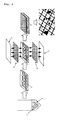
- the reference numeral 1 represents a pancreatic islet cells
- 2 represents a PVA+EC solution
- 3 represents PVA+EC gel
- 4 and 6 represent mesh sheets
- 5 and 7 represent glass plates
- 8 represents a sheet-like pancreatic islet cell-containing cellular preparation.
- the present invention is characterized in that a cellular preparation contains PVA mixed with a cell preservative and contains cells producing/secreting biologically active factors such as hormones and proteins useful for patients in the PVA.
- the PVA to be used in the present invention is not particularly limited without departing from the purpose of the invention and is preferable to have purity enough to be used, for example, corneal protectors/moisturizers in eye drops, medical materials such as intestinal adhesion preventing membranes, and food contact materials such as food wrapping films. It may also include commercialized products, and freeze-dry products obtained by producing polyvinyl acetate from vinyl acetate followed by hydrolysis. In the case of producing the PVA, the production method and the freeze-drying method may be carried out according to the known conventional methods.
- the PVA has preferably an average molecular weight of about 5,000 to 16,600 and a saponification degree of about 85% or higher.
- the PVA is preferably a fine granular freeze-dried product which is easily suspended in a solution such as physiological saline and phosphate buffers
- the cell preservative to be used in the present invention is not particularly limited and may include solutions to be employed generally for preservation of animal cells and commercialized preservation solutions, for example, Euro-Collins solution, Cell Banker (code 630-01601: Wako Pure Chemical Industries, Ltd. Osaka, Japan) and UW solution (ViaSpan: Bristol-Myers Squibb Company, USA). They may be commercialized ones or compositions prepared in laboratories. In the case of preparation in laboratories, it is preferable to carry out the preparation by adding and mixing each component in distilled water and disinfecting the obtained solutions by filtration with a filter.
- the composition of the Euro-Collins solution is shown in the following Table 1.
- Table 1 Component g/L D-glucose 35.0 K 2 HPO 4 7.4 KH 2 PO 4 2.05 KCl 1.12 NaHCO 3 0.84 Nicotinamide 1.22
- the PVA containing a cell preservative in the PVA can be produced, for example, by mixing PVA with a cell preservative.
- the resulting PVA containing the cell preservative is desirably aseptic.
- the method of mixing PVA and a cell preservative is not particularly limited, however, there can be, for example, exemplified a method of suspending a commercialized granular PVA in distilled water, etc. dissolving and sterilizing the resulting PVA suspension by heating and disinfecting (autoclaving or the like), and adding and mixing a cell preservative to and with the obtained aseptic aqueous PVA solution.
- the heating and disinfecting treatment and the aseptic treatment themselves may be carried out according to the known conventional methods.
- PVA containing the cell preservative produced by any means other than the above-mentioned mixing means may be called as PVA treated with the cell preservative and therefore, it should go without saying that PVA treated with the cell preservative is also included within the scope of the present invention.
- the content of PVA in the mixed solution (hereinafter, called as a PVA-cell preservative solution) obtained by mixing an aqueous suspended PVA solution and a cell preservative is generally about 2 to 5% by weight, more preferably about 2.5 to 3.5% by weight, and especially preferably about 2.5 to 3% by weight based on the PVA-cell preservative solution. It is because gelation of PVA is most easily caused in this concentration range and also PVA gel with preferable strength for encapsulating the cell in the cellular preparation of the present invention.
- a concentrated cell preservative solution with a concentration of about 2 to 10 times as high as that for normal cell preservative solution is prepared or purchased, and then properly mixed with the above-mentioned aqueous suspended PVA solution (e.g.
- the cellular preparation of the present invention may further contain dimethyl sulfoxide (DMSO) and serum albumin for protecting the cells, antibiotics, etc. for inhibiting contamination of germ, and also vitamins such as nicotinamide for cell activity retention.
- DMSO dimethyl sulfoxide
- serum albumin for protecting the cells, antibiotics, etc. for inhibiting contamination of germ
- vitamins such as nicotinamide for cell activity retention.
- other additives which are generally allowed to add under Pharmaceutical Affairs Law, e. g. an agent for proving sustained release property, an isotonic agent, a pH adjustment agent, and the like may be supplemented.
- the addition method of these other additives to the cellular preparation is not particularly limited, however, it is preferable to add them to the above-mentioned PVA-cell preservative solution in aseptic manner at the time of production of the solution.
- the contents thereof are preferably within a range of not inhibiting the growth/survival of the cells contained therein and/or the secretion
- the PVA treated with the cell preservative is produced in aseptic manner as described above and therefore, the cellular preparation of the present invention is seldom to be contaminated with germ and possible to be preserved at room temperature (about 15 to 35°C) for a long period of time.
- the cells to be used for the present invention may include pancreatic islet cells, pancreatic endocrine cells, hepatocyte, anterior pituitary cells, growth hormone-secreting cells, osteocytes, thyroid hormone-secreting cells, and parathyroid-hormone-secreting cells. These cells are preferably derived from a mammal such as human being, pig, rat, and mouse, and those which produce/secret biologically active factors such as hormones and proteins effective for patients. Selection of cell types is preferably determined depending on the type of diseases of patients receiving the cellular preparation.
- pancreatic islet cells or pancreatic endocrine cells, etc., producing insulin are preferable for diabetic patients, etc.; hepatocytes, etc., producing blood coagulation factors for hemophiliac patients, etc.; anterior pituitary cells and growth hormone-secreting cells, etc., producing growth hormones for pituitarydwarfismpatients, etc . ; and osteocytes producing bone morphogenetic proteins (BMP) for patients suffering from bone diseases such as bone fracture.
- BMP bone morphogenetic proteins
- cells having functions of selectively metabolizing and detoxifying harmful metabolites may be used for patients suffering from diseases which accumulates harmful metabolites due to hepatic insufficiency, renal insufficiency, or inborn errors of metabolism.
- a patient needs a plurality of biologically active factors since he suffers from a plurality of diseases
- two or more kinds of the above-mentioned cells may be combined.
- Biologically active factors efficacious to the above-mentioned patients can be provided since the present invention contains these cells. Accordingly, use of the cellular preparation of the present invention makes it possible to prevent or treat various diseases whose remediation is possible, through supply of biologically active factors or detoxification of harmful metabolites.
- the above-mentioned cells to be employed in the present invention may be an established cell line for laboratories or cells isolated from tissues of a living body, however, they are preferably differentiated, non-dividing cells since differentiated, non-dividing cells are more capable of producing /secreting target hormones and proteins than non-differentiated, dividing cells.
- the isolation method of the cells from living tissues is not particularly limited, and known conventional techniques may be employed.
- the known methods include a method of extracting tissues by proper means, treating the tissues with Dispase, EDTA, or the like and then with trypsin to finally isolate single cells.
- isolation of the cells from living tissues may be carried out by a separation method through the known collagenase treatment, e.g. J. Adam Van Der Vliet et al., Transplantation, 45(2), p493-495 (1988).
- the isolation may be carried out, for example, according to the method described in Japanese Patent Application Laid-Open No. 2001-231548.
- the isolated cells from the living tissues in the above-mentioned manner are preferably free from pathogens such as pathogenic virus.
- the cells already established for laboratories and single cells obtained from living tissues are cultured in a suitable media until they become confluent and to be employed as cells for the present invention after passage culture is repeated 2 or 3 times.
- the cells to be employed in the present invention are pancreatic islet cells or pancreatic endocrine cells (hereinafter, referred to as pancreatic endocrine cells or the like)
- these cells are preferably subjected to passage culture in a CMRL-1066 culture medium supplemented with 10% fetal bovine serum (hereinafter abbreviated as FBS) and nicotinamide (Sigma, St. Louis, MO, USA).
- FBS fetal bovine serum
- nicotinamide Sigma, St. Louis, MO, USA
- the cells to be employed in the present invention may be transformed cells into which gene coding peptides of biologically active factors such as hormones or proteins necessary for patients to be administered are introduced. Use of the transformed cells makes it possible to produce the target biologically active factors efficiently in a large amount.
- the base sequence of genes encoding peptides of such biologically active factors has already been opened, and such genes include, for example, genes available from American Type Culture Collection (ATCC) or from commercially easily available sources , and synthetic genes obtained by producing oligonucleotide probes from the known sequences and carrying out the synthesis using said probes by known conventional methods, for example, PCR amplification method and DNA synthesis method.
- ATCC American Type Culture Collection
- synthetic genes obtained by producing oligonucleotide probes from the known sequences and carrying out the synthesis using said probes by known conventional methods, for example, PCR amplification method and DNA synthesis method.
- use of transformed cells containing a gene encoding tumor growth suppression proteins makes it possible to use the
- pancreatic islet cells pancreatic endocrine cells
- hepatocytes pancreatic endocrine cells
- anterior pituitary cells growth hormone-secreting cells
- osteocytes there can be exemplified monkey COS-7 cells, Vero cells, Chinese hamster CHO cells (hereinafter abbreviated as CHO cell), dhfr gene-defective Chinese hamster CHO cells (hereinafter abbreviated as CHO(dhfr - )cell), mouse BALB/3T3 cells, mouse L cells, mouse AtT-20 cells, mouse C127 cells, mouse myeloma cells, rat GH3 cells, human HeLa cells, human FL cells, and the like.
- CHO cell Chinese hamster CHO cells
- CHO(dhfr - )cell dhfr gene-defective Chinese hamster CHO cells
- mouse BALB/3T3 cells mouse L cells
- mouse AtT-20 cells mouse C127 cells
- the method for introducing the gene into the cells to be used in the present invention is not particularly limited and may be carried out by known conventional technique.
- conventionally known methods of introducing the gene into recombinant expression vectors such as plasmids or viruses, and artificial vectors such as liposomes and microcapsules can be exemplified.
- examples of applicable methods are competent cell method (J. Mol. Biol. , 53, p154 (1970)), DEAE dextran method (Science, 215, p166 (1982)), In-vitro packaging method (Proc. Natl. Acad. Sci.
- any expression vectors so long as it can express desired gene in the cells into which they are introduced and can produce efficiently the peptides of biologically active factors may be used.
- animal viruses such as bacteriophage, e.g.
- the cells to be employed in the present invention are preferably contained in the PVA by the following techniques.
- Cells are added and mixed in the above produced PVA-cell preservative solution.
- the cells to be added and mixed are preferably treated previously with a cell preservative. Practically, the cells are preferably suspended previously in the cell preservative and recovered by centrifugation, and then mixed with the PVA-cell preservative solution.
- the number of the cells to be contained in the cellular preparation of the present invention cannot be generalized since it depends on the types and the extent of the diseases of patients to whom the cellular preparation is administered, and it is therefore preferable to determine the number of cells by a doctor.
- the number of cells is preferably about 1 ⁇ 10 7 to 5 ⁇ 10 7 cells/ml in the PVA-cell preservative solution. Adjustment of the number of the cells within the range makes the cells dispersed evenly in the PVA gel and the cells can stably survive for a long duration without inhibition of supply of oxygen and nutrients to the cells due to cell coagulation.
- the cellular preparation of the present invention may further contain mucopolysaccharides such as hyaluronic acid, chondroithin sulfuric acid, and dermatanic acid; extracellular matrix containing one or more substances such as erastin, collagen, and fibrin; and/or growth factors such as hepatocyte growth factor (HGF), vascular endothelial cell growth factor (VEGF), human basic fibroblast growth factor (bFGF), fibroblast growth factor (FGF), platelet-derived growth factor (PDGF), insulin-like growth factor (IGF), and growth hormone (GH).
- HGF hepatocyte growth factor
- VEGF vascular endothelial cell growth factor
- bFGF human basic fibroblast growth factor
- FGF fibroblast growth factor
- PDGF platelet-derived growth factor
- IGF insulin-like growth factor
- GH growth hormone
- the method for adding the extracellular matrix and/or growth factors in the present invention may be a method involving, for example, immersing the cells in a cell culture medium containing the extracellular matrix and the growth factor for forming the extracellular matrix on the surfaces of the cells before mixing the above PVA-cell preservative solution with the cells, or a method of previously adding the extracellular matrix and the growth factor in the PVA-cell preservative solution, or the like.
- the content of the above-mentioned extracellular matrix and/growth factor in the cellular preparation of the present invention is not particularly limited, however, it is within the range without inhibition of the cell retention and the secretion function of the biologically active factors and preferably in the range for causing effects on prolongation of the cell survival period and extension of the secretion period of the biologically active factors.
- the cellular preparation of the present invention can be produced by mixing the PVA-cell preservative solution with the cells, then cooling the cell mixed solution (hereinafter, referred to as cell-containing PVA-cell preservative solution), and gelling the PVA . Cooling is preferably carried out by keeping the solution in a housing at a temperature as ultra low as about -20 to -80°C for about a half of a day to 3 days or in a housing at a temperature as ultra low as about -80°C for about 18 to 30 hours.
- the cell preservative is previously mixed with the PVA, even if the quenching is carried out at the above-mentioned temperature, necrosis or damage of the cells can be suppressed. Moreover, the cells can be contained in the PVA gel in dispersed manner and coagulation of the cells can be prevented.
- the cellular preparation of the present invention may be formed in various shapes such as sheet, plate, board, rod, tube, or beads.
- the above-mentioned cell-containing PVA-cell preservative solution is applied to a glass plate and cooled together with the glass plate to produce a cellular preparation in the form of a thin sheet.
- the cellular preparation with a desired thickness can be produced by properly adjusting the application dose of the cell-containing PVA-cell preservative solution per unit surface area of the glass plate and it is generally about 100 to 300 ⁇ l/mm 2 .
- the cellular preparation of the present invention may be used in combination with a reinforcing material for reinforcement and/or simplicity in the handling.
- a reinforcing material for reinforcement and/or simplicity in the handling.
- gelation is preferably carried out by fixing the cell-containing PVA-cell preservative solution in a mesh sheet made of a resin or the like.
- a mesh sheet (4) made of PET polyethylene terephthalate
- the above-mentioned cell-containing PVA-cell preservative solution is applied to the mesh sheet (4).
- Another mesh sheet (6) is put thereon and further another glass plate (7) is put thereon to sandwich the cell-containing PVA-cell preservative solution and the mesh sheets (4) and (6) with glass plates (5) and (7). While being kept as it is, the assembled body is cooled to about -20 to -80°C to form it into a sheet-like gel.
- the above-mentioned mesh sheet is preferably immersed previously in the PVA-cell preservative solution.
- the above-mentioned reinforcing material e.g. the mesh sheets
- the cellular preparation in the frozen state is preferably administered to a living body after the cells in the frozen state are activated.
- the activation of the cells in the frozen cellular preparation is preferably carried out by thawing the cells and culturing them again in a proper culture medium. To be more specific, such activation is carried out by quickly immersing the frozen state cellular preparation in a cell culture medium of about 37°C, such as CMRL - 1966, and culturing the thawed cells again for about 24 hours in a new culture medium.
- a new cell preservation solution e.g. an UW solution, etc.
- the administration manner of the cellular preparation obtained by the above-mentioned method to a living body differs depending on the type, the affected part, and severity of diseases of patients to be administered, and it is therefore preferably determined by a doctor.
- a preferable administration manner will be described.
- the subjects to which the cellular preparation of the present invention are administered include human being and also a mammal other than human being, e.g. dog, cat, monkey, rabbit, mouse, and the like.
- the cellular preparation of the present invention can be administered in the form suitable to the application site of a living body.
- the cellular preparation of the present invention may be transplanted to the application site in a direct contact manner to the affected tissues, and also it can be transplanted in the subcutaneous tissue or the muscle with relatively slight invasion.
- the cellular preparation in tubular shape is cut into small fragments and transplanted in the subcutaneous tissue by using a surgical suture needle, or the cellular preparation can be injected in the muscle and subcutaneous tissue by filling an injector with the cellular preparation.
- the cellular preparation is easy to be recovered from the administration site.
- transplantation may be carried out together with known proper angiogenesis inducers.
- the angiogenesis inducers may be added previously in the cellular preparation or may be administered separately.
- the dose of the biologically active factors such as hormones and proteins to be supplied to a patient from the cellular preparation of the present invention may properly be set by determination by a doctor in consideration of the secretion of the biologically active factors of the cells to be employed and the change of the cell survival ratio during the transplantation.
- the insulin secretion of the cells to be used e. g. pancreatic islet secretion cells
- the insulin supply amount of the cellular preparation of the present invention can be determined.
- the present invention Since the cellular preparation of the present invention can stably preserve the cells contained therein for a long duration, deterioration ratio of the secretion of the biologically active factors with the lapse of time is low. Accordingly, the present invention is suitable to supply the biologically active factors needed for patients stably for a long duration, resulting in decreased frequency of the transplantation of the cellular preparation.
- the secretion of the biologically active factors of the cells to be used in the cellular preparation of the present invention may quantitatively be determined according to the known quantitative determination methods suitable for measurement of such biologically active factors.
- a quantitative determination method of insulin in the case where the cells used are pancreatic islet secretion cells will be described in detail in the following Examples.
- Pancreatic islet cells were isolated from Wister rats with 8 to 10 week-old and 300 to 350 g weight (Shimadzu animal Co. Ltd. Kyoto, Japan) by the following method.
- the centrifugation was carried out at 1,000 rpm for 1 minute.
- the tissue suspension was filtered through an 800 ⁇ m size mesh sheet.
- the filtrate was recovered in a 50 ml conical tube and centrifuged (1,500 rpm for 3 minutes) to obtain tissue pellets.
- the pellets were suspended in Hank's solution containing 27% dextran (Sigma, St. Louis, MO, USA, MW 70.000) and centrifuged by discontinuous density-gradient centrifugation.
- the lowermost layer was controlled to be 27% dextran (density 1.094 g/ml), and the upper layer was made into two layers of 23% dextran (density 1.081 g/ml) and 11% dextran (density 1.041 g/ml).
- the discontinuous density-gradient centrifugation was carried out at 400 rpm for 4 minutes and further at 1,700 rpm for 10 minutes. After centrifugation, the pancreatic islet cells-containing fraction was sampled in the interface between two uppermost layers with a Pustule pipette. The obtained pancreatic islet cells were washed two times with CMRL-1066 culture medium (1,000 rpm for 3 minutes).
- PVA 3 g (manufactured by Gunse Co.: PVA-180H, Lot37435) was suspended in distilled water 75 ml and then subjected to heating dissolution and sterilization treatment through autoclaving (121°C, 15 minutes) two times.
- Ten times concentration of Euro-Collin solution (hereinafter abbreviated as EC solution) 10 ml, DMSO 5 ml, FBS 10 ml, and nicotinamide 0.122 g were aseptically added to and mixed with the obtained aseptic aqueous PVA solution, thereby to obtain an aseptic 3% PVA-EC solution 100 ml.
- the above-mentioned 10 times concentration of EC solution was prepared by admixing the components shown in Table 1 in distilled water 100 ml, and filtered and disinfected through 0.22 ⁇ m mesh. Thereafter, it was preserved at a room temperature.
- pancreatic islet cells obtained in (1) were cultured for 24 hours in a CMRL-1066 culture medium containing 10% FBS and nicotinamide 1.22 g/L in a carbon dioxide gas incubator (5% CO 2 , 37°C).
- the culture solution containing 1,000 pancreatic islet cells was placed in a 1.5 ml centrifugation tube and centrifuged at 800 rpm for 1 minute.
- Cell Banker (code 630-01601: Wako Pure Chemical Industries, Ltd. Osaka, Japan) 0.1 ml was added to the obtained cell pellets and the cells were suspended, and then kept still for 5 minutes. The cells were recovered by centrifugation again.
- the recovered cells were suspended in the aseptic 3% PVA-EC solution prepared in (2) 100 ⁇ l to obtain a pancreatic islet cells-containing PVA-EC solution.
- the frozen sheet-like pancreatic islet cellular preparation obtained in (4) was thawed quickly in CMRL-1066 culture medium at 37°C.
- the thawed sheet-like pancreatic islet cellular preparation was immersed in a UW solution 5 ml (ViaSpan: Bristol-Myers Squibb Company, USA) for 5 minutes. This procedure was repeated 3 times to remove DMSO contained in the sheet.
- the sheet was further immersed in a UW solution 5 ml at 4°C for 24 hours.
- pancreatic islet cellular preparation containing activated cells (Production Example 1) in a sheet from.
- Production Example 1 was employed for the following tests.
- An aseptic 3% aqueous PVA solution was prepared as described in (2) without using EC solution 10 ml, DMSO 5 ml, FBS 10 ml, and nicotinamide 0.122 g and a pancreatic islet cellular preparation produced from the solution in the form of sheet was employed as Comparative Example 1.
- CMRL-1066 culture medium for 24 hours (5% CO 2 , 37°C) .
- the number of the viable cells in Production Example 1 and Comparative Example 1, and the number of free pancreatic islet cells after 1-day culture were counted using a microscope to calculate the recovery ratio (%) of the viable cells.
- the recovery ratio was a relative value to 100 defined as the number of the cells before the culture.
- the results are shown in Fig. 2.
- the recovery ratio of the viable cells in Production Example 1 was 82%, which was extremely higher than that of Comparative Example 1 (about 35%), and the value was higher than that of the free cells.
- pancreatic isolated islet cells 200 cells were cultured respectively in CMRL-1066 culture medium for one day (5% CO 2 , 37°C). The respective insulin amounts after 1-day culture were investigated.
- the cells of Production Example 1 and Comparative Example 1, and the free pancreatic islet cells were recovered in hydrochloric acid-EtOH and mashed respectively with a spatula, and the gel and the cells in the preparation were stirred and suspended in hydrochloric acid-EtOH with a vortex mixer (-20°C, 24 hours).
- the insulin concentrations in the respective cell suspensions were measured. Insulin was measured by using a commercialized insulin measurement ELISA kit (Lbis Insulin Kit, Shibayagi).
- the results are shown in Fig. 3.
- the insulin content in Production Example 1 was about 205 ng/ml which was extremely higher than that of Comparative Example 1 (about 20 ng/ml), and the value was approximately the same as that of the free cells.
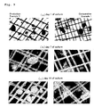
- the cells of Production Example 1 and Comparative Example 1, and isolated islet cells were cultured in CMRL-1066 culture medium (5% CO 2 , 37°C) and recovered after 1 day, 7 days, and 14 days. After the recovery, the recovered culture products were subjected to a glucose reaction test.
- the glucose reaction test was carried out as follows. At first the recovered cells of Production Example 1 and Comparative Example 1, and free pancreatic islet cells were cultured respectively in CMRL-1066 culture medium containing 3.3 mM glucose and 0.1% bovine serum albumin (BSA) for 1 hour (5% CO 2 , 37°C) and then washed with glucose-free CMRL-1066 culture medium.
- BSA bovine serum albumin
- Fig. 4 shows the amount (concentration) of insulin secretion after 1 day-culture; (b) shows the result after 7 days-culture; and (c) shows the result after 14 days-culture.
- Comparative Example 1 no increase in the insulin secretion depending on the glucose concentration was observed.
- Production Example 1 increase in the insulin secretion (concentration) depending on the glucose concentration was observed after 1 day-culture, 7 days-culture, and 14 days-culture.
- free cells no increase in the insulin secretion depending on the glucose concentration was observed after 7 days-culture, the increase was observed in the case of Production Example 1 even after 14 days-culture.
- pancreas was extracted, placed in a flask, immersed in a thermostatic bath of 37°C and digested in about 18 minutes. After digestion, the flask was shaken several times to destruct the pancreas and washed three times with Hank's solution containing 0.1% bovine serum albumin. After the cells were floated in the Hank's solution, the solution was filtered through an 850 ⁇ m-diameter mesh filter to recover the contents.
- the islet separation was carried out using specific gravity gradation of Dextran 70. That is, the pancreatic islet cell components from which the supernatant solution was removed were stirred uniformly in Dextran with 1.094 specific gravity + Hank' s solution, and then Dextran with 1.094 specific gravity was slowly poured, and Dextran with 1. 081 specific gravity and Dextran with 1.041 specific gravity were slowly poured successively on the cell layer to form 4 layers. The layered sample was centrifuged at 400 rpm for 4 minutes and further at 1700 rpm for 13 minutes. The islet separated between the first layer and the second layer was recovered with a hand pick, washed and purified with a hand pick.
- pancreatic islet cells were cultured overnight in an incubator under environments at 37°C, 5% CO 2 , 95% air.
- the culture medium was CMRL 1066 + 10% inactivated fetal bovine serum + antibiotic-antimycotic solution (Gibco, BRL) + nicotinamide.
- Fig. 6 The results are shown in Fig. 6. As can be seen from Fig. 6, the islet cellular preparation according to the present invention was found to secrete insulin corresponding to the glucose stimulation and was not inferior to the free islet cells at the same period.
- Streptozotocin 200 mg/Kg dissolved in a citric acid buffer (pH 4.5) was administered to C57BL/6 male mice of 8 to 10-week-old by intraabdominal administration.
- One week after the administration blood sugar measurement was carried out three times, and mice showing blood sugar 350 mg/dl or higher two or more times were determined to be diabetic and used for the experiment. Mice without streptozotocin were used as a normal group of mice.
- the transplantation of the islet cellular preparation was carried out as followings. That is, diabetic model mice (after 1 week or longer from diabetes development) were undergone abdominal section by median incision under ether anesthesia, and the islet cellular preparation (Production Example 2) obtained in Production Example 2 was kept in the abdomen and the abdominal wall was closed in two layers. These mice were defined as a transplanted mice group. Mice undergone transplantation of the preparation containing no pancreatic islet cells (produced in the same manner as in Comparative Example 1) were defined as a diabetic mice group.
- the survival period after the transplantation defined as 0 day was observed.
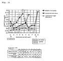
- Fig. 7 It is apparent from Fig. 7 that a half or more of the diabetic mice group died within 4 weeks after the transplantation and no mouse could survive up to 8 weeks during the passage observation period. On the other hand, except two mice which showed continuously high blood sugar state and finally died among the group of the mice with the islet cells, others survived 8 weeks or longer (survival ratio up to 8 weeks: 0% vs. 81.8%; p ⁇ 0.001).
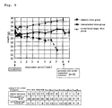
- the average weight fluctuation of each group is shown in Fig. 9.
- the weight was decreased after surgical transplantation, and it is supposed to be due to the poor dietary, and no significant difference from the diabetic mice group was observed in the Repeated-Measure Analysis of Variance carried out up to 3 weeks after the transplantation.
- the weight was turned to increase, and as compared with that in the first week when the average weight was most decreased during the passage, significant increase was observed after 3 weeks (p ⁇ 0.05) .
- the average weight significantly exceeded the average weight of the diabetic mice group in 3 weeks and 4 weeks from the transplantation.
- BUN blood urea nitrogen
- creatinine was measured once a week before the transplantation and once a week after the transplantation.
- the pancreatic islet cellular preparation was extracted 8 weeks after the transplantation and thereafter the measurement was carried out until 1 week after the extraction.
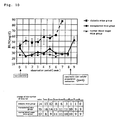
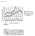
- the fluctuation of BUN is shown in Fig. 10.
- the average ⁇ SE of BUN of mice with normal blood sugar measured before the transplantation of the islet cellular preparation was 25.53 mg/dl ⁇ 1.804 (20 to 38.9).
- 41.3% showed BUN increase (24/58, diabetic mice group: 13/35 vs. transplanted mice group: 11/23, BUN > 35 mg/dl).
- 14 diabetic mice in the diabetic mice group 13 mice showed high BUN value in the passage.
- mice showing the BUN increase before the surgery was found normalized after 1 week from the transplantation.
- the BUN value in average decreased after 1 week from the transplantation were approximately normal value and found significantly different from that of the diabetic mice group (p 0.002).
- the BUN value measurement was carried out for 4 mice in one week after the extraction and all showed increase as compared with the value before extraction.
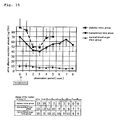
- the change in creatinine level is shown in Fig. 11.
- the normal value of creatinine was 0.351 mg/dl ⁇ 0.032 (0.16 to 0. 5).
- Almost a half of mice in the diabetic mice group showed as high as 0.5 mg/dl 1 week after the streptozotocin administration and all of the mice were found abnormal in the creatinine value 4 weeks after the administration.
- the creatinine increase all of the mice were dead in several weeks and no mouse survived up to 10 weeks from the streptozotocin administration.
- 6 mice in the transplanted mice group showed the increase after implantation of the creatinine, the increase was slight and mice could survive during the observation passage.
- Urine was collected for 24 hours and the urine amount and 24-hour urine sugar excretion amount, 24-hour urine albumin excretion amount, and urine ketone were measured. All of the group of transplanted mice, the group of diabetic mice and the group of normal blood sugar mice were fed in free drinking and eating dietary environments for 24 hours, and the 24-hour urine was collected. Urine sugar was measured by Fuji DRI-CHEM system and the value calculated by multiplying the urine amount was defined to be the urine sugar excretion amount. The 24-hour urine albumin excretion amount was calculated by measuring the urine albumin concentration by ELISA (Albuwell M (Exocill)) and multiplying the 24-hour urine amount. The urine ketone was evaluated by using Urotape (Eiken) and evaluated by numeration of the color tone.
- the change of 24-hour urine amount is shown in Fig. 12.
- the group of the transplanted mice did not show any significant difference, though slightly lower, from the group of diabetic mice.
- the change of urine ketone amount is shown in Fig. 14 .
- both of the group of diabetic mice and the group of the transplanted mice showed increase before transplantation.
- the change of urine albumin amount is shown in Fig. 15.
- the urine albumin amount was lower in the group of the transplanted mice than in the group of diabetic mice, however no significant difference was found.
- the 1, 5-AG is a polyol having extremely similar structure to that of glucose and mainly taken from food and absorbed and accumulated abundantly in a living body and reabsorbed in the kidney. It has competitive action against glucose, and in diabetes, the reabsorption of the urine sugar AG is inhibited to increase excretion of AG in urine and decrease AG in blood. Accordingly, 1,5-AG is very sensitively reacted to the dynamism of glucose and therefore, 1,5-AG can be a marker reflecting real time condition of diabetes.
- the pancreatic islet cellular preparation of the present invention is effective for improving the diabetic state and preventing the death from diabetes and diabetic renal insufficiency by transplantation of the preparation.
- the transplantation of the pancreatic islet cellular preparation of the present invention is not only effective in treatment for diabetes but also in prevention of diabetic complication.
- the present invention can provide a cellular preparation capable of stably and durably retaining cells having capabilities of producing/secreting biologically active factors such as hormones and proteins useful for patients in PVA, which is a proper material as a high molecular weight polymer to be used for bioartificial organs. Further, by administration of the cellular preparation to patients, endocrine/ metabolic disease, hemophilia, systemic bone disease or cancer may be prevented or treated. Since the pancreatic cellular preparation of the present invention can retain cells stably for a long duration, resulting in lessened frequency of transplantation and may be formed in various shapes, it is possible to transplant such cellular preparation by subcutaneous or intramuscular injection.
Landscapes
- Health & Medical Sciences (AREA)
- Life Sciences & Earth Sciences (AREA)
- Engineering & Computer Science (AREA)
- Biomedical Technology (AREA)
- Chemical & Material Sciences (AREA)
- Epidemiology (AREA)
- Veterinary Medicine (AREA)
- Public Health (AREA)
- General Health & Medical Sciences (AREA)
- Animal Behavior & Ethology (AREA)
- Zoology (AREA)
- Medicinal Chemistry (AREA)
- Immunology (AREA)
- Pharmacology & Pharmacy (AREA)
- Cell Biology (AREA)
- Virology (AREA)
- Developmental Biology & Embryology (AREA)
- Biotechnology (AREA)
- Gastroenterology & Hepatology (AREA)
- Bioinformatics & Cheminformatics (AREA)
- Proteomics, Peptides & Aminoacids (AREA)
- Neurology (AREA)
- Orthopedic Medicine & Surgery (AREA)
- Botany (AREA)
- Chemical Kinetics & Catalysis (AREA)
- Dermatology (AREA)
- Oral & Maxillofacial Surgery (AREA)
- Transplantation (AREA)
- Micro-Organisms Or Cultivation Processes Thereof (AREA)
- Medicines Containing Material From Animals Or Micro-Organisms (AREA)
- Medicinal Preparation (AREA)
Applications Claiming Priority (2)
| Application Number | Priority Date | Filing Date | Title |
|---|---|---|---|
| JP2003112103 | 2003-04-16 | ||
| PCT/JP2003/014271 WO2004091679A1 (fr) | 2003-04-16 | 2003-11-10 | Preparation cellulaire |
Publications (1)
| Publication Number | Publication Date |
|---|---|
| EP1614436A1 true EP1614436A1 (fr) | 2006-01-11 |
Family
ID=33296023
Family Applications (1)
| Application Number | Title | Priority Date | Filing Date |
|---|---|---|---|
| EP03816652A Withdrawn EP1614436A1 (fr) | 2003-04-16 | 2003-11-10 | Preparation cellulaire |
Country Status (7)
| Country | Link |
|---|---|
| US (1) | US20060246043A1 (fr) |
| EP (1) | EP1614436A1 (fr) |
| KR (1) | KR20050120712A (fr) |
| CN (1) | CN1764481A (fr) |
| AU (1) | AU2003277650A1 (fr) |
| CA (1) | CA2522106A1 (fr) |
| WO (1) | WO2004091679A1 (fr) |
Cited By (2)
| Publication number | Priority date | Publication date | Assignee | Title |
|---|---|---|---|---|
| WO2013117925A1 (fr) * | 2012-02-08 | 2013-08-15 | University Of Warwick | Cryoconservation de cellules en l'absence d'agents induisant la vitrification |
| EP2491957A4 (fr) * | 2009-08-02 | 2013-10-02 | Univ Tokyo Womens Medical | Feuillet de cellules de langerhans, procédé de production associé, et application associée |
Families Citing this family (3)
| Publication number | Priority date | Publication date | Assignee | Title |
|---|---|---|---|---|
| US8703428B2 (en) * | 2007-10-06 | 2014-04-22 | Corning Incorporated | Single-cell label-free assay |
| AR111216A1 (es) * | 2017-02-23 | 2019-06-19 | Japan Vam & Poval Co Ltd | Dispositivo de integración celular o tisular |
| US20210283308A1 (en) * | 2020-03-13 | 2021-09-16 | City Of Hope | Implantable device for retaining live cells and providing nutrients thereto |
Family Cites Families (4)
| Publication number | Priority date | Publication date | Assignee | Title |
|---|---|---|---|---|
| US4879283A (en) * | 1985-10-03 | 1989-11-07 | Wisconsin Alumni Research Foundation | Solution for the preservation of organs |
| US5804178A (en) * | 1986-11-20 | 1998-09-08 | Massachusetts Institute Of Technology | Implantation of cell-matrix structure adjacent mesentery, omentum or peritoneum tissue |
| CA2214088A1 (fr) * | 1995-03-03 | 1996-09-12 | Metabolex Inc. | Nouveau processus d'encapsulation par un polymere gelifiant |
| JP2001270831A (ja) * | 2000-03-24 | 2001-10-02 | Lion Corp | 健胃消臭剤 |
-
2003
- 2003-11-10 CA CA002522106A patent/CA2522106A1/fr not_active Abandoned
- 2003-11-10 WO PCT/JP2003/014271 patent/WO2004091679A1/fr not_active Ceased
- 2003-11-10 CN CNA2003801102619A patent/CN1764481A/zh active Pending
- 2003-11-10 AU AU2003277650A patent/AU2003277650A1/en not_active Abandoned
- 2003-11-10 EP EP03816652A patent/EP1614436A1/fr not_active Withdrawn
- 2003-11-10 KR KR1020057019282A patent/KR20050120712A/ko not_active Withdrawn
- 2003-11-10 US US10/551,990 patent/US20060246043A1/en not_active Abandoned
Non-Patent Citations (1)
| Title |
|---|
| See references of WO2004091679A1 * |
Cited By (2)
| Publication number | Priority date | Publication date | Assignee | Title |
|---|---|---|---|---|
| EP2491957A4 (fr) * | 2009-08-02 | 2013-10-02 | Univ Tokyo Womens Medical | Feuillet de cellules de langerhans, procédé de production associé, et application associée |
| WO2013117925A1 (fr) * | 2012-02-08 | 2013-08-15 | University Of Warwick | Cryoconservation de cellules en l'absence d'agents induisant la vitrification |
Also Published As
| Publication number | Publication date |
|---|---|
| WO2004091679A1 (fr) | 2004-10-28 |
| US20060246043A1 (en) | 2006-11-02 |
| CA2522106A1 (fr) | 2004-10-28 |
| KR20050120712A (ko) | 2005-12-22 |
| CN1764481A (zh) | 2006-04-26 |
| AU2003277650A1 (en) | 2004-11-04 |
Similar Documents
| Publication | Publication Date | Title |
|---|---|---|
| DE69635940T2 (de) | Hydrogelmatrix für Zellgewebelagerung | |
| Lanza et al. | Treatment of severely diabetic pancreatectomized dogs using a diffusion-based hybrid pancreas | |
| Tobias et al. | Grafting of encapsulated BDNF-producing fibroblasts into the injured spinal cord without immune suppression in adult rats | |
| JP5538217B2 (ja) | 組織修復用無細胞組織基質構成体 | |
| US10251915B2 (en) | Co-encapsulation of live cells with oxygen-generating particles | |
| US20050180957A1 (en) | Method of using fibrin-bound angiogenic factors to stimulate vascularization of transplant site of encapsulated cells | |
| US9012404B2 (en) | Compositions and methods for cardiac tissue protection and regeneration | |
| US20060127873A1 (en) | Composition for cytocompatible, injectable, self-gelling chitosan solutions for encapsulating and delivering live cells or biologically active factors | |
| US20020187182A1 (en) | Biocompatible fleece for hemostasis and tissue engineering | |
| JP5456692B2 (ja) | カプセル化された腎臓組織 | |
| EP3848058A1 (fr) | Mélange biologique inactivé viral | |
| EP1614436A1 (fr) | Preparation cellulaire | |
| RU2425645C1 (ru) | Способ и трансплантат для лечения печеночной недостаточности | |
| JP2025513507A (ja) | 組織伝導性足場材料 | |
| Lahooti et al. | Agarose enhances the viability of intraperitoneally implanted microencapsulated L929 fibroblasts | |
| JP2004331643A (ja) | 細胞製剤 | |
| Burke et al. | Phase-changing citrate macromolecule combats oxidative pancreatic islet damage, enables islet engraftment and function in the omentum | |
| Aoki et al. | Intrasplenic transplantation of encapsulated cells: a novel approach to cell therapy | |
| CN102657874B (zh) | 用于修复神经损伤的水凝胶包裹慢病毒Lingo-1 RNAi复合物及其制备方法 | |
| CN112203667A (zh) | 大包封的治疗性细胞、装置及其使用方法 | |
| KR102572188B1 (ko) | 매크로-캡슐화 치료 세포 및 이의 사용 방법 | |
| AU2003238760B2 (en) | Porcine islets cultured with porcine sertoli cells for xenotransplantation | |
| RU2425646C1 (ru) | Способ и трансплантат для лечения печеночной недостаточности | |
| US20250361482A1 (en) | Apoptosis-induced stem cells, production method therefor, and composition containing same for preventing or treating inflammatory or renal diseases | |
| de Vries et al. | Selecting biocompatible biomaterials for stem cell-derived β-cell transplantation |
Legal Events
| Date | Code | Title | Description |
|---|---|---|---|
| PUAI | Public reference made under article 153(3) epc to a published international application that has entered the european phase |
Free format text: ORIGINAL CODE: 0009012 |
|
| 17P | Request for examination filed |
Effective date: 20050922 |
|
| AK | Designated contracting states |
Kind code of ref document: A1 Designated state(s): AT BE BG CH CY CZ DE DK EE ES FI FR GB GR HU IE IT LI LU MC NL PT RO SE SI SK TR |
|
| AX | Request for extension of the european patent |
Extension state: AL LT LV MK |
|
| DAX | Request for extension of the european patent (deleted) | ||
| STAA | Information on the status of an ep patent application or granted ep patent |
Free format text: STATUS: THE APPLICATION HAS BEEN WITHDRAWN |
|
| 18W | Application withdrawn |
Effective date: 20070518 |