EP1622085A1 - Procédé pour la reconstruction d'images CT à partir de données incomplètes d'un objet - Google Patents
Procédé pour la reconstruction d'images CT à partir de données incomplètes d'un objet Download PDFInfo
- Publication number
- EP1622085A1 EP1622085A1 EP04016967A EP04016967A EP1622085A1 EP 1622085 A1 EP1622085 A1 EP 1622085A1 EP 04016967 A EP04016967 A EP 04016967A EP 04016967 A EP04016967 A EP 04016967A EP 1622085 A1 EP1622085 A1 EP 1622085A1
- Authority
- EP
- European Patent Office
- Prior art keywords
- image
- interest
- region
- data
- image object
- Prior art date
- Legal status (The legal status is an assumption and is not a legal conclusion. Google has not performed a legal analysis and makes no representation as to the accuracy of the status listed.)
- Withdrawn
Links
Images


Classifications
-
- G—PHYSICS
- G06—COMPUTING OR CALCULATING; COUNTING
- G06T—IMAGE DATA PROCESSING OR GENERATION, IN GENERAL
- G06T12/00—Tomographic reconstruction from projections
- G06T12/20—Inverse problem, i.e. transformations from projection space into object space
-
- G—PHYSICS
- G06—COMPUTING OR CALCULATING; COUNTING
- G06T—IMAGE DATA PROCESSING OR GENERATION, IN GENERAL
- G06T2211/00—Image generation
- G06T2211/40—Computed tomography
- G06T2211/412—Dynamic
-
- Y—GENERAL TAGGING OF NEW TECHNOLOGICAL DEVELOPMENTS; GENERAL TAGGING OF CROSS-SECTIONAL TECHNOLOGIES SPANNING OVER SEVERAL SECTIONS OF THE IPC; TECHNICAL SUBJECTS COVERED BY FORMER USPC CROSS-REFERENCE ART COLLECTIONS [XRACs] AND DIGESTS
- Y10—TECHNICAL SUBJECTS COVERED BY FORMER USPC
- Y10S—TECHNICAL SUBJECTS COVERED BY FORMER USPC CROSS-REFERENCE ART COLLECTIONS [XRACs] AND DIGESTS
- Y10S378/00—X-ray or gamma ray systems or devices
- Y10S378/901—Computer tomography program or processor
Definitions
- the present invention is directed to a method for producing x-ray computer tomography (CT) images from a limited set of computer tomography data.
- CT computer tomography
- an X-ray source is collimated to form a fan beam with a defined fan beam angle and fan beam width.
- the fan beam is oriented to lie within the X-Y plane of a Cartesian coordinate system, termed the "imaging plane", and to be transmitted through an imaged object to an X-ray detector array oriented within the imaging plane.
- the detector array is comprised of detector elements, which each measure the intensity of transmitted radiation along a ray projected from the X-ray source to that particular detector element.
- the intensity of transmitted radiation received by each detector element in the detector array is dependent on the attenuation of the X-ray beam along a ray by the imaged object.
- Each detector element produces an intensity signal dependent on the intensity of transmitted radiation striking the detector element.
- the X-ray source and detector array may be rotated on a gantry within the imaging plane so that the fan beam intercepts the imaged object at different angles.
- a projection is acquired, comprised of the intensity signals of each of the detector elements.
- the projection at each of these different angles together form a tomographic projection set. Normally a projection set will be taken over 360° of gantry rotation. An attempt to reconstruct an image with less than a complete projection set will normally lead to image artifacts caused by the missing data and image blurring.
- the main object of radiotherapy is to deliver the prescribed dosage of radiation to a tumor in a patient while minimizing the damage to surrounding, healthy tissue. Since very high energy radiation is normally used to destroy tumors in radiotherapy, the high energy is also destructive to the normal tissue surrounding the tumor. Therefore, it is essential that the delivery of radiation be limited precisely to the prescribed target volume (i.e. the tumor plus adequate margins).
- the main sources of the problem result from the fact that there is a natural motion of organs inside the body, which can range from approximately a millimetre in the case of the brain inside the skull to several centimetres for the organs in the trunk. Another factor relates to changes which occur in the tumor over time as a result of successful treatment.
- one method taught in the prior art is to utilize a low dose, low energy X-ray source in conjunction with the therapy beam source.
- US 5,233,990 A relates to a method for producing corrected verification images of anatomical portions of a patient being treated with conventional radiation therapy equipment consisting of a gantry having a high-energy radiation source capable of emitting a high-energy radiation beam along a high energy beam axis, said gantry being disposed for irradiation of a stationary patient positioned on a gurney at a fixed distance from the high-energy radiation source.
- the method comprises the steps of providing a low-energy radiation source mounted on a side of the gantry opposite to the high-energy radiation source, said low-energy radiation source being capable of emitting a low-energy radiation beam along a low-energy beam axis coaxial with the axis of the beam from the high-energy radiation source; providing a radiation detector mounted between the high-energy radiation source and the patient's gurney, said detector being positioned so that it can be exposed to both the high-energy beam and the low-energy beam; irradiating the patient with said coaxial low-energy radiation source during treatment to form verification images on said radiation detector and comparing the verification images with the diagnostic images during treatment to ensure that the anatomical portions of the patient being irradiated by high-energy radiation treatment correspond to the anatomical portions delineated on the diagnostic images.
- WO 03/076016 A1 is directed to a device for performing and verifying therapeutic radiation.
- An X-ray is arranged across from a target volume of the beam source for a high-energy beam in such a way that the beams run in essentially opposite directions.
- the invention also relates to a computer program and a control method for operating said device.
- the inventive device makes it possible to exactly verify areas that are subjected to different levels of radiation, the entire anatomy of the target volume and the surroundings thereof in addition to the contour of the therapy beam.
- the X-ray detects the anatomy and position of the patient within the range of the target volume before the high-energy beam is applied and then detects the shape of the applied high-energy beam and areas that are subjected to different levels of radiation as well as at least one partial segment of the target volume during the emission breaks of the high-energy beam.
- the detected data is used for correcting the treatment plan.
- the prior art systems for radiation therapy contain a limited accessible space for the placement of the verification imaging equipment. Furthermore, there is little time during the emission breaks of the therapy beam to collect data for the verification imaging. Therefore no complete data sets can be acquired. With an incomplete data set, an image reconstruction with reasonable quality is not possible using conventional image reconstruction techniques.
- CT-images are either reconstructed analytically or iteratively.
- filtered back projection is accomplished by a projection reconstruction algorithm, where the projection measurements are filtered by an appropriate convolution kernel and then back projected. For this method the complete data set is required. 3D-medical imaging using the method of filtered back projection of multi-phase objects with an incomplete data set yields artifacts and image blurring.
- Bayesian image reconstruction i.e. Maximum Likelihood methods
- Bayesian approach provides the means to incorporate prior knowledge in data analysis.
- Bayesian analysis revolves around the posterior probability, which summarizes the degree of one's certainty concerning a given situation.
- Bayes's law states that the posterior probability is proportional to the product of the likelihood and the prior probability.
- the likelihood encompasses the information contained in the new data.
- the prior expresses the degree of certainty concerning the situation before the data is taken.
- This prior art reconstruction technique is also not suitable for a satisfactory image reconstruction from the limited data, particularly a limited observation angle.
- An object of the present invention is to provide a method for 3D-image reconstruction from limited CT-data.
- Another object of the present invention is to provide a method for on-line 3D-image reconstruction during a radiation therapy session from a limited set of X-ray projections, made within a limited observation angle, allowing the recognition of changes of the image object during the session.
- This method is using the preliminary first CT-image (source image) as a reference and algorithms for image deformation in compliance with the limited data for the reconstruction of the new image (target image), the algorithms being selected according to expected image changes.
- a source image of the region of interest is reconstructed from CT-data using a standard CT image reconstruction technique, like filtered back projection or the method of Bayesian image reconstruction.
- the region of interest can be a 2D-region or a 3D-volume of interest.
- the source image is reconstructed from a sufficient set of projection data, which allows reconstructing the source image substantially without artifacts.
- step b) of the method according to the present invention a transformation function or a combination of transformation functions is selected, which is able to transform the source image into the target image.
- the transformation function is chosen depending on expected changes of the image object.
- the matching transformation function for each different image object with different possible changes has to be investigated once and can be selected for similar image objects every time the method according to the invention is used.
- step c) an incomplete CT-data set of the image object is acquired, which would not be sufficient for image reconstruction with reasonable quality using for example conventional back projection techniques.
- the observation angle is the angle of the X-ray source, rotating around a rotation axis which crosses the image object, under which the data from a CT-projection is acquired.
- step d) of the method according to the invention the target image of the image object, which has possibly undergone a change since the source image has been produced, is reconstructed in an iterative optimization procedure under the constraint of preserving the characteristic morphology of the source image to a large extent.
- the number of iterations depends on the robustness to noise of the selected algorithm.
- the transformation function is used, deforming the source image.
- the target image is optimized in view of the limited CT-data acquired in step c).
- the selected transformation function is based upon a basis function.
- the voxel representation of an unknown map of attenuation coefficients (target image) is used.
- a voxel is a 3-dimensional pixel (a volume pixel), a small box-shaped part of a 3-dimensional space.
- the discrete values (target image) can be calculated with the help of eq. (I) as the values of function ⁇ ( r ⁇ ) in the points, which correspond to the pixel centers.
- the basis functions can be selected from the group of common polynomial, Bernstein polynomial, tri-linear interpolator, 3D cubic spline, 3D thin-plate and others. These basis functions are:
- the observation angles in step c) are chosen from a range of 0° to 90° about a rotation axis which crosses the region of interest of the image object.
- the necessary range of observation angles depends on the complexity of the image to be reconstructed. It can be smaller than 90°, in some applications even smaller than 30°.
- the possible changes, on which the selection of the transformation function in step b) of the method according to the present invention depends, may be at least one of the group of changes in position, shape, size and orientation.
- the target image is reconstructed for an on-line verification during a radiation therapy of a patient with a high energy therapy beam.
- steps c) and d) are repeated during the radiation therapy, acquiring the CT-data from the limited number of projections during emission breaks of the high-energy therapy beam. If steps c) and d) are repeated, the last target image can be used instead of the source image for the reconstruction of the new target image.
- the image object can be an organ of the patient and the region of interest can be a tumor in the organ and the tumor's surroundings.
- the transformation function can be selected depending on the organ having a tumor and the possible changes of the tumor for example as a result of the organ moving within the body of the patient.
- All organs of the human body can be classified as to their expected changes and mobility during a radiation therapy session (local or global changes). In its turn this main property defines the respective image reconstruction algorithm.
- An example for a possible classification of organs is given in the following table: Organ Basis function Skull, lungs Common polynomial, Bernstein polynomial Prostate 3D thin-plate spline, cubic spline Thorax Bernstein polynomial Rectum Tri-linear interpolator
- the CT-data from a limited number of projections in step c) is acquired by moving a radiation source emitting X-rays on a circle or a straight line in a plane, which is situated perpendicularly to the high energy therapy beam on one side of the image object, and detecting a number of projections of the region of interest with the aid of a detector on the other side of the image object.
- a device which can be used for detecting the projections, can be constructed in the same way as the device for performing and verifying a therapeutic treatment, which is disclosed in WO 03/076016 A1.
- the reconstructed target image of the region of interest can be used during the radiation therapy for correcting the treatment plan for the radiation therapy of the patient or even for stopping the therapy, if the positioning of the therapy beam cannot be adapted to a detected change of the image object to be treated. For example if the patient moves, the target image will reveal a change in the area being radiated compared to the source image, so that immediate steps can be taken to minimize any localization error. Therefore, this invention is suitable for on-line, real-time application, which renders it particularly valuable for radiation therapy.
- the Shepp-Logan phantom is an analytic phantom used in tomographic application due to the simple expression of its Fourier transform.
- the phantom consists of ellipses resembling the human brain features and the cranial contour.
- This phantom head is well known because of its use in testing the accuracy of reconstruction algorithms with regard to their ability to reconstruct cross-sections of the human head with X-ray tomography.
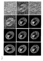
- Fig. 1 line 1, column 1, a 2D-image of the central cross-section of a Shepp-Logan phantom is presented. After rotation of this phantom as shown in Fig. 1, line 1, column 2, the changed phantom was "irradiated" with X-rays, taking 20 projections of it within an observation angle range of 0° to 90°. Using this limited data of the 20 projections, the rotated phantom was reconstructed using prior art reconstruction methods and the method according to the present invention with different transformation functions.
- Line 2 shows the result of filtered back projection.
- the image contains artifacts and is blurred. Accuracy of the construction can only be improved with more projections.
- This state of the art reconstruction method is thus not suitable for image reconstructions using limited projection data under a limited range of observation angles.
- the reconstruction result applying filtered back projections is even worse, when white Gausian noise with a mean value of 40 % of the maximum of the projected grey level is imposed on each projection before image reconstruction, as can be seen in line 3, column 4 of Fig. 1.
- the original Shepp-Logan phantom as shown in line 1, column 1, was used as the source image.
- the selected transformation functions were based on the basis functions Bernstein Polynomials, bi-linear interpolation and cubic spline.
- the image of the rotated phantom was reconstructed with or without imposed noise (Bernstein Polynomials - line 2, column 1 and line 3, column 1; bi-linear interpolation - line 2, column 2 and line 3, column 2; cubic spline - line 2, column 3 and line 3, column 3).
- the reconstructed images using the method according to the present invention are of a much higher quality than the reconstructed images using prior art methods, with small differences resulting from the different selected transformation functions.
- the method according to the present invention is also very suitable for the image reconstruction of the Shepp-Logan phantom after a local deformation as shown in Fig. 3, line 1, column 1.
- Fig. 3 line 1, column 1.
- the images were reconstructed starting with data of 20 projections applying the following algorithms with and without noise:
- the considered reconstruction results in all three cases of a changed image object illustrate substantial capability of the proposed fast image reconstruction techniques based on image deformation, if a source image, given by a preliminary CT-session, is available.
Landscapes
- Physics & Mathematics (AREA)
- General Physics & Mathematics (AREA)
- Engineering & Computer Science (AREA)
- Theoretical Computer Science (AREA)
- Apparatus For Radiation Diagnosis (AREA)
- Image Analysis (AREA)
Priority Applications (6)
| Application Number | Priority Date | Filing Date | Title |
|---|---|---|---|
| EP04016967A EP1622085A1 (fr) | 2004-07-19 | 2004-07-19 | Procédé pour la reconstruction d'images CT à partir de données incomplètes d'un objet |
| PCT/EP2005/007734 WO2006008089A1 (fr) | 2004-07-19 | 2005-07-15 | Procede pour produire des images de tomographie par ordinateur aux rayons x, a partir de donnees limitees d'un objet image |
| EP05764143A EP1769461B1 (fr) | 2004-07-19 | 2005-07-15 | Procédé pour la reconstruction d'images ct à partir de données incomplètes d'un objet |
| DE602005019740T DE602005019740D1 (de) | 2004-07-19 | 2005-07-15 | Verfahren zur erzeugung von ct bildern aus unvollständigen projektionsdaten eines untersuchungsobjektes |
| AT05764143T ATE459944T1 (de) | 2004-07-19 | 2005-07-15 | Verfahren zur erzeugung von ct bildern aus unvollständigen projektionsdaten eines untersuchungsobjektes |
| US11/632,724 US7616729B2 (en) | 2004-07-19 | 2005-07-15 | Method for producing X-ray computer tomography images from limited data of an image object |
Applications Claiming Priority (1)
| Application Number | Priority Date | Filing Date | Title |
|---|---|---|---|
| EP04016967A EP1622085A1 (fr) | 2004-07-19 | 2004-07-19 | Procédé pour la reconstruction d'images CT à partir de données incomplètes d'un objet |
Publications (1)
| Publication Number | Publication Date |
|---|---|
| EP1622085A1 true EP1622085A1 (fr) | 2006-02-01 |
Family
ID=34925807
Family Applications (2)
| Application Number | Title | Priority Date | Filing Date |
|---|---|---|---|
| EP04016967A Withdrawn EP1622085A1 (fr) | 2004-07-19 | 2004-07-19 | Procédé pour la reconstruction d'images CT à partir de données incomplètes d'un objet |
| EP05764143A Expired - Lifetime EP1769461B1 (fr) | 2004-07-19 | 2005-07-15 | Procédé pour la reconstruction d'images ct à partir de données incomplètes d'un objet |
Family Applications After (1)
| Application Number | Title | Priority Date | Filing Date |
|---|---|---|---|
| EP05764143A Expired - Lifetime EP1769461B1 (fr) | 2004-07-19 | 2005-07-15 | Procédé pour la reconstruction d'images ct à partir de données incomplètes d'un objet |
Country Status (5)
| Country | Link |
|---|---|
| US (1) | US7616729B2 (fr) |
| EP (2) | EP1622085A1 (fr) |
| AT (1) | ATE459944T1 (fr) |
| DE (1) | DE602005019740D1 (fr) |
| WO (1) | WO2006008089A1 (fr) |
Families Citing this family (14)
| Publication number | Priority date | Publication date | Assignee | Title |
|---|---|---|---|---|
| US7602359B2 (en) * | 2004-02-02 | 2009-10-13 | Seiko Epson Corporation | Image signal correcting method, correcting circuit, electro-optical device, and electronic apparatus |
| US8767917B2 (en) * | 2005-07-22 | 2014-07-01 | Tomotherapy Incorpoated | System and method of delivering radiation therapy to a moving region of interest |
| KR20100016039A (ko) * | 2007-03-30 | 2010-02-12 | 코닌클리케 필립스 일렉트로닉스 엔.브이. | 분계 불확실성의 확률론적 분석에 의한 방사선 요법시 개선된 치료 계획 평가 |
| US7978191B2 (en) | 2007-09-24 | 2011-07-12 | Dolphin Imaging Systems, Llc | System and method for locating anatomies of interest in a 3D volume |
| US8467497B2 (en) * | 2007-10-25 | 2013-06-18 | Tomotherapy Incorporated | System and method for motion adaptive optimization for radiation therapy delivery |
| JP2011502010A (ja) * | 2007-10-25 | 2011-01-20 | トモセラピー・インコーポレーテッド | 放射線療法送達の運動適応最適化のためのシステム及び方法 |
| ATE534189T1 (de) * | 2008-04-08 | 2011-12-15 | Austriamicrosystems Ag | Verstärkeranordnung und signalerzeugungsverfahren |
| WO2012106460A2 (fr) | 2011-02-01 | 2012-08-09 | L-3 Communications Security and Detection Systems Inc. | Procédé de reconstruction utilisant des techniques directes et itératives |
| EP2792303A4 (fr) * | 2011-12-18 | 2015-08-05 | Nat Univ Corp Kyoto Univ | Procédé de traitement d'image de tomodensitométrie à suivi de mouvement et dispositif de traitement d'image de tomodensitométrie à suivi de mouvement |
| WO2013116701A1 (fr) * | 2012-02-03 | 2013-08-08 | State University Of New York, The | Procédés et systèmes de reconstruction par problème inverse et application à une reconstruction ect |
| US9069092B2 (en) | 2012-02-22 | 2015-06-30 | L-3 Communication Security and Detection Systems Corp. | X-ray imager with sparse detector array |
| JP6412020B2 (ja) | 2013-02-26 | 2018-10-24 | アキュレイ インコーポレイテッド | 電磁作動式のマルチリーフコリメーター |
| CN106530366B (zh) * | 2015-09-09 | 2019-04-16 | 清华大学 | 能谱ct图像重建方法及能谱ct成像系统 |
| FI20155856A7 (fi) * | 2015-11-18 | 2017-05-19 | Teknologian Tutkimuskeskus Vtt Oy | Menetelmiä ja laitteita läpitunkevaa kuvantamista varten |
Family Cites Families (18)
| Publication number | Priority date | Publication date | Assignee | Title |
|---|---|---|---|---|
| US4861669A (en) * | 1987-03-26 | 1989-08-29 | Ppg Industries, Inc. | Sputtered titanium oxynitride films |
| US4952904A (en) * | 1988-12-23 | 1990-08-28 | Honeywell Inc. | Adhesion layer for platinum based sensors |
| US5070036A (en) * | 1989-01-04 | 1991-12-03 | Quality Microcircuits Corporation | Process for contacting and interconnecting semiconductor devices within an integrated circuit |
| US5227196A (en) * | 1989-02-16 | 1993-07-13 | Semiconductor Energy Laboratory Co., Ltd. | Method of forming a carbon film on a substrate made of an oxide material |
| US5233990A (en) | 1992-01-13 | 1993-08-10 | Gideon Barnea | Method and apparatus for diagnostic imaging in radiation therapy |
| US5609948A (en) * | 1992-08-21 | 1997-03-11 | Minnesota Mining And Manufacturing Company | Laminate containing diamond-like carbon and thin-film magnetic head assembly formed thereon |
| US5895266A (en) * | 1996-02-26 | 1999-04-20 | Applied Materials, Inc. | Titanium nitride barrier layers |
| SE9704607D0 (sv) * | 1997-12-09 | 1997-12-09 | Chemfilt R & D Ab | A method and apparatus for magnetically enhanced sputtering |
| US6517956B1 (en) * | 1999-05-03 | 2003-02-11 | Seagate Technology Llc | Magneto-resistance recording media comprising aluminum nitride corrosion barrier layer and a c-overcoat |
| US6569295B2 (en) * | 2001-03-20 | 2003-05-27 | International Business Machines Corporation | Method for grading surface topography for improved step coverage and planarization |
| SE521095C2 (sv) * | 2001-06-08 | 2003-09-30 | Cardinal Cg Co | Förfarande för reaktiv sputtring |
| EP1483022B1 (fr) | 2002-03-12 | 2008-02-13 | Deutsches Krebsforschungszentrum Stiftung des öffentlichen Rechts | Dispositif pour mettre en oeuvre et verifier un traitement therapeutique et programme informatique associes |
| US6904118B2 (en) * | 2002-07-23 | 2005-06-07 | General Electric Company | Method and apparatus for generating a density map using dual-energy CT |
| US6915796B2 (en) * | 2002-09-24 | 2005-07-12 | Chien-Min Sung | Superabrasive wire saw and associated methods of manufacture |
| US20040254448A1 (en) * | 2003-03-24 | 2004-12-16 | Amies Christopher Jude | Active therapy redefinition |
| KR100725690B1 (ko) * | 2003-07-08 | 2007-06-07 | 마츠시타 덴끼 산교 가부시키가이샤 | 반도체장치 및 그 제조방법 |
| US7300556B2 (en) * | 2003-08-29 | 2007-11-27 | Hitachi Global Storage Technologies Netherlands B.V. | Method for depositing a thin film adhesion layer |
| JP4407904B2 (ja) * | 2004-02-06 | 2010-02-03 | Hoya株式会社 | 磁気ディスクの製造方法 |
-
2004
- 2004-07-19 EP EP04016967A patent/EP1622085A1/fr not_active Withdrawn
-
2005
- 2005-07-15 DE DE602005019740T patent/DE602005019740D1/de not_active Expired - Fee Related
- 2005-07-15 WO PCT/EP2005/007734 patent/WO2006008089A1/fr not_active Ceased
- 2005-07-15 EP EP05764143A patent/EP1769461B1/fr not_active Expired - Lifetime
- 2005-07-15 AT AT05764143T patent/ATE459944T1/de not_active IP Right Cessation
- 2005-07-15 US US11/632,724 patent/US7616729B2/en not_active Expired - Fee Related
Non-Patent Citations (3)
| Title |
|---|
| BANSAL R ET AL: "A novel approach for the registration of 2D portal and 3D CT images for treatment setup verification in radiotherapy", MEDICAL IMAGE COMPUTING AND COMPUTER-ASSISTED INTERVENTION - MICCAI'98. FIRST INTERNATIONAL CONFERENCE. PROCEEDINGS SPRINGER-VERLAG BERLIN, GERMANY, 11 October 1998 (1998-10-11), pages 1075 - 1086, XP002298243, ISBN: 3-540-65136-5 * |
| CLARKSON M J ET AL: "A multiple 2D video-3D medical image registration algorithm", PROCEEDINGS OF THE SPIE - THE INTERNATIONAL SOCIETY FOR OPTICAL ENGINEERING SPIE-INT. SOC. OPT. ENG USA, vol. 3979, 14 February 2000 (2000-02-14), pages 342 - 352, XP002298244, ISSN: 0277-786X * |
| PENNEY G P ET AL: "Deforming a preoperative volume to better represent the intraoperative scene", PROCEEDINGS OF THE SPIE - THE INTERNATIONAL SOCIETY FOR OPTICAL ENGINEERING SPIE-INT. SOC. OPT. ENG USA, vol. 3979, 14 February 2000 (2000-02-14), pages 482 - 492, XP002298242, ISSN: 0277-786X * |
Also Published As
| Publication number | Publication date |
|---|---|
| WO2006008089A1 (fr) | 2006-01-26 |
| US7616729B2 (en) | 2009-11-10 |
| ATE459944T1 (de) | 2010-03-15 |
| EP1769461B1 (fr) | 2010-03-03 |
| US20070242796A1 (en) | 2007-10-18 |
| EP1769461A1 (fr) | 2007-04-04 |
| DE602005019740D1 (de) | 2010-04-15 |
Similar Documents
| Publication | Publication Date | Title |
|---|---|---|
| US7187792B2 (en) | Apparatus and method for determining measure of similarity between images | |
| JP4271941B2 (ja) | 患者の断層撮影投影画像を増強するための方法 | |
| Thibault et al. | A three‐dimensional statistical approach to improved image quality for multislice helical CT | |
| US7532705B2 (en) | Systems and methods for localizing a target for radiotherapy based on digital tomosynthesis | |
| US11127174B2 (en) | Medical imaging system with a fixed array of x-ray detectors and a fixed array of x-ray emitters for producing a digital 3-dimensional image | |
| US20150213633A1 (en) | System, method and computer-accessible medium for providing a panoramic cone beam computed tomography (cbct) | |
| US8229199B2 (en) | Method for image reconstruction using sparsity-constrained correction | |
| US8532350B2 (en) | Dose reduction and image enhancement in tomography through the utilization of the object's surroundings as dynamic constraints | |
| US20160287906A1 (en) | Portal dosimetry systems, devices, and methods | |
| US7616729B2 (en) | Method for producing X-ray computer tomography images from limited data of an image object | |
| JP2010500151A (ja) | Drr発生及び画像登録のための画像セグメント化 | |
| US20110019791A1 (en) | Selection of optimal views for computed tomography reconstruction | |
| Park et al. | A fully GPU-based ray-driven backprojector via a ray-culling scheme with voxel-level parallelization for cone-beam CT reconstruction | |
| Jang et al. | Head motion correction based on filtered backprojection for x‐ray CT imaging | |
| US20160335785A1 (en) | Method of repeat computer tomography scanning and system thereof | |
| EP3629294A1 (fr) | Procédé de fourniture d'un ensemble de données d'apprentissage | |
| Anastasio et al. | A preliminary investigation of local tomography for megavoltage CT imaging | |
| Chang et al. | Panoramic cone beam computed tomography | |
| US10573029B2 (en) | Fast iterative image reconstruction method for emission tomography | |
| Balogh et al. | Comparison of iterative reconstruction implementations for multislice helical CT | |
| KR102351367B1 (ko) | 다중비공명호형기반 콘빔 전산화 단층 촬영 시스템 및 영상 재구성 방법 | |
| Nielsen et al. | MR-based CT metal artifact reduction using Bayesian modelling | |
| Chan | Generative AI for cone-beam CT dose reduction and intensity correction in adaptive radiotherapy | |
| Yu et al. | Few-view and limited-angle cone-beam megavoltage CT for breast localization in radiation therapy | |
| Staub | Time dependent cone-beam CT reconstruction via a motion model optimized with forward iterative projection matching |
Legal Events
| Date | Code | Title | Description |
|---|---|---|---|
| PUAI | Public reference made under article 153(3) epc to a published international application that has entered the european phase |
Free format text: ORIGINAL CODE: 0009012 |
|
| AK | Designated contracting states |
Kind code of ref document: A1 Designated state(s): AT BE BG CH CY CZ DE DK EE ES FI FR GB GR HU IE IT LI LU MC NL PL PT RO SE SI SK TR |
|
| AX | Request for extension of the european patent |
Extension state: AL HR LT LV MK |
|
| AKX | Designation fees paid | ||
| STAA | Information on the status of an ep patent application or granted ep patent |
Free format text: STATUS: THE APPLICATION IS DEEMED TO BE WITHDRAWN |
|
| 18D | Application deemed to be withdrawn |
Effective date: 20060802 |
|
| REG | Reference to a national code |
Ref country code: DE Ref legal event code: 8566 |