EP1649829A1 - Einführsystem mit einem Ballon für einen selbstexpandierbaren Stent - Google Patents
Einführsystem mit einem Ballon für einen selbstexpandierbaren Stent Download PDFInfo
- Publication number
- EP1649829A1 EP1649829A1 EP05022438A EP05022438A EP1649829A1 EP 1649829 A1 EP1649829 A1 EP 1649829A1 EP 05022438 A EP05022438 A EP 05022438A EP 05022438 A EP05022438 A EP 05022438A EP 1649829 A1 EP1649829 A1 EP 1649829A1
- Authority
- EP
- European Patent Office
- Prior art keywords
- stent
- delivery system
- sheath
- balloon
- stent delivery
- Prior art date
- Legal status (The legal status is an assumption and is not a legal conclusion. Google has not performed a legal analysis and makes no representation as to the accuracy of the status listed.)
- Withdrawn
Links
- 238000000034 method Methods 0.000 claims abstract description 26
- 239000000463 material Substances 0.000 claims description 52
- 239000003814 drug Substances 0.000 claims description 42
- 229940079593 drug Drugs 0.000 claims description 40
- 208000037803 restenosis Diseases 0.000 claims description 20
- 239000002184 metal Substances 0.000 claims description 13
- 229910052751 metal Inorganic materials 0.000 claims description 13
- 210000001715 carotid artery Anatomy 0.000 claims description 12
- 230000003073 embolic effect Effects 0.000 claims description 10
- 230000004044 response Effects 0.000 claims description 8
- 229930012538 Paclitaxel Natural products 0.000 claims description 7
- 229960001592 paclitaxel Drugs 0.000 claims description 7
- RCINICONZNJXQF-MZXODVADSA-N taxol Chemical compound O([C@@H]1[C@@]2(C[C@@H](C(C)=C(C2(C)C)[C@H](C([C@]2(C)[C@@H](O)C[C@H]3OC[C@]3([C@H]21)OC(C)=O)=O)OC(=O)C)OC(=O)[C@H](O)[C@@H](NC(=O)C=1C=CC=CC=1)C=1C=CC=CC=1)O)C(=O)C1=CC=CC=C1 RCINICONZNJXQF-MZXODVADSA-N 0.000 claims description 7
- 208000007536 Thrombosis Diseases 0.000 claims description 6
- 230000002401 inhibitory effect Effects 0.000 claims description 6
- BSYNRYMUTXBXSQ-UHFFFAOYSA-N Aspirin Chemical compound CC(=O)OC1=CC=CC=C1C(O)=O BSYNRYMUTXBXSQ-UHFFFAOYSA-N 0.000 claims description 5
- 239000005552 B01AC04 - Clopidogrel Substances 0.000 claims description 5
- 229960001138 acetylsalicylic acid Drugs 0.000 claims description 5
- GKTWGGQPFAXNFI-HNNXBMFYSA-N clopidogrel Chemical compound C1([C@H](N2CC=3C=CSC=3CC2)C(=O)OC)=CC=CC=C1Cl GKTWGGQPFAXNFI-HNNXBMFYSA-N 0.000 claims description 5
- 229940020573 plavix Drugs 0.000 claims description 5
- 230000000452 restraining effect Effects 0.000 claims description 2
- 238000003384 imaging method Methods 0.000 description 31
- 238000002591 computed tomography Methods 0.000 description 27
- 210000001519 tissue Anatomy 0.000 description 14
- YXTWADZIBGVHPZ-UHFFFAOYSA-N 2-chloro-n-(2-chloroethyl)-n-[(5-methylthiophen-2-yl)methyl]ethanamine;hydrochloride Chemical compound Cl.CC1=CC=C(CN(CCCl)CCCl)S1 YXTWADZIBGVHPZ-UHFFFAOYSA-N 0.000 description 13
- 238000003780 insertion Methods 0.000 description 12
- 230000037431 insertion Effects 0.000 description 12
- 230000002792 vascular Effects 0.000 description 11
- 210000001367 artery Anatomy 0.000 description 10
- 239000011248 coating agent Substances 0.000 description 10
- 238000000576 coating method Methods 0.000 description 10
- 238000011156 evaluation Methods 0.000 description 10
- 238000012545 processing Methods 0.000 description 10
- 210000004204 blood vessel Anatomy 0.000 description 9
- 238000010968 computed tomography angiography Methods 0.000 description 9
- 238000011282 treatment Methods 0.000 description 9
- 229920000642 polymer Polymers 0.000 description 8
- 230000002829 reductive effect Effects 0.000 description 8
- 208000031481 Pathologic Constriction Diseases 0.000 description 7
- 230000006378 damage Effects 0.000 description 7
- 230000010102 embolization Effects 0.000 description 7
- 238000005516 engineering process Methods 0.000 description 7
- 210000002216 heart Anatomy 0.000 description 7
- 239000011159 matrix material Substances 0.000 description 7
- 230000002980 postoperative effect Effects 0.000 description 7
- 230000036262 stenosis Effects 0.000 description 7
- 208000037804 stenosis Diseases 0.000 description 7
- 238000002583 angiography Methods 0.000 description 6
- 150000001875 compounds Chemical class 0.000 description 6
- 230000007423 decrease Effects 0.000 description 6
- 238000013461 design Methods 0.000 description 6
- 208000006011 Stroke Diseases 0.000 description 5
- 210000004351 coronary vessel Anatomy 0.000 description 5
- 208000014674 injury Diseases 0.000 description 5
- 229910001220 stainless steel Inorganic materials 0.000 description 5
- 239000010935 stainless steel Substances 0.000 description 5
- 238000012800 visualization Methods 0.000 description 5
- 206010002329 Aneurysm Diseases 0.000 description 4
- 230000008901 benefit Effects 0.000 description 4
- HLXZNVUGXRDIFK-UHFFFAOYSA-N nickel titanium Chemical compound [Ti].[Ti].[Ti].[Ti].[Ti].[Ti].[Ti].[Ti].[Ti].[Ti].[Ti].[Ni].[Ni].[Ni].[Ni].[Ni].[Ni].[Ni].[Ni].[Ni].[Ni].[Ni].[Ni].[Ni].[Ni] HLXZNVUGXRDIFK-UHFFFAOYSA-N 0.000 description 4
- 229910001000 nickel titanium Inorganic materials 0.000 description 4
- 230000008733 trauma Effects 0.000 description 4
- 229920002988 biodegradable polymer Polymers 0.000 description 3
- 239000004621 biodegradable polymer Substances 0.000 description 3
- 238000003745 diagnosis Methods 0.000 description 3
- 239000013013 elastic material Substances 0.000 description 3
- 206010020718 hyperplasia Diseases 0.000 description 3
- 238000002513 implantation Methods 0.000 description 3
- 229910001026 inconel Inorganic materials 0.000 description 3
- 238000007917 intracranial administration Methods 0.000 description 3
- 238000002595 magnetic resonance imaging Methods 0.000 description 3
- UNEIHNMKASENIG-UHFFFAOYSA-N para-chlorophenylpiperazine Chemical compound C1=CC(Cl)=CC=C1N1CCNCC1 UNEIHNMKASENIG-UHFFFAOYSA-N 0.000 description 3
- 230000035479 physiological effects, processes and functions Effects 0.000 description 3
- 239000004033 plastic Substances 0.000 description 3
- 229920003023 plastic Polymers 0.000 description 3
- 230000008569 process Effects 0.000 description 3
- 210000000813 small intestine Anatomy 0.000 description 3
- 230000001225 therapeutic effect Effects 0.000 description 3
- 238000002560 therapeutic procedure Methods 0.000 description 3
- XLYOFNOQVPJJNP-UHFFFAOYSA-N water Substances O XLYOFNOQVPJJNP-UHFFFAOYSA-N 0.000 description 3
- 206010003210 Arteriosclerosis Diseases 0.000 description 2
- 208000037260 Atherosclerotic Plaque Diseases 0.000 description 2
- 229920000049 Carbon (fiber) Polymers 0.000 description 2
- 208000032382 Ischaemic stroke Diseases 0.000 description 2
- 206010028980 Neoplasm Diseases 0.000 description 2
- 206010061876 Obstruction Diseases 0.000 description 2
- 206010057469 Vascular stenosis Diseases 0.000 description 2
- 208000027418 Wounds and injury Diseases 0.000 description 2
- 239000008280 blood Substances 0.000 description 2
- 210000004369 blood Anatomy 0.000 description 2
- 210000000988 bone and bone Anatomy 0.000 description 2
- 210000004556 brain Anatomy 0.000 description 2
- 239000004917 carbon fiber Substances 0.000 description 2
- NEHMKBQYUWJMIP-UHFFFAOYSA-N chloromethane Chemical compound ClC NEHMKBQYUWJMIP-UHFFFAOYSA-N 0.000 description 2
- 239000002131 composite material Substances 0.000 description 2
- 239000002872 contrast media Substances 0.000 description 2
- 230000007547 defect Effects 0.000 description 2
- 238000011161 development Methods 0.000 description 2
- 238000002059 diagnostic imaging Methods 0.000 description 2
- 238000012377 drug delivery Methods 0.000 description 2
- 230000000694 effects Effects 0.000 description 2
- 230000003628 erosive effect Effects 0.000 description 2
- 238000002594 fluoroscopy Methods 0.000 description 2
- 229920001477 hydrophilic polymer Polymers 0.000 description 2
- 230000003902 lesion Effects 0.000 description 2
- 210000004072 lung Anatomy 0.000 description 2
- 238000004519 manufacturing process Methods 0.000 description 2
- 150000002739 metals Chemical class 0.000 description 2
- VNWKTOKETHGBQD-UHFFFAOYSA-N methane Chemical compound C VNWKTOKETHGBQD-UHFFFAOYSA-N 0.000 description 2
- 230000007310 pathophysiology Effects 0.000 description 2
- 230000000306 recurrent effect Effects 0.000 description 2
- 238000007634 remodeling Methods 0.000 description 2
- 238000009877 rendering Methods 0.000 description 2
- 230000008439 repair process Effects 0.000 description 2
- 230000035945 sensitivity Effects 0.000 description 2
- 238000003860 storage Methods 0.000 description 2
- 210000004876 tela submucosa Anatomy 0.000 description 2
- 238000002604 ultrasonography Methods 0.000 description 2
- 208000020446 Cardiac disease Diseases 0.000 description 1
- 201000000054 Coronary Restenosis Diseases 0.000 description 1
- 206010056489 Coronary artery restenosis Diseases 0.000 description 1
- 229910052688 Gadolinium Inorganic materials 0.000 description 1
- 208000016988 Hemorrhagic Stroke Diseases 0.000 description 1
- 206010052428 Wound Diseases 0.000 description 1
- 230000005856 abnormality Effects 0.000 description 1
- 230000004075 alteration Effects 0.000 description 1
- 238000004458 analytical method Methods 0.000 description 1
- 238000002399 angioplasty Methods 0.000 description 1
- 239000003242 anti bacterial agent Substances 0.000 description 1
- 230000003466 anti-cipated effect Effects 0.000 description 1
- 230000010100 anticoagulation Effects 0.000 description 1
- 229910052788 barium Inorganic materials 0.000 description 1
- DSAJWYNOEDNPEQ-UHFFFAOYSA-N barium atom Chemical compound [Ba] DSAJWYNOEDNPEQ-UHFFFAOYSA-N 0.000 description 1
- 230000009286 beneficial effect Effects 0.000 description 1
- 230000003115 biocidal effect Effects 0.000 description 1
- 230000000903 blocking effect Effects 0.000 description 1
- 230000017531 blood circulation Effects 0.000 description 1
- 210000002302 brachial artery Anatomy 0.000 description 1
- DQXBYHZEEUGOBF-UHFFFAOYSA-N but-3-enoic acid;ethene Chemical compound C=C.OC(=O)CC=C DQXBYHZEEUGOBF-UHFFFAOYSA-N 0.000 description 1
- 230000000747 cardiac effect Effects 0.000 description 1
- 230000015556 catabolic process Effects 0.000 description 1
- 239000003795 chemical substances by application Substances 0.000 description 1
- 229940044683 chemotherapy drug Drugs 0.000 description 1
- 210000000275 circle of willis Anatomy 0.000 description 1
- 230000004087 circulation Effects 0.000 description 1
- 229920001577 copolymer Polymers 0.000 description 1
- 238000006731 degradation reaction Methods 0.000 description 1
- 238000002716 delivery method Methods 0.000 description 1
- 238000001514 detection method Methods 0.000 description 1
- 238000011982 device technology Methods 0.000 description 1
- 238000002405 diagnostic procedure Methods 0.000 description 1
- 235000014113 dietary fatty acids Nutrition 0.000 description 1
- 238000009792 diffusion process Methods 0.000 description 1
- 230000000916 dilatatory effect Effects 0.000 description 1
- 230000003292 diminished effect Effects 0.000 description 1
- 238000006073 displacement reaction Methods 0.000 description 1
- 238000004090 dissolution Methods 0.000 description 1
- 230000002708 enhancing effect Effects 0.000 description 1
- 239000005038 ethylene vinyl acetate Substances 0.000 description 1
- 229930195729 fatty acid Natural products 0.000 description 1
- 239000000194 fatty acid Substances 0.000 description 1
- 150000004665 fatty acids Chemical class 0.000 description 1
- 210000001105 femoral artery Anatomy 0.000 description 1
- 238000001914 filtration Methods 0.000 description 1
- 238000004108 freeze drying Methods 0.000 description 1
- 230000006870 function Effects 0.000 description 1
- UIWYJDYFSGRHKR-UHFFFAOYSA-N gadolinium atom Chemical compound [Gd] UIWYJDYFSGRHKR-UHFFFAOYSA-N 0.000 description 1
- 239000011521 glass Substances 0.000 description 1
- PCHJSUWPFVWCPO-UHFFFAOYSA-N gold Chemical compound [Au] PCHJSUWPFVWCPO-UHFFFAOYSA-N 0.000 description 1
- 239000010931 gold Substances 0.000 description 1
- 229910052737 gold Inorganic materials 0.000 description 1
- 230000035876 healing Effects 0.000 description 1
- 208000019622 heart disease Diseases 0.000 description 1
- 229920001600 hydrophobic polymer Polymers 0.000 description 1
- 208000015181 infectious disease Diseases 0.000 description 1
- 238000002347 injection Methods 0.000 description 1
- 239000007924 injection Substances 0.000 description 1
- 208000020658 intracerebral hemorrhage Diseases 0.000 description 1
- 238000011835 investigation Methods 0.000 description 1
- PNDPGZBMCMUPRI-UHFFFAOYSA-N iodine Chemical compound II PNDPGZBMCMUPRI-UHFFFAOYSA-N 0.000 description 1
- 230000000302 ischemic effect Effects 0.000 description 1
- 238000002955 isolation Methods 0.000 description 1
- 210000003734 kidney Anatomy 0.000 description 1
- 230000000670 limiting effect Effects 0.000 description 1
- 150000002632 lipids Chemical class 0.000 description 1
- 210000004185 liver Anatomy 0.000 description 1
- 230000007774 longterm Effects 0.000 description 1
- 238000012792 lyophilization process Methods 0.000 description 1
- 238000005259 measurement Methods 0.000 description 1
- 238000013160 medical therapy Methods 0.000 description 1
- 229940050176 methyl chloride Drugs 0.000 description 1
- 238000012986 modification Methods 0.000 description 1
- 230000004048 modification Effects 0.000 description 1
- 238000012544 monitoring process Methods 0.000 description 1
- 210000003205 muscle Anatomy 0.000 description 1
- 230000001338 necrotic effect Effects 0.000 description 1
- 235000015097 nutrients Nutrition 0.000 description 1
- 239000003605 opacifier Substances 0.000 description 1
- -1 p-carboxyphenoxy Chemical group 0.000 description 1
- 230000036961 partial effect Effects 0.000 description 1
- 230000010412 perfusion Effects 0.000 description 1
- 230000002093 peripheral effect Effects 0.000 description 1
- 229920001200 poly(ethylene-vinyl acetate) Polymers 0.000 description 1
- 230000002265 prevention Effects 0.000 description 1
- 230000005855 radiation Effects 0.000 description 1
- 230000009467 reduction Effects 0.000 description 1
- 230000000717 retained effect Effects 0.000 description 1
- 210000004872 soft tissue Anatomy 0.000 description 1
- 239000000243 solution Substances 0.000 description 1
- 208000023516 stroke disease Diseases 0.000 description 1
- 229910052715 tantalum Inorganic materials 0.000 description 1
- GUVRBAGPIYLISA-UHFFFAOYSA-N tantalum atom Chemical compound [Ta] GUVRBAGPIYLISA-UHFFFAOYSA-N 0.000 description 1
- 229940124597 therapeutic agent Drugs 0.000 description 1
- 230000000451 tissue damage Effects 0.000 description 1
- 231100000827 tissue damage Toxicity 0.000 description 1
- 230000001988 toxicity Effects 0.000 description 1
- 231100000419 toxicity Toxicity 0.000 description 1
- 230000000472 traumatic effect Effects 0.000 description 1
- WFKWXMTUELFFGS-UHFFFAOYSA-N tungsten Chemical compound [W] WFKWXMTUELFFGS-UHFFFAOYSA-N 0.000 description 1
- 229910052721 tungsten Inorganic materials 0.000 description 1
- 239000010937 tungsten Substances 0.000 description 1
- 210000003462 vein Anatomy 0.000 description 1
- 230000037303 wrinkles Effects 0.000 description 1
Images



Classifications
-
- A—HUMAN NECESSITIES
- A61—MEDICAL OR VETERINARY SCIENCE; HYGIENE
- A61F—FILTERS IMPLANTABLE INTO BLOOD VESSELS; PROSTHESES; DEVICES PROVIDING PATENCY TO, OR PREVENTING COLLAPSING OF, TUBULAR STRUCTURES OF THE BODY, e.g. STENTS; ORTHOPAEDIC, NURSING OR CONTRACEPTIVE DEVICES; FOMENTATION; TREATMENT OR PROTECTION OF EYES OR EARS; BANDAGES, DRESSINGS OR ABSORBENT PADS; FIRST-AID KITS
- A61F2/00—Filters implantable into blood vessels; Prostheses, i.e. artificial substitutes or replacements for parts of the body; Appliances for connecting them with the body; Devices providing patency to, or preventing collapsing of, tubular structures of the body, e.g. stents
- A61F2/02—Prostheses implantable into the body
- A61F2/04—Hollow or tubular parts of organs, e.g. bladders, tracheae, bronchi or bile ducts
- A61F2/06—Blood vessels
- A61F2/07—Stent-grafts
-
- A—HUMAN NECESSITIES
- A61—MEDICAL OR VETERINARY SCIENCE; HYGIENE
- A61B—DIAGNOSIS; SURGERY; IDENTIFICATION
- A61B17/00—Surgical instruments, devices or methods
- A61B17/12—Surgical instruments, devices or methods for ligaturing or otherwise compressing tubular parts of the body, e.g. blood vessels or umbilical cord
- A61B17/12022—Occluding by internal devices, e.g. balloons or releasable wires
-
- A—HUMAN NECESSITIES
- A61—MEDICAL OR VETERINARY SCIENCE; HYGIENE
- A61B—DIAGNOSIS; SURGERY; IDENTIFICATION
- A61B17/00—Surgical instruments, devices or methods
- A61B17/12—Surgical instruments, devices or methods for ligaturing or otherwise compressing tubular parts of the body, e.g. blood vessels or umbilical cord
- A61B17/12022—Occluding by internal devices, e.g. balloons or releasable wires
- A61B17/12099—Occluding by internal devices, e.g. balloons or releasable wires characterised by the location of the occluder
- A61B17/12109—Occluding by internal devices, e.g. balloons or releasable wires characterised by the location of the occluder in a blood vessel
- A61B17/12113—Occluding by internal devices, e.g. balloons or releasable wires characterised by the location of the occluder in a blood vessel within an aneurysm
-
- A—HUMAN NECESSITIES
- A61—MEDICAL OR VETERINARY SCIENCE; HYGIENE
- A61B—DIAGNOSIS; SURGERY; IDENTIFICATION
- A61B17/00—Surgical instruments, devices or methods
- A61B17/12—Surgical instruments, devices or methods for ligaturing or otherwise compressing tubular parts of the body, e.g. blood vessels or umbilical cord
- A61B17/12022—Occluding by internal devices, e.g. balloons or releasable wires
- A61B17/12131—Occluding by internal devices, e.g. balloons or releasable wires characterised by the type of occluding device
- A61B17/1214—Coils or wires
- A61B17/12145—Coils or wires having a pre-set deployed three-dimensional shape
-
- A—HUMAN NECESSITIES
- A61—MEDICAL OR VETERINARY SCIENCE; HYGIENE
- A61F—FILTERS IMPLANTABLE INTO BLOOD VESSELS; PROSTHESES; DEVICES PROVIDING PATENCY TO, OR PREVENTING COLLAPSING OF, TUBULAR STRUCTURES OF THE BODY, e.g. STENTS; ORTHOPAEDIC, NURSING OR CONTRACEPTIVE DEVICES; FOMENTATION; TREATMENT OR PROTECTION OF EYES OR EARS; BANDAGES, DRESSINGS OR ABSORBENT PADS; FIRST-AID KITS
- A61F2/00—Filters implantable into blood vessels; Prostheses, i.e. artificial substitutes or replacements for parts of the body; Appliances for connecting them with the body; Devices providing patency to, or preventing collapsing of, tubular structures of the body, e.g. stents
- A61F2/95—Instruments specially adapted for placement or removal of stents or stent-grafts
- A61F2/9522—Means for mounting a stent or stent-graft onto or into a placement instrument
-
- A—HUMAN NECESSITIES
- A61—MEDICAL OR VETERINARY SCIENCE; HYGIENE
- A61F—FILTERS IMPLANTABLE INTO BLOOD VESSELS; PROSTHESES; DEVICES PROVIDING PATENCY TO, OR PREVENTING COLLAPSING OF, TUBULAR STRUCTURES OF THE BODY, e.g. STENTS; ORTHOPAEDIC, NURSING OR CONTRACEPTIVE DEVICES; FOMENTATION; TREATMENT OR PROTECTION OF EYES OR EARS; BANDAGES, DRESSINGS OR ABSORBENT PADS; FIRST-AID KITS
- A61F2/00—Filters implantable into blood vessels; Prostheses, i.e. artificial substitutes or replacements for parts of the body; Appliances for connecting them with the body; Devices providing patency to, or preventing collapsing of, tubular structures of the body, e.g. stents
- A61F2/95—Instruments specially adapted for placement or removal of stents or stent-grafts
- A61F2/958—Inflatable balloons for placing stents or stent-grafts
-
- A—HUMAN NECESSITIES
- A61—MEDICAL OR VETERINARY SCIENCE; HYGIENE
- A61F—FILTERS IMPLANTABLE INTO BLOOD VESSELS; PROSTHESES; DEVICES PROVIDING PATENCY TO, OR PREVENTING COLLAPSING OF, TUBULAR STRUCTURES OF THE BODY, e.g. STENTS; ORTHOPAEDIC, NURSING OR CONTRACEPTIVE DEVICES; FOMENTATION; TREATMENT OR PROTECTION OF EYES OR EARS; BANDAGES, DRESSINGS OR ABSORBENT PADS; FIRST-AID KITS
- A61F2/00—Filters implantable into blood vessels; Prostheses, i.e. artificial substitutes or replacements for parts of the body; Appliances for connecting them with the body; Devices providing patency to, or preventing collapsing of, tubular structures of the body, e.g. stents
- A61F2/95—Instruments specially adapted for placement or removal of stents or stent-grafts
- A61F2/962—Instruments specially adapted for placement or removal of stents or stent-grafts having an outer sleeve
- A61F2/97—Instruments specially adapted for placement or removal of stents or stent-grafts having an outer sleeve the outer sleeve being splittable
-
- A—HUMAN NECESSITIES
- A61—MEDICAL OR VETERINARY SCIENCE; HYGIENE
- A61L—METHODS OR APPARATUS FOR STERILISING MATERIALS OR OBJECTS IN GENERAL; DISINFECTION, STERILISATION OR DEODORISATION OF AIR; CHEMICAL ASPECTS OF BANDAGES, DRESSINGS, ABSORBENT PADS OR SURGICAL ARTICLES; MATERIALS FOR BANDAGES, DRESSINGS, ABSORBENT PADS OR SURGICAL ARTICLES
- A61L31/00—Materials for other surgical articles, e.g. stents, stent-grafts, shunts, surgical drapes, guide wires, materials for adhesion prevention, occluding devices, surgical gloves, tissue fixation devices
- A61L31/14—Materials characterised by their function or physical properties, e.g. injectable or lubricating compositions, shape-memory materials, surface modified materials
- A61L31/18—Materials at least partially X-ray or laser opaque
-
- A—HUMAN NECESSITIES
- A61—MEDICAL OR VETERINARY SCIENCE; HYGIENE
- A61B—DIAGNOSIS; SURGERY; IDENTIFICATION
- A61B17/00—Surgical instruments, devices or methods
- A61B17/12—Surgical instruments, devices or methods for ligaturing or otherwise compressing tubular parts of the body, e.g. blood vessels or umbilical cord
- A61B17/12022—Occluding by internal devices, e.g. balloons or releasable wires
- A61B2017/1205—Introduction devices
-
- A—HUMAN NECESSITIES
- A61—MEDICAL OR VETERINARY SCIENCE; HYGIENE
- A61B—DIAGNOSIS; SURGERY; IDENTIFICATION
- A61B90/00—Instruments, implements or accessories specially adapted for surgery or diagnosis and not covered by any of the groups A61B1/00 - A61B50/00, e.g. for luxation treatment or for protecting wound edges
- A61B90/39—Markers, e.g. radio-opaque or breast lesions markers
- A61B2090/3954—Markers, e.g. radio-opaque or breast lesions markers magnetic, e.g. NMR or MRI
-
- A—HUMAN NECESSITIES
- A61—MEDICAL OR VETERINARY SCIENCE; HYGIENE
- A61B—DIAGNOSIS; SURGERY; IDENTIFICATION
- A61B90/00—Instruments, implements or accessories specially adapted for surgery or diagnosis and not covered by any of the groups A61B1/00 - A61B50/00, e.g. for luxation treatment or for protecting wound edges
- A61B90/39—Markers, e.g. radio-opaque or breast lesions markers
-
- A—HUMAN NECESSITIES
- A61—MEDICAL OR VETERINARY SCIENCE; HYGIENE
- A61F—FILTERS IMPLANTABLE INTO BLOOD VESSELS; PROSTHESES; DEVICES PROVIDING PATENCY TO, OR PREVENTING COLLAPSING OF, TUBULAR STRUCTURES OF THE BODY, e.g. STENTS; ORTHOPAEDIC, NURSING OR CONTRACEPTIVE DEVICES; FOMENTATION; TREATMENT OR PROTECTION OF EYES OR EARS; BANDAGES, DRESSINGS OR ABSORBENT PADS; FIRST-AID KITS
- A61F2/00—Filters implantable into blood vessels; Prostheses, i.e. artificial substitutes or replacements for parts of the body; Appliances for connecting them with the body; Devices providing patency to, or preventing collapsing of, tubular structures of the body, e.g. stents
- A61F2/82—Devices providing patency to, or preventing collapsing of, tubular structures of the body, e.g. stents
- A61F2/852—Two or more distinct overlapping stents
-
- A—HUMAN NECESSITIES
- A61—MEDICAL OR VETERINARY SCIENCE; HYGIENE
- A61F—FILTERS IMPLANTABLE INTO BLOOD VESSELS; PROSTHESES; DEVICES PROVIDING PATENCY TO, OR PREVENTING COLLAPSING OF, TUBULAR STRUCTURES OF THE BODY, e.g. STENTS; ORTHOPAEDIC, NURSING OR CONTRACEPTIVE DEVICES; FOMENTATION; TREATMENT OR PROTECTION OF EYES OR EARS; BANDAGES, DRESSINGS OR ABSORBENT PADS; FIRST-AID KITS
- A61F2/00—Filters implantable into blood vessels; Prostheses, i.e. artificial substitutes or replacements for parts of the body; Appliances for connecting them with the body; Devices providing patency to, or preventing collapsing of, tubular structures of the body, e.g. stents
- A61F2/82—Devices providing patency to, or preventing collapsing of, tubular structures of the body, e.g. stents
- A61F2/86—Stents in a form characterised by the wire-like elements; Stents in the form characterised by a net-like or mesh-like structure
-
- A—HUMAN NECESSITIES
- A61—MEDICAL OR VETERINARY SCIENCE; HYGIENE
- A61F—FILTERS IMPLANTABLE INTO BLOOD VESSELS; PROSTHESES; DEVICES PROVIDING PATENCY TO, OR PREVENTING COLLAPSING OF, TUBULAR STRUCTURES OF THE BODY, e.g. STENTS; ORTHOPAEDIC, NURSING OR CONTRACEPTIVE DEVICES; FOMENTATION; TREATMENT OR PROTECTION OF EYES OR EARS; BANDAGES, DRESSINGS OR ABSORBENT PADS; FIRST-AID KITS
- A61F2/00—Filters implantable into blood vessels; Prostheses, i.e. artificial substitutes or replacements for parts of the body; Appliances for connecting them with the body; Devices providing patency to, or preventing collapsing of, tubular structures of the body, e.g. stents
- A61F2/82—Devices providing patency to, or preventing collapsing of, tubular structures of the body, e.g. stents
- A61F2/86—Stents in a form characterised by the wire-like elements; Stents in the form characterised by a net-like or mesh-like structure
- A61F2/90—Stents in a form characterised by the wire-like elements; Stents in the form characterised by a net-like or mesh-like structure characterised by a net-like or mesh-like structure
- A61F2/91—Stents in a form characterised by the wire-like elements; Stents in the form characterised by a net-like or mesh-like structure characterised by a net-like or mesh-like structure made from perforated sheets or tubes, e.g. perforated by laser cuts or etched holes
- A61F2/915—Stents in a form characterised by the wire-like elements; Stents in the form characterised by a net-like or mesh-like structure characterised by a net-like or mesh-like structure made from perforated sheets or tubes, e.g. perforated by laser cuts or etched holes with bands having a meander structure, adjacent bands being connected to each other
-
- A—HUMAN NECESSITIES
- A61—MEDICAL OR VETERINARY SCIENCE; HYGIENE
- A61F—FILTERS IMPLANTABLE INTO BLOOD VESSELS; PROSTHESES; DEVICES PROVIDING PATENCY TO, OR PREVENTING COLLAPSING OF, TUBULAR STRUCTURES OF THE BODY, e.g. STENTS; ORTHOPAEDIC, NURSING OR CONTRACEPTIVE DEVICES; FOMENTATION; TREATMENT OR PROTECTION OF EYES OR EARS; BANDAGES, DRESSINGS OR ABSORBENT PADS; FIRST-AID KITS
- A61F2/00—Filters implantable into blood vessels; Prostheses, i.e. artificial substitutes or replacements for parts of the body; Appliances for connecting them with the body; Devices providing patency to, or preventing collapsing of, tubular structures of the body, e.g. stents
- A61F2/02—Prostheses implantable into the body
- A61F2/04—Hollow or tubular parts of organs, e.g. bladders, tracheae, bronchi or bile ducts
- A61F2/06—Blood vessels
- A61F2/07—Stent-grafts
- A61F2002/075—Stent-grafts the stent being loosely attached to the graft material, e.g. by stitching
-
- A—HUMAN NECESSITIES
- A61—MEDICAL OR VETERINARY SCIENCE; HYGIENE
- A61F—FILTERS IMPLANTABLE INTO BLOOD VESSELS; PROSTHESES; DEVICES PROVIDING PATENCY TO, OR PREVENTING COLLAPSING OF, TUBULAR STRUCTURES OF THE BODY, e.g. STENTS; ORTHOPAEDIC, NURSING OR CONTRACEPTIVE DEVICES; FOMENTATION; TREATMENT OR PROTECTION OF EYES OR EARS; BANDAGES, DRESSINGS OR ABSORBENT PADS; FIRST-AID KITS
- A61F2/00—Filters implantable into blood vessels; Prostheses, i.e. artificial substitutes or replacements for parts of the body; Appliances for connecting them with the body; Devices providing patency to, or preventing collapsing of, tubular structures of the body, e.g. stents
- A61F2/02—Prostheses implantable into the body
- A61F2/30—Joints
- A61F2002/30001—Additional features of subject-matter classified in A61F2/28, A61F2/30 and subgroups thereof
- A61F2002/30003—Material related properties of the prosthesis or of a coating on the prosthesis
- A61F2002/3006—Properties of materials and coating materials
- A61F2002/3008—Properties of materials and coating materials radio-opaque, e.g. radio-opaque markers
-
- A—HUMAN NECESSITIES
- A61—MEDICAL OR VETERINARY SCIENCE; HYGIENE
- A61F—FILTERS IMPLANTABLE INTO BLOOD VESSELS; PROSTHESES; DEVICES PROVIDING PATENCY TO, OR PREVENTING COLLAPSING OF, TUBULAR STRUCTURES OF THE BODY, e.g. STENTS; ORTHOPAEDIC, NURSING OR CONTRACEPTIVE DEVICES; FOMENTATION; TREATMENT OR PROTECTION OF EYES OR EARS; BANDAGES, DRESSINGS OR ABSORBENT PADS; FIRST-AID KITS
- A61F2/00—Filters implantable into blood vessels; Prostheses, i.e. artificial substitutes or replacements for parts of the body; Appliances for connecting them with the body; Devices providing patency to, or preventing collapsing of, tubular structures of the body, e.g. stents
- A61F2/95—Instruments specially adapted for placement or removal of stents or stent-grafts
- A61F2002/9505—Instruments specially adapted for placement or removal of stents or stent-grafts having retaining means other than an outer sleeve, e.g. male-female connector between stent and instrument
-
- A—HUMAN NECESSITIES
- A61—MEDICAL OR VETERINARY SCIENCE; HYGIENE
- A61F—FILTERS IMPLANTABLE INTO BLOOD VESSELS; PROSTHESES; DEVICES PROVIDING PATENCY TO, OR PREVENTING COLLAPSING OF, TUBULAR STRUCTURES OF THE BODY, e.g. STENTS; ORTHOPAEDIC, NURSING OR CONTRACEPTIVE DEVICES; FOMENTATION; TREATMENT OR PROTECTION OF EYES OR EARS; BANDAGES, DRESSINGS OR ABSORBENT PADS; FIRST-AID KITS
- A61F2/00—Filters implantable into blood vessels; Prostheses, i.e. artificial substitutes or replacements for parts of the body; Appliances for connecting them with the body; Devices providing patency to, or preventing collapsing of, tubular structures of the body, e.g. stents
- A61F2/95—Instruments specially adapted for placement or removal of stents or stent-grafts
- A61F2/958—Inflatable balloons for placing stents or stent-grafts
- A61F2002/9583—Means for holding the stent on the balloon, e.g. using protrusions, adhesives or an outer sleeve
-
- A—HUMAN NECESSITIES
- A61—MEDICAL OR VETERINARY SCIENCE; HYGIENE
- A61F—FILTERS IMPLANTABLE INTO BLOOD VESSELS; PROSTHESES; DEVICES PROVIDING PATENCY TO, OR PREVENTING COLLAPSING OF, TUBULAR STRUCTURES OF THE BODY, e.g. STENTS; ORTHOPAEDIC, NURSING OR CONTRACEPTIVE DEVICES; FOMENTATION; TREATMENT OR PROTECTION OF EYES OR EARS; BANDAGES, DRESSINGS OR ABSORBENT PADS; FIRST-AID KITS
- A61F2210/00—Particular material properties of prostheses classified in groups A61F2/00 - A61F2/26 or A61F2/82 or A61F9/00 or A61F11/00 or subgroups thereof
- A61F2210/0014—Particular material properties of prostheses classified in groups A61F2/00 - A61F2/26 or A61F2/82 or A61F9/00 or A61F11/00 or subgroups thereof using shape memory or superelastic materials, e.g. nitinol
- A61F2210/0019—Particular material properties of prostheses classified in groups A61F2/00 - A61F2/26 or A61F2/82 or A61F9/00 or A61F11/00 or subgroups thereof using shape memory or superelastic materials, e.g. nitinol operated at only one temperature whilst inside or touching the human body, e.g. constrained in a non-operative shape during surgery, another temperature only occurring before the operation
-
- A—HUMAN NECESSITIES
- A61—MEDICAL OR VETERINARY SCIENCE; HYGIENE
- A61F—FILTERS IMPLANTABLE INTO BLOOD VESSELS; PROSTHESES; DEVICES PROVIDING PATENCY TO, OR PREVENTING COLLAPSING OF, TUBULAR STRUCTURES OF THE BODY, e.g. STENTS; ORTHOPAEDIC, NURSING OR CONTRACEPTIVE DEVICES; FOMENTATION; TREATMENT OR PROTECTION OF EYES OR EARS; BANDAGES, DRESSINGS OR ABSORBENT PADS; FIRST-AID KITS
- A61F2230/00—Geometry of prostheses classified in groups A61F2/00 - A61F2/26 or A61F2/82 or A61F9/00 or A61F11/00 or subgroups thereof
- A61F2230/0002—Two-dimensional shapes, e.g. cross-sections
- A61F2230/0028—Shapes in the form of latin or greek characters
- A61F2230/0054—V-shaped
-
- A—HUMAN NECESSITIES
- A61—MEDICAL OR VETERINARY SCIENCE; HYGIENE
- A61F—FILTERS IMPLANTABLE INTO BLOOD VESSELS; PROSTHESES; DEVICES PROVIDING PATENCY TO, OR PREVENTING COLLAPSING OF, TUBULAR STRUCTURES OF THE BODY, e.g. STENTS; ORTHOPAEDIC, NURSING OR CONTRACEPTIVE DEVICES; FOMENTATION; TREATMENT OR PROTECTION OF EYES OR EARS; BANDAGES, DRESSINGS OR ABSORBENT PADS; FIRST-AID KITS
- A61F2250/00—Special features of prostheses classified in groups A61F2/00 - A61F2/26 or A61F2/82 or A61F9/00 or A61F11/00 or subgroups thereof
- A61F2250/0058—Additional features; Implant or prostheses properties not otherwise provided for
- A61F2250/006—Additional features; Implant or prostheses properties not otherwise provided for modular
- A61F2250/0063—Nested prosthetic parts
-
- A—HUMAN NECESSITIES
- A61—MEDICAL OR VETERINARY SCIENCE; HYGIENE
- A61F—FILTERS IMPLANTABLE INTO BLOOD VESSELS; PROSTHESES; DEVICES PROVIDING PATENCY TO, OR PREVENTING COLLAPSING OF, TUBULAR STRUCTURES OF THE BODY, e.g. STENTS; ORTHOPAEDIC, NURSING OR CONTRACEPTIVE DEVICES; FOMENTATION; TREATMENT OR PROTECTION OF EYES OR EARS; BANDAGES, DRESSINGS OR ABSORBENT PADS; FIRST-AID KITS
- A61F2250/00—Special features of prostheses classified in groups A61F2/00 - A61F2/26 or A61F2/82 or A61F9/00 or A61F11/00 or subgroups thereof
- A61F2250/0058—Additional features; Implant or prostheses properties not otherwise provided for
- A61F2250/0071—Additional features; Implant or prostheses properties not otherwise provided for breakable or frangible
-
- A—HUMAN NECESSITIES
- A61—MEDICAL OR VETERINARY SCIENCE; HYGIENE
- A61F—FILTERS IMPLANTABLE INTO BLOOD VESSELS; PROSTHESES; DEVICES PROVIDING PATENCY TO, OR PREVENTING COLLAPSING OF, TUBULAR STRUCTURES OF THE BODY, e.g. STENTS; ORTHOPAEDIC, NURSING OR CONTRACEPTIVE DEVICES; FOMENTATION; TREATMENT OR PROTECTION OF EYES OR EARS; BANDAGES, DRESSINGS OR ABSORBENT PADS; FIRST-AID KITS
- A61F2250/00—Special features of prostheses classified in groups A61F2/00 - A61F2/26 or A61F2/82 or A61F9/00 or A61F11/00 or subgroups thereof
- A61F2250/0058—Additional features; Implant or prostheses properties not otherwise provided for
- A61F2250/0096—Markers and sensors for detecting a position or changes of a position of an implant, e.g. RF sensors, ultrasound markers
- A61F2250/0098—Markers and sensors for detecting a position or changes of a position of an implant, e.g. RF sensors, ultrasound markers radio-opaque, e.g. radio-opaque markers
Definitions
- the invention relates to stent delivery system and method using a balloon for a self-expandable stent.
- endovascular therapy has ushered in a new age of minimally invasive vascular treatment.
- Endovascular devices have been rapidly developed and refined.
- Present technologies have enabled precise deployment of stents in much smaller arteries and have become more flexible and compliant so they can be navigated through tortuosities.
- the monitoring of these patients has become suboptimal because it relies on conventional angiography which is invasive and expensive. It also requires the patient to spend a full day removed from their daily activities. It also requires that some patients on anticoagulation briefly discontinue their therapy or be admitted to the hospital for an extended period of time.
- New MDCTA technology has not been widely used or validated for follow up.
- MDCTA is also non-invasive, requires a minimal amount of time and is less costly. MDCTA now has an axial resolution less than 0.5 mm and with the proposed development of new protocols and algorithms for image processing, this will be a superior tool to evaluate stenting and the etiology of any restenosis or stent failures. In particular, it will likely be able to separate negative remodeling from neointimal growth. It will also be able to evaluate for stent deformity and wall apposition as well as remodeling. MDCTA should also be applicable to other endovascular procedures such as follow up for aneurysm coilings.
- MDCTA reflects a number of advances in medical imaging that allow real time and/or three-dimensional image gathering under Computed Tomography ("CT”), Magnetic Resonance Imaging (“MRI”) or the like.
- CT scanners such as the Toshiba Acquillion multi detector are capable of generating images in three different areas at frame rates of 13 frames a second, to thereby generate a three-dimensional rendering of the target area.
- CT Angiography CTA
- CTP CT Perfusion
- MDCTA Due to these present limitations using MDCTA, it is common to rely on classical angiography for postoperative evaluation of endovascular procedures, yet such angiographic methods are invasive and expensive. In the USA, an angiogram can cost up to $8000.00, yet a corresponding MDCTA could be offered for as little as $400.00. Additionally, endovascular ultrasound has significant associated risks and is not suitable for the small intracranial vessels. In the end, it is believed that MDCTA has the potential to provide good visualization of the lumen as well as the arterial wall and stent. MDCTA actually visualizes the stent better than fluoroscopy and will likely prove to be the preferred technique when background subtraction is used to increase vascular conspicuity. It is also believed that MDCTA would also enable more precise outcome evaluation and allow for investigation of the underlying pathophysiology as well as evaluation of the stents and devices used.
- Biodegradable polymers release their loaded agents as they break down, while the matrix of non-biodegradable polymers remains intact even after all of the therapeutic agent has been released. These polymers release their loaded material by a process of either bulk erosion or surface erosion and diffusion or degradation.
- the polymers and co-polymers that are available at the present time include ethylene vinyl acetate (“EVAc”), a hydrophilic non biodegradable polymer, and biodegradeable polymers such as hydrophobic polymers such as poly[BIS (p-carboxyphenoxy)]propane-sebacic acid (“PCPP:SA”), hydrophilic polymers and fatty acid dimer-sebacic acid (“FAD:SA”) polymers that deliver drugs including hydrophilic drugs and compounds.
- EVAc ethylene vinyl acetate
- PCPP:SA poly[BIS (p-carboxyphenoxy)]propane-sebacic acid
- FAD:SA fatty acid dimer-sebacic acid
- a process such as lyophilization can be used to load the polymer with the desired compound or drug or compounds or drugs.
- a desired compound such as iodinated contrast material, and methyl chloride may undergo the lyophilization process to load the PCPP:SA with a material with the ability to attenuate x-ray radiation and be visible on a radiographic image.
- stent delivery systems generally use either a balloon-expandable stent or a self-expandable stent
- stent delivery systems are delivered to a predetermined deployment site, for example a stenosed area of a blood vessel by a catheter.
- Balloon-expandable stents are usually expanded at the deployment site by inflation of a balloon mounted on the tip of the catheter. After the stent has been expanded, the balloon is deflated and the catheter and balloon are removed from the deployment site.
- self-expandable stents are capable of expanding without a balloon.
- Self-expandable stents are usually made of a spring metal, such as nitinol or stainless steel.
- Self-expandable stents are compressed onto the core of a catheter and retained by a sheath of the catheter. Once delivered to the deployment site, the self-expandable stent is expanded by retracting the sheath rearward, thereby releasing the stent. The self-expandable stent then expands against the vessel wall.
- balloon-expandable stents are generally made of ductile material.
- balloon-expandable stents are more suitable for coronary arteries where external traumas usually do not reach the stent.
- any deformation of a self-expandable stent is temporary because the stent returns to its original shape once the pressure is removed.
- self-expandable stents generally act like a spring and have a shape memory feature. Accordingly, self-expandable stents are more suitable for peripheral vessels including carotid arteries.
- the stent implantation using conventional stent delivery systems involves multiple passages through the host blood vessel.
- the number of passages of the balloon, the protection device and the stent delivery system through the host vessel usually amounts to about five passes.
- a balloon is often inserted into the stent deployment site prior to the stent implantation to predilate the vessel.
- protection devices or filters are generally introduced into the vessel at the distal side of the deployment site to trap debris that may be released during the stent deployment.
- debris is substantially released at the time of (1) passage of the protection device across the lesion, (2) deployment of the stent, (3) the post stenting angioplasty, and (4) the removal of the protection device.
- the largest burst amount of debris release occurs at the time of passage of the balloon and protection devices.
- Debris released from the stent deployment may lead to embolization, which causes blockage of an artery or a vein. If the embolization results in blocking of blood flow in the heart, brain, liver, lungs or kidney, it can be critical.
- protection devices have been used to prevent or reduce embolization of debris released during stent deployment.
- Protection devices are typically disposed at a distal end of a catheter and released on the distal side of the deployment site in order to prevent distal flow of released debris, which later causes embolization.
- protection devices have not sufficiently shown the anticipated effects, i.e., reducing embolization, to justify the costs of inserting the protection devices.
- the protection device may cause a stroke during removal of the device.
- protection devices are used with the carotid vessel of a patient's brain, he or she often experiences traumatic feelings.
- a stent delivery system that overcomes the drawbacks of conventional stent delivery systems. Specifically, a stent delivery system allowing a postoperative evaluation under an imaging beam such as in a CT system is needed. In addition, a stent delivery system which can minimize embolic releases at the time of implanting a stent is needed.
- a medical device made from a material operable to perform a therapeutic function of the device and wherein the material allows three-dimensional visualization of a surrounding tissue when the medical device is inserted into the tissue and viewed under an imaging beam.
- the medical device can be a stent and the surrounding tissue can be a lumen of a blood vessel.
- the stent can have a coating of a radiopaque material prior to insertion such that the stent that can be viewed during a conventional angiographic x-ray DA/DSA insertion and wherein the coating diminishes after insertion such that the stent can be viewed under CT post insertion.
- the stent can be coated with at least one of an antibiotic and a chemotherapy drug.
- the stent can be coated with at least one drug selected from the group consisting of a drug that is therapeutically effective to decrease attachment of platelets to the stent and a drug that is therapeutically effective to decrease restenosis.
- the drug can be selected from the group consisting of aspirin, plavix or paclitaxel.
- the device can be selected from the group of devices for the treatment of obstruction due to clot, plaque, atheroma, tumours, and treatments involving intimal hyperplasia and recurrent stenosis.
- the material used to manufacture the medical device can be selected from the group consisting of plastic, composite carbon fiber and Inconel, nitinol, stainless steel, or a radiolucent material.
- the imaging system can be a substantially real-time CT machine, such as the Toshiba Acquillon.
- the medical device can have an image density of less than about 1200 Hounsfield Units.
- the image density can be less than about 900 Hounsfield Units.
- the image density can be less than about 700 Hounsfield Units.
- the image density can be less than about 400 Hounsfield Units.
- the medical device can be a microcoil and the surrounding tissue is an aneurysm repaired with the microcoil.
- the configuration and structure of the medical device can be chosen to combine with the properties of the chosen material to provide a reduced beam hardened artifact.
- the device is a stent and the struts of the stent can be aligned or otherwise configured to reduce the beam hardened artifact.
- a stent delivery system of a self-expandable stent mounted on a balloon is provided.
- the stent delivery system comprises a self-expandable stent expanding from a compressed state to an expanded state and a sheath configured to constrain the stent having the compressed state and release the stent having the expanded state.
- the stent and the sheath are delivered by a catheter into a predetermined deployment site.
- the catheter also includes a balloon mounted on its tip and the balloon inflates to expand the stent.
- the sheath is fractured to release the stent.
- the sheath may be expanded along with the stent rather than fractured.
- a sheath may be made of SIS (Small Intestine Submucosa) material.
- an imaging processing unit for a CT machine comprising:
- the database of known medical devices can include at least one of a stent and a microcoil.
- the associated properties in the database can include a Hounsfield unit measurement of the device.
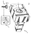
- Imaging system 30 comprises a patient chamber 34, an image processing unit 38 and a display 42.
- Imaging system 30 can be based on any known or established imaging technology, but in a present embodiment is based on computed tomography (CT) having substantially the same functionality as a machine like the Toshiba Acquillon.
- CT computed tomography
- patient chamber 34 is operable to capture images of a patient P in at least three planes
- processing unit 38 is operable to assemble those captured images to present a three-dimensional rendering of a target area within patient P on display 42.
- Images on display 42 can be navigated and/or viewed using the mouse and keyboard attached to processing unit 38, allowing the user to view a target area within patient P from any number of views.
- image processing unit 38 can also be attached to other output devices in addition to display 42, such as a printer. Further, image processing unit 38 also typically includes a fixed storage device (such as a hard drive), a removable storage device (such as CD-Rewriter, or a tape drive) and a network interface card or other network interface means for connecting processing unit 38 to a network such as an intranet and/or the internet over which captured images can be delivered.
- a fixed storage device such as a hard drive
- a removable storage device such as CD-Rewriter, or a tape drive
- network interface card or other network interface means for connecting processing unit 38 to a network such as an intranet and/or the internet over which captured images can be delivered.
- FIG 2 shows stent 50 in isolation, however, for purposes of explaining the prior art, it is to be assumed that stent 50 has been implanted in a coronary artery of patent P.
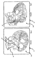
- Figure 3 shows an image 54 rendered on display 42 of system 30 of patient P.
- Image 54 shows a beam hardened artifact52 as it is implanted inside a coronary artery 58 inside a heart 62 of patient P.
- the area identified as beam hardened artifact52 is an inaccurate reproduction of stent 50 as it is implanted inside artery 58.
- the beam hardening artifact52 is created by the material of stent 50. Accordingly, system 30 is of limited value in performing post-operative evaluations of stent 50 and for determining whether any restenosis has occurred of coronary artery 58.
- Figures 4 and 5 show additional images 54a and 54b, respectively, of different orientations of heart 62, which are readily produced on display 42 due to the imaging capability of system 30.
- stent 50 and the surrounding artery 58 are inaccurately reproduced due to beam hardening artifact52 of stent 50.
- system 30 in its current form stent 50 and system 30 do not provide meaningful images for post-operative evaluation of artery 58 and the progress of any restenosis that may be occurring in the lumen of artery 58 surrounding stent 50.
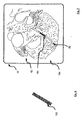
- FIG. 6 shows a medical device in accordance with an embodiment of the invention as a stent 150.
- Stent 150 from outward appearances is substantially the same as prior art stent 50, and indeed, in the present embodiment is designed to provide substantially the same mechanical and therapeutic functionality as prior art stent 50.
- stent 150 is made from a material that has a selected radiopacity such that the appearance of stent 150 is preserved when stent 150 is exposed to the imaging beam of system 30 and presented on display 42.
- image 154 has no beam hardened artefacts, it is now possible to examine the lumen of artery 58 surrounding stent 150, and thereby allow for an examination thereof for restenosis.
- H ⁇ Material ⁇ ⁇ Water ⁇ Water ⁇ 1000
- the value of the Hounsfield unit varies from -1000 (for air) to 1000 (for bone) to 3000, as more particularly shown in Table I.
- Tissue Range of Hounsfield units Table I Material Hounsfield Unit Air -1000 Lung -500 to -200 Fat -200 to -50 Water 0 Blood 25 Muscle 25 to 40 Bone 200 to 1000.
- the foregoing equation and table is found in Principles of Computerized Tomographic Imaging Parallel CT, Fanbeam CT, Helical CT and Multislice CT by Marjolein van der Glas, August 29, 2000, http://www.ph.tn.tudelft.nl/ ⁇ marlein/pdf/CT.pdf.
- stent 150 in certain imaging systems materials with Hounsfield units exceeding about 1000 can be prone to creating beam hardening artifacts.
- presently preferred materials from which stent 150 can be manufactured to have reduced beam hardening artifacts include certain plastic, composite carbon fiber and Inconel metals that have similar mechanical properties to prior art stent 50 such that substantially the same therapeutic effect in stent 150 is achieved as was available in prior art stent 50.
- the chosen material for stent 150 has a level of Hounsfield density that diminish beam hardening artifacts to substantially preserve the appearance of the device under CT or other corresponding imaging beam.
- stent 150 (or other medical devices according to the present invention) be made from a material or materials to have an overall image density of less than about 1200 Hounsfield Units. Such medical devices can also have an overall image density of less than about 900 Hounsfield Units. Such medical devices can also have an overall image density of less than about 700 Hounsfield Units. Such medical devices can also have an overall image density of less than about 400 Hounsfield Units.
- the medical devices within the scope of the invention include devices for the treatment of obstruction due to clot, plaque, atheroma, tumours or the like, and/or treatments involving intimal hyperplasia and recurrent stenosis after stent placement.
- An appropriate device is delivered into the vascular or bilary system under image guidance. The post placement follow up of the lumen is enabled by the diminished density and beam hardening artifact of the construct and coating of the stent.
- FIG. 8 A specific example of another medical device within the scope of the invention is shown in Figures 8 and 9, which shows a microcoil 250 for treatment of an aneurysm and which is introduced via a guiding cathether 240 and a microcatheter 245.
- guiding cathether 240 is inserted through an incision 260 near the femoral artery or brachial artery or other suitable location and passed through the venous system of the patient until it reaches a blood vessel 264 proximal to an aneurysm 268 in the patient's head.
- Figure 10 shows an image 254 of the resulting beam hardened artifact252 when a prior art microcoil (not shown) is post-operatively examined using imaging system 30 has been previously inserted in the patient according to the method described in reference to Figure 8.
- the beam hardened artifact252 thus renders it difficult, if not impossible, to accurately examine the prior art microcoil using imaging system 30.
- microcoil 250 when microcoil 250 is inserted according to the method described with reference to Figure 8, then microcoil 250, the now-repaired aneuryism 268 and blood vessel 264 leading thereto are all visible on display 42 and therefore capable of post-operative evaluation.
- FIG. 12 shows an image 454 of a sagittal view of patient along a plane that includes the carotid artery 470 of the patient.
- Image 454 is characterized by a beam hardened artifact 452 through which the lumen of an implanted prior art stent can be identified, but artifact452 is severe enough to obscure the lumen of the carotid artery 470, therefore preventing a determination as to whether restenosis is occuring in the lumen of artery 470 surrounding the prior art stent.
- the specific structure and/or configuration and/or shape of stent 150 is chosen to further reduce the device's overall radiopacity.
- the weave of the stent's structure can be chosen to reduce the radiopacity, and therefore the measured level of Hounsfield units associated with the stent.
- a stent delivery system having a reduced number of passages of the stent or devices across the stenosis before dilating and deploying the stent in the stenosis.
- it is necessary to cross the wire, pre-dilate, and deploy the stent posteriorly.
- a stent delivery system includes a self-expanding yet balloon mounted and intelligently be restrained.
- the stent can be mounted on a balloon and deployed by inflation of the balloon.
- Such a stent is self-expanding but is delivered on the balloon.
- the inflation of the balloon breaks the restraining polymeric bands and results in the self-expansion of the stent once the initial stimulus has been given.
- This polymeric material is drug-coated and thrombosis resistant. This polymeric material helps restrain plaque and potential embolic material behind the stent.
- the overall configuration of the stent has reduced beam hardened artifacts post insertion when viewed under a CT system. Referring to Figures 14-20, various embodiments of the stent delivery system are described in detail below.
- FIG 14 illustrates a cross sectional view of a stent delivery system 500.
- the stent delivery system 500 is delivered into a predetermined carotid artery 505 or other vessel being treated.
- the carotid artery 505 is located in a patient's neck.
- Stenosis 506 has developed on the wall of the carotid artery and the stent delivery system 500 is to be deployed to dilate the vessel 505.
- the stent delivery system 500 includes the stent 501 and the sheath 502.
- a balloon 503 mounted on the distal tip of a catheter 504 is surrounded by the stent 501 and the sheath 502.
- the balloon 503 referred to herein is typical of the type of balloon usually used with balloon-expandable stents.
- the catheter 504 is introduced into the blood vessel 505 with the stent 501, the sheath 502 and the balloon 503 mounted thereon.
- the stent 501 is a self-expandable stent.
- a carotid artery for example carotid artery 505
- the stent 501 may be temporarily deformed by external traumas, but returns to the original shape as a result of its spring-like characteristics.
- the stent 501 is delivered into the vessel 505 in a compressed state as shown in Fig. 14.
- the sheath 502 constrains the stent 501 in a collapsed state on the balloon tip of the catheter 504.
- the balloon 503 is inflated to expand the stent 501 and the sheath 502 as illustrated in Fig. 15.
- the sheath 502 releases the stent 501 so that the stent 501 deploys into and supports the vessel 505.
- the sheath 502 is configured to fracture in order to release the stent 501.
- the stent 501 provides structural support to the wall of the vessel 505.
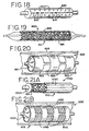
- Figures 16-18 illustrate various structures and designs of a sheath that is configured to fracture in order to release the stent 501.
- the sheath is designed to include a plurality of bands 602.
- the bands 602 are arranged parallel to one another and spaced apart according to a longitudinal direction of the stent 501.
- the bands 602 constrain the stent 501 in the compressed state.
- the bands 602 are configured to fracture to release the stent 501.
- the bands 602 may be selected from an elastic material that has a predetermined threshold level of stretching.
- the bands 602 may be made of various polymers compatible with human physiology. If the bands 602 are expanded above the threshold level, they are configured to fracture. As shown in Figure 16, at least one portion 601 of the stent 501 is exposed between the bands 602. This facilitates breaking or fracturing of the bands 602 because the stent 501 is less restricted by a sheath. In addition, other structures such as perforations may be added to encourage the fracturing of the bands 602. As shown in Figure 16, the bands 602 may be sutured to the struts of the stent 501. The sutures 604 prevent the bands 602 from drifting away from the stent 501 into the vessel 505 during the deployment.
- Figure 17 depicts a plurality of strips 702 that surround the stent 501.
- the strips 702 are designed to have a narrower width than the bands 602.
- the strips 702 are arranged parallel to each other and angled relative to a longitudinal direction of the stent 501.
- the range of the angle of the strips 702 may be between 0° and 90°.
- the strips 702 constrain the stent 501 to keep the stent 501 in the compressed state.
- the strips 702 may be fractured upon expansion of the stent 501.
- wires may be used to restrict the stent 501 instead of the strips 702.
- the wires may be made from metal and may be selected from a group of metals compatible with human physiology. For example, stainless steel or nitinol may be used to form the wires.
- the wires may surround the stent 501 in a similar manner that the strips 702 restrict the stent 501 as shown in Figure 17. A certain portion of the wires may be thinner than the remaining portions so that each wire can be easily fractured upon inflation of the balloon 503.
- the wires as a constraining structure may be simple and convenient to design and manufacture.
- Figure 18 illustrates a sheath 820 that is continuous and that includes at least one perforation 810 formed on an outer surface thereof. Various shapes, sizes and designs of perforations are available. Due to the perforations 810, the sheath 820 can be easily fractured upon the expansion of the stent 501.
- the catheter 504 having a balloon mounted tip is introduced into the carotid vessel 505 developing stenosis 506.
- the self-expandable stent 501 in the compressed state and the sheath 502 constraining the stent 501 are disposed on the balloon mounted tip of the catheter 504.
- no insertion of a balloon-tipped catheter or protection devices is performed either to predilate the vessel 505 or capture any embolic material prior to the insertion of the stent delivery system 500. Accordingly, the insertion of the stent delivery device 500 is the first passage into the host vessel 505.
- the balloon 503 is inflated as shown in Figure 15.
- the stent 501 as well as the sheath 502 expand in response to the inflation of the balloon 503.
- the sheath 502 is fractured to release the stent 501.
- the sheath 502 may include the plurality of bands 602 or strips 702 as shown in Figures 16 and 17.
- the sheath 502 may have perforations such as perforations 810 shown in Figure 18.
- the bands 602, the strips 702 and the perforations 810 facilitate the fractures of the sheath 502 to release the stent 501.
- the sheath 502 remains captured between the expanded stent 501 and the vessel wall.
- sutures 604 may be provided to secure the sheath to the stent 501.
- Emboli captured between the sheath 502 and the vessel wall is also trapped along with the sheath 502.
- the balloon 503 is deflated and removed from the vessel by removing the catheter 504.
- the stent 501 remains within the vessel 505 and provides the structural support. Accordingly, the stent delivery system 500 passes into the vessel 505 only twice to deploy the stent 501 and remove the catheter 504 after the deployment of the stent 501.
- the stent delivery system 500 has the advantage that release of emboli and debris and damage to the vessel 505 arising out of passage of the stent delivery system 500, balloon 503 and protection device are substantially minimized.
- FIG 19 illustrates a second embodiment of a stent delivery system 800.
- balloon-expandable stents 860 and 861 are used as a sheath instead of the sheath 502, the bands 602, the strips 702 or the wires.
- the balloon-expandable stents 860, 861 are mounted on the self-expandable stent 501.
- the balloon-expandable stents 860, 861 are different from each other, but two identical balloon-expandable stents may be used.
- a single, long balloon-expandable stent or three or more balloon-expandable stents may be used.
- the self-expandable stent 501 is mounted on the balloon 503.
- the stent delivery system 800 has a three layered structure, that is, the structure of the balloon 503, the self-expandable stent 501, and the balloon-expandable stent 860.
- the balloon-expandable stents 860, 861 are generally made from a ductile material, such as metal. For example, stainless steel or nitinol may be used, but any biocompatible metal is possibly used.
- the balloon-expandable stents 860, 861 restrict the self-expandable stent 501 as a sheath as shown in Figure 19.
- the balloon-expandable stents 860 and 861 are made from a ductile material, it maintains an initial, compressed state. Upon inflation of the balloon 503, the balloon-expandable stents 860, 861 are expanded along with the self-expandable stent 501.
- the balloon-expandable stents 860, 861 may also be designed to have minimal struts, which result in smaller or fewer openings between the struts. This design makes it possible for the balloon-expandable stents 860, 861 to effectively retain embolic material released from deployment of the stent 501.
- balloon-expandable stents 860, 861 as long as such stents restrict the stent 501 in a compressed state and expands in response to the inflation of the balloon 503.
- a balloon-expandable stent such as the balloon-expandable stents 860, 861 are longer in its collapsed form and shorten as they are expanded. This results in a larger space for a self-expandable stent such as the self-expandable stent 501 to be exposed.
- a double stent area i.e., where the self-expandable stent 501 and the balloon-expandable stents 860 and .861 are overlapped, is smaller.
- Figure 20 shows a third embodiment of a stent delivery system.
- a sheath constrains the stent 501 to keep the stent 501 compressed and is fractured to release the stent 501 when the stent 501 is expanded by the inflation of the balloon 503.
- Figure 20 depicts another embodiment of a sheath used in the stent delivery system 900.
- a sheath 903 does not constrain the stent 501.
- the sheath 903 is disposed on the stent 501 to trap debris behind the stent 501.
- the sheath 903 simply surrounds the stent 501 and does not impose any pressure on stent 501 to keep the stent 501 collapsed on the balloon 901.
- the sheath 903 is made of a non-elastic material, but it is plastically deformable and expands in response to the inflation of the balloon. Once the sheath 903 changes its shape, it is deformed and does not return to its original shape.
- the balloon 901 is inflated to expand the stent 501.
- the sheath 903 expands along with the stent 501 as shown in Figure 20. After the expansion, the sheath 903 stays within the blood vessel 505 along with the stent 501 that provides the structural support to the vessel 505.
- the sheath 903 effectively traps debris released as a result of the stent deployment, thereby minimizing embolization and/or restenosis caused by such debris.
- the stent delivery device 900 includes another structure for constraining the stent 501.
- at least one constraining structure 905 constrains both the stent 501 and the sheath 903.
- the constraining structure 905 is fractured upon the inflation of the balloon 901 and releases both the stent 501 and the sheath 903.
- the constraining structure 905 includes at least one band 905 as illustrated in Figure 20, but other shapes and designs are possible.
- the constraining structure 905 is made of elastic material that has a predetermined threshold level of stretching. When the constraining structure 905 is expanded to exceed the threshold stretch level, it is configured to fracture.
- the fractured structure 905 is trapped between the vessel wall and the sheath 903 and does not drift within the vessel 505. Sutures that secure the constraining structure 905 to the stent may be included. Accordingly, the fractured structure 903 does not cause any embolization or restenosis.
- the stent delivery system 900 operates as follows.
- the stent delivery system 900 is introduced into the vessel 505. Prior to insertion of the stent delivery system 900, no predilation of the vessel 505 by a balloon or an insertion of a protection device is performed.
- the stent 501. is restrained in the compressed state by the constraining structure 905.
- the balloon 901 is inflated to expand the stent 501.
- the sheath 903 is expanded and plastically deformed.
- the constraining structure 905 is expanded to reach its threshold level and ultimately is fractured as shown in Figure 20. As a result, the debris or emboli released during the stent deployment is trapped between the sheath 903 and the vessel wall.
- the sheath 903 is trapped between the stent 501 and the vessel wall. Accordingly, embolic or debris release is substantially minimized even though no separate protection device is used. As a result, passages of the host vessel 505 for implanting the stent 501 are limited to two passes, thereby substantially minimizing embolic release and damage to the vessel arising out of such passages. Additionally, the sheath 903 continuously surrounds the expanded stent 501 throughout the deployment process, and therefore the vessel wall is protected from damage resulting from contact with the struts of the stent 501.
- FIG 21 shows a fourth embodiment of a stent delivery system.
- a stent delivery system 1000 includes a sheath 1010 that is made of SIS material (Small Intestine Submucosa).
- Figure 21A illustrates the stent delivery system 1000 when the stent 501 is in a compressed state
- Figure 21B illustrates that the stent 501 is expanded by inflation of a balloon 1003.
- the SIS material is derived from the small intestine of a pig and may be used to heal wounds or repair tissue damage.
- the SIS material is mechanically and chemically processed to be implanted into a patient's body.
- the SIS material typically has a matrix structure.
- the SIS material When the SIS material is implanted, tissues adjacent to the SIS matrix begin to deliver cells and nutrients. Cells rapidly occupy the SIS matrix, and eventually, the SIS matrix is replaced by newly grown tissues. The SIS remains at the implantation site and provides support to the new tissues.
- the SIS material When used with a stent delivery system, it may repair the defect or lesion of the vessel walls.
- the SIS material is used as a sheath covering a stent and the SIS material works as a smooth buffer between the stent and the vessel walls upon deployment of the stent.
- the stent is generally made of metal and upon expansion of the stent, the structure of the stent such as the struts may damage the vessel wall tissues.
- the SIS material is soft and thick, thereby protecting the vessel wall from damage. Tissues of the vessel wall grow into a matrix structure of the SIS sheath, thereby minimizing release of embolic materials such as necrotic tissues.
- the matrix structure of the SIS sheath is readily configured to embed at least one drug such as a restenosis inhibiting drug.
- the SIS material does not readily stretch in response to the stent expansion.
- a plurality of wrinkles 1020 are provided to make the sheath 1010 stretch in response to the expansion of the stent 501.
- the sheath 1010 does not constrain the stent 501. It is simply disposed on the stent 501.
- at least one constraining structure 1050 is provided in Figure 21B. The constraining structure 1050 restrains the stent 501 to be compressed. Upon expansion of the stent 501, the constraining structure 1050 is fractured and releases the stent 1001.
- the sheath 1010 remains with the stent 501 after the expansion, to enclose debris between the sheath 1010 and the vessel wall.
- the constraining structure 1050 is trapped between the sheath 1010 and the vessel 505. Sutures or other structures may be used to secure the sheath 1010 and constraining structure 1050 to the stent 1001.
- the constraining structure 905, 1050 includes a plurality of bands.
- the constraining structure 905, 1050 may include strips and/or wires.
- Various shapes are possible and two or more shapes may be used together for the constraining structure 905, 1050.
- the constraining structure 905, 1050 may be made from polymeric material, metal or any material that is compatible with human physiology.
- the stent delivery system 1000 provides additional advantages such as healing any damage to the vessel wall.
- the number of passages of hardware across the stent or devices across the stent is reduced from as many as five (as found in prior art delivery methods) to two passes (according to the described method passes).
- material that would otherwise become potentially an embolic source is restrained against the vessel wall. This can be helpful in reducing the risks of stroke after carotid stenting and in some circumstances can reduce the need for distal flow protection devices which themselves have stroke risk.
- shunt 150 is coated (either in its entirety or in particular locations) with an opacifier to temporarily increase the Hounsfield units associated with shunt 150 during its insertion, to allow shunt 150 to be inserted using traditional means.
- a coating would be configured to gradually abate and dissolve into the patient's blood stream, such that the radiopacity and associated Hounsfield units of stent 150 would decrease over time, such that under a post-operative CT evaluation, the Hounsfield units associated with stent 150 are low enough to allow proper visualization of the lumen of artery 58 surrounding stent 150.
- Suitable materials for coating shunt 150 include gold, iodine, ionic and non ionic iodinated compounds, ethiodol, and lipiodol, barium, tungsten, tantalum, gadolinium. Whatever coating is chosen, the amount and rate of dissolving of the coating is chosen to reduce toxicity experience by the patient during dissolution.
- the aforementioned coating is a hydrophilic polymer containing a restenosis inhibiting drug and a density enhancing radiologic material such as lyophilizied iodinated contrast material, which is embedded into the polymer.
- This coating is then placed over a stent 150 that is made from a suitable material such as a plastic or metal, such as stainless steel, inconel or metal glass (materials already approved by The United States of America Food and Drug Administration), or an optimal arrangement of strands of another metal can be used.
- stent 150 is both drug eluting and density eluting (i.e. the level of Hounsfield units associated with the stent decreases over time.)
- the stent 150 may be used in the stent delivery systems 500, 900 and 1000 described in conjunction with Figures 14-21.
- the stent 150 may be a self-expandable stent and disposed on the balloon mounted catheter like the stent 501.
- the stent 501 is a self-expandable stent, it is suitable for carotid arteries that are often subject to external traumas.
- the balloon is inflated to expand the stent 150 and the sheath 502, 903 and 1010.
- the sheath 502 is configured to be fractured in response to the expansion of the stent 150.
- sheaths 903 and 1010 expand and remain within the carotid artery 505 to trap the embolic release between the sheath 502, 903 and 1010 and the vessel wall.
- the sheath 903 is plastically deformable and the sheath 1010 is made of SIS material.
- Separate constraining structures 905 and 1050 are provided to constrain the stent 150 instead of the sheaths 903 and 1010.
- certain post processing software is provided in image processing unit 38 to maximize vascular conspicuity in conjunction with the known Hounsfield units and other imaging properties associated with stent 150 or other medical device in accordance with the present invention.
- image processing unit 38 to maximize vascular conspicuity in conjunction with the known Hounsfield units and other imaging properties associated with stent 150 or other medical device in accordance with the present invention.
- a level of Hounsfield units associated with stent 150 is known, then upon detection by system 30 of an item within the patient at that particular level of Hounsfield units, then that information can be used to identify the item as stent 150 and then to further enhance the image of the surrounding vascular region based on the known imaging properties (i.e. radiopacity, structure, etc.) and using known signal processing an filtering techniques.
- known imaging properties i.e. radiopacity, structure, etc.
- the stents, coils and other medical devices according to the present invention can be coated with a material to decrease the risk of infection and restenosis, using techniques and compounds described in EP0797988A2 and EP1155689A2 to Angiotech Pharmaceuticals Inc. of Canada, and the University of British Columbia.
- the present invention also provides certain novel methods for evaluating cervical and intracranial vascular stents using CT, including MDCTA, that is reliable and low cost and then to use these techniques for long term evaluation and outcome analysis of stenting. Sensitivity and specificity can then be determined for MDCTA by comparison to conventional catheter angiogram results.
- the radiographic density of the stent, coil or other device can be altered to enhance CT, X Ray, Ultrasound and MRI visibility of the lumen. For the purpose of enhanced accuracy of CT diagnostic imaging beam hardening artifacts will be reduced and/or minimized.
- the devices in the present invention are in contrast to prior art devices that have been developed for conventional fluoroscopy guidance and thus are of a radiodensity or radiopacity that exceeds the needs of CT for clear visualization, this excess density creates unwanted beam hardening artifact.
- the present invention allows for a relatively non-invasive means to visualize the lumen of a blood vessel surrounding a previously installed stent (or other site of an implanted medical device). Due to the reduced beam hardening artifacts of the stent, obscuration of the lumen is reduced. This results in the ability to visualize the lumen non-invasively as compared follow-ups conducted by invasive repeat catheter angiography, with its resultant risk of stroke, death and/or injury to an important vessel or to otherwise obscure a critical finding.
- CTA and CTP are relatively less invasive imaging modalities that have been shown to aid in the diagnosis and treatment of acute ischemic stroke. Both utilize highspeed spiral CT scanning and three-dimensional volumetric reconstruction software to create various types of images following injection of lV contrast solution.
- CTA can provide three-dimensional vascular delineation similar to other non-invasive techniques as well as visualization of adjacent non-vascular soft-tissue.
- CTA can also offer rapid volume acquisition, limited reconstruction artifact and scan completion during the period of peak intravascular contrast enhancement. Using CTA, it is often possible to see filling defect in a vessel as a result of contrast displacement by clot or thrombus.
- the sensitivity for detecting flow abnormality in vessels in the circle of Willis by CTA can be at least 89% when compared to digital subtraction angiography ("DSA”), and CTA does not carry the up to 5% risk of complication, and the up to 0.5% risk of permanent stroke that DSA has been shown to carry.
- DSA digital subtraction angiography
Landscapes
- Health & Medical Sciences (AREA)
- Life Sciences & Earth Sciences (AREA)
- Biomedical Technology (AREA)
- Engineering & Computer Science (AREA)
- General Health & Medical Sciences (AREA)
- Veterinary Medicine (AREA)
- Vascular Medicine (AREA)
- Heart & Thoracic Surgery (AREA)
- Animal Behavior & Ethology (AREA)
- Public Health (AREA)
- Surgery (AREA)
- Transplantation (AREA)
- Oral & Maxillofacial Surgery (AREA)
- Cardiology (AREA)
- Molecular Biology (AREA)
- Reproductive Health (AREA)
- Medical Informatics (AREA)
- Nuclear Medicine, Radiotherapy & Molecular Imaging (AREA)
- Physics & Mathematics (AREA)
- Optics & Photonics (AREA)
- Epidemiology (AREA)
- Gastroenterology & Hepatology (AREA)
- Pulmonology (AREA)
- Neurosurgery (AREA)
- Materials For Medical Uses (AREA)
Applications Claiming Priority (1)
| Application Number | Priority Date | Filing Date | Title |
|---|---|---|---|
| US10/965,590 US20050049672A1 (en) | 2003-03-24 | 2004-10-14 | Stent delivery system and method using a balloon for a self-expandable stent |
Publications (1)
| Publication Number | Publication Date |
|---|---|
| EP1649829A1 true EP1649829A1 (de) | 2006-04-26 |
Family
ID=35589308
Family Applications (1)
| Application Number | Title | Priority Date | Filing Date |
|---|---|---|---|
| EP05022438A Withdrawn EP1649829A1 (de) | 2004-10-14 | 2005-10-14 | Einführsystem mit einem Ballon für einen selbstexpandierbaren Stent |
Country Status (2)
| Country | Link |
|---|---|
| US (1) | US20050049672A1 (de) |
| EP (1) | EP1649829A1 (de) |
Cited By (6)
| Publication number | Priority date | Publication date | Assignee | Title |
|---|---|---|---|---|
| WO2009100210A1 (en) * | 2008-02-05 | 2009-08-13 | Silk Road Medical, Inc. | Interventional catheter system and methods |
| US7998104B2 (en) | 2003-11-21 | 2011-08-16 | Silk Road Medical, Inc. | Method and apparatus for treating a carotid artery |
| WO2011123852A1 (en) * | 2010-04-02 | 2011-10-06 | Cappella, Inc. | Systems and methods for delivering a stent to a body lumen |
| US8157760B2 (en) | 2007-07-18 | 2012-04-17 | Silk Road Medical, Inc. | Methods and systems for establishing retrograde carotid arterial blood flow |
| US8545432B2 (en) | 2009-06-03 | 2013-10-01 | Silk Road Medical, Inc. | System and methods for controlling retrograde carotid arterial blood flow |
| US8858490B2 (en) | 2007-07-18 | 2014-10-14 | Silk Road Medical, Inc. | Systems and methods for treating a carotid artery |
Families Citing this family (32)
| Publication number | Priority date | Publication date | Assignee | Title |
|---|---|---|---|---|
| US8034100B2 (en) | 1999-03-11 | 2011-10-11 | Endologix, Inc. | Graft deployment system |
| US6261316B1 (en) | 1999-03-11 | 2001-07-17 | Endologix, Inc. | Single puncture bifurcation graft deployment system |
| US20040193178A1 (en) * | 2003-03-26 | 2004-09-30 | Cardiomind, Inc. | Multiple joint implant delivery systems for sequentially-controlled implant deployment |
| EP1898771A2 (de) * | 2005-06-24 | 2008-03-19 | Power Ten, LLC | Expandierbares zugangssystem für einen chirurgischen situs |
| US8202311B2 (en) * | 2005-07-27 | 2012-06-19 | Cook Medical Technologies Llc | Stent/graft device and method for open surgical placement |
| US20080039928A1 (en) * | 2006-08-08 | 2008-02-14 | Medlogics Device Corporation | Slotted Self-Expanding Stent Delivery System |
| US20080039920A1 (en) * | 2006-08-08 | 2008-02-14 | Medlogics Device Corporation | Tethered Self-Expanding Stent Delivery System |
| WO2008084286A2 (en) * | 2006-10-25 | 2008-07-17 | Arterial Remodeling Technologies, S.A. | Method for expansion and deployment of polymeric structures including stents |
| US8372131B2 (en) * | 2007-07-16 | 2013-02-12 | Power Ten , LLC | Surgical site access system and deployment device for same |
| US20090024158A1 (en) * | 2007-07-16 | 2009-01-22 | Zimmer Spine, Inc. | Access Port Expander And Method |
| US20090043330A1 (en) * | 2007-08-09 | 2009-02-12 | Specialized Vascular Technologies, Inc. | Embolic protection devices and methods |
| US20090043380A1 (en) * | 2007-08-09 | 2009-02-12 | Specialized Vascular Technologies, Inc. | Coatings for promoting endothelization of medical devices |
| US20090112239A1 (en) * | 2007-10-31 | 2009-04-30 | Specialized Vascular Technologies, Inc. | Sticky dilatation balloon and methods of using |
| WO2009091968A1 (en) * | 2008-01-18 | 2009-07-23 | Med Institute, Inc. | Intravascular device attachment system having struts |
| US8221494B2 (en) | 2008-02-22 | 2012-07-17 | Endologix, Inc. | Apparatus and method of placement of a graft or graft system |
| US8236040B2 (en) | 2008-04-11 | 2012-08-07 | Endologix, Inc. | Bifurcated graft deployment systems and methods |
| EP2520320B1 (de) | 2008-07-01 | 2016-11-02 | Endologix, Inc. | Kathetersystem |
| WO2010054121A2 (en) * | 2008-11-07 | 2010-05-14 | Specialized Vascular Technologies, Inc. | Extracellular matrix modulating coatings for medical devices |
| JP5629871B2 (ja) | 2009-04-28 | 2014-11-26 | エンドロジックス、インク | 移植片あるいは移植片システムを配置する装置および方法 |
| WO2011044533A2 (en) | 2009-10-09 | 2011-04-14 | Specialized Vascular Technologies, Inc. | Coating system and method for drug elution management |
| US10751206B2 (en) | 2010-06-26 | 2020-08-25 | Scott M. Epstein | Catheter or stent delivery system |
| US20110319902A1 (en) * | 2010-06-26 | 2011-12-29 | Scott Epstein | Catheter delivery system |
| EP2635241B1 (de) | 2010-11-02 | 2019-02-20 | Endologix, Inc. | Vorrichtung zur positionierung einer gefässprothese bzw. eines gefässprothesensystems |
| CN103561807B (zh) | 2011-03-01 | 2015-11-25 | 恩朵罗杰克斯股份有限公司 | 导管系统及其使用方法 |
| US8998936B2 (en) | 2011-06-30 | 2015-04-07 | The Spectranetics Corporation | Reentry catheter and method thereof |
| US9814862B2 (en) | 2011-06-30 | 2017-11-14 | The Spectranetics Corporation | Reentry catheter and method thereof |
| US8956376B2 (en) | 2011-06-30 | 2015-02-17 | The Spectranetics Corporation | Reentry catheter and method thereof |
| US10109058B2 (en) | 2015-05-17 | 2018-10-23 | Lightlab Imaging, Inc. | Intravascular imaging system interfaces and stent detection methods |
| US9996921B2 (en) | 2015-05-17 | 2018-06-12 | LIGHTLAB IMAGING, lNC. | Detection of metal stent struts |
| CN111529126A (zh) | 2015-06-30 | 2020-08-14 | 恩朵罗杰克斯股份有限公司 | 用于将第一细长构件固定至第二细长构件的系统及方法 |
| US10338795B2 (en) * | 2015-07-25 | 2019-07-02 | Lightlab Imaging, Inc. | Intravascular data visualization and interface systems and methods |
| CN107510518B (zh) * | 2016-06-16 | 2019-11-26 | 上海微创医疗器械(集团)有限公司 | 支架系统以及支架的存放方法 |
Citations (7)
| Publication number | Priority date | Publication date | Assignee | Title |
|---|---|---|---|---|
| US5980565A (en) * | 1997-10-20 | 1999-11-09 | Iowa-India Investments Company Limited | Sandwich stent |
| US6179878B1 (en) * | 1996-10-22 | 2001-01-30 | Thomas Duerig | Composite self expanding stent device having a restraining element |
| US6352561B1 (en) * | 1996-12-23 | 2002-03-05 | W. L. Gore & Associates | Implant deployment apparatus |
| US20020052640A1 (en) * | 2000-08-04 | 2002-05-02 | Steve Bigus | Sheath for self-expanding stents |
| US6533806B1 (en) * | 1999-10-01 | 2003-03-18 | Scimed Life Systems, Inc. | Balloon yielded delivery system and endovascular graft design for easy deployment |
| US20040073286A1 (en) * | 2001-01-22 | 2004-04-15 | Armstrong Joseph P. | Deployment system for intraluminal devices |
| US20040176833A1 (en) * | 2002-11-22 | 2004-09-09 | Cook Incorporated | Stent tissue graft prosthesis |
Family Cites Families (53)
| Publication number | Priority date | Publication date | Assignee | Title |
|---|---|---|---|---|
| US3721997A (en) * | 1972-07-17 | 1973-03-27 | Sterling L O Dell | Protective garment |
| US3984696A (en) * | 1974-12-11 | 1976-10-05 | Medi-Ray, Inc. | Radiation guard for X-ray table |
| US4062518A (en) * | 1976-11-10 | 1977-12-13 | General Electric Company | X-ray shielding device |
| DK558178A (da) * | 1978-12-12 | 1980-06-13 | Uno Plast As | Kirurgisk instrument |
| US4686962A (en) * | 1986-07-03 | 1987-08-18 | Habley Medical Technology Corporation | Disposable cartridge assembly for hypodermically implanting a genitourinary prosthesis |
| US5308352A (en) * | 1989-11-17 | 1994-05-03 | Koutrouvelis Panos G | Stereotactic device |
| US5292339A (en) * | 1991-06-14 | 1994-03-08 | Telectronics Pacing Systems, Inc. | Implantable pacemaker/cardioverter/defibrillator device and method incorporating multiple bradycardia support pacing rates |
| US5211165A (en) * | 1991-09-03 | 1993-05-18 | General Electric Company | Tracking system to follow the position and orientation of a device with radiofrequency field gradients |
| WO1993010695A1 (en) * | 1991-11-25 | 1993-06-10 | Sze Wee Tan | Body support for a baby |
| US5311883A (en) * | 1992-11-06 | 1994-05-17 | Eleanor Sherman | Sanitary shield for dedicated mammography apparatus |
| US5335663A (en) * | 1992-12-11 | 1994-08-09 | Tetrad Corporation | Laparoscopic probes and probe sheaths useful in ultrasonic imaging applications |
| WO1996011624A2 (en) * | 1994-10-07 | 1996-04-25 | St. Louis University | Surgical navigation systems including reference and localization frames |
| US5590655A (en) * | 1993-09-20 | 1997-01-07 | Hussman; Karl L. | Frameless laser guided stereotactic localization system |
| US5989280A (en) * | 1993-10-22 | 1999-11-23 | Scimed Lifesystems, Inc | Stent delivery apparatus and method |
| US5549635A (en) * | 1994-01-24 | 1996-08-27 | Solar, Rita & Gaterud, Ltd. | Non-deformable self-expanding parallel flow endovascular stent and deployment apparatus therefore |
| US5545150A (en) * | 1994-05-06 | 1996-08-13 | Endoscopic Concepts, Inc. | Trocar |
| US5482043A (en) * | 1994-05-11 | 1996-01-09 | Zulauf; David R. P. | Method and apparatus for telefluoroscopy |
| US6978166B2 (en) * | 1994-10-07 | 2005-12-20 | Saint Louis University | System for use in displaying images of a body part |
| US6120536A (en) * | 1995-04-19 | 2000-09-19 | Schneider (Usa) Inc. | Medical devices with long term non-thrombogenic coatings |
| US5609629A (en) * | 1995-06-07 | 1997-03-11 | Med Institute, Inc. | Coated implantable medical device |
| US5592952A (en) * | 1995-08-18 | 1997-01-14 | Bohn; William W. | Infection control surgical drape and method of making surgical incision |
| US5638819A (en) * | 1995-08-29 | 1997-06-17 | Manwaring; Kim H. | Method and apparatus for guiding an instrument to a target |
| US5902254A (en) * | 1996-07-29 | 1999-05-11 | The Nemours Foundation | Cathether guidewire |
| US5676159A (en) * | 1996-11-05 | 1997-10-14 | Janin Group | Ultrasound cover |
| DE69737208T2 (de) * | 1996-11-15 | 2007-11-08 | Cook Inc., Bloomington | Stentanbringungsvorrichtung mit einer auftrennbaren hülle |
| US5957974A (en) * | 1997-01-23 | 1999-09-28 | Schneider (Usa) Inc | Stent graft with braided polymeric sleeve |
| AU6534098A (en) * | 1997-02-20 | 1998-09-09 | Johns Hopkins University, The | Friction transmission with axial loading and a radiolucent surgical needle driver |
| US5892238A (en) * | 1997-05-02 | 1999-04-06 | Bionix Development Corp. | Radiation therapy shielding assembly |
| US5899935A (en) * | 1997-08-04 | 1999-05-04 | Schneider (Usa) Inc. | Balloon expandable braided stent with restraint |
| TR200000865T2 (tr) * | 1997-09-25 | 2000-08-21 | Minrad Inc. | Steril örtü |
| FR2772267A1 (fr) * | 1997-12-16 | 1999-06-18 | Oreal | Compositions de teinture des fibres keratiniques contenant des derives imidazo-pyridines et procede |
| US6740113B2 (en) * | 1998-05-29 | 2004-05-25 | Scimed Life Systems, Inc. | Balloon expandable stent with a self-expanding portion |
| US6475234B1 (en) * | 1998-10-26 | 2002-11-05 | Medinol, Ltd. | Balloon expandable covered stents |
| US6285902B1 (en) * | 1999-02-10 | 2001-09-04 | Surgical Insights, Inc. | Computer assisted targeting device for use in orthopaedic surgery |
| US6456684B1 (en) * | 1999-07-23 | 2002-09-24 | Inki Mun | Surgical scanning system and process for use thereof |
| US6544041B1 (en) * | 1999-10-06 | 2003-04-08 | Fonar Corporation | Simulator for surgical procedures |
| US6481888B1 (en) * | 1999-10-12 | 2002-11-19 | R. Hank Morgan | Scatter ban drape |
| US6379302B1 (en) * | 1999-10-28 | 2002-04-30 | Surgical Navigation Technologies Inc. | Navigation information overlay onto ultrasound imagery |
| US6475235B1 (en) * | 1999-11-16 | 2002-11-05 | Iowa-India Investments Company, Limited | Encapsulated stent preform |
| US6355058B1 (en) * | 1999-12-30 | 2002-03-12 | Advanced Cardiovascular Systems, Inc. | Stent with radiopaque coating consisting of particles in a binder |
| US6453185B1 (en) * | 2000-03-17 | 2002-09-17 | Integra Lifesciences, Inc. | Ventricular catheter with reduced size connector and method of use |
| US6325538B1 (en) * | 2000-03-17 | 2001-12-04 | Christian M. Heesch | Radiation field isolator apparatus |
| US6689125B1 (en) * | 2000-04-04 | 2004-02-10 | Spinalabs, Llc | Devices and methods for the treatment of spinal disorders |
| US6490475B1 (en) * | 2000-04-28 | 2002-12-03 | Ge Medical Systems Global Technology Company, Llc | Fluoroscopic tracking and visualization system |
| CA2403276C (en) * | 2000-05-04 | 2009-10-20 | Oregon Health Sciences University | Endovascular stent graft |
| SE522805C2 (sv) * | 2000-06-22 | 2004-03-09 | Jan Otto Solem | Stentappliceringssystem |
| US6412851B1 (en) * | 2000-08-29 | 2002-07-02 | Jay Burks | Retractable cover for small vehicles |
| US6585677B2 (en) * | 2000-08-30 | 2003-07-01 | John A. Cowan, Jr. | Shunt |
| US6917827B2 (en) * | 2000-11-17 | 2005-07-12 | Ge Medical Systems Global Technology Company, Llc | Enhanced graphic features for computer assisted surgery system |
| US6716238B2 (en) * | 2001-05-10 | 2004-04-06 | Scimed Life Systems, Inc. | Stent with detachable tethers and method of using same |
| US6749628B1 (en) * | 2001-05-17 | 2004-06-15 | Advanced Cardiovascular Systems, Inc. | Stent and catheter assembly and method for treating bifurcations |
| US6961606B2 (en) * | 2001-10-19 | 2005-11-01 | Koninklijke Philips Electronics N.V. | Multimodality medical imaging system and method with separable detector devices |
| US20040148000A1 (en) * | 2003-01-24 | 2004-07-29 | Bilge Fertac H. | Self expanding stent delivery system with balloon |
-
2004
- 2004-10-14 US US10/965,590 patent/US20050049672A1/en not_active Abandoned
-
2005
- 2005-10-14 EP EP05022438A patent/EP1649829A1/de not_active Withdrawn
Patent Citations (7)
| Publication number | Priority date | Publication date | Assignee | Title |
|---|---|---|---|---|
| US6179878B1 (en) * | 1996-10-22 | 2001-01-30 | Thomas Duerig | Composite self expanding stent device having a restraining element |
| US6352561B1 (en) * | 1996-12-23 | 2002-03-05 | W. L. Gore & Associates | Implant deployment apparatus |
| US5980565A (en) * | 1997-10-20 | 1999-11-09 | Iowa-India Investments Company Limited | Sandwich stent |
| US6533806B1 (en) * | 1999-10-01 | 2003-03-18 | Scimed Life Systems, Inc. | Balloon yielded delivery system and endovascular graft design for easy deployment |
| US20020052640A1 (en) * | 2000-08-04 | 2002-05-02 | Steve Bigus | Sheath for self-expanding stents |
| US20040073286A1 (en) * | 2001-01-22 | 2004-04-15 | Armstrong Joseph P. | Deployment system for intraluminal devices |
| US20040176833A1 (en) * | 2002-11-22 | 2004-09-09 | Cook Incorporated | Stent tissue graft prosthesis |
Cited By (41)
| Publication number | Priority date | Publication date | Assignee | Title |
|---|---|---|---|---|
| US9526504B2 (en) | 2003-11-21 | 2016-12-27 | Silk Road Medical, Inc. | Method and apparatus for treating a carotid artery |
| US7998104B2 (en) | 2003-11-21 | 2011-08-16 | Silk Road Medical, Inc. | Method and apparatus for treating a carotid artery |
| US8002728B2 (en) | 2003-11-21 | 2011-08-23 | Silk Road Medical, Inc. | Method and apparatus for treating a carotid artery |
| US11918226B2 (en) | 2003-11-21 | 2024-03-05 | Silk Road Medical, Inc. | Method and apparatus for treating an arterial lesion |
| US11849954B2 (en) | 2003-11-21 | 2023-12-26 | Silk Road Medical, Inc. | Method and apparatus for treating an arterial lesion |
| US8343089B2 (en) | 2003-11-21 | 2013-01-01 | Silk Road Medical, Inc. | Method and apparatus for treating a carotid artery |
| US8414516B2 (en) | 2003-11-21 | 2013-04-09 | Silk Road Medical, Inc. | Method and apparatus for treating a carotid artery |
| US10779835B2 (en) | 2003-11-21 | 2020-09-22 | Silk Road Medical, Inc. | Method and apparatus for treating a carotid artery |
| US10722239B2 (en) | 2003-11-21 | 2020-07-28 | Silk Road Medical, Inc. | Method and apparatus for treating an arterial lesion |
| US10188399B2 (en) | 2003-11-21 | 2019-01-29 | Silk Road Medical, Inc. | Method and apparatus for treating a carotid artery |
| US9662118B2 (en) | 2003-11-21 | 2017-05-30 | Silk Road Medical, Inc. | Method and apparatus for treating a carotid artery |
| US8870805B2 (en) | 2003-11-21 | 2014-10-28 | Silk Road Medical, Inc. | Method and apparatus for treating a carotid artery |
| US8858490B2 (en) | 2007-07-18 | 2014-10-14 | Silk Road Medical, Inc. | Systems and methods for treating a carotid artery |
| US10485917B2 (en) | 2007-07-18 | 2019-11-26 | Silk Road Medical, Inc. | Methods and systems for establishing retrograde carotid arterial blood flow |
| US9259215B2 (en) | 2007-07-18 | 2016-02-16 | Silk Road Medical, Inc. | Systems and methods for treating a carotid artery |
| US12296082B2 (en) | 2007-07-18 | 2025-05-13 | Boston Scientific Scimed, Inc. | Methods and systems for establishing retrograde carotid arterial blood flow |
| US9011364B2 (en) | 2007-07-18 | 2015-04-21 | Silk Road Medical, Inc. | Methods and systems for establishing retrograde carotid arterial blood flow |
| US9655755B2 (en) | 2007-07-18 | 2017-05-23 | Silk Road Medical, Inc. | Systems and methods for treating a carotid artery |
| US12194219B2 (en) | 2007-07-18 | 2025-01-14 | Silk Road Medical, Inc. | Methods and systems for establishing retrograde carotid arterial blood flow |
| US12156960B2 (en) | 2007-07-18 | 2024-12-03 | Silk Road Medical, Inc. | Systems and methods for treating a carotid artery |
| US9789242B2 (en) | 2007-07-18 | 2017-10-17 | Silk Road Medical, Inc. | Methods and systems for establishing retrograde carotid arterial blood flow |
| US9833555B2 (en) | 2007-07-18 | 2017-12-05 | Silk Road Medical, Inc. | Methods and systems for establishing retrograde carotid arterial blood flow |
| US10085864B2 (en) | 2007-07-18 | 2018-10-02 | Silk Road Medical, Inc. | Systems and methods for treating a carotid artery |
| US8784355B2 (en) | 2007-07-18 | 2014-07-22 | Silk Road Medical, Inc. | Methods and systems for establishing retrograde carotid arterial blood flow |
| US12042593B2 (en) | 2007-07-18 | 2024-07-23 | Silk Road Medical, Inc. | Methods and systems for establishing retrograde carotid arterial blood flow |
| US10286139B2 (en) | 2007-07-18 | 2019-05-14 | Silk Road Medical, Inc. | Methods and systems for establishing retrograde carotid arterial blood flow |
| US10426885B2 (en) | 2007-07-18 | 2019-10-01 | Silk Road Medical, Inc. | Methods and systems for establishing retrograde carotid arterial blood flow |
| US8157760B2 (en) | 2007-07-18 | 2012-04-17 | Silk Road Medical, Inc. | Methods and systems for establishing retrograde carotid arterial blood flow |
| US10543307B2 (en) | 2007-07-18 | 2020-01-28 | Silk Road Medical, Inc. | Methods and systems for establishing retrograde carotid arterial blood flow |
| US10709832B2 (en) | 2007-07-18 | 2020-07-14 | Silk Road Medical, Inc. | Methods and systems for establishing retrograde carotid arterial blood flow |
| US8740834B2 (en) | 2007-07-18 | 2014-06-03 | Silk Road Medical, Inc. | Methods and systems for establishing retrograde carotid arterial blood flow |
| US11364332B2 (en) | 2007-07-18 | 2022-06-21 | Silk Road Medical, Inc. | Methods and systems for establishing retrograde carotid arterial blood flow |
| US10952882B2 (en) | 2007-07-18 | 2021-03-23 | Silk Road Medical, Inc. | Systems and methods for treating a carotid artery |
| US11364369B2 (en) | 2008-02-05 | 2022-06-21 | Silk Road Medical, Inc. | Interventional catheter system and methods |
| US10226598B2 (en) | 2008-02-05 | 2019-03-12 | Silk Road Medical, Inc. | Interventional catheter system and methods |
| US9669191B2 (en) | 2008-02-05 | 2017-06-06 | Silk Road Medical, Inc. | Interventional catheter system and methods |
| WO2009100210A1 (en) * | 2008-02-05 | 2009-08-13 | Silk Road Medical, Inc. | Interventional catheter system and methods |
| US8545432B2 (en) | 2009-06-03 | 2013-10-01 | Silk Road Medical, Inc. | System and methods for controlling retrograde carotid arterial blood flow |
| US9138527B2 (en) | 2009-06-03 | 2015-09-22 | Silk Road Medical, Inc. | System and methods for controlling retrograde carotid arterial blood flow |
| WO2011123852A1 (en) * | 2010-04-02 | 2011-10-06 | Cappella, Inc. | Systems and methods for delivering a stent to a body lumen |
| US9468548B2 (en) | 2010-04-02 | 2016-10-18 | Cappella, Inc. | Systems and methods for delivering a stent to a body lumen |
Also Published As
| Publication number | Publication date |
|---|---|
| US20050049672A1 (en) | 2005-03-03 |
Similar Documents
| Publication | Publication Date | Title |
|---|---|---|
| EP1649829A1 (de) | Einführsystem mit einem Ballon für einen selbstexpandierbaren Stent | |
| US8465539B2 (en) | Device viewable under an imaging beam | |
| Eggebrecht et al. | Nonsurgical retrieval of embolized coronary stents | |
| CN111278367B (zh) | 阻塞装置 | |
| CN113891696A (zh) | 用于与血管内移植物相关的治疗的方法和装置 | |
| US20030204248A1 (en) | Device viewable under an imaging beam | |
| Orend et al. | Endovascular treatment (EVT) of acute traumatic lesions of the descending thoracic aorta–7 years' experience | |
| EP1505907B1 (de) | Anwendungsspezifisches material mit verbesserter strahlungsundurchlässigkeit | |
| Bruining et al. | Quantitative multi-modality imaging analysis of a bioabsorbable poly-L-lactic acid stent design in the acute phase: a comparison between 2-and 3D-QCA, QCU and QMSCT-CA. | |
| EP1295615A1 (de) | Strahlungsundurchlässiger Stent | |
| Görich et al. | Displaced metallic biliary stents: technique and rationale for interventional radiologic retrieval. | |
| EP1691721B1 (de) | Unter einem darstellungsstrahl sichtbare vorrichtung | |
| RU2639815C1 (ru) | Способ двойной защиты головного мозга при стентировании каротидных артерий у пациентов с высоким риском эмболии | |
| Priestley et al. | Iatrogenic vertebral arteriovenous fistula treated with a hemobahn stent-graft | |
| US20220296249A1 (en) | Endovascular coil device for embolization of blood vessels | |
| Peterson et al. | Coil embolization of an inferior pancreaticoduodenal artery aneurysm associated with celiac artery occlusion | |
| Peynircioglu et al. | Stent-graft applications in peripheral non-atherosclerotic arterial lesions | |
| CA2455439C (en) | Device viewable under an imaging beam | |
| Mirchia et al. | A successful endovascular repositioning of migrated transjugular intrahepatic portosystemic shunt (TIPS) stent | |
| RU229789U1 (ru) | Стент-графт временной установки в кровеносных сосудах | |
| RU230271U1 (ru) | Стент-графт временной установки в кровеносных сосудах | |
| RU230317U1 (ru) | Стент-графт временной установки в кровеносных сосудах | |
| RU228182U1 (ru) | Стент-графт временной установки в кровеносных сосудах | |
| Tsoumakidou et al. | Detection of complications after aortic stent grafting | |
| Hendriks et al. | Spontaneous pseudo-aneurysm of the deep femoral artery treated by a covered stent |
Legal Events
| Date | Code | Title | Description |
|---|---|---|---|
| PUAI | Public reference made under article 153(3) epc to a published international application that has entered the european phase |
Free format text: ORIGINAL CODE: 0009012 |
|
| AK | Designated contracting states |
Kind code of ref document: A1 Designated state(s): AT BE BG CH CY CZ DE DK EE ES FI FR GB GR HU IE IS IT LI LT LU LV MC NL PL PT RO SE SI SK TR |
|
| AX | Request for extension of the european patent |
Extension state: AL BA HR MK YU |
|
| 17P | Request for examination filed |
Effective date: 20060509 |
|
| AKX | Designation fees paid |
Designated state(s): DE GB IE |
|
| 17Q | First examination report despatched |
Effective date: 20060609 |
|
| STAA | Information on the status of an ep patent application or granted ep patent |
Free format text: STATUS: THE APPLICATION IS DEEMED TO BE WITHDRAWN |
|
| 18D | Application deemed to be withdrawn |
Effective date: 20080927 |