EP1656883A1 - Tragbares Gerät zur Messung eines EMG-Signals - Google Patents
Tragbares Gerät zur Messung eines EMG-Signals Download PDFInfo
- Publication number
- EP1656883A1 EP1656883A1 EP04447248A EP04447248A EP1656883A1 EP 1656883 A1 EP1656883 A1 EP 1656883A1 EP 04447248 A EP04447248 A EP 04447248A EP 04447248 A EP04447248 A EP 04447248A EP 1656883 A1 EP1656883 A1 EP 1656883A1
- Authority
- EP
- European Patent Office
- Prior art keywords
- signal
- emg
- acquisition
- stimulation
- electrodes
- Prior art date
- Legal status (The legal status is an assumption and is not a legal conclusion. Google has not performed a legal analysis and makes no representation as to the accuracy of the status listed.)
- Withdrawn
Links
Images


Classifications
-
- A—HUMAN NECESSITIES
- A61—MEDICAL OR VETERINARY SCIENCE; HYGIENE
- A61B—DIAGNOSIS; SURGERY; IDENTIFICATION
- A61B5/00—Measuring for diagnostic purposes; Identification of persons
- A61B5/48—Other medical applications
- A61B5/4821—Determining level or depth of anaesthesia
-
- A—HUMAN NECESSITIES
- A61—MEDICAL OR VETERINARY SCIENCE; HYGIENE
- A61B—DIAGNOSIS; SURGERY; IDENTIFICATION
- A61B5/00—Measuring for diagnostic purposes; Identification of persons
- A61B5/24—Detecting, measuring or recording bioelectric or biomagnetic signals of the body or parts thereof
- A61B5/30—Input circuits therefor
- A61B5/307—Input circuits therefor specially adapted for particular uses
- A61B5/313—Input circuits therefor specially adapted for particular uses for electromyography [EMG]
-
- A—HUMAN NECESSITIES
- A61—MEDICAL OR VETERINARY SCIENCE; HYGIENE
- A61B—DIAGNOSIS; SURGERY; IDENTIFICATION
- A61B5/00—Measuring for diagnostic purposes; Identification of persons
- A61B5/24—Detecting, measuring or recording bioelectric or biomagnetic signals of the body or parts thereof
- A61B5/316—Modalities, i.e. specific diagnostic methods
- A61B5/389—Electromyography [EMG]
- A61B5/395—Details of stimulation, e.g. nerve stimulation to elicit EMG response
-
- A—HUMAN NECESSITIES
- A61—MEDICAL OR VETERINARY SCIENCE; HYGIENE
- A61B—DIAGNOSIS; SURGERY; IDENTIFICATION
- A61B5/00—Measuring for diagnostic purposes; Identification of persons
- A61B5/40—Detecting, measuring or recording for evaluating the nervous system
- A61B5/4029—Detecting, measuring or recording for evaluating the nervous system for evaluating the peripheral nervous systems
- A61B5/4041—Evaluating nerves condition
-
- A—HUMAN NECESSITIES
- A61—MEDICAL OR VETERINARY SCIENCE; HYGIENE
- A61B—DIAGNOSIS; SURGERY; IDENTIFICATION
- A61B5/00—Measuring for diagnostic purposes; Identification of persons
- A61B5/72—Signal processing specially adapted for physiological signals or for diagnostic purposes
- A61B5/7203—Signal processing specially adapted for physiological signals or for diagnostic purposes for noise prevention, reduction or removal
- A61B5/7217—Signal processing specially adapted for physiological signals or for diagnostic purposes for noise prevention, reduction or removal of noise originating from a therapeutic or surgical apparatus, e.g. from a pacemaker
-
- H—ELECTRICITY
- H03—ELECTRONIC CIRCUITRY
- H03M—CODING; DECODING; CODE CONVERSION IN GENERAL
- H03M1/00—Analogue/digital conversion; Digital/analogue conversion
- H03M1/12—Analogue/digital converters
- H03M1/18—Automatic control for modifying the range of signals the converter can handle, e.g. gain ranging
- H03M1/181—Automatic control for modifying the range of signals the converter can handle, e.g. gain ranging in feedback mode, i.e. by determining the range to be selected from one or more previous digital output values
- H03M1/183—Automatic control for modifying the range of signals the converter can handle, e.g. gain ranging in feedback mode, i.e. by determining the range to be selected from one or more previous digital output values the feedback signal controlling the gain of an amplifier or attenuator preceding the analogue/digital converter
-
- A—HUMAN NECESSITIES
- A61—MEDICAL OR VETERINARY SCIENCE; HYGIENE
- A61B—DIAGNOSIS; SURGERY; IDENTIFICATION
- A61B2560/00—Constructional details of operational features of apparatus; Accessories for medical measuring apparatus
- A61B2560/04—Constructional details of apparatus
- A61B2560/0443—Modular apparatus
- A61B2560/045—Modular apparatus with a separable interface unit, e.g. for communication
-
- A—HUMAN NECESSITIES
- A61—MEDICAL OR VETERINARY SCIENCE; HYGIENE
- A61B—DIAGNOSIS; SURGERY; IDENTIFICATION
- A61B5/00—Measuring for diagnostic purposes; Identification of persons
- A61B5/0002—Remote monitoring of patients using telemetry, e.g. transmission of vital signals via a communication network
Definitions
- the present invention relates to a new apparatus for measuring electromyograms.
- EMG electromyogram
- the goal of EMG analysis is to obtain information on the state and function of muscles by quantifying electromuscular activity. This measurement is performed by means of electrodes applied on or under the skin. A signal is detected, reflecting the activity of the underlying muscle.
- Electro-stimulation which consists of exciting a peripheral motor nerve with the help of electrical impulses to provoke, externally, therefore without the intermediary of the brain, the reaction of the muscle which is associated with it.
- EMG is used to evaluate the rate of muscle relaxation.
- US-A-4,291,705 discloses an apparatus for determining the degree of neuromuscular palsy by performing "supra-maximal” stimulation (see below, ⁇ [0157] and following) of a peripheral motor nerve.
- This device incorporates the rectified EMG signal and displays the result.
- the state of paralysis is indicated by measuring the ratio of the EMG surface curarized on the surface of the EMG of reference, that is to say that obtained before the first curare injections.
- the system described is entirely analog and provides an output signal proportional to the ratio of the surfaces of the rectified EMG signals. This analog signal is directed to the display and to the recorder.
- the stimulator can deliver a single pulse, a rapid series of pulses or a number predetermined pulse at regular time intervals but is not able to provide other waveforms than rectangles (eg 200 ⁇ s duration).
- This system includes a module for managing the delay between the pacemaker and the acquisition chain in order to control the beginning of the integration. There is no trigger by level.
- GB-A-2,113,846 describes a system for monitoring the state of a patient during anesthesia performing EEG measurement (electroencephalogram) coupled with EMG measurements.
- EEG measurement electroencephalogram
- a stimulation-response system measures the rate of muscle relaxation by stimulating a motor nerve and measuring the response of the corresponding muscle. The curves are recorded and displayed in real time.
- a " Four of a Train” ( TOF) is used, that is, four electrical pulses separated by half a second. For each electrical pulse, the system displays a histogram bar representing the (integral) surface of the corresponding rectified EMG.
- the system includes an analog-to-digital converter (ADC) and a memory for storing the information displayed
- US-A-4,595,018 discloses a software method for measuring neuromuscular transmission (NMT).
- NMT neuromuscular transmission
- the measurements are tainted with a stimulating artifact.
- the instrument makes a first measurement and stores the value of the stimulation artifact.
- the measurement error is eliminated mathematically. This method presents the disadvantage that the artifact still disturbs the measurement chain.
- Document KR-A-9,004,899 describes an apparatus comprising an EMG sensor for detecting an electrical signal along a muscle tissue, an EMG amplifier for filtering and amplifying the detected signal, a display, a signal controller for the real-time analysis of the EMG signal and for performing high-speed calculations on the signal, as well as a stimulator for the transmission of a stimulus signal to the surface of the muscle tissue.
- US-A-5,300,096 discloses an electrical muscle stimulator for obtaining digital electromyographic (EMG) signals for analysis and display by a computer program.
- EMG digital electromyographic
- the system works by stimulation-response, that is to say, electrical excitation of a motor nerve followed by the acquisition of the associated EMG signal.
- the stimulator delivers a pre-selected therapeutic pulse train.
- An interconnecting circuit connects the triggerable acquisition system by level, the stimulator and the computer.
- the software manages the period, the intensity, etc., of the electrical pulses, the triggering threshold of the acquisition system and the real-time display.
- the user interface includes a set of panels that display one or more signals. There are a number of display options (trigger, averaging, spectral analysis).
- the displayed signal can be generated from an auditory, visual or electrical stimulator.
- the system adapts the bandwidth according to the type of signal measured (EMG, EEG, ).
- the user can choose the type of stimulator (electrical, auditory or visual).
- International Application WO-A-02 053012 discloses a digital clinical electrodiagnostic apparatus for the measurement of electromyography (EMG) and / or nerve conduction.
- the system includes a pair of measuring electrodes, an amplifier, a filter and a stimulator.
- the pacemaker and the acquisition system are connected to the stereo input of a multimedia card integrated into a PC. Galvanic isolation exists between the PC and the acquisition system.
- the system is triggerable by level.
- a disadvantage is also the need to have a PC, resulting in loss of portability, security, etc. (see section 1.7).
- International application WO-A-99 41682 discloses a program running on a PDA and allowing to enter data relating to a patient.
- the PDA is also able to connect to a computer network to exchange information with a database.
- the system has an interface that allows to collect signals on the patient.
- This system is used to measure the effect of high altitudes on EEG and ECG parameters.
- DATAQ Instruments Inc. offers a modular hardware and software acquisition chain including a low noise preamplifier for electrophysiological signal conditioning, in particular EMG.
- This apparatus has the disadvantage of manual adjustment of the amplifier gain and does not include a stimulator.
- Signals can be sent immediately via a wired connection to a computer for further analysis, or recorded to avoid the use of a connection cable between the operator and the computer.
- This device has the unique feature that synchronization with digital video images is possible. In this way, one can examine precisely which position involves a static muscular load.
- the disadvantages of this device are the lack of stimulator, wireless link and display on the device, as well as lack of autonomy.
- the stimulator can supply the wave trains usually used in anesthesia (eg ST, TOF, TETANOS, DBS), an accelerometer can measure the acceleration of the thumb (measurement indirect force) and the device displays the ratio between the acceleration of the fourth and the first bending.
- anesthesia eg ST, TOF, TETANOS, DBS
- an accelerometer can measure the acceleration of the thumb (measurement indirect force) and the device displays the ratio between the acceleration of the fourth and the first bending.
- the present invention aims to propose a solution that makes it possible to overcome the drawbacks of the state of the art.
- the object of the invention is to provide a device for measuring electrophysiological signals of EMG type consecutive to electro-stimulation, which is portable, autonomous, very compact, reliable, flexible, easy to use, compliant with safety standards electrical (fault current limitation) and inexpensive manufacturing.
- An additional object of the invention is to provide an apparatus that can perform reliable measurements despite the decrease in amplitude of the EMG signals during curarization during anesthesia.
- Another object of the invention is to allow easy and automatic control of the correct laying of the measuring electrodes.
- Another object of the invention is to allow a quick calibration of the device, with regard to a possible stimulation artefact, as well as an accurate determination of the amplitude of the supra-maximal excitation.
- a further object of the invention is to provide a device capable of being connected or controlled by a remote network of computers, possibly through a wireless connection.
- a first object of the present invention relates to an integrated, portable and autonomous apparatus for the direct measurement, display, processing and remote transmission of electromyographic (EMG) signals described in the terms of claim 1.
- EMG electromyographic
- the innovation lies in the automatic adaptation of the amplification gain of the measured EMG signal in combination with the processing of the inevitable stimulation artifact, so as to optimize the use of the resolution of the analog / digital converter of the system.
- the invention provides automatic gain control with maximum accuracy (i.e., minimum relative quantization error).
- a preferred use of this device is the evaluation of the rate of muscle relaxation during the curarization performed during anesthesia.
- the resolution is maintained even when the amplitude of the EMG signal decreases over time, as is the case in this application.
- the invention contributes to solving the problem of the decrease in the signal-to-noise ratio related to the decrease in the amplitude of the EMG signal during curarization. More generally, the invention allows effective measurement in a noisy environment (electromagnetic pollution).
- a second object of the present invention is described in claims 22 and 23 which relate to a method for automatically adjusting the gain applied to the input signal and maintaining the maximum resolution of the analog-to-digital converter in the aforementioned measuring apparatus, while suppressing or attenuating the pacing artifact, depending on whether this device is used respectively in synchronized trigger mode on pacing or in level trigger mode.
- FIG. 1 represents the block diagram of the measuring system according to the invention.
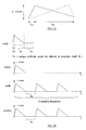
- Figure 2A schematically shows a triangular signal shape.
- Figure 2B schematically shows the parameterization of a stimulation sequence.
- FIG. 3 represents the schematic diagram of the acquisition system according to the invention.
- Figure 4 schematically shows the method of automatic gain adjustment.
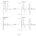
- Figure 5 schematically shows an EMG signal with stimulation artifact such that V EMG > V ARTEFACT .
- FIG. 6 schematically represents an EMG signal with stimulation artifact such that V EMG ⁇ V ARTEFACT .
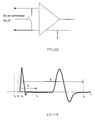
- Figure 7 shows the saturation of the amplifier due to excessive amplification of the stimulation artifact.
- Figure 8 shows the gain adjustment for EMG with stimulus artifact such as V ARTEFACT > V EMG .
- FIG. 9 shows the detailed block diagram of the acquisition chain.
- Figure 10A schematically shows the short-circuiting of the measuring electrodes.
- FIG. 10B schematically represents the EMG signal with stimulation artifact and short-circuiting of the measurement electrodes (synchronized acquisition at the end of the stimulation).
- FIG. 10C corresponds to the signal of FIG. 10B according to whether the system operates in synchronized mode or in level trigger mode.
- Figures 10D and 10E schematically show the triggering of the acquisition when exceeding a programmable voltage threshold independent of the stimulator.
- Figure 10F shows schematically the initiation of the acquisition at the request of the user (continuous acquisition).
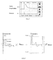
- Figure 11 schematically shows networking for a polytopic measurement.
- Figure 12 schematically shows an open loop acquisition.
- Figure 13 schematically shows a closed loop acquisition in the operating room.
- Figure 14 graphically represents an intensity search leading to "supra-maximal" excitation.
- Figure 15 shows the amplitude of the EMG signal as a function of the intensity of the electrical pulses.
- the system according to the invention schematically illustrated in FIG. 1 comprises a current source 1 enabling the excitation of a peripheral motor nerve and an acquisition chain 2 specially adapted to the measurement of evoked electromyographic potentials.
- a microcontroller 3 responsible for controlling and synchronizing the various modules of the system in real time and communicating with the computer 4 via a standardized interface 5 (RS-232, USB, RS-485 , etc.).
- the EMG signal and / or its parameters can be viewed on the display screen 6.
- the system can be controlled from any workstation with a standard communication port (RS-232, USB, RS-485, etc.), it is best to use a PDA (Personal Digital Assistant) 4 to achieve the user interface to ensure portability, autonomy and system security (see below, section 1.7).
- PDA Personal Digital Assistant
- the system is capable of establishing a wireless communication with a central computer 8 via a "Bluetooth" transmitter / receiver 7 integrated in the PDA.
- the EMG responses can be stored in the non-volatile memory 9 available on board the PDA (SD, CompactFlash, etc.).
- the system includes a stimulator that can work in two different modes.
- the stimulator delivers user-programmable stimulation sequences.
- the second it delivers the trains of pulses usually used in anesthesia.
- the electro-stimulator contains a series of rectangular pulse sequences prerecorded in its memory, such as ST (Single Twich), TOF (Train Of Four), TS (Tetanic Stimulation) or DBS (Double Burst Stimulation) .
- ST Single Twich
- TOF Train Of Four
- TS Tetanic Stimulation
- DBS Double Burst Stimulation
- Figure 2B illustrates the parameterization of a stimulation sequence.
- the pacemaker performs regular or on demand measurement of impedance between stimulation electrodes according to medical standards for the fault current.
- the acquisition chain 2 shown diagrammatically in FIG. 3, serves to amplify the signal coming from the measurement electrodes 21 in “V 1 " using a differential amplifier 22, to filter it through a filter 25 to extract the unwanted frequencies and perform the analog / digital conversion 26 of the signal "V 2 " and conditioned.
- the automatic gain control system 23 controlled 24 from the microcontroller amplifies the input signal 21 to best exploit the voltage range of the converter.
- Stimulation causes an annoying artifact for low amplitude EMG measurement.
- the EMG signals being of relatively low amplitude and collected in a rather noisy environment, they should be amplified as much as possible and as close as possible to the measurement site.
- Figure 5 shows that for EMGs of suitable amplitude (when the patient is not curarized), the amplitude of the muscular response is generally higher than the amplitude of the stimulation artifact.
- the maximum gain of the amplifier is therefore inversely proportional to the amplitude of the EMG signal.
- the maximum gain of the amplifier therefore no longer depends on the amplitude of the EMG signal but on the amplitude of the stimulation artifact, resulting in a bad signal-to-noise ratio (SNR or signal-to- noise ratio). Ratio) for small EMGs.
- Figure 8 shows the gain adjustment for EMG with stimulation artifact such as V ARTEFACT > V EMG .
- the connector 31 can be distinguished towards the measuring electrodes and the reference electrode.
- the system performs differential pre-amplification 33 to minimize common-mode noises sensed by the human body.
- the preamplified signal "V 0 " passes through a bandpass filter 34 to keep only the useful frequencies thereof (10 - 1000 Hz).
- the filtered signal "V F " may optionally be re-amplified to best fit within the input voltage range of the analog-to-digital converter 310 (see “Mode 2" below).
- Two separate modules 311 and 312 make it possible to independently adjust the gain of the preamplifier 33 and that of the amplifier 35 via certain output tabs 38 of the microcontroller 39.
- the signal is masked.
- This method involves shorting the acquisition electrodes for the duration of the stimulation artifact. It requires that the stimulator is coupled to the EMG acquisition chain and that it provides a synchronization signal.
- the gain of the preamplifier can be optimized immediately.
- the delay between the end of the stimulation and the opening of the relay bypassing the electrodes is made programmable by the user.
- the amplification is bi-staged with intermediate filtering of the stimulation artifact.
- the system operates in synchronized mode (with short-circuiting of the measuring electrodes) or in tripping mode (without short-circuiting of the measuring electrodes) and regardless of the relative amplitude of the EMG relative to the stimulus artefact, we can always amplify the signal optimally, that is, by keeping the relative quantization error constant.
- the system is designed to operate in three different modes and its architecture is adapted to keep the quantization error constant in each mode.
- the current source and the acquisition chain being driven by the same microcontroller, it is easy to synchronize the start of the acquisition on the end of the stimulation.
- An output from the microcontroller drives a relay that short-circuits the measurement electrodes during the first microseconds after the stimulation, which has the effect of masking the artifact of stimulation and allowing the increase of the amplifier gain without risk of saturation.
- the triggering of the acquisition takes place when a programmable voltage threshold (independent of the stimulator) is exceeded.
- a circular buffer of programmable size contains the samples preceding the trigger condition.
- the system performs two-stage amplification with intermediate filtering of the stimulation artifact.
- the triggering of the acquisition is done at the request of the user.
- the system according to the invention is designed to perform regularly or on demand the impedance measurement at the levels of the acquisition electrodes.
- the system has protection resistors to limit the fault current in the event that the supply voltage is accidentally applied to the measuring electrodes.
- the system performs the acquisition of the EMG, saves the response on the non-volatile memory, displays the curve in accordance with the selected display mode, performs a curve processing and displays the determining parameters.
- the user reads the registered EMGs.
- the system reads from the device's memory, displays the signal in the selected display mode, processes the curve and displays the relevant parameters.
- the user chooses the type of sequence to be delivered (ST, TOF, %), the intensity and the width of the pulses.
- the user draws his own waveforms using the appropriate graphical tools provided.
- the gain adjustment of the amplifiers is transparent to the user.
- the system automatically adjusts the gain as seen previously.
- the user can choose both the gain of the pre-amp and the gain of the amp of the acquisition chain. 1.5.5 Display Mode The various usual display modes of the state of the art are provided. 1.5.6 Choice of critical parameters
- the evaluation of muscle relaxation can be done in particular by measuring the ratio of the peak-to-peak amplitude of the curarized EMG on the peak-to-peak amplitude of a reference EMG (for example T 1 / T 0 , T 4 / T 1 , etc.) or by measuring the ratio of the surfaces (eg S 1 / S 0 , S 4 / S 1 , etc.) of the rectified EMGs (i.e. the potentials taken in ultimate value).
- a reference EMG for example T 1 / T 0 , T 4 / T 1 , etc.
- S 1 / S 0 , S 4 / S 1 , etc. the ratio of the surfaces of the rectified EMGs (i.e. the potentials taken in ultimate value).
- the software allows for complete analysis, both in real time and post-processing.
- the device is designed to work as a "slave" of a central computer via a wireless connection.
- the central computer is also able to establish communication with several EMG stimulation-response systems and interrogate them in turn ( Figure 11).
- the EMGs are saved on the PDA memory card.
- the apparatus of the invention is first of all useful to appreciate the effect of new molecules that appear on the market and whose effects must be estimated at different sites, on different muscles.
- the time constant and the inertia of the effects of the curative agents depend on the type of drug administered to the patient and, on the other hand, the Paralysis rate is not uniform in all parts of the body.
- the device described is also useful in a neurophysiological examination for assessing muscle tone. It is often necessary to appreciate the muscular tone of a given muscle or the relation of paralysis or recovery between two muscles. Indeed, during a curare injection, the patient's paralysis begins at the central level and ends with the peripheral muscles. Likewise, muscles like the diaphragm paralyze before the muscles at the extremities, like the adductor of the thumb. The process of decurarization is done in the same order.
- the apparatus of the invention also has the possibility of networking for a polytopic measurement (see FIG. 11).
- the device is designed to slave to a central computer via a wireless connection.
- the central computer is also able to establish communication with several EMG stimulation-response systems and interrogate them in turn.
- the apparatus operates completely autonomously and provides the display of the response curve as well as a numerical value representative of the state of muscle relaxation of the patient (T 4 / T 1 or T 1 / T 0 , etc.). .
- the user may choose to configure the PDA to schedule stimulation sequences to be delivered during surgery or to administer pacing sequences on demand.
- a command 41 is sent to the microcontroller which then provides real-time management.
- the stimulator delivers the selected pulse train 42 to a peripheral motor nerve and the acquisition system measures the response of the corresponding muscle.
- the PDA receives the burst of bytes 44 from the system and provides the processing of the response (saving the response, displaying the curve and evaluating the parameters estimating the degree of neuromuscular block).
- the PDA is able to communicate with a central computer via a wireless connection "Bluetooth” or a wired connection with galvanic isolation.
- the device can thus work as slave of the master computer and integrate into a closed control loop.
- the PDA After performing the measurement of the EMG, the PDA sends information (total curve or pre-processed response) to the central computer that controls the curare injection pumps.
- the PDA operates as a slave of a WorkStation.
- the central computer periodically interrogates the stimulation-response system to control the degree of neuromuscular blockage of the patient during the surgical procedure.
- the regulation loop is of closed type.
- the central computer also controls the injection of the curare pumps.
- EMG electromyography
- Stimulation of sufficient intensity will cause the reaction of all the muscle fibers and the response obtained will be maximal in amplitude.
- Figure 14 shows the EMG obtained as the intensity of the stimulation current source is gradually increased.
- the amplitude of the response signal increases with the intensity of the current pulses until saturation is reached. This saturation indicates that all the muscle fibers are actually excited and the response reaches a maximum amplitude.
- the weakening of the maximal response can be related to the state of relaxation of the muscle.
- the intensity of this stimulation will therefore be 20 to 25 percent higher than that for which we obtain a maximum response, hence the qualifier "supra-maximal".
- Figure 15 shows the amplitude of the EMG signal as a function of the intensity of the electrical pulses
- the threshold characterized by the saturation of the amplitude, depends strongly on one muscle to the other and even from one patient to another.
- Some devices such as the currently used TOF-Watch (in accelerometry) are programmed by default to 50 mA to ensure that they are above the saturation threshold and that the muscle is properly excited.
- the microcontroller can determine very precisely the intensity of the electrical pulses leading to a super-maximum excitation.
- EMG muscle relaxation rate evaluation
- the apparatus of the invention can be advantageously used in intensive care unit (ICU); the reduction in the size of the device (portability) allows measurements of evoked potentials directly in the patient's bed, during the postoperative period in the recovery room for example.
- ICU intensive care unit
- the apparatus of the invention may be useful in the home of the patient who is recovering muscle.
- patients can have a portable electro-stimulator they use at home (example: belt to build muscles).
Landscapes
- Health & Medical Sciences (AREA)
- Life Sciences & Earth Sciences (AREA)
- Engineering & Computer Science (AREA)
- General Health & Medical Sciences (AREA)
- Veterinary Medicine (AREA)
- Biophysics (AREA)
- Biomedical Technology (AREA)
- Heart & Thoracic Surgery (AREA)
- Medical Informatics (AREA)
- Molecular Biology (AREA)
- Surgery (AREA)
- Animal Behavior & Ethology (AREA)
- Physics & Mathematics (AREA)
- Public Health (AREA)
- Pathology (AREA)
- Signal Processing (AREA)
- Neurology (AREA)
- Physiology (AREA)
- Neurosurgery (AREA)
- Artificial Intelligence (AREA)
- Computer Vision & Pattern Recognition (AREA)
- Psychiatry (AREA)
- Theoretical Computer Science (AREA)
- Anesthesiology (AREA)
- Measurement And Recording Of Electrical Phenomena And Electrical Characteristics Of The Living Body (AREA)
Priority Applications (6)
| Application Number | Priority Date | Filing Date | Title |
|---|---|---|---|
| EP04447248A EP1656883A1 (de) | 2004-11-10 | 2004-11-10 | Tragbares Gerät zur Messung eines EMG-Signals |
| CA002586729A CA2586729A1 (fr) | 2004-11-10 | 2005-11-10 | Appareil et procede de mesure d'un signal emg |
| JP2007539428A JP2008519609A (ja) | 2004-11-10 | 2005-11-10 | Emg信号を測定するための装置および方法 |
| US11/667,439 US20070270918A1 (en) | 2004-11-10 | 2005-11-10 | Appliance and Method for Measuring an Emg Signal |
| EP05810936A EP1814452A1 (de) | 2004-11-10 | 2005-11-10 | Gerät und verfahren zur messung eines emg-signals |
| PCT/BE2005/000162 WO2006050586A1 (fr) | 2004-11-10 | 2005-11-10 | Appareil et procede de mesure d'un signal emg |
Applications Claiming Priority (1)
| Application Number | Priority Date | Filing Date | Title |
|---|---|---|---|
| EP04447248A EP1656883A1 (de) | 2004-11-10 | 2004-11-10 | Tragbares Gerät zur Messung eines EMG-Signals |
Publications (1)
| Publication Number | Publication Date |
|---|---|
| EP1656883A1 true EP1656883A1 (de) | 2006-05-17 |
Family
ID=34933110
Family Applications (2)
| Application Number | Title | Priority Date | Filing Date |
|---|---|---|---|
| EP04447248A Withdrawn EP1656883A1 (de) | 2004-11-10 | 2004-11-10 | Tragbares Gerät zur Messung eines EMG-Signals |
| EP05810936A Withdrawn EP1814452A1 (de) | 2004-11-10 | 2005-11-10 | Gerät und verfahren zur messung eines emg-signals |
Family Applications After (1)
| Application Number | Title | Priority Date | Filing Date |
|---|---|---|---|
| EP05810936A Withdrawn EP1814452A1 (de) | 2004-11-10 | 2005-11-10 | Gerät und verfahren zur messung eines emg-signals |
Country Status (5)
| Country | Link |
|---|---|
| US (1) | US20070270918A1 (de) |
| EP (2) | EP1656883A1 (de) |
| JP (1) | JP2008519609A (de) |
| CA (1) | CA2586729A1 (de) |
| WO (1) | WO2006050586A1 (de) |
Cited By (6)
| Publication number | Priority date | Publication date | Assignee | Title |
|---|---|---|---|---|
| US11026627B2 (en) | 2013-03-15 | 2021-06-08 | Cadwell Laboratories, Inc. | Surgical instruments for determining a location of a nerve during a procedure |
| US11177610B2 (en) | 2017-01-23 | 2021-11-16 | Cadwell Laboratories, ino. | Neuromonitoring connection system |
| US11253182B2 (en) | 2018-05-04 | 2022-02-22 | Cadwell Laboratories, Inc. | Apparatus and method for polyphasic multi-output constant-current and constant-voltage neurophysiological stimulation |
| US11443649B2 (en) | 2018-06-29 | 2022-09-13 | Cadwell Laboratories, Inc. | Neurophysiological monitoring training simulator |
| US11510603B2 (en) | 2014-11-26 | 2022-11-29 | Safeop Surgical, Inc. | Device and means of assessing neuromuscular junction status with higher fidelity |
| US11992339B2 (en) | 2018-05-04 | 2024-05-28 | Cadwell Laboratories, Inc. | Systems and methods for dynamic neurophysiological stimulation |
Families Citing this family (31)
| Publication number | Priority date | Publication date | Assignee | Title |
|---|---|---|---|---|
| US8073503B2 (en) * | 2007-11-06 | 2011-12-06 | Qualcomm Incorporated | Personal health modules supported by portable communication devices |
| TWI406149B (zh) * | 2009-08-24 | 2013-08-21 | Univ Nat Cheng Kung | EMG signal processing chip |
| FI20106337A0 (fi) * | 2010-12-17 | 2010-12-17 | Polar Electro Oy | Häiriönvaimennuspiiri biometrisiä mittauksia varten |
| FI20106338A0 (fi) * | 2010-12-17 | 2010-12-17 | Polar Electro Oy | Häiriön vaimennus biometrisissä mittauksissa |
| JP5624669B2 (ja) * | 2011-02-28 | 2014-11-12 | 日本光電工業株式会社 | 生体電気信号計測装置 |
| KR101941171B1 (ko) * | 2011-09-26 | 2019-01-23 | 삼성전자주식회사 | 생체신호를 측정하는 장치 및 방법 |
| CN103006199B (zh) * | 2011-09-26 | 2016-09-28 | 三星电子株式会社 | 用于测量生物信号的设备和方法 |
| TWI459929B (zh) * | 2011-10-17 | 2014-11-11 | Jia Jin Chen | 即時自主肌力與電刺激誘發肌力量測電路 |
| JP2015506246A (ja) * | 2012-01-27 | 2015-03-02 | ティー4・アナリティクス・リミテッド・ライアビリティ・カンパニー | 運動神経の刺激に対する筋肉の電気的活動を評価する方法およびシステム |
| JP2015506245A (ja) * | 2012-01-27 | 2015-03-02 | ティー4・アナリティクス・リミテッド・ライアビリティ・カンパニー | 麻酔モニタリングシステムおよび麻酔をモニタリングする方法 |
| EP2953568A4 (de) * | 2013-02-06 | 2016-11-09 | Ronny Kafiluddi | Identifikation peripherer nerven |
| JP2016508400A (ja) | 2013-02-15 | 2016-03-22 | アカシア・デザインズ・ベスローテン・フェンノートシャップ | 医療用モニタリング・システムに使用する電極システム |
| JP6418750B2 (ja) * | 2014-02-07 | 2018-11-07 | オージー技研株式会社 | 筋力増強システム |
| JP2015164510A (ja) * | 2014-02-07 | 2015-09-17 | パナソニックIpマネジメント株式会社 | 筋力サポータおよび筋力サポート方法 |
| US10398369B2 (en) | 2014-08-08 | 2019-09-03 | Medtronic Xomed, Inc. | Wireless stimulation probe device for wireless nerve integrity monitoring systems |
| US12582344B2 (en) | 2014-08-08 | 2026-03-24 | Medtronic Xomed, Inc. | Wireless stimulation probe device for wireless nerve integrity monitoring systems |
| US12201436B2 (en) | 2014-08-08 | 2025-01-21 | Medtronic Xomed, Inc. | Wireless nerve integrity monitoring systems and devices |
| WO2016026100A1 (zh) * | 2014-08-20 | 2016-02-25 | 华为技术有限公司 | 一种肌电信号采集装置 |
| US10039915B2 (en) | 2015-04-03 | 2018-08-07 | Medtronic Xomed, Inc. | System and method for omni-directional bipolar stimulation of nerve tissue of a patient via a surgical tool |
| US11980465B2 (en) | 2015-04-03 | 2024-05-14 | Medtronic Xomed, Inc. | System and method for omni-directional bipolar stimulation of nerve tissue of a patient via a bipolar stimulation probe |
| US10849517B2 (en) | 2016-09-19 | 2020-12-01 | Medtronic Xomed, Inc. | Remote control module for instruments |
| US20180263521A1 (en) * | 2017-03-17 | 2018-09-20 | Tribe Private Company | System and method for emg signal acquisition |
| SE1850295A1 (en) * | 2018-03-16 | 2019-09-17 | Senzime Ab Publ | Anesthetizing monitoring system, unit and method therefore |
| SE1850294A1 (en) | 2018-03-16 | 2019-09-17 | Senzime Ab Publ | Anesthetizing monitoring system, unit and method therefore |
| US11510628B2 (en) * | 2018-09-28 | 2022-11-29 | Case Western Reserve University | Elimination of artifacts due to delivery of an electrical signal from neural recordings |
| CN112773380B (zh) * | 2019-11-07 | 2023-09-22 | 深圳市理邦精密仪器股份有限公司 | 一种肌电信号的处理方法、处理设备以及存储介质 |
| US12543998B2 (en) | 2020-01-24 | 2026-02-10 | Medtronic Xomed, Inc. | Conductive instrument |
| WO2022217358A1 (en) * | 2021-04-13 | 2022-10-20 | University Health Network | Point-of-care prediction of muscle responsiveness to therapy during neurorehabilitation |
| CN113933351B (zh) * | 2021-09-30 | 2023-12-22 | 深圳市中金岭南有色金属股份有限公司凡口铅锌矿 | 矿浆pH值检测方法、装置及计算机可读存储介质 |
| CZ2022110A3 (cs) * | 2022-03-09 | 2023-09-27 | Deymed Diagnostic S.R.O. | Zařízení pro elektrickou stimulaci organismu snižující stimulační artefakt |
| AU2024329098A1 (en) * | 2023-08-21 | 2026-02-26 | Saluda Medical Pty Limited | Improved measurement of evoked responses to neural stimulation |
Citations (11)
| Publication number | Priority date | Publication date | Assignee | Title |
|---|---|---|---|---|
| US4291705A (en) | 1979-09-10 | 1981-09-29 | The Regents Of The University Of California | Neuromuscular block monitor |
| GB2113846B (en) | 1982-01-29 | 1985-08-29 | Instrumentarium Oy | Measuring depth of anaesthesia |
| US4595018A (en) | 1983-06-10 | 1986-06-17 | Instrumentarium Corp. | Method of further developing the measuring of a neuro-muscular junction |
| KR900004899A (ko) | 1988-09-26 | 1990-04-13 | 노만 에드워드 루이스 | 실리콘 실란트의 모듈러스 조절 방법 |
| US5010888A (en) * | 1988-03-25 | 1991-04-30 | Arzco Medical Electronics, Inc. | Method and apparatus for detection of posterior ischemia |
| US5300096A (en) | 1992-06-03 | 1994-04-05 | Hall H Eugene | Electromyographic treatment device |
| WO1999041982A1 (de) | 1998-02-20 | 1999-08-26 | Bayer Aktiengesellschaft | Perlpolymerisat-formulierungen |
| US6083156A (en) * | 1998-11-16 | 2000-07-04 | Ronald S. Lisiecki | Portable integrated physiological monitoring system |
| US6195585B1 (en) * | 1998-06-26 | 2001-02-27 | Advanced Bionics Corporation | Remote monitoring of implantable cochlear stimulator |
| US6224549B1 (en) | 1999-04-20 | 2001-05-01 | Nicolet Biomedical, Inc. | Medical signal monitoring and display |
| WO2002053012A2 (en) | 2001-01-04 | 2002-07-11 | Indian Institute Of Technology | A CLINICAL ELECTRODIAGNOSTIC DIGITAL INSTRUMENT FOR ELECTROMYOGRAPHY (emg) AND/OR NERVE CONDUCTION MEASUREMENT |
Family Cites Families (14)
| Publication number | Priority date | Publication date | Assignee | Title |
|---|---|---|---|---|
| JPH0448203Y2 (de) * | 1987-11-30 | 1992-11-13 | ||
| KR900004899B1 (ko) | 1988-05-18 | 1990-07-09 | 김성환 | 근전도(emg)신호 처리 시스템 electromyograph signal processing system |
| US5131401A (en) * | 1990-09-10 | 1992-07-21 | Axon Medical Inc. | Method and apparatus for monitoring neuromuscular blockage |
| US5549656A (en) * | 1993-08-16 | 1996-08-27 | Med Serve Group, Inc. | Combination neuromuscular stimulator and electromyograph system |
| US5892472A (en) * | 1997-06-30 | 1999-04-06 | Harris Corporation | Processor controlled analog-to-digital converter circuit |
| US5851191A (en) * | 1997-07-01 | 1998-12-22 | Neurometrix, Inc. | Apparatus and methods for assessment of neuromuscular function |
| US6146335A (en) * | 1997-07-01 | 2000-11-14 | Neurometrix, Inc. | Apparatus for methods for the assessment of neuromuscular function of the lower extremity |
| AU2762899A (en) | 1998-02-17 | 1999-08-30 | Southern Research Institute | Patient data acquisition unit and data support system |
| US6334068B1 (en) * | 1999-09-14 | 2001-12-25 | Medtronic Xomed, Inc. | Intraoperative neuroelectrophysiological monitor |
| ATE394137T1 (de) * | 1999-10-29 | 2008-05-15 | Compex Medical Sa | Neuromuskuläres stimulationsgerät mit aufnahme der muskelreaktion auf den elektrischen stimulationsimpuls |
| US6201489B1 (en) * | 2000-03-21 | 2001-03-13 | International Business Machines Corporation | Method and circuit for temporal cancellation of DC offset |
| US6414164B1 (en) * | 2000-07-12 | 2002-07-02 | International Business Machines Corporation | Synthesis of soluble derivatives of sexithiophene and their use as the semiconducting channels in thin-film filed-effect transistors |
| US7016731B2 (en) * | 2002-06-28 | 2006-03-21 | Harbinger Medical, Inc. | Sensing artifact reduction for cardiac diagnostic system |
| KR100513169B1 (ko) * | 2002-07-22 | 2005-09-07 | 모승기 | 전기 측정 및 자극 장치 |
-
2004
- 2004-11-10 EP EP04447248A patent/EP1656883A1/de not_active Withdrawn
-
2005
- 2005-11-10 CA CA002586729A patent/CA2586729A1/fr not_active Abandoned
- 2005-11-10 EP EP05810936A patent/EP1814452A1/de not_active Withdrawn
- 2005-11-10 WO PCT/BE2005/000162 patent/WO2006050586A1/fr not_active Ceased
- 2005-11-10 JP JP2007539428A patent/JP2008519609A/ja active Pending
- 2005-11-10 US US11/667,439 patent/US20070270918A1/en not_active Abandoned
Patent Citations (11)
| Publication number | Priority date | Publication date | Assignee | Title |
|---|---|---|---|---|
| US4291705A (en) | 1979-09-10 | 1981-09-29 | The Regents Of The University Of California | Neuromuscular block monitor |
| GB2113846B (en) | 1982-01-29 | 1985-08-29 | Instrumentarium Oy | Measuring depth of anaesthesia |
| US4595018A (en) | 1983-06-10 | 1986-06-17 | Instrumentarium Corp. | Method of further developing the measuring of a neuro-muscular junction |
| US5010888A (en) * | 1988-03-25 | 1991-04-30 | Arzco Medical Electronics, Inc. | Method and apparatus for detection of posterior ischemia |
| KR900004899A (ko) | 1988-09-26 | 1990-04-13 | 노만 에드워드 루이스 | 실리콘 실란트의 모듈러스 조절 방법 |
| US5300096A (en) | 1992-06-03 | 1994-04-05 | Hall H Eugene | Electromyographic treatment device |
| WO1999041982A1 (de) | 1998-02-20 | 1999-08-26 | Bayer Aktiengesellschaft | Perlpolymerisat-formulierungen |
| US6195585B1 (en) * | 1998-06-26 | 2001-02-27 | Advanced Bionics Corporation | Remote monitoring of implantable cochlear stimulator |
| US6083156A (en) * | 1998-11-16 | 2000-07-04 | Ronald S. Lisiecki | Portable integrated physiological monitoring system |
| US6224549B1 (en) | 1999-04-20 | 2001-05-01 | Nicolet Biomedical, Inc. | Medical signal monitoring and display |
| WO2002053012A2 (en) | 2001-01-04 | 2002-07-11 | Indian Institute Of Technology | A CLINICAL ELECTRODIAGNOSTIC DIGITAL INSTRUMENT FOR ELECTROMYOGRAPHY (emg) AND/OR NERVE CONDUCTION MEASUREMENT |
Non-Patent Citations (1)
| Title |
|---|
| MILLARD R E ET AL: "A gated differential amplifier for recording physiological responses to electrical stimulation", JOURNAL OF NEUROSCIENCE METHODS NETHERLANDS, vol. 44, no. 1, 1992, pages 81 - 84, XP002322320, ISSN: 0165-0270 * |
Cited By (10)
| Publication number | Priority date | Publication date | Assignee | Title |
|---|---|---|---|---|
| US11026627B2 (en) | 2013-03-15 | 2021-06-08 | Cadwell Laboratories, Inc. | Surgical instruments for determining a location of a nerve during a procedure |
| US12178606B2 (en) | 2013-03-15 | 2024-12-31 | Cadwell Laboratories, Inc. | Neuromonitoring systems and methods |
| US11510603B2 (en) | 2014-11-26 | 2022-11-29 | Safeop Surgical, Inc. | Device and means of assessing neuromuscular junction status with higher fidelity |
| US11177610B2 (en) | 2017-01-23 | 2021-11-16 | Cadwell Laboratories, ino. | Neuromonitoring connection system |
| US11949188B2 (en) | 2017-01-23 | 2024-04-02 | Cadwell Laboratories, Inc. | Methods for concurrently forming multiple electrical connections in a neuro-monitoring system |
| US11253182B2 (en) | 2018-05-04 | 2022-02-22 | Cadwell Laboratories, Inc. | Apparatus and method for polyphasic multi-output constant-current and constant-voltage neurophysiological stimulation |
| US11992339B2 (en) | 2018-05-04 | 2024-05-28 | Cadwell Laboratories, Inc. | Systems and methods for dynamic neurophysiological stimulation |
| US11998338B2 (en) | 2018-05-04 | 2024-06-04 | Cadwell Laboratories, Inc. | Systems and methods for dynamically switching output port cathode and anode designations |
| US11443649B2 (en) | 2018-06-29 | 2022-09-13 | Cadwell Laboratories, Inc. | Neurophysiological monitoring training simulator |
| US11978360B2 (en) | 2018-06-29 | 2024-05-07 | Cadwell Laboratories, Inc. | Systems and methods for neurophysiological simulation |
Also Published As
| Publication number | Publication date |
|---|---|
| US20070270918A1 (en) | 2007-11-22 |
| CA2586729A1 (fr) | 2006-05-18 |
| JP2008519609A (ja) | 2008-06-12 |
| EP1814452A1 (de) | 2007-08-08 |
| WO2006050586A1 (fr) | 2006-05-18 |
Similar Documents
| Publication | Publication Date | Title |
|---|---|---|
| EP1656883A1 (de) | Tragbares Gerät zur Messung eines EMG-Signals | |
| US11826170B2 (en) | Artificial intelligence models for wireless patch data acquisition for gastrointestinal electrodiagnostics | |
| Guo et al. | Development of a multi-channel compact-size wireless hybrid sEMG/NIRS sensor system for prosthetic manipulation | |
| Matthews et al. | A wearable physiological sensor suite for unobtrusive monitoring of physiological and cognitive state | |
| EP2615972B1 (de) | Vorrichtung zur automatisierten messung der leitungsgeschwindigkeit und der amplitude der suralnerven | |
| EP0892618B1 (de) | Vorrichtung zur bestimmung der narkose-tiefe | |
| EP2327360B1 (de) | Verfahren und Vorrichtung zur Verbesserung des Signal-Rauschverhältnisses von EKG-Signalen zur Erleichterung der Herzschlagerkennung | |
| WO2006132958A2 (en) | Neurophysiological wireless bio-sensor | |
| JP2001509721A (ja) | 神経筋機能のアセスメントのための装置および方法 | |
| WO2009065006A2 (en) | Determination of biosensor contact quality | |
| CN105982642A (zh) | 一种基于体震信号的睡眠检测方法及检测系统 | |
| CN208709857U (zh) | 一种呼吸暂停检测系统 | |
| KR20210117230A (ko) | 패치형전극을 이용한 수면상태 판단장치 | |
| US9968301B2 (en) | Body-driven pseudorandom signal injection for biomedical acquisition channel calibration | |
| Estepp et al. | Validation of a dry electrode system for EEG | |
| WO2009150765A1 (ja) | 睡眠状態モニタリング装置、モニタリングシステムおよびコンピュータプログラム | |
| CN113951905A (zh) | 一种用于日常动态监测的多通道胃电采集系统 | |
| Affanni et al. | Wearable instrument for skin potential response analysis in AAL applications | |
| Romero et al. | Motion artifact reduction in ambulatory ECG monitoring: an integrated system approach | |
| US20090118597A1 (en) | Neural Signal Processing | |
| WO2016185931A1 (ja) | 生体情報測定装置 | |
| CA2580758A1 (fr) | Procede de traitement d'une serie rr et son application a l'analyse de la variabilite du rythme cardiaque, et en particulier a l'evaluation de la douleur ou du stress chez un etrevivant | |
| CN110680339A (zh) | 一种低负荷多维度智能睡眠监护筛查方法 | |
| WO1994016610A2 (fr) | Dispositif de determination d'informations physiologiques, et utilisation correspondante | |
| US9486154B2 (en) | Device and method for recording physiological signal |
Legal Events
| Date | Code | Title | Description |
|---|---|---|---|
| PUAI | Public reference made under article 153(3) epc to a published international application that has entered the european phase |
Free format text: ORIGINAL CODE: 0009012 |
|
| AK | Designated contracting states |
Kind code of ref document: A1 Designated state(s): AT BE BG CH CY CZ DE DK EE ES FI FR GB GR HU IE IS IT LI LU MC NL PL PT RO SE SI SK TR |
|
| AX | Request for extension of the european patent |
Extension state: AL HR LT LV MK YU |
|
| AKX | Designation fees paid | ||
| REG | Reference to a national code |
Ref country code: DE Ref legal event code: 8566 |
|
| STAA | Information on the status of an ep patent application or granted ep patent |
Free format text: STATUS: THE APPLICATION IS DEEMED TO BE WITHDRAWN |
|
| 18D | Application deemed to be withdrawn |
Effective date: 20061118 |