EP1656884A2 - Methode zur Beurteilung der Haut und der allgemeinen Gesundheit einer Person - Google Patents
Methode zur Beurteilung der Haut und der allgemeinen Gesundheit einer Person Download PDFInfo
- Publication number
- EP1656884A2 EP1656884A2 EP05257026A EP05257026A EP1656884A2 EP 1656884 A2 EP1656884 A2 EP 1656884A2 EP 05257026 A EP05257026 A EP 05257026A EP 05257026 A EP05257026 A EP 05257026A EP 1656884 A2 EP1656884 A2 EP 1656884A2
- Authority
- EP
- European Patent Office
- Prior art keywords
- skin
- area
- intensity
- fluorescent emission
- exposure radiation
- Prior art date
- Legal status (The legal status is an assumption and is not a legal conclusion. Google has not performed a legal analysis and makes no representation as to the accuracy of the status listed.)
- Withdrawn
Links
Images


Classifications
-
- G—PHYSICS
- G01—MEASURING; TESTING
- G01N—INVESTIGATING OR ANALYSING MATERIALS BY DETERMINING THEIR CHEMICAL OR PHYSICAL PROPERTIES
- G01N21/00—Investigating or analysing materials by the use of optical means, i.e. using sub-millimetre waves, infrared, visible or ultraviolet light
- G01N21/62—Systems in which the material investigated is excited whereby it emits light or causes a change in wavelength of the incident light
- G01N21/63—Systems in which the material investigated is excited whereby it emits light or causes a change in wavelength of the incident light optically excited
- G01N21/64—Fluorescence; Phosphorescence
- G01N21/6486—Measuring fluorescence of biological material, e.g. DNA, RNA, cells
-
- A—HUMAN NECESSITIES
- A61—MEDICAL OR VETERINARY SCIENCE; HYGIENE
- A61B—DIAGNOSIS; SURGERY; IDENTIFICATION
- A61B5/00—Measuring for diagnostic purposes; Identification of persons
- A61B5/0059—Measuring for diagnostic purposes; Identification of persons using light, e.g. diagnosis by transillumination, diascopy, fluorescence
- A61B5/0071—Measuring for diagnostic purposes; Identification of persons using light, e.g. diagnosis by transillumination, diascopy, fluorescence by measuring fluorescence emission
-
- A—HUMAN NECESSITIES
- A61—MEDICAL OR VETERINARY SCIENCE; HYGIENE
- A61B—DIAGNOSIS; SURGERY; IDENTIFICATION
- A61B5/00—Measuring for diagnostic purposes; Identification of persons
- A61B5/44—Detecting, measuring or recording for evaluating the integumentary system, e.g. skin, hair or nails
- A61B5/441—Skin evaluation, e.g. for skin disorder diagnosis
- A61B5/442—Evaluating skin mechanical properties, e.g. elasticity, hardness, texture, wrinkle assessment
-
- G—PHYSICS
- G01—MEASURING; TESTING
- G01N—INVESTIGATING OR ANALYSING MATERIALS BY DETERMINING THEIR CHEMICAL OR PHYSICAL PROPERTIES
- G01N21/00—Investigating or analysing materials by the use of optical means, i.e. using sub-millimetre waves, infrared, visible or ultraviolet light
- G01N21/62—Systems in which the material investigated is excited whereby it emits light or causes a change in wavelength of the incident light
- G01N21/63—Systems in which the material investigated is excited whereby it emits light or causes a change in wavelength of the incident light optically excited
- G01N21/64—Fluorescence; Phosphorescence
-
- G—PHYSICS
- G01—MEASURING; TESTING
- G01N—INVESTIGATING OR ANALYSING MATERIALS BY DETERMINING THEIR CHEMICAL OR PHYSICAL PROPERTIES
- G01N33/00—Investigating or analysing materials by specific methods not covered by groups G01N1/00 - G01N31/00
- G01N33/48—Biological material, e.g. blood, urine; Haemocytometers
- G01N33/50—Chemical analysis of biological material, e.g. blood, urine; Testing involving biospecific ligand binding methods; Immunological testing
- G01N33/5005—Chemical analysis of biological material, e.g. blood, urine; Testing involving biospecific ligand binding methods; Immunological testing involving human or animal cells
- G01N33/5091—Chemical analysis of biological material, e.g. blood, urine; Testing involving biospecific ligand binding methods; Immunological testing involving human or animal cells for testing the pathological state of an organism
Definitions
- the present invention relates to a method of assessing skin and the overall health of an individual using fluorescence.
- the major fluorescence bands that have been detected by in vivo fluorescence spectroscopy include: a) a band assigned to tryptophan (maximum at 295 nm excitation, 345 nm emission), b) a band assigned to pepsin digestible collagen cross-links (335 nm excitation, 390 nm emission), c) a band assigned to collagenase digestible collagen cross-links (370 nm excitation, 460 nm emission), and d) a band most likely due to elastin and collagen cross-links (390-420 nm broad band excitation, 500 nm emission). See Gillies et al. J. Invest. Dermatol, 115(4):704-707, 2000.
- the fluorescence signal assigned to tryptophan moieties measured in situ was found to increase when epidermal proliferation increases. See Kollias et al., J. Invest. Dermatol, 111(5):776-780,1998 and Zhang et al., Lasers Surg. Med. 20(3):319-331, 1997. This was verified by inducing epidermal repair after mechanical insult, e.g. tape stripping. See Brancaleon et al., J. Invest. Dermatol. 113(6):977-982, 1999. Furthermore, ⁇ -hydroxy-acid-induced increases of cellular turnover in human epidermis caused the 295 nm excitation band to increase in a dose dependent manner. See Doukas et al., Photochem.
- Non-enzymatic glycosilation of proteins occurs spontaneously with aging (See Monnier et al., Clin Endocrinol Metab 11(2):431-452, 1982; Njoroge et al., J. Biol. Chem. 263(22):10646-10652, 1988; Sell et al., J. Biol Chem 264(36):21597-21602, 1989; and Shaklai et al., J. Biol Chem 259(6):3812-3817, 1984) resulting in increased protein absorbance and fluorescence (Maillard reaction).
- the glucose-protein adduct rearranges and dehydrates to form brown and fluorescent pigments, which may form cross-links resulting in decreased protein solubility and altered mechanical properties.
- cross-links are evident in long-lived proteins, such as elastin and collagen.
- the accumulation of fluorescing cross-links in collagen has been used as a marker for the observed accelerated rate of aging in diabetes. See Monnier et al., Clin. Endocrinol. Metab 11(2):431-452, 1982.
- SKH mice the magnitude of the pepsin digestible collagen cross-link fluorescence maximum increases with chronological aging, whereas the increase in the magnitude of the collagenase digestible collagen cross-link and the elastin-associated fluorescence maxima is modest.
- Applicants have surprisingly found that skin native autofluorescence is a tool to evaluate skin health and the effects of aging (e.g., chronological aging as well as photoaging) on skin health.
- the present invention features a method of determining skin health of an area of skin by (i) exposing the area of skin to a first exposure radiation to induce the area of skin to emit a first fluorescent emission, wherein the first exposure radiation comprises primarily of wavelengths of from about 290 nm to about 300 nm; (ii) measuring the intensity of the first fluorescent emission having a wavelength of from about 320 to about 350; (iii) exposing the area of skin to a second exposure radiation to induce the area of skin to emit a second fluorescent emission, wherein the second exposure radiation comprises primarily of wavelengths of from about 330-420 nm; (iv) measuring the intensity of the second fluorescent emission having a wavelength of from about 380-470; (v) calculating a ratio of the intensity measured in step (ii) to the intensity measured in step (iv); and (vi) comparing the ratio to a control ratio.
- the present invention features a method of determining the effect of a treatment to the skin of a subject by: (i) exposing a first area of skin to a first exposure radiation to induce the area of skin to emit a first fluorescent emission, wherein the first exposure radiation comprises primarily of wavelengths of from about 290 nm to about 300 nm and wherein the first area of skin was exposed to the composition; (ii) measuring the intensity of the first fluorescent emission having a wavelength of from about 320 to about 350;(iii) exposing the first area of skin to a second exposure radiation to induce the area of skin to emit a second fluorescent emission, wherein the second exposure radiation comprises primarily of wavelengths of from about 330-420 nm; (iv) measuring the intensity of the second fluorescent emission having a wavelength of from about 380-470; (v) calculating a ratio of the intensity measured in step (ii) to the intensity measured in step (iv);(iv) repeating steps (i) to (v) for a second area of skin, where
- the present invention relates to a method of assessing the overall health of an individual including creating a standard curve for a plurality of healthy individuals by I) exposing an area of skin of each healthy individual to a first exposure radiation to induce said area of skin to emit a first fluorescent emission, wherein said first exposure radiation comprises primarily of wavelengths of from about 290 nm to about 300 nm; II) measuring the intensity of said first fluorescent emission having a wavelength of from about 320 nm to about 350 nm; (III)exposing said area of skin to a second exposure radiation to induce said area of skin to emit a second fluorescent emission, wherein said second exposure radiation comprises primarily of wavelengths of from about 330 nm to about 420 nm; IV) measuring the intensity of said second fluorescent emission having a wavelength of from about 380 nm to about 470 nm; V) calculating a ratio of said intensity measured in step (II) to said intensity measured in step (IV); plotting a standard curve for age of individual versus the ratio of step
- the average fluorescence value by age can be determined by following the same steps described above.
- the fluorescence value of an individual whose overall health is in question may then be compared to the average fluorescence value for that age. If the fluorescence value of the individual is below the average fluorescence value for the age, it is an indication that there may be a health problem, such as diabetes.
- the area(s) of skin are exposed to at least two exposure radiations (e.g., from UV radiation sources such as xenon arc lamps or mercury lamps).
- the first exposure radiation includes primarily wavelengths of from about 290 nm to about 300 nm and the second exposure radiation includes primarily wavelengths of from about 330-420 nm. What is meant by "primarily" is at least half of the wavelengths of the exposure radiation.
- the first exposure radiation includes primarily wavelengths of about 295 nm and the second exposure radiation includes primarily wavelengths of from about 390 to about 410 nm.
- the exposure radiations are directed to the skin in order emit a fluorescent emission and to measure the intensity of such emission (e.g., a specific wavelength or wavelength range).
- the method includes measuring the intensity of the first fluorescent emission having a wavelength of from about 330 nm to about 350 nm (e.g., about 340 nm) and measuring the intensity of the second fluorescent emission having a wavelength of from about 380 nm to about 470 nm (e.g., 440 nm).
- control ratio is an established standard ratio (e.g., an established norm for the subject's age, sex, and/or race) or a ratio obtained from the subject (e.g., previously obtained from the same area of skin or obtained from another area skin such as an area of skin not readily exposed to UV radiation such as the underarm or buttocks).
- the method thus, is able to determine the skin health of the subject (e.g., by comparing the ratio to the control ratio).
- the difference of the ratio value between exposed areas of skin and protected areas of skin has been found to generally decline with age. This difference is believed to be indicative of the ability of skin to react to external stimuli by repairing itself.
- the method is used to determine the effect of effect of a treatment to the skin of a subject.
- treatments include, but are not limited to, cosmetic and pharmaceutical treatments (e.g, topical, parenteral, or oral), laser treatment, or abrasive treatment (e.g., microderm abrasion).
- the treatment is a topical composition, such as a topical lotion or cream containing an anti-aging agent such as a retinoid (e.g., retinoic acid or retinol).
- In vivo fluorescence spectroscopy can be performed for example using a fiber optic probe attached to a spectrofluorimeter (e.g. the model SkinSkan (JY Horiba, Edison, NJ)).
- the method requires: a) a UV radiation source (e.g., a Xenon arc lamp or a mercury lamp), b) a method of selecting the radiation wavelength (e.g. a monochromator, a prism, or a grating), c) a method of delivery of the radiation to the tissue (e.g. a fiber bundle), d) a method of collection of the emitted radiation from the tissue (e.g. a fiber bundle), e) a method of selecting the emitted radiation wavelength (e.g.
- a UV radiation source e.g., a Xenon arc lamp or a mercury lamp
- a method of selecting the radiation wavelength e.g. a monochromator, a prism, or a grating
- a monochromator e.g. a prism, or a grating
- a method of detecting the emitted radiation e.g. a photomultiplier, a single photodiode, a photodiode array, or a CCD array. See, e.g., Stamatas GN, et al., J Invest Dermatol 118(2):295-302, 2002.
- Measurements were preformed by placing the fiber optic probe in contact with the skin site of interest. Before each set of measurements, the instrument was spectrally calibrated for excitation and emission in the region 250-650 nm. The chromatic resolution of the spectrofluorimeter was ⁇ 1 nm.
- excitation spectra was the preferred method of measuring in vivo skin fluorescence. The reason for this choice over acquisition of emission spectra was that excitation spectra were similar to absorption spectra and bands tend to be narrower than in emission acquisition, both of which help in the identification of the individual fluorophores in a complex spectrum.
- excitation spectra that were used in this study were the following: a) excitation scanned from 240 nm to 320 nm with emission set at 340 nm (tryptophan excitation maximum at 295 nm), b) excitation scanned from 240 nm to 380 nm with emission set at 390 nm (pepsin digestible collagen cross-link excitation maximum at 335 nm), c) excitation scanned from 240 nm to 410 nm with emission at 420 nm (collagenase digestible collagen cross-link excitation maximum at 360 nm), d) excitation scanned from 260 nm to 490 nm with emission set at 500 nm (elastin cross-links - desmosine and isodesmosine - excitation maximum at about 390 nm).
- fluorescence intensity was normalized with the diffuse reflectance signal of the same skin site at the corresponding wavelength. See, e.g., Stamatas GN, et al., J Invest Dermatol 118(2):295-302, 2002.
- a diffuse reflectance spectrum can be acquired by synchronizing the excitation and emission monochromators to select the same wavelength, scanning the range from 240 nm to 500 nm. The correction was necessary especially for wavelengths greater than 315 nm.
- the measured fluorescence in this wavelength region arises from the dermis (Gillies et al, 2000, Kollias et al, 1998), which means that excitation light has to travel through the whole epidermis where it is attenuated by epidermal melanin and proteins, both of which absorb strongly in the UV. Then the emitted light has to travel again through the whole epidermis to the collection fibers. This means that both excitation and emission intensities are compromised.
- fluorophores that reside in the epidermis i.e.
- the attenuation effect is not as severe.
- the intensity of the light source is low below 300 nm and normalization of the fluorescence by the diffuse reflectance signal in this wavelength range amplified the noise.
- This problem arises only for the tryptophan band (295 nm excitation).
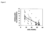
- the tryptophan fluorescence signal may be normalized to another fluorescence band, rather than to the diffuse reflectance value at 295 nm. Normalizing the tryptophan band to the 390 nm excitation band was used since the latter was found to change the slowest with aging. Other bands can also be used for the normalization. Also if the radiation source intensity is sufficient at about 295 nm normalization with the diffuse reflectance signal at this wavelength can be used.
- the intensity of skin fluorescence was found to change with age.
- a series of typical excitation spectra taken on the cheek area of two individuals of 30 and 60 years of age, both of skin type II is shown in Fig. 1.
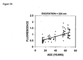
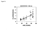
- the fluorescence excitation band ascribed to the tryptophan moieties (295 nm) decreases with age, whereas the bands of collagen and elastin cross-links (335 nm, 360 nm, and 390 nm) increase.
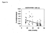
- the age distribution of the fluorescence intensity for the 295 nm, 335 nm, 360 nm, and 390 nm excitation bands taken from 108 individuals in Shanghai, China is shown in Figs. 2a, 2b, 2c, and 2d correspondingly.
- the data has been fitted with linear regressions and the intervals between the average ⁇ one standard deviation are shown. It is evident that the value of the standard deviation of the data distribution was higher for younger ages for the 295 nm excitation band. The opposite was found for all the other bands.
- the 295 nm excitation band was the only one declining with age (at -0.002 units/year).
- the slopes of the best linear fit of the data represent the rates of change of the fluorescence intensities and are shown in Table I. Rates of change (units/year) for the skin fluorescence bands and the normalized tryptophan fluorescence (I 295nm / I 390nm ). All measurements were preformed on the face (cheek). The rates of change were calculated from the slopes of the best linear fit of the data. The values are given in fluorescence units per year for the fluorescence bands and in ratio units per year for the normalized tryptophan fluorescence.
- PDCXL pepsin digestible collagen cross-links
- CDCXL collagenase digestible collagen cross-links
- NTF normalized tryptophan fluorescence.
- Table I Tryptophan PDCXL CDCXL Elastin NTF Ratio Geographical Area Season n 295 nm 335 nm 360 nm 390 nm 295 nm/390 nm Guangzhou, China Summer 108 -0.0021 0.010 0.013 0.0053 -0.103 Harbin, China Summer 106 -0.0007 0.012 0.015 0.0062 -0.074 Harbin, China Winter 64 -0.0025 0.014 0.016 0.0047 -0.091 Shanghai, China Summer 100 -0.0017 0.012 0.018 0.0060 -0.119 Shanghai, China Winter 100 -0.0024 0.012 0.013 0.0053 -0.135 Sendai, Japan Summer 108 -0.0019 0.010 0.016 0.0047 -0.128 Manila, Philippines Summer 100 -0.000
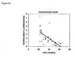
- the cheek area was selected as it was expected to have received solar UV radiation that results in cumulative skin damage over a lifetime.
- sun exposure photoaging
- the results are shown in Figs. 4a and 4b.
- the fluorescence ratio I 295nm / I 390nm acquired from the face decreased with age (Fig. 4a).
- the rate of decrease (0.087 units/year) was close to the values from other regions noted in Table I.
- the normalized tryptophan fluorescence was also decreasing with age (Fig. 4b), although at a much slower rate (0.010 units/year).
- Fluorescence acquisition consisted of a synchronous scan of excitation and emission wavelengths in the range 250-600 nm with constant Stokes shift of 50 nm and was preformed with a spectrofluorimeter (model SkinSkan, JY Horiba, Edison, NJ). This scan provides information for the tryptophan fluorescence (excitation 295 nm, emission 345 nm) and for the collagen and elastin cross-link related fluorescence, including the band where the tryptophan fluorescence is normalized to (excitation 390 nm, emission 440 nm).
- the low numbers of NFT for the diabetic group indicates a reduced capacity for epidermal repair compared to the control healthy group.
- the reduced NTF values in diabetic skin may be related to the poor wound healing and repair capacity of diabetic skin.
- the data (Table 2) were compared with previous plotted data. Based on the NTF values from the "exposed” areas collected from the previous study it was demonstrated that the control group falls within the age-matched "healthy” curve, while the diabetic group assumes lower values.
- the data indicate that overall health of an individual may be assessed by creating a standard curve for healthy individuals and comparing the ratio for individual whose health is in question to those on the curve. As indicated above, a ratio that falls below the standard curve may be an indication that the individual has a health problem, such as diabetes.
Landscapes
- Health & Medical Sciences (AREA)
- Life Sciences & Earth Sciences (AREA)
- Engineering & Computer Science (AREA)
- Molecular Biology (AREA)
- Pathology (AREA)
- Biomedical Technology (AREA)
- Physics & Mathematics (AREA)
- General Health & Medical Sciences (AREA)
- Immunology (AREA)
- Chemical & Material Sciences (AREA)
- Biophysics (AREA)
- Animal Behavior & Ethology (AREA)
- Medical Informatics (AREA)
- Public Health (AREA)
- Veterinary Medicine (AREA)
- Surgery (AREA)
- Heart & Thoracic Surgery (AREA)
- Analytical Chemistry (AREA)
- Biochemistry (AREA)
- General Physics & Mathematics (AREA)
- Urology & Nephrology (AREA)
- Hematology (AREA)
- Nuclear Medicine, Radiotherapy & Molecular Imaging (AREA)
- Dermatology (AREA)
- Microbiology (AREA)
- Cell Biology (AREA)
- Biotechnology (AREA)
- Tropical Medicine & Parasitology (AREA)
- Physiology (AREA)
- Food Science & Technology (AREA)
- Medicinal Chemistry (AREA)
- Investigating, Analyzing Materials By Fluorescence Or Luminescence (AREA)
- Measurement Of The Respiration, Hearing Ability, Form, And Blood Characteristics Of Living Organisms (AREA)
- Measuring And Recording Apparatus For Diagnosis (AREA)
Applications Claiming Priority (1)
| Application Number | Priority Date | Filing Date | Title |
|---|---|---|---|
| US10/986,941 US8620411B2 (en) | 2003-12-12 | 2004-11-15 | Method of assessing skin and overall health of an individual |
Publications (2)
| Publication Number | Publication Date |
|---|---|
| EP1656884A2 true EP1656884A2 (de) | 2006-05-17 |
| EP1656884A3 EP1656884A3 (de) | 2006-09-06 |
Family
ID=35788426
Family Applications (1)
| Application Number | Title | Priority Date | Filing Date |
|---|---|---|---|
| EP05257026A Withdrawn EP1656884A3 (de) | 2004-11-15 | 2005-11-14 | Methode zur Beurteilung der Haut und der allgemeinen Gesundheit einer Person |
Country Status (4)
| Country | Link |
|---|---|
| US (1) | US8620411B2 (de) |
| EP (1) | EP1656884A3 (de) |
| BR (1) | BRPI0505274A (de) |
| CA (1) | CA2526581C (de) |
Cited By (3)
| Publication number | Priority date | Publication date | Assignee | Title |
|---|---|---|---|---|
| WO2008003146A3 (en) * | 2006-07-03 | 2008-07-03 | Ilan Karavani | Method for determination of the type of skin of the face of a person and method for the determination of the aging of the skin of person' s face. |
| BE1017797A4 (nl) * | 2007-05-09 | 2009-07-07 | Karavani Ilan | Werkwijze voor het bepalen van het huidtype van het gelaat van personen. |
| US7711799B2 (en) | 2004-11-22 | 2010-05-04 | Alcatel-Lucent Usa Inc. | Method and apparatus for pre-packetized caching for network servers |
Families Citing this family (13)
| Publication number | Priority date | Publication date | Assignee | Title |
|---|---|---|---|---|
| US20070276199A1 (en) * | 2002-04-04 | 2007-11-29 | Ediger Marwood N | Determination of a Measure of a Glycation End-Product or Disease State Using Tissue Fluorescence |
| US7139598B2 (en) * | 2002-04-04 | 2006-11-21 | Veralight, Inc. | Determination of a measure of a glycation end-product or disease state using tissue fluorescence |
| US8131332B2 (en) * | 2002-04-04 | 2012-03-06 | Veralight, Inc. | Determination of a measure of a glycation end-product or disease state using tissue fluorescence of various sites |
| US8140147B2 (en) * | 2002-04-04 | 2012-03-20 | Veralight, Inc. | Determination of a measure of a glycation end-product or disease state using a flexible probe to determine tissue fluorescence of various sites |
| US20070004972A1 (en) * | 2005-06-29 | 2007-01-04 | Johnson & Johnson Consumer Companies, Inc. | Handheld device for determining skin age, proliferation status and photodamage level |
| FR2891641B1 (fr) * | 2005-10-04 | 2007-12-21 | Lvmh Rech | Procede et appareil de caracterisation des imperfections de la peau et procede d'appreciation de l'effet anti-vieillissement d'un produit cosmetique. |
| US8849380B2 (en) * | 2007-11-26 | 2014-09-30 | Canfield Scientific Inc. | Multi-spectral tissue imaging |
| CA3194784A1 (en) | 2008-05-20 | 2009-11-26 | University Health Network | Device and method for fluorescence-based imaging and monitoring |
| US9615747B2 (en) * | 2010-01-19 | 2017-04-11 | Access Business Group International Llc | Method for determining skin glycation |
| US20150078642A1 (en) * | 2012-04-24 | 2015-03-19 | The General Hospital Corporation | Method and system for non-invasive quantification of biologial sample physiology using a series of images |
| DK3171765T3 (da) | 2014-07-24 | 2021-11-01 | Univ Health Network | Indsamling og analyse af data til diagnostiske formål |
| JP6793944B2 (ja) * | 2016-11-24 | 2020-12-02 | Nsマテリアルズ株式会社 | 携帯型測定器 |
| AU2019307872A1 (en) * | 2018-07-16 | 2021-01-28 | Skin Rejuvenation Technologies (Pty) Ltd | A method and system for cosmetic recommendations |
Family Cites Families (8)
| Publication number | Priority date | Publication date | Assignee | Title |
|---|---|---|---|---|
| US4894547A (en) | 1987-09-28 | 1990-01-16 | Yale University | Optical method and apparatus for detecting and measuring aging, photoaging, dermal disease and pigmentation in skin |
| US5131398A (en) | 1990-01-22 | 1992-07-21 | Mediscience Technology Corp. | Method and apparatus for distinguishing cancerous tissue from benign tumor tissue, benign tissue or normal tissue using native fluorescence |
| US5701902A (en) | 1994-09-14 | 1997-12-30 | Cedars-Sinai Medical Center | Spectroscopic burn injury evaluation apparatus and method |
| US6091985A (en) | 1998-01-23 | 2000-07-18 | Research Foundation Of City College Of New York | Detection of cancer and precancerous conditions in tissues and/or cells using native fluorescence excitation spectroscopy |
| US6721582B2 (en) * | 1999-04-06 | 2004-04-13 | Argose, Inc. | Non-invasive tissue glucose level monitoring |
| US20020091324A1 (en) | 1998-04-06 | 2002-07-11 | Nikiforos Kollias | Non-invasive tissue glucose level monitoring |
| US20050049467A1 (en) | 2003-08-28 | 2005-03-03 | Georgios Stamatas | Method for assessing pigmented skin |
| US9750449B2 (en) * | 2003-12-12 | 2017-09-05 | Johnson & Johnson Consumer Inc. | Method of assessing skin |
-
2004
- 2004-11-15 US US10/986,941 patent/US8620411B2/en not_active Expired - Fee Related
-
2005
- 2005-11-14 EP EP05257026A patent/EP1656884A3/de not_active Withdrawn
- 2005-11-14 CA CA2526581A patent/CA2526581C/en not_active Expired - Lifetime
- 2005-11-16 BR BRPI0505274-2A patent/BRPI0505274A/pt not_active Application Discontinuation
Cited By (3)
| Publication number | Priority date | Publication date | Assignee | Title |
|---|---|---|---|---|
| US7711799B2 (en) | 2004-11-22 | 2010-05-04 | Alcatel-Lucent Usa Inc. | Method and apparatus for pre-packetized caching for network servers |
| WO2008003146A3 (en) * | 2006-07-03 | 2008-07-03 | Ilan Karavani | Method for determination of the type of skin of the face of a person and method for the determination of the aging of the skin of person' s face. |
| BE1017797A4 (nl) * | 2007-05-09 | 2009-07-07 | Karavani Ilan | Werkwijze voor het bepalen van het huidtype van het gelaat van personen. |
Also Published As
| Publication number | Publication date |
|---|---|
| US8620411B2 (en) | 2013-12-31 |
| US20050203355A1 (en) | 2005-09-15 |
| BRPI0505274A (pt) | 2006-07-11 |
| EP1656884A3 (de) | 2006-09-06 |
| CA2526581C (en) | 2015-01-27 |
| CA2526581A1 (en) | 2006-05-15 |
Similar Documents
| Publication | Publication Date | Title |
|---|---|---|
| CA2489915C (en) | Method of assessing skin and overall health of an individual | |
| CA2526581C (en) | Method of assessing skin and overall health of an individual | |
| Kollias et al. | Endogenous skin fluorescence includes bands that may serve as quantitative markers of aging and photoaging | |
| Na et al. | Autofluorescence of human skin is age-related after correction for skin pigmentation and redness | |
| Stamatas et al. | Facial skin fluorescence as a marker of the skin's response to chronic environmental insults and its dependence on age | |
| Gillies et al. | Fluorescence excitation spectroscopy provides information about human skin in vivo | |
| Ou-Yang et al. | A chemiluminescence study of UVA-induced oxidative stress in human skin in vivo | |
| US20050049467A1 (en) | Method for assessing pigmented skin | |
| Heerfordt et al. | Protoporphyrin IX in the skin measured noninvasively predicts photosensitivity in patients with erythropoietic protoporphyria | |
| NouhzadehMalekshah et al. | Evaluation of laser fluorescence in combination with photosensitizers for detection of demineralized lesions | |
| Drakaki et al. | Laser-induced fluorescence and reflectance spectroscopy for the discrimination of basal cell carcinoma from the surrounding normal skin tissue | |
| JP2003522579A (ja) | 非侵襲性組織グルコース濃度モニタリング | |
| US20040219684A1 (en) | In vitro prediction of sunscreen PFA values | |
| Pearse et al. | Portable erythema meter and its application to use in human skin | |
| JP7214679B2 (ja) | 肌ダメージの判定方法 | |
| EP3733076B1 (de) | Verfahren zur bestimmung der uv-lichtempfindlichkeit | |
| JP7239400B2 (ja) | 皮膚抗酸化能の判定方法 | |
| Ravnbak et al. | Skin pigmentation kinetics after exposure to ultraviolet A | |
| Togsverd-Bo et al. | Organ transplant recipients express enhanced skin autofluorescence and pigmentation at skin cancer sites | |
| Makmatov-Rys et al. | Optical Technology for Ultraviolet Erythema Assessment and Minimal Erythema Dose Determination in Healthy Volunteers. | |
| Utz et al. | In vivo evaluation of sunscreens by spectroscopic methods | |
| Utz et al. | Optical and imaging techniques for in-vivo sunscreens investigation | |
| Calabro et al. | Variations in the optical scattering properties of skin in murine animal models | |
| Kawada et al. | Determination of UVA Protection Factor with SPEX SkinSkan | |
| Brian Riordan et al. | M Department of |
Legal Events
| Date | Code | Title | Description |
|---|---|---|---|
| PUAI | Public reference made under article 153(3) epc to a published international application that has entered the european phase |
Free format text: ORIGINAL CODE: 0009012 |
|
| AK | Designated contracting states |
Kind code of ref document: A2 Designated state(s): AT BE BG CH CY CZ DE DK EE ES FI FR GB GR HU IE IS IT LI LT LU LV MC NL PL PT RO SE SI SK TR |
|
| AX | Request for extension of the european patent |
Extension state: AL BA HR MK YU |
|
| PUAL | Search report despatched |
Free format text: ORIGINAL CODE: 0009013 |
|
| AK | Designated contracting states |
Kind code of ref document: A3 Designated state(s): AT BE BG CH CY CZ DE DK EE ES FI FR GB GR HU IE IS IT LI LT LU LV MC NL PL PT RO SE SI SK TR |
|
| AX | Request for extension of the european patent |
Extension state: AL BA HR MK YU |
|
| 17P | Request for examination filed |
Effective date: 20070223 |
|
| 17Q | First examination report despatched |
Effective date: 20070321 |
|
| AKX | Designation fees paid |
Designated state(s): DE FR GB IT |
|
| STAA | Information on the status of an ep patent application or granted ep patent |
Free format text: STATUS: THE APPLICATION IS DEEMED TO BE WITHDRAWN |
|
| 18D | Application deemed to be withdrawn |
Effective date: 20090915 |