EP1676918A2 - G-beta-gamma regulierte Phosphatidylinositil-3' Kinase - Google Patents
G-beta-gamma regulierte Phosphatidylinositil-3' Kinase Download PDFInfo
- Publication number
- EP1676918A2 EP1676918A2 EP06005399A EP06005399A EP1676918A2 EP 1676918 A2 EP1676918 A2 EP 1676918A2 EP 06005399 A EP06005399 A EP 06005399A EP 06005399 A EP06005399 A EP 06005399A EP 1676918 A2 EP1676918 A2 EP 1676918A2
- Authority
- EP
- European Patent Office
- Prior art keywords
- protein
- subunit
- regulatory subunit
- gene
- regulatory
- Prior art date
- Legal status (The legal status is an assumption and is not a legal conclusion. Google has not performed a legal analysis and makes no representation as to the accuracy of the status listed.)
- Withdrawn
Links
Images








Classifications
-
- C—CHEMISTRY; METALLURGY
- C12—BIOCHEMISTRY; BEER; SPIRITS; WINE; VINEGAR; MICROBIOLOGY; ENZYMOLOGY; MUTATION OR GENETIC ENGINEERING
- C12N—MICROORGANISMS OR ENZYMES; COMPOSITIONS THEREOF; PROPAGATING, PRESERVING, OR MAINTAINING MICROORGANISMS; MUTATION OR GENETIC ENGINEERING; CULTURE MEDIA
- C12N9/00—Enzymes; Proenzymes; Compositions thereof; Processes for preparing, activating, inhibiting, separating or purifying enzymes
- C12N9/10—Transferases (2.)
- C12N9/12—Transferases (2.) transferring phosphorus containing groups, e.g. kinases (2.7)
- C12N9/1205—Phosphotransferases with an alcohol group as acceptor (2.7.1), e.g. protein kinases
-
- A—HUMAN NECESSITIES
- A61—MEDICAL OR VETERINARY SCIENCE; HYGIENE
- A61P—SPECIFIC THERAPEUTIC ACTIVITY OF CHEMICAL COMPOUNDS OR MEDICINAL PREPARATIONS
- A61P11/00—Drugs for disorders of the respiratory system
-
- A—HUMAN NECESSITIES
- A61—MEDICAL OR VETERINARY SCIENCE; HYGIENE
- A61P—SPECIFIC THERAPEUTIC ACTIVITY OF CHEMICAL COMPOUNDS OR MEDICINAL PREPARATIONS
- A61P11/00—Drugs for disorders of the respiratory system
- A61P11/06—Antiasthmatics
-
- A—HUMAN NECESSITIES
- A61—MEDICAL OR VETERINARY SCIENCE; HYGIENE
- A61P—SPECIFIC THERAPEUTIC ACTIVITY OF CHEMICAL COMPOUNDS OR MEDICINAL PREPARATIONS
- A61P19/00—Drugs for skeletal disorders
-
- A—HUMAN NECESSITIES
- A61—MEDICAL OR VETERINARY SCIENCE; HYGIENE
- A61P—SPECIFIC THERAPEUTIC ACTIVITY OF CHEMICAL COMPOUNDS OR MEDICINAL PREPARATIONS
- A61P19/00—Drugs for skeletal disorders
- A61P19/02—Drugs for skeletal disorders for joint disorders, e.g. arthritis, arthrosis
-
- A—HUMAN NECESSITIES
- A61—MEDICAL OR VETERINARY SCIENCE; HYGIENE
- A61P—SPECIFIC THERAPEUTIC ACTIVITY OF CHEMICAL COMPOUNDS OR MEDICINAL PREPARATIONS
- A61P25/00—Drugs for disorders of the nervous system
- A61P25/28—Drugs for disorders of the nervous system for treating neurodegenerative disorders of the central nervous system, e.g. nootropic agents, cognition enhancers, drugs for treating Alzheimer's disease or other forms of dementia
-
- A—HUMAN NECESSITIES
- A61—MEDICAL OR VETERINARY SCIENCE; HYGIENE
- A61P—SPECIFIC THERAPEUTIC ACTIVITY OF CHEMICAL COMPOUNDS OR MEDICINAL PREPARATIONS
- A61P29/00—Non-central analgesic, antipyretic or antiinflammatory agents, e.g. antirheumatic agents; Non-steroidal antiinflammatory drugs [NSAID]
-
- A—HUMAN NECESSITIES
- A61—MEDICAL OR VETERINARY SCIENCE; HYGIENE
- A61P—SPECIFIC THERAPEUTIC ACTIVITY OF CHEMICAL COMPOUNDS OR MEDICINAL PREPARATIONS
- A61P35/00—Antineoplastic agents
-
- A—HUMAN NECESSITIES
- A61—MEDICAL OR VETERINARY SCIENCE; HYGIENE
- A61P—SPECIFIC THERAPEUTIC ACTIVITY OF CHEMICAL COMPOUNDS OR MEDICINAL PREPARATIONS
- A61P37/00—Drugs for immunological or allergic disorders
-
- A—HUMAN NECESSITIES
- A61—MEDICAL OR VETERINARY SCIENCE; HYGIENE
- A61P—SPECIFIC THERAPEUTIC ACTIVITY OF CHEMICAL COMPOUNDS OR MEDICINAL PREPARATIONS
- A61P37/00—Drugs for immunological or allergic disorders
- A61P37/08—Antiallergic agents
-
- A—HUMAN NECESSITIES
- A61—MEDICAL OR VETERINARY SCIENCE; HYGIENE
- A61P—SPECIFIC THERAPEUTIC ACTIVITY OF CHEMICAL COMPOUNDS OR MEDICINAL PREPARATIONS
- A61P39/00—General protective or antinoxious agents
-
- A—HUMAN NECESSITIES
- A61—MEDICAL OR VETERINARY SCIENCE; HYGIENE
- A61P—SPECIFIC THERAPEUTIC ACTIVITY OF CHEMICAL COMPOUNDS OR MEDICINAL PREPARATIONS
- A61P39/00—General protective or antinoxious agents
- A61P39/06—Free radical scavengers or antioxidants
-
- A—HUMAN NECESSITIES
- A61—MEDICAL OR VETERINARY SCIENCE; HYGIENE
- A61P—SPECIFIC THERAPEUTIC ACTIVITY OF CHEMICAL COMPOUNDS OR MEDICINAL PREPARATIONS
- A61P43/00—Drugs for specific purposes, not provided for in groups A61P1/00-A61P41/00
-
- A—HUMAN NECESSITIES
- A61—MEDICAL OR VETERINARY SCIENCE; HYGIENE
- A61P—SPECIFIC THERAPEUTIC ACTIVITY OF CHEMICAL COMPOUNDS OR MEDICINAL PREPARATIONS
- A61P7/00—Drugs for disorders of the blood or the extracellular fluid
-
- A—HUMAN NECESSITIES
- A61—MEDICAL OR VETERINARY SCIENCE; HYGIENE
- A61P—SPECIFIC THERAPEUTIC ACTIVITY OF CHEMICAL COMPOUNDS OR MEDICINAL PREPARATIONS
- A61P9/00—Drugs for disorders of the cardiovascular system
-
- A—HUMAN NECESSITIES
- A61—MEDICAL OR VETERINARY SCIENCE; HYGIENE
- A61P—SPECIFIC THERAPEUTIC ACTIVITY OF CHEMICAL COMPOUNDS OR MEDICINAL PREPARATIONS
- A61P9/00—Drugs for disorders of the cardiovascular system
- A61P9/10—Drugs for disorders of the cardiovascular system for treating ischaemic or atherosclerotic diseases, e.g. antianginal drugs, coronary vasodilators, drugs for myocardial infarction, retinopathy, cerebrovascula insufficiency, renal arteriosclerosis
-
- C—CHEMISTRY; METALLURGY
- C07—ORGANIC CHEMISTRY
- C07K—PEPTIDES
- C07K2319/00—Fusion polypeptide
-
- G—PHYSICS
- G01—MEASURING; TESTING
- G01N—INVESTIGATING OR ANALYSING MATERIALS BY DETERMINING THEIR CHEMICAL OR PHYSICAL PROPERTIES
- G01N2333/00—Assays involving biological materials from specific organisms or of a specific nature
- G01N2333/90—Enzymes; Proenzymes
- G01N2333/91—Transferases (2.)
- G01N2333/912—Transferases (2.) transferring phosphorus containing groups, e.g. kinases (2.7)
- G01N2333/91205—Phosphotransferases in general
- G01N2333/9121—Phosphotransferases in general with an alcohol group as acceptor (2.7.1), e.g. general tyrosine, serine or threonine kinases
- G01N2333/91215—Phosphotransferases in general with an alcohol group as acceptor (2.7.1), e.g. general tyrosine, serine or threonine kinases with a definite EC number (2.7.1.-)
-
- G—PHYSICS
- G01—MEASURING; TESTING
- G01N—INVESTIGATING OR ANALYSING MATERIALS BY DETERMINING THEIR CHEMICAL OR PHYSICAL PROPERTIES
- G01N2500/00—Screening for compounds of potential therapeutic value
- G01N2500/10—Screening for compounds of potential therapeutic value involving cells
-
- Y—GENERAL TAGGING OF NEW TECHNOLOGICAL DEVELOPMENTS; GENERAL TAGGING OF CROSS-SECTIONAL TECHNOLOGIES SPANNING OVER SEVERAL SECTIONS OF THE IPC; TECHNICAL SUBJECTS COVERED BY FORMER USPC CROSS-REFERENCE ART COLLECTIONS [XRACs] AND DIGESTS
- Y10—TECHNICAL SUBJECTS COVERED BY FORMER USPC
- Y10S—TECHNICAL SUBJECTS COVERED BY FORMER USPC CROSS-REFERENCE ART COLLECTIONS [XRACs] AND DIGESTS
- Y10S530/00—Chemistry: natural resins or derivatives; peptides or proteins; lignins or reaction products thereof
- Y10S530/827—Proteins from mammals or birds
- Y10S530/829—Blood
Definitions
- the present invention relates to novel G-protein regulated phosphoinositide 30H-kinase enzymes isolated from cells of hematopoietic lineage which are involved in cellular signal transduction pathways, and to the use of these novel enzymes in the treatment and diagnosis of disease.
- Phosphoinositide 30H-kinases are a large family of enzymes capable of 3-phosphorylating at least one of the cellular phosphoinositides (Whitman et al., 1988, Nature 332:644-646; Auger et al., 1989, Cell 57:167-175). 3-phosphorylated phosphoinositides are found in all higher eukaryotic cells. A growing body of evidence implicates PI3K and a lipid product of this enzyme, phosphatidylinositol (3,4,5)-triphosphate (hereinafter "PtdIns(3,4,5)P 3 "), as part of a novel and important second messenger system in cellular signal transduction.
- PtdIns(3,4,5)P 3 phosphatidylinositol
- PtdIns(3,4,5)P 3 -based signalling system appear to be independent of the previously characterized signalling pathway based on inositol phospholipids, in which a phosphoinositidase C (PIC) hydrolyses PtdIns(4,5)P 2 to release the structurally distinct second messengers inositol (1,4,5)-triphosphate (Ins(1,4,5)P 3 ) and diacylglycerol.
- PIC phosphoinositidase C
- tyrosine kinase receptors which, when activated, result in increased accumulation of PtdIns(3,4,5)P 3 are the PDGF receptor, the EGF receptor, members of the FGF receptor family, the CSF-1 receptor, the insulin receptor, the IGF-1 receptor, and the NGF receptor.
- Receptors associated with src family non-receptor tyrosine kinases which stimulate PtdIns(3,4,5)P 3 accumulation are the Il-2 receptor, Il-3 receptor, mIgM receptor, the CD4 receptor, the CD2 receptor, and the CD3/T cell receptor.
- PtdIns(3,4,5)P 3 appears to be a second messenger in extremely diverse signalling pathways.
- PI3K activity and production of PtdIns(3,4,5)P 3 is a physiological relevant pathway of signal transduction for these diverse receptors is derived, inter alia , from two different lines of experimental evidence: inhibition of PI3K activity by fungal metabolites and observations of direct protein associations.
- Wortmannin a fungal metabolite, irreversibly inhibits PI3K activity by binding covalently to the catalytic domain of this enzyme. Inhibition of PI3K activity by wortmannin eliminates the subsequent cellular response to the extracellular factor.
- neutrophils respond to the chemokine fMet-Leu-Phe (fMLP) by stimulating PI3K and synthesizing PtdIns(3,4,5)P 3 .
- fMLP chemokine fMet-Leu-Phe
- PI3K enzymes interact directly with, and may be co-puritied with, activated forms of several receptor tyrosine kinases.
- receptor tyrosine kinase associated PI3K was found to consist of 170-200 kD heterodimers (Otsu et al., 1991, Cell 65:91-104, Pons et al., 1995, Mol. Cell. Biol. 15:4453-4465, Inukai et al., 1996, J. Biol. Chem. 271:5317-5320) comprising a catalytic subunit and an adaptor (or regulatory) subunit.
- the catalytic subunit which irreversibly binds wortmannin, tightly associates with one or other members of a small family of highly related regulatory subunits, p55 ⁇ , pSSPIK, p85 ⁇ and p85 ⁇ , to form the 170-200 kD heterodimers.
- the known regulatory subunits contain a large collection of protein:protein interaction domains, including two SH2 domains (Cantley et al., 1991, Cell 64:281-302).
- the presence of the SH2 domains are thought to be responsible for the binding and stimulation of PI3K heterodimers to activated receptor tyrosine kinases.
- Activated receptors are phosphorylated at key tyrosine residues within local consensus sequences preferred by the SH2 domains found in the 55-87 kD PI3K adaptors (Songyang et al., 1993, Cell 72:767-778).
- the PI3K heterodimer binds , it directly activates the PI3K catalytic subunit (although this effect is relatively small in vitro, Carpenter et al., 1993, J. Biol. Chem. 268:9478-9483, Backer et al., 1992, EMBO J.
- PI3Ks p110/p85 sub-family of PI3Ks do not seem to be involved in the production of PtdIns(3,4,5)P 3 that can occur as a result of activation of cell surface receptors which utilize heterotrimeric GTPases to transduce their signals (e.g. , fMLP, PAF, ATP, and thrombin).
- cell surface receptors which utilize heterotrimeric GTPases to transduce their signals (e.g. , fMLP, PAF, ATP, and thrombin).
- Platelets also contain a G ⁇ -sensitive PI3K, although it is unclear whether this is a p85/p110 PI3K family member (Thomason et al., 1994, J. Biol. Chem. 269:16525-16528). It seemed likely that this poorly characterized, G ⁇ -sensitive PI3K might be responsible for production of PtdIns(3,4,5)P 3 in response to agonists like ATP, fMLP etc.
- p110 ⁇ wortmannin-sensitive PtdIns(4,5)P 2 -selective PI3K
- p110 ⁇ was amplified by PCR using primers designed to target potential PI3K's as well as PtdIns4-kinase's catalytic centers. It is clearly distinct from p110 ⁇ and p110 ⁇ , as it lacks, for example, an amino-terminus binding domain for a member of the p85 adaptor family.
- the present invention relates to the discovery, identification, purification, and cloning of nucleotides that encode the trimeric G protein regulated PI3K, a novel protein that produces accumulation of the second messenger PtdIns(3,4,5)P 3 in response to activation of G protein-linked receptors.
- This novel G-protein regulated PI3K is comprised of a catalytic subunit, p120, and a regulatory subunit, p101.
- the p120 catalytic subunit shares partial amino acid sequence homology with the PI3K catalytic subunits p110 ⁇ , p110 ⁇ , and p110 ⁇ .
- p101 is a completely new protein with no identifiable homology to any other sequence previously available.
- activated G protein subunits induce a mild stimulation of catalytic activity by the p120 catalytic subunit.
- the PI3K activity of the catalytic subunit is stimulated over 100 fold by activated G proteins.
- porcine p101 and p120 cDNAs encoding both porcine p101 and p120 and human p101 and p120 have been cloned and sequenced, and are described herein.
- the porcine p101 and p120 cDNAs encode proteins of about 877 amino acids and about 1102 amino acids, respectively (FIG. 2 and FIG. 4), while the human p101 and p120 cDNAs encode proteins of about 880 amino acids and about 1102 amino acids, respectively (FIG. 11 and FIG. 13).
- amino acid sequences of the p120 proteins are homologous to those of other known PI3K catalytic subunits
- the p120 cDNAs described herein diverge from the cDNAs encoding the known PI3K catalytic subunits, particularly at, for example, the carboxyl terminus (amino acid residues 1075 to 1102).
- the p101 transcript is found primarily in cells of hematopoietic lineage. p120, and other PI3K catalytic subunit proteins, appear to have a far broader tissue and cell type distribution. Notably, the presence of a trimeric G protein sensitive PI3K activity has only been found in a limited number of cells of hematopoietic lineage (e.g. , neutrophils, platelets, etc.). Thus, the ability to activate PI3K enzymes in response to stimulation of trimeric G protein linked receptors appears largely dependent on the presence of the p101 subunit.
- the invention encompasses the following nucleotides, host cells expressing such nucleotides, and the expression products of such nucleotides: (a) nucleotides that encode mammalian p101 and p120 proteins, including the porcine p101 and p120 and the human p101 and p120 , and the p101 and p120 gene products, including the porcine and human gene products; (b) nucleotides that encode portions of p101 and p120 that correspond to its functional domains, and the polypeptide products specified by such nucleotide sequences, including but not limited to the p101 nucleotides encoding, for example, the p101 G ⁇ interaction domain, or the catalytic subunit associating domain, or amino acid residues from porcine p101 extending from about 1 to 160, 80-120, 161 to 263, 264 to 414, 415 to 565, 566 to 706, 707-832, and/or 833 to 877, or amino acid residues from human
- the G ⁇ interaction domain, or the catalytic subunit associating domain or the domains described by amino acid residues from about I to 160, 80-120, 161 to 263, 264 to 414, 415 to 565, 566 to 706, 707-832, and/or 833 to 877), or the p120 protein or one of its domains ( e.g. , the membrane binding domain, or the regulatory subunit domain, or amino acid residues from about 173 to 302 and 310 to 315) fused to another polypeptide.
- the invention also encompasses agonists and antagonists of G protein regulated PI3K, including small molecules, large molecules, mutant p101 proteins that compete with native p101, and antibodies, as well as nucleotide sequences that can be used to inhibit p101 gene expression (e.g. , antisense and ribozyme molecules, and gene or regulatory sequence replacement constructs) or to enhance p101 gene expression (e.g. , expression constructs that place the p101 gene under the control of a strong promoter system), and transgenic cells and animals that express a p101 transgene or "knock-outs" that do not express p101.
- G protein regulated PI3K including small molecules, large molecules, mutant p101 proteins that compete with native p101, and antibodies, as well as nucleotide sequences that can be used to inhibit p101 gene expression (e.g. , antisense and ribozyme molecules, and gene or regulatory sequence replacement constructs) or to enhance p101 gene expression (e.g
- the present invention encompasses methods and compositions for the diagnostic evaluation, typing and prognosis of immune system disorders, including inflammation, and for the identification of subjects having a predisposition to such conditions.
- p101 nucleic acid molecules of the invention can be used as diagnostic hybridization probes or as primers for diagnostic PCR analysis for the identification of p101 gene mutations, allelic variations and regulatory defects in the p101 gene.
- the present invention further provides for diagnostic kits for the practice of such methods.
- the present invention also relates to methods for the use of the p101 gene and/or p120 gene products for the identification of compounds which modulate, i.e., act as agonists or antagonists, of G protein-regulated PI3K gene expression and or p101 and/or p120 gene product activity.
- Such compounds can be used as agents to control immune system disorders and, in particular, as therapeutic agents for the treatment of inflammation disorders such as arthritis, septic shock, adult respiratory distress syndrome (ARDS), pneumonia, asthma, allergies, reperfusion injury, atherosclerosis, Alzheimer's disease, and cancer.
- the invention encompasses methods and compositions for the treatment of inflammation disorders such as arthritis, septic shock, adult respiratory distress syndrome (ARDS), pneumonia, asthma, allergies, reperfusion injury, atherosclerosis, Alzheimer's disease, and cancer.
- inflammation disorders such as arthritis, septic shock, adult respiratory distress syndrome (ARDS), pneumonia, asthma, allergies, reperfusion injury, atherosclerosis, Alzheimer's disease, and cancer.
- ARDS adult respiratory distress syndrome
- Such methods and compositions are capable of modulating the level of p101 gene expression and/or the level of p101 or p120 gene product activity.
- This invention is based, in part, on the surprising discovery of a G protein stimulated PI3K activity in isolated neutrophils; the purification and characterization of this activity as a heterodimeric PI3K protein comprised of a p101 subunit and a p120 subunit; the identification and cloning of p101 cDNA from libraries prepared from porcine neutrophil mRNA, human monocyte mRNA, and human leukocyte mRNA; the identification and cloning of p120 cDNA from libraries prepared from porcine neutrophil mRNA and human Leukocyte mRNA; characterization of the novel sequences; and studies of isolated recombinantly expressed p101 and p120 protein in insect cells, U937 cells, and Cos-7 cells.
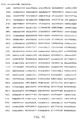
- FIGS. 1A, 1B, 1C, and 1D Porcine p101 adaptor subunit nucleotide sequence. (SEQ ID NO:1)
- FIG. 1 Deduced Porcine p101 amino acid sequence. (SEQ ID NO:2) Tryptic peptides identified by protein sequencing are underlined with a solid line.
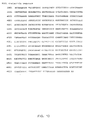
- FIGS. 3A, 3B and 3C Porcine p120 catalytic subunit nucleotide sequence. (SEQ ID NO:3)
- FIG. 4 Deduced Porcine p120 catalytic subunit sequence. (SEQ ID NO:4) Tryptic peptides identified by protein sequencing are underlined with a solid line. The region of divergence from the published amino acid sequence of p110 ⁇ is underscored with a broken line.
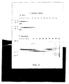
- G ⁇ -sensitive PI3K in neutrophil cytosol is distinct to p85/p110 PI3Ks. Aliquots of each of the fractions derived from a Q-sepharose chromatographic profile were either Western blotted and probed with ⁇ p85 ⁇ or ⁇ p85 ⁇ monoclonal antibodies and visualized with an HRP-linked second antibody (upper panel in Figure 5), or incubated with 100nM [ 3 H]-17-hydroxy-wortmannin, resolved by SDS-PAGE (6% acrylamide and fluorographed (lower panel in Figure 5). G ⁇ -sensitive PI3K activity eluted in fractions 20-24.
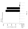
- Peak B After Final Mini Q Column Purification Contains a G ⁇ -sensitive PI3K Activity.
- the final purification product of the Peak B activity was analyzed after isolation from a Mini Q column in the presence and absence of activated G ⁇ subunits ( ⁇ ), a tyrosine phosphorylated peptide (YP-peptide), wortmannin, and/or G ⁇ i -GDP subunits ( ⁇ i -GDP).
- FIG. 7 Pharmacological and regulatory properties of free p120 and p101/p120 PI3Ks recombinantly expressed in Sf9 cells.
- Assays contained either 7 nM p101/p120 or 36 nM p120 alone (final concentrations) which were incubated with various reagents; 10 mM NaF and 30 ⁇ M AlCl 3 (A/F); 1 ⁇ M G ⁇ subunits ( ⁇ ); 2 ⁇ M G ⁇ -GDP; 100 nM wortmannin (W) or 50 ⁇ M tyrosine phosphorylated peptide (PY) for a total of 15 minutes (at 0°C) prior to starting the assays by adding [ ⁇ 32 P]-ATP; [ 32 P]-incorporated into [ 32 P]-Ptdlns(3,4,5)P 3 was quantified.
- p101 can associate with a G ⁇ -stimulated PI3K activity in U937 cells.
- U937 cells were transiently transfected with mammalian expression vectors encoding either (EE)120 or (EE)101, co-transfected in combination with mammalian expression vectors encoding myc101, myc120, or an irrelevant control myc fusion protein, as indicated.
- a total of 40 ⁇ g of vector DNA was used.
- the cells lysed, precleaned and immunoprecipitated with protein G sepharose covalently cross-linked to ⁇ -(EE) monoclonal antibody as described more fully in the Examples herein.
- the resulting immunoprecipitates were washed and G ⁇ -activated (1 ⁇ M, final concentration) PI3K activity was assayed.
- the PI3K activity detected in immunoprecipitates from cells transfected with irrelevant (EE)-tagged protein, either with or without G ⁇ s was subtracted from the data shown (these were means of 1896 dpm and 2862 dpm in the absence and presence of G ⁇ s, respectively).
- Parallel transfections labelled with [ 35 S]-m ⁇ ethionine showed the amount of [ 35 S]-p101 and [ 35 S]-p120 in the immunoprecipitates fell by 40% when they were co-transfected (data not shown).
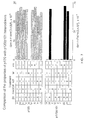
- FIG. 9 Regulation of p101/p120 by G ⁇ s in vivo .
- Cos-7 cells were transfected with mammalian expression vectors encoding G ⁇ subunit (" ⁇ 1 ⁇ 2 "), p101 ("101"), and/or p120 ("120"), as indicated. After 48 hours of transient expression, cells from each transfection were either Western blotted to determine "Relative Expression", or assayed for the accumulation of PtdIns(3,4,5)P 3 accumulation (shaded bars). For Western blotting, samples were probed with an ⁇ -(myc) monoclonal antibody to quantitate the expression of the various (myc)-tagged proteins.
- Results are given relative to the expression obtained in the presence of all 4 key vectors; the absolute levels of (myc)-p120 and (myc)-p101 were very similar and about 10 x greater than that of (myc)- ⁇ 2 .
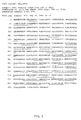
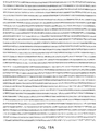
- FIGS. 10A and 10B Human p101 adaptor subunit nucleotide sequence. (SEQ ID NO:11)
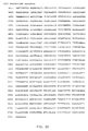
- Figures 12A and 12B Human p120 catalytic subunit nucleotide sequence. (SEQ ID NO:13)
- the present invention relates to the identification, purification, and cloning of a specific form of PI3K which is activated by ⁇ subunits of trimeric G-proteins.
- p101 described for the first time herein, is a novel subunit of the G-protein regulated PI3K. Also described herein is the identification, cloning, and correct sequence of p120, the catalytic subunit of the trimeric G-protein regulated PI3K.
- PI3K are enzymes which phosphorylate phosphatidyl inositols at the 3d position to generate the intracellular signaling molecule PtdIns(3,4,5)P 3 .
- PtdIns(3,4,5)P 3 the intracellular signaling molecule
- neutrophils are activated by, inter alia, the chemokine fMLP which is released in response to infection by microorganisms.
- This agonist binds to a pertussis toxin sensitive G protein coupled receptor and activates the neutrophil respiratory burst, resulting in superoxide production and cytotoxicity.
- the respiratory burst response correlates with a rapid and transient intracellular increase in PI3K activity.
- wortmannin which selectively inhibits the catalytic component of PI3K's, causes a shutdown of superoxide production. Since the vast majority of agonists which activate this respiratory burst bind to G protein-linked receptors, the G protein regulated PI3K activity represents a candidate for a dominant effector pathway whose inhibition will reduce the destructive cellular effects of inflammation. However, wortmannin is not a clinically appropriate inhibitor of this pathway since wortmannin inhibits all PI3K catalytic activities, including those involved in other cellular pathways such as growth factors.
- the G-protein regulated PI3K is composed of two subunits: the catalytic subunit (p110 ⁇ , p117 or p120); and the p101 regulatory subunit.
- the ability of PI3K to respond to cell surface receptors which stimulate the release of G ⁇ subunits from activated trimeric G-proteins is largely dependent on the presence of the p101 regulatory subunit.
- catalytic subunits exhibit a small stimulation (approximately 1.7 fold) of PI3K activity in vitro in the presence of G ⁇ subunits, addition of p101 proteins will increase G ⁇ stimulation of PI3K activity 100 fold.
- the invention also encompasses the use of p101 and p120 nucleotides, p101 and p120 proteins and peptides, as well as antibodies to p101 and p120 (which can, for example, act as p101 or p120 agonists or antagonists), antagonists that inhibit G protein-activated PI3K activity or expression, or agonists that activate G protein-activated PI3K activity or increase its expression in the treatment of hematopoietic lineage cell activation disorders, including, but not limited to immune responses associated with acute and chronic inflammation, in animals such as humans.
- antagonists of G ⁇ -activated PI3K will be useful in the treatment of arthritis, including rheumatoid arthritis, septic shock, adult respiratory distress syndrome (ARDS), pneumonia, asthma and other lung conditions, allergies, reperfusion injury, atherosclerosis and other cardiovascular diseases, Alzheimer's disease, and cancer, to name just a few inflammatory disorders.
- p 101 nucleotides and p101 regulatory subunits, as well as p120 nucleotides and p120 catalytic subunits are useful for the identification of compounds effective in the treatment of hematopoietic lineage cell activation disorders involving G protein-activated PI3Ks.
- the invention encompasses the use of p101 and p120 nucleotides, p101 and p120 proteins and peptides, as well as antibodies to p101 and p120 in the diagnosis of hematopoietic lineage cell activation disorders.
- the diagnosis of a p101 regulatory subunit or p120 catalytic subunit abnormality in a patient, or an abnormality in the G protein activated PI3K signal transduction pathway, will also assist in devising a proper treatment or therapeutic regimen.
- the invention described in the subsections below encompasses p101 regulatory subunit, polypeptides or peptides corresponding to functional domains of the p101 regulatory subunit (e.g. , the catalytic subunit association domain, or the domain which interacts with activated G proteins), mutated, truncated or deleted p101 regulatory subunits (e.g. a p101 regulatory subunit with one or more functional domains or portions thereof deleted), p101 regulatory subunit fusion proteins (e.g. a p101 regulatory subunit or a functional domain of p101 regulatory subunit, fused to an unrelated protein or peptide such as an epitope tag, i.e. , the myc epitope), nucleotide sequences encoding such products, and host cell expression systems that can produce such p101 regulatory subunit products.
- p101 regulatory subunit e.g. , the catalytic subunit association domain, or the domain which interacts with activated G proteins
- the invention encompasses p120 catalytic subunit proteins, polypeptides, functional domains of the p120 subunit ( e.g. , the catalytic domain), mutated, truncated or deleted p120 subunit proteins, p120 fusion proteins, nucleotide sequences encoding such products, and host cell expression systems that can produce such p120 catalytic subunit products.
- the invention also encompasses antibodies and anti-idiotypic antibodies (including Fab fragments), antagonists and agonists of the G protein-activated PI3K, as well as compounds or nucleotide constructs that inhibit expression of the p101 or p120 gene (transcription factor inhibitors, antisense and ribozyme molecules, or gene or regulatory sequence replacement constructs), or promote expression of p101 regulatory subunit (e.g. , expression constructs in which p101 coding sequences are operatively associated with expression control elements such as promoters, promoter/enhancers, etc.).
- the invention also relates to host cells and animals genetically engineered to express the human p101 regulatory subunit (or mutants thereof) or to inhibit or "knock-out" expression of the animal's endogenous p101 regulatory subunit.
- the invention particularly encompasses antagonists which prevent the association of p101 regulatory subunits with their binding partners, including p120 and other PI3K catalytic subunit proteins such as p117 and p110 ⁇ , as well as activated trimeric G protein proteins, including G ⁇ subunits.
- the p101 regulatory subunit proteins or peptides, p101 regulatory subunit fusion proteins, p101 nucleotide sequences, antibodies, antagonists and agonists can be useful for the detection of mutant p101 regulatory subunits or inappropriately expressed p101 regulatory subunits for the diagnosis of immune disorders.
- the p101 and p120 subunit proteins or peptides, p101 and p120 subunit fusion proteins, p101 and p120 nucleotide sequences, host cell expression systems, antibodies, antagonists, agonists and genetically engineered cells and animals can be used for screening for drugs effective in the treatment of such immune disorders.
- engineered host cells and/or animals may offer an advantage in that such systems allow not only for the identification of compounds that bind to the p101 regulatory subunit, but can also identify compounds that affect the signal transduced by the activated p101 regulatory subunit, specifically, production of the intracellular signaling molecule PtdIns(3,4,5)P 3 .
- the p101 regulatory subunit protein products and fusion protein products, antibodies and anti-idiotypic antibodies (including Fab fragments), antagonists or agonists can be used for therapy of such diseases.
- nucleotide constructs encoding functional p101 regulatory subunits, mutant p101 regulatory subunits, as well as antisense and ribozyme molecules can be used in "gene therapy" approaches for the modulation of p101 regulatory subunit expression and/or activity in the treatment of hematopoietic lineage cell activation disorders.
- the invention also encompasses pharmaceutical formulations and methods for treating hematopoietic lineage cell activation disorders.
- the invention is based, in part, on the surprising discovery of novel PI3K enzymes in porcine neutrophils. Like other PI3K proteins, these enzymes 3-phosphorylate PtdIns, PtdIns4P and PtdIns(4,5)P 2 substrates, and were completely inhibited by 100 nM wortmannin. However, unlike the previously described PI3K p85/p101 protein complexes, the PI3K activity was stimulated over 100 fold by incubation with G ⁇ subunits. Addition of G ⁇ -GDP subunits could inhibit this G ⁇ activation. Furthermore, the PI3K activity was not stimulated by phosphorylated tyrosine peptides.
- p117/p101 complex When purified, two distinct heterodimeric protein complex were identified: a p117/p101 complex and a p120/p101 complex. Peptide sequencing revealed that the p101 protein was identical in each complex. The p120 and p117 proteins were homologous with the exception of the amino terminus (see Examples below). Porcine p101 and p120 cDNAs were then cloned using degenerate probes based upon the peptide sequence to screen an expression library of cDNAs synthesized from porcine neutrophil mRNA. Human p101 and p120 were cloned using probes derived from the porcine cDNA sequences to screen human monocyte and leukocyte cDNA libraries.
- p101 and/or p120 fusion proteins in insect cells demonstrated that p101 binds tightly to p120 in a 1:1 molar stoichiometry.
- Free purified p120 exhibited PI3K activity which was insensitive to the presence of G ⁇ subunits and tyrosine phosphorylated peptides, and only mildly stimulated by G ⁇ subunits.
- the PI3K activity of p120 was stimulated 100 fold by the presence of G ⁇ subunits.
- p101 was expressed alone in insect cells, p101 did not exhibit PI3K activity.
- transient transfections were performed in Cos-7 cells, which do not normally stimulate PI3K activity in response to activated G-proteins, with constructs encoding p101, p120 and/or G ⁇ subunits. Transfection of a construct which expressed p120 only produced significant increases in cellular PtdIns(3,4,5)P 3 levels in a G ⁇ dependent fashion when co-expressed in the presence of p101.
- the cDNA sequence (SEQ ID NO:1) and deduced amino acid sequence (SEQ ID NO:2) of porcine p101 regulatory subunit are shown in FIGs. 1 and 2, respectively.
- a relatively common allelic variation occurs at amino acid residue 483 in the open reading frame; a serine may be replaced by a glycine at this position.
- the cDNA sequence (SEQ ID NO:11) and deduced amino acid sequence (SEQ ID NO:12) of human p101 regulatory subunit are shown in FIGs. 10 and 11, respectively.
- the nucleotide sequence encoding the first 733 amino acids of the porcine p101 regulatory subunit, corresponding to the first 736 amino acids of the human subunit, is believed to be sufficient to bind the catalytic subunit.
- this truncated p101 bound to p120 will not stimulate PI3K activity in response to G ⁇ subunits. Therefore, the catalytic subunit associating domain is believed to be contained within the first 732 amino acids (735 for human), and the carboxyl terminal amino acids 733 to 877 (736 to 880 for human) could be involved in the response to G ⁇ subunits.
- porcine p101 are described by amino acid residues from about 1 to 160, 80-120, 161 to 263, 264 to 414, 415 to 565, 566 to 706, 707-832, and/or 833 to 877.
- the corresponding domains of human p101 are described by amino acid residues from about 1 to 160, 80-120, 161 to 263, 264 to 416, 417 to 567, 568 to 709, 710-835, and/or 836 to 880.
- the nucleotide sequences which encode amino acid residues from about 161 to 263 define a pleckstrin homology (“PH") domain (PROSITE:PS50003).
- PH domains may be involved in binding of proteins to G ⁇ subunits (Touhara et al., 1994, J. Biol. Chem. 269:10217) and to membrane phospholipids ( see Shaw, 1996, Bioessays 18:35-46). Thus, the PH domain of p101 may be involved in both of these events.
- the nucleotide sequences encoding amino acids 1 to 160 of p101 may be responsible for binding to the p120 catalytic subunit.
- this region of the p101 protein is analyzed for secondary structure, it is predicted to have a self-contained alternating ⁇ -helix/ ⁇ -sheet structure. Within this structure is found homology to a "WW domain" (Staub et al., 1996, Structure 4:495-499 and TIBS 21:161-163).
- This WW domain may bind to a proline-rich domain found within the N-terminus of p120 protein (residues 310 to 315).
- the WW domain of p101 may be involved in mediating the interaction between the regulatory subunit and the catalytic subunit.
- the cDNA sequence (SEQ ID NO:3) and deduced amino acid sequence (SEQ ID NO:4) of porcine p120 are shown in FIGs. 3 and 4, respectively.
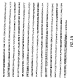
- the cDNA sequence (SEQ ID NO:13) and deduced amino acid sequence (SEQ ID NO:14) of human p120 are shown in FIGs. 12 and 13, respectively.
- a cryptic thrombin cleavage site is present after the first approximately 40 amino acid residues.
- the truncated p120 protein lacking these approximately 40 amino terminal residues is still able to bind to p101, but PI3K activity is reduced approximately 20 to 30%.
- a protein having the amino acid sequence of the section of SEQ ID NO:4 (porcine p120 deduced amino acid sequence) extending from about amino acid residue 41 to residue 1102 retains the functionality of full-length p120.
- a protein having the amino acid sequence of the section of SEQ ID NO:14 (human p120 deduced amino acid sequence) extending from about amino acid residue 41 to residue 1102 would also retain the functionality of full-length p120.
- the p120 protein was highly homologous to the previously cloned p110 ⁇ protein reported by Stoyanov et al.
- nucleotides encoding a proline-rich region including p120 residues 310 to 315 may be involved in the interaction between p120 and p101. Additionally, the nucleotides encoding p120 amino acid residues from about 173 to 302 define a weak PH domain which may be a candidate for membrane binding and/or G ⁇ subunit interaction of the p101/p120 complex.
- p120 cDNA encodes the catalytic subunit of the G ⁇ -activated PI3K.
- the p101 cDNA encodes a novel regulatory subunit protein which binds to the p120 subunit. This heterodimer 3-phosphorylates PtdIns, PtdIns4P and PtdIns(4,5)P 2 in response to activation of trimeric G-protein linked receptors.
- the p101 nucleotide sequences of the invention include: (a) the DNA sequence shown in FIG. 1 or FIG.10 or contained in the cDNA clone pCMV3mycp101 as deposited with the American Type Culture Collection (ATCC) under accession number 97636; (b) nucleotide sequence that encodes the amino acid sequence shown in FIG. 2 or FIG.11, or the p101 regulatory subunit amino acid sequence encoded by the cDNA clone pCMV3mycp101 as deposited with the ATCC; (c) any nucleotide sequence that hybridizes to the complement of the DNA sequence shown in FIG. 1 or FIG.
- Functional equivalents of the p101 regulatory subunit include naturally occurring p101 regulatory subunit present in other species, and mutant p101 regulatory subunits whether naturally occurring or engineered which retain at least some of the functional activities of p101 (i.e. , binding to the p120 or p117 catalytic subunit, stimulation of catalytic activity in response to G ⁇ subunits, and/or interaction with G ⁇ subunits).
- the invention also includes degenerate variants of sequences (a) through (d).
- the invention also includes nucleic acid molecules, preferably DNA molecules, that hybridize to, and are therefore the complements of, the nucleotide sequences (a) through (d), in the preceding paragraph.
- Such hybridization conditions may be highly stringent or less highly stringent, as described above.
- highly stringent conditions may refer, e.g. , to washing in 6xSSC/0.05% sodium pyrophosphate at 37°C (for 14-base oligos), 48°C (for 17-base oligos), 55°C (for 20-base oligos), and 60°C (for 23-base oligos).
- nucleic acid molecules may encode or act as p101 antisense molecules, useful, for example, in p101 gene regulation (for and/or as antisense primers in amplification reactions of p101 gene nucleic acid sequences).
- p101 gene regulation such techniques can be used to regulate, for example, inflammatory immune responses.
- sequences may be used as part of ribozyme and/or triple helix sequences, also useful for p101 gene regulation.
- such molecules may be used as components of diagnostic methods whereby, for example, the presence of a particular p101 allele responsible for causing an inflammatory response, such as arthritis, may be detected.
- p101 proteins are conserved in different mammalian species.
- human p101 was cloned from monocyte and leukocyte libraries using probes derived from the porcine p101 cDNA sequence, and the deduced amino acid sequences of the human and porcine were found to be highly conserved.
- homologs of p101 and p120 can be isolated from a variety of mammalian cells known or suspected to contain a trimeric G-protein regulated PI3K, particularly cells of hematopoietic origin, and more particularly platelets, monocytes, leukocytes, osteoclasts. and neutrophils.
- homologs of p101 in related species can be useful for developing animal model systems more closely related to humans for purposes of drug discovery.
- expression libraries of cDNAs synthesized from neutrophil mRNA derived from the organism of interest can be screened using labeled catalytic subunit derived from that species, e.g. , a p120, p117, or p110 ⁇ catalytic subunit fusion protein.
- cDNA libraries, or genomic DNA libraries derived from the organism of interest can be screened by hybridization using the nucleotides described herein as hybridization or amplification probes.
- genes at other genetic loci within the genome that encode proteins which have extensive homology to one or more domains of the p101 gene product can also be identified via similar techniques.
- screening techniques can identify clones derived from alternatively spliced transcripts in the same or different species.
- the labeled probe can contain at least 15-30 base pairs of the human or porcine p101 nucleotide sequence, as shown in FIGS. 1 and 10, respectively.
- the hybridization washing conditions used should be of a lower stringency when the cDNA library is derived from an organism different from the type of organism from which the labeled sequence was derived.
- hybridization can, for example, be performed at 65°C overnight in Church's buffer (7% SDS, 250 mM NaHPO 4 , 2 ⁇ M EDTA, 1% BSA). Washes can be done with 2XSSC, 0.1 % SDS at 65°C and then at 0.1XSSC, 0.1% SDS at 65°C.
- the labeled p101 nucleotide probe may be used to screen a genomic library derived from the organism of interest, again, using appropriately stringent conditions.
- the identification and characterization of human genomic clones is helpful for designing diagnostic tests and clinical protocols for treating hematopoietic lineage cell activation disorders in human patients.
- sequences derived from regions adjacent to the intron/exon boundaries of the human gene can be used to design primers for use in amplification assays to detect mutations within the exons, introns, splice sites ( e.g. splice acceptor and/or donor sites), etc., that can be used in diagnostics.
- a p101 gene homolog may be isolated from nucleic acid of the organism of interest by performing PCR using two degenerate oligonucleotide primer pools designed on the basis of amino acid sequences within the p101 gene product disclosed herein.
- the template for the reaction may be cDNA obtained by reverse transcription of mRNA prepared from, for example, human or non-human cell lines or cell types, such as neutrophils, known or suspected to express a p101 gene allele.
- the PCR product may be subcloned and sequenced to ensure that the amplified sequences represent the sequences of a p101 gene.
- the PCR fragment may then be used to isolate a full length cDNA clone by a variety of methods.
- the amplified fragment may be labeled and used to screen a cDNA library, such as a bacteriophage cDNA library.
- the labeled fragment may be used to isolate genomic clones via the screening of a genomic library.
- RNA may be isolated, following standard procedures, from an appropriate cellular source (i.e., one known, or suspected, to express the p101 gene, such as, for example, neutrophils or other types of leukocytes).
- a reverse transcription reaction may be performed on the RNA using an oligonucleotide primer specific for the most 5' end of the amplified fragment for the priming of first strand synthesis.
- the resulting RNA/DNA hybrid may then be "tailed" with guanines using a standard terminal transferase reaction, the hybrid may be digested with RNAase H, and second strand synthesis may then be primed with a poly-C primer.
- cDNA sequences upstream of the amplified fragment may easily be isolated.
- the p101 gene sequences may additionally be used to isolate mutant p101 gene alleles. Such mutant alleles may be isolated from individuals either known or proposed to have a genotype which contributes to the symptoms of hematopoietic lineage cell activation disorders such as inflammation. Mutant alleles and mutant allele products may then be utilized in the therapeutic and diagnostic systems described below. Additionally, such p101 gene sequences can be used to detect p101 gene regulatory (e.g. , promoter or promotor/enhancer) defects which can affect hematopoietic lineage cell activation.
- p101 gene regulatory e.g. , promoter or promotor/enhancer
- a cDNA of a mutant p101 gene may be isolated, for example, by using PCR, a technique which is well known to those of skill in the art.
- the first cDNA strand may be synthesized by hybridizing an oligo-dT oligonucleotide to mRNA isolated from cells known or suspected to be expressed in an individual putatively carrying the mutant p101 allele, and by extending the new strand with reverse transcriptase.
- the second strand of the cDNA is then synthesized using an oligonucleotide that hybridizes specifically to the 5' end of the normal gene.
- the product is then amplified via PCR, cloned into a suitable vector, and subjected to DNA sequence analysis through methods well known to those of skill in the art.
- DNA sequence analysis By comparing the DNA sequence of the mutant p101 allele to that of the normal p101 allele, the mutation(s) responsible for the loss or alteration of function of the mutant p101 gene product can be ascertained.
- a genomic library can be constructed using DNA obtained from an individual suspected of or known to carry the mutant p101 allele, or a cDNA library can be constructed using RNA from a cell type known, or suspected, to express the mutant p101 allele.
- the normal p101 gene or any suitable fragment thereof may then be labeled and used as a probe to identify the corresponding mutant p101 allele in such libraries.
- Clones containing the mutant p101 gene sequences may then be purified and subjected to sequence analysis according to methods well known to those of skill in the art.
- an expression library can be constructed utilizing cDNA synthesized from, for example, RNA isolated from a cell type known, or suspected, to express a mutant p101 allele in an individual suspected of or known to carry such a mutant allele.
- gene products made by the putatively mutant cell type may be expressed and screened using standard antibody screening techniques in conjunction with antibodies raised against the normal p101 gene product, as described, below, in Section 5.3.
- screening techniques see, for example, Harlow, E. and Lane, eds., 1988, "Antibodies: A Laboratory Manual", Cold Spring Harbor Press, Cold Spring Harbor.
- screening can be accomplished by screening with labeled fusion proteins, such as, for example, the (EE)120 or the myc120 fusion proteins.
- a polyclonal set of antibodies to p101 regulatory subunit are likely to cross-react with the mutant p101 regulatory subunit gene product.
- Library clones detected via their reaction with such labeled antibodies can be purified and subjected to sequence analysis according to methods well known to those of skill in the art.
- the invention also encompasses nucleotide sequences that encode mutant p101 regulatory subunits, peptide fragments of the p101 regulatory subunit, truncated p101 regulatory subunits, and p101 regulatory subunit fusion proteins. These include, but are not limited to nucleotide sequences encoding mutant p101 regulatory subunits described in section 5.2 infra ; polypeptides or peptides corresponding to the catalytic binding, or G ⁇ subunit binding domains of the p101 regulatory subunit or portions of these domains; truncated p101 regulatory subunits in which one or two of the domains is deleted, or a truncated, nonfunctional p101 regulatory subunit.
- Nucleotides encoding fusion proteins may include but are not limited to full length p101 regulatory subunit, truncated p101 regulatory subunit or peptide fragments of p101 regulatory subunit fused to an unrelated protein or peptide, such as for example, an epitope tag which aids in purification or detection of the resulting fusion protein; or an enzyme, fluorescent protein, luminescent protein which can be used as a marker.
- the invention also encompasses (a) DNA vectors that contain any of the foregoing p101 regulatory subunit coding sequences and/or their complements ( i.e. , antisense); (b) DNA expression vectors that contain any of the foregoing p101 regulatory subunit coding sequences operatively associated with a regulatory element that directs the expression of the coding sequences; and (c) genetically engineered host cells that contain any of the foregoing p101 regulatory subunit coding sequences operatively associated with a regulatory element that directs the expression of the coding sequences in the host cell.
- regulatory elements include but are not limited to inducible and non-inducible promoters, enhancers, operators and other elements known to those skilled in the art that drive and regulate expression.
- Such regulatory elements include but are not limited to the baculovirus promoter, cytomegalovirus hCMV immediate early gene promoter, the early or late promoters of SV40 adenovirus, the lac system, the trp system, the TAC system, the TRC system, the major operator and promoter regions of phage A, the control regions of fd coat protein, the promoter for 3-phosphoglycerate kinase, the promoters of acid phosphatase, and the promoters of the yeast ⁇ -mating factors.
- the invention also encompasses nucleotides encoding p120 subunit proteins including the newly described carboxyl terminus of this catalytic subunit, deletion variants of p120 subunit proteins, nucleotides which hybridize to these nucleotides under highly stringent conditions and which encode functionally equivalent products, including the cDNA clone pCMV3mycp120 deposited with the ATCC under accession number 97637, allelic variants of p120 ( e.g. , mutant alleles or the naturally occurring alleles such as the allelic variation at amino acid residue 483), and equivalent p120 nucleotides from different organisms isolated as described above for the p101 nucleotides of the invention. Additionally, the invention encompasses expression vectors and host cells for the recombinant production of p120.
- p101 regulatory subunit and p120 catalytic subunit polypeptides and peptide fragments, mutated, truncated or deleted forms of the p101 regulatory subunit and/or p101 regulatory subunit fusion proteins and the p120 catalytic subunit and/or p120 catalytic subunit fusion proteins can be prepared for a variety of uses, including but not limited to the generation of antibodies, as reagents in diagnostic assays, the identification of other cellular gene products involved in the regulation of hematopoietic lineage cell activation, as reagents in assays for screening for compounds that can be used in the treatment of hematopoietic lineage cell activation disorders, and as pharmaceutical reagents useful in the treatment of hematopoietic lineage cell activation disorders related to G ⁇ -activated PI3K.
- FIGS. 2 and 11 show the amino acid sequences of porcine and human p101 regulatory subunit protein, respectively.
- FIGS. 4 and 13 show the amino acid sequence of porcine and human p120 catalytic subunit protein, respectively.
- the broken line on FIG. 4 underscores the region of p120 which diverges from the published sequences of the PI3K catalytic subunits p110 ⁇ , p110 ⁇ , and p110 ⁇ .
- the p101 regulatory subunit sequence begins with a methionine in a DNA sequence context consistent with a translation initiation site.
- the predicted molecular mass of both porcine and human p101 regulatory subunits is 97 kD.
- the p101 regulatory subunit amino acid sequences of the invention include the amino acid sequences shown in FIG. 2 (SEQ ID NO:2) or FIG. 4 (SEQ ID NO:12), or the amino acid sequence encoded by the cDNA clone pCMV3mycp101, as deposited with the ATCC. Further, p101 regulatory subunits of other species are encompassed by the invention. In fact, any p101 regulatory subunit protein encoded by the p101 nucleotide sequences described above, are within the scope of the invention.
- the p120 catalytic subunit amino acid sequences of the invention include the cDNA clone pCMV3mycp120, as deposited with the ATCC.
- the invention also encompasses p120 catalytic subunits of other species, and the p120 proteins encoded by the p120 nucleotide sequences described above in the previous section.
- the invention also encompasses proteins that are functionally equivalent to the p101 regulatory subunit encoded by the nucleotide sequences described above, as judged by any of a number of criteria, including but not limited to the ability to bind catalytic subunit, the binding affinity for catalytic subunit, the ability to stimulate PI3K activity of the catalytic subunit in response to activated trimeric G proteins, the resulting biological effect of catalytic subunit binding and response to activation of trimeric G proteins, e.g. , signal transduction, a change in cellular metabolism (e.g.
- Such functionally equivalent p101 regulatory subunit proteins include but are not limited to additions or substitutions of amino acid residues within the amino acid sequence encoded by the p101 nucleotide sequences described, above, but which result in a silent change, thus producing a functionally equivalent gene product. Amino acid substitutions may be made on the basis of similarity in polarity, charge, solubility, hydrophobicity, hydrophilicity, and/or the amphipathic nature of the residues involved.
- nonpolar (hydrophobic) amino acids include alanine, leucine, isoleucine, valine, proline, phenylalanine, tryptophan, and methionine; polar neutral amino acids include glycine, serine, threonine, cysteine, tyrosine, asparagine, and glutamine; positively charged (basic) amino acids include arginine, lysine, and histidine; and negatively charged (acidic) amino acids include aspartic acid and glutamic acid.
- the invention also encompasses functional equivalents of p120 protein, as described above.
- site-directed mutations of the p101 coding sequence can be engineered (using site-directed mutagenesis techniques well known to those skilled in the art) to generate mutant p101 regulatory subunits with increased function, e.g. , higher binding affinity for catalytic subunit, and/or greater signalling capacity; or decreased function, e.g. , lower binding affinity for catalytic subunit, and/or decreased signal transduction capacity.
- porcine p101 amino acid sequence may be aligned with that of human p101 regulatory subunit.
- Mutant p101 regulatory subunits can be engineered so that regions of interspecies identity are maintained, whereas the variable residues are altered, e.g. , by deletion or insertion of an amino acid residue(s) or by substitution of one or more different amino acid residues.
- Conservative alterations at the variable positions can be engineered in order to produce a mutant p101 regulatory subunit that retains function; e.g., catalytic subunit binding affinity or activated G protein transduction capability or both.
- Non-conservative changes can be engineered at these variable positions to alter function, e.g. , catalytic subunit binding affinity or signal transduction capability, or both.
- deletion or non-conservative alterations of the conserved regions can be engineered.
- One of skill in the art may easily test such mutant or deleted p101 regulatory subunits for these alterations in function using the teachings presented herein.
- mutations to the p101 coding sequence can be made to generate p101 regulatory subunits that are better suited for expression, scale up, etc. in the host cells chosen.
- the triplet code for each amino acid can be modified to conform more closely to the preferential codon usage of the host cell's translational machinery.
- Peptides corresponding to one or more domains (or a portion of a domain) of the p101 regulatory subunit e.g. , the p120 binding domain, the G protein interacting domain, or the domains defined by amino acid residues from about 1 to 150, 151 to 300, 301 to 450, 451 to 600, 601-732 (porcine) or 601-735 (human), and 733-877 (porcine) or 736-881 (human)), truncated or deleted p101 regulatory subunits (e.g., p101 regulatory subunit in which portions of one or more of the above domains are deleted) as well as fusion proteins in which the full length p101 regulatory subunit, a p101 regulatory subunit peptide or truncated p101 regulatory subunit is fused to an unrelated protein are also within the scope of the invention and can be designed on the basis of the p101 nucleotide and p101 regulatory subunit amino acid sequences disclosed in this Section and above. Such fusion
- p101 regulatory subunit polypeptides and peptides can be chemically synthesized (e.g. , see Creighton, 1983, Proteins: Structures and Molecular Principles, W.H. Freeman & Co., N.Y.), large polypeptides derived from the p101 regulatory subunit and the full length p101 regulatory subunit itself may advantageously be produced by recombinant DNA technology using techniques well known in the art for expressing nucleic acid containing p101 gene sequences and/or coding sequences. Such methods can be used to construct expression vectors containing the p101 nucleotide sequences described above and appropriate transcriptional and translational control signals.
- RNA capable of encoding p101 nucleotide sequences may be chemically synthesized using, for example, synthesizers. See, for example, the techniques described in "Oligonucleotide Synthesis", 1984, Gait, M.J. ed., IRL Press, Oxford, which is incorporated by reference herein in its entirety.
- a variety of host-expression vector systems may be utilized to express the p101 nucleotide sequences of the invention.
- the p101 regulatory subunit peptide or polypeptide is a soluble derivative the peptide or polypeptide can be recovered from the culture, i.e. , from the host cell in cases where the p101 regulatory subunit peptide or polypeptide is not secreted, and from the culture media in cases where the p101 regulatory subunit peptide or polypeptide is secreted by the cells.
- engineered host cells themselves may be used in situations where it is important not only to retain the structural and functional characteristics of the p101 regulatory subunit, but to assess biological activity, e.g. , in drug screening assays.
- the expression systems that may be used for purposes of the invention include but are not limited to microorganisms such as bacteria (e.g. , E. coli , B. subtilis ) transformed with recombinant bacteriophage DNA, plasmid DNA or cosmid DNA expression vectors containing p101 nucleotide sequences; yeast ( e.g. , Saccharomyces , Pichia ) transformed with recombinant yeast expression vectors containing the p101 nucleotide sequences; insect cell systems infected with recombinant virus expression vectors (e.g.
- microorganisms such as bacteria (e.g. , E. coli , B. subtilis ) transformed with recombinant bacteriophage DNA, plasmid DNA or cosmid DNA expression vectors containing p101 nucleotide sequences; yeast ( e.g. , Saccharomyces , Pichia ) transformed with recombinant yeast expression vectors
- baculovirus containing the p101 sequences
- plant cell systems infected with recombinant virus expression vectors e.g. , cauliflower mosaic virus, CaMV; tobacco mosaic virus, TMV
- recombinant plasmid expression vectors e.g. , Ti plasmid
- mammalian cell systems e.g. , COS, CHO, BHK, 293, 3T3, U937 harboring recombinant expression constructs containing promoters derived from the genome of mammalian cells (e.g. , metallothionein promoter) or from mammalian viruses (e.g. , the adenovirus late promoter; the vaccinia virus 7.5K promoter).
- a number of expression vectors may be advantageously selected depending upon the use intended for the p101 gene product being expressed. For example, when a large quantity of such a protein is to be produced, for the generation of pharmaceutical compositions of p101 regulatory subunit protein or for raising antibodies to the p101 regulatory subunit protein, for example, vectors which direct the expression of high levels of fusion protein products that are readily purified may be desirable.
- vectors include, but are not limited, to the E. coli expression vector pUR278 (Ruther et al., 1983, EMBO J.
- p101 coding sequence may be ligated individually into the vector in frame with the lacZ coding region so that a fusion protein is produced;
- pIN vectors Inouye & Inouye, 1985, Nucleic Acids Res. 13:3101-3109; Van Heeke & Schuster, 1989, J. Biol. Chem. 264:5503-5509; and the like.
- pGEX vectors may also be used to express foreign polypeptides as fusion proteins with glutathione S-transferase (GST).
- the inserted sequence encodes a relatively small polypeptide (less than 25 kD)
- such fusion proteins are generally soluble and can easily be purified from lysed cells by adsorption to glutathione-agarose beads followed by elution in the presence of free glutathione.
- the pGEX vectors are designed to include thrombin or factor Xa protease cleavage sites so that the cloned target gene product can be released from the GST moiety.
- the inclusion bodies may be purified and the recombinant protein solubilized using techniques well known to one of skill in the art.
- Autographa californica nuclear polyhidrosis virus may be used as a vector to express foreign genes.
- AcNPV Autographa californica nuclear polyhidrosis virus
- Sf9 insect cells are infected with a baculovirus vectors expressing either a 6 x HIS-tagged p120 construct, or an (EE)-tagged p101 construct.
- a number of viral-based expression systems may be utilized. Specific embodiments described more fully below express tagged p101 or p120 cDNA sequences using a CMV promoter to transiently express recombinant protein in U937 cells or in Cos-7 cells.
- retroviral vector systems well known in the art may be used to insert the recombinant expression construct into host cells. For example, retroviral vector systems for transducing hematopoietic cells are described in published PCT applications WO 96/09400 and WO 94/29438.
- the p101 nucleotide sequence of interest may be ligated to an adenovirus transcription/translation control complex, e.g. , the late promoter and tripartite leader sequence.
- This chimeric gene may then be inserted in the adenovirus genome by in vitro or in vivo recombination. Insertion in a non-essential region of the viral genome (e.g. , region E1 or E3) will result in a recombinant virus that is viable and capable of expressing the p101 gene product in infected hosts.
- a non-essential region of the viral genome e.g. , region E1 or E3
- Specific initiation signals may also be required for efficient translation of inserted p101 nucleotide sequences. These signals include the ATG initiation codon and adjacent sequences. In cases where an entire p101 gene or cDNA, including its own initiation codon and adjacent sequences, is inserted into the appropriate expression vector, no additional translational control signals may be needed. However, in cases where only a portion of the p101 coding sequence is inserted, exogenous translational control signals, including, perhaps, the ATG initiation codon, must be provided. Furthermore, the initiation codon must be in phase with the reading frame of the desired coding sequence to ensure translation of the entire insert.
- exogenous translational control signals and initiation codons can be of a variety of origins, both natural and synthetic.
- the efficiency of expression may be enhanced by the inclusion of appropriate transcription enhancer elements, transcription terminators, etc. (See Bittner et al., 1987, Methods in Enzymol. 153:516-544).
- a host cell strain may be chosen which modulates the expression of the inserted sequences, or modifies and processes the gene product in the specific fashion desired. Such modifications (e.g. , glycosylation) and processing (e.g. , cleavage) of protein products may be important for the function of the protein.
- Different host cells have characteristic and specific mechanisms for the post-translational processing and modification of proteins and gene products. Appropriate cell lines or host systems can be chosen to ensure the correct modification and processing of the foreign protein expressed.
- eukaryotic host cells which possess the cellular machinery for proper processing of the primary transcript may be used. Such mammalian host cells include but are not limited to CHO, VERO, BHK, HeLa, COS, MDCK, 293, 3T3, WI38, and U937 cells.
- cell lines which stably express the p101 sequences described above may be engineered.
- host cells can be transformed with DNA controlled by appropriate expression control elements (e.g. , promoter, enhancer sequences, transcription terminators, polyadenylation sites, etc.), and a selectable marker.
- appropriate expression control elements e.g. , promoter, enhancer sequences, transcription terminators, polyadenylation sites, etc.
- engineered cells may be allowed to grow for 1-2 days in an enriched media, and then are switched to a selective media.
- the selectable marker in the recombinant plasmid confers resistance to the selection and allows cells to stably integrate the plasmid into their chromosomes and grow to form foci which in turn can be cloned and expanded into cell lines.
- This method may advantageously be used to engineer cell lines which express the p101 gene product. Such engineered cell lines may be particularly useful in screening and evaluation of compounds that affect the endogenous activity of the p101 gene product.
- a number of selection systems may be used, including but not limited to the herpes simplex virus thymidine kinase (Wigler et al., 1977, Cell 11:223), hypoxanthine-guanine phosphoribosyltransferase (Szybalska & Szybalski, 1962, Proc. Natl. Acad. Sci. USA 48:2026), and adenine phosphoribosyltransferase (Lowy et al., 1980, Cell 22:817) genes can be employed in tk - , hgprt - or aprt - cells, respectively.
- antimetabolite resistance can be used as the basis of selection for the following genes: dhfr, which confers resistance to methotrexate (Wigler et al., 1980, Natl. Acad. Sci. USA 77:3567; O'Hare et al., 1981, Proc. Natl. Acad. Sci. USA 78:1527); gpt, which confers resistance to mycophenolic acid (Mulligan & Berg, 1981, Proc. Natl. Acad. Sci. USA 78:2072); neo, which confers resistance to the aminoglycoside G-418 (Colberre-Garapin et al., 1981, J. Mol. Biol. 150: 1); and hygro, which confers resistance to hygromycin (Santerre et al., 1984, Gene 30:147).
- the p101 gene products can also be expressed in transgenic animals.
- Animals of any species including, but not limited to, mice, rats, rabbits, guinea pigs, pigs, micro-pigs, goats, and non-human primates, e.g. , baboons, monkeys, and chimpanzees may be used to generate p101 transgenic animals.
- Any technique known in the art may be used to introduce the p101 transgene into animals to produce the founder lines of transgenic animals.
- Such techniques include, but are not limited to pronuclear microinjection (Hoppe, P.C. and Wagner, T.E., 1989, U.S. Pat. No. 4,873,191); retrovirus mediated gene transfer into germ lines (Van der Putten et al., 1985, Proc. Natl. Acad. Sci., USA 82:6148-6152); gene targeting in embryonic stem cells (Thompson et al., 1989, Cell 56:313-321); electroporation of embryos (Lo, 1983, Mol Cell. Biol.
- the present invention provides for transgenic animals that carry the p101 transgene in all their cells, as well as animals which carry the transgene in some, but not all their cells, i.e. , mosaic animals.
- the transgene may be integrated as a single transgene or in concatamers, e.g. , head-to-head tandems or head-to-tail tandems.
- the transgene may also be selectively introduced into and activated in a particular cell type by following, for example, the teaching of Lasko et al. (Lasko, M. et al., 1992, Proc. Natl. Acad. Sci. USA 89: 6232-6236).
- the regulatory sequences required for such a cell-type specific activation will depend upon the particular cell type of interest, and will be apparent to those of skill in the art.
- vectors containing some nucleotide sequences homologous to the endogenous p101 gene are designed for the purpose of integrating, via homologous recombination with chromosomal sequences, into and disrupting the function of the nucleotide sequence of the endogenous p101 gene. In this way, the expression of the endogenous p101 gene may also be eliminated by inserting non-functional sequences into the endogenous gene.
- the transgene may also be selectively introduced into a particular cell type, thus inactivating the endogenous p101 gene in only that cell type, by following, for example, the teaching of Gu et al. (Gu et al., 1994, Science 265: 103-106).
- the regulatory sequences required for such a cell-type specific inactivation will depend upon the particular cell type of interest, and will be apparent to those of skill in the art.
- the expression of the recombinant p101 gene may be assayed utilizing standard techniques. Initial screening may be accomplished by Southern blot analysis or PCR techniques to analyze animal tissues to assay whether integration of the transgene has taken place. The level of mRNA expression of the transgene in the tissues of the transgenic animals may also be assessed using techniques which include but are not limited to Northern blot analysis of cell type samples obtained from the animal, in situ hybridization analysis, and RT-PCR. Samples of p101 gene-expressing tissue, may also be evaluated immunocytochemically using antibodies specific for the p101 transgene product, as described below.
- Antibodies that specifically recognize one or more epitopes of p101 regulatory subunit, or epitopes of conserved variants of p101 regulatory subunit, or peptide fragments of the p101 regulatory subunit are also encompassed by the invention.
- antibodies which recognize one or more epitopes of the p120 protein, particularly, the novel carboxyl terminus include but are not limited to polyclonal antibodies, monoclonal antibodies (mAbs), humanized or chimeric antibodies, single chain antibodies, Fab fragments, F(ab') 2 fragments, fragments produced by a Fab expression library, anti-idiotypic (anti-Id) antibodies, and epitope-binding fragments of any of the above.
- the antibodies of the invention may be used, for example, in the detection of the p101 regulatory subunit or p120 in a biological sample and may, therefore, be utilized as part of a diagnostic or prognostic technique whereby patients may be tested for abnormal amounts of these proteins.
- Such antibodies may also be utilized in conjunction with, for example, compound screening schemes, as described, below, in Section 5.5, for the evaluation of the effect of test compounds on expression and/or activity of the p101 or p120 gene products.
- such antibodies can be used in conjunction with the gene therapy techniques described, below, in Section 5.6, to, for example, evaluate the normal and/or engineered p101 regulatory subunit-expressing cells prior to their introduction into the patient.
- Such antibodies may additionally be used as a method for the inhibition of abnormal p101 regulatory subunit or p120 activity.
- Such antibodies may, therefore, be utilized as part of inflammatory disorder treatment methods.
- various host animals may be immunized by injection with the p101 regulatory subunit, a p101 regulatory subunit peptide, truncated p101 regulatory subunit polypeptides, functional equivalents of the p101 regulatory subunit or mutants of the p101 regulatory subunit.
- host animals may be immunized by injection with p120 catalytic subunit or peptides of the p120 subunit.
- Such host animals may include but are not limited to rabbits, mice, and rats, to name but a few.
- adjutants may be used to increase the immunological response, depending on the host species, including but not limited to Freund's (complete and incomplete), mineral gels such as aluminum hydroxide, surface active substances such as lysolecithin, pluronic polyols, polyanions, peptides, oil emulsions, keyhole limpet hemocyanin, dinitrophenol, and potentially useful human adjutants such as BCG (bacille Calmette-Guerin) and Corynebacterium parvum.
- BCG Bacille Calmette-Guerin
- Monoclonal antibodies which are homogeneous populations of antibodies to a particular antigen, may be obtained by any technique which provides for the production of antibody molecules by continuous cell lines in culture. These include, but are not limited to, the hybridoma technique of Kohler and Milstein, (1975, Nature 256:495-497; and U.S. Patent No. 4,376,110), the human B-cell hybridoma technique (Kosbor et al., 1983, Immunology Today 4:72; Cole et al., 1983, Proc. Natl. Acad. Sci. USA 80:2026-2030), and the EBV-hybridoma technique (Cole et al., 1985, Monoclonal Antibodies And Cancer Therapy, Alan R.
- Such antibodies may be of any immunoglobulin class including IgG, IgM, IgE, IgA, IgD and any subclass thereof.
- the hybridoma producing the mAb of this invention may be cultivated in vitro or in vivo. Production of high titers of mAbs in vivo makes this the presently preferred method of production.
- chimeric antibodies In addition, techniques developed for the production of "chimeric antibodies" (Morrison et al., 1984, Proc. Natl. Acad. Sci. USA, 81:6851-6855; Neuberger et al., 1984, Nature, 312:604-608; Takeda et al., 1985, Nature, 314:452-454) by splicing the genes from a mouse antibody molecule of appropriate antigen specificity together with genes from a human antibody molecule of appropriate biological activity can be used.
- a chimeric antibody is a molecule in which different portions are derived from different animal species, such as those having a variable region derived from a porcine mAb and a human immunoglobulin constant region.
- Single chain antibodies are formed by linking the heavy and light chain fragments of the Fv region via an amino acid bridge, resulting in a single chain polypeptide.
- Antibody fragments which recognize specific epitopes may be generated by known techniques.
- such fragments include but are not limited to: the F(ab') 2 fragments which can be produced by pepsin digestion of the antibody molecule and the Fab fragments which can be generated by reducing the disulfide bridges of the F(ab') 2 fragments.
- Fab expression libraries may be constructed (Huse et al., 1989, Science, 246:1275-1281) to allow rapid and easy identification of monoclonal Fab fragments with the desired specificity.
- Antibodies to the p101 regulatory subunit or the p120 catalytic subunit can, in turn, be utilized to generate anti-idiotype antibodies that "mimic" the p101 regulatory subunit or p120 subunit, respectively, using techniques well known to those skilled in the art. (See, e.g. , Greenspan & Bona, 1993, FASEB J 7(5):437-444; and Nissinoff, 1991, J. Immunol. 147(8):2429-2438).
- antibodies which bind to the p101 regulatory subunit and competitively inhibit the binding of catalytic subunit to the p101 regulatory subunit can be used to generate anti-idiotypes that "mimic" the p101 regulatory subunit and, therefore, bind and neutralize catalytic subunit.
- Such neutralizing anti-idiotypes or Fab fragments of such anti-idiotypes can be used in therapeutic regimens to neutralize catalytic subunit and reduce inflammation.
- a variety of methods can be employed for the diagnostic and prognostic evaluation of hematopoietic lineage cell activation disorders, including inflammatory disorders, and for the identification of subjects having a predisposition to such disorders.
- Such methods may, for example, utilize reagents such as the p101 and p120 nucleotide sequences described above, and p101 regulatory subunit and p120 antibodies, as described, in Section 5.3.
- reagents may be used, for example, for: (1) the detection of the presence of p101 or p120 gene mutations, or the detection of either over- or under-expression of p101 or p120 mRNA relative to the non-hematopoietic lineage cell activation disorder state; (2) the detection of either an over- or an under-abundance of p101 or p120 gene product relative to the non-hematopoietic lineage cell activation disorder state; and (3) the detection of perturbations or abnormalities in the signal transduction pathway mediated by p101 regulatory subunit and p120 catalytic subunit.
- the methods described herein may be performed, for example, by utilizing pre-packaged diagnostic kits comprising at least one specific p101 or p120 nucleotide sequence or p101 or p120 regulatory subunit antibody reagent described herein, which may be conveniently used, e.g. , in clinical settings, to diagnose patients exhibiting hematopoietic lineage cell activation disorder abnormalities.
- any nucleated cell can be used as a starting source for genomic nucleic acid.
- any cell type or tissue in which the p101 or p120 gene is expressed such as, for example, neutrophil cells, may be utilized.
- Mutations within the p101 or p120 genes can be detected by utilizing a number of techniques. Nucleic acid from any nucleated cell can be used as the starting point for such assay techniques, and may be isolated according to standard nucleic acid preparation procedures which are well known to those of skill in the art.
- DNA may be used in hybridization or amplification assays of biological samples to detect abnormalities involving gene structure, including point mutations, insertions, deletions and chromosomal rearrangements.
- assays may include, but are not limited to, Southern analyses, single stranded conformational polymorphism analyses (SSCP), and PCR analyses.
- Such diagnostic methods for the detection of p101 or p120 gene-specific mutations can involve for example, contacting and incubating nucleic acids including recombinant DNA molecules, cloned genes or degenerate variants thereof, obtained from a sample, e.g. , derived from a patient sample or other appropriate cellular source, with one or more labeled nucleic acid reagents including recombinant DNA molecules, cloned genes or degenerate variants thereof, as described above, under conditions favorable for the specific annealing of these reagents to their complementary sequences within the p101 or p120 gene.
- the lengths of these nucleic acid reagents are at least 15 to 30 nucleotides.
- nucleic acid from the cell type or tissue of interest can be immobilized, for example, to a solid support such as a membrane, or a plastic surface such as that on a microtiter plate or polystyrene beads.
- a solid support such as a membrane, or a plastic surface such as that on a microtiter plate or polystyrene beads.
- Detection of the remaining, annealed, labeled p101 or n120 nucleic acid reagents is accomplished using standard techniques well-known to those in the art.
- the p101 or p120 gene sequences to which the nucleic acid reagents have annealed can be compared to the annealing pattern expected from a normal gene sequence in order to determine whether a gene mutation is present.
- Alternative diagnostic methods for the detection of p101 or p120 gene specific nucleic acid molecules, in patient samples or other appropriate cell sources may involve their amplification, e.g. , by PCR (the experimental embodiment set forth in Mullis, K.B., 1987, U.S. Patent No. 4,683,202), followed by the detection of the amplified molecules using techniques well known to those of skill in the art.
- the resulting amplified sequences can be compared to those which would be expected if the nucleic acid being amplified contained only normal copies of the p101 or p120 gene in order to determine whether a gene mutation exists.
- genotyping techniques can be performed to identify individuals carrying p101 or p120 gene mutations. Such techniques include, for example, the use of restriction fragment length polymorphisms (RFLPs), which involve sequence variations in one of the recognition sites for the specific restriction enzyme used.
- RFLPs restriction fragment length polymorphisms
- the level of p101 or p120 gene expression can also be assayed by detecting and measuring p101 or p120 transcription.
- RNA from a cell type or tissue known, or suspected to express the p101 or p120 gene such as hematopoietic lineage cells, especially myeloid cells and platelets, may be isolated and tested utilizing hybridization or PCR techniques such as are described, above.
- the isolated cells can be derived from cell culture or from a patient.
- the analysis of cells taken from culture may be a necessary step in the assessment of cells to be used as part of a cell-based gene therapy technique or, alternatively, to test the effect of compounds on the expression of the p101 or p120 gene.
- analyses may reveal both quantitative and qualitative aspects of the expression pattern of the p101 or p120 gene, including activation or inactivation of p101 or p120 gene expression.
- Antibodies directed against wild type or mutant p101 or p120 gene products or conserved variants or peptide fragments thereof, which are discussed, above, in Section 5.3, may also be used as hematopoietic lineage cell activation disorder diagnostics and prognostics, as described herein.
- diagnostic methods may be used to detect abnormalities in the level of p101 or p120 gene expression, or abnormalities in the structure and/or temporal, tissue, cellular, or subcellular location of the p101 regulatory subunit, and may be performed in vivo or in vitro , such as, for example, on biopsy tissue.
- the tissue or cell type to be analyzed will generally include those which are known, or suspected, to contain cells express the p101 or p120 gene, such as, for example, neutrophil cells which have infiltrated an inflamed tissue.
- the protein isolation methods employed herein may, for example, be such as those described in Harlow and Lane (Harlow, E. and Lane, D., 1988, “Antibodies: A Laboratory Manual", Cold Spring Harbor Laboratory Press, Cold Spring Harbor, New York), which is incorporated herein by reference in its entirety.
- the isolated cells can be derived from cell culture or from a patient.
- the analysis of cells taken from culture may be a necessary step in the assessment of cells that could be used as part of a cell-based gene therapy technique or, alternatively, to test the effect of compounds on the expression of the p101 or p120 gene.
- antibodies, or fragments of antibodies, such as those described, above, in Section 5.3, useful in the present invention may be used to quantitatively or qualitatively detect the presence of p101 or p120 gene products or conserved variants or peptide fragments thereof. This can be accomplished, for example, by immunofluorescence techniques employing a fluorescently labeled antibody (see below, this Section) coupled with light microscopic, flow cytometric, or fluorimetric detection.
- the antibodies (or fragments thereof) or fusion or conjugated proteins useful in the present invention may, additionally, be employed histologically, as in immunofluorescence, immunoelectron microscopy or non-immuno assays, for in situ detection of p101 or p120 gene products or conserved variants or peptide fragments thereof, or for catalytic subunit binding (in the case of labeled catalytic subunit fusion protein).
- In situ detection may be accomplished by removing a histological specimen from a patient, and applying thereto a labeled antibody or fusion protein of the present invention.
- the antibody (or fragment) or fusion protein is preferably applied by overlaying the labeled antibody (or fragment) onto a biological sample.
- Immunoassays and non-immunoassays for p101 or p120 gene products or conserved variants or peptide fragments thereof will typically comprise incubating a sample, such as a biological fluid, a tissue extract, freshly harvested cells, or lysates of cells which have been incubated in cell culture, in the presence of a detectably labeled antibody capable of identifying p101 or p120 gene products or conserved variants or peptide fragments thereof, and detecting the bound antibody by any of a number of techniques well-known in the art.
- the biological sample may be brought in contact with and immobilized onto a solid phase support or carrier such as nitrocellulose, or other solid support which is capable of immobilizing cells, cell particles or soluble proteins.
- a solid phase support or carrier such as nitrocellulose, or other solid support which is capable of immobilizing cells, cell particles or soluble proteins.
- the support may then be washed with suitable buffers followed by treatment with the detectably labeled p101 regulatory subunit or p120 subunit antibody or fusion protein.
- the solid phase support may then be washed with the buffer a second time to remove unbound antibody or fusion protein.
- the amount of bound label on solid support may then be detected by conventional means.
- Solid phase support or carrier is intended to encompass any support capable of binding an antigen or an antibody.
- Well-known supports or carriers include glass, polystyrene, polypropylene, polyethylene, dextran, nylon, amylases, natural and modified celluloses, polyacrylamides, gabbros, and magnetite.
- the nature of the carrier can be either soluble to some extent or insoluble for the purposes of the present invention.
- the support material may have virtually any possible structural configuration so long as the coupled molecule is capable of binding to an antigen or antibody.
- the support configuration may be spherical, as in a bead, or cylindrical, as in the inside surface of a test tube, or the external surface of a rod.
- the surface may be flat such as a sheet, test strip, etc.
- Preferred supports include polystyrene beads. Those skilled in the art will know many other suitable carriers for binding antibody or antigen, or will be able to ascertain the same by use of routine experimentation.
- binding activity of a given lot of p101 regulatory subunit or p120 subunit antibody or fusion protein may be determined according to well known methods. Those skilled in the art will be able to determine operative and optimal assay conditions for each determination by employing routine experimentation.
- one of the ways in which the antibody can be detectably labeled is by linking the same to an enzyme and use in an enzyme immunoassay (EIA) (Voller, "The Enzyme Linked Immunosorbent Assay (ELISA)", 1978, Diagnostic Horizons 2 :1-7, Microbiological Associates Quarterly Publication, Walkersville, MD); Voller et al., 1978, J. Clin. Pathol. 31 :507-520; Butler, 1981, Meth. Enzymol.
- EIA enzyme immunoassay
- the enzyme which is bound to the antibody will react with an appropriate substrate, preferably a chromogenic substrate, in such a manner as to produce a chemical moiety which can be detected, for example, by spectrophotometric, fluorimetric or by visual means.
- Enzymes which can be used to detectably label the antibody include, but are not limited to, malate dehydrogenase, staphylococcal nuclease, delta-5-steroid isomerase, yeast alcohol dehydrogenase, alpha-glycerophosphate, dehydrogenase, triose phosphate isomerase, horseradish peroxidase, alkaline phosphatase, asparaginase, glucose oxidase, beta-galactosidase, ribonuclease, urease, catalase, glucose-6-phosphate dehydrogenase, glucoamylase and acetylcholinesterase.
- the detection can be accomplished by colorimetric methods which employ a chromogenic substrate for the enzyme. Detection may also be accomplished by visual comparison of the extent of enzymatic reaction of a substrate in comparison with similarly prepared standards.
- Detection may also be accomplished using any of a variety of other immunoassays.
- a radioimmunoassay RIA
- the radioactive isotope can be detected by such means as the use of a gamma counter or a scintillation counter or by autoradiography.
- fluorescent labeling compounds fluorescein isothiocyanate, rhodamine, phycoerythrin, phycocyanin, allophycocyanin and fluorescamine.
- the antibody can also be detectably labeled using fluorescence emitting metals such as 152 Eu, or others of the lanthanide series. These metals can be attached to the antibody using such metal chelating groups as diethylenetriaminepentacetic acid (DTPA) or ethylenediaminetetraacetic acid (EDTA).
- DTPA diethylenetriaminepentacetic acid
- EDTA ethylenediaminetetraacetic acid
- the antibody also can be detectably labeled by coupling it to a chemiluminescent compound.
- the presence of the chemiluminescent-tagged antibody is then determined by detecting the presence of luminescence that arises during the course of a chemical reaction.
- chemiluminescent labeling compounds are luminol, isoluminol, theromatic acridinium ester, imidazole, acridinium salt and oxalate ester.
- Bioluminescence is a type of chemiluminescence found in biological systems in, which a catalytic protein increases the efficiency of the chemiluminescent reaction. The presence of a bioluminescent protein is determined by detecting the presence of luminescence. Important bioluminescent compounds for purposes of labeling are luciferin, luciferase and aequorin.
- the following assays are designed to identify compounds that interact with ( e.g. , bind to) p101 regulatory subunit or the p120 catalytic subunit, compounds that interact with ( e.g. , bind to) intracellular proteins that interact with p101 regulatory subunit and/or the p120 catalytic subunit, compounds that interfere with the interaction of p101 regulatory subunit with the p120 catalytic subunit or with other intracellular proteins involved in G protein stimulated PI3K mediated signal transduction, and to compounds which modulate the activity of p101 or p120 gene ( i.e. , modulate the level of p101 or p120 gene expression) or modulate the level of p101 or p120.
- Assays may additionally be utilized which identify compounds which bind to p101 or p120 gene regulatory sequences (e.g. , promoter sequences) and which may modulate p101 or p120 gene expression. See e.g. , Platt, K.A., 1994, J. Biol. Chem. 269 :28558-28562, which is incorporated herein by reference in its entirety.
- the compounds which may be screened in accordance with the invention include but are not limited to peptides, antibodies and fragments thereof, prostaglandins, lipids and other organic compounds (e.g. , terpines, peptidomimetics) that bind to the p101 regulatory subunit or p120 catalytic subunit and either mimic the activity triggered by the natural ligand ( i.e. , agonists) or inhibit the activity triggered by the natural ligand (i.e. , antagonists); as well as peptides, antibodies or fragments thereof, and other organic compounds that mimic the p101 regulatory subunit or the p120 catalytic subunit (or a portion thereof) and bind to and "neutralize" natural ligand.
- organic compounds e.g. , terpines, peptidomimetics
- Such compounds may include, but are not limited to, peptides such as, for example, soluble peptides, including but not limited to members of random peptide libraries (see, e.g. , Lam, K.S. et al., 1991, Nature 354:82-84; Houghten, R. et al., 1991, Nature 354:84-86), and combinatorial chemistry-derived molecular library peptides made of D- and/or L- configuration amino acids, phosphopeptides (including, but not limited to members of random or partially degenerate, directed phosphopeptide libraries; see, e.g. , Songyang, Z. et al.
- antibodies including, but not limited to, polyclonal, monoclonal, humanized, anti-idiotypic, chimeric or single chain antibodies, and FAb, F(ab') 2 and FAb expression library fragments, and epitope-binding fragments thereof); and small organic or inorganic molecules.
- Other compounds which can be screened in accordance with the invention include but are not limited to small organic molecules that are able to gain entry into an appropriate cell (e.g. , in the neutrophil) and affect the expression of the p101 or p120 gene or some other gene involved in the p101 regulatory subunit signal transduction pathway ( e.g. , by interacting with the regulatory region or transcription factors involved in gene expression); or such compounds that affect the activity of the p101 regulatory subunit, e.g. , by inhibiting or enhancing the binding of p101 to the catalytic subunit of the PI3K or the binding of p101 to some other intracellular factor involved in the p101 regulatory subunit signal transduction pathway, such as, for example, G ⁇ .
- Computer modelling and searching technologies permit identification of compounds, or the improvement of already identified compounds, that can modulate p101 regulatory subunit or p120 catalytic subunit expression or activity. Having identified such a compound or composition, the active sites or regions are identified. Such active sites might typically be the binding partner sites, such as, for example, the interaction domains of the p120 catalytic subunit with p101 regulatory subunit itself.
- the active site can be identified using methods known in the art including, for example, from the amino acid sequences of peptides, from the nucleotide sequences of nucleic acids, or from study of complexes of the relevant compound or composition with its natural ligand. In the latter case, chemical or X-ray crystallographic methods can be used to find the active site by finding where on the factor the complexed ligand is found.
- the three dimensional geometric structure of the active site is determined. This can be done by known methods, including X-ray crystallography, which can determine a complete molecular structure. On the other hand, solid or liquid phase NMR can be used to determine certain intra-molecular distances. Any other experimental method of structure determination can be used to obtain partial or complete geometric structures.
- the geometric structures may be measured with a complexed ligand, natural or artificial, which may increase the accuracy of the active site structure determined.
- the methods of computer based numerical modelling can be used to complete the structure or improve its accuracy.
- Any recognized modelling method may be used, including parameterized models specific to particular biopolymers such as proteins or nucleic acids, molecular dynamics models based on computing molecular motions, statistical mechanics models based on thermal ensembles, or combined models.
- standard molecular force fields representing the forces between constituent atoms and groups, are necessary, and can be selected from force fields known in physical chemistry.
- the incomplete or less accurate experimental structures can serve as constraints on the complete and more accurate structures computed by these modeling methods.
- candidate modulating compounds can be identified by searching databases containing compounds along with information on their molecular structure. Such a search seeks compounds having structures that match the determined active site structure and that interact with the groups defining the active site. Such a search can be manual, but is preferably computer assisted. These compounds found from this search are potential G protein activated PI3K modulating compounds.
- these methods can be used to identify improved modulating compounds from an already known modulating compound or ligand.
- the composition of the known compound can be modified and the structural effects of modification can be determined using the experimental and computer modelling methods described above applied to the new composition.
- the altered structure is then compared to the active site structure of the compound to determine if an improved fit or interaction results. In this manner systematic variations in composition, such as by varying side groups, can be quickly evaluated to obtain modified modulating compounds or ligands of improved specificity or activity.
- CHARMm performs the energy minimization and molecular dynamics functions.
- QUANTA performs the construction, graphic modelling and analysis of molecular structure. QUANTA allows interactive construction, modification, visualization, and analysis of the behavior of molecules with each other.
- Compounds identified via assays such as those described herein may be useful, for example, in elaborating the biological function of the p101 or p120 gene product, and for ameliorating hematopoietic lineage cell activation disorders.
- Assays for testing the effectiveness of compounds identified by, for example, techniques such as those described in Section 5.5.1 through 5.5.3, are discussed, below, in Section 5.5.4.
- In vitro systems may be designed to identify compounds capable of interacting with (e.g., binding to) p101 regulatory subunit or p120 catalytic subunit.
- Compounds identified may be useful, for example, in modulating the activity of wild type and/or mutant p101 or p120 gene products; may be utilized in screens for identifying compounds that disrupt normal p101 regulatory subunit/catalytic subunit interactions; or may in themselves disrupt such interactions.
- the principle of the assays used to identify compounds that bind to the p101 regulatory subunit involves preparing a reaction mixture of the p101 regulatory subunit and the test compound under conditions and for a time sufficient to allow the two components to interact and bind, thus forming a complex which can be removed and/or detected in the reaction mixture.
- the p101 regulatory subunit species used can vary depending upon the goal of the screening assay. For example, where agonists of the natural ligand are sought, the full length p101 regulatory subunit, or a fusion protein containing the p101 regulatory subunit fused to a protein or polypeptide that affords advantages in the assay system ( e.g. , labeling, isolation of the resulting complex, etc.) can be utilized.
- the screening assays can be conducted in a variety of ways.
- one method to conduct such an assay would involve anchoring the p101 regulatory subunit protein, polypeptide, peptide or fusion protein or the test substance onto a solid phase and detecting p101 regulatory subunit/test compound complexes anchored on the solid phase at the end of the reaction.
- the p101 regulatory subunit reactant may be anchored onto a solid surface, and the test compound, which is not anchored, may be labeled, either directly or indirectly.
- a p101 regulatory subunit protein anchored on the solid phase is complexed with labeled catalytic subunit such as p120. Then, a test compound could be assayed for its ability to disrupt the association of the p101/p120 complex.
- microtiter plates may conveniently be utilized as the solid phase.
- the anchored component may be immobilized by non-covalent or covalent attachments.
- Non-covalent attachment may be accomplished by simply coating the solid surface with a solution of the protein and drying.
- an immobilized antibody preferably a monoclonal antibody, specific for the protein to be immobilized may be used to anchor the protein to the solid surface.
- the surfaces may be prepared in advance and stored.
- the nonimmobilized component is added to the coated surface containing the anchored component. After the reaction is complete, unreacted components are removed ( e.g. , by washing) under conditions such that any complexes formed will remain immobilized on the solid surface.
- the detection of complexes anchored on the solid surface can be accomplished in a number of ways. Where the previously nonimmobilized component is pre-labeled, the detection of label immobilized on the surface indicates that complexes were formed. Where the previously nonimmobilized component is not pre-labeled, an indirect label can be used to detect complexes anchored on the surface; e.g. , using a labeled antibody specific for the previously nonimmobilized component (the antibody, in turn, may be directly labeled or indirectly labeled with a labeled anti-Ig antibody).
- a reaction can be conducted in a liquid phase, the reaction products separated from unreacted components, and complexes detected; e.g., using an immobilized antibody specific for p101 regulatory subunit protein, polypeptide, peptide or fusion protein, or the catalytic subunit protein or fusion protein, or the test compound to anchor any complexes formed in solution, and a labeled antibody specific for the other component of the possible complex to detect anchored complexes.
- any method suitable for detecting protein-protein interactions may be employed for identifying intracellular proteins that interact with p101 regulatory subunit and/or the catalytic subunit p120.
- traditional methods which may be employed are co-immunoprecipitation, crosslinking and co-purification through gradients or chromatographic columns of cell lysates or proteins obtained from cell lysates and the p101 regulatory subunit to identify proteins in the lysate that interact with the p101 regulatory subunit.
- the p101 regulatory subunit component used can be a full length p101 regulatory subunit, or a truncated peptide.
- the component may be a p120 catalytic subunit, or a complex of the p101 regulatory subunit with the p120 catalytic subunit.
- an intracellular protein can be identified and can, in turn, be used, in conjunction with standard techniques, to identify proteins with which it interacts. For example, at least a portion of the amino acid sequence of an intracellular protein which interacts with the p101 regulatory subunit, PI3K (p101/p120 complex), or p120 catalytic subunit, can be ascertained using techniques well known to those of skill in the art, such as via the Edman degradation technique. (See, e.g. , Creighton, 1983, "Proteins: Structures and Molecular Principles", W.H.
- the amino acid sequence obtained may be used as a guide for the generation of oligonucleotide mixtures that can be used to screen for gene sequences encoding such intracellular proteins. Screening may be accomplished, for example, by standard hybridization or PCR techniques. Techniques for the generation of oligonucleotide mixtures and the screening are well-known. (See, e.g. , Ausubel, supra. , and PCR Protocols: A Guide to Methods and Applications, 1990, Innis, M. et al., eds. Academic Press, Inc., New York).
- methods may be employed which result in the simultaneous identification of genes which encode the intracellular proteins interacting with p101 regulatory subunit and/or the p120 catalytic subunit and/or the PI3K.
- These methods include, for example, probing expression, libraries, in a manner similar to the well known technique of antibody probing of ⁇ gt11 libraries, using labeled p101 regulatory subunit protein, or a p101 regulatory subunit polypeptide, peptide or fusion protein, e.g. , a p101 regulatory subunit polypeptide or p101 regulatory subunit domain fused to a marker (e.g. , an enzyme, fluor, luminescent protein, or dye), or an Ig-Fc domain.
- a marker e.g. , an enzyme, fluor, luminescent protein, or dye
- plasmids are constructed that encode two hybrid proteins: one plasmid consists of nucleotides encoding the DNA-binding domain of a transcription activator protein fused to a p101 nucleotide sequence encoding p101 regulatory subunit, a p101 regulatory subunit polypeptide, peptide or fusion protein, and the other plasmid consists of nucleotides encoding the transcription activator protein's activation domain fused to a cDNA encoding an unknown protein which has been recombined into this plasmid as part of a cDNA library.
- the DNA-binding domain fusion plasmid and the cDNA library are transformed into a strain of the yeast Saccharomyces cerevisiae that contains a reporter gene (e.g., HBS or lacZ) whose regulatory region contains the transcription activator's binding site.
- a reporter gene e.g., HBS or lacZ
- Either hybrid protein alone cannot activate transcription of the reporter gene; the DNA-binding domain hybrid cannot because it does not provide activation function, and the activation domain hybrid cannot because it cannot localize to the activator's binding sites. Interaction of the two hybrid proteins reconstitutes the functional activator protein and results in expression of the reporter gene, which is detected by an assay for the reporter gene product.
- the two-hybrid system or related methodology may be used to screen activation domain libraries for proteins that interact with the "bait" gene product.
- p101 regulatory subunit may be used as the bait gene product.
- Total genomic or cDNA sequences are fused to the DNA encoding an activation domain.
- This library and a plasmid encoding a hybrid of a bait p101 gene product fused to the DNA-binding domain are cotransformed into a yeast reporter strain, and the resulting transformants are screened for those that express the reporter gene.
- a bait p101 gene sequence such as the open reading frame of p101 (or a domain of p101 ), as depicted in FIG.
- a cDNA library of the cell line from which proteins that interact with bait p101 gene product are to be detected can be made using methods routinely practiced in the art. According to the particular system described herein, for example, the cDNA fragments can be inserted into a vector such that they are translationally fused to the transcriptional activation domain of GAL4.
- This library can be co-transfected along with the bait p101 gene-GAL4 fusion plasmid into a yeast strain which contains a lacZ gene driven by a promoter which contains GAL4 activation sequence.
- a cDNA encoded protein, fused to GAL4 transcriptional activation domain, that interacts with bait p101 gene product will reconstitute an active GAL4 protein and thereby drive expression of the HIS3 gene.
- Colonies which express HIS3 can be detected by their growth on petri dishes containing semi-solid agar based media lacking histidine. The cDNA can then be purified from these strains, and used to produce and isolate the bait p101 gene-interacting protein using techniques routinely practiced in the art.
- binding partners The macromolecules that interact with the p101 regulatory subunit are referred to, for purposes of this discussion, as "binding partners". These binding partners are likely to be involved in the p101 regulatory subunit signal transduction pathway, and therefore, in the role of p101 regulatory subunit in hematopoietic lineage cell activation regulation.
- Known binding partners are catalytic subunits of the PI3K kinase such as p120, p117, and perhaps certain p110 proteins.
- Other binding partners are likely to be activated trimeric G proteins such as G ⁇ subunits, and or lipids.
- the basic principle of the assay systems used to identify compounds that interfere with the interaction between the p101 regulatory subunit and its binding partner or partners involves preparing a reaction mixture containing p101 regulatory subunit protein, polypeptide, peptide or fusion protein as described in Sections 5.5.1 and 5.5.2 above, and the binding partner under conditions and for a time sufficient to allow the two to interact and bind, thus forming a complex.
- the reaction mixture is prepared in the presence and absence of the test compound.
- the test compound may be initially included in the reaction mixture, or may be added at a time subsequent to the addition of the p101 regulatory subunit moiety and its binding partner. Control reaction mixtures are incubated without the test compound or with a placebo.
- any complexes between the p101 regulatory subunit moiety and the binding partner is then detected.
- the formation of a complex in the control reaction, but not in the reaction mixture containing the test compound, indicates that the compound interferes with the interaction of the p101 regulatory subunit and the interactive binding partner.
- complex formation within reaction mixtures containing the test compound and normal p101 regulatory subunit protein may also be compared to complex formation within reaction mixtures containing the test compound and a mutant p101 regulatory subunit. This comparison may be important in those cases wherein it is desirable to identify compounds that disrupt interactions of mutant but not normal p101 regulatory subunits.
- the assay for compounds that interfere with the interaction of the p101 regulatory subunit and binding partners can be conducted in a heterogeneous or homogeneous format.
- Heterogeneous assays involve anchoring either the p101 regulatory subunit moiety product or the binding partner onto a solid phase and detecting complexes anchored on the solid phase at the end of the reaction.
- homogeneous assays the entire reaction is carried out in a liquid phase.
- the order of addition of reactants can be varied to obtain different information about the compounds being tested. For example, test compounds that interfere with the interaction by competition can be identified by conducting the reaction in the presence of the test substance; i.e.
- test compounds that disrupt preformed complexes e.g. compounds with higher binding constants that displace one of the components from the complex, can be tested by adding the test compound to the reaction mixture after complexes have been formed.
- test compounds that disrupt preformed complexes e.g. compounds with higher binding constants that displace one of the components from the complex.
- either the p101 regulatory subunit moiety or the interactive binding partner is anchored onto a solid surface, while the non-anchored species is labeled, either directly or indirectly.
- the anchored species may be immobilized by non-covalent or covalent attachments. Non-covalent attachment may be accomplished simply by coating the solid surface with a solution of the p101 or p120 gene product or binding partner and drying. Alternatively, an immobilized antibody specific for the species to be anchored may be used to anchor the species to the solid surface. The surfaces may be prepared in advance and stored.
- the partner of the immobilized species is exposed to the coated surface with or without the test compound. After the reaction is complete, unreacted components are removed ( e.g. , by washing) and any complexes formed will remain immobilized on the solid surface.
- the detection of complexes anchored on the solid surface can be accomplished in a number of ways. Where the non-immobilized species is pre-labeled, the detection of label immobilized on the surface indicates that complexes were formed. Where the non-immobilized species is not pre-labeled, an indirect label can be used to detect complexes anchored on the surface; e.g.
- the antibody in turn, may be directly labeled or indirectly labeled with a labeled anti-Ig antibody.
- test compounds which inhibit complex formation or which disrupt preformed complexes can be detected.
- the reaction can be conducted in a liquid phase in the presence or absence of the test compound, the reaction products separated from unreacted components, and complexes detected; e.g. , using an immobilized antibody specific for one of the binding components to anchor any complexes formed in solution, and a labeled antibody specific for the other partner to detect anchored complexes.
- test compounds which inhibit complex or which disrupt preformed complexes can be identified.
- a homogeneous assay can be used.
- a preformed complex of the p101 regulatory subunit moiety and the interactive binding partner is prepared in which either the p101 regulatory subunit or its binding partners is labeled, but the signal generated by the label is quenched due to formation of the complex (see, e.g. , U.S. Patent No. 4,109,496 by Rubenstein which utilizes this approach for immunoassays).
- the addition of a test substance that competes with and displaces one of the species from the preformed complex will result in the generation of a signal above background. In this way, test substances which disrupt p101 regulatory subunit/intracellular binding partner interaction can be identified.
- a p101 regulatory subunit fusion can be prepared for immobilization.
- the p101 regulatory subunit or a peptide fragment, e.g. , corresponding to the CD can be fused to a glutathione-S-transferase (GST) gene using a fusion vector, such as pGEX-5X-1, in such a manner that its binding activity is maintained in the resulting fusion protein.
- GST glutathione-S-transferase
- the interactive binding partner can be purified and used to raise a monoclonal antibody, using methods routinely practiced in the art and described above, in Section 5.3.
- This antibody can be labeled with the radioactive isotope 125 I, for example, by methods routinely practiced in the art.
- the GST-p101 regulatory subunit fusion protein can be anchored to glutathione-agarose beads.
- the interactive binding partner can then be added in the presence or absence of the test compound in a manner that allows interaction and binding to occur.
- unbound material can be washed away, and the labeled monoclonal antibody can be added to the system and allowed to bind to the complexed components.
- the interaction between the p101 or p120 gene product and the interactive binding partner can be detected by measuring the amount of radioactivity that remains associated with the glutathione-agarose beads. A successful inhibition of the interaction by the test compound will result in a decrease in measured radioactivity.
- the GST-p101 regulatory subunit fusion protein and the interactive binding partner can be mixed together in liquid in the absence of the solid glutathione-agarose beads.
- the test compound can be added either during or after the species are allowed to interact. This mixture can then be added to the glutathione-agarose beads and unbound material is washed away. Again the extent of inhibition of the p101 regulatory subunit/binding partner interaction can be detected by adding the labeled antibody and measuring the radioactivity associated with the beads.
- these same techniques can be employed using peptide fragments that correspond to the binding domains of the p101 regulatory subunit and/or the interactive or binding partner (in cases where the binding partner is a protein), in place of one or both of the full length proteins.
- Any number of methods routinely practiced in the art can be used to identify and isolate the binding sites. These methods include, but are not limited to, mutagenesis of the gene encoding one of the proteins and screening for disruption of binding in a co-immunoprecipitation assay. Compensating mutations in the gene encoding the second species in the complex can then be selected. Sequence analysis of the genes encoding the respective proteins will reveal the mutations that correspond to the region of the protein involved in interactive binding.
- one protein can be anchored to a solid surface using methods described above, and allowed to interact with and bind to its labeled binding partner, which has been treated with a proteolytic enzyme, such as trypsin. After washing, a short, labeled peptide comprising the binding domain may remain associated with the solid material, which can be isolated and identified by amino acid sequencing. Also, once the gene coding for the intracellular binding partner is obtained, short gene segments can be engineered to express peptide fragments of the protein, which can then be tested for binding activity and purified or synthesized.
- a proteolytic enzyme such as trypsin
- a p101 gene product can be anchored to a solid material as described, above, by making a GST-p101 regulatory subunit fusion protein and allowing it to bind to glutathione agarose beads.
- the interactive binding partner can be labeled with a radioactive isotope, such as 35 S, and cleaved with a proteolytic enzyme such as trypsin. Cleavage products can then be added to the anchored GST- p101 fusion protein and allowed to bind. After washing away unbound peptides, labeled bound material, representing the intracellular binding partner binding domain, can be eluted, purified, and analyzed for amino acid sequence by well-known methods. Peptides so identified can be produced synthetically or fused to appropriate facilitative proteins using recombinant DNA technology.
- Compounds including but not limited to binding compounds identified via assay techniques such as those described, above, in Sections 5.5.1 through 5.5.3, can be tested for the ability to ameliorate immune system disorder symptoms, including inflammation.
- the assays described above can identify compounds which affect p101 regulatory subunit activity (e.g. , compounds that bind to the p101 regulatory subunit, inhibit binding of the natural ligands, and either activate signal transduction (agonists) or block activation (antagonists), and compounds that bind to a natural ligand of the p101 regulatory subunit and neutralize the ligand activity); or compounds that affect p101 or p120 gene activity (by affecting p101 or p120 gene expression, including molecules, e.g.
- the assays described herein can also identify compounds that modulate p101 regulatory subunit signal transduction (e.g. , compounds which affect downstream signaling events, such as inhibitors or enhancers of activities which participate in transducing the PtdIns(4,5)P 3 signal which is generated by catalytic subunit binding to the p101 regulatory subunit).
- Such compounds which affect another step in the p101 regulatory subunit signal transduction pathway in which the p101 or p120 gene and/or p101 or p120 gene product is involved and, by affecting this same pathway may modulate the effect of p101 regulatory subunit on the development of hematopoietic lineage cell activation disorders are within the scope of the invention.
- Such compounds can be used as part of a therapeutic method for the treatment of hematopoietic lineage cell activation disorders.
- the invention encompasses cell-based and animal model-based assays for the identification of compounds exhibiting such an ability to ameliorate hematopoietic lineage cell activation disorder symptoms.
- Such cell-based assay systems can also be used as the standard to assay for purity and potency of the natural ligand, catalytic subunit, including recombinantly or synthetically produced catalytic subunit and catalytic subunit mutants.
- Cell-based systems can be used to identify compounds which may act to ameliorate hematopoietic lineage cell activation disorder symptoms.
- Such cell systems can include, for example, recombinant or non-recombinant cells, such as cell lines, which express the p101 and/or p120 gene.
- recombinant or non-recombinant cells such as cell lines, which express the p101 and/or p120 gene.
- leukocyte cells or cell lines derived from leukocyte cells can be used.
- expression host cells e.g. , COS cells, CHO cells, fibroblasts, Sf9 cells
- genetically engineered to express a functional p101/p120 PI3K and to respond to activation by the natural ligand G ⁇ subunits e.g.
- induction of another host cell gene e.g. , induction of another host cell gene, change in intracellular messenger levels (e.g. , PtdIns(3,4,5)P 3 , etc.), can be used as an end point in the assay.
- change in intracellular messenger levels e.g. , PtdIns(3,4,5)P 3 , etc.
- cells may be exposed to a compound suspected of exhibiting an ability to ameliorate hematopoietic lineage cell activation disorder symptoms, at a sufficient concentration and for a time sufficient to elicit such an amelioration of hematopoietic lineage cell activation disorder symptoms in the exposed cells.
- the cells can be assayed to measure alterations in the expression of the p101 or p120 gene, e.g. , by assaying cell lysates for p101 or p120 mRNA transcripts (e.g. , by Northern analysis) or for p101 or p120 protein expressed in the cell; compounds which regulate or modulate expression of the p101 or p120 gene are valuable candidates as therapeutics.
- the cells are examined to determine whether one or more hematopoietic lineage cell activation disorder-like cellular phenotypes has been altered to resemble a more normal or more wild type phenotype, or a phenotype more likely to produce a lower incidence or severity of disorder symptoms.
- the expression and/or activity of components of the signal transduction pathway of which p101 regulatory subunit is a part, or the activity of the p101 regulatory subunit signal transduction pathway itself can be assayed.
- cell lysates can be assayed for the presence of increased levels of the second messenger PtdIns(3,4,5)P 3 , compared to lysates derived from unexposed control cells.
- the ability of a test compound to inhibit production of second messenger in these assay systems indicates that the test compound inhibits signal transduction initiated by p101 regulatory subunit activation.
- the cell lysates can be readily assayed using anion-exchange HPLC.
- levels of superoxide production or O 2 may be assayed by monitoring chemiluminescence from horseradish-peroxidase catalyzed luminol oxidation as described in Wymann et al., 1987, Anal. Biochem. 165:371-378, incorporated herein by reference in its entirety.
- a change in cellular adhesion of intact cells may be assayed using techniques well known to those of skill in the art.
- animal-based hematopoietic lineage cell activation disorder systems which may include, for example, mice, may be used to identify compounds capable of ameliorating hematopoietic lineage cell activation disorder-like symptoms.
- animal models may be used as test systems for the identification of drugs, pharmaceuticals, therapies and interventions which may be effective in treating such disorders.
- animal models may be exposed to a compound, suspected of exhibiting an ability to ameliorate hematopoietic lineage cell activation disorder symptoms, at a sufficient concentration and for a time sufficient to elicit such an amelioration of hematopoietic lineage cell activation disorder symptoms in the exposed animals.
- the response of the animals to the exposure may be monitored by assessing the reversal of disorders associated with hematopoietic lineage cell activation disorders such as inflammation.
- any treatments which reverse any aspect of hematopoietic lineage cell activation disorder-like symptoms should be considered as candidates for human hematopoietic lineage cell activation disorder therapeutic intervention.
- Dosages of test agents may be determined by deriving dose-response curves, as discussed below.
- the invention also encompasses methods and compositions for modifying hematopoietic lineage cell activation and treating hematopoietic lineage cell activation disorders, including inflammatory disorders.
- hematopoietic lineage cell activation disorders including inflammatory disorders.
- by decreasing the level of p101 gene expression, and/or p101 regulatory subunit gene activity, and/or downregulating activity of the p101 regulatory subunit pathway e.g. , by interfering with the interaction of p101 regulatory subunit with the p120 catalytic subunit, or by targeting downstream signalling events
- the response of leukocyte cells to factors which activate trimeric G protein associated receptors, such as cytokines may be reduced and the symptoms of chronic inflammatory diseases ameliorated.
- the response of leukocyte cells to activation of G protein associated receptors may be augmented by increasing p101 regulatory subunit activity. For example, such augmentation may serve to boost the response of the immune system to infections. Different approaches are discussed below.
- Any method which neutralizes catalytic subunit or inhibits expression of the p101 or p120 gene can be used to reduce the inflammatory response.
- Such approaches can be used to treat inflammatory response disorders such as arthritis, including rheumatoid arthritis, septic shock, adult respiratory distress syndrome (ARDS), pneumonia, asthma and other lung conditions, allergies, reperfusion injury, atherosclerosis and other cardiovascular diseases, Alzheimer's disease, and cancer, to name just a few inflammatory disorders.
- immuno therapy can be designed to reduce the level of endogenous p101 or p120 gene expression, e.g. , using antisense or ribozyme approaches to inhibit or prevent translation of p101 or p120 mRNA transcripts; triple helix approaches to inhibit transcription of the p101 or p120 gene; or targeted homologous recombination to inactivate or "knock out" the p101 or p120 gene or its endogenous promoter.
- Antisense approaches involve the design of oligonucleotides (either DNA or RNA) that are complementary to p101 or p120 regulatory subunit mRNA.
- the antisense oligonucleotides will bind to the complementary p101 or p120 mRNA transcripts and prevent translation. Absolute complementarity, although preferred, is not required.
- a sequence "complementary" to a portion of an RNA, as referred to herein, means a sequence having sufficient complementarity to be able to hybridize with the RNA, forming a stable duplex. In the case of double-stranded antisense nucleic acids, a single strand of the duplex DNA may thus be tested, or triplex formation may be assayed.
- the ability to hybridize will depend on both the degree of complementarity and the length of the antisense nucleic acid. Generally, the longer the hybridizing nucleic acid, the more base mismatches with an RNA it may contain and still form a stable duplex (or triplex, as the case may be). One skilled in the art can ascertain a tolerable degree of mismatch by use of standard procedures to determine the melting point of the hybridized complex.
- Oligonucleotides that are complementary to the 5' end of the message should work most efficiently at inhibiting translation.
- sequences complementary to the 3' untranslated sequences of mRNAs have recently shown to be effective at inhibiting translation of mRNAs as well. See generally, Wagner, R., 1994, Nature 372:333-335.
- oligonucleotides complementary to either the 5'- or 3'- non-translated, non-coding regions of the p101 or p120 shown in FIG. 1 and FIG. 3 could be used in an antisense approach to inhibit translation of endogenous p101 or p120 mRNA.
- Oligonucleotides complementary to the 5' untranslated region of the mRNA should include the complement of the AUG start codon.
- Antisense oligonucleotides complementary to mRNA coding regions are less efficient inhibitors of translation but could be used in accordance with the invention.
- antisense nucleic acids should be at least six nucleotides in length, and are preferably oligonucleotides ranging from 6 to about 50 nucleotides in length. In specific aspects the oligonucleotide is at least 10 nucleotides, at least 17 nucleotides, at least 25 nucleotides or at least 50 nucleotides.
- in vitro studies are first performed to quantitate the ability of the antisense oligonucleotide to inhibit gene expression. It is preferred that these studies utilize controls that distinguish between antisense gene inhibition and nonspecific biological effects of oligonucleotides. It is also preferred that these studies compare levels of the target RNA or protein with that of an internal control RNA or protein. Additionally, it is envisioned that results obtained using the antisense oligonucleotide are compared with those obtained using a control oligonucleotide.
- control oligonucleotide is of approximately the same length as the test oligonucleotide and that the nucleotide sequence of the oligonucleotide differs from the antisense sequence no more than is necessary to prevent specific hybridization to the target sequence.
- the oligonucleotides can be DNA or RNA or chimeric mixtures or derivatives or modified versions thereof, single-stranded or double-stranded.
- the oligonucleotide can be modified at the base moiety, sugar moiety, or phosphate backbone, for example, to improve stability of the molecule, hybridization, etc.
- the oligonucleotide may include other appended groups such as peptides (e.g. , for targeting host cell receptors in vivo), or agents facilitating transport across the cell membrane (see, e.g., Letsinger et al., 1989, Proc. Natl. Acad. Sci. U.S.A.
- the oligonucleotide may be conjugated to another molecule, e.g ., a peptide, hybridization triggered cross-linking agent, transport agent, hybridization-triggered cleavage agent, etc.
- the antisense oligonucleotide may comprise at least one modified base moiety which is selected from the group including but not limited to 5-fluorouracil, 5-bromouracil, 5-chlorouracil, 5-iodouracil, hypoxanthine, xantine, 4-acetylcytosine, 5-(carboxyhydroxylmethyl) uracil, 5-carboxymethylaminomethyl-2-thiouridine, 5-carboxymethylaminomethyluracil, dihydrouracil, beta-D-galactosylqueosine, inosine, N6-isopentenyladenine, 1-methylguanine, 1-methylinosine, 2,2-dimethylguanine, 2-methyladenine, 2-methylguanine, 3-methylcytosine, 5-methylcytosine, N6-adenine, 7-methylguanine, 5-methylaminomethyluracil, 5-methoxyaminomethyl-2-thiouracil, beta-D-mannosylqueos
- the antisense oligonucleotide may also comprise at least one modified sugar moiety selected from the group including but not limited to arabinose, 2-fluoroarabinose, xylulose, and hexose.
- the antisense oligonucleotide comprises at least one modified phosphate backbone selected from the group consisting of a phosphorothioate, a phosphorodithioate, a phosphoramidothioate, a phosphoramidate, a phosphordiamidate, a methylphosphonate, an alkyl phosphotriester, and a formacetal or analog thereof.
- the antisense oligonucleotide is an ⁇ -anomeric oligonucleotide.
- An ⁇ -anomeric oligonucleotide forms specific double-stranded hybrids with complementary RNA in which, contrary to the usual ⁇ -units, the strands run parallel to each other (Gautier et al., 1987, Nucl. Acids Res. 15:6625-6641).
- the oligonucleotide is a 2'-0-methylribonucleotide (Inoue et al., 1987, Nucl. Acids Res. 15:6131-6148), or a chimeric RNA-DNA analogue (Inoue et al., 1987, FEBS Lett. 215:327-330).
- Oligonucleotides of the invention may be synthesized by standard methods known in the art, e.g. by use of an automated DNA synthesizer (such as are commercially available from Biosearch, Applied Biosystems, etc.).
- an automated DNA synthesizer such as are commercially available from Biosearch, Applied Biosystems, etc.
- phosphorothioate oligonucleotides may be synthesized by the method of Stein et al., 1988, Nucl. Acids Res. 16:3209.
- Methylphosphonate oligonucleotides can be prepared by use of controlled pore glass polymer supports (Sarin et al., 1988, Proc. Natl. Acad. Sci. U.S.A. 85:7448-7451).
- antisense nucleotides complementary to the p101 or p120 coding region sequence could be used, those complementary to the transcribed untranslated region are most preferred.
- the antisense molecules should be delivered to cells which express the p101 regulatory subunit in vivo, e.g. , cells of hempatopoetic origin such as platelet, and neutrophils and other leukocytes.
- a number of methods have been developed for delivering antisense DNA or RNA to cells; e.g. , antisense molecules can be injected directly into the tissue or cell derivation site, or modified antisense molecules, designed to target the desired cells (e.g. , antisense linked to peptides or antibodies that specifically bind receptors or antigens expressed on the target cell surface) can be administered systemically.
- the use of such a construct to transfect target cells in the patient will result in the transcription of sufficient amounts of single stranded RNAs that will form complementary base pairs with the endogenous p101 or p120 transcripts and thereby prevent translation of the p101 or p120 mRNA.
- a vector can be introduced in vivo such that it is taken up by a cell and directs the transcription of an antisense RNA.
- Such a vector can remain episomal or become chromosomally integrated, as long as it can be transcribed to produce the desired antisense RNA.
- Such vectors can be constructed by recombinant DNA technology methods standard in the art.
- Vectors can be plasmid, viral, or others known in the art, used for replication and expression in mammalian cells.
- Expression of the sequence encoding the antisense RNA can be by any promoter known in the art to act in mammalian, preferably human cells. Such promoters can be inducible or constitutive.
- Such promoters include but are not limited to: the SV40 early promoter region (Bernoist and Chambon, 1981, Nature 290:304-310), the promoter contained in the 3' long terminal repeat of Rous sarcoma virus (Yamamoto et al. , 1980, Cell 22:787-797), the herpes thymidine kinase promoter (Wagner et al., 1981, Proc. Natl. Acad. Sci. U.S.A. 78:1441-1445), the regulatory sequences of the metallothionein gene (Brinster et al., 1982, Nature 296:39-42), etc.
- plasmid, cosmid, YAC or viral vector can be used to prepare the recombinant DNA construct which can be introduced directly into the tissue or cell derivation site; e.g., the bone marrow.
- viral vectors can be used which selectively infect the desired tissue or cell type; (e.g. , viruses which infect cells of hematopoietic lineage), in which case administration may be accomplished by another route ( e.g. , systemically).
- Ribozyme molecules designed to catalytically cleave p101 or p120 mRNA transcripts can also be used to prevent translation of p101 or p120 mRNA and expression of p101 regulatory subunit.
- ribozymes that cleave mRNA at site specific recognition sequences can be used to destroy p101 or p120 mRNAs
- the use of hammerhead ribozymes is preferred.
- Hammerhead ribozymes cleave mRNAs at locations dictated by flanking regions that form complementary base pairs with the target mRNA.
- the target mRNA have the following sequence of two bases: 5'-UG-3'.
- the construction and production of hammerhead ribozymes is well known in the art and is described more fully in Haseloff and Gerlach, 1988, Nature, 334:585-591.
- the ribozyme is engineered so that the cleavage recognition site is located near the 5' end of the p101 or p120 mRNA; i.e., to increase efficiency and minimize the intracellular accumulation of non-functional mRNA transcripts.
- the ribozymes of the present invention also include RNA endoribonucleases (hereinafter "Cech-type ribozymes”) such as the one which occurs naturally in Tetrahymena Thermophila (known as the IVS, or L-19 IVS RNA) and which has been extensively described by Thomas Cech and collaborators (Zaug et al., 1984, Science, 224:574-578; Zaug and Cech, 1986, Science, 231:470-475; Zaug et al., 1986, Nature, 324:429-433; published International Patent Application No. WO 88/04300 by University Patents Inc.; Been and Cech, 1986, Cell, 47:207-216).
- Cech-type ribozymes such as the one which occurs naturally in Tetrahymena Thermophila (known as the IVS, or L-19 IVS RNA) and which has been extensively described by Thomas Cech and collaborators (Zaug et al., 1984, Science, 224:57
- the Cech-type ribozymes have an eight base pair active site which hybridizes to a target RNA sequence whereafter cleavage of the target RNA takes place.
- the invention encompasses those Cech-type ribozymes which target eight base-pair active site sequences that are present in p101 or p120 .
- the ribozymes can be composed of modified oligonucleotides (e.g. for improved stability, targeting, etc.) and should be delivered to cells which express the p101 regulatory subunit in vivo, e.g. , neutrophils.
- a preferred method of delivery involves using a DNA construct "encoding" the ribozyme under the control of a strong constitutive pol III or pol II promoter, so that transfected cells will produce sufficient quantities of the ribozyme to destroy endogenous p101 or p120 messages and inhibit translation. Because ribozymes unlike antisense molecules, are catalytic, a lower intracellular concentration is required for efficiency.
- Endogenous p101 or p120 gene expression can also be reduced by inactivating or "knocking out" the p101 or p120 gene or its promoter using targeted homologous recombination.
- endogenous p101 or p120 gene expression can also be reduced by inactivating or "knocking out" the p101 or p120 gene or its promoter using targeted homologous recombination.
- endogenous p101 or p120 gene expression can also be reduced by inactivating or "knocking out" the p101 or p120 gene or its promoter using targeted homologous recombination.
- a mutant, non-functional p101 regulatory subunit flanked by DNA homologous to the endogenous p101 or p120 gene (either the coding regions or regulatory regions of the p101 or p120 gene) can be used, with or without a selectable marker and/or a negative selectable marker, to transfect cells that express p101 regulatory subunit in vivo. Insertion of the DNA construct, via targeted homologous recombination, results in inactivation of the p101 or p120 gene.
- Such approaches are particularly suited in the agricultural field where modifications to ES (embryonic stem) cells can be used to generate animal offspring with an inactive p101 regulatory subunit (e.g. , see Thomas & Capecchi 1987 and Thompson 1989, supra).
- this approach can be adapted for use in humans provided the recombinant DNA constructs are directly administered or targeted to the required site in vivo using appropriate viral vectors.
- endogenous p101 or p120 gene expression can be reduced by targeting deoxyribonucleotide sequences complementary to the regulatory region of the p101 or p120 gene (i.e. , the p101 or p120 promoter and/or enhancers) to form triple helical structures that prevent transcription of the p101 or p120 gene in target cells in the body.
- deoxyribonucleotide sequences complementary to the regulatory region of the p101 or p120 gene i.e. , the p101 or p120 promoter and/or enhancers
- the activity of p101 regulatory subunit can be reduced using a "dominant negative" approach to interfere with trimeric G protein activation of PI3K.
- constructs which encode defective p101 regulatory subunits can be used in gene therapy approaches to diminish the activity of the p101 regulatory subunit in appropriate target cells.
- nucleotide sequences that direct host cell expression of p101 regulatory subunits in which the G ⁇ interacting domain is deleted or mutated can be introduced into hematopoietic cells (either by in vivo or ex vivo gene therapy methods described above).
- nucleotide sequences which encode only a functional domain of p101 could be used as an inhibitor of native p101/p120 interactions.
- targeted homologous recombination can be utilized to introduce such deletions or mutations into the subject's endogenous p101 or p120 gene in the bone marrow.
- the engineered cells will express non-functional receptors (i.e. , a regulatory subunit that is capable of binding the catalytic subunit, but incapable of stimulating the catalytic activity in response to G protein activation).
- non-functional receptors i.e. , a regulatory subunit that is capable of binding the catalytic subunit, but incapable of stimulating the catalytic activity in response to G protein activation.
- Such engineered cells i.e. neutrophils or other leukocyte lineages, should demonstrate a diminished response to activation of G protein linked receptors to extracellular chemokines, resulting in reduction of the inflammatory phenotype.
- p101 or p120 nucleic acid sequences can be utilized for the treatment of hematopoietic lineage cell activation disorders, including reduced immune system responses to chemokines.
- treatment can be administered, for example, in the form of gene replacement therapy.
- one or more copies of a normal p101 gene or a portion of the p101 gene that directs the production of a p101 gene product exhibiting normal function may be inserted into the appropriate cells within a patient or animal subject, using vectors which include, but are not limited to adenovirus, adeno-associated virus, retrovirus and herpes virus vectors, in addition to other particles that introduce DNA into cells, such as liposomes.
- vectors include, but are not limited to adenovirus, adeno-associated virus, retrovirus and herpes virus vectors, in addition to other particles that introduce DNA into cells, such as liposomes.
- p101 or p120 gene is expressed in the hematopoietic lineage cells, including the neutrophils and other leukocytes
- gene replacement therapy techniques should be capable of delivering p101 or p120 gene sequences to these cell types within patients.
- targeted homologous recombination can be utilized to correct the defective endogenous p101 or p120 gene in the appropriate cell type; e.g. , bone marrow cells or neutrophils and/or other leukocytes.
- targeted homologous recombination can be used to correct the defect in ES cells in order to generate offspring with a corrected trait.
- compounds identified in the assays described above that stimulate, enhance, or modify the signal transduced by activated p101 regulatory subunit e.g. , by activating downstream signalling proteins in the p101 regulatory subunit cascade and thereby by-passing the defective p101 regulatory subunit, can be used to achieve immune system stimulation.
- the formulation and mode of administration will depend upon the physico-chemical properties of the compound.
- the compounds that are determined to affect p101 or p120 gene expression or p101 regulatory subunit activity, or the interaction of p101 with any of its binding partners including but not limited to the catalytic subunit, can be administered to a patient at therapeutically effective doses to treat or ameliorate hematopoietic cell activation disorders, including inflammatory response disorders such as arthritis, including rheumatoid arthritis, septic shock, adult respiratory distress syndrome (ARDS), pneumonia, asthma and other lung conditions, allergies, reperfusion injury, atherosclerosis and other cardiovascular diseases, Alzheimer's disease, and cancer.
- a therapeutically effective dose refers to that amount of the compound sufficient to result in amelioration of symptoms of hematopoietic lineage cell activation disorders.
- Toxicity and therapeutic efficacy of such compounds can be determined by standard pharmaceutical procedures in cell cultures or experimental animals, e.g. , for determining the LD 50 (the dose lethal to 50% of the population) and the ED 50 (the dose therapeutically effective in 50% of the population).
- the dose ratio between toxic and therapeutic effects is the therapeutic index and it can be expressed as the ratio LD 50 /ED 50 .
- Compounds which exhibit large therapeutic indices are preferred. While compounds that exhibit toxic side effects may be used, care should be taken to design a delivery system that targets such compounds to the site of affected tissue in order to minimize potential damage to uninfected cells and, thereby, reduce side effects.
- the data obtained from the cell culture assays and animal studies can be used in formulating a range of dosage for use in humans.
- the dosage of such compounds lies preferably within a range of circulating concentrations that include the ED 50 with little or no toxicity.
- the dosage may vary within this range depending upon the dosage form employed and the route of administration utilized.
- the therapeutically effective dose can be estimated initially from cell culture assays.
- a dose may be formulated in animal models to achieve a circulating plasma concentration range that includes the IC 50 (i.e. , the concentration of the test compound which achieves a half-maximal inhibition of symptoms) as determined in cell culture.
- IC 50 i.e. , the concentration of the test compound which achieves a half-maximal inhibition of symptoms
- levels in plasma may be measured, for example, by high performance liquid chromatography.
- compositions for use in accordance with the present invention may be formulated in conventional manner using one or more physiologically acceptable carriers or excipients.
- the compounds and their physiologically acceptable salts and solvates may be formulated for administration by inhalation or insufflation (either through the mouth or the nose) or oral, buccal, parenteral or rectal administration.
- the pharmaceutical compositions may take the form of, for example, tablets or capsules prepared by conventional means with pharmaceutically acceptable excipients such as binding agents (e.g. , pregelatinised maize starch, polyvinylpyrrolidone or hydroxypropyl methylcellulose); fillers ( e.g. , lactose, microcrystalline cellulose or calcium hydrogen phosphate); lubricants (e.g. , magnesium stearate, talc or silica); disintegrants ( e.g. , potato starch or sodium starch glycolate); or wetting agents (e.g. , sodium lauryl sulphate).
- binding agents e.g. , pregelatinised maize starch, polyvinylpyrrolidone or hydroxypropyl methylcellulose
- fillers e.g. , lactose, microcrystalline cellulose or calcium hydrogen phosphate
- lubricants e.g. , magnesium stearate, talc or silica
- Liquid preparations for oral administration may take the form of, for example, solutions, syrups or suspensions, or they may be presented as a dry product for constitution with water or other suitable vehicle before use.
- Such liquid preparations may be prepared by conventional means with pharmaceutically acceptable additives such as suspending agents (e.g. , sorbitol syrup, cellulose derivatives or hydrogenated edible fats); emulsifying agents (e.g. , lecithin or acacia); non-aqueous vehicles ( e . g. , almond oil, oily esters, ethyl alcohol or fractionated vegetable oils); and preservatives (e,g., methyl or propyl-p-hydroxybenzoates or sorbic acid).
- the preparations may also contain buffer salts, flavoring, coloring and sweetening agents as appropriate.
- Preparations for oral administration may be suitably formulated to give controlled release of the active compound.
- compositions may take the form of tablets or lozenges formulated in conventional manner.
- the compounds for use according to the present invention are conveniently delivered in the form of an aerosol spray presentation from pressurized packs or a nebulizer, with the use of a suitable propellant, e.g. , dichlorodifluoromethane, trichlorotluoromethane, dichlorotetrafluoroethane, carbon dioxide or other suitable gas.
- a suitable propellant e.g. , dichlorodifluoromethane, trichlorotluoromethane, dichlorotetrafluoroethane, carbon dioxide or other suitable gas.
- a suitable propellant e.g. , dichlorodifluoromethane, trichlorotluoromethane, dichlorotetrafluoroethane, carbon dioxide or other suitable gas.
- the dosage unit may be determined by providing a valve to deliver a metered amount.
- the compounds may be formulated for parenteral administration by injection, e.g. , by bolus injection or continuous infusion.
- Formulations for injection may be presented in unit dosage form, e.g. , in ampoules or in multi-dose containers, with an added preservative.
- the compositions may take such forms as suspensions, solutions or emulsions in oily or aqueous vehicles, and may contain formulatory agents such as suspending, stabilizing and/or dispersing agents.
- the active ingredient may be in powder form for constitution with a suitable vehicle, e.g. , sterile pyrogen-free water, before use.
- the compounds may also be formulated in rectal compositions such as suppositories or retention enemas, e.g. , containing conventional suppository bases such as cocoa butter or other glycerides.
- the compounds may also be formulated as a depot preparation. Such long acting formulations may be administered by implantation (for example subcutaneously or intramuscularly) or by intramuscular injection.
- the compounds may be formulated with suitable polymeric or hydrophobic materials (for example as an emulsion in an acceptable oil) or ion exchange resins, or as sparingly soluble derivatives, for example, as a sparingly soluble salt.
- compositions may, if desired, be presented in a pack or dispenser device which may contain one or more unit dosage forms containing the active ingredient.
- the pack may for example comprise metal or plastic foil, such as a blister pack.
- the pack or dispenser device may be accompanied by instructions for administration.
- the dried lipid was redissolved in chloroform: methanol/2:1, v/v), applied to a potassium oxalate-impregnated TLC plated and resolved in a mixture of chloroform: acetone: methanol: acetic acid: H 2 O (40: 15: 13: 12: 8, v/v/v/v/v; Traynor-Kaplan et al., 1988).
- the lipid/G protein subunit mixtures were prepared as follows:
- G ⁇ subunits an equimolar mixture of G ⁇ :o, i, i, and i 3 prepared as described in, and stored in the same buffer as the G ⁇ subunits except supplemented with 10 ⁇ M GDP
- Pigs blood (42 batches) was collected directly into anti-coagulant in 21 containers. Within 40 minute of collection the blood/anti-coagulant was mixed with 3% (w/v) polyvinylpyrrolidone (PVP, 360 kD) in isotonic saline (4.2 of blood mixture: 0.8 of PVP). After 35 minutes standing (in 25 containers) the erythrocytes had settled adequately to allow the supernatant to be siphoned off and centrifuged (8 minutes, 1000 x g) in 1 liter containers. Approximately 28 liters of this primary supernatant was recovered. The sedimented cells were resuspended in Hank's saline such that the final accumulated volume was contained in two 1 liter centrifuge bottles.
- PVP polyvinylpyrrolidone
- the cells were sedimented by centrifugation (8 minute 1000 x g). Supernatants were aspirated and the cell pellet was hypotonically shocked (to lyse any residual, contaminating erythrocytes) by the addition of 70 mls of ice-cold H 2 O. After 25-30 seconds of mixing, 77 mls of 10 x Hank's saline (without calcium) was added. After one wash with Hank's saline, the cells were combined into one centrifuge bottle, pelleted and resuspended in 500 mls of ice-cold; 40 mM TRIS, pH 7.5, 4°C; 0.12 M NaCl; 2.5 mM MgCl 2 .
- Di-isopropylfluorophosphate was added (final concentration 0.5 mM), after 5 minute on ice the cells were pelleted (approximately 80-90 mls packed volume) and resuspended in 300 mls of ice-cold 40 mM TRIS, pH 7.5; 0.1 M NaCl; 2.5 mM MgCl 2 ; 1 mM EGTA; 0.2 mM EDTA and antiproteases I.
- the cell suspension was sonicated (Heat Systems Probe sonicator, setting 9.25, 4 x 15 seconds with 1 minute mixing, on ice, between each burst) then centrifuged (2000 x g 10 min) to sediment unbroken cells and nuclei (less than 5% of cells remained intact). The supernatant was centrifuged (at 100 000 x g 60 min, 4°C) and the supernatants were decanted, pooled, mixed with EDTA, betaine and DTT (final concentrations of 5 mM, 1% and 1 mM; respectively) and finally frozen in liquid nitrogen and stored at -80°C.
- Cytosolic fractions prepared in this manner typically had a protein concentration of 8 to 9 mg-ml -1 (about 2.5 g protein per preparation). Once the cytosol from the equivalent of 750 liters of blood (18 to 42 preparations, 9 x 10 12 cells, 40 g protein) had been collected and stored at -80°C, they were thawed in three batches separated by 5 hr. From this point onwards all procedures were carried out at 2-4°C.
- the freshly thawed cytosol was supplemented with Tween-20 (0.05%, w/v, final concentration), centrifuged (20 000 x g for 30 min, 2°C) filtered (5 ⁇ m cellulose nitrate, 4.5 cms diameter; Whatman) diluted approximately 2.5 x with buffer K (see below) to a final conductivity of 200 ⁇ S (at 4°C), then loaded (12.5 ml-min -1 with a peristaltic pump) onto a 800 ml (5 cms diameter) column of fast flow Q Sepharose equilibrated with buffer K.
- the total volume of diluted cytosol was approximately 15.5 liters.
- the column was washed with 1 liter of buffer K then eluted with a 4.5 liter, linear gradient, of 0.1 to 0.6M NaCl in buffer K at 8 ml-min -1 . Fifty 1 ml fractions were collected. The conductivity and Absorbance (at 280 nm) of the column eluate were monitored continuously (and in all subsequent steps).
- Buffer K had the following composition: 40 mM TRIS/H1, 1 mM EGTA, 0.2 mM EDTA, 1% betaine; 0.05% w/v Tween 20, 5 mM ⁇ - glycerophosphate pH 7.5 at 4°C, 15 mM ⁇ -mercaptoethanol with 4 ⁇ gml -1 each of antipain, leupeptin, bestatin, pepstatin A and aprotinin and 0.1 mM PMSF ('antiproteases II'). This solution, as well as those that follow, was 0.2 ⁇ m filtered.
- the desalted pool from Q sepharose was immediately loaded (5 ml-min -1 ) onto 80 ml of hydroxylapatite (2.6 cms diameter; Macroprep-ceramic, BioRad) equilibrated with 1 liter of buffer M (5 mM ⁇ -glycerophosphate; 10 mM potassium phosphate, pH 7.0, 4°C, 0.05% w/v Tween 20, 1% Betaine, 15 mM ⁇ - mercaptoethanol) at a flow rate of 10 ml-min -1 , and then with 100 mls of buffer N, (comprised of buffer M supplemented with 0.1 mM EDTA and antiproteases I) immediately prior to loading the sample.
- buffer M 5 mM ⁇ -glycerophosphate; 10 mM potassium phosphate, pH 7.0, 4°C, 0.05% w/v Tween 20, 1% Betaine, 15 mM ⁇ - mercaptoethanol
- Buffer O was: 20 mM HEPES, 1 mM EGTA, 0.2 EDTA, 0.05% w/v/ Tween 20, 1.0% butane, 1 mM ⁇ -glycerophosphate pH 7.2, 4°C, 15 MM ⁇ - mercaptoethanol, plus antiproteases II.
- peaks A and B G ⁇ -stimulated PI3K activity eluted from Heparin sepharose HR in two peaks, designated peaks A and B. Both A and B were further purified by sequential use of the same combination of columns. Peak A was in 15 mls (0.4 M KCl) and was diluted 8 fold into buffer P (see below) to a final conductivity of 200 ⁇ S, 4°C), peak B was in 15 mls (0.6 M KCl) and was diluted 10 x into buffer P (to a final conductivity of 200 ⁇ S, 4°C). Dilution was immediately prior to loading at 1 ml-min -1 onto a Mono Q 5/5 HR column pre-equilibrated with 20 mls of buffer P.
- Buffer P contained: 10 mM Tris, 1 mM EGTA, 0.2 EDTA, 0.05% w/v Tween 20, 1% betaine, 1 mM ⁇ -glycerophosphate, pH 7.1, 4°C, 15 mM ⁇ -mercaptoethanol plus antiproteases II.
- the relevant fractions from Mono Q (A) and (B) were pooled independently (both had a total volume of 3 mls) concentrated with an ultrafiltration unit (50 kD cut-off pre-washed with buffer P) to 0.8 mls, centrifuged (10,000 x g for 10 minutes, 0°C) and loaded (0.25 ml-min -1 ) directly onto a high performance size exclusion chromatography column (V. 72 mls, V, 172 mls) pre-equilibrated with buffer Q (see below; 2 liters without antiproteases II, then 150 mls with antiproteases II) 1.5 ml fractions were collected just prior to the V.
- Buffer Q contained: 0.17 M KCl, 1% betaine, 0.05% w/v Tween 20, 1 mM ⁇ - glycerophosphate, 1 mM EGTA, 0.2 mM EDTA, 1.5 mM potassium phosphate, 40 mM HEPES, pH 6.9 at 4°C, 15 mM ⁇ -mercaptoethanol.
- Buffer R contained: 1% betaine, 0.05% w/v Tween 20, 1 mM EGTA, 0.2 mM EDTA, 20 mM HEPES, pH 6.8 at 4°C, 15 mM ⁇ -mercaptoethanol plus antiproteases II.
- Buffer S contained: I % betaine, 0.05% w/v Tween 20, 1 mM EGTA, 0.2 mM EDTA, 2 mM ⁇ -glycerophosphate, 10 mM TRIS, pH 7.7, 4°C, 1 mM DTT (without antiproteases).
- Protein concentrations throughout the purification were was estimated in four ways: (a) with a protein binding dye (BioRad; this was only used on lysates, cytosolic and Q sepharose fractions); (b) by integration of Abs 280 nm peak areas (this was calibrated by using the dye binding assay); (c) proteins on filters were stained with Ponceau S and compared with the staining intensity of defined aliquots of a similarly immobilized standard; and (d) proteins resolved by SDS-PAGE and stained with Coomassie R250 were compared with aliquots of proteins of known concentration run on the same gel.
- a protein binding dye BioRad; this was only used on lysates, cytosolic and Q sepharose fractions
- Abs 280 nm peak areas this was calibrated by using the dye binding assay
- proteins on filters were stained with Ponceau S and compared with the staining intensity of defined aliquots of a similarly immobilized standard
- porcine neutrophil cytosol by an ion-exchange chromatography showed it contained a G ⁇ -activated PI3K activity of similar properties to ones already described in U937 and osteosarcoma cells.
- Use of [ 3 H]-17-hydroxy-wortmannin as a probe identified a doublet of proteins of apparent size 117 kD and 120 kD which eluted in the fractions containing G ⁇ -activated PI3K activity, and further, that they were at 2-4% of the levels of [ 3 H]-17-hydroxy-wortmannin bound by p110 ⁇ and/or p110 ⁇ (see FIGs. 5).
- PI3K activities were p117/p101 and p120/p101 heterodimers in their native state.
- PI3Ks A and B had been purified approximately 180,000 X and 380,000 X from neutrophil lysates (1,000,000,000 X from blood) with 5.5% overall recovery of activity.
- Table 1 defines the recoveries of protein and PI3K activity through each step).
- PI3Ks A and B were indistinguishable on the basis of their lipid kinase activities. Both preparations were (a) activated over 100 X by G ⁇ subunits, in a G ⁇ -GDP-sensitive fashion, (b) completely inhibited by 100 nM wortmannin (with 5 ⁇ M ATP in the assays), (c) insensitive, either in the presence or absence of G ⁇ s, to tyrosine phosphorylated peptides which activate p85/p110 family PI3Ks five fold (see FIG.
- both porcine PI3K proteins were analyzed by amino acid sequencing.
- PI3Ks A and B purified from the equivalent of 40g of cytosolic protein, were Western blotted onto nitrocellulose, stained with Ponceau S, the bands corresponding to all four subunits were excised, treated with trypsin and processed for internal amino acid sequence analysis.
- the filters were stained with 0.1 % Ponceau S in 1% acetic acid for 1 min, then destained for 1 minute in 1% acetic acid. Approximately 85-90% of the protein loaded on the gel was recovered the filter.
- the bands of interest were excised from the nitrocellulose and processed for internal amino acid sequence analysis as described (Tempst et al., 1990, Electrophoresis 11:537-552), with modifications (Lui et al., 1996). Briefly, in situ proteolytic cleavage was done using 0.5 ⁇ g trypsin (Promega, Madison, WI) in 25 ⁇ l 100 mM NH 4 HCO 3 (supplemented with 1% Zwittergent 3-16) at 37°C for 2 hours.
- the resulting peptide mixture was reduced and S-alkylated with, respectively 0.1% ⁇ -mercaptoethanol (BioRad, Richmond, CA) and 0.3 % 4-vinyl pyridine (Aldrich, Milwaukee, WI), and fractionated by reversed phase HPLC.
- An enzyme blank was done on an equally sized strip of nitrocellulose.
- HPLC solvents and system configuration were as described (Elicone et al., 1994), except that an 2.1 mm 214 TP54 Vydac C4 (Separations Group, Hesperia, CA) column was used with gradient elution at a flow rate of 100 ⁇ l/min.
- Identification of Trp-containing peptides was done by manual ratio analysis of absorbances at 297 and 277 nm, monitored in real time using an Applied Biosystems (Foster City, CA) model 1000S diodarray detector (Erdjument-Bromage et al., 1994). Fractions were collected by hand, kept on ice for the duration of the run and then stored at -70°C before analysis.
- Mass analysis (on 2% aliquots) was carried out using a model Voyager RP MALDI-TOF instrument (Vestec/PerSeptive, Framingham, MA) in the linear mode, with a 337 nm output nitrogen laser, an 1.3 m flight tube and ⁇ - cyano-4-hydroxy cinnamic acid (premade solution obtained from Linear Sci., Reno, NV) as the matrix.
- a 30 kV ion acceleration voltage grid voltage at 70%, guide wire voltage at 0.1 %) and -2.0 kV multiplier voltage were used.
- Stepwise liberated PTH-amino acids were identified using an 'on-line' 120A HPLC system (AB) equipped with a PTH C18 (2/1 x 220 mm; 5 micron particle size) column (AB). Instruments and procedures were optimized for femtomole level phenyl thihydantoin amino acid analysis as described (Erdjument-Bromage et al., 1994, Protein Sci. 3:2435-2446; Tempst et al., 1994, Methods CompanionMeth. Enzymol. 6:284-261).
- Peptide average isotopic masses were summed from the identified residues (including the presumed ones) using ProComp version 1.2 software (obtained from Dr. P.C. Andrews, University of Michigan, Ann Arbor, MI).
- a doubly tyrosine phosphorylated peptide (18 residues) based on the sequence surrounding tyrosines 740 and 751 in the PDGF ⁇ R was prepared by the microchemical facility at the Babraham Institute.
- Peptide sequence data immediately resolved several issues regarding the relationships between these proteins.
- the p101s derived from both PI3Ks A and B were identical and further a relatively common allelic-variant was identified at 483 in the ORF, (marked in fig. 4) such that a serine was replaced by a glycine.
- p117 and p120 displayed virtually identical tryptic HPLC profiles and all apparently common peptides that were sequenced from both species were identical with the exception of a amino-terminal blocked peptide from p 117 (see below). Peptide sequence information was then used to design probes for retrieving the nucleotide sequence encoding these proteins.
- oligonucleotide probes based on the sequence of a peptide from porcine p120 and a peptide from porcine p101 were used to screen an oligo-dT-primed, amplified, cDNA library (made from pig neutrophil poly A-selected RNA). Described blow is the cloning and characterization of cDNA's encoding both the p101 and p120 proteins.
- RNA was used by Stratagene (San Diego, CA) to produce PolyA-selected mRNA from which they prepared oligodT- and random-primed cDNA libraries in ⁇ ZAPII (approximately 5.4 x 10 -6 and 3.2 x 10 6 primary p.f.u. respectively). Amplified libraries were constructed from approximately 2 x 10 6 original recombinants and these were used to screen for p120 and p101 cDNAs by standard procedures.
- plaques derived from the oligodT-primed library were screened using a [ 32 P]-labelled oligo based on peptide sequence from p120 [CA(T/C) GA(T/C) TT(T/C) ACI CA(A/G) CA(A/G) GTI CA(A/G) GTI AT(T/C/A) GA(T/C) ATG] (SEQ ID NO:5). Twelve positive clones were identified isolated as BluescriptTM based plasmids and both DBA strands sequenced (using the ABI automatic sequencing facility at the Babraham Institute). The inserts of these plasmids represented a series of overlapping clones with two clones defining a full length ORF encoding all of the peptide sequence derived from p117/p120 tryptic digests (FIG. 4).
- 0.9 x 10 6 plaques derived from the oligodT-primed library were screened using a [ 32 P]-labelled oligo based on peptide sequence from p101 [GCITA(T/C)ATGGA(A/G)GA(T/C)ATIGA(A/G)GA] (SEQ ID NO:6). 1 positive clone was identified, isolated and sequenced. The 5' end of this clone (D11) represented part of the sequence for one of the tryptic peptides, thus identifying it as a partial clone. A further 0.6 x 10 6 plaques from the oligo T-primed library were screened using a [ 32 P]-labelled Cla-1 restriction fragment derived from D11.
- Radiolabeled PCR products derived from the porcine p101 and p120 cDNA sequences were successfully used to screen human cDNA libraries for their human homologs. The cloning and characterization of cDNA's encoding human p101 and p120 is described below.
- a human monocyte cDNA library (Clontech, Palo Alto, CA; mRNA source: U937 cell line, PMA treated at 50 ng/ml for 3.5 days) was screened with a radiolabeled PCR product corresponding to bp 887-1287 of the porcine P101 cDNA sequence.
- ⁇ gt1 phages containing the human cDNA library were plated, as described in the Clontech Lambda Library Protocol Handbook, and transferred to nylon filters.
- Filter-bound DNA was denatured by autoclaving the filters for one minute, after which the filters were prehybridized for 2 hours at 42°C in 50% formaldehyde, 5x Denhardts, 5x SSC, 0.05 mg/ml salmon sperm DNA, 0.05M NaPO 4 at pH6.8, 1mM sodium pyrophosphate and 1 % SDS.
- the radiolabeled PCR probe was denatured by boiling for 5 minutes, cooled on ice for 15 minutes, and then added to the filters in the hybridization buffer at a final concentration of 1 million cpm/ml. Hybridization proceeded overnight at 42°C with constant agitation.
- the porcine sequence is very GC rich at the 5' end, the probability of finding a full length clone in an oligo-dT primed library is low. Therefore, we decided to use a library that was both oligo dT and random primed in order to increase the probability of finding a clone that contained the GC rich 5' end.
- a human leukocyte 5'-Stretch Plus cDNA Library (Clontech; mRNA source: normal peripheral blood leukocytes) and isolated several clones, one of which contained the 5' end of the human p101 sequence.
- This clone included roughly 300 bp upstream of the coding sequence and the first 1612 bp of the coding sequence.
- a full length cDNA clone containing the entire coding region of the human p101 sequence was then constructed by fusing the 5' end of the leukocyte clone (from a HindIII site 77 bp upstream of the start codon to a SacI site at position 1262) with the monocyte clone (from the SacI site to an XbaI site at position 3014, 305 bp downstream of the stop codon). The entire sequence of the full-length cDNA clone was determined, and the coded amino acid sequence deduced.
- the human homolog of porcine p120 was cloned by screening a leukocyte cDNA library (Clontech; mRNA source: normal peripheral blood leukocytes) with a radioactively labeled PCR fragment of the porcine sequence from bp 875 to 1315, according to the protocol described above for the cloning of human p101.
- a leukocyte cDNA library (Clontech; mRNA source: normal peripheral blood leukocytes)
- mRNA source normal peripheral blood leukocytes
- the human sequence contains an EcoRI site at bp 2408, and since the cDNA library had been constructed with EcoRI linkers, the full length human sequence had to be contained in two separate clones, one containing the 5' EcoRI fragment, the other containing the 3' EcoRI fragment.
- the library was therefore rescreened under the same conditions as above with a radioactively labelled PCR fragment from the 3' end (bp 2801 to 3395) of the porcine p120 sequence.
- the DNA inserts of the two phages containing the 5' and the 3' EcoRI fragment were isolated and fused at the EcoRI site to give a full length cDNA clone (1-5128) containing the total coding region (84 to 3390) of the human p120 homolog.
- the entire sequence of the full-length cDNA clone was determined, and the coded amino acid sequence deduced.
- Recombinant, clonal, baculo-viruses harboring either a amino-terminal, 6 x HIS-tagged p120 (pAcHLT p120) cDNA or a amino-terminal, (EE)-tagged p101 cDNA (pAcO-Gl) p101) were prepared and used to drive production of the above proteins in insect (Sf9) cells.
- N-terminal PCR and appropriate restriction sites allowed the porcine p120 and p101 ORFs to be manipulated into a form where they could be inserted in frame into various expression vectors.
- the first amino acid encoded after the N-terminal tag was the start methionine.
- the 3'-untranslated region of the p120 cDNA was used in full and that of the p101 cDNA truncated at a BamHI site (nucleotide 192, FIG. 1).
- the vectors used for baculovirus-driven expression in Sf9 cells were pAcHLT (which encodes an N-terminal '6 x His-tag' followed by a thrombin cleavage site, Pharminogen; the p120 ORF was inserted into the XhoI-EcoRI sites) and pAcO-G1 (which encodes an N-terminal 'EE-tag', ONYX Pharmaceuticals; the p101 ORF was inserted into the EcoRI-NotI sites). All vectors were N-terminally sequenced before use.
- Sf9 cells were grown in TNM FH with 11 % HI-FBS in a spinner flask and were maintained at between 0.5 and 2 x 10 6 cells ml -1 .
- Sf9 cells were transfected with InsectinTM (Invitrogen) liposomes with linearized baculo-gold DNA (Pharminogen) and relevant baculo-virus transfer vectors as recommended (Invitrogen).
- InsectinTM Invitrogen
- the resulting recombinant baculo viruses were plaque-purified and amplified to yield high-titre viral stocks. The optimal (for production of protein) dilutions were determined for each high-titre stock.
- pAcO-Gl p101 virus were allowed to infect adherent Sf9 cells for 2.2 days at 27°C; pAcHLT pl20 virus were allowed to infect in a spinner culture (usually) for 1.9 days at 27°C.
- Cells were harvested into ice-cold 0.41% KCl; 2.66% sucrose; 20 mM MgCl 2 ; 8 mM NaH 2 PO 4 , pH 6.2, 25°C; treated with 1 mM di-isopropylfluorophosphate (5 minutes on ice), and washed once in the harvesting solution. Centrifugally packed aliquots of cells were frozen in liquid N, and stored at -80°C.
- Porcine p120 was purified using a metal-ion chelation column (Talon, Clontech). Cell pellets were thawed into 0.10 M NaCl; 50 mM sodium phosphate, pH 8.0, 4°C; 10 mM Tris.HCl, pH 8.0, 4°C; 1 mM MgCl 2 and antiproteases I (see above) at a ratio of 1 liter of infected Sf9 cell culture into 50 mls of sonication buffer, probe-sonicated (4 x 15 second bursts on ice), and centrifuged (120,000 x g for 40 minutes, 4°C).
- the supernatant was removed and pooled (cloudy supernatant at the top of the tube was removed separately, 0.45 ⁇ M filtered with a low-protein binding filter and then pooled with the remainder).
- the cytosolic fraction was supplemented with Tween-20 and betaine (0.05%, w/v, and 1%, respectively) then pumped onto a column of Talon resin equilibrated in equivalent buffer (1.2 mls of resin per liter of original infected Sf9 culture at a linear flow velocity of 20 cms hr -1 ). This, and all subsequent steps were carried out at 4°C.
- the column was sequentially washed (same flow) with 20 column volumes each of: Buffer A, 50 mM sodium phosphate, pH 8.0, 4°C; 10 mM Tris/HCl, pH 8.0, 4°C; 0.15 M NaCl; 1% betaine; 0.05%, w/v, Tween-20; buffer B, 1%, w/v, Triton X-100; 0.15 M NaCl; 50 mM sodium phosphate, pH 8.0, 4°C; 10 mM Tris, pH 8.0, 4°C; 1% betaine 0.05%, w/v, Tween-20; buffer C, 0.15 M NaCl; 50 mM sodium phosphate, pH 7.1, 4°C; 1% betaine; 0.05%, w/v, Tween-20; buffer D; 0.15 M NaCl; 30 mM Tris, pH 7.5, 4°C%, 1% betaine, 0.02%, w/v, Tween-20; 10%
- buffer F which was comprised of buffer D supplemented with 70 mM imidazole (pH 7.5; final concentration). Typically 1 ml fractions were collected. With experience fractions were pooled on the basis of the Abs 280 nm trace (continuously recorded) and supplemented with 1 mM DTT and 1 mM EGTA (final concentrations) immediately. Typically this process yielded 4 mg of p120 per liter of Sf9 culture. The p120 prepared in this manner was usually greater than 90% pure.
- the final pool of p120 was desalted via a 15 ml column of G-25 superfine equilibrated in buffer G, which was comprised of buffer D supplemented with 1 mM DTT and I mM EGTA (final concentrations).
- buffer G was comprised of buffer D supplemented with 1 mM DTT and I mM EGTA (final concentrations).
- 'p120 blank' preparations used in some experiments were prepared in precise parallel to a normal p120 preparations except the starting cells were either infected with wild type baculo-virus, or were uninfected.
- the final fractions derived from these 'blank' preparations were pooled on a 'parallel volume' basis because they contained virtually no protein.
- the p120 6 x HIS tag contained a thrombin cleavage recognition motif.
- Careful titration with thrombin and analysis with 6% polyacrylamide SDS-gels revealed two thrombin sites, with similar sensitivities to thrombin, both close to the amino-terminal (because an ⁇ COOH-terminal antibody still immunoprecipitated the twice-cut p120).
- One site was at the expected location for the site engineered into the tag; the other site was approximately 40-residues in from the amino-terminal (in a region with no favored thrombin recognition sequences).
- thrombin Under optimized conditions (2 U ml -1 , thrombin; 0.2 mg-ml -1 p120; 4 hours, 4°C, with 1 mM EGTA and 1 mM MgCl 2 ) it was possible to generate preparations of thrombin cleaved p120 which contained 15% uncut p120, 50% cut at the authentic amino-terminal thrombin site and 35% with an additional approximately 40 residues cleaved. In these experiments thrombin action was terminated by the addition of 100 nM N-acetyl D-Phe Pro-2-amido-5 guanidino butane boronic acid; a potent thrombin inhibitor (Sigma). Throughout this work.
- Porcine (EE)-p101 was purified from frozen pellets of Sf9 cells as follows. Cells from 2 liters of infected Sf9 culture were sonicated into 50 mls of 0.12 M NaCl; 1 mM MgCl 2 ; 25 mM HEPES, pH 7.4, 4°C; 1 mM EGTA plus antiproteases I as described above.
- p101/p120 heterodimers formed in vivo by co-infection of Sf9 cells with both forms of recombinant baculo-virus were purified as described for (EE)-p101 except the immunoprecipitates were washed 4 x with buffer I, 1%, w/v, Triton X-100; 0.15M NaCl; 20 mM HEPES, pH 7.4, 4°C; 1 mM EGTA; 2 x with buffer J, comprised of buffer I supplemented with 0.4M NaCl (final concentration), then 3 x with buffer G before being eluted with 1 bed volume of 150 ⁇ g ml -1 (final concentration) of (EE)-peptide in buffer G.
- the (EE)-peptide amino-terminal-acetylated EYMPTD, has a very high affinity for the ⁇ -(EE) monoclonal antibody. After the beads were incubated with (EE)-peptide, on ice, for 40 minutes, the supernatant was removed. Aliquots of the eluted proteins were diluted and assayed for PI3K activity (see above) or mixed with SDS sample buffer directly.
- p120 bound tightly, and in 1:1 molar stoichiometry, to p101 both (a) in vitro, when both proteins had been independently purified, mixed (with p101 still associated with the protein G matrix used to isolate it) then washed stringently before being eluted with (EE)-peptide or, (b) in vivo , when Sf9 cells were co-infected with both forms of rbv and proteins were purified via the p101 tag (data not shown).
- Free, purified p120 was a PtdIns(4,5)P 2 -selective, wortmannin-sensitive PI3K.
- G ⁇ s had a small and bi-modal effect on free p120 PI3K activity.
- An equimolar mixture (final total concentration of 2 ⁇ M) of G ⁇ 's o, i,, i 2 and i 3 bound to GDP and in the presence of aluminum fluoride had no significant effect on free p120 PI3K activity (this was a preparation of Gas which, when added in a 1.5 fold molar excess, could completely inhibit the effects of G ⁇ s, on PI3K).
- Tyrosine phosphorylated peptides able to activate p85/p110-PI3K family members also had no detectable effect on p120 PI3K activity.
- PI3K activity could be activated greater than 100 x by G ⁇ subunits.
- Tyrosine phosphorylated peptides and G ⁇ -GDP/aluminum fluoride had no effect on G ⁇ activated, or basal, p101/p120 PI3K activity.
- the specific activity of p120 in a p101/p120 complex is lower than the specific activity of free p120 but is increased greater than 50x in their presence (see FIG. 7).
- a family of mammalian expression vectors were constructed that enabled transient expression of amino-terminal epitope-tagged forms (either (myc) or (EE)) of porcine p101 and p120.
- This example describes the production of purified recombinant p101 and p120 fusion proteins from mammalian cells, and subsequent analysis of their properties.
- U937 cells were grown in RPMI 1640 with 10%, v/v, heat-inactivated (HI)-FBS and diluted 4 fold every 2 days.
- Cos-7 cells were grown in DMEM 10% HI-FBS.
- N-terminal PCR and appropriate restriction sites allowed the porcine p120 and p101 ORFs to be manipulated into a form where they could be inserted in frame into various expression vectors (in each case the first amino acid encoded after the N-terminal tag was the start methionine.
- the 3'-untranslated resin of the p120 cDNA was used in full and that of the p101 cDNA truncated at a BamHI site (nucleotide 192, FIG. 1).
- the vectors used for cytomegalovirus-driven (CMV) expression in mammalian cells were (A) pCMV(EE) (encoding an N-terminal MEEEEFMPMPMEF (SEQ ID NO:7) or MEEEEFMPMEFSS (SEQ ID NO:8) 'EE-tag' for p101 or p120 expression, respectively) and (B) pCMV (myc) (encoding an N-terminal MEQKLISEEDLEF (SEQ ID NO:9)or MEQKLISEEDLEFSS (SEQ ID NO:10) 'myc-tag' for p101 or p120 expression, respectively). All vectors were N-terminally sequenced before use.
- Exponentially growing U937 cells were washed 2 x with PBS and resuspended in sterile electroporation medium (EM) containing; 30 mM NaCl, 0.12 M KCl, 8.1 mM Na 2 HPO 4 , 1.46 mM KH 2 PO 4 and 5 mM MgCl 2 , at room temperature.
- EM electroporation medium
- Circular plasmid DNA was added in 1 x EM to the cells to produce a 0.5 ml final volume containing 1.4 x 10 7 cells and 40 ⁇ g total DNA (usually made up to 40 ⁇ g with an expression plasmid with the same promoter and expressing a similarly tagged but irrelevant protein) and were transferred to a cuvette (0.4 cms gap, BioRad). After 15 minutes standing at room temperature the cells were electroporated (1 pulse at 280V and 960 ⁇ F, with a BioRad Gene pulser; time constants were typically 18 msec) then placed on ice for a further 8 minutes before being diluted into 5 mls of RPMI, 10% HI-FBS.
- the cells were diluted with 35 ml of RPMI, 10% HI-FBS, supplemented with penicillin and streptomycin, then TPA and ZnCl 2 were added (both of which substantially amplify expression from CMV promoters in U937 cells) to final concentrations of 5 x 10 -8 M and 200 ⁇ M, respectively.
- the RPMI used after the electroporation was methionine-, and leucine-free and contained 2 mM NaHCO 3 and 25 mM HEPES and 10% dialyzed HI-FBS and the cells resuspended in a final volume of 10 mls with 20 ⁇ Ci/ml [ 31 S]-methionine/leucine (phs TPA and ZnCl 2 , as above).
- Cells from 1 electroporation were lysed into 1.25 mls of lysis buffer containing 1%, w/v, Triton X-100, 25 mM HEPES, pH 7.4, 4°C; 1 mM EGTA; 1 mM MgCl 2 ; 0.15 m NaCl and 0.1 mM PMSF, 10 ⁇ g ml -1 leupeptin, 10 ⁇ gml -1 aprotinin, 10 ⁇ gml -1 antipain 10 ⁇ gml -1 pepstatin A and 10 ⁇ gml -1 of bestatin (henceforth known as antiproteases I).
- the lysates were centrifuged (12,000 rpm, 0°C, 15 minutes). The resulting supernatants was removed and mixed with 4M NaCl (0.1 ml), 20% sodium cholate (25 ⁇ l) and protein G sepharose (40 ⁇ l packed volume) covalently coupled to an irrelevant, isotype-matched, mouse monoclonal antibody (at approx. 5-10 mg-ml -1 sepharose). The supernatants were mixed end over end for 20 minutes, then the beads were sedimented and the supernatant transferred to another tube with protein G sepharose beads (10 ⁇ l, packed) covalently crosslinked to ⁇ -(EE)-mouse monoclonal antibody (at approx.
- Exponentially growing Cos-7 cells were trypsinized/replated at about 50-70% confluence 3 hours prior to transfection. At the time of transfection they were again trypsinized, diluted into DMEM 10% FBS, counted, washed 2x in PBS and resuspended in EM (1 x 10 7 per cuvette) mixed with circular plasmid DNA (40 ⁇ g total per cuvette, made up of combinations of 10 ⁇ g of EXV-(EE)- ⁇ 1 , 10 ⁇ g of EXV-(myc)- ⁇ 2 , 10 ⁇ g of pCMV-(myc)-p120 or 10-40 ⁇ g of an irrelevant mammalian expression vector), to give a final volume of 500 ⁇ l and then transferred to an electroporation cuvette (0.4 cms gap, BioRad).
- the cells were electroporated (250V, 960 ⁇ F), placed on ice for 8 minutes then diluted into DMEM 10% FBS. Aggregated cells were allowed to clump and were avoided as the cells were aliquoted into 6 cm dishes (four from each cuvette).
- (EE)-p101 and (EE)-p120 could be specifically immunoprecipitated from [ 35 S]-methionine labelled cells in approximately equal amounts (allowing for their relative compliment of methionines; 8:25 respectively, data not shown).
- cells transfected with (EE)-p120 ⁇ - (EE)-immunoprecipitates contained barely detectable PI3K activity either in the presence or absence of G ⁇ s.
- These cells contained comparable moles of [ 35 S]-methionine labelled p120 as there was (EE)-p101 in ⁇ -(EE)-immunoprecipitates from (EE)-p101 transfected cells.
- Co-transfection with (myc)-p101 resulted in a substantial increase in, specifically, the G ⁇ -stimulated PI3K activity that could be recovered, despite a fall in the expression of (EE)-p120 (as judged by [ 35 S]-methionine labelling) (see FIG. 8).
Landscapes
- Health & Medical Sciences (AREA)
- Life Sciences & Earth Sciences (AREA)
- Chemical & Material Sciences (AREA)
- Organic Chemistry (AREA)
- Engineering & Computer Science (AREA)
- General Health & Medical Sciences (AREA)
- Bioinformatics & Cheminformatics (AREA)
- Medicinal Chemistry (AREA)
- Veterinary Medicine (AREA)
- Chemical Kinetics & Catalysis (AREA)
- Animal Behavior & Ethology (AREA)
- Pharmacology & Pharmacy (AREA)
- Public Health (AREA)
- General Chemical & Material Sciences (AREA)
- Nuclear Medicine, Radiotherapy & Molecular Imaging (AREA)
- Biomedical Technology (AREA)
- Pulmonology (AREA)
- Wood Science & Technology (AREA)
- Genetics & Genomics (AREA)
- Zoology (AREA)
- Molecular Biology (AREA)
- Immunology (AREA)
- Biochemistry (AREA)
- Biotechnology (AREA)
- Physical Education & Sports Medicine (AREA)
- General Engineering & Computer Science (AREA)
- Rheumatology (AREA)
- Toxicology (AREA)
- Microbiology (AREA)
- Neurology (AREA)
- Neurosurgery (AREA)
- Cardiology (AREA)
- Heart & Thoracic Surgery (AREA)
- Vascular Medicine (AREA)
- Psychiatry (AREA)
- Hematology (AREA)
- Hospice & Palliative Care (AREA)
- Urology & Nephrology (AREA)
- Diabetes (AREA)
- Pain & Pain Management (AREA)
Applications Claiming Priority (2)
| Application Number | Priority Date | Filing Date | Title |
|---|---|---|---|
| US08/672,211 US5874273A (en) | 1996-06-27 | 1996-06-27 | G-beta-gamma regulated phosphatidylinositol-3' kinase |
| EP97931459A EP0939821B1 (de) | 1996-06-27 | 1997-06-26 | G-beta-gamma regulierte phosphatidylinositil-3' kinase |
Related Parent Applications (1)
| Application Number | Title | Priority Date | Filing Date |
|---|---|---|---|
| EP97931459A Division EP0939821B1 (de) | 1996-06-27 | 1997-06-26 | G-beta-gamma regulierte phosphatidylinositil-3' kinase |
Publications (2)
| Publication Number | Publication Date |
|---|---|
| EP1676918A2 true EP1676918A2 (de) | 2006-07-05 |
| EP1676918A3 EP1676918A3 (de) | 2006-07-12 |
Family
ID=24697610
Family Applications (2)
| Application Number | Title | Priority Date | Filing Date |
|---|---|---|---|
| EP97931459A Expired - Lifetime EP0939821B1 (de) | 1996-06-27 | 1997-06-26 | G-beta-gamma regulierte phosphatidylinositil-3' kinase |
| EP06005399A Withdrawn EP1676918A3 (de) | 1996-06-27 | 1997-06-26 | G-beta-gamma regulierte Phosphatidylinositil-3' Kinase |
Family Applications Before (1)
| Application Number | Title | Priority Date | Filing Date |
|---|---|---|---|
| EP97931459A Expired - Lifetime EP0939821B1 (de) | 1996-06-27 | 1997-06-26 | G-beta-gamma regulierte phosphatidylinositil-3' kinase |
Country Status (12)
| Country | Link |
|---|---|
| US (6) | US5874273A (de) |
| EP (2) | EP0939821B1 (de) |
| JP (1) | JP4240537B2 (de) |
| CN (1) | CN1306576A (de) |
| AT (1) | ATE346154T1 (de) |
| AU (1) | AU722324B2 (de) |
| CA (1) | CA2259143C (de) |
| DE (1) | DE69736983T2 (de) |
| DK (1) | DK0939821T3 (de) |
| ES (1) | ES2278392T3 (de) |
| PT (1) | PT939821E (de) |
| WO (1) | WO1997049818A2 (de) |
Families Citing this family (21)
| Publication number | Priority date | Publication date | Assignee | Title |
|---|---|---|---|---|
| CA2242311A1 (en) * | 1997-09-01 | 1999-03-01 | Smithkline Beecham P.L.C. | Novel compounds |
| JP2002503470A (ja) * | 1998-02-16 | 2002-02-05 | スミスクライン・ビーチャム・パブリック・リミテッド・カンパニー | P101タンパク質のスプライス変異体 |
| US6238886B1 (en) * | 1998-02-16 | 2001-05-29 | Smithkline Beecham Plc | Compounds |
| WO2000011143A1 (en) * | 1998-08-24 | 2000-03-02 | Smithkline Beecham Plc | Human phosphatidylinositol 3-kinase regulatory p101 subunit (ec 2.7.1.137) |
| US6667299B1 (en) * | 2000-03-16 | 2003-12-23 | Hollis-Eden Pharmaceuticals, Inc. | Pharmaceutical compositions and treatment methods |
| GB9903416D0 (en) * | 1999-02-15 | 1999-04-07 | Smithkline Beecham Plc | Novel compounds |
| GB9908897D0 (en) * | 1999-04-19 | 1999-06-16 | Smithkline Beecham Plc | Novel compounds |
| CA2387500C (en) * | 1999-10-20 | 2007-07-31 | Gentra Systems, Inc. | Mixing and pouring apparatus with rotatable arm and related vessel |
| IL135310A0 (en) * | 2000-03-28 | 2001-05-20 | Compugen Ltd | Novel nucleic acid and amino acid sequences |
| US20030044803A1 (en) * | 2000-09-22 | 2003-03-06 | Pedersen Finn Skou | Methods for diagnosis and treatment of diseases associated with altered expression of JAK1 |
| US20030077590A1 (en) * | 2000-09-22 | 2003-04-24 | Pedersen Finn Skou | Methods for diagnosis and treatment of diseases associated with altered expression of neurogranin |
| CN1539021A (zh) * | 2001-06-11 | 2004-10-20 | Ӧ���о�ϵͳARS�ɷݹ�˾ | 氨基糖苷结合分子的新型亲近闪烁检测法 |
| US20070098728A1 (en) * | 2001-09-24 | 2007-05-03 | Pedersen Finn S | Novel compositions and methods in cancer |
| US6933246B2 (en) * | 2002-06-14 | 2005-08-23 | Trikon Technologies Limited | Dielectric film |
| AU2003248927A1 (en) * | 2002-07-10 | 2004-01-23 | Massachusetts Institute Of Technology | Solid-phase and solution-phase synthesis of glycosylphosphatidylinositol glycans |
| WO2009100445A2 (en) * | 2008-02-07 | 2009-08-13 | The Children's Hospital Of Philadelphia | Compositions and methods which modulate g-protein signaling for the treatment of asthma |
| US8569229B2 (en) | 2008-02-07 | 2013-10-29 | The Children's Hospital Of Philadelphia | Compositions and methods which modulate G-protein signaling for the treatment of inflammatory disorders such as asthma and allergic conjunctivitis |
| JP2013006772A (ja) * | 2009-10-14 | 2013-01-10 | Hokkaido Univ | ウイルスの感染を抑制する物質のスクリーニング方法およびウイルス感染抑制剤 |
| WO2012173984A2 (en) * | 2011-06-13 | 2012-12-20 | Albert Einstein College Of Medicine Of Yeshiva University | Gβγ BINDING SITE ON THE PIK3CB GENE PRODUCT AND METHODS OF USE |
| CN108559715A (zh) * | 2018-04-19 | 2018-09-21 | 西南大学 | 筛选与G蛋白βγ二聚体相互作用蛋白质的酵母转化子及其筛选方法 |
| KR20240017937A (ko) * | 2021-06-07 | 2024-02-08 | 플래그쉽 파이어니어링 이노베이션스 Vii, 엘엘씨 | 치료제의 표적화된 전달을 위한 조성물 및 방법 |
Family Cites Families (2)
| Publication number | Priority date | Publication date | Assignee | Title |
|---|---|---|---|---|
| GB9208135D0 (en) * | 1992-04-13 | 1992-05-27 | Ludwig Inst Cancer Res | Polypeptides having kinase activity,their preparation and use |
| WO1996012024A1 (de) * | 1994-10-13 | 1996-04-25 | Max-Planck-Gesellschaft zur Förderung der Wissenschaften e.V., Berlin | Klonierung, expression und charakterisierung einer neuen form der phosphatidylinositol-3-kinase |
-
1996
- 1996-06-27 US US08/672,211 patent/US5874273A/en not_active Expired - Lifetime
-
1997
- 1997-06-26 EP EP97931459A patent/EP0939821B1/de not_active Expired - Lifetime
- 1997-06-26 AT AT97931459T patent/ATE346154T1/de active
- 1997-06-26 DE DE69736983T patent/DE69736983T2/de not_active Expired - Lifetime
- 1997-06-26 ES ES97931459T patent/ES2278392T3/es not_active Expired - Lifetime
- 1997-06-26 JP JP50356098A patent/JP4240537B2/ja not_active Expired - Fee Related
- 1997-06-26 AU AU35083/97A patent/AU722324B2/en not_active Ceased
- 1997-06-26 CN CN97197004A patent/CN1306576A/zh active Pending
- 1997-06-26 PT PT97931459T patent/PT939821E/pt unknown
- 1997-06-26 DK DK97931459T patent/DK0939821T3/da active
- 1997-06-26 WO PCT/US1997/011219 patent/WO1997049818A2/en not_active Ceased
- 1997-06-26 CA CA2259143A patent/CA2259143C/en not_active Expired - Fee Related
- 1997-06-26 EP EP06005399A patent/EP1676918A3/de not_active Withdrawn
- 1997-08-15 US US08/916,917 patent/US5856132A/en not_active Expired - Lifetime
- 1997-11-18 US US08/972,631 patent/US5856133A/en not_active Expired - Lifetime
- 1997-11-18 US US08/972,629 patent/US5859201A/en not_active Expired - Lifetime
- 1997-11-18 US US08/972,630 patent/US5869271A/en not_active Expired - Lifetime
-
1999
- 1999-01-04 US US09/225,170 patent/US6017763A/en not_active Expired - Lifetime
Also Published As
| Publication number | Publication date |
|---|---|
| CA2259143A1 (en) | 1997-12-31 |
| EP1676918A3 (de) | 2006-07-12 |
| ATE346154T1 (de) | 2006-12-15 |
| DE69736983D1 (de) | 2007-01-04 |
| CA2259143C (en) | 2010-05-18 |
| DK0939821T3 (da) | 2007-03-26 |
| WO1997049818A2 (en) | 1997-12-31 |
| US5869271A (en) | 1999-02-09 |
| US5859201A (en) | 1999-01-12 |
| JP2000511062A (ja) | 2000-08-29 |
| US5874273A (en) | 1999-02-23 |
| PT939821E (pt) | 2007-02-28 |
| AU722324B2 (en) | 2000-07-27 |
| JP4240537B2 (ja) | 2009-03-18 |
| US5856133A (en) | 1999-01-05 |
| US6017763A (en) | 2000-01-25 |
| AU3508397A (en) | 1998-01-14 |
| US5856132A (en) | 1999-01-05 |
| DE69736983T2 (de) | 2007-07-26 |
| EP0939821B1 (de) | 2006-11-22 |
| WO1997049818A3 (en) | 1998-02-26 |
| ES2278392T3 (es) | 2007-08-01 |
| EP0939821A2 (de) | 1999-09-08 |
| CN1306576A (zh) | 2001-08-01 |
Similar Documents
| Publication | Publication Date | Title |
|---|---|---|
| EP0939821B1 (de) | G-beta-gamma regulierte phosphatidylinositil-3' kinase | |
| WO1997049818A9 (en) | G-beta-gamma regulated phosphatidylinositol-3' kinase | |
| AU749198B2 (en) | A novel human G-protein coupled receptor | |
| EP1019432B1 (de) | Ob-rezeptor und verfahren zur diagnose und behandlung von gewicht | |
| US7144730B2 (en) | Lipoxygenase proteins and polynucleotides encoding the same | |
| US6762284B1 (en) | Nucleotide sequences that encode phosphatidylinositol-3′ kinase associated proteins and uses thereof | |
| JP2001520512A (ja) | ヒト・ホスホリパーゼインヒビター | |
| US7547515B2 (en) | Methods for detection of the Ob receptor and the diagnosis of body weight disorders, including obesity and cachexia | |
| US6287782B1 (en) | Methods of using the Ob receptor to identify therapeutic compounds | |
| WO2001064887A2 (en) | 32705, 23224, 27423, 32700, 32712, novel human g-proteins | |
| US20030138890A1 (en) | Novel G protein-coupled receptor family members, human thioredoxin family members, human leucine-rich repeat family members, and human ringfinger family member | |
| WO2001059116A2 (en) | 19459 protein, a human g-protein coupled seven transmembrane receptor | |
| MXPA98004158A (es) | El receptor ob y metodos de diagnosticar y tratarel peso | |
| WO2001057195A1 (en) | 25204, a human short chain dehydrogenase/reductase | |
| KR19990071688A (ko) | Ob 수용체 및 비만의 진단 및 치료방법 |
Legal Events
| Date | Code | Title | Description |
|---|---|---|---|
| PUAI | Public reference made under article 153(3) epc to a published international application that has entered the european phase |
Free format text: ORIGINAL CODE: 0009012 |
|
| PUAL | Search report despatched |
Free format text: ORIGINAL CODE: 0009013 |
|
| AC | Divisional application: reference to earlier application |
Ref document number: 0939821 Country of ref document: EP Kind code of ref document: P |
|
| AK | Designated contracting states |
Kind code of ref document: A2 Designated state(s): AT BE CH DE DK ES FI FR GB GR IE IT LI LU MC NL PT SE |
|
| AK | Designated contracting states |
Kind code of ref document: A3 Designated state(s): AT BE CH DE DK ES FI FR GB GR IE IT LI LU MC NL PT SE |
|
| 17P | Request for examination filed |
Effective date: 20060810 |
|
| 17Q | First examination report despatched |
Effective date: 20061113 |
|
| AKX | Designation fees paid |
Designated state(s): AT BE CH DE DK ES FI FR GB GR IE IT LI LU MC NL PT SE |
|
| 17Q | First examination report despatched |
Effective date: 20061113 |
|
| STAA | Information on the status of an ep patent application or granted ep patent |
Free format text: STATUS: THE APPLICATION IS DEEMED TO BE WITHDRAWN |
|
| 18D | Application deemed to be withdrawn |
Effective date: 20091006 |