EP1688740A1 - Procede et appareil de determination de la viabilite cellulaire - Google Patents
Procede et appareil de determination de la viabilite cellulaire Download PDFInfo
- Publication number
- EP1688740A1 EP1688740A1 EP04798219A EP04798219A EP1688740A1 EP 1688740 A1 EP1688740 A1 EP 1688740A1 EP 04798219 A EP04798219 A EP 04798219A EP 04798219 A EP04798219 A EP 04798219A EP 1688740 A1 EP1688740 A1 EP 1688740A1
- Authority
- EP
- European Patent Office
- Prior art keywords
- gravitational field
- setup
- cells
- cell viability
- cell
- Prior art date
- Legal status (The legal status is an assumption and is not a legal conclusion. Google has not performed a legal analysis and makes no representation as to the accuracy of the status listed.)
- Withdrawn
Links
- 238000000034 method Methods 0.000 title claims abstract description 77
- 230000003833 cell viability Effects 0.000 title claims abstract description 26
- 238000001995 gravitational field-flow fractionation Methods 0.000 claims abstract description 67
- 238000000684 flow cytometry Methods 0.000 claims abstract description 37
- 239000007788 liquid Substances 0.000 claims abstract description 14
- 238000001917 fluorescence detection Methods 0.000 claims abstract description 12
- 210000004027 cell Anatomy 0.000 claims description 76
- 239000000523 sample Substances 0.000 claims description 37
- CHADEQDQBURGHL-UHFFFAOYSA-N (6'-acetyloxy-3-oxospiro[2-benzofuran-1,9'-xanthene]-3'-yl) acetate Chemical compound O1C(=O)C2=CC=CC=C2C21C1=CC=C(OC(C)=O)C=C1OC1=CC(OC(=O)C)=CC=C21 CHADEQDQBURGHL-UHFFFAOYSA-N 0.000 claims description 16
- 238000011088 calibration curve Methods 0.000 claims description 15
- 239000012086 standard solution Substances 0.000 claims description 13
- LOKCTEFSRHRXRJ-UHFFFAOYSA-I dipotassium trisodium dihydrogen phosphate hydrogen phosphate dichloride Chemical compound P(=O)(O)(O)[O-].[K+].P(=O)(O)([O-])[O-].[Na+].[Na+].[Cl-].[K+].[Cl-].[Na+] LOKCTEFSRHRXRJ-UHFFFAOYSA-I 0.000 claims description 9
- 239000002953 phosphate buffered saline Substances 0.000 claims description 9
- 238000010186 staining Methods 0.000 claims description 8
- 239000000243 solution Substances 0.000 claims description 7
- XJMOSONTPMZWPB-UHFFFAOYSA-M propidium iodide Chemical compound [I-].[I-].C12=CC(N)=CC=C2C2=CC=C(N)C=C2[N+](CCC[N+](C)(CC)CC)=C1C1=CC=CC=C1 XJMOSONTPMZWPB-UHFFFAOYSA-M 0.000 claims description 6
- 210000005253 yeast cell Anatomy 0.000 claims description 6
- 239000004793 Polystyrene Substances 0.000 claims description 5
- XAGFODPZIPBFFR-UHFFFAOYSA-N aluminium Chemical compound [Al] XAGFODPZIPBFFR-UHFFFAOYSA-N 0.000 claims description 5
- 229910052782 aluminium Inorganic materials 0.000 claims description 5
- 239000004411 aluminium Substances 0.000 claims description 5
- 239000004033 plastic Substances 0.000 claims description 5
- 229920000728 polyester Polymers 0.000 claims description 5
- 229920002223 polystyrene Polymers 0.000 claims description 5
- 239000004809 Teflon Substances 0.000 claims description 4
- 229920006362 Teflon® Polymers 0.000 claims description 4
- 238000002347 injection Methods 0.000 claims description 4
- 239000007924 injection Substances 0.000 claims description 4
- 238000012545 processing Methods 0.000 claims description 4
- 241000894006 Bacteria Species 0.000 claims description 3
- FAPWRFPIFSIZLT-UHFFFAOYSA-M Sodium chloride Chemical compound [Na+].[Cl-] FAPWRFPIFSIZLT-UHFFFAOYSA-M 0.000 claims description 3
- 239000007975 buffered saline Substances 0.000 claims description 3
- 239000012488 sample solution Substances 0.000 claims description 3
- BZTDTCNHAFUJOG-UHFFFAOYSA-N 6-carboxyfluorescein Chemical compound C12=CC=C(O)C=C2OC2=CC(O)=CC=C2C11OC(=O)C2=CC=C(C(=O)O)C=C21 BZTDTCNHAFUJOG-UHFFFAOYSA-N 0.000 claims description 2
- DEGAKNSWVGKMLS-UHFFFAOYSA-N calcein Chemical compound O1C(=O)C2=CC=CC=C2C21C1=CC(CN(CC(O)=O)CC(O)=O)=C(O)C=C1OC1=C2C=C(CN(CC(O)=O)CC(=O)O)C(O)=C1 DEGAKNSWVGKMLS-UHFFFAOYSA-N 0.000 claims description 2
- 230000010354 integration Effects 0.000 claims description 2
- 229960002378 oftasceine Drugs 0.000 claims description 2
- 238000000527 sonication Methods 0.000 claims description 2
- 239000000463 material Substances 0.000 claims 3
- 240000004808 Saccharomyces cerevisiae Species 0.000 abstract description 52
- 238000001514 detection method Methods 0.000 abstract description 7
- 235000014680 Saccharomyces cerevisiae Nutrition 0.000 description 51
- 238000000926 separation method Methods 0.000 description 9
- 230000035899 viability Effects 0.000 description 9
- 238000004458 analytical method Methods 0.000 description 8
- 238000012512 characterization method Methods 0.000 description 6
- 238000000855 fermentation Methods 0.000 description 5
- 230000004151 fermentation Effects 0.000 description 5
- 238000001825 field-flow fractionation Methods 0.000 description 5
- 241000235070 Saccharomyces Species 0.000 description 4
- 238000000540 analysis of variance Methods 0.000 description 4
- 230000004044 response Effects 0.000 description 4
- LFQSCWFLJHTTHZ-UHFFFAOYSA-N Ethanol Chemical compound CCO LFQSCWFLJHTTHZ-UHFFFAOYSA-N 0.000 description 3
- 238000011534 incubation Methods 0.000 description 3
- 238000004519 manufacturing process Methods 0.000 description 3
- 229940057059 monascus purpureus Drugs 0.000 description 3
- 238000007427 paired t-test Methods 0.000 description 3
- 238000000528 statistical test Methods 0.000 description 3
- 238000012360 testing method Methods 0.000 description 3
- WRMNZCZEMHIOCP-UHFFFAOYSA-N 2-phenylethanol Chemical compound OCCC1=CC=CC=C1 WRMNZCZEMHIOCP-UHFFFAOYSA-N 0.000 description 2
- 241000235072 Saccharomyces bayanus Species 0.000 description 2
- 241000582914 Saccharomyces uvarum Species 0.000 description 2
- 230000001476 alcoholic effect Effects 0.000 description 2
- 238000003556 assay Methods 0.000 description 2
- 230000008901 benefit Effects 0.000 description 2
- 238000005251 capillar electrophoresis Methods 0.000 description 2
- 230000000052 comparative effect Effects 0.000 description 2
- 238000010828 elution Methods 0.000 description 2
- 238000002474 experimental method Methods 0.000 description 2
- 239000007850 fluorescent dye Substances 0.000 description 2
- ZSIAUFGUXNUGDI-UHFFFAOYSA-N hexan-1-ol Chemical compound CCCCCCO ZSIAUFGUXNUGDI-UHFFFAOYSA-N 0.000 description 2
- 238000005259 measurement Methods 0.000 description 2
- 150000007523 nucleic acids Chemical class 0.000 description 2
- 102000039446 nucleic acids Human genes 0.000 description 2
- 108020004707 nucleic acids Proteins 0.000 description 2
- 239000002245 particle Substances 0.000 description 2
- 238000010200 validation analysis Methods 0.000 description 2
- 238000011514 vinification Methods 0.000 description 2
- RBTBFTRPCNLSDE-UHFFFAOYSA-N 3,7-bis(dimethylamino)phenothiazin-5-ium Chemical compound C1=CC(N(C)C)=CC2=[S+]C3=CC(N(C)C)=CC=C3N=C21 RBTBFTRPCNLSDE-UHFFFAOYSA-N 0.000 description 1
- 102000004144 Green Fluorescent Proteins Human genes 0.000 description 1
- 108010043121 Green Fluorescent Proteins Proteins 0.000 description 1
- 239000000980 acid dye Substances 0.000 description 1
- 230000009471 action Effects 0.000 description 1
- 239000000654 additive Substances 0.000 description 1
- 239000011324 bead Substances 0.000 description 1
- 235000013405 beer Nutrition 0.000 description 1
- 230000015556 catabolic process Effects 0.000 description 1
- 239000003153 chemical reaction reagent Substances 0.000 description 1
- 235000019987 cider Nutrition 0.000 description 1
- 230000001332 colony forming effect Effects 0.000 description 1
- 239000012141 concentrate Substances 0.000 description 1
- 238000006731 degradation reaction Methods 0.000 description 1
- 230000001066 destructive effect Effects 0.000 description 1
- 230000000694 effects Effects 0.000 description 1
- 238000011156 evaluation Methods 0.000 description 1
- 230000005284 excitation Effects 0.000 description 1
- GNBHRKFJIUUOQI-UHFFFAOYSA-N fluorescein Chemical compound O1C(=O)C2=CC=CC=C2C21C1=CC=C(O)C=C1OC1=CC(O)=CC=C21 GNBHRKFJIUUOQI-UHFFFAOYSA-N 0.000 description 1
- 235000013305 food Nutrition 0.000 description 1
- 235000019674 grape juice Nutrition 0.000 description 1
- 238000004128 high performance liquid chromatography Methods 0.000 description 1
- XMBWDFGMSWQBCA-UHFFFAOYSA-N hydrogen iodide Chemical compound I XMBWDFGMSWQBCA-UHFFFAOYSA-N 0.000 description 1
- 230000007062 hydrolysis Effects 0.000 description 1
- 238000006460 hydrolysis reaction Methods 0.000 description 1
- 230000005764 inhibitory process Effects 0.000 description 1
- 230000003993 interaction Effects 0.000 description 1
- 238000001499 laser induced fluorescence spectroscopy Methods 0.000 description 1
- 239000004816 latex Substances 0.000 description 1
- 229920000126 latex Polymers 0.000 description 1
- 229920002521 macromolecule Polymers 0.000 description 1
- 238000012423 maintenance Methods 0.000 description 1
- 239000011159 matrix material Substances 0.000 description 1
- 229960000907 methylthioninium chloride Drugs 0.000 description 1
- 230000007483 microbial process Effects 0.000 description 1
- 238000013508 migration Methods 0.000 description 1
- 230000005012 migration Effects 0.000 description 1
- 239000003068 molecular probe Substances 0.000 description 1
- 230000003287 optical effect Effects 0.000 description 1
- 239000011236 particulate material Substances 0.000 description 1
- 239000011148 porous material Substances 0.000 description 1
- 238000001303 quality assessment method Methods 0.000 description 1
- 238000007430 reference method Methods 0.000 description 1
- 230000010076 replication Effects 0.000 description 1
- 238000011160 research Methods 0.000 description 1
- 238000002398 sedimentation field-flow fractionation Methods 0.000 description 1
- 238000007619 statistical method Methods 0.000 description 1
- 239000000126 substance Substances 0.000 description 1
- 239000000725 suspension Substances 0.000 description 1
- 230000002195 synergetic effect Effects 0.000 description 1
- 230000009466 transformation Effects 0.000 description 1
- 239000011800 void material Substances 0.000 description 1
- 239000003643 water by type Substances 0.000 description 1
Images




Classifications
-
- G—PHYSICS
- G01—MEASURING; TESTING
- G01N—INVESTIGATING OR ANALYSING MATERIALS BY DETERMINING THEIR CHEMICAL OR PHYSICAL PROPERTIES
- G01N15/00—Investigating characteristics of particles; Investigating permeability, pore-volume or surface-area of porous materials
- G01N15/10—Investigating individual particles
- G01N15/14—Optical investigation techniques, e.g. flow cytometry
- G01N15/1429—Signal processing
- G01N15/1433—Signal processing using image recognition
-
- G—PHYSICS
- G01—MEASURING; TESTING
- G01N—INVESTIGATING OR ANALYSING MATERIALS BY DETERMINING THEIR CHEMICAL OR PHYSICAL PROPERTIES
- G01N15/00—Investigating characteristics of particles; Investigating permeability, pore-volume or surface-area of porous materials
- G01N15/10—Investigating individual particles
- G01N15/1023—Microstructural devices for non-optical measurement
-
- G—PHYSICS
- G01—MEASURING; TESTING
- G01N—INVESTIGATING OR ANALYSING MATERIALS BY DETERMINING THEIR CHEMICAL OR PHYSICAL PROPERTIES
- G01N15/00—Investigating characteristics of particles; Investigating permeability, pore-volume or surface-area of porous materials
- G01N15/10—Investigating individual particles
- G01N15/14—Optical investigation techniques, e.g. flow cytometry
- G01N15/1484—Optical investigation techniques, e.g. flow cytometry microstructural devices
-
- G—PHYSICS
- G01—MEASURING; TESTING
- G01N—INVESTIGATING OR ANALYSING MATERIALS BY DETERMINING THEIR CHEMICAL OR PHYSICAL PROPERTIES
- G01N30/00—Investigating or analysing materials by separation into components using adsorption, absorption or similar phenomena or using ion-exchange, e.g. chromatography or field flow fractionation
- G01N30/0005—Field flow fractionation
-
- G—PHYSICS
- G01—MEASURING; TESTING
- G01N—INVESTIGATING OR ANALYSING MATERIALS BY DETERMINING THEIR CHEMICAL OR PHYSICAL PROPERTIES
- G01N15/00—Investigating characteristics of particles; Investigating permeability, pore-volume or surface-area of porous materials
- G01N15/10—Investigating individual particles
- G01N15/14—Optical investigation techniques, e.g. flow cytometry
- G01N2015/1486—Counting the particles
Definitions
- This invention relates to the field of cell analysis and in particular to a method and an apparatus for the determination of cell viability.
- FFF field flow fractionation
- FFF allows the separation of the sample components inside a narrow, ribbon-like channel, placed on a typical liquid chromatographic system.
- the channel comprises two plane parallel plates which undergo the action of a perpendicular externally generated field.
- Different interactions of the sample components with the external field allow to concentrate or drive these components toward one plate in carrier flow streamlines of different velocities.
- the migration along the channel of sample components at different velocities produces the separation.
- GrFFF gravitational field flow fractionation
- a sedimentation FFF type that employs the Earth's gravitational field applied perpendicularly to the channel.
- GrFFF gravitational field flow fractionation
- This technique is very advantageous for cell sorting and posterior characterization, due to its simplicity, reduced risk of sample degradation and low cost.
- yeasts are frequently used in fermentation processes such as baking, beer brewing, and the production of diary products and wine.
- New methodologies have recently been developed for the characterization of commercial active dry wine yeast using field flow fractionation (cf. e.g. R. Sanz et al., "Gravitational Field-Flow Fractionation for the characterisation of active dry wine yeast", J. Chromatogr. A 2001, vol. 919, pp. 339-347).
- the most important improvements in the yeast industry are related to the developing of selected yeast strains.
- the transformation of grape juice into wine is essentially a microbial process because yeasts, usually of the genus 5 Saccharomyces , generally conduct the alcoholic fermentation.
- the present invention provides a method and an apparatus specifically designed for using this method, which overcome some of the problems found in the current art. In the invention great accuracy and simplicity are combined while maintaining a low cost.
- a new method of determining cell viability using gravitational field flow fractionation with fluorescence detection which comprises the steps of: a) dispersing a sample containing said cells in a carrier liquid and staining the cells with a detectable fluorescence probe; b) injecting the treated sample into the gravitational field flow fractionation setup 10 (cf. FIG.10); c) detecting the cells by measuring the fluorescence emitted; and d) processing emission data to determine the number of viable cells.
- the sample is dispersed in a carrier liquid by sonication.
- the carrier liquid is a buffered saline solution, and more preferably, it is a phosphate buffered saline solution (PBS).
- PBS phosphate buffered saline solution
- the carrier liquid is also used as mobile phase.
- the method of the present invention uses propidiun iodide (PI), fluorescein diacetate (FDA), carboxy-fluorescein diacetate (cFDA), calcein, or SYTO 13, a well-known greenfluorescent nucleic acid dye (cf. R.P. Haughland, "Nucleic acid detection"; In M.T.Z.
- fluorescence probes As fluorescence probes.
- the most preferred fluorescence probe is propidium iodide.
- the gravitational field flow fractionation setup 10 used for carrying out the method of the invention comprises: a) two parallel plates 35 1 and 2 , separated by a sheet 15 where an inner channel 3 is located, and clamped together with a rigid and non-deformable frame; b) a channel inlet 5 ; and c) a channel outlet 6 .
- the processing of the emission data is done with a calibration curve.
- the calibration curve is obtained by plotting peak area versus concentration of non-viable cells corresponding to several standard solutions. More preferably, the number of viable cells per gram is calculated using the non-viable cells results and the concentration of the sample solutions in cell number.
- the concentration data of the sample solutions can be obtained by Coulter counter.
- the calibration curve is obtained by plotting peak area versus concentration of viable cells corresponding to several standard solutions. More preferably, the number of viable cells per gram is directly obtained from the calibration curve. Even more preferably, the standard solutions have well-established cell viability. Still even more preferably, the cell viability of the standard solutions is determined by combining flow cytometry and Coulter counter techniques. Accurate cell viability results are obtained using both calibration curves, without significant differences between them.
- the cells are yeast cells. In another specific embodiment the cells are bacteria cells.
- the suitability of the new method was tested with several commercial yeast strains. The validation of the method was performed by comparing GrFFF viability values with those obtained using flow cytometry/Coulter counter techniques. Accurate linearity (0.15-6 g/l), good repeatability (relative standard deviation (RSD): 2.4%) and detection limit low enough to detect 2 x10 4 non-viable cells were obtained.
- the method of the present invention can be carried out with an apparatus that permits the determination of cell viability, the apparatus being also subject-matter of the invention.
- the apparatus is illustrated by the example of FIG.11 and it has the following parts: a) a pump system 7 ; b) a carrier liquid reservoir compartment 30 8 ; c) a chamber 9 which contains the gravitational field flow fractionation setup 10 ; d) an injection system to introduce the sample and the carrier liquid 11 ; e) a fluorescence detection system comprising a flow cell 12 and a fluorescence detector 13 ; and f) an acquisition and treatment data system 14 to obtain cell viability results.
- the gravitational field flow fractionation setup 10 comprises: a) two parallel of a fluorescence detection system. plates 1 and 2 , separated by a band where a sheet 15 with an inner channel 3 is located, and clamped together with a rigid and a non-deformable frame; b) a channel inlet 5 ; and c) a channel outlet 6.
- the plates 1 and 2 are made of plastic and the sheet 15 is made of polyester, polystyrene or teflon.
- the rigid and non-deformable frame is formed by several longitudinal bars 4 and several transverse bars 16 .
- the longitudinal bars 4 and the transverse bars 16 of the frame are made of aluminium since a more homogeneous distribution around the channel contour is obtained when the channel is tightened up. Even more preferably, the channel outlet 6 is connected to a flow cell 12 of a fluorescence detection system.
- Samples were commercial active dry wine yeast packaged under vacuum and kept at 4 °C for its good conservation. Twelve types of Saccharomyces were analysed: 30 S. cerevisiae (Intec Red, Intec Cerevisiae, Uvaferm BM45, Uvaferm VRB, Uvaferm BC, Uvaferm ALB, Enoferm BDX, Actiflore, Bourgoblanc), S. bayanus (Intec Bayanus, Enoferm QA23) and S. uvarum (Uvaferm UVA).
- S. cerevisiae Intec Red, Intec Cerevisiae, Uvaferm BM45, Uvaferm VRB, Uvaferm BC, Uvaferm ALB, Enoferm BDX, Actiflore, Bourgoblanc
- S. bayanus Intec Bayanus, Enoferm QA23
- S. uvarum Uvaferm UVA
- FIG. 11 shows a scheme of an apparatus according to the present invention.
- the GrFFF setup employed basically corresponds to the one in FIG. 10. It consists of two plastic plates 1 and 2 , separated by a polyester band 15 where the channel 3 is located, and clamped together with an aluminium frame formed by several longitudinal bars 4 and several transverse bars 16 .
- the dimensions of the ribbon-like channel used were 0.0151 cm thick, 2 cm wide and 30 cm long with a total void volume of 831 ⁇ l.
- the injected amount was 20 ⁇ l introduced through an injector valve (mod. 7125 Rheodyne, Cotati, CA, USA) with an injection flow of 0.2 ml/min generated by an HPLC pump (mod. 510, Waters, Milford, MA, USA) during 45 seconds.
- the flow was stopped after the injection time to permit relaxation of the yeast cells; the stopflow time was 2 minutes.
- the elution flow was 0.2 ml/min.
- the channel outlet 5 10 6 was connected to a fluorescence detection system comprising a flow cell 12 and a fluorescence detector 13 . All experiments were performed using a fluorescence detector (mod. FP-1520, Jasco Corporation, Tokyo, Japan) at gain 100 and attenuation 64. Isotonic phosphate buffered saline (PBS) at pH 7.4 was used as mobile phase. The experiments were done at room temperature.
- PBS Isotonic phosphate buffered saline
- Sample treatment The yeast samples were resuspended in PBS at pH 7.4 and sonicated for 2 minutes. Sample concentration ranged from 1 to 3 g/l in agreement with the values currently used in wineries.
- PI propidium iodide
- 960 ⁇ l of yeast solution were spiked with 40 ⁇ l of a PI solution of 1 mg/ml. Incubation time was 1 minute at room temperature. After this time, the samples were immediately injected into the GrFFF system to be analyzed.
- FC Flow cytometry technique
- the calibration of the instrument consists of an optical alignment based on optimized signal from 10 nm fluorescent beads (Flowcheck, Epics Division, Coulter Corp., Hialeah, FL, USA). Time vs. fluorescence was used as a control of the stability of the instrument. Excitation of the sample was done using a 488 nm air-cooled argon-ion laser at 15 mW power. The instrument was set up with the standard configuration: forward scatter (FS), side scatter (SS), green (525 nm) fluorescence for FDA and red (675 nm) fluorescence for PI were collected.
- FS forward scatter
- SS side scatter
- Green 525 nm
- Red fluorescence emitted by PI was collected through a 675-band pass filter; photomultiplier tube voltage was set up at 886 V. Green fluorescence was collected using a 550 dichroic long pass filter and a 525-band pas filter; photomultiplier tube voltage was set at 654 V. Fluorescence was represented in logarithmic histograms.
- Sample treatment yeast samples were resuspended in phosphate buffered saline (PBS, pH 7.4) to obtain a final concentration of 10 6 cell/ml. An aliquot of 1 ml of each yeast solution was placed in a test tube, and then FDA was added to each test tube to achieve a final concentration of 10 ⁇ g/ml. The incubation time was 10 minutes at room temperature. After that, a PI solution (1 mg/ml) was also added to each test tube to achieve a final concentration of 40 ⁇ g/ml. Finally, a 1-minute additional of incubation time at room temperature was applied.
- PBS phosphate buffered saline
- Sample treatment suspensions of active dry wine yeast from 0.2 to 3.5 g/l in PBS were sonicated for 2 minutes and then diluted 1:1000 with a specific isotonic reagent (Isoton II, Coulter Corp.) previously filtered on 0.2 ⁇ m pore filter.
- the analytical volume was 500 ⁇ l and all measurements were performed three times at room temperature.
- yeast standard solutions were prepared from a Saccharomyces 5 cerevisiae strain at different cell concentrations.
- the yeast strain used as standard for the calibration was a commercial yeast strain available in an active dry yeast form, Intec Red, and sample concentration ranged from 0.2 to 3.5 g/l within the linearity range of the method (0.15 to 6 g/l).
- sample concentration ranged from 0.2 to 3.5 g/l within the linearity range of the method (0.15 to 6 g/l).
- Coulter counter was used and results were expressed in cell/ml.
- the same standard was stained with FDA and PI to obtain the percentage of cell viability by flow cytometry. As an example, FIG.
- GrFFF calibration was performed by injecting into the system the seven yeast standard solutions after staining with PI.
- GrFFF allowed an efficient separation of the fluorescent dead volume preventing the interference of the fluorophore excess in the sample matrix.
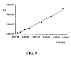
- FDA was not used as GrFFF fluorophore probe because it was not stable enough in the mobile phase. A high signal due to the hydrolysis of FDA prevented the detection of the stained cells. Peak areas of the fractograms obtained by GrFFF with PI probe were obtained and plotted against non-viable cells, calculated from FC and CC as commented above, in a X,Y-chart obtaining the calibration curve of the GrFFF system. Peak area values are also given in 25 30 Table 1 .
- FIG. 2 shows the calibration curve obtained for the Intec Red yeast standard.
- Table 1 Data obtained for the calibration of the GrFFF method with Intec Red yeast strain as a standard. PI was used as a staining probe. Intec Red yeast standard Concentration (g/l) FC / CC data GrFFF data Concentration (viable cells/ml) Concentration (non-viable cells/ml) Peak area S1 0.2032 8.96E+06 5.44E+06 2.01E+06 S2 0.4024 1.44E+07 8.77E+06 3.88E+06 S3 0.6040 2.27E+07 1.38E+06 9.31E+06 S4 0.8040 3.18E+07 1.93E+06 1.17E+07 S5 1.4008 5.68E+07 3.45E+06 2.47E+07 S6 2.5024 7.61E+07 4.62E+06 3.67E+07 S7 3.4988 1.01E+08 6.11E+06 5.28E+07
- FIG. 6 shows as an example the fractograms obtained for three commercial yeasts, Enoferm BDX, Uvaferm ALB and Uvaferm BC analyzed by GrFFF.
- the highest area corresponds to Uvaferm ALB yeast strain and means a high number of stained non-viable cells in the sample. Integration of sample peaks was performed to obtain by external calibration the non-viable cell/g for each sample. Viable cells/g were calculated for each sample using the non-viable results and CC data. Viability results are expressed as viable cells/g.
- FIG. 3, FIG. 4 and FIG. 5 illustrate the results obtained by FC. Flow cytometry shows the viable cell region (+ FDA) and the non-viable cell region (+ PI). For these samples a viability of 35.9%, 52.8% and 43.8% was respectively obtained. For the total cell concentration Coulter counter was used and results were expressed in cell/ml.
- the limit of detection of the proposed GrFFF method was calculated from the calibration curve as the lowest level of concentration that gave an area equal to the intercept value plus three times the estimated standard deviation. The obtained value was 10 6 non-viable cells/ml, corresponding to 2 x 10 4 cells injected into the GrFFF setup.
- Two statistical tests, paired t -test and analysis of variance (ANOVA two-factor without replication) were applied to compare both analytical methods. For paired t -test calculated t (n 11) was 0.47, which was lower than t critical value (0.05, 11), 2.23. Moreover, the ANOVA calculated F value for the methods was 0.22, in contrast to the F critical value, which was 4.96. Both statistical tests indicated that no significant differences occurred at 95% confidence level between results obtained using GrFFF and FC/CC. 5
Landscapes
- Chemical & Material Sciences (AREA)
- General Health & Medical Sciences (AREA)
- Life Sciences & Earth Sciences (AREA)
- Health & Medical Sciences (AREA)
- Analytical Chemistry (AREA)
- Biochemistry (AREA)
- Physics & Mathematics (AREA)
- General Physics & Mathematics (AREA)
- Immunology (AREA)
- Pathology (AREA)
- Dispersion Chemistry (AREA)
- Engineering & Computer Science (AREA)
- Signal Processing (AREA)
- Measuring Or Testing Involving Enzymes Or Micro-Organisms (AREA)
Applications Claiming Priority (2)
| Application Number | Priority Date | Filing Date | Title |
|---|---|---|---|
| ES200302654A ES2239886B1 (es) | 2003-11-05 | 2003-11-05 | Metodo y aparato para la determinacion de la viabilidad celular. |
| PCT/ES2004/000492 WO2005045425A1 (fr) | 2003-11-05 | 2004-11-04 | Procede et appareil de determination de la viabilite cellulaire |
Publications (1)
| Publication Number | Publication Date |
|---|---|
| EP1688740A1 true EP1688740A1 (fr) | 2006-08-09 |
Family
ID=34566015
Family Applications (1)
| Application Number | Title | Priority Date | Filing Date |
|---|---|---|---|
| EP04798219A Withdrawn EP1688740A1 (fr) | 2003-11-05 | 2004-11-04 | Procede et appareil de determination de la viabilite cellulaire |
Country Status (3)
| Country | Link |
|---|---|
| EP (1) | EP1688740A1 (fr) |
| ES (1) | ES2239886B1 (fr) |
| WO (1) | WO2005045425A1 (fr) |
Cited By (6)
| Publication number | Priority date | Publication date | Assignee | Title |
|---|---|---|---|---|
| WO2007128737A3 (fr) * | 2006-05-04 | 2008-01-17 | Univ Bologna Alma Mater | Méthode et dispositif de fractionnement de cellules souches |
| WO2008065529A3 (fr) * | 2006-09-15 | 2008-09-04 | Multigene Vascular Systems Inc | Appareil et méthodes d'évaluation de produits à base de cellules |
| WO2011156249A3 (fr) * | 2010-06-07 | 2012-04-12 | Nexcelom Bioscience Llc | Mesure de viabilité et de concentration de levures |
| US8993259B2 (en) | 2012-05-02 | 2015-03-31 | Charles River Laboratories, Inc. | Method of viability staining with membrane permeable fluorescent dye and membrane impermeable fluorescence quencher |
| US9709500B2 (en) | 2012-05-02 | 2017-07-18 | Charles River Laboratories, Inc. | Optical method for detecting viable microorganisms in a cell sample |
| US10324036B2 (en) | 2012-05-02 | 2019-06-18 | Charles River Laboratories, Inc. | Porous planar cell capture system |
Family Cites Families (6)
| Publication number | Priority date | Publication date | Assignee | Title |
|---|---|---|---|---|
| US5193688A (en) * | 1989-12-08 | 1993-03-16 | University Of Utah | Method and apparatus for hydrodynamic relaxation and sample concentration NIN field-flow fraction using permeable wall elements |
| WO2001096025A2 (fr) * | 2000-06-14 | 2001-12-20 | Board Of Regents, The University Of Texas System | Systeme et procedes d'analyse de sous-populations de cellules |
| AU2001296516A1 (en) * | 2000-10-09 | 2002-04-22 | Aviva Biosciences Coropration | Compositions and methods for separation of moieties on chips |
| US6403378B1 (en) * | 2001-04-26 | 2002-06-11 | Guava Technologies, Inc. | Cell viability assay reagent |
| JP2002323490A (ja) * | 2001-04-26 | 2002-11-08 | Japan Science & Technology Corp | 藻類の生死判定試薬及び判定方法 |
| WO2003066661A2 (fr) * | 2001-07-03 | 2003-08-14 | Genentech, Inc. | Anticorps humains du dr4 et utilisations |
-
2003
- 2003-11-05 ES ES200302654A patent/ES2239886B1/es not_active Expired - Fee Related
-
2004
- 2004-11-04 EP EP04798219A patent/EP1688740A1/fr not_active Withdrawn
- 2004-11-04 WO PCT/ES2004/000492 patent/WO2005045425A1/fr not_active Ceased
Non-Patent Citations (1)
| Title |
|---|
| See references of WO2005045425A1 * |
Cited By (12)
| Publication number | Priority date | Publication date | Assignee | Title |
|---|---|---|---|---|
| WO2007128737A3 (fr) * | 2006-05-04 | 2008-01-17 | Univ Bologna Alma Mater | Méthode et dispositif de fractionnement de cellules souches |
| US8263359B2 (en) | 2006-05-04 | 2012-09-11 | Alma Mater Studiorum—Universita di Bologna | Method and device to fractionate stem cells |
| WO2008065529A3 (fr) * | 2006-09-15 | 2008-09-04 | Multigene Vascular Systems Inc | Appareil et méthodes d'évaluation de produits à base de cellules |
| WO2011156249A3 (fr) * | 2010-06-07 | 2012-04-12 | Nexcelom Bioscience Llc | Mesure de viabilité et de concentration de levures |
| CN103154264A (zh) * | 2010-06-07 | 2013-06-12 | 耐克思乐生物科学有限责任公司 | 酵母浓度和存活率的测定 |
| CN103154264B (zh) * | 2010-06-07 | 2015-11-25 | 耐克思乐生物科学有限责任公司 | 酵母浓度和存活率的测定 |
| US11473121B2 (en) * | 2010-06-07 | 2022-10-18 | Nexcelom Bioscience Llc | Yeast concentration and viability measurement |
| US8993259B2 (en) | 2012-05-02 | 2015-03-31 | Charles River Laboratories, Inc. | Method of viability staining with membrane permeable fluorescent dye and membrane impermeable fluorescence quencher |
| US8993260B2 (en) | 2012-05-02 | 2015-03-31 | Charles River Laboratories, Inc. | Fluorescence-based viability staining method using a membrane permeable flourescent dye and membrane impermeable fluorescence quencher |
| US9709500B2 (en) | 2012-05-02 | 2017-07-18 | Charles River Laboratories, Inc. | Optical method for detecting viable microorganisms in a cell sample |
| US10324036B2 (en) | 2012-05-02 | 2019-06-18 | Charles River Laboratories, Inc. | Porous planar cell capture system |
| US10976258B2 (en) | 2012-05-02 | 2021-04-13 | Charles River Laboratories, Inc. | Porous planar cell capture system and method of use |
Also Published As
| Publication number | Publication date |
|---|---|
| WO2005045425A1 (fr) | 2005-05-19 |
| ES2239886A1 (es) | 2005-10-01 |
| ES2239886B1 (es) | 2006-12-16 |
Similar Documents
| Publication | Publication Date | Title |
|---|---|---|
| EP1250585B1 (fr) | Procede et appareil d'analyse des globules dans un echantillon de sang entier | |
| US10585023B2 (en) | Method and devices for treating biological samples | |
| US7214299B2 (en) | Method for separation, identification and evaluation of microbes and cells | |
| US8748159B2 (en) | Method and apparatus for viable and nonviable prokaryotic and eukaryotic cell quantitation | |
| CN104880439B (zh) | 样本分析装置及样本分析方法 | |
| Karlberg et al. | Extraction based on the flow-injection principle: Part 3. Fluorimetric Determination of Vitamin B1 (Thiamine) by the Thiochrome Method | |
| US6803208B2 (en) | Automated epifluorescence microscopy for detection of bacterial contamination in platelets | |
| EP2326727A2 (fr) | Systèmes à base de cytométrie en flux, et procédés de détection de microbes | |
| Campbell | Flow cytometric analysis of autotrophic picoplankton | |
| Kotiaho et al. | Membrane inlet ion mobility spectrometry for on-line measurement of ethanol in beer and in yeast fermentation | |
| EP1688740A1 (fr) | Procede et appareil de determination de la viabilite cellulaire | |
| JPH02503747A (ja) | 微生物の定性および/または定量試験方法およびその方法を実施するための装置 | |
| Davey et al. | On the determination of the size of microbial cells using flow cytometry | |
| US20020018211A1 (en) | System and method to detect the presence of a target organism within an air sample using flow cytometry | |
| Pettipher | Preliminary evaluation of flow cytometry for the detection of yeasts in soft drinks | |
| EP1984700B1 (fr) | Ensemble de normes et procede de production | |
| Bruetschy et al. | Use of flow cytometry in oenology to analyse yeasts | |
| Sharrow | Overview of flow cytometry | |
| US20210053065A1 (en) | Methods for Screening and Subsequent Processing of Samples Taken from Non-Sterile Sites | |
| Sanz et al. | Determination of viable yeast cells by gravitational field‐flow fractionation with fluorescence detection | |
| Hughes et al. | Identification of immobilized bacteria by aminopeptidase profiling | |
| Garcia et al. | Use of fluorescent probes for determination of yeast cell viability by gravitational field‐flow fractionation | |
| Carvell et al. | Developments in using off-line radio frequency impedance methods for measuring the viable cell concentration in the brewery | |
| US20240167072A1 (en) | Method for the detection of particles | |
| Chunxiang et al. | Microbial sensor for on-line determination of microbial population in a fermenter |
Legal Events
| Date | Code | Title | Description |
|---|---|---|---|
| PUAI | Public reference made under article 153(3) epc to a published international application that has entered the european phase |
Free format text: ORIGINAL CODE: 0009012 |
|
| 17P | Request for examination filed |
Effective date: 20060602 |
|
| AK | Designated contracting states |
Kind code of ref document: A1 Designated state(s): AT BE BG CH CY CZ DE DK EE ES FI FR GB GR HU IE IS IT LI LU MC NL PL PT RO SE SI SK TR |
|
| DAX | Request for extension of the european patent (deleted) | ||
| 17Q | First examination report despatched |
Effective date: 20071213 |
|
| GRAP | Despatch of communication of intention to grant a patent |
Free format text: ORIGINAL CODE: EPIDOSNIGR1 |
|
| RAP1 | Party data changed (applicant data changed or rights of an application transferred) |
Owner name: UNIVERSIDAD DE BARCELONA |
|
| STAA | Information on the status of an ep patent application or granted ep patent |
Free format text: STATUS: THE APPLICATION IS DEEMED TO BE WITHDRAWN |
|
| 18D | Application deemed to be withdrawn |
Effective date: 20101231 |