EP1750131B1 - Procédé de jugement de la caractéristique de la tumeur maligne - Google Patents
Procédé de jugement de la caractéristique de la tumeur maligne Download PDFInfo
- Publication number
- EP1750131B1 EP1750131B1 EP06015779A EP06015779A EP1750131B1 EP 1750131 B1 EP1750131 B1 EP 1750131B1 EP 06015779 A EP06015779 A EP 06015779A EP 06015779 A EP06015779 A EP 06015779A EP 1750131 B1 EP1750131 B1 EP 1750131B1
- Authority
- EP
- European Patent Office
- Prior art keywords
- parameter
- threshold value
- recurrence
- risk
- expression level
- Prior art date
- Legal status (The legal status is an assumption and is not a legal conclusion. Google has not performed a legal analysis and makes no representation as to the accuracy of the status listed.)
- Not-in-force
Links
Images




Classifications
-
- C—CHEMISTRY; METALLURGY
- C12—BIOCHEMISTRY; BEER; SPIRITS; WINE; VINEGAR; MICROBIOLOGY; ENZYMOLOGY; MUTATION OR GENETIC ENGINEERING
- C12Q—MEASURING OR TESTING PROCESSES INVOLVING ENZYMES, NUCLEIC ACIDS OR MICROORGANISMS; COMPOSITIONS OR TEST PAPERS THEREFOR; PROCESSES OF PREPARING SUCH COMPOSITIONS; CONDITION-RESPONSIVE CONTROL IN MICROBIOLOGICAL OR ENZYMOLOGICAL PROCESSES
- C12Q1/00—Measuring or testing processes involving enzymes, nucleic acids or microorganisms; Compositions therefor; Processes of preparing such compositions
- C12Q1/48—Measuring or testing processes involving enzymes, nucleic acids or microorganisms; Compositions therefor; Processes of preparing such compositions involving transferase
- C12Q1/485—Measuring or testing processes involving enzymes, nucleic acids or microorganisms; Compositions therefor; Processes of preparing such compositions involving transferase involving kinase
-
- A—HUMAN NECESSITIES
- A61—MEDICAL OR VETERINARY SCIENCE; HYGIENE
- A61P—SPECIFIC THERAPEUTIC ACTIVITY OF CHEMICAL COMPOUNDS OR MEDICINAL PREPARATIONS
- A61P35/00—Antineoplastic agents
-
- G—PHYSICS
- G01—MEASURING; TESTING
- G01N—INVESTIGATING OR ANALYSING MATERIALS BY DETERMINING THEIR CHEMICAL OR PHYSICAL PROPERTIES
- G01N33/00—Investigating or analysing materials by specific methods not covered by groups G01N1/00 - G01N31/00
- G01N33/48—Biological material, e.g. blood, urine; Haemocytometers
- G01N33/50—Chemical analysis of biological material, e.g. blood, urine; Testing involving biospecific ligand binding methods; Immunological testing
- G01N33/53—Immunoassay; Biospecific binding assay; Materials therefor
- G01N33/575—Immunoassay; Biospecific binding assay; Materials therefor for cancer
-
- G—PHYSICS
- G01—MEASURING; TESTING
- G01N—INVESTIGATING OR ANALYSING MATERIALS BY DETERMINING THEIR CHEMICAL OR PHYSICAL PROPERTIES
- G01N2333/00—Assays involving biological materials from specific organisms or of a specific nature
- G01N2333/90—Enzymes; Proenzymes
- G01N2333/91—Transferases (2.)
- G01N2333/912—Transferases (2.) transferring phosphorus containing groups, e.g. kinases (2.7)
Definitions
- the present invention relates to a method for judging a risk of recurrence of breast cancer based on an analysis result obtained through analysis of protein of a tumor cell collected from a patient having the breast cancer.
- a feature of a malignant tumor can be judged based on a measured expression level of HER2 of a malignant tumor cell.
- the expression level of the HER2 is used to predict prognosis of early breast cancer.
- the HER2 is a growth factor receptor having a tyrosine kinase activity, and it is generally interpreted that the malignant tumor in which HER2 is expressed at a high level involves a high risk of recurrence, while the risk of recurrence is low in the malignant tumor in which HER2 is expressed at a low level.
- the malignant tumor undergoing the low HER2 expression level still involves some risk of recurrence, and the prognosis of the malignant tumor based on the expression level of the HER2 has failed to be very accurate.
- Ishihara et al. (Biochimica et Biophysica Acta, vol. 1741, no. 3, 2005, 226-233 ) describes a cancer diagnostic system based on a cyclin dependent kinase (CDK) profiling technology by which the expression and activities of the CDK molecules in lysate of tumor tissue is analysed.
- CDK cyclin dependent kinase
- a main object of the present invention is to provide a method for judging a risk of recurrence of breast cancer with a high accuracy based on an analysis result obtained through analysis of molecules expressed in a tumor cell collected from a patient having the breast cancer.
- a first aspect of the present invention relates to methods for judging risk of recurrence of breast cancer, comprising the steps of:
- the method capable of accurately judging the risk of recurrence of breast cancer based on the analysis results of the cyclin dependent kinases (CDKs) and the growth factor receptor expressed in the tumor cell collected from the patient having the breast cancer can be provided as recited in claim 1.
- CDKs cyclin dependent kinases
- the first parameter, the second parameter, the third parameter and the fourth parameter are obtained as an initial step.
- the first parameter is based on the activity value and the expression level of the first cyclin dependent kinase (first CDK) contained in the tumor cell collected from the patient having the malignant tumor as recited in claim 1.
- the second parameter is based on the activity value and the expression level of the second cyclin dependent kinase (second CDK) contained in the tumor cell as recited in claim 1.
- the third parameter is calculated from the first parameter and the second parameter as recited in claim 1.
- the fourth parameter is based on the expression level of the growth factor receptor contained in the tumor cell as recited in claim 1.
- the method according to the present embodiment includes the first comparing step for comparing the third parameter to the first threshold value, the second comparing step for comparing the fourth parameter to the second threshold value, and the step for judging the risk of recurrence of breast cancer based on the results of these comparing steps as recited in claim 1.
- Reference examples of the malignant tumor to which the foregoing judging method is applied include a malignant tumor derived from a hematopoietic organ, carcinoma derived from an epithelial cell, sarcoma and the like.
- Examples of the malignant tumor derived from the hematopoietic organ include leukemia, malignant lymphoma and the like.
- Examples of the carcinoma include breast cancer, stomach cancer, large intestine cancer, esophagus cancer, prostate cancer and the like.
- Examples of the sarcoma include osteogenic sarcoma, sarcoma developed in soft parts and the like.
- the tumor cell of the breast cancer used in the method is preferably a malignant tumor cell in "stage IIB" in stage classification.
- the stage classification is a type of classification in which a malignancy of the malignant tumor is shown, wherein the malignant tumor is classified into, from lower levels of the malignancy, stages I, IIA, IIB, IIIA, IIIB and IV
- the stage classification is based on TNM classification.
- the TNM classification is a stage classification of the malignant tumor according to the Unio Internationalis Contra Cancrum (UICC, International Union against Cancer). "T” denotes a size of a primary tumor and ranges from T0 (primary lesion cannot be confirmed) through T4 (tumor is exposed out of body).
- N denotes a degree of invasion into lymphonodus in vicinity and ranges from N0 (no metastasis into lymphonodus) through N3 (metastasis into lymphonodus in vicinity of a median part of a body (lymphonodus in vicinity of breast bone) is suspected).
- M denotes presence or absence of remote metastasis and ranges from M0 (no remote metastasis) through M1 (remote metastasis is confirmed).
- the malignant tumor in the stage IIB indicates the malignant tumor in "T2, N1, M0" or "T3, N0, M0" in the TNM classification.
- This judging method is used for judging the risk of recurrence.
- the recurrence is diagnosed when the malignant tumor recurs in a body part from which the tumor is extirpated after a predetermined length of time has passed since the extirpation and when the tumor cell, which is separated from the primary tumor, is transported into a remote tissue to independently proliferate therein.
- the possibility of the recurrence depends on the proliferation potency, survival potency and migratory aptitude of the tumor cell, and the like.
- the first parameter is obtained from the ratio of the activation value and the expression level of CDK1, while the second parameter is obtained from the ratio of the activation value and the expression level of CDK2.
- a ratio of the activation value and the expression level a value obtained by dividing the activation value by the expression level (specific activity), a value obtained by dividing the expression level by the activation value (inverse number of specific activity), or the like, can be used.
- the third parameter is obtained based on the ratio of the first parameter and the second parameter.
- the ratio of the first parameter and the second parameter can adopt a value obtained by dividing the first parameter by the second parameter, a value obtained by dividing the second parameter by the first parameter, or the like.
- the fourth parameter is obtained based on the expression level of the growth factor receptor HER2 included in the tumor cell.
- the expression level may be solely used, or a value calculated from addition, subtraction, multiplication and division of the expression level and the activation value may be used as the fourth parameter.
- the expression level is preferably solely used.
- the third parameter is compared to the corresponding threshold value (hereinafter, referred to as first threshold value).
- the first threshold value is appropriately set for the method of claim 1.
- the first threshold value can be set as a value capable of dividing a group of breast cancer patients in to a high-value group and a low value group in a certain proportion.
- the proportion of the separation of the high-value group is preferably at least 30% of the group of patients, for example.
- the proportion higher than 30% is more preferably set to the high-value group so that the recurrence does not occur in the patients divided into the low-value group.
- the first threshold value can adopt such values that the proportion of the high-value group: low-value group in the group of patients results in 30 : 70, preferably 40 : 60, or more preferably 50 : 50 (median) or 60 : 40. It is unnecessary to so strictly set the proportion of the separation because such values that can divide the group of patients as exactly 50 : 50 or 60 : 40 may not possibly exist. For example, when the number of the patients constituting the group of patients is 99, there does not exist such a value that can divide the group of patients as 50 : 50, in which case, the proportion of the separation may be set to 50 : 49.
- the first parameter or the second parameter, or the both parameters may have a very small value or a very large value, and the third parameter thereby may show a value significantly different to the first threshold value.
- the fourth parameter is compared to the threshold value corresponding thereto (hereinafter, referred to as the second threshold value).
- the second threshold value is appropriately set for the method of claim 1.
- the second threshold value corresponding to the presence or absence of the recurrence with a high possibility can be set when the recurrence risk of the tumor cell whose presence or absence of the recurrence after a predetermined length of time is known is judged in combination with the result of the first comparing step.
- the tumor cell collected from the patient is used to prepare a measurement sample used for the measurements of the activation value and the expression level.
- a method of preparing the sample is not particularly limited as far as the activation values and the expression levels of the CDKs and the growth factor receptor specified in claim 1 can be measured.
- a cell lysate can be used as the measurement sample.
- the cell lysate is a sample obtained in such a manner that a buffer is added to the tumor cell collected from the patient so that the tumor cell is physically and/or chemically ground in the buffer.
- a supernatant obtained through centrifugation of the cell lysate is preferably used as the measurement sample.
- HER2 precipitates obtained through the centrifugation of the cell lysate are preferably used as the measurement sample.
- the buffer used in this specification may appropriately include a surface active agent or a protease inhibitor.
- a method of measuring the activation values of the CDKs or the growth factor receptor specified in claim 1 is not particularly limited, and any publicly known method can be adopted.
- any publicly known method can be mentioned.
- ATP Y -[ 32 P]-ATP
- the measurement sample, a substrate and ( Y -[ 32 P]-ATP are mixed with one another, and 32 P is introduced into the substrate through reactions of the kinases.
- a label amount of the phosphorylated substrate labeled with 32 P is measured, and the phosphorylated substrate is determined based on a calibration curve generated by means of a calibrator.
- the activations values of the kinases are calculated based on an amount of the determined phosphorylated substrate.
- a method recited in No. 2002-335997 of the Japanese Patent Application Laid-Open, in which any radioactive material is not used can be adopted.
- a sample including one or both of the kinases, a substrate and adenosine 5'-O-(3-thiotriphosphate) (ATP- Y S) are mixed with one another, and a monothiophosphoric group is introduced into the substrate through the reactions of the kinases.
- a labeled fluorescent material or a labeled enzyme is bound with a sulfuric atom of the introduced monothiophosphoric group so that the substrate is labeled.
- a reagent kit comprising a reagent including an anti-CDK antibody, a reagent including the CDK substrate, a reagent including ATP- Y S and a reagent including a labeled substance which can be bound with the phosphoric substrate can be used.
- the CDK substrate and ATP- Y S may be housed in one container.
- a method of measuring the expression levels of the CDKs or the growth factor receptor specified in claim 1 is not particularly limited, and any publicly known method can be used for the measurement.
- any publicly known method can be used for the measurement.
- ELISA, western blotting, a method recited in No. 2003-130871 of the Japanese Patent Application Laid-Open, and the like, can be used.
- the measured value of the expression level of the protein is variable depending on if the number of the tumor cells included in the measurement sample is large or small. Therefore, the measured value of the expression level is preferably corrected.
- an expression level of protein coded by a housekeeping gene (hereinafter, referred to as housekeeping protein) can be used for the correction of the measured value of the expression level.
- the housekeeping protein is a protein constantly expressed at a certain level in any cell, examples of which include glyceraldehyde-3-phosphate dehydrogenase (GAPDH), actin and the like.
- the measured value of the expression level of the measurement sample can be corrected in such a manner that not only the expression level of the protein of the measurement sample but also the expression level of the housekeeping protein is measured, and the expression level of the protein of the measurement sample is divided by the expression level of the housekeeping protein.
- the expression level or the ratio of the activation value and the expression level is used as the first parameter
- the expression level or the ratio of the activation value and the expression level is used as the second parameter
- the ratio of the first and second parameters is used as the third parameter
- the expression levels of the first and second CDKs are measured.
- the expression level of the housekeeping protein is offset in the calculation, which makes the foregoing correction unnecessary.
- the feature of the malignant tumor is determined based on the comparison result of the first comparing step and the comparison result of the second comparing step.
- the malignant tumor can be classified into any of three groups respectively having the recurrence risks of different degrees (, high, medium and low) based on these results.
- any of methods 1 - 5 described below can be used, for example, so that the recurrence risk can be judged.
- the result of the comparison of the third parameter to the first threshold value and the result of the comparison of the fourth parameter to the second threshold value are combined so that the recurrence risk is judged.
- the recurrence risk appears to be low, which is judged as "A1".
- the parameter in one of the comparison results is at least the relevant threshold value, and the parameter in the other is below the relevant threshold value, the recurrence risk is judged as "B1" having the recurrence risk higher than that of the tumor cell judged as "A1".
- the recurrence risk is judged as "C1" having the recurrence risk higher than that of the tumor cell judged as "B1".
- the method 2 is a judging method based on a flow chart shown in Fig. 1 .
- the third parameter is compared to the first threshold value (Step 1), and the recurrence risk is judged as "C2" when the third parameter is at least the threshold value.
- the fourth parameter is compared to the second threshold value (Step 2). Then, the recurrence risk is judged as "B2" having the recurrence risk lower than that of the tumor cell judged as "C2" when the fourth parameter is at least the threshold value.
- the recurrence risk is judged as "A2" having the recurrence risk lower than that of the tumor cell judged as "B2". This method places an emphasis on the comparison in which the CDKs are used.
- the method 3 is a judging method based on a flow chart shown in Fig. 2 .
- the fourth parameter is compared to the second threshold value (Step 1), and the recurrence risk is judged as "C3" when the fourth parameter is at least the threshold value.
- the third parameter is compared to the first threshold value (Step 2). Then, the recurrence risk is judged as "B3" having the recurrence risk lower than that of the tumor cell judged as "C3" when the third parameter is at least the threshold value.
- the recurrence risk is judged as "A3" having the recurrence risk lower than that of the tumor cell judged as "B3". This method places an emphasis on the comparison in which the growth factor is used.
- the third parameter is compared to the first threshold value, and the recurrence risk is judged as "A4" when the third parameter is below the threshold value.
- the fourth parameter is compared to the second threshold value. Then, the recurrence risk is judged as "B4" having the recurrence risk higher than that of the tumor cell judged as "A4" when the fourth parameter is below the threshold value.
- the recurrence risk is judged as "C4" having the recurrence risk higher than that of the tumor cell judged as "B4".
- the fourth parameter is compared to the second threshold value, and the recurrence risk is judged as "A5" when the fourth parameter is below the threshold value.
- the third parameter is compared to the first threshold value. Then, the recurrence risk is judged as "B5" having the recurrence risk higher than that of the tumor cell judged as "A5" when the fourth parameter is below the threshold value.
- the recurrence risk is judged as "C5" having the recurrence risk higher than that of the tumor cell judged as "B5".
- Which of the foregoing methods 1 - 5 is used can be decided in consideration of the type of the malignant cell, the type of the parameter, comparison to clinical data and the like. Any of the methods 1 - 3 may be used in the case of judging the recurrence risk of the malignant tumor.
- the CDK is deeply involved in the growth of the cell. Therefore, the proliferation potency of the tumor cell can be accurately grasped when the comparing steps using the CDKs are implemented as recited in claim 1. As described earlier, the proliferation potency of the cell is a key factor which influences the recurrence risk of the cell.
- the growth factor receptor HER2 mainly relates to activation of a growth signal path of the cell. Therefore, the recurrence risk of the tumor cell can be predicted when the comparing steps using the growth factor receptor are implemented. However, the growth signal path is subjected to complicated adjustments while transmitting a growth signal, and the risk of recurrence thereby cannot be accurately predicted.
- the judgement result of the malignant tumor can be a barometer in selecting a therapeutic method.
- the method can assist a decision making if an aggressive therapy such as a treatment in which an anticancer agent is used should be conducted or a relatively mild therapy such as a hormone therapy should be conducted to the patient diagnosed of the malignant tumor. Further, a given dose and a type of the anticancer agent to be used can be adjusted depending on the judgement result.
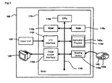
- Fig. 3 is a block diagram showing the structure of the computer system 100.
- This system 100 is mainly configured by body 110, input unit 130 for inputting necessary data to the body 110, and display unit 120.
- This system 100 can include a portable recording medium 140.
- a program 140a may be recorded in the portable recording medium 140, or may be stored in a memory 110b, a memory 110c and/or a memory 110c.
- CPU 110a, memories 110b to 110d, Input/Ourput Interface (I/O interface) 110f, image output interface 110h, and reading device 110e are connected by a bus 110i.
- I/O interface Input/Ourput Interface
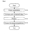
- Fig. 4 is a flow chart showing the processing sequence of the computer program for executing above-mentioned Method 1. This program is stored in memory 110d.
- CPU110a reads out a threshold corresponding to the third parameter and a threshold corresponding to the fourth parameter which are preliminarily stored in memory 110d as data of program. CPU110a executes comparison of the third parameter and the threshold, and comparison of the fourth parameter and the threshold (Step S11).
- CPU110a executes a determination based on comparison results (Step S12). Concretely, CPU110a determines the sample as "High” risk of recurrence in the case that the third parameter is equal to or more than the threshold and the fourth parameter is equal to or more than the threshold. CPU110a determines the sample as "Intermediate” risk of recurrence in the case that the third parameter is equal to or more than the threshold and the fourth parameter is less than the threshold. CPU110a determines the sample as "Intermediate” risk of recurrence in the case that the third parameter is less than the threshold and the fourth parameter is equal to or more than the threshold. CPU110a determines the sample as "Low” risk of recurrence in the case that the third parameter is less than the threshold and the fourth parameter is less than the threshold.
- CPU110a stores the determination result in the RAM 110c, and output to the display unit 120 through the I/O interface 110h (Step S13).
- activity, expression level, and parameter data is input with the input unit 130. It is not limited to this embodiment however, and for instance, the body 110 may automatically obtain the activity, expression level, and parameter data through I/O interface from measuring apparatus(es). Moreover, in this embodiment, although ration of CDK2 specific activity and CDK1 specific activity is input, expression level and activity of CDK2, and expression level and activity of CDK1 may be input. In this case, CPU110a can calculate ration of CDK2 specific activity and CDK1 specific activity, and compare the ration to the threshold.
- Measurement samples i - xii were prepared from 12 specimens of tumor cell blocks (13 - 92 mg) extirpated from 12 breast cancer patients (patients i - xii) in the stage IIB as follows.
- a buffer A including 0.1w / v% of Nonidet P-40 (Calbiochem Inc.), 50 mM of tris-hydrochloric acid (pH 7.4), 5 mM of EDTA, 50 mM of sodium fluoride, 1 mM of sodium orthovanadate, 100 ⁇ l/ml of protease inhibitor cocktail (SIGMA CORPORATION)), and the tumor cell block were housed in a tube so that the tumor cell block in the buffer was approximately 150 mg/ml.
- a buffer A including 0.1w / v% of Nonidet P-40 (Calbiochem Inc.), 50 mM of tris-hydrochloric acid (pH 7.4), 5 mM of EDTA, 50 mM of sodium fluoride, 1 mM of sodium orthovanadate, 100 ⁇ l/ml of protease inhibitor cocktail (SIGMA CORPORATION)
- the tumor cell block was homogenized in the buffer A by means of an electric homogenizer so that the tumor cells were ground, and a cell lysate was thereby prepared.
- the cell lysate was centrifuged at 4°C and 15,000 rpm for five minutes, and precipitates were used as the HER2 measurement sample, while a supernatant was used as the CDK measurement sample.
- a buffer B (including 1% of TritonX-100, 0.1% of sodium deoxycholic acid, 0.1% of SDS, 50 mM of tris-hydrochloric acid (pH 8.0), 150 mM of NaCl, 0.5 mM of EDTA, and 0.2% of protease inhibitor cocktail (SIGMA CORPORATION)) was added to the HER2 measurement sample, and the resulting substance was centrifuged at 15,000 rpm for five minutes.
- TritonX-100 0.1% of sodium deoxycholic acid, 0.1% of SDS, 50 mM of tris-hydrochloric acid (pH 8.0), 150 mM of NaCl, 0.5 mM of EDTA, and 0.2% of protease inhibitor cocktail (SIGMA CORPORATION)
- the supernatant was obtained, and a SDS loading buffer (including 200 mM of tris-hydrochloric acid (pH 6.8), 40% of glycerol, 8% of SDS, 10% of 2-melcaptoethanol) in the amount of 1/3 of the supernatant was added to the supernatant. Then, the resulting substance was heated at 100°C for five minutes.
- a SDS loading buffer including 200 mM of tris-hydrochloric acid (pH 6.8), 40% of glycerol, 8% of SDS, 10% of 2-melcaptoethanol
- Protein in the gel was transferred onto the Immobilon FL membrane (Millipore Corporation) at 100V and 4°C for an hour by means of the Mini Trans-Blot cell transfer system (Bio-Rad Laboratories, Inc.).
- the membrane was blocked with a blocking reagent A (including 4% of BSA, 25 mM of tris-hydrochloric acid (pH 7.4), 150 mM of NaCl and 0.02% of Tween 20) at 37°C for an hour, and washed with a washing solution A (including 25 mM of tris-hydrochloric acid (pH 7.4), 150 mM of NaCl, and 0.02% of Tween 20).
- a blocking reagent A including 4% of BSA, 25 mM of tris-hydrochloric acid (pH 7.4), 150 mM of NaCl and 0.02% of Tween 20
- a rabbit anti-HER2 antibody (first antibody: Upstate Group, Inc.) solution was blotted in the membrane so that the HER2 of the membrane and the first antibody were reacted with each other.
- the membrane was washed with 20 ml of the washing solution A, and thereafter, 5 ml of a biotin-labeled goat anti-rabbit IgG-B (second antibody: Santa Cruz Biotechnology Inc.) solution was blotted in the membrane so that the first and second antibodies in the membrane were reacted with each other.
- a biotin-labeled goat anti-rabbit IgG-B second antibody: Santa Cruz Biotechnology Inc.
- the membrane was washed with 20 ml of the washing solution A, and thereafter, 5 ml of an Alexa fluor 488 conjugated streptabidin (Molecular Probes Inc.) solution as fluorochrome was blotted in the membrane so that the second antibody and the fluorochrome were reacted with each other.
- an Alexa fluor 488 conjugated streptabidin Molecular Probes Inc.
- the membrane was washed with 20 ml of the washing solution A, and thereafter, an amount of HER2 adsorbed to the membrane was analyzed and measured by the Molecular Imager FX (Bio-Rad Laboratories, Inc.) which was a fluorescence image analyzer.
- the expression level of the GAPDH was measured in an experimental procedure similar that of the measurement of the expression level of the HER2 except for the use of the supernatant of the cell lysate as the measurement sample and the use of not the rabbit anti-HER2 antibody but a rabbit anti-GAPDH antibody as the first antibody.
- the GAPDH expression level was used to correct the measured value of the HER2 expression level.
- a value shown below as the HER2 expression level is a value after the correction using the GAPDH expression level (value of HER2 expression level / GAPDH expression level).
- the measurement result of the HER2 expression level is shown in the following tables 1 and graph of Fig. 5 .
- each of the CDK measurement samples was housed in each well of a blotter set in a PVDF membrane (Millipore Corporation). Then, the CDK measurement sample was suctioned from a bottom surface of the well, which is a rear surface of the membrane, at a negative pressure of approximately 250 mmHg for approximately 30 seconds so that protein in the CDK measurement sample was adsorbed to the membrane.
- washing solution B including 25 mM of tris-hydrochloric acid (pH 7.4) and 150 mM of NaCl was housed in each well and suctioned at the negative pressure of 500 mmHg for 15 seconds so that the membrane was washed.
- a blocking reagent B 4% of BSA, 25 mM of tris-hydrochloric acid (pH 7.4) and 150 mM of NaCl was housed in each well and left in a stationary state for 15 minutes, and suctioned at the negative pressure of 500 mmHg for 15 seconds so that the membrane was blocked.
- washing solution B 100 ⁇ l of the washing solution B was housed in each well and suctioned at the negative pressure of 500 mmHg for 15 seconds so that the membrane was washed.
- washing solution B 100 ⁇ l of the washing solution B was housed in each well and suctioned at the negative pressure of 500 mmHg for 15 seconds so that the membrane was washed.
- FITC-labeled streptavidin 50 ⁇ l of a labeled solution including FITC-labeled streptavidin was housed in each well and left in the stationary state at room temperature for approximately 30 minutes so that the second antibody in the membrane was FITC-labeled. Thereafter, the suction was applied thereto from the bottom surface of the well at the negative pressure of 500 mmHg for approximately 15 minutes.
- washing solution B 50 ⁇ l of the washing solution B was housed in each well and suctioned at the negative pressure of 500 mmHg for 15 seconds, which was repeated five times so that the membrane was washed.
- the membrane was removed from the blotter, washed with 20% methanol for approximately five minutes and dried at room temperature for approximately 20 minutes. Thereafter, fluorescence intensity of the protein adsorbed to the membrane was analyzed and measured by the Molecular Imager FX (Bio-Rad Laboratories Inc.) which was the fluorescence image analyzer. The measurement value was calculated based on the calibration curve.
- the calibration curve was generated in such a manner that 50 ⁇ l of a solution obtained by dissolving recombined CDK1s of five different concentrations in a washing solution B including 0.005% of Nonidet P-40 and 50 ⁇ g/ml of BSA was injected into each well processed in a manner similar to the foregoing description and FITC-labeled in an experimental procedure similar to the foregoing description and fluorescence intensity thereof was measured so that a relationship between the fluorescence intensity and the expression level of the CDK1 was expressed.
- the expression level of the CDK2 was measured in an experimental procedure similar to the measurement of the expression level of the CDK1 described earlier except for the use of not the rabbit anti-CDK1 antibody but a rabbit anti-CDK2 antibody as the first antibody.
- the buffer A 500 ⁇ l of the buffer A was housed in an Eppendorf tube of 1.5 ml, and the CDK measurement sample was added thereto.
- the CDK measurement sample was prepared so that a total protein amount in the mixed solution housed in the tube was 100 ⁇ g and then added.
- the beads were washed three times with a beads washing buffer (including 0.1 w/v% of Nonidet P-40 and 50 mM of tris-hydrochloric acid (pH 7.0)) and suspended in 15 ⁇ l of the lysis buffer A again.
- a beads washing buffer including 0.1 w/v% of Nonidet P-40 and 50 mM of tris-hydrochloric acid (pH 7.0)
- a sample including Sepharose beads with which the CDK1 is bound via the anti-CDK1 antibody could be obtained.
- a substrate solution of the CDK1 including 10 ⁇ g of histone H1 (Upstate Biotechnology Inc.), 5 mM of ATP- Y S (SIGMA CORPORATION), and 20mM of tris-hydrochloric acid (pH 7.4) and 0.1% of TritonX-100 was added to the sample.
- the substrate solution was prepared so that a total amount of the mixed solution housed in the tube was 50 ⁇ l and then added.
- the resulting substance was shaken at 37°C for 10 minutes to induce the kinase reaction so that the monothiophosphoric acid group was introduced into the histone H1.
- the foregoing substance was centrifuged at 2,000 rpm for 20 seconds so that the beads were precipitated. As a result, a supernatant in the amount of 18 ⁇ l was obtained.
- 15 ⁇ l of a binding buffer including 150 mM of tris-hydrochloric acid (pH 9.2) and 5mM of EDTA
- 10 mM of an iodoacetylbiotin solution including 100 mM of tris-hydrochloric acid (pH 7.5) and 1 mM of EDTA
- a binding buffer including 150 mM of tris-hydrochloric acid (pH 9.2) and 5mM of EDTA
- 10 mM of an iodoacetylbiotin solution including 100 mM of tris-hydrochloric acid (pH 7.5) and 1 mM of EDTA
- a sample including 0.4 ⁇ g of the monothiophosphoric acid substrate with which the iodoacetylbiotin was bound was blotted onto the PVDF membrane by means of the slot blotter.
- the PVDF membrane was blocked with a solution including 1w/v% of BSA, and streptavidin-FITC (Vector Laboratories Inc.) was added thereto, which was reacted at 37°C for an hour.
- the PVDF membrane was washed with 50 mM of the washing solution B three times.
- the calibration curve was generated in such a manner that a solution including proteins having two different concentrations (biotin-labeled immunoglobulin) were blotted onto the PVDF membrane, FITC-labeled in a method similar to the foregoing description, and the fluorescence intensities of the proteins were measured with the fluorescence image analyzer. Therefore, an activation IU (unit) of the measured CDK1 denotes a value indicating fluorescence intensity equal to the fluorescence amount when the protein is 1ng.
- the activation value of the CDK2 was measured in a manner similar to the measurement of the activation value of the CDK1 except for the use of the anti-CDK2 antibody (Santa Cruz Biotechnology Inc.).
- CDK specific activity (mU/ng) was calculated from the CDK activation value and the CDK expression value calculated above in the following formula.
- CDK specific activity CDK activation value / CDK expression level
- a threshold value corresponding to a ratio of the specific activities of the CDK1 and the CDK2 (CDK2 specific activity / CDK1 specific activity; hereinafter, referred to as ratio of specific activities) was set, and a threshold value corresponding to the expression level of the HER2 was set. Then, a result obtained from comparing the ratio of the specific activities to the relevant threshold value and a result obtained from comparing the expression level of the HER2 to the relevant threshold value were combined so that the feature of the tumor cell was judged.
- the threshold value for the ratio of the specific activities was set to 46.
- the value is capable of separating 129 specimens as 65 : 64 when the ratios of the specific activities of the 129 tumor cell specimens collected from 129 breast cancer patients are calculated and listed from the smaller values of the ratio of the specific activities.
- the value of the ratio of the specific activities being at least 46 was judged as "High", while the value below 46 was judged as "Low”.
- the judgement was "Low” irrespective of the ratio of the specific activities.
- the judgement was "High” irrespective of the ratio of the specific activities.
- the threshold value for the expression level of the HER2 was set to 0.7.
- the value was set in terms of the presence or absence of the recurrence in the patients i - xii and the comparison results of the ratio of the specific activities and the corresponding threshold value. The judgement was "High” when the expression level of HER2 is at least 0.7, while “Low” when below 0.7.
- the proliferation potency, survival potency and migratory aptitude of the cell appear to be low, and the recurrence risk is judged as low.
- the tumor cell judged as "recurrence risk is INTERMEDIATE” is judged as having the recurrence risk higher than that of the tumor cell judged as “recurrence risk is LOW'.
- the tumor cell judged as "recurrence risk is HIGH” is judged as having the recurrence risk higher than that the tumor cell judged as "recurrence risk is INTERMEDIATE".
- the presence or absence of the recurrence (in the table, "-" denotes “no recurrence", and “+” denotes “recurrence is detected"), measurement results of the foregoing molecules, comparison results with respect to the threshold values and judgement result of the recurrence risk in relation to the patients i - xii are collectively shown in the following table 1. "The presence or absence of the recurrence” indicates that the follow research is conducted to the respective patients i - xii after the tumor cell blocks are extirpated from them to confirm whether or not the recurrence is diagnosed in five years from the extirpation.
- Table 1 recurrence specific activity of CDK1 specific activity of CDK2 comparison of ratio with threshold expression level of HER2 comparison of expression level with threshold risk of recurrence patient i - 0.0 26.0 Low 1.344 High INTERMEDIATE patient ii - 0.0 228.7 Low 0.055 Low LOW patient iii - 41.6 4219.4 High 0.048 Low INTERMEDIATE patient iv - 0.0 2167.3 High 0.108 Low INTERMEDIATE patient v - 38.4 2632.3 High 0.004 Low INTERMEDIATE patient vi - 0.0 3666.9 High 0.303 Low INTERMEDIATE patient vii + 0.0 8275.4 High 5.565 High HIGH patient viii + 322.0 5392.6 Low 2.257 High INTERMEDIATE patient ix + 290.5 30643.5 High 0.000 Low INTERMEDIATE patient x - 313.2 482.3 Low 0.022 Low LOW patient xi - 0.0 5621.0 High 0.633 Low INTERMEDIATE patient xii + 226.2
- the patients can be classified into the three groups respectively having the recurrence risks at different degrees, and the presence or absence of the recurrence can be thereby predicted. Further, the prediction result can be used as a key factor in deciding the therapeutic policy for the malignant tumor patient.
Landscapes
- Health & Medical Sciences (AREA)
- Life Sciences & Earth Sciences (AREA)
- Chemical & Material Sciences (AREA)
- Engineering & Computer Science (AREA)
- Immunology (AREA)
- Organic Chemistry (AREA)
- Molecular Biology (AREA)
- General Health & Medical Sciences (AREA)
- Biomedical Technology (AREA)
- Biochemistry (AREA)
- Microbiology (AREA)
- Zoology (AREA)
- Wood Science & Technology (AREA)
- Physics & Mathematics (AREA)
- Analytical Chemistry (AREA)
- Proteomics, Peptides & Aminoacids (AREA)
- Hematology (AREA)
- Biotechnology (AREA)
- Urology & Nephrology (AREA)
- Medicinal Chemistry (AREA)
- Genetics & Genomics (AREA)
- Cell Biology (AREA)
- Pathology (AREA)
- General Physics & Mathematics (AREA)
- Food Science & Technology (AREA)
- Bioinformatics & Cheminformatics (AREA)
- General Engineering & Computer Science (AREA)
- Biophysics (AREA)
- Nuclear Medicine, Radiotherapy & Molecular Imaging (AREA)
- Pharmacology & Pharmacy (AREA)
- General Chemical & Material Sciences (AREA)
- Chemical Kinetics & Catalysis (AREA)
- Animal Behavior & Ethology (AREA)
- Public Health (AREA)
- Veterinary Medicine (AREA)
- Measuring Or Testing Involving Enzymes Or Micro-Organisms (AREA)
- Investigating Or Analysing Biological Materials (AREA)
- Apparatus For Radiation Diagnosis (AREA)
Claims (5)
- Procédé in vitro pour juger du risque de récurrence de cancer du sein, comprenant les étapes de :obtenir en tant qu'un premier paramètre une première activité spécifique représentant un rapport d'une activité par rapport à un niveau d'expression de la kinase dépendante d'une cycline 1 (CDK1) contenue dans une cellule tumorale du cancer du sein, en tant qu'un deuxième paramètre une deuxième activité spécifique représentant un rapport d'une activité par rapport à un niveau d'expression de CDK2 contenue dans la cellule tumorale, en tant qu'un troisième paramètre un rapport de la première activité spécifique par rapport à la deuxième activité spécifique, et un quatrième paramètre basé sur le niveau d'expression de HER2 contenue dans la cellule tumorale;comparer une première valeur seuil au troisième paramètre, et une deuxième valeur seuil au quatrième paramètre; etjuger si le risque de récurrence est un risque élevé, un risque intermédiaire ou un risque faible sur la base des résultats de comparaison de l'étape de comparaison.
- Procédé selon la revendication 1, dans lequel le troisième paramètre est la deuxième activité spécifique/la première activité spécifique, dans lequel l'étape de jugement est effectuée en jugeant que le risque de récurrence est un risque élevé lorsque le troisième paramètre est supérieur à la première valeur seuil et le quatrième paramètre est supérieur à la deuxième valeur seuil.
- Procédé selon la revendication 1, dans lequel le troisième paramètre est la deuxième activité spécifique/la première activité spécifique, dans lequel l'étape de jugement est effectuée en jugeant que le risque de récurrence est un risque faible lorsque le troisième paramètre n'est pas supérieur à la première valeur seuil et le quatrième paramètre n'est pas supérieur à la deuxième valeur seuil.
- Produit de programme informatique agencé pour amener un processeur à exécuter le procédé des revendications 1 à 3.
- Support de stockage lisible par ordinateur, dans lequel un programme informatique est incorporé, où le programme consiste à amener un processeur à exécuter le procédé des revendications 1 à 3.
Applications Claiming Priority (1)
| Application Number | Priority Date | Filing Date | Title |
|---|---|---|---|
| JP2005223039 | 2005-08-01 |
Publications (2)
| Publication Number | Publication Date |
|---|---|
| EP1750131A1 EP1750131A1 (fr) | 2007-02-07 |
| EP1750131B1 true EP1750131B1 (fr) | 2008-07-09 |
Family
ID=37074189
Family Applications (1)
| Application Number | Title | Priority Date | Filing Date |
|---|---|---|---|
| EP06015779A Not-in-force EP1750131B1 (fr) | 2005-08-01 | 2006-07-28 | Procédé de jugement de la caractéristique de la tumeur maligne |
Country Status (4)
| Country | Link |
|---|---|
| US (1) | US7634362B2 (fr) |
| EP (1) | EP1750131B1 (fr) |
| AT (1) | ATE400815T1 (fr) |
| DE (1) | DE602006001701D1 (fr) |
Families Citing this family (3)
| Publication number | Priority date | Publication date | Assignee | Title |
|---|---|---|---|---|
| JP2007259846A (ja) * | 2006-02-28 | 2007-10-11 | Sysmex Corp | 悪性腫瘍の性質の判定方法 |
| JP5111902B2 (ja) * | 2007-03-14 | 2013-01-09 | シスメックス株式会社 | 癌の診断支援装置 |
| JP2010057486A (ja) * | 2008-09-02 | 2010-03-18 | Sysmex Corp | 化学療法に対するがん患者の応答の予測方法 |
Family Cites Families (13)
| Publication number | Priority date | Publication date | Assignee | Title |
|---|---|---|---|---|
| US6218529B1 (en) * | 1995-07-31 | 2001-04-17 | Urocor, Inc. | Biomarkers and targets for diagnosis, prognosis and management of prostate, breast and bladder cancer |
| GB2334578A (en) | 1998-02-18 | 1999-08-25 | Univ Liverpool | Diagnosis of cancer involving assay of levels of cyclin-dependent kinase (CDK) isoenzymes |
| WO2000047998A1 (fr) * | 1999-02-10 | 2000-08-17 | Cell Works Inc. | Caracterisation de classes de cellules cancereuses circulantes isolees a partir de fluides corporels et methodes d'utilisation |
| EP1512755A3 (fr) * | 1999-06-10 | 2007-11-21 | Sloan-Kettering Institute For Cancer Research | Marqueurs moléculaires du cancer de la prostate |
| EP1233060B1 (fr) * | 2001-02-14 | 2007-11-14 | Sysmex Corporation | Procédé pour la détérmination de l'activité du facteur de régulation du cycle cellulaire et procédé pour la diagnose de cancer utilisant le même |
| EP1412327A2 (fr) * | 2001-05-30 | 2004-04-28 | LG Biomedical Institute | Inhibiteurs de la proteine kinase destines au traitement d'une maladie |
| US20040018525A1 (en) * | 2002-05-21 | 2004-01-29 | Bayer Aktiengesellschaft | Methods and compositions for the prediction, diagnosis, prognosis, prevention and treatment of malignant neoplasma |
| US20040229294A1 (en) * | 2002-05-21 | 2004-11-18 | Po-Ying Chan-Hui | ErbB surface receptor complexes as biomarkers |
| JP4317188B2 (ja) * | 2003-02-26 | 2009-08-19 | シスメックス株式会社 | 細胞の検査方法 |
| EP3170906B1 (fr) * | 2003-06-24 | 2018-08-22 | Genomic Health, Inc. | Prédiction de probabilité de récurrence de cancer |
| US8021831B2 (en) | 2003-08-25 | 2011-09-20 | Board Of Regents, The University Of Texas System | Taxane chemosensitivity prediction test |
| FR2863275B1 (fr) * | 2003-12-09 | 2007-08-10 | Biomerieux Sa | Procede pour le diagnostic/pronostic du cancer du sein |
| WO2006133460A2 (fr) * | 2005-06-09 | 2006-12-14 | Yale University | Methodes de diagnostic et de traitement du cancer du sein d'apres le rapport her/er |
-
2006
- 2006-07-28 EP EP06015779A patent/EP1750131B1/fr not_active Not-in-force
- 2006-07-28 AT AT06015779T patent/ATE400815T1/de not_active IP Right Cessation
- 2006-07-28 DE DE602006001701T patent/DE602006001701D1/de active Active
- 2006-08-01 US US11/496,397 patent/US7634362B2/en not_active Expired - Fee Related
Also Published As
| Publication number | Publication date |
|---|---|
| EP1750131A1 (fr) | 2007-02-07 |
| US7634362B2 (en) | 2009-12-15 |
| DE602006001701D1 (de) | 2008-08-21 |
| ATE400815T1 (de) | 2008-07-15 |
| US20070141651A1 (en) | 2007-06-21 |
Similar Documents
| Publication | Publication Date | Title |
|---|---|---|
| Kawahara et al. | A targeted proteomic strategy for the measurement of oral cancer candidate biomarkers in human saliva | |
| Gharbi et al. | Evaluation of two-dimensional differential gel electrophoresis for proteomic expression analysis of a model breast cancer cell system | |
| US20150079078A1 (en) | Biomarkers for triple negative breast cancer | |
| Daily et al. | Using tears as a non-invasive source for early detection of breast cancer | |
| Phillips et al. | Novel prognostic markers in triple-negative breast cancer discovered by MALDI-mass spectrometry imaging | |
| Kim et al. | Proteomic analysis of primary colon cancer and synchronous solitary liver metastasis | |
| Zawadzka et al. | Phosphoprotein secretome of tumor cells as a source of candidates for breast cancer biomarkers in plasma | |
| Chen et al. | Elevation of serum l-lactate dehydrogenase B correlated with the clinical stage of lung cancer | |
| WO2012116229A2 (fr) | Compositions et procédés pour le traitement de profilage de tumeur personnalisé | |
| Pan et al. | Serum thymidine kinase 1 concentration as a prognostic factor of chemotherapy-treated non-Hodgkin’s lymphoma patients | |
| EP2371968B1 (fr) | Procédé de jugement de risque de récurrence de cancer, programme informatique et système informatique | |
| EP1826568B1 (fr) | Procédé de jugement d'un risque de récidive de la tumeur maligne | |
| Rentoft et al. | Transcriptional profiling of formalin fixed paraffin embedded tissue: pitfalls and recommendations for identifying biologically relevant changes | |
| Huang et al. | Total oxidant/antioxidant status in sera of patients with esophageal cancer | |
| Tenzer et al. | Integrated quantitative proteomic and transcriptomic analysis of lung tumor and control tissue: a lung cancer showcase | |
| Li et al. | Use of tryptic peptide MALDI mass spectrometry imaging to identify the spatial proteomic landscape of colorectal cancer liver metastases. | |
| Lee et al. | Proteomic heterogeneity of the extracellular matrix identifies histologic subtype-specific fibroblast in gastric cancer | |
| Gámez-Pozo et al. | MALDI profiling of human lung cancer subtypes | |
| Steiner et al. | Relative quantification of proteins in formalin-fixed paraffin-embedded breast cancer tissue using multiplexed mass spectrometry assays | |
| US20150254433A1 (en) | Methods and Models for Determining Likelihood of Cancer Drug Treatment Success Utilizing Predictor Biomarkers, and Methods of Diagnosing and Treating Cancer Using the Biomarkers | |
| US7634362B2 (en) | Method for judging feature of malignant tumor | |
| EP2764082B1 (fr) | Dosage srm/mrm de la protéine récepteur 2 de l'éphrine de type a | |
| EP1686377B1 (fr) | Procédé de prédiction de l'efficacité d'une chimiothérapie | |
| Diederiks et al. | Development of Tier 2 LC-MRM-MS protein quantification methods for liquid biopsies | |
| CN119757585A (zh) | 一种基于蛋白质组学诊断结直肠癌的产品及应用 |
Legal Events
| Date | Code | Title | Description |
|---|---|---|---|
| PUAI | Public reference made under article 153(3) epc to a published international application that has entered the european phase |
Free format text: ORIGINAL CODE: 0009012 |
|
| AK | Designated contracting states |
Kind code of ref document: A1 Designated state(s): AT BE BG CH CY CZ DE DK EE ES FI FR GB GR HU IE IS IT LI LT LU LV MC NL PL PT RO SE SI SK TR |
|
| AX | Request for extension of the european patent |
Extension state: AL BA HR MK YU |
|
| 17P | Request for examination filed |
Effective date: 20070807 |
|
| AKX | Designation fees paid |
Designated state(s): AT BE BG CH CY CZ DE DK EE ES FI FR GB GR HU IE IS IT LI LT LU LV MC NL PL PT RO SE SI SK TR |
|
| 17Q | First examination report despatched |
Effective date: 20071105 |
|
| GRAP | Despatch of communication of intention to grant a patent |
Free format text: ORIGINAL CODE: EPIDOSNIGR1 |
|
| GRAS | Grant fee paid |
Free format text: ORIGINAL CODE: EPIDOSNIGR3 |
|
| GRAA | (expected) grant |
Free format text: ORIGINAL CODE: 0009210 |
|
| AK | Designated contracting states |
Kind code of ref document: B1 Designated state(s): AT BE BG CH CY CZ DE DK EE ES FI FR GB GR HU IE IS IT LI LT LU LV MC NL PL PT RO SE SI SK TR |
|
| REG | Reference to a national code |
Ref country code: GB Ref legal event code: FG4D |
|
| REG | Reference to a national code |
Ref country code: CH Ref legal event code: EP |
|
| REF | Corresponds to: |
Ref document number: 602006001701 Country of ref document: DE Date of ref document: 20080821 Kind code of ref document: P |
|
| REG | Reference to a national code |
Ref country code: IE Ref legal event code: FG4D |
|
| NLV1 | Nl: lapsed or annulled due to failure to fulfill the requirements of art. 29p and 29m of the patents act | ||
| PG25 | Lapsed in a contracting state [announced via postgrant information from national office to epo] |
Ref country code: LT Free format text: LAPSE BECAUSE OF FAILURE TO SUBMIT A TRANSLATION OF THE DESCRIPTION OR TO PAY THE FEE WITHIN THE PRESCRIBED TIME-LIMIT Effective date: 20080709 Ref country code: NL Free format text: LAPSE BECAUSE OF FAILURE TO SUBMIT A TRANSLATION OF THE DESCRIPTION OR TO PAY THE FEE WITHIN THE PRESCRIBED TIME-LIMIT Effective date: 20080709 Ref country code: IS Free format text: LAPSE BECAUSE OF FAILURE TO SUBMIT A TRANSLATION OF THE DESCRIPTION OR TO PAY THE FEE WITHIN THE PRESCRIBED TIME-LIMIT Effective date: 20081109 |
|
| PG25 | Lapsed in a contracting state [announced via postgrant information from national office to epo] |
Ref country code: BG Free format text: LAPSE BECAUSE OF FAILURE TO SUBMIT A TRANSLATION OF THE DESCRIPTION OR TO PAY THE FEE WITHIN THE PRESCRIBED TIME-LIMIT Effective date: 20081009 Ref country code: ES Free format text: LAPSE BECAUSE OF FAILURE TO SUBMIT A TRANSLATION OF THE DESCRIPTION OR TO PAY THE FEE WITHIN THE PRESCRIBED TIME-LIMIT Effective date: 20081020 Ref country code: FI Free format text: LAPSE BECAUSE OF FAILURE TO SUBMIT A TRANSLATION OF THE DESCRIPTION OR TO PAY THE FEE WITHIN THE PRESCRIBED TIME-LIMIT Effective date: 20080709 Ref country code: LV Free format text: LAPSE BECAUSE OF FAILURE TO SUBMIT A TRANSLATION OF THE DESCRIPTION OR TO PAY THE FEE WITHIN THE PRESCRIBED TIME-LIMIT Effective date: 20080709 Ref country code: SI Free format text: LAPSE BECAUSE OF FAILURE TO SUBMIT A TRANSLATION OF THE DESCRIPTION OR TO PAY THE FEE WITHIN THE PRESCRIBED TIME-LIMIT Effective date: 20080709 Ref country code: PT Free format text: LAPSE BECAUSE OF FAILURE TO SUBMIT A TRANSLATION OF THE DESCRIPTION OR TO PAY THE FEE WITHIN THE PRESCRIBED TIME-LIMIT Effective date: 20081209 Ref country code: AT Free format text: LAPSE BECAUSE OF FAILURE TO SUBMIT A TRANSLATION OF THE DESCRIPTION OR TO PAY THE FEE WITHIN THE PRESCRIBED TIME-LIMIT Effective date: 20080709 |
|
| PG25 | Lapsed in a contracting state [announced via postgrant information from national office to epo] |
Ref country code: MC Free format text: LAPSE BECAUSE OF NON-PAYMENT OF DUE FEES Effective date: 20080731 Ref country code: BE Free format text: LAPSE BECAUSE OF FAILURE TO SUBMIT A TRANSLATION OF THE DESCRIPTION OR TO PAY THE FEE WITHIN THE PRESCRIBED TIME-LIMIT Effective date: 20080709 |
|
| PG25 | Lapsed in a contracting state [announced via postgrant information from national office to epo] |
Ref country code: EE Free format text: LAPSE BECAUSE OF FAILURE TO SUBMIT A TRANSLATION OF THE DESCRIPTION OR TO PAY THE FEE WITHIN THE PRESCRIBED TIME-LIMIT Effective date: 20080709 Ref country code: DK Free format text: LAPSE BECAUSE OF FAILURE TO SUBMIT A TRANSLATION OF THE DESCRIPTION OR TO PAY THE FEE WITHIN THE PRESCRIBED TIME-LIMIT Effective date: 20080709 |
|
| PLBE | No opposition filed within time limit |
Free format text: ORIGINAL CODE: 0009261 |
|
| STAA | Information on the status of an ep patent application or granted ep patent |
Free format text: STATUS: NO OPPOSITION FILED WITHIN TIME LIMIT |
|
| PG25 | Lapsed in a contracting state [announced via postgrant information from national office to epo] |
Ref country code: RO Free format text: LAPSE BECAUSE OF FAILURE TO SUBMIT A TRANSLATION OF THE DESCRIPTION OR TO PAY THE FEE WITHIN THE PRESCRIBED TIME-LIMIT Effective date: 20080709 Ref country code: CZ Free format text: LAPSE BECAUSE OF FAILURE TO SUBMIT A TRANSLATION OF THE DESCRIPTION OR TO PAY THE FEE WITHIN THE PRESCRIBED TIME-LIMIT Effective date: 20080709 Ref country code: SK Free format text: LAPSE BECAUSE OF FAILURE TO SUBMIT A TRANSLATION OF THE DESCRIPTION OR TO PAY THE FEE WITHIN THE PRESCRIBED TIME-LIMIT Effective date: 20080709 |
|
| 26N | No opposition filed |
Effective date: 20090414 |
|
| PG25 | Lapsed in a contracting state [announced via postgrant information from national office to epo] |
Ref country code: IE Free format text: LAPSE BECAUSE OF NON-PAYMENT OF DUE FEES Effective date: 20080728 |
|
| PG25 | Lapsed in a contracting state [announced via postgrant information from national office to epo] |
Ref country code: IT Free format text: LAPSE BECAUSE OF FAILURE TO SUBMIT A TRANSLATION OF THE DESCRIPTION OR TO PAY THE FEE WITHIN THE PRESCRIBED TIME-LIMIT Effective date: 20080709 |
|
| PG25 | Lapsed in a contracting state [announced via postgrant information from national office to epo] |
Ref country code: SE Free format text: LAPSE BECAUSE OF FAILURE TO SUBMIT A TRANSLATION OF THE DESCRIPTION OR TO PAY THE FEE WITHIN THE PRESCRIBED TIME-LIMIT Effective date: 20081009 |
|
| PG25 | Lapsed in a contracting state [announced via postgrant information from national office to epo] |
Ref country code: PL Free format text: LAPSE BECAUSE OF FAILURE TO SUBMIT A TRANSLATION OF THE DESCRIPTION OR TO PAY THE FEE WITHIN THE PRESCRIBED TIME-LIMIT Effective date: 20080709 |
|
| PG25 | Lapsed in a contracting state [announced via postgrant information from national office to epo] |
Ref country code: HU Free format text: LAPSE BECAUSE OF FAILURE TO SUBMIT A TRANSLATION OF THE DESCRIPTION OR TO PAY THE FEE WITHIN THE PRESCRIBED TIME-LIMIT Effective date: 20090110 Ref country code: CY Free format text: LAPSE BECAUSE OF FAILURE TO SUBMIT A TRANSLATION OF THE DESCRIPTION OR TO PAY THE FEE WITHIN THE PRESCRIBED TIME-LIMIT Effective date: 20080709 Ref country code: LU Free format text: LAPSE BECAUSE OF NON-PAYMENT OF DUE FEES Effective date: 20080728 |
|
| PG25 | Lapsed in a contracting state [announced via postgrant information from national office to epo] |
Ref country code: TR Free format text: LAPSE BECAUSE OF FAILURE TO SUBMIT A TRANSLATION OF THE DESCRIPTION OR TO PAY THE FEE WITHIN THE PRESCRIBED TIME-LIMIT Effective date: 20080709 |
|
| PG25 | Lapsed in a contracting state [announced via postgrant information from national office to epo] |
Ref country code: GR Free format text: LAPSE BECAUSE OF FAILURE TO SUBMIT A TRANSLATION OF THE DESCRIPTION OR TO PAY THE FEE WITHIN THE PRESCRIBED TIME-LIMIT Effective date: 20081010 |
|
| REG | Reference to a national code |
Ref country code: CH Ref legal event code: PL |
|
| PG25 | Lapsed in a contracting state [announced via postgrant information from national office to epo] |
Ref country code: LI Free format text: LAPSE BECAUSE OF NON-PAYMENT OF DUE FEES Effective date: 20100731 Ref country code: CH Free format text: LAPSE BECAUSE OF NON-PAYMENT OF DUE FEES Effective date: 20100731 |
|
| REG | Reference to a national code |
Ref country code: FR Ref legal event code: PLFP Year of fee payment: 11 |
|
| REG | Reference to a national code |
Ref country code: FR Ref legal event code: PLFP Year of fee payment: 12 |
|
| REG | Reference to a national code |
Ref country code: FR Ref legal event code: PLFP Year of fee payment: 13 |
|
| PGFP | Annual fee paid to national office [announced via postgrant information from national office to epo] |
Ref country code: FR Payment date: 20200611 Year of fee payment: 15 |
|
| PGFP | Annual fee paid to national office [announced via postgrant information from national office to epo] |
Ref country code: GB Payment date: 20200716 Year of fee payment: 15 |
|
| GBPC | Gb: european patent ceased through non-payment of renewal fee |
Effective date: 20210728 |
|
| PG25 | Lapsed in a contracting state [announced via postgrant information from national office to epo] |
Ref country code: GB Free format text: LAPSE BECAUSE OF NON-PAYMENT OF DUE FEES Effective date: 20210728 |
|
| PG25 | Lapsed in a contracting state [announced via postgrant information from national office to epo] |
Ref country code: FR Free format text: LAPSE BECAUSE OF NON-PAYMENT OF DUE FEES Effective date: 20210731 |
|
| PGFP | Annual fee paid to national office [announced via postgrant information from national office to epo] |
Ref country code: DE Payment date: 20230531 Year of fee payment: 18 |
|
| REG | Reference to a national code |
Ref country code: DE Ref legal event code: R119 Ref document number: 602006001701 Country of ref document: DE |
|
| PG25 | Lapsed in a contracting state [announced via postgrant information from national office to epo] |
Ref country code: DE Free format text: LAPSE BECAUSE OF NON-PAYMENT OF DUE FEES Effective date: 20250201 |