EP1786471B1 - Traitement de tissu cardio-vasculaire - Google Patents
Traitement de tissu cardio-vasculaire Download PDFInfo
- Publication number
- EP1786471B1 EP1786471B1 EP05777528.0A EP05777528A EP1786471B1 EP 1786471 B1 EP1786471 B1 EP 1786471B1 EP 05777528 A EP05777528 A EP 05777528A EP 1786471 B1 EP1786471 B1 EP 1786471B1
- Authority
- EP
- European Patent Office
- Prior art keywords
- stem cells
- cells
- stem cell
- protein
- cardiac
- Prior art date
- Legal status (The legal status is an assumption and is not a legal conclusion. Google has not performed a legal analysis and makes no representation as to the accuracy of the status listed.)
- Expired - Lifetime
Links
Images


Classifications
-
- A—HUMAN NECESSITIES
- A61—MEDICAL OR VETERINARY SCIENCE; HYGIENE
- A61K—PREPARATIONS FOR MEDICAL, DENTAL OR TOILETRY PURPOSES
- A61K38/00—Medicinal preparations containing peptides
- A61K38/16—Peptides having more than 20 amino acids; Gastrins; Somatostatins; Melanotropins; Derivatives thereof
- A61K38/17—Peptides having more than 20 amino acids; Gastrins; Somatostatins; Melanotropins; Derivatives thereof from animals; from humans
- A61K38/22—Hormones
- A61K38/30—Insulin-like growth factors, i.e. somatomedins, e.g. IGF-1, IGF-2
-
- A—HUMAN NECESSITIES
- A61—MEDICAL OR VETERINARY SCIENCE; HYGIENE
- A61K—PREPARATIONS FOR MEDICAL, DENTAL OR TOILETRY PURPOSES
- A61K35/00—Medicinal preparations containing materials or reaction products thereof with undetermined constitution
- A61K35/12—Materials from mammals; Compositions comprising non-specified tissues or cells; Compositions comprising non-embryonic stem cells; Genetically modified cells
- A61K35/48—Reproductive organs
- A61K35/54—Ovaries; Ova; Ovules; Embryos; Foetal cells; Germ cells
- A61K35/545—Embryonic stem cells; Pluripotent stem cells; Induced pluripotent stem cells; Uncharacterised stem cells
-
- A—HUMAN NECESSITIES
- A61—MEDICAL OR VETERINARY SCIENCE; HYGIENE
- A61K—PREPARATIONS FOR MEDICAL, DENTAL OR TOILETRY PURPOSES
- A61K38/00—Medicinal preparations containing peptides
- A61K38/16—Peptides having more than 20 amino acids; Gastrins; Somatostatins; Melanotropins; Derivatives thereof
- A61K38/17—Peptides having more than 20 amino acids; Gastrins; Somatostatins; Melanotropins; Derivatives thereof from animals; from humans
- A61K38/18—Growth factors; Growth regulators
- A61K38/1825—Fibroblast growth factor [FGF]
-
- A—HUMAN NECESSITIES
- A61—MEDICAL OR VETERINARY SCIENCE; HYGIENE
- A61K—PREPARATIONS FOR MEDICAL, DENTAL OR TOILETRY PURPOSES
- A61K38/00—Medicinal preparations containing peptides
- A61K38/16—Peptides having more than 20 amino acids; Gastrins; Somatostatins; Melanotropins; Derivatives thereof
- A61K38/17—Peptides having more than 20 amino acids; Gastrins; Somatostatins; Melanotropins; Derivatives thereof from animals; from humans
- A61K38/18—Growth factors; Growth regulators
- A61K38/1841—Transforming growth factor [TGF]
-
- A—HUMAN NECESSITIES
- A61—MEDICAL OR VETERINARY SCIENCE; HYGIENE
- A61K—PREPARATIONS FOR MEDICAL, DENTAL OR TOILETRY PURPOSES
- A61K38/00—Medicinal preparations containing peptides
- A61K38/16—Peptides having more than 20 amino acids; Gastrins; Somatostatins; Melanotropins; Derivatives thereof
- A61K38/17—Peptides having more than 20 amino acids; Gastrins; Somatostatins; Melanotropins; Derivatives thereof from animals; from humans
- A61K38/18—Growth factors; Growth regulators
- A61K38/1875—Bone morphogenic factor; Osteogenins; Osteogenic factor; Bone-inducing factor
-
- A—HUMAN NECESSITIES
- A61—MEDICAL OR VETERINARY SCIENCE; HYGIENE
- A61K—PREPARATIONS FOR MEDICAL, DENTAL OR TOILETRY PURPOSES
- A61K38/00—Medicinal preparations containing peptides
- A61K38/16—Peptides having more than 20 amino acids; Gastrins; Somatostatins; Melanotropins; Derivatives thereof
- A61K38/17—Peptides having more than 20 amino acids; Gastrins; Somatostatins; Melanotropins; Derivatives thereof from animals; from humans
- A61K38/19—Cytokines; Lymphokines; Interferons
- A61K38/20—Interleukins [IL]
- A61K38/204—IL-6
-
- A—HUMAN NECESSITIES
- A61—MEDICAL OR VETERINARY SCIENCE; HYGIENE
- A61K—PREPARATIONS FOR MEDICAL, DENTAL OR TOILETRY PURPOSES
- A61K38/00—Medicinal preparations containing peptides
- A61K38/16—Peptides having more than 20 amino acids; Gastrins; Somatostatins; Melanotropins; Derivatives thereof
- A61K38/17—Peptides having more than 20 amino acids; Gastrins; Somatostatins; Melanotropins; Derivatives thereof from animals; from humans
- A61K38/19—Cytokines; Lymphokines; Interferons
- A61K38/20—Interleukins [IL]
- A61K38/2093—Leukaemia inhibitory factor [LIF]
-
- A—HUMAN NECESSITIES
- A61—MEDICAL OR VETERINARY SCIENCE; HYGIENE
- A61K—PREPARATIONS FOR MEDICAL, DENTAL OR TOILETRY PURPOSES
- A61K38/00—Medicinal preparations containing peptides
- A61K38/16—Peptides having more than 20 amino acids; Gastrins; Somatostatins; Melanotropins; Derivatives thereof
- A61K38/43—Enzymes; Proenzymes; Derivatives thereof
- A61K38/46—Hydrolases (3)
- A61K38/48—Hydrolases (3) acting on peptide bonds (3.4)
- A61K38/482—Serine endopeptidases (3.4.21)
- A61K38/4833—Thrombin (3.4.21.5)
-
- A—HUMAN NECESSITIES
- A61—MEDICAL OR VETERINARY SCIENCE; HYGIENE
- A61P—SPECIFIC THERAPEUTIC ACTIVITY OF CHEMICAL COMPOUNDS OR MEDICINAL PREPARATIONS
- A61P9/00—Drugs for disorders of the cardiovascular system
Definitions
- This document relates to methods and materials involved in treating cardiovascular tissue with, for example, stem cells.
- Myocardial injury leads to cardiomyocyte loss, ventricular remodeling and consequent impairment of myocardial function.
- the mitotic capacity of cardiomyocytes is too limited to support adequate myocardial regeneration.
- current therapeutic modalities attenuate disease progression without contributing significantly to myocardial repair.
- Pluripotent cells such as embryonic stem cells can proliferate indefinitely in vitro and can differentiate, in vitro, into cells that recapitulate the cardiac phenotype by expressing characteristic cardiac markers and demonstrating functional excitation-contraction coupling.
- embryonic stem cells When transplanted into injured hearts, embryonic stem cells can generate cardiomyocytes that repopulate regions of dysfunctional myocardium and improve contractile performance.
- Pluripotent stem cells can form tumor cells once removed from the native hematopoietic environment.
- the terms “embryonic cells”, “stem cells” and “pluripotent cells” shall exclude those derived using human embryos.
- the invention relates to subject matter as defined in the appended claims.
- This document describes methods and materials related to treating cardiovascular tissue (e.g., heart tissue or vascular tissue).
- cardiovascular tissue e.g., heart tissue or vascular tissue.
- this document describes stem cells, compositions containing stem cells, methods for obtaining stem cells, compositions for generating stem cells expressing particular markers, and methods for repairing cardiovascular tissue.
- the stem cells and compositions containing stem cells described herein can be used to repair cardiovascular tissue.
- Such stem cells can allow clinicians to treat, for example, myocardial injury since the stem cells can have the ability to differentiate into cardiomyocytes.
- stem cells and compositions containing stem cells can be used to repair cardiovascular tissue without producing tumors within the tissue.
- the compositions described herein for generating stem cells can allow medical professionals to produce large amounts of stem cells that can express particular markers and can be used to repair cardiovascular tissue.
- this document describes a method for treating heart or vascular tissue.
- the method includes administering stem cells to the heart or vascular tissue under conditions wherein the stem cells do not develop tumor cells in the heart or vascular tissue.
- the stem cells can be injected into the heart of a mammal (e.g., a human).
- the stem cells can be embryonic stem cells or stem cells expressing one or more of the polypeptides listed in the third column of Table 1.
- the stem cells can be injected in an amount less than about 1500 stem cells per mg of heart or vascular tissue.
- the heart or vascular tissue can be tissue that was contacted with TNF- ⁇ , TGF- ⁇ , or ⁇ -interferon.
- the method can include contacting the heart or vascular tissue with TNF- ⁇ , TGF- ⁇ , or ⁇ -interferon. This contacting step can occur before, during, or after the stem cells are administered.
- Embryonic stem cells can be administered to heart tissue or vascular tissue such that tumors do not develop.
- less than 1000 embryonic stem cells per 1 mg of tissue can be injected into a mammal (e.g., human, primate, horse, dog, cat, pig, cow, or rodent).
- about 50 to 1500 cells e.g., 100 to 1500, 200 to 1500, 500 to 1500, 200 to 1000, or 500 to 1000 cells
- 3 x 10 5 cells can be administered. Injecting 3300 cells per 1 mg of heart tissue can result in a 15 percent tumor risk, while injecting 10,000 cells per 1 mg of heart tissue can result in a 72 percent tumor risk.
- This document further describes a stem cell that expresses (a) at least one polypeptide selected from the group consisting of Oct4; DEK; BRCA1; Ect2; and MYC; (b) at least one polypeptide selected from the group consisting of Fosb; NRAP; MEF2A; Furin; TGF ⁇ 1; fibronectin receptor ⁇ ; discoidin domain receptor 1; bag2; cystein rich protein; CUGBP2; NDRG4; CBP/p300 inhibitory protein 1; interferon inducible protein 1; tropomyosin 1, alpha; Rho GTPase activating protein 1; carboxypeptidase D; profilin 2; transforming growth factor beta 1 induced transcript 1; tropomyosin 1, alpha; growth hormone receptor; vinculin; adenylate cyclase 6; S100 calcium binding protein A1; tropomyosin 2, beta; retinol binding protein 1, cellular; Moesin; matrix metalloproteinase
- stem cells are described, wherein the stem cells exhibit nuclear translocation of cardiac transcription factors such as NKX2.5, Mes2C, and GATA4; and wherein the stem cells do not comprise sarcomeric proteins such as Annexin A6, Connective Tissue Growth Factor, Smad6, Na+ channel, L-Type Ca 2+ channel, Ca 2+ ATPase, MLC2V, MLC2a, I-MHC, I-actin, I-actinin, Troponin T2, or Titin.
- cardiac transcription factors such as NKX2.5, Mes2C, and GATA4
- sarcomeric proteins such as Annexin A6, Connective Tissue Growth Factor, Smad6, Na+ channel, L-Type Ca 2+ channel, Ca 2+ ATPase, MLC2V, MLC2a, I-MHC, I-actin, I-actinin, Troponin T2, or Titin.
- composition i.e., a cardiogenic cocktail
- the cardiogenic cocktail can contain caspase-4; chemokine ligand 1; chemokine ligand 2; chemokine ligand 5; chemokine ligand 7; chemokine ligand 11; chemokine ligand 20; haptoglobin; colony stimulating factor-1; lectin; cholesterol 25-hydroxylase; syntaxin-8; syntaxin-11; ceruloplasmin; complement component 1; complement component 3; platelet derived growth factor; integrin alpha 6; lysosomal acid lipase 1; ⁇ -2 microglobulin; ubiquitin; macrophage migration inhibitory factor; retinoic acid; cofilin; cyclophillin A; FKBP12; NDPK; profilin 1; cystatin C; calcyclin, or any combination thereof.
- the cardiogenic cocktail upon exposure to embryonic stem cells, can causes the embryonic stem cells to lose their tumorigenicity.
- the cardiogenic cocktail, upon exposure to embryonic stem cells can causes the embryonic stem cells to commit
- the method includes contacting embryonic stem cells with a cardiogenic cocktail.
- the stem cells can express one or more of the polypeptides listed in the third column of Table 1.
- the method can include isolating cardiopoietic stem cells from embryonic stem cells or cardiomyocytes.
- the method includes determining whether or not the cells express (a) at least one polypeptide selected from the group consisting of Oct4; DEK; BRCA1; Ect2; and MYC; (b) at least one polypeptide selected from the group consisting of Fosb; NRAP; MEF2A; Furin; TGF ⁇ 1; fibronectin receptor ⁇ ; discoidin domain receptor 1; bag2; cystein rich protein; CUGBP2; NDRG4; CBP/p300 inhibitory protein 1; interferon inducible protein 1; tropomyosin 1, alpha; Rho GTPase activating protein 1; carboxypeptidase D; profilin 2; transforming growth factor beta 1 induced transcript 1; tropomyosin 1, alpha; growth hormone receptor; vinculin; adenylate cyclase 6; S100 calcium binding protein A1; tropomyosin 2, beta; retinol binding protein 1,
- the invention features a method of making a cardiogenic cocktail.
- the method includes (a) culturing ventral endodermalcells in a medium, wherein said cells are obtained from an embryo treated with TNF-alpha with the proviso that the embryo is not a human embryo; thereby generating conditioned culture medium; and (b) harvesting the conditioned culture medium, thereby obtaining a cardiogenic cocktail.
- the method can include adding one or more of the following components: IGF-1, IL-6, FGF-4, TGF- ⁇ , BMP, LIF, and h ⁇ -thrombin.
- the ventral endodermal-like cells can be embryonal carcinoma cells.
- compositions containing cells where at least 5 percent (e.g., at least 10, 15, 20, 25, 30, 40, 50, 60, 70, 80, 90, 95, or 99 percent) of the cells of the composition are stem cells that express (a) at least one polypeptide selected from the group consisting of Oct4; DEK; BRCA1; Ect2; and MYC; (b) at least one polypeptide selected from the group consisting of Fosb; NRAP; MEF2A; Furin; TGF ⁇ 1; fibronectin receptor ⁇ ; discoidin domain receptor 1; bag2; cystein rich protein; CUGBP2; NDRG4; CBP/p300 inhibitory protein 1; interferon inducible protein 1; tropomyosin 1, alpha; Rho GTPase activating protein 1; carboxypeptidase D; profilin 2; transforming growth factor beta 1 induced transcript 1; tropomyosin 1, alpha; growth hormone receptor; vinculin; adenylate cyclas
- cardiovascular tissue e.g., heart tissue or vascular tissue.
- cardiovascular tissue e.g., heart tissue or vascular tissue.
- stem cells compositions containing stem cells
- methods for obtaining stem cells compositions for generating stem cells expressing particular markers
- methods for repairing cardiovascular tissue e.g., cardiovascular tissue, cardiovascular tissue, cardiovascular tissue, cardiovascular tissue, cardiovascular tissue, cardiovascular tissue, etc.
- stem cell refers to cells that are unspecialized, can renew themselves, and can develop into more mature, specialized cells.
- a stem cell can be unspecialized while being committed to differentiate into a particular type of specialized cell to the exclusion of other types of specialized cells.
- a stem cell can be committed to develop into cardiomyocytes and not neurons.
- Stem cells can be obtained from various tissues.
- stem cells such as embryonic stem cells can be obtained from non-human embryos, embryoid bodies, or fetal tissue, while stem cells such as adult stem cells can be obtained from adult tissue.
- the stem cells described herein can have the ability to differentiate into cardiomyocytes. Such stem cells can be used to replace diseased or damaged heart tissue (e.g., myocardium). In some cases, the stem cells described herein can result in the production of cardiomyocytes without producing tumor cells for a period of time following, for example, implantation. Such a time period can be one, two, three, four, five, six, seven, eight, nine, ten, or more months. In some cases, the time period can be one, two, three, four, five, six, or more years. For example, the stem cells described herein can result in the production of cardiomyocytes without producing tumor cells for at least one, two, three, or more years following implantation within a human.
- the stem cells described herein can express any combination of polypeptides.
- the stem cells described herein can express one or more polypeptides (e.g., one, two, three, four, or five polypeptides) that are also expressed by embryonic stem cells but not cardiomyocytes.
- polypeptides include, without limitation, Oct4; DEK; BRCA1; Ect2; and MYC polypeptides.
- the stem cells described herein can contain all of these polypeptides or any combination thereof (e.g., Oct4, BRCA1, Ect2, and MYC polypeptides; or Oct4, DEK, BRCA1, and MYC polypeptides; or Oct4, DEK, BRCAI, and Ect2 polypeptides).
- the stem cells described herein can express one or more polypeptides (e.g., one, two, three, four, five, six, seven, eight, nine, ten, or more polypeptides) that are also expressed by cardiomyocytes but not embryonic stem cells.
- polypeptides e.g., one, two, three, four, five, six, seven, eight, nine, ten, or more polypeptides
- Such polypeptides include, without limitation, Fosb; NRAP; MEF2A; Furin; TGF[1; fibronectin receptor ⁇ ; discoidin domain receptor 1; bag2; cystein rich protein; CUGBP2; NDRG4; CBP/p300 inhibitory protein 1; interferon inducible protein 1; tropomyosin 1, alpha; Rho GTPase activating protein 1; carboxypeptidase D; profilin 2; transforming growth factor beta 1 induced transcript 1; tropomyosin 1, alpha; growth hormone receptor; vinculin; adenylate cyclase 6; S100 calcium binding protein A1; tropomyosin 2, beta; retinol binding protein 1, cellular; Moesin; matrix metalloproteinase 2; secreted acidic cysteine rich glycoprotein; mannosidase 1, alpha; lectin, galactose binding, soluble 1; S100 calcium binding protein A6 (calcyclin); ep
- the stem cells described herein can contain all of these polypeptides or any combination thereof (e.g., vinculin, annexin A5, CUGBP2, NDRG4, and profilin 2 polypeptides; or NRAP, MEF2A, Furin, TGF ⁇ 1, and fibronectin receptor ⁇ polypeptides; or Fosb, Rho GTPase activating protein 1, FK506 binding protein 10, annexin A3, and NDRG4 polypeptides).
- these polypeptides or any combination thereof e.g., vinculin, annexin A5, CUGBP2, NDRG4, and profilin 2 polypeptides
- NRAP MEF2A, Furin, TGF ⁇ 1, and fibronectin receptor ⁇ polypeptides
- Fosb Rho GTPase activating protein 1, FK506 binding protein 10, annexin A3, and NDRG4 polypeptides.
- the stem cells described herein can express one or more polypeptides (e.g., one, two, three, four, five, six, seven, eight, or more polypeptides) that are not expressed by cardiomyocytes nor embryonic stem cells.
- polypeptides include, without limitation, integral membrane protein 2A; insulin-like growth factor binding protein 4; thymus cell antigen 1, theta; selenoprotein P, plasma, 1; glycoprotein 38; epidermal growth factor receptor pathway substrate 8; heat shock protein 1A; cellular retinoic acid binding protein I; and placenta-specific 8 polypeptides.
- the stem cells described herein can contain all of these polypeptides or any combination thereof (e.g., insulin-like growth factor binding protein 4, heat shock protein 1A, and integral membrane protein 2A polypeptides; or integral membrane protein 2A, cellular retinoic acid binding protein I, and placenta-specific 8 polypeptides; or glycoprotein 38, epidermal growth factor receptor pathway substrate 8, cellular retinoic acid binding protein I, and placenta-specific 8 polypeptides).
- these polypeptides or any combination thereof e.g., insulin-like growth factor binding protein 4, heat shock protein 1A, and integral membrane protein 2A polypeptides; or integral membrane protein 2A, cellular retinoic acid binding protein I, and placenta-specific 8 polypeptides; or glycoprotein 38, epidermal growth factor receptor pathway substrate 8, cellular retinoic acid binding protein I, and placenta-specific 8 polypeptides).
- the stem cells described herein can express any number of different polypeptides and can express polypeptides at any level as compared to the levels observed in other cells.
- a stem cell having the ability to differentiate into cardiomyocytes can express Oct4, DEK, BRCA1, Ect2, and MYC polypeptides at a level less than the levels observed in embryonic stem cells.
- a stem cell having the ability to differentiate into cardiomyocytes can express matrix metalloproteinase 2; secreted acidic cysteine rich glycoprotein; mannosidase 1, alpha; lectin, galactose binding, soluble 1; S100 calcium binding protein A6 (calcyclin); epoxide hydrolase 1, microsomal; pleiomorphic adenoma gene-like 1; insulin-like growth factor 2; tubby like protein 4; and prion protein polypeptides at a level less than the levels observed in cardiomyocytes.
- the level of other polypeptides with respect to the levels observed in other cells are provided in Table 1.
- stem cells having the ability to produce cardiomyocytes without resulting in the production of tumor cells as well as stem cells expressing one or more of the polypeptides listed as present in the third column of Table 1 can be obtained by contacting stem cells such as embryonic stem cells with a cardiogenic cocktail.
- stem cells such as embryonic stem cells can be contacted with a cardiogenic cocktail for one, two, three, four, five, or more hours (e.g., one, two, three, four, five, or more days) under culture conditions.
- a cardiogenic cocktail can be a solution that contains, without limitation, one or more of the following polypeptides: TGF- ⁇ 1; TGF- ⁇ 2; BMP-1; BMP-2; BMP-5; BMP-6; FGF-4; FGF-5; FGF-12; FGF-13; FGF-15; FGF-20; leukemia inhibitory factor (LIF); VEGF-C; interleukin 6; caspase-4; chemokine ligand 1; chemokine ligand 2; chemokine ligand 5; chemokine ligand 7; chemokine ligand 11; chemokine ligand 20; haptoglobin; colony stimulating factor-1; lectin; cholesterol 25-hydroxylase; syntaxin-8; syntaxin-11; ceruloplasmin; complement component 1; complement component 3; platelet derived growth factor; integrin alpha 6; lysosomal acid lipase 1; interleukin 6; ⁇ -2 microglobulin; ubiquitin; macrophage migration inhibitor
- a cardiogenic cocktail can contain TGF-p1, TGF-p2, BMP-1, BMP-2, BMP-5, BMP-6, FGF-4, FGF-5, FGF-12, FGF-13, FGF-15, FGF-20, leukemia inhibitory factor (LIF), VEGF-C, and interleukin 6.
- LIF leukemia inhibitory factor
- VEGF-C interleukin 6
- a cardiogenic cocktail can be obtained by culturing ventral endodermal cells or ventral endodermal-like cells and collecting the resulting conditioned medium, which can contain a cocktail of polypeptides that has the ability to cause, for example, embryonic stem cells to lose their tumorigenicity and to commit to differentiating into cardiomyocytes.
- Ventral endodermal cells can be obtained from a non-human embryo such as a mouse, rat, bovine, or porcine embryo.
- the endoderm can be cardiotrophically primed by treatment of the embryo with a cardiotrophic-enhancing agent (e.g., a TNF-a polypeptide) to induce an increased expression of cardiogenic cocktail components.
- a cardiotrophic-enhancing agent e.g., a TNF-a polypeptide
- Representative ventral endodermal-like cells include, without limitation, embryonal carcinoma cells.
- exogenous IGF-1, IL-6, FGF-4, TGF-p, BMP, LIF, and ha-thrombin polypeptides (or any combination thereof) can be added to the conditioned medium.
- conditioned medium to which about 50 ng/mL IGF-1, about 100 ng/mL IL-6, about 10 ng/mL FGF-4, about 25 ng/mL TGFp, about 5 ng/mL BMP, about 100 U/mL LIF, and about 40 nM hathrombin has been added can be used as a cardiogenic cocktail to drive cardiogenesis of stem cells (e.g., embryonic stem cells).
- stem cells e.g., embryonic stem cells.
- the stem cells After contacting stem cells (e.g., embryonic stem cells) with a cardiogenic cocktail, the stem cells can differentiate into stem cells having the ability to produce cardiomyocytes and not another cell type (e.g., a tumor cell). Such cells can be referred to as cardiopoietic stem cells.
- Cardiopoietic stem cells can exhibit nuclear translocation of cardiac transcription factors (e.g., Nkx2.5, MEF2C, and GATA4), demonstrative of definitive commitment to the cardiac fate. Cardiopoietic stem cells also can lack cellular plasticity, which can be demonstrated, in part, by down-regulated expression of markers of both pluripotency and oncogenesis. Cardiopoietic stem cells can exhibit contact inhibition in culture, a phenomenon not observed with pluripotent embryonic stem cells.
- Cardiopoietic stem cells can represent a transitional state during cardiogenic metamorphosis from a phenotype of high nucleus to cytosol ratio, typical of embryonic stem cells, towards acquisition of a striated cardiomyocyte structure. Distinct from their pluripotent source or cardiomyocyte progeny, cardiopoietic stem cells can exhibit features of definitive commitment to the cardiac program including upregulation of cardiac transcription factors in the absence of excitation-contraction components. Cardiopoietic stem cells can be highly proliferative, likely because they are not impeded by the mitotic burden of sarcomeric organization.
- a cardiopoietic stem cell can be obtained by culturing embryonic stem cells with a cardiogenic cocktail. Once obtained, the cardiopoietic stem cell can be treated with antisense oligonucleotides, rybozymes, RNA interference constructs, or a combination thereof to reduce the expression of one or more particular polypeptides.
- standard gene knock-out technology can be used to make cells, non-human embryos, or non-human mammals lacking expression of one or more particular polypeptides. Such cells, non-human embryos, and non-human mammals then can be used as a source for embryonic stem cells that can be treated with a cardiogenic cocktail to obtain, for example, cardiopoietic stem cells lacking expression of one or more particular polypeptides.
- any method can be used to determine whether or not a sample (e.g., biological sample such as an embryoid body) contains a stem cell having one or more of the characteristics described herein.
- a sample e.g., biological sample such as an embryoid body
- cells can be examined using RT-PCR or immunocytochemistry to determine whether or not the cells express one or more of the polypeptides listed in the third column of Table 1.
- RT-PCR or immunocytochemistry to determine whether or not the cells express one or more of the polypeptides listed in the third column of Table 1.
- In vitro or in vivo experiments similar to those described herein can be used to identify stem cells capable of differentiating into cardiomyocytes without producing tumor cells.
- the stem cells described herein can be used to make a composition containing stem cells.
- a composition can contain an enriched population of the stem cells described herein.
- a composition can contain cells such that at least 3,5, 10, 20, 25, 30, 35, 40, 50, 60, 70, 80, 90, 95, 99, or 100 percent of the cells of the composition are the stem cells described herein.
- Any method can be used to make an enriched population of stem cells.
- culturing techniques can be used to expand a stem cell population as opposed to other cells within the culture.
- separation techniques e.g., Percoll gradients
- a composition containing an enriched population of the stem cells described herein can contain additional components such as a cardiogenic cocktail or culture media designed to maintain the differentiation state of the stem cells.
- the stem cells described herein can be used to treat cardiovascular tissue.
- the stem cells described herein can be administered to injured cardiovascular tissue (e.g., cardiac tissue) within a mammal such as a human, monkey, horse, sheep, cow, pig, dog, cat, or rodent.
- injured tissue e.g., cardiac tissue
- a mammal such as a human, monkey, horse, sheep, cow, pig, dog, cat, or rodent.
- Such injured tissue can be tissue damaged by ischemia and/or infarction.
- the injured tissue can be tissue damaged congenitally.
- the stem cells described herein can be administered immediately after birth or prenatally.
- the administered stem cells can differentiate into cardiomyocytes that incorporate into injured cardiac or vascular tissue, thereby repairing the injury.
- the stem cells can express the polypeptides listed in the third column of Table 1. Any method can be used to administer the stem cells to cardiovascular tissue.
- a catheter can be used to deliver the stem cells to an injured region of cardiac or vascular tissue.
- the stem cells can be directly injected into the injured tissue or can be placed directly onto or into the heart if the recipient is, for example, already undergoing an open chest procedure.
- embryonic stem cells can be administered to a recipient using a relatively small number of embryonic stem cells so that the administered embryonic stem cells result in the production of cells (e.g., cardiomyocytes) without producing tumor cells.
- embryonic stem cells can be administered in an amount less than about 1500 embryonic stem cells per mg of cardiovascular tissue (e.g., less than about 1400, 1200, 1000, 800, 500, or 300 embryonic stem cells per mg of cardiovascular tissue).
- any of the methods described herein for treating cardiovascular tissue can include additional treatments.
- growth factors or cytokines such as TNF ⁇ or ⁇ -interferon, or any combination thereof can be administered to the cardiovascular tissue being treated.
- the growth factors or cytokines can be administered to cardiovascular tissue before or after stem cells (e.g., the stem cells described herein, embryonic stem cells, or a combination thereof) are administered.
- the growth factors or cytokines can be administered with the stem cells (e.g., the stem cells described herein, embryonic stem cells, or a combination thereof).
- growth factors and cytokines such as TNF ⁇ or ⁇ -interferon can cause cardiovascular tissue (e.g., heart tissue) to produce polypeptides that create an environment favorable for the differentiation of stem cells into cardiomyocytes without producing tumor cells.
- cardiovascular tissue e.g., heart tissue
- the CGR8 murine embryonic stem cell line was propagated in BHK21 or Glasgow MEM medium supplemented with pyruvate, non-essential amino acids, mercaptoethanol, 7.5% fetal calf serum, and the leukemia inhibitory factor ( Meyer et al., 2000, FEBS Lett., 478:151-158 ; Perez-Terzic et al., 2003, Circ. Res., 92:444-452 ).
- a CGR8 cell clone was engineered to express the yellow fluorescent protein (YFP) or the enhanced cyan fluorescent protein (ECFP) under the control of the cardiac-specific ⁇ -actinpromoter subcloned upstream of ECFP using Xho I and Hin dIII restriction sites of the promoterless pECFP vector (Clontech).
- This ⁇ -actin promoter construct was linearized using Xho I, and electroporated into CGR8 stem cells as described ( Behfar et al., 2002, FASEB J., 16:1558-1566 ; Meyer et al., 2000, FEBS Lett., 478:151-158 ).
- stem cells were post-fixed in phosphate-buffered 1% OsO 4 , stained en bloc with 2% uranyl acetate, dehydrated in ethanol and propylene oxide, and embedded in low viscosity epoxy resin. Thin (90-nm) sections were stained with lead citrate, and micrographs taken on a JEOL 1200 EXII electron microscope ( Hodgson et al., 2003, EMBO J., 22:1732-1742 ).
- CGR8 embryonic stem cells were differentiated in vitro using the previously established hanging-drop method to generate embryoid bodies ( Behfar et al., 2002, FASEB J., 16:1558-1566 ; Maltsev et al., 1994, Circ. Res., 75:233-244 ). Following enzyme dissociation of embryoid bodies, Percoll gradient was used to isolate a highly enriched population of stem cell-derived cardiomyocytes as described ( Perez-Terzic et al., 2003, Circ. Res., 92:444-452 ).
- Action potential profiles and voltage/current relationship were acquired and analyzed with the Bioquest software from cells superfused with Tyrode solution (in mM: NaCl 137, KCl 5.4, CaCl 2 2, MgCl 2 1, HEPES 10, glucose 10; pH 7.4 with NaOH) using patch pipettes (5-10 MW) containing (in mM) KCl 140, MgCl 2 1, HEPES 10, EGTA 5, and supplemented with 5 mM ATP (pH 7.2 adjusted with KOH). Electrophysiological measurements were performed at 31 ⁇ 1°C using a temperature controller (HCC-100A, Dagan Corp.) equipped with a Peltier thermocouple ( Zingman et al., 2002, J. Biol.
- Myocardial infarction model Myocardial infarction was induced at 6-weeks of age by ligation of the left anterior descending coronary artery in male Sprague Dawley rats resulting in an established model with an approximately 30% infarcted left ventricle (Charles River). Consequently, left ventricular ejection fraction was depressed from 75 ⁇ 2% at baseline to 47 ⁇ 3% post-infarct. Infarcted animals were randomized into sham- and embryonic stem cell-treatment groups. Eight weeks after infarct, animals were anesthetized with isoflurane (3% induction, 1.5% maintenance), 12-lead electrocardiography was performed, and the heart exposed by thoracotomy.
- Electrocardiography Twelve-lead electrocardiography was performed under isoflurane anesthesia using subcutaneous needle electrodes (Grass Instruments) and a differential electrocardiographic amplifier (Model RPS312, Grass Instruments). Standard and augmented limb leads (I, II, III, aVR, aVL, aVF) as well as precordial leads (V1 - V6) were recorded prior to sham/stem cell injection and serially thereafter.
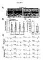
- Fig. 1A The CGR8 embryonic stem cell colony, used herein ( Fig. 1A ), demonstrated typical features of undifferentiated cells including a high nucleus to cytosol ratio, prominent nucleoli and mitochondria with few cristae ( Fig. 1 B) .
- the cardiogenic capacity of this embryonic stem cell line was probed by in vitro differentiation, with cells readily derived that express the cardiac transcription factor MEF2C ( Lin et al., 1997, Science, 276:1404-1407 ), the cardiac contractile protein ⁇ -actinin and sarcomeric striations ( Fig. 1C ).
- stem cell-derived cardiomyocytes demonstrated action potential activity associated with prominent inward Na + and Ca 2+ currents ( Fig. 1D ), critical for excitation-contraction coupling manifested as rhythmic intracellular Ca 2+ transients ( Fig. 1E ).
- Injection of CGR8 cells into myocardium resulted in local retention of these embryonic stem cells ( Fig. 1F ) without detectable dispersal into non-cardiac tissues ( Fig. 1G ).
- rats were randomly assigned to stem cell or sham treatment groups.
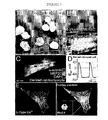
- stem cell-derived cardiomyocytes revealed distinct sarcomeric striations indicating development of the contractile apparatus ( FIG. 6B and 6C ).
- Sarcomeres in the infarct area of stem cell-treated hearts showed normal cardiac ultrastucture on electron microscopy, in contrast to acellular infarct areas of sham-treated hearts ( Fig. 6D and 6E ).
- embryonic stem cells were able to incorporate within host infarct territory, demonstrate cardiogenic differentiation, and contribute to myocardial repair.
- Embryonic stem cell-derived cardiomyocytes through electrical and mechanical coupling with native myocardium, could contribute to a net increase in contractile tissue.
- Stem cell-derived cardiomyocytes aligned with and integrated within host myocardial fibers.
- the host myocardium has been shown to secrete cardiogenic growth factors that interact in a paracrine fashion with receptors on stem cells supporting cardiac differentiation with expression of cardiac contractile and gap junction proteins ( Behfar et al., 2002, FASEB J., 16:1558-1566 ; Mery et al., 2003, J. Muscle Res. Cell.
- stem cell-derived cardiomyocytes The present failure to observe ectopy is further consistent with electrical integration of stem cell-derived cardiomyocytes and host tissue.
- the stem cell-derived cardiomyocyte effect on active myocardial properties is moreover evidenced here by improved inotropic response to ⁇ -adrenergic challenge.
- a synergistic potential mechanism for functional improvement by stem cell-derived cardiomyocytes is through alteration of myocardial passive mechanical properties ( Askari et al., 2003, Lancet, 362:697-703 ), as shown herein by the limited appearance of scar and less dilation of the left ventricle compared to sham treated infarcted hearts.
- This lack of host versus graft reaction may be graft- and/or host-dependent due to low expression of immunogenic antigens by stem cells, generation of mixed chimerism, downregulation of host immune response and/or induction of improved tolerance ( Drukker et al., 2002, Proc. Natl. Acad. Sci. USA, 99:9864-9869 ; Frandrich et al., 2002, Nat. Med., 8:171-178 ; Lila et al., 2002, Circulation, 105:1949-1954 ).
- embryonic stem cell therapy on myocardial structure and function in this experimental model supports the potential for stem cell-based reparative treatment of myocardial infarction.
- embryonic stem cells provide a unique therapeutic modality that has the potential to reduce the morbidity and mortality of this prevalent heart disease.
- Embryonic stem cells and derived cardiomyocytes were used to visualize murine embryonic stem cells in culture ( Perez-Terzic et al., 2003, Circ. Res., 92:444-452 ; Hodgson et al., 2004, Am. J. Physiol., 287:H471-H479 ).
- embryonic stem cells were engineered to express the enhanced cyan fluorescent protein (ECFP) under control of the cardiac-specific ⁇ -actin promoter ( Behfar et al., 2002, FASEB J., 16:1558-1566 ; Meyer et al., 2000, FEBS Lett., 478:151-158 ).
- ECFP enhanced cyan fluorescent protein
- engineered stem cells were delivered directly into healthy or infarcted left ventricular walls of isoflurane-anesthetized mice or rats, respectively ( Hodgson et al., 2004, Am. J. Physiol., 287:H471-H479 ).
- embryonic stem cells have the capacity to differentiate from a pluripotent to a cardiac phenotype ( FIG. 7A-B ). This is demonstrated herein as derived cells recapitulated typical cardiomyocyte features including nuclear translocation of cardiac transcription factors, the Myocyte Enhancer Factor 2C (MEF2C) and the homeodomain transcription factor (Nkx2.5) leading to sarcomerogenesis ( Fig. 7B-C ) and action potential formation associated with L-type Ca 2+ channel expression ( Fig. 7C-E ). Thus, embryonic stem cells serve as a reliable cell-based source for de novo cardiogenesis.
- MEF2C Myocyte Enhancer Factor 2C
- Nkx2.5 homeodomain transcription factor leading to sarcomerogenesis
- Fig. 7B-C action potential formation associated with L-type Ca 2+ channel expression
- Fig. 7C-E action potential formation associated with L-type Ca 2+ channel expression
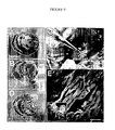
- FIG. 8A-B Titrated stem cell delivery secures cardiogenesis in vivo. Delivery of embryonic stem cells, engineered for in vivo fluorescence tracking, resulted in incorporation into host heart of new cyan fluorescing cardiac cells within the area of stem cell transplantation ( Fig. 8A-B ). Engraftment of stem cell-derived cardiomyocytes was associated with normal heart morphology ( Fig. 8A ) and function ( Fig. 8C , inset). A threshold in the capacity of the host heart to accept stem cell implantation was established, above which excessive stem cell load compromised cardiogenesis post-transplantation due to uncontrolled differentiation resulting in tumorigenesis ( Fig. 8C-D ). Thus, the host heart has a finite capacity for driving cardiogenesis in support of differentiation and engraftment of stem cells in vivo ( Fig. 8E ).
- FIG. 9A-B A myocardial infarct model was generated by coronary ligation, resulting in an aneurysmal and scarred anterior wall with significant compromise in contractile performance.
- Fig. 9C embryonic stem cells engineered to fluoresce upon cardiogenesis were delivered by direct myocardial transplantation into peri-infarct area. Histopathological examination showed benefit in stem cell-treated infarcted hearts with areas of repair populated by fluorescent cardiac cells indicating stem cell-based de novo cardiogenesis ( Fig. 9D-E ).
- stem cell-treated hearts demonstrated synchronous functionally recovered heart muscle on the echocardiographic follow-up ( Fig. 10A ).
- Structural repair translated into improvement in the overall ejection fraction in the stem cell-treated group, a benefit maintained over the five month observation period, with no evidence for inflammatory infiltrates ( Fig. 10B-C ) or arrhythmogenesis ( Fig. 10D ).
- Murine embryonic stem cells were differentiated into embryoid bodies, with cardiogenesis monitored by epifluorescence using ⁇ -actinin antibody (1:1,000) and live microscopy ( Behfar et al., 2002, FASEB J., 16:1558-1566 ; Perez-Terzic et al., 2003, Circ. Res., 92:444-452 ).
- a visceral endoderm-like population ( Mummery et al., 2003, Circulation, 107:2733-2740 ) was derived from F9 cells (ATCC) with retinoic acid (1 ⁇ M), dbcAMP (0.5 mM) and theophylline (0.5 mM).
- Conditioned medium was obtained after 24 h of culture to stimulate cardiogenesis of embryonic stem cells (cultured at 100 cells/cm 2 ) monitored by confocal microscopy.
- Cardiopoietic stem cell isolation From day 7 embryoid bodies, cardiopoietic stem cells were isolated by Percoll purification and visualized through laser confocal examination using MEF2C (1:400, Cell Signaling Technologies), Nkx2.5 (1:300), GATA4 (1:300, Santa Cruz Biotech), ⁇ -actinin (1:1,000, Sigma) and phospho-Smad3 (1:2,000) antibodies.
- MEF2C Cell Signaling Technologies
- Nkx2.5 (1:300
- GATA4 (1:300, Santa Cruz Biotech
- ⁇ -actinin (1:1,000, Sigma
- phospho-Smad3 phospho-Smad3
- Cardiopoietic stem cell proliferation and purity was assessed by ArrayScan high-throughput multichannel fluorescence automated microscopy (Cellomics) using MEF2C and ⁇ -actinin antibodies, along with DAPI staining.
- Action potential profiles and voltage-current relationships were acquired by patch-clamp electrophysiology ( Hodgson et al., 2004, Am. J. Physiol., 287:H471-H479 ).
- Calcium dynamics were tracked, in Fluo 4-AM loaded cells, using laser confocal line scanning ( Perez-Terzic et al., 2003, Circ. Res., 92:444-452 ; Hodgson et al., 2004, Am. J. Physiol., 287:H471-H479 ).
- Genomics Comparative gene expression profiles of embryonic versus cardiopoietic stem cells or cardiomyocytes, as well as unprimed versus TNF ⁇ -primed endoderm were attained by labelled cRNA hybridization to the mouse genome 430 2.0 array using standard protocols (Affymetrix). Data was acquired with a GeneChip Scanner 3000 (Affymetrix), and analyzed with the GeneSpring software (Silicon Genetics). Data population sets were normalized to the undifferentiated or unprimed phenotype, and quality filtered to eliminate background noise prior to hierarchical clustering.
- Endodermal cells were cultured with serum-free GMEM. Derived conditioned medium was centrifuged, filtered, quantified (Bradford assay), concentrated (Amicon Ultra 5 kDa cut-off), and re-quantified for volumetric normalization. The protein equivalent of 5 ml conditioned medium was resuspended in isoelectric focusing (IEF) buffer containing urea (7 M), thiourea (2 M), CHAPS (2% w/v) and DeStreak (15 mg/ml, Amersham). Proteins were resolved in the first dimension using immobilized pH gradient IEF strips (BioRad) at pH 3-10, 4-7 and 6-11, and in the second dimension by 7.5% and 15% SDS-PAGE.
- IEF isoelectric focusing
- Proteins visualized by silver staining, were isolated, destained and trypsin digested ( Arrell et al., 2001, Circ. Res., 89:480-487 ) with extracted peptides subjected to high performance liquid chromatography-electrospray ionization tandem mass spectrometry (ThermoFinnigan LTQ). Proteins were identified using SEQUEST and Mascot search algorithms for in silico mining of the SwissProt database. Identified proteins were further quantified with enzyme-linked immunosorbent assay.
- mice echocardiography with a 15-MHz probe was used to guide myocardial delivery of embryonic or cardiopoietic stem cells engineered for in situ tracking ( Behfar et al., 2002, FASEB J., 16:1558-1566 ). Cardiac perfomance was monitored by ultrasound imaging in the parasternal short axis with 2-D M-mode probing in the long axis, Doppler pulse wave analysis and by twelve-lead electrocardiography ( Hodgson et al., 2004, Am. J. Physiol., 287:H471-H479 ), and invasively with intraventricular microcatheterization (Millar).
- harvested heart tissue was fixed for 1 h in 3% paraformaldehyde, paraffin sectioned, and subjected to antigen retrieval followed by confocal examination using CFP antibody for cell tracking (1:500, Molecular Probes) in combination with ⁇ -actinin for sarcomere visualization and DAPI nuclear stain.
- Transgenic cardiac-restricted overexpression of the cytokine TNF ⁇ was achieved using the ⁇ -myosin heavy chain promoter linked to the TNF ⁇ transgene ( Sivasubramanian et al., 2001, Circulation, 104:826-831 ).
- Disrupting the kinase domain of the TGF- ⁇ receptor ( ⁇ TGF ⁇ RII) or overexpression of the BMP inhibitor noggin was used to ablate the capacity of embryonic stem cells to respond to cardiogenic cues ( Behfar et al., 2002, FASEB J., 16:1558-1566 ).
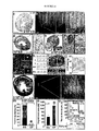
- the captured cardiopoietic stem cells were en route to maturation, undergoing myofibrillogenesis to form contracting cardiomyocytes (CM; Fig. 11D ).
- Ultrastructural dissection with nanoscale deconvolution underscored the transitional state of cardiopoietic stem cells during cardiogenic metamorphosis from a phenotype of high nucleus to cytosol ratio, typical of embryonic stem cells, towards acquisition of a striated cardiac structure ( Fig. 11e-f ).
- the molecular fingerprint of cardiopoietic stem cells indicated definitive commitment to the cardiac program ( Fig. 11G-H ).
- the TNF ⁇ effect on cardiogenesis manifested as a doubling of the protein concentration within the endodermal secretome ( Fig. 12F ).
- Application of the conditioned medium derived from the TNF ⁇ -primed endoderm guided vigorous differentiation of pluripotent embryonic stem cells directly into cardiopoietic stem cells, eliminating the need for transit through an embryoid body ( Fig. 12G-J ).
- Cardiopoietic stem cells derived extra-embryo maintained mitotic activity, clonal expansion and resilience to hypoxic stress (5% O 2 ), remnants of their embryonic source, yet acquired contact inhibition and could reproducibly generate sarcomeres to complete the cardiac program ( Fig. 12K ).
- TNF ⁇ amplification of cardiopoiesis was due to a re-distribution of the endodermal secreted protein content ( FIG. 13A-D ).
- a cocktail of over twenty proteins within the endodermal secretome were found up-regulated, ranging from factors involved in sarcomerogenesis (profilin, cofilin) ( Obinata et al., 1997, Cell Struct. Funct., 22:181-189 ; Mohri et al., 2000, J. Muscle Res. Cell.
- cardiopoietic stem cell use was assessed in vivo upon echocardiography-guided transplantation. Loads of ⁇ 3 ⁇ x 10 5 pluripotent cells per heart led to incorporation of embryonic stem cell-derived cardiomyocytes into recipient heart muscle ( Fig. 14a-c ), but increases in cell dose precipitated neoplastic invasion ( Fig. 14D-F ) demonstrating the low therapeutic index of embryonic stem cell-based treatment. In contrast, delivery of ⁇ 3 x ⁇ 10 6 cardiopoietic stem cells, recruited in vitro from a monolayer of embryonic stem cells using the here identified cardiogenic cocktail ( Fig.
- TNF ⁇ -TG cardiac-restricted transgenic TNF ⁇ overexpression
- Fig. 14o inset the host heart cardiogenic capacity
- Fig. 14o reduced the risk for carcinogenesis upon embryonic stem cell transplantation
- a cardiogenic cocktail was generated containing the indicated amount of each component: TGF- ⁇ 1 (2.5 ng/ml); TGF- ⁇ 2 (2.5 ng/ml); BMP-1 (5 ng/ml); BMP-2 (5 ng/ml); BMP-5 (5 ng/ml); BMP-6 (5 ng/ml); FGF-4 (10 ng/ml); FGF-5 (10 ng/ml); FGF-12 (10 ng/ml); FGF-13 (10 ng/ml); FGF-15 (10 ng/ml); FGF-20 (10 ng/ml); leukemia inhibitory factor (1000 U/ml); VEGF-C (15 ng/ml); and interleukin 6 (100 ng/ml).
- IGF-1 50 ng/ml
- IL-6 100 ng/ml
- FGF-4 10 ng/ml
- TGF ⁇ 25 ng/ml
- BMP 5 ng/ml
- LIF 100 U/ml
- h ⁇ -thrombin 40 nM
- the cardiogenic cocktail described herein contains 54 components; 47 components that are endoderm derived and 7 components that are exogenously added.
- the pluripotent murine embryonic stem cell lines i.e., CGR8, D3, 129 and R29
- the pluripotent murine embryonic stem cell lines were propagated in GMEM medium with pyruvate, non-essential amino acids, mercaptoethanol, 7.5% fetal calf serum (FCS) and leukemia inhibitory factor (LIF, ESGRO), as described ( Terzic et al., 2003, Circ Res., 92:444-452 ; Behfar et al., 2002, FASEB J., 16:1558-1566 ).
- Embryoid dissociation and characterization Embryoid dissociation and characterization. Embryoid bodies, at different stages of differentiation, were detached from dishes with 0.05% trypsin, and dissociated using 1 mg/ml collagenase (CLSII, Worthington) and 0.25 mg/ml pancreatin in (in mmol/L) NaCl 117, HEPES 20, NaH 2 PO 4 1.2, KCl 5.4, MgSO 4 1 and glucose 5 (pH 7.35) ( Terzic et al., 2003, Circ Res., 92:444-452 ). Embryoid body cell suspensions were characterized for progenitor content by confocal immunofluorescent probing in addition to video microscopy.
- the same approach can be applied to purify cardiopoietic stem cells and cardiomyocytes following guided differentiation of embryonic stem cells monolayers.
- purified cells were cultured in a temperature- and gas-controlled chamber (37°C, 5% CO 2 ), set on a stage of a CCD camera-coupled microscope, with phase contrast images acquired serially.
- PBS phosphate buffered saline
- PFA paraformaldehyde
- coverslips were incubated with a blocking buffer (SuperblockTM, Pierce Biotech) following fixation.
- Blocking buffer was removed, and antibodies were added (Nkx2.5 1:400; MEF2C 1:500; GATA4 1:300; ⁇ -Actinin 1:1000; L-type Ca 2+ channel 1:400; Cx43 1:500; Kir6.2 1:500; GFP 1:400) overnight at 4°C.
- primary antibodies were rinsed-off using wash buffer, and ALEXA-conjugated secondary antibodies (1:500) was applied. Secondary antibody was rinsed off using wash buffer, and the DNA-stain DAPI was incubated prior to mounting.
- Fluorescent excitation of stained cellular components were accomplished by concurrent use of the UV (350-370 nm), Arg/Kry (488 nm), and HeNe (568, 633 nm) laser lines for multi-color Meta-based confocal microscopy (Zeiss). Acquired images were analyzed using Axioplan software (Zeiss). Additionally, cardiopoietic stem cell proliferation and purity was assessed by ArrayScan high-throughput multichannel fluorescence automated microscopy (Cellomics) using MEF2C and ⁇ -actinin antibodies, along with DAPI staining.
- RNA isolation and genomic profiling Total RNA was isolated from embryoid bodies, as well as from embryonic stem cells and when derived from cardiopoietic stem cells and cardiac cell progeny using the Micro-to-Midi isolation kit (Invitrogen). Total RNA was also be attained from TNF ⁇ -primed and unprimed endodermal cells. RNA samples was analyzed using Affymetrix microarrays where comparative gene expression profiles of embryoid bodies, as well as embryonic versus cardiopoietic stem cells or cardiomyocytes, and in separate studies unprimed versus TNF ⁇ -primed endoderm was attained by labelled cRNA hybridization to the mouse genome 430 2.0 array using standard protocols (Affymetrix).
- Data was acquired with a GeneChip Scanner 3000 (Affymetrix), and analyzed with the GeneSpring software (Silicon Genetics). Data population sets were normalized to the undifferentiated or unprimed phenotype, and quality filtered to eliminate background noise prior to hierarchical clustering. Data was confirmed by subjecting sample total RNA to real time Q-RT-PCR, using primers specifying each gene identity ( Behfar et al., 2002, FASEB J., 16:1558-1566 ).
- Two-dimensional gel electrophoresis To resolve the proteomic phenotype of cardiopoietic stem cells versus their source and progeny, cellular proteins was solubilized in isoelectric focusing (IEF) buffer (7 M urea, 2 M thiourea, 2% CHAPS, with protein concentrations determined by protein assay. Two-dimensional gel electrophoresis (2-DE) was carried out in the first dimension (IEF) using a Protean ® IEF cell (Bio-Rad).
- IEF isoelectric focusing
- Protein samples will be added to 300 ⁇ l of IEF buffer, supplemented with DTT (dithiothreitol) and ampholytes (Bio-Rad), and then taken up by active rehydration into immobilized pH gradient (IPG) Ready StripsTM (170 mm linear gradient strips, pH 3-10 or 4-7, Bio-Rad) at 50 volts (V) for 10 h. IEF then followed, using a series of rapid ramping voltage steps for 15 min each at 100, 500, and 1000 V, followed by 10000 V for 60 kiloV-h. A variation of this process using passive rehydration was used for IEF of basic proteins (pH 6-11).
- SDS-PAGE buffer 25 mM Tris, 192 mM glycine, pH 8.3, 0.1% w/v SDS
- proteins resolved by 12.5% SDS-PAGE using a Protean ® II Xi system (Bio-Rad).
- 2-D gels were silver-stained for compatibility with subsequent analysis of protein by mass spectrometry ( Shevchenko et al., 1996, Anal. Chem., 68:850-858 ).
- Protein spots of interest were isolated and destained ( Gharahdaghi et al., 1999, Electrophoresis, 20:601-605 ) to ensure removal of silver, then digested with trypsin, extracted, dried under vacuum, and stored at -20°C until mass spectrometric analysis.
- Tandem mass spectrometry Tryptic peptides isolated from individual 2-D gel spots was reconstituted and resolved by high performance liquid chromatography (HPLC) on a C18 reversed phase (RP) column. Peptides were eluted from the RP-HPLC column with an increasing acetonitrile gradient, and electrosprayed into a Finnigan LCQ Deca ion trap mass spectrometer (Thermo Finnigan).
- MS/MS spectra of eluting peptides were acquired in a data-dependent fashion by first acquiring a full MS scan from m/z (mass/charge ratio) 150 to 2000 followed by MS/MS scans up to m/z 2000 to determine amino acid sequence following collision-induced dissociation of peptides that provide the most intense ions of the previous full MS scan.
- MS/MS spectra were searched against the SwissProt or NCBI non-redundant databases using both the SEQUEST and Mascot search algorithms to search both the 2+ and 3+ charge states of fragmented peptides. Results from each algorithm was cross referenced, providing more robust analysis than would either algorithm alone ( Sadygov et al., 2004, Nature Meth., 1:195-202 ).
- membranes were incubated with 10 ml of blocking buffer (1x TBS, 0.1% Tween-20, 5% (w/v) skim milk powder, pH 7.5) at room temperature and then incubated with a primary antibody (1:1,000) diluted in primary solution (1x TBS, 0.1% Tween-20, 1% (w/v) skim milk powder, pH 7.5). Secondary antibody (1:10,000) made up in primary solution was added the following day after rinsing the primary off with wash buffer (1x TBS, 0.1% Tween-20, pH 7.5). Western blots were developed using the Pierce chemiluminescence kit and visualized using a UVP Bioimager.
- Patch-clamp Membrane electrical activity was determined by patch-clamp recording in the whole cell configuration using the current- or voltage-clamp mode (Axopatch 1C, Axon Instruments). Action potential profiles and voltage-current relationship was acquired and analyzed with the Bioquest software from cells, at different stages of cardiac differentiation and maturation, superfused with Tyrode solution (in mM: NaCl 137, KCl 5.4, CaCl 2 2, MgCl 2 1, HEPES 10, glucose 10; pH 7.4 with NaOH) using patch pipettes (5-10 M ⁇ ) containing (in mM) KCl 140, MgCl 2 1, HEPES 10, EGTA 5, and supplemented with 5 mM ATP (pH 7.2 adjusted with KOH). Electrophysiological measurements were performed at 31 ⁇ 1°C using a temperature controller (HCC-100A, Dagan Corp.) equipped with a Peltier thermocouple (1,10).
- HCC-100A Dagan Corp.
- Transmitted / field-emission scanning electron and atomic force microscopy Cells at different stages of cardiac differentiation were fixed in PBS with 1% glutaraldehyde and 4% formaldehyde (pH 7.2).
- TEM transmitted electron microscopy
- cells were processed in phosphate-buffered 1% OsO 4 , stained with 2% uranyl acetate, dehydrated in ethanol and propylene oxide, and embedded in epoxy resin. Thin (90-nm) sections were placed on copper grids, stained with lead citrate, and micrographs were taken with a JEOL electron microscope ( Behfar et al., 2002, FASEB J., 16:1558-1566 ).
- FESEM field-emission scanning electron
- AFM Contact-mode atomic force microscopy
- Embryonic stem cell clones were engineered to express either the green fluorescent protein (GFP), the enhanced cyan fluorescent protein (ECFP) or the cytosolic (or nuclear) lacZ under the control of the cardiac-specific ⁇ -actinor ⁇ -myosin promoter subcloned upstream of the reporter gene (Clontech).
- GFP green fluorescent protein
- ECFP enhanced cyan fluorescent protein
- cytosolic (or nuclear) lacZ under the control of the cardiac-specific ⁇ -actinor ⁇ -myosin promoter subcloned upstream of the reporter gene (Clontech).
- reporter genes were subcloned downstream of 5'LTR promoter of the modified murine stem cell virus (MSCV) or the murine phosphoglycerate kinase (PGK) promoter, for stable and robust post-transplantational expression.
- MSCV modified murine stem cell virus
- PGK murine phosphoglycerate kinase
- Constructs were linearized, and introduced into embryonic stem cells by lipofectamine (Invitrogen) transfection ( Terzic et al., 2003, Circ Res., 92:444-452 ; Behfar et al., 2002, FASEB J., 16:1558-1566 ).
- the reporter gene was packaged into an MMLV retroviral system for high-yield embryonic stem cell incorporation.
- Infarction was generated by ligation of the left coronary artery (LCA) following endotracheal intubation, ventilation and thoracotomy in C57BL/6 mice. Coronary occlusion was confirmed by acute inspection of color change of the left ventricle wall, and ST elevation on the electrocardiogram before chest closure. Sham-operated mice underwent the same surgical procedure without LCA ligation. Our experience reveals a ⁇ 10% surgery-related mortality. Infarcted mice will receive various stem cell regimens, and followed from 1 to 12 months.
- LCA left coronary artery
- mice deficient in K ATP channels generated by targeted disruption of the Kir6.2 gene and backcrossed for five generations to a C57BL/6 background, will be compared to age- and sex-matched control mice ( Seino & Miki, 2003, Prog. Biophys. Mol. Biol., 81:133-176 ). All mice receive standard chow, with a 12-hour day/night cycle and observed daily until termination of studies.
- Myocyte cross-sectional area, interstitial and perivascular fibrosis was quantified using MetaMorph software; ii) myocyte ultrastructure was examined by transmitted/scanning electron microscopy; iii) myocytes were stained for terminal deoxynucletidyl transferase-mediated dUTP nick end-labeling (TUNEL) and caspase-3, and undergo a DNA laddering assay to assess for apoptosis; iv) myocardial collagen content was measured by hydroxyproline assay.
- Electrocardiography Electrocardiograms were recorded telemetrically in conscious, untethered mice from surgically implanted transmitters (Data Sciences International) ( Terzic et al., 2003, Circ Res., 92:444-452 ; 10). In addition 12-lead surface electrocardiograms were recorded in lightly isoflurane-anethetized mice.
Landscapes
- Health & Medical Sciences (AREA)
- Life Sciences & Earth Sciences (AREA)
- Medicinal Chemistry (AREA)
- Animal Behavior & Ethology (AREA)
- Chemical & Material Sciences (AREA)
- Engineering & Computer Science (AREA)
- Veterinary Medicine (AREA)
- Public Health (AREA)
- General Health & Medical Sciences (AREA)
- Pharmacology & Pharmacy (AREA)
- Epidemiology (AREA)
- Immunology (AREA)
- Bioinformatics & Cheminformatics (AREA)
- Proteomics, Peptides & Aminoacids (AREA)
- Gastroenterology & Hepatology (AREA)
- Zoology (AREA)
- Developmental Biology & Embryology (AREA)
- Cell Biology (AREA)
- Reproductive Health (AREA)
- Endocrinology (AREA)
- Molecular Biology (AREA)
- Diabetes (AREA)
- Virology (AREA)
- Gynecology & Obstetrics (AREA)
- Biotechnology (AREA)
- Orthopedic Medicine & Surgery (AREA)
- Biomedical Technology (AREA)
- Heart & Thoracic Surgery (AREA)
- Chemical Kinetics & Catalysis (AREA)
- Nuclear Medicine, Radiotherapy & Molecular Imaging (AREA)
- General Chemical & Material Sciences (AREA)
- Organic Chemistry (AREA)
- Cardiology (AREA)
- Micro-Organisms Or Cultivation Processes Thereof (AREA)
- Medicines Containing Material From Animals Or Micro-Organisms (AREA)
- Medicines That Contain Protein Lipid Enzymes And Other Medicines (AREA)
- Peptides Or Proteins (AREA)
Claims (2)
- Procédé de préparation d'un cocktail cardiogénique, comprenant :(a) la culture de cellules endodermiques ventrales dans un milieu, dans lequel lesdites cellules sont obtenues à partir d'un embryon traité avec du TNF-alpha, à condition que l'embryon ne soit pas un embryon humain ;
un milieu de culture conditionné étant ainsi généré ; et(b) la collecte du milieu de culture conditionné, un cocktail cardiogénique étant ainsi obtenu. - Procédé selon la revendication 1, comprenant en outre : l'ajout d'un ou de plusieurs composants audit cocktail cardiogénique, dans lequel ledit un ou plusieurs composants sont choisis dans le groupe constitué de IGF-I, IL-6, FGF-4, TGF-β, BMP, LIF et la h-αthrombine.
Priority Applications (1)
| Application Number | Priority Date | Filing Date | Title |
|---|---|---|---|
| EP10179541.7A EP2269461B1 (fr) | 2004-07-30 | 2005-07-29 | Traitement de tissu cardiovasculaire |
Applications Claiming Priority (3)
| Application Number | Priority Date | Filing Date | Title |
|---|---|---|---|
| US59287104P | 2004-07-30 | 2004-07-30 | |
| US68077505P | 2005-05-12 | 2005-05-12 | |
| PCT/US2005/026800 WO2006015127A2 (fr) | 2004-07-30 | 2005-07-29 | Traitement de tissu cardio-vasculaire |
Related Child Applications (2)
| Application Number | Title | Priority Date | Filing Date |
|---|---|---|---|
| EP10179541.7A Division-Into EP2269461B1 (fr) | 2004-07-30 | 2005-07-29 | Traitement de tissu cardiovasculaire |
| EP10179541.7A Division EP2269461B1 (fr) | 2004-07-30 | 2005-07-29 | Traitement de tissu cardiovasculaire |
Publications (3)
| Publication Number | Publication Date |
|---|---|
| EP1786471A2 EP1786471A2 (fr) | 2007-05-23 |
| EP1786471A4 EP1786471A4 (fr) | 2008-01-23 |
| EP1786471B1 true EP1786471B1 (fr) | 2015-02-11 |
Family
ID=35787826
Family Applications (2)
| Application Number | Title | Priority Date | Filing Date |
|---|---|---|---|
| EP05777528.0A Expired - Lifetime EP1786471B1 (fr) | 2004-07-30 | 2005-07-29 | Traitement de tissu cardio-vasculaire |
| EP10179541.7A Expired - Lifetime EP2269461B1 (fr) | 2004-07-30 | 2005-07-29 | Traitement de tissu cardiovasculaire |
Family Applications After (1)
| Application Number | Title | Priority Date | Filing Date |
|---|---|---|---|
| EP10179541.7A Expired - Lifetime EP2269461B1 (fr) | 2004-07-30 | 2005-07-29 | Traitement de tissu cardiovasculaire |
Country Status (4)
| Country | Link |
|---|---|
| US (2) | US8173118B2 (fr) |
| EP (2) | EP1786471B1 (fr) |
| ES (2) | ES2532909T3 (fr) |
| WO (1) | WO2006015127A2 (fr) |
Families Citing this family (19)
| Publication number | Priority date | Publication date | Assignee | Title |
|---|---|---|---|---|
| EP1786471B1 (fr) | 2004-07-30 | 2015-02-11 | Mayo Foundation For Medical Education And Research | Traitement de tissu cardio-vasculaire |
| US9765298B2 (en) | 2006-07-24 | 2017-09-19 | Mayo Foundation For Medical Education And Research | Methods and materials for providing cardiac cells |
| US20090246179A1 (en) * | 2008-02-11 | 2009-10-01 | The Cleveland Clinic Foundation | Method of treating myocardial injury |
| US8808185B2 (en) * | 2008-03-28 | 2014-08-19 | General Electric Company | System and method for generating a patient diagnosis |
| WO2009145761A1 (fr) * | 2008-05-27 | 2009-12-03 | Mayo Foundation For Medical Education And Research | Procédés et matériaux destinés à utiliser des cellules dans le traitement du tissu cardiaque |
| US10047346B2 (en) * | 2008-08-08 | 2018-08-14 | Mayo Foundation For Medical Education And Research | Method of treating heart tissue using induced pluripotent stem cells |
| WO2010037224A1 (fr) * | 2008-10-03 | 2010-04-08 | St. Michael's Hospital | Procédé de prévention et de traitement de maladies cardiovasculaires avec brca1 |
| WO2010042856A2 (fr) * | 2008-10-09 | 2010-04-15 | The General Hospital Corporation | Myocarde obtenu par génie tissulaire et procédés de production et utilisations de celui-ci |
| EP2432482B1 (fr) | 2009-05-20 | 2015-04-15 | Cardio3 Biosciences S.A. | Composition pharmaceutique pour le traitement de cardiopathies |
| AU2010251151B2 (en) * | 2009-05-20 | 2015-08-20 | Cardio3 Biosciences S.A. | Pharmaceutical Composition for the Treatment of Heart Diseases |
| WO2011032025A2 (fr) * | 2009-09-10 | 2011-03-17 | The Salk Institute For Biological Studies | Cellules souches pluripotentes induites d'origine adipocytaire |
| NZ599930A (en) * | 2009-12-02 | 2014-04-30 | Cardio3 Biosciences Sa | Pharmaceutical compositions for the stimulation of stem cells. |
| WO2012051515A2 (fr) * | 2010-10-14 | 2012-04-19 | University Of Central Florida Research Foundation, Inc. | Cellules souches pluripotentes cardio-induites et procédés d'utilisation pour la réparation et la régénération du myocarde |
| US20130029416A1 (en) | 2011-07-22 | 2013-01-31 | Tayaramma Thatava | Differentiating induced pluripotent stem cells into glucose-responsive, insulin-secreting progeny |
| AU2015355188B2 (en) * | 2014-12-01 | 2021-08-26 | Northeast Ohio Medical University | CAMKK1 as a novel regenerative therapeutic |
| RU2644650C2 (ru) * | 2014-12-01 | 2018-02-13 | Общество с ограниченной ответственностью "Т-Хелпер Клеточные Технологии" | Материал стволовых клеток и способ его получения |
| EP3267965A4 (fr) | 2015-03-11 | 2018-11-14 | Atta Behfar | Technologie d'administration d'exosomes |
| WO2018055235A1 (fr) | 2016-09-21 | 2018-03-29 | University Of Helsinki | Isoxazole-amides pour le traitement de maladies cardiaques |
| EP3518941B1 (fr) | 2016-09-30 | 2022-03-09 | Mayo Foundation for Medical Education and Research | Vecteurs viraux pour la reprogrammation nucléaire |
Family Cites Families (30)
| Publication number | Priority date | Publication date | Assignee | Title |
|---|---|---|---|---|
| US7696404B2 (en) * | 1996-08-19 | 2010-04-13 | Advanced Cell Technology, Inc. | Embryonic or stem-like cell lines produced by cross species nuclear transplantation and methods for enhancing embryonic development by genetic alteration of donor cells or by tissue culture conditions |
| US20020061837A1 (en) | 1996-09-05 | 2002-05-23 | Lough John W. | Bone morphogenetic protein and fibroblast growth factor compositions and methods for the induction of cardiogenesis |
| US5839438A (en) | 1996-09-10 | 1998-11-24 | Neuralmed, Inc. | Computer-based neural network system and method for medical diagnosis and interpretation |
| CA2296704C (fr) | 1997-07-14 | 2010-10-19 | Osiris Therapeutics, Inc. | Regeneration du muscle cardiaque a l'aide de cellules souche mesenchymateuses |
| JP5943533B2 (ja) * | 2000-05-17 | 2016-07-06 | アステリアス バイオセラピューティクス インコーポレイテッド | 神経前駆細胞の集団 |
| CA2411102A1 (fr) * | 2000-06-20 | 2001-12-27 | Idec Pharmaceutical Corporation | Traitement de maladies associees aux cellules beta telles que les maladies auto-immunes a caractere malin par combinaison anticorps anti-cd20 froid/anticorps anti-cd22 radiomarque |
| WO2003006950A2 (fr) | 2001-07-12 | 2003-01-23 | Geron Corporation | Cellules de la lignee des cardiomyocytes produites a partir de cellules souches humaines pluripotentielles |
| US7732199B2 (en) * | 2001-07-12 | 2010-06-08 | Geron Corporation | Process for making transplantable cardiomyocytes from human embryonic stem cells |
| CA2458575C (fr) * | 2001-08-24 | 2014-07-08 | Advanced Cell Technology, Inc. | Essais de criblage pour l'identification d'agents induisant la differenciation, et production de cellules differenciees pour la therapie cellulaire |
| AU2004249340B2 (en) | 2003-06-25 | 2011-04-21 | Ottawa Hospital Research Institute | Use of cardiotrophin to modulate stem cell proliferation |
| CN1886149A (zh) * | 2003-09-23 | 2006-12-27 | 路德维格癌症研究院 | Vegf-c或vegf-d物质及刺激神经干细胞的方法 |
| DE10347436B4 (de) | 2003-10-13 | 2007-08-02 | Johann Wolfgang Goethe-Universität Frankfurt am Main | In vitro Verfahren zur Diagnose der kardiovaskulären Funktionalität von Blut-abgeleiteter zirkulierender Vorläuferzellen (BDP) |
| GB0329449D0 (en) * | 2003-12-19 | 2004-01-28 | Omnicyte Ltd | Stem cells |
| BRPI0506839A (pt) | 2004-01-16 | 2007-06-12 | Novartis Ag | composições e métodos para induzir a cardiomiogênese |
| CN1969040B (zh) | 2004-03-19 | 2014-12-10 | 阿斯特利亚斯生物治疗股份公司 | 制备适合用于再生医学的高纯度心肌细胞的方法 |
| US7452718B2 (en) * | 2004-03-26 | 2008-11-18 | Geron Corporation | Direct differentiation method for making cardiomyocytes from human embryonic stem cells |
| CA2569242A1 (fr) * | 2004-06-01 | 2005-12-15 | Es Cell International Pte Ltd | Differenciation en cardiomyocytes amelioree |
| EP1786471B1 (fr) | 2004-07-30 | 2015-02-11 | Mayo Foundation For Medical Education And Research | Traitement de tissu cardio-vasculaire |
| EP1805300A4 (fr) | 2004-09-14 | 2009-09-09 | Univ Columbia | Différenciation de cellules souches mésenchateuses de cellules souches cardiaques qui favorisent la cure cardiaque |
| WO2006081190A2 (fr) | 2005-01-25 | 2006-08-03 | Five Prime Therapeutics, Inc. | Compositions et methodes de traitement de troubles cardiaques |
| CA2615396A1 (fr) | 2005-07-15 | 2007-01-25 | Primegen Biotech, Llc | Reprogrammation therapeutique de cellules souches germinatives |
| EP1991664B1 (fr) | 2006-02-16 | 2018-03-28 | Ospedale San Raffaele S.r.l. | Periangioblastes du muscle squelettique et mesangioblastes du muscle cardiaque, procédé d'isolation et leurs utilisations |
| EP2502988A1 (fr) | 2006-07-13 | 2012-09-26 | Cellartis AB | Nouvelle population de cellules précurseurs cardiaques multipotentes dérivées de balstocystes humains |
| US9765298B2 (en) * | 2006-07-24 | 2017-09-19 | Mayo Foundation For Medical Education And Research | Methods and materials for providing cardiac cells |
| US20100166714A1 (en) | 2006-11-02 | 2010-07-01 | The General Hospital Corporation | Cardiovascular stem cells, methods for stem cell isolation, and uses thereof |
| US20100189697A1 (en) | 2007-03-07 | 2010-07-29 | Andre Terzic | Cardiac-specific progenitor cells |
| WO2009145761A1 (fr) | 2008-05-27 | 2009-12-03 | Mayo Foundation For Medical Education And Research | Procédés et matériaux destinés à utiliser des cellules dans le traitement du tissu cardiaque |
| AU2010251151B2 (en) | 2009-05-20 | 2015-08-20 | Cardio3 Biosciences S.A. | Pharmaceutical Composition for the Treatment of Heart Diseases |
| US20120100533A1 (en) | 2009-05-20 | 2012-04-26 | Cardio3 Biosciences Sa | Method for determining the cardio-generative potential of mammalian cells |
| CN102498399A (zh) | 2009-05-20 | 2012-06-13 | 卡迪欧参生物科技有限公司 | 哺乳动物细胞的心脏生长潜能的确定方法 |
-
2005
- 2005-07-29 EP EP05777528.0A patent/EP1786471B1/fr not_active Expired - Lifetime
- 2005-07-29 WO PCT/US2005/026800 patent/WO2006015127A2/fr not_active Ceased
- 2005-07-29 US US11/572,874 patent/US8173118B2/en not_active Expired - Lifetime
- 2005-07-29 ES ES05777528.0T patent/ES2532909T3/es not_active Expired - Lifetime
- 2005-07-29 ES ES10179541.7T patent/ES2626234T3/es not_active Expired - Lifetime
- 2005-07-29 EP EP10179541.7A patent/EP2269461B1/fr not_active Expired - Lifetime
-
2012
- 2012-03-28 US US13/433,095 patent/US8962320B2/en not_active Expired - Lifetime
Also Published As
| Publication number | Publication date |
|---|---|
| ES2626234T3 (es) | 2017-07-24 |
| US8173118B2 (en) | 2012-05-08 |
| WO2006015127A9 (fr) | 2014-07-31 |
| US20120178164A1 (en) | 2012-07-12 |
| US8962320B2 (en) | 2015-02-24 |
| WO2006015127A3 (fr) | 2007-05-24 |
| WO2006015127A2 (fr) | 2006-02-09 |
| EP2269461B1 (fr) | 2017-03-22 |
| EP2269461A1 (fr) | 2011-01-05 |
| EP1786471A2 (fr) | 2007-05-23 |
| ES2532909T3 (es) | 2015-04-01 |
| EP1786471A4 (fr) | 2008-01-23 |
| US20080213214A1 (en) | 2008-09-04 |
Similar Documents
| Publication | Publication Date | Title |
|---|---|---|
| US8962320B2 (en) | Treating cardiovascular tissue | |
| Dorn et al. | CTGF/CCN2 is an autocrine regulator of cardiac fibrosis | |
| Behfar et al. | Cardiopoietic programming of embryonic stem cells for tumor-free heart repair | |
| Laflamme et al. | Cardiomyocytes derived from human embryonic stem cells in pro-survival factors enhance function of infarcted rat hearts | |
| Hong et al. | Cardiac stem cell therapy for cardiac repair | |
| Leri et al. | Cardiac stem cells and mechanisms of myocardial regeneration | |
| US20110280834A1 (en) | Methods and compositions for cardiac tissue regeneration | |
| US11918609B2 (en) | Stem cells for wound healing | |
| AU2012351977B2 (en) | Stimulation of ovarian follicle development and oocyte maturation | |
| EP2184068A1 (fr) | Population de cellules souches adultes issues du tissu adipeux cardiaque et son utilisation dans la régénération cardiaque | |
| JP6806683B2 (ja) | 哺乳動物の卵丘細胞卵母細胞複合体の体外成熟 | |
| Mykhaylichenko et al. | Experimental induction of reparative morphogenesis and adaptive reserves in the ischemic myocardium using multipotent mesenchymal bone marrow-derived stem cells | |
| Yang et al. | Does pretreatment of bone marrow mesenchymal stem cells with 5-azacytidine or double intravenous infusion improve their therapeutic potential for dilated cardiomyopathy? | |
| EP3280429B1 (fr) | Fstl1 pour la réparation de tissu cardiaque | |
| Van Laake et al. | Cardiomyocytes derived from stem cells | |
| Rao et al. | Harnessing epicardial progenitor cells and their derivatives for rescue and repair of cardiac tissue after myocardial infarction | |
| Formigli et al. | Skeletal myoblasts for heart regeneration and repair: state of the art and perspectives on the mechanisms for functional cardiac benefits | |
| Tsao et al. | Muscle derived stem cells stimulate muscle myofiber repair and counteract fat infiltration in a diabetic mouse model of critical limb ischemia | |
| US8221740B2 (en) | Side population cells in cardiac repair | |
| EP2776057B1 (fr) | Une population de cellules comprenant des cellules dérivées du placenta exprimant cdx2 pour une utilisation dans le traitement d'un tissu cardiaque endommagé ou dégénéré en induisant la régénération cardiaque | |
| EP2830711A1 (fr) | Procédés et compositions à l'aide de fgf-9 pour améliorer la néovascularisation et la régénération | |
| CN115417921B (zh) | 用于修复心脏组织的心外膜衍生的旁分泌因子 | |
| HK40082668A (en) | Epicardial-derived paracrine factors for repairing cardiac tissue | |
| Benissan-Messan | MG53 improves regeneration of satellite cells and healing following volumetric muscle loss injury by decreasing fibrosis and modulating the inflammatory environment | |
| Gerbin | Tissue Engineering Strategies to Improve Post-MI Engraftment of hESC-Derived Cardiomyocytes |
Legal Events
| Date | Code | Title | Description |
|---|---|---|---|
| PUAI | Public reference made under article 153(3) epc to a published international application that has entered the european phase |
Free format text: ORIGINAL CODE: 0009012 |
|
| 17P | Request for examination filed |
Effective date: 20070228 |
|
| AK | Designated contracting states |
Kind code of ref document: A2 Designated state(s): AT BE BG CH CY CZ DE DK EE ES FI FR GB GR HU IE IS IT LI LT LU LV MC NL PL PT RO SE SI SK TR |
|
| AX | Request for extension of the european patent |
Extension state: AL BA HR MK YU |
|
| R17D | Deferred search report published (corrected) |
Effective date: 20070524 |
|
| RIC1 | Information provided on ipc code assigned before grant |
Ipc: A61K 48/00 20060101ALI20070615BHEP Ipc: A01N 63/00 20060101AFI20070615BHEP |
|
| DAX | Request for extension of the european patent (deleted) | ||
| A4 | Supplementary search report drawn up and despatched |
Effective date: 20080102 |
|
| 17Q | First examination report despatched |
Effective date: 20080924 |
|
| 111L | Licence recorded |
Free format text: 0101 CARDIO3 BIOSCIENCES SA Effective date: 20090327 |
|
| GRAP | Despatch of communication of intention to grant a patent |
Free format text: ORIGINAL CODE: EPIDOSNIGR1 |
|
| GRAJ | Information related to disapproval of communication of intention to grant by the applicant or resumption of examination proceedings by the epo deleted |
Free format text: ORIGINAL CODE: EPIDOSDIGR1 |
|
| GRAP | Despatch of communication of intention to grant a patent |
Free format text: ORIGINAL CODE: EPIDOSNIGR1 |
|
| INTG | Intention to grant announced |
Effective date: 20140814 |
|
| INTG | Intention to grant announced |
Effective date: 20140901 |
|
| GRAS | Grant fee paid |
Free format text: ORIGINAL CODE: EPIDOSNIGR3 |
|
| GRAA | (expected) grant |
Free format text: ORIGINAL CODE: 0009210 |
|
| 111L | Licence recorded |
Designated state(s): AT BE BG CH CY CZ DE DK EE ES FI FR GB GR HU IE IS IT LT LU LV MC NL PL PT RO SE SI SK TR Free format text: EXCLUSIVE LICENSE Name of requester: CARDIO3 BIOSCIENCES SA, BE Effective date: 20090327 |
|
| AK | Designated contracting states |
Kind code of ref document: B1 Designated state(s): AT BE BG CH CY CZ DE DK EE ES FI FR GB GR HU IE IS IT LI LT LU LV MC NL PL PT RO SE SI SK TR |
|
| REG | Reference to a national code |
Ref country code: GB Ref legal event code: FG4D |
|
| REG | Reference to a national code |
Ref country code: CH Ref legal event code: EP |
|
| REG | Reference to a national code |
Ref country code: IE Ref legal event code: FG4D |
|
| REG | Reference to a national code |
Ref country code: AT Ref legal event code: REF Ref document number: 709638 Country of ref document: AT Kind code of ref document: T Effective date: 20150315 |
|
| REG | Reference to a national code |
Ref country code: DE Ref legal event code: R096 Ref document number: 602005045811 Country of ref document: DE Effective date: 20150326 |
|
| REG | Reference to a national code |
Ref country code: ES Ref legal event code: FG2A Ref document number: 2532909 Country of ref document: ES Kind code of ref document: T3 Effective date: 20150401 |
|
| REG | Reference to a national code |
Ref country code: NL Ref legal event code: VDEP Effective date: 20150211 |
|
| REG | Reference to a national code |
Ref country code: AT Ref legal event code: MK05 Ref document number: 709638 Country of ref document: AT Kind code of ref document: T Effective date: 20150211 |
|
| REG | Reference to a national code |
Ref country code: LT Ref legal event code: MG4D |
|
| PG25 | Lapsed in a contracting state [announced via postgrant information from national office to epo] |
Ref country code: LT Free format text: LAPSE BECAUSE OF FAILURE TO SUBMIT A TRANSLATION OF THE DESCRIPTION OR TO PAY THE FEE WITHIN THE PRESCRIBED TIME-LIMIT Effective date: 20150211 Ref country code: FI Free format text: LAPSE BECAUSE OF FAILURE TO SUBMIT A TRANSLATION OF THE DESCRIPTION OR TO PAY THE FEE WITHIN THE PRESCRIBED TIME-LIMIT Effective date: 20150211 Ref country code: SE Free format text: LAPSE BECAUSE OF FAILURE TO SUBMIT A TRANSLATION OF THE DESCRIPTION OR TO PAY THE FEE WITHIN THE PRESCRIBED TIME-LIMIT Effective date: 20150211 |
|
| PG25 | Lapsed in a contracting state [announced via postgrant information from national office to epo] |
Ref country code: LV Free format text: LAPSE BECAUSE OF FAILURE TO SUBMIT A TRANSLATION OF THE DESCRIPTION OR TO PAY THE FEE WITHIN THE PRESCRIBED TIME-LIMIT Effective date: 20150211 Ref country code: NL Free format text: LAPSE BECAUSE OF FAILURE TO SUBMIT A TRANSLATION OF THE DESCRIPTION OR TO PAY THE FEE WITHIN THE PRESCRIBED TIME-LIMIT Effective date: 20150211 Ref country code: IS Free format text: LAPSE BECAUSE OF FAILURE TO SUBMIT A TRANSLATION OF THE DESCRIPTION OR TO PAY THE FEE WITHIN THE PRESCRIBED TIME-LIMIT Effective date: 20150611 Ref country code: AT Free format text: LAPSE BECAUSE OF FAILURE TO SUBMIT A TRANSLATION OF THE DESCRIPTION OR TO PAY THE FEE WITHIN THE PRESCRIBED TIME-LIMIT Effective date: 20150211 Ref country code: GR Free format text: LAPSE BECAUSE OF FAILURE TO SUBMIT A TRANSLATION OF THE DESCRIPTION OR TO PAY THE FEE WITHIN THE PRESCRIBED TIME-LIMIT Effective date: 20150512 |
|
| PG25 | Lapsed in a contracting state [announced via postgrant information from national office to epo] |
Ref country code: EE Free format text: LAPSE BECAUSE OF FAILURE TO SUBMIT A TRANSLATION OF THE DESCRIPTION OR TO PAY THE FEE WITHIN THE PRESCRIBED TIME-LIMIT Effective date: 20150211 Ref country code: DK Free format text: LAPSE BECAUSE OF FAILURE TO SUBMIT A TRANSLATION OF THE DESCRIPTION OR TO PAY THE FEE WITHIN THE PRESCRIBED TIME-LIMIT Effective date: 20150211 Ref country code: SK Free format text: LAPSE BECAUSE OF FAILURE TO SUBMIT A TRANSLATION OF THE DESCRIPTION OR TO PAY THE FEE WITHIN THE PRESCRIBED TIME-LIMIT Effective date: 20150211 Ref country code: RO Free format text: LAPSE BECAUSE OF FAILURE TO SUBMIT A TRANSLATION OF THE DESCRIPTION OR TO PAY THE FEE WITHIN THE PRESCRIBED TIME-LIMIT Effective date: 20150211 Ref country code: CZ Free format text: LAPSE BECAUSE OF FAILURE TO SUBMIT A TRANSLATION OF THE DESCRIPTION OR TO PAY THE FEE WITHIN THE PRESCRIBED TIME-LIMIT Effective date: 20150211 |
|
| REG | Reference to a national code |
Ref country code: DE Ref legal event code: R097 Ref document number: 602005045811 Country of ref document: DE |
|
| PG25 | Lapsed in a contracting state [announced via postgrant information from national office to epo] |
Ref country code: PL Free format text: LAPSE BECAUSE OF FAILURE TO SUBMIT A TRANSLATION OF THE DESCRIPTION OR TO PAY THE FEE WITHIN THE PRESCRIBED TIME-LIMIT Effective date: 20150211 |
|
| PLBE | No opposition filed within time limit |
Free format text: ORIGINAL CODE: 0009261 |
|
| STAA | Information on the status of an ep patent application or granted ep patent |
Free format text: STATUS: NO OPPOSITION FILED WITHIN TIME LIMIT |
|
| 26N | No opposition filed |
Effective date: 20151112 |
|
| PG25 | Lapsed in a contracting state [announced via postgrant information from national office to epo] |
Ref country code: MC Free format text: LAPSE BECAUSE OF FAILURE TO SUBMIT A TRANSLATION OF THE DESCRIPTION OR TO PAY THE FEE WITHIN THE PRESCRIBED TIME-LIMIT Effective date: 20150211 Ref country code: SI Free format text: LAPSE BECAUSE OF FAILURE TO SUBMIT A TRANSLATION OF THE DESCRIPTION OR TO PAY THE FEE WITHIN THE PRESCRIBED TIME-LIMIT Effective date: 20150211 |
|
| REG | Reference to a national code |
Ref country code: CH Ref legal event code: PL |
|
| PG25 | Lapsed in a contracting state [announced via postgrant information from national office to epo] |
Ref country code: LU Free format text: LAPSE BECAUSE OF FAILURE TO SUBMIT A TRANSLATION OF THE DESCRIPTION OR TO PAY THE FEE WITHIN THE PRESCRIBED TIME-LIMIT Effective date: 20150729 |
|
| REG | Reference to a national code |
Ref country code: IE Ref legal event code: MM4A |
|
| PG25 | Lapsed in a contracting state [announced via postgrant information from national office to epo] |
Ref country code: LI Free format text: LAPSE BECAUSE OF NON-PAYMENT OF DUE FEES Effective date: 20150731 Ref country code: CH Free format text: LAPSE BECAUSE OF NON-PAYMENT OF DUE FEES Effective date: 20150731 |
|
| REG | Reference to a national code |
Ref country code: DE Ref legal event code: R082 Ref document number: 602005045811 Country of ref document: DE Representative=s name: HERNANDEZ, YORCK, DIPL.-ING., DE |
|
| REG | Reference to a national code |
Ref country code: FR Ref legal event code: PLFP Year of fee payment: 12 |
|
| PG25 | Lapsed in a contracting state [announced via postgrant information from national office to epo] |
Ref country code: IE Free format text: LAPSE BECAUSE OF NON-PAYMENT OF DUE FEES Effective date: 20150729 |
|
| PG25 | Lapsed in a contracting state [announced via postgrant information from national office to epo] |
Ref country code: BG Free format text: LAPSE BECAUSE OF FAILURE TO SUBMIT A TRANSLATION OF THE DESCRIPTION OR TO PAY THE FEE WITHIN THE PRESCRIBED TIME-LIMIT Effective date: 20150211 Ref country code: HU Free format text: LAPSE BECAUSE OF FAILURE TO SUBMIT A TRANSLATION OF THE DESCRIPTION OR TO PAY THE FEE WITHIN THE PRESCRIBED TIME-LIMIT; INVALID AB INITIO Effective date: 20050729 |
|
| PG25 | Lapsed in a contracting state [announced via postgrant information from national office to epo] |
Ref country code: CY Free format text: LAPSE BECAUSE OF FAILURE TO SUBMIT A TRANSLATION OF THE DESCRIPTION OR TO PAY THE FEE WITHIN THE PRESCRIBED TIME-LIMIT Effective date: 20150211 |
|
| REG | Reference to a national code |
Ref country code: FR Ref legal event code: PLFP Year of fee payment: 13 |
|
| PG25 | Lapsed in a contracting state [announced via postgrant information from national office to epo] |
Ref country code: TR Free format text: LAPSE BECAUSE OF FAILURE TO SUBMIT A TRANSLATION OF THE DESCRIPTION OR TO PAY THE FEE WITHIN THE PRESCRIBED TIME-LIMIT Effective date: 20150211 |
|
| PG25 | Lapsed in a contracting state [announced via postgrant information from national office to epo] |
Ref country code: PT Free format text: LAPSE BECAUSE OF FAILURE TO SUBMIT A TRANSLATION OF THE DESCRIPTION OR TO PAY THE FEE WITHIN THE PRESCRIBED TIME-LIMIT Effective date: 20150211 |
|
| REG | Reference to a national code |
Ref country code: FR Ref legal event code: PLFP Year of fee payment: 14 |
|
| PGFP | Annual fee paid to national office [announced via postgrant information from national office to epo] |
Ref country code: IT Payment date: 20180724 Year of fee payment: 14 |
|
| PGFP | Annual fee paid to national office [announced via postgrant information from national office to epo] |
Ref country code: ES Payment date: 20190801 Year of fee payment: 15 |
|
| PG25 | Lapsed in a contracting state [announced via postgrant information from national office to epo] |
Ref country code: IT Free format text: LAPSE BECAUSE OF NON-PAYMENT OF DUE FEES Effective date: 20190729 |
|
| REG | Reference to a national code |
Ref country code: ES Ref legal event code: FD2A Effective date: 20220103 |
|
| PG25 | Lapsed in a contracting state [announced via postgrant information from national office to epo] |
Ref country code: ES Free format text: LAPSE BECAUSE OF NON-PAYMENT OF DUE FEES Effective date: 20200730 |
|
| P01 | Opt-out of the competence of the unified patent court (upc) registered |
Effective date: 20230612 |
|
| PGFP | Annual fee paid to national office [announced via postgrant information from national office to epo] |
Ref country code: DE Payment date: 20240702 Year of fee payment: 20 |
|
| PGFP | Annual fee paid to national office [announced via postgrant information from national office to epo] |
Ref country code: GB Payment date: 20240701 Year of fee payment: 20 |
|
| PGFP | Annual fee paid to national office [announced via postgrant information from national office to epo] |
Ref country code: BE Payment date: 20240703 Year of fee payment: 20 |
|
| PGFP | Annual fee paid to national office [announced via postgrant information from national office to epo] |
Ref country code: FR Payment date: 20240702 Year of fee payment: 20 |
|
| REG | Reference to a national code |
Ref country code: DE Ref legal event code: R071 Ref document number: 602005045811 Country of ref document: DE |
|
| REG | Reference to a national code |
Ref country code: GB Ref legal event code: PE20 Expiry date: 20250728 |
|
| REG | Reference to a national code |
Ref country code: BE Ref legal event code: MK Effective date: 20250729 |