EP1815388B1 - Generation et affichage geometriques de mise en correspondance de cliches mammaires et d'images de tomosynthese - Google Patents
Generation et affichage geometriques de mise en correspondance de cliches mammaires et d'images de tomosynthese Download PDFInfo
- Publication number
- EP1815388B1 EP1815388B1 EP05824734A EP05824734A EP1815388B1 EP 1815388 B1 EP1815388 B1 EP 1815388B1 EP 05824734 A EP05824734 A EP 05824734A EP 05824734 A EP05824734 A EP 05824734A EP 1815388 B1 EP1815388 B1 EP 1815388B1
- Authority
- EP
- European Patent Office
- Prior art keywords
- images
- tomosynthesis
- image
- tomosynthesis images
- breast
- Prior art date
- Legal status (The legal status is an assumption and is not a legal conclusion. Google has not performed a legal analysis and makes no representation as to the accuracy of the status listed.)
- Expired - Lifetime
Links
Images



Classifications
-
- G—PHYSICS
- G06—COMPUTING OR CALCULATING; COUNTING
- G06T—IMAGE DATA PROCESSING OR GENERATION, IN GENERAL
- G06T3/00—Geometric image transformations in the plane of the image
- G06T3/02—Affine transformations
-
- A—HUMAN NECESSITIES
- A61—MEDICAL OR VETERINARY SCIENCE; HYGIENE
- A61B—DIAGNOSIS; SURGERY; IDENTIFICATION
- A61B6/00—Apparatus or devices for radiation diagnosis; Apparatus or devices for radiation diagnosis combined with radiation therapy equipment
- A61B6/50—Apparatus or devices for radiation diagnosis; Apparatus or devices for radiation diagnosis combined with radiation therapy equipment specially adapted for specific body parts; specially adapted for specific clinical applications
- A61B6/502—Apparatus or devices for radiation diagnosis; Apparatus or devices for radiation diagnosis combined with radiation therapy equipment specially adapted for specific body parts; specially adapted for specific clinical applications for diagnosis of breast, i.e. mammography
-
- G—PHYSICS
- G06—COMPUTING OR CALCULATING; COUNTING
- G06F—ELECTRIC DIGITAL DATA PROCESSING
- G06F18/00—Pattern recognition
- G06F18/20—Analysing
- G06F18/22—Matching criteria, e.g. proximity measures
-
- G—PHYSICS
- G06—COMPUTING OR CALCULATING; COUNTING
- G06T—IMAGE DATA PROCESSING OR GENERATION, IN GENERAL
- G06T11/00—Two-dimensional [2D] image generation
- G06T11/60—Creating or editing images; Combining images with text
-
- G—PHYSICS
- G06—COMPUTING OR CALCULATING; COUNTING
- G06T—IMAGE DATA PROCESSING OR GENERATION, IN GENERAL
- G06T12/00—Tomographic reconstruction from projections
- G06T12/20—Inverse problem, i.e. transformations from projection space into object space
-
- G—PHYSICS
- G06—COMPUTING OR CALCULATING; COUNTING
- G06T—IMAGE DATA PROCESSING OR GENERATION, IN GENERAL
- G06T7/00—Image analysis
- G06T7/30—Determination of transform parameters for the alignment of images, i.e. image registration
-
- G—PHYSICS
- G06—COMPUTING OR CALCULATING; COUNTING
- G06T—IMAGE DATA PROCESSING OR GENERATION, IN GENERAL
- G06T2207/00—Indexing scheme for image analysis or image enhancement
- G06T2207/10—Image acquisition modality
- G06T2207/10072—Tomographic images
- G06T2207/10081—Computed x-ray tomography [CT]
-
- G—PHYSICS
- G06—COMPUTING OR CALCULATING; COUNTING
- G06T—IMAGE DATA PROCESSING OR GENERATION, IN GENERAL
- G06T2207/00—Indexing scheme for image analysis or image enhancement
- G06T2207/30—Subject of image; Context of image processing
- G06T2207/30004—Biomedical image processing
- G06T2207/30068—Mammography; Breast
Definitions
- This patent specification is in the field of x-ray imaging of patients for screening or other purposes, and more specifically is directed to methods and systems for generating and displaying mammograms and tomosynthesis x-ray images in ways that improve their usefulness.
- X-ray mammograms have long been a standard in screening patients for breast cancer or other abnormalities and also are widely used in diagnosis and treatment planning.
- X-ray mammography typically records the breast image on x-ray film but more recently digital x-ray image receptors have come into use, as in the SeleniaTM mammography system available from Hologic Inc. of Bedford, MA and its division Lorad of Danbury, CT.
- a cone-shaped or pyramid-shaped x-ray beam passes through the compressed breast and forms a two-dimensional projection image. Any one of a number of orientations can be used, such as cranial-caudal (CC) or MLO (mediolateral-oblique) orientation.
- CC cranial-caudal
- MLO mediumolateral-oblique
- breast x-ray tomosynthesis has been proposed.
- the technology typically involves taking two-dimensional (2D) projection images of the immobilized breast at each of a number of angles of the x-ray beam relative to the breast and processing the resulting x-ray measurements to reconstruct images of breast slices that typically are in planes transverse to the x-ray beam axis, such as parallel to the image plane of a mammogram of the same breast.
- the range of angles is substantially less than in computerized tomography, i.e. substantially less than 180°, e.g. ⁇ 15°.
- Tomosynthesis technology is described in U.S. Patent Application Ser. No.
- tomosynthesis images In clinical use, it can be desirable for a number of reasons to assess both tomosynthesis images and conventional mammograms of the patient's breasts.
- Mammograms may offer good visualization of microcalcifications, and can offer higher spatial resolution compared with tomosynthesis.
- Tomosynthesis images may have different desirable characteristics - e.g., they may offer better visualization of structures that can be obscured by overlying or underlying tissue in a conventional mammogram.
- the inventors named herein have recognized that a challenge arises in assessing tomosynthesis images, either alone or in conjunction with mammograms of the same breast.
- Tomosynthesis images tend to look different from mammograms in that a given tomosynthesis image may not show anatomical structure seen in a mammogram or in another tomosynthesis image of the same breast; and, to the extent a tomosynthesis image shows structure that also is seen in the mammogram or in another tomosynthesis image, that structure may be at different relative places in the images. This can make it difficult to apply to tomosynthesis images the expertise built over years of experience reading mammograms, and difficult to visualize and assess the same structure from the different types of images. For these and other reasons, the inventors believe that a need exists for further improvements in the generation and presentation of such images to make them more useful to health professionals.
- US 2004/008809 A1 disusses a method for creating three-dimensional displays or images from a multiplicity of two-dimensional projected images and, more specifically, to a method and system for producing task-dependent radiographic images of an object of interest which are substantially free of blurring artifacts.
- US 5668844 discusses a calibrated radiographic-imaging device for use as part of a self-calibrating tomosynthetic X-ray system in which random relative positional geometries between the source of radiation and the radiographic imager affixed to the object of interest may be utilized for recording radiographic images for tomosynthesis.
- the disclosed process and system generate and display tomosynthesis slice images of a patient's breast such that an object in the breast is at same or at least matching relative places in each slice image in which it is visible and, preferably, also at the same or at least matching place as in a conventional mammogram of the same breast.
- the method and system obtain 2D x-ray projection data for tomosynthesis images and, preferably, at least one 2D x-ray projection mammogram of a patient's breast, preferably using in each case a cone-shaped or pyramid-shaped imaging x-ray beam, and generate tomosynthesis images such that they conform to the same geometric coordinate system and, preferably, to the same coordinate system as a 2D projection mammogram.
- the tomosynthesis images can be generated in a two-step computer-implemented process that first reconstructs tomosynthesis images in an initial coordinate system in which objects are not or may not be at matching positions in different tomosynthesis images or in the mammogram, and then projects those images into another coordinate system, such as the coordinate system of the mammogram.
- the reconstruction can directly generate tomosynthesis images that match the appropriate coordinate system, e.g. the cone beam geometry of the mammogram.
- the cone-shaped or pyramid-shaped beam can be simulated by scanning the breast with a fan-shaped x-ray beam or a beam having some other geometry.
- the term x-ray beam as used in this patent specification includes such simulated cone-shaped or pyramid-shaped beams.
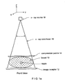
- Figs. 1a and 1b illustrate in simplified view an example of geometry for CC (cranial-caudal) imaging of a patient's breast 10.
- Breast 10 is compressed between an image receptor 12, such as a flat panel digital imager, and a compression paddle 14, and is imaged with a cone-shaped or pyramid-shaped x-ray beam 16 from an x-ray source 18.
- Fig. 1a illustrates a front view where the long axis of the compressed breast 10 is normal to the sheet
- Fig. 1b illustrates a side view where the long axis of the breast is in the plane of the sheet and where the patient's chest (not shown) is to the right of the illustrated geometry.
- Respective coordinate systems are shown above Figs. 1a and 1b .
- the illustrated geometry is similar to that used in the SeleniaTM mammography system identified above as well as in the tomosynthesis system disclosed in said patent application Ser. No. 10/723,486 .
- the breast can be compressed and imaged in other orientations as well.
- Fig. 2 illustrates the same geometry as Fig. 1b , and the same coordinate system, except that it highlights a single raypath or x-ray trajectory 20 rather than illustrate the entire x-ray beam 16.
- Fig. 2 illustrates two objects in breast 10, object #1 at slice A and object #2 at slice B.
- object is used here to refer to any structure that can be imaged in a mammogram or a tomosynthesis image, such as a lesion in the breast
- slice is used to refer to a layer of the breast of a finite thickness, e.g. thickness in the z-direction, that is less than the total breast thickness.
- a slice can be a few mm thick, or thinner or thicker.
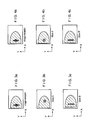
- Figs. 3a-3c illustrate a mammogram that can be obtained with the geometry of Fig. 2
- Figs. 3b and 3c illustrate tomosynthesis images of slices A and B, respectively, obtained with a system as disclosed in said application Ser. No. 10/723,486 . Because objects #1 and #2 are along the same x-ray trajectory 20, they appear superimposed in the mammogram of Fig. 3a .
- x-ray trajectory 20 is not normal to the image plane of receptor 12, as is the general case with x-ray trajectories when using such cone-beam or pyramid-beam x-ray geometry, the two objects appear at different xy locations in the tomosynthesis images of Figs. 3b and 3c .
- These tomosynthesis images can be conceptualized as vertical projections of slices A and B onto the image plane of the mammogram, e.g. the image plane of x-ray receptor 12.
- Another way to conceptualize such a tomosynthesis image is to imagine that slice A alone, with no other breast tissue above or below it, were laid directly on x-ray receptor 12 and imaged.
- the same object #2 shows up at different xy locations in Figs. 3a and 3c .
- Fig. 4a is the same as Fig. 3a - a mammogram that shows the same objects #1 and #2 superimposed.
- Fig. 4b differs significantly from Fig. 3b
- Fig. 4c differs significantly from Fig. 3c .
- Figs. 4b and 4c show the images of objects #1 and #2 at xy locations that are in the same coordinate system.
- this is the same coordinate system as that of the mammogram of Fig. 4a (and 3a ).
- Both Figs. 4b and 3b are tomosynthesis images of slice A, but Fig. 4c shows object #2 in slice image A' at the correct xy location that matches the location of the same object in the mammogram of Fig. 4a .
- both Figs. 4c and 3c are tomosynthesis images of slice B, but Fig. 4c shows object #2 in slice image B' at the correct xy location that matches the location of the same object in the mammogram of Fig. 4a .
- those pixel values can be reconstructed into a rectangular Cartesian coordinate system (30 in Fig 5a ) using known reconstruction algorithms such as filtered back projection, iterative reconstruction, maximum likelihood reconstruction, or others, for example as taught in said patent application Ser. No. 10/723,486 .
- the voxels (elemental volume elements) that are imaged as respective pixels in the tomosynthesis slice images are aligned along lines normal to the image plane of receptor 12.
- the result can be conceptualized as a set of pixel values representing x-ray properties of the voxels that are in the 3D space bound by the image plane of receptor 12 at the bottom, compression paddle 14 on top, and on the sides by the boundaries of x-ray beam 16 that impinges on receptor 12, and are uniformly spaced in xy planes.
- x-ray beam 16 is cone-shaped for pyramid-shaped, the sides of this 3D space slope at least on three sides of the beam, and the x-ray trajectories from source 18 to receptor 12 diverge in the general case.
- each x-ray trajectory such as trajectory 20 is non-normal to the image plane of receptor 12.
- the height of an object in breast 10 influences where the image of that object will be in a mammogram taken with receptor 12. Stated differently, if two objects in the breast are along the same line normal to the image plane, in general they will appear spaced from each other in the mammogram but if the same two objects are along one of the sloping x-ray trajectory, they will appear superimposed in the mammogram.
- tomosynthesis reconstruction directly or indirectly calculates a pixel image of a slice that is both parallel to the mammogram image plane and is in the same coordinate system as the mammogram, as disclosed in this patent specification, the resulting tomosynthesis image in general can show the image of an object in the breast at the same position relative to other tissue in the same slice in the breast in all tomosynthesis slice images and will better match the mammogram image.
- images such as in Figs. 4b and 4c can be obtained by projecting each of several horizontal breast slices separately onto the image plane of the mammography image, along the actual x-ray trajectories included in x-ray beam 16.
- This can be conceptualized by imagining that a slice such as slice A keeps its physical position illustrated in Fig. 2 , all other breast tissue is absent, and a projection image is taken of slice A alone, using the geometry of Fig. 2 .
- this cannot be done literally because of the presence of breast tissue above and/or below the slice.
- the reconstruction geometry can be a cone beam coordinate system 31 shown in Fig 5b , where the voxels that correspond to pixels in the tomosynthesis slice images are at different xy spacings (and differ in size at least in the xy plane) in different slices and corresponding voxels of different slices are along the same (generally sloping) x-ray trajectory.
- the desired result can be achieved indirectly, by first reconstructing tomosynthesis images that together represent a three dimensional space having at least three sloping sides matching the geometry of the imaging x-ray beam (as in the coordinate system of Fig. 5a ), and then geometrically projecting the pixel values of such tomosynthesis images onto the image plane of the mammogram along the directions of respective x-ray trajectories in the x-ray beam, again using a computer-implemented process adapted without undue experimentation to a particular x-ray data acquisition geometry by a programmer of ordinary skill in the art.
- Tomosynthesis slice images such as in Figs. 4b and 4c can facilitate assessment of breast features by allowing more direct and simplified comparison between different tomosynthesis slice images and between tomosynthesis slice images on the one hand and conventional mammograms on the other hand.
- the health professional can read a mammogram ( Fig. 4a ) in a conventional manner, but can also display and view any one or several of a number of tomosynthesis slice images of the same breast to visualize and assess structures that can be at different heights in the breast but appear at the same or at least matching relative locations in each image in which they show.
- the mammogram and the tomosynthesis slice images can be displayed on the same monitor or screen, displaying one image at a time by alternating from one image to another with a suitable switch or other interface controlled by the health professional.
- one or more tomosynthesis and/or mammogram images can be displayed on one monitor or screen while one or more other images can be displayed on another monitor or screen, to allow for simultaneous viewing.
- two or more tomosynthesis and/or mammogram images can displayed at respective locations on the same monitor or screen.
- a control interface can allow the health professional to select the images for display and the locations for display of those images.
- Reconstructing and displaying tomosynthesis slice images ( Figs. 4b and 4c ) as described above is particularly suitable for use in conjunction with computer-aided diagnosis (CAD) of breast images.
- CAD computer-aided diagnosis
- an x-ray breast image is computer-analyzed and image markers are generated and displayed to indicate the location of suspected abnormalities and, in some cases, the likely type of abnormality.
- the tomosynthesis slice images are as in Figs. 4b and 4c , such markers can be accurately and easily displayed at correct locations relative to the tomosynthesis slice image, even when such markers are generated based solely or mainly on the appropriate mammogram.
- fusion images can be generated and selectively displayed under the control of a health professional.
- a mammogram such as in Fig. 4a and a slice image such as in Fig. 4b or 4c can be superimposed for display, for example with the mammogram in gray scale or in a first selected color or set of colors and the tomosynthesis image in a second selected color or set of colors.
- the fused image can further include CAD markers displayed at the appropriate locations.
- Another display method is to select a region of interest in a mammogram, for example by the health professional drawing or otherwise indicating a region of interest (ROI), and replacing the ROI with the corresponding portion of a selected tomosynthesis slice image.
- ROI region of interest
- the particular tomosynthesis slice image or succession of such images can be selected by the health professions through an appropriate interface such a track ball or mouse buttons or wheel.
- the health professional can scroll up and down the height of the imaged breast and see tomosynthesis images within the ROI without losing landmark orientation relative to other parts of the breast that are still seen in the portion of the mammogram outside the ROI.
- the tomosynthesis x-ray measurements and/or images described above can be used to reconstruct or reformat slice images conforming to planes that are not parallel to the image plane of a mammogram, using image processing techniques known in technologies such as CT (computerized tomography) scanning , and to reconstruct or reformat 3D displays of the imaged breast or selected portions of the breast, for display alone or in conjunction with the display of one or more mammograms and/or 3D tomosynthesis slice images.
- CT computerized tomography
- FIG. 6 illustrates in block diagram form an example of a system implementing technology described above.
- An x-ray data acquisition unit 50 acquires x-ray measurements for tomosynthesis and/or mammogram images, for example as described in patent application Ser. No. 10/723,486 .
- a pre-processing unit 52 applies known gain and offset corrections to the raw x-ray measurements from unit 50, and known normalization/log conversion of the corrected data.
- Image reconstruction unit 54 uses the pre-processed x-ray measurements to generate appropriate tomosynthesis and/or mammographic images. Such images are displayed at unit 56, under the control of a user interface 58 that includes controls such a keyboard, mouse, etc. to select and manipulate the displayed images as well as to control units 50-58 for other purposes.
- a geometry matrix can be defined from a geometry calibration file and input projection angles appropriate to unit 50 for use in backprojection, from fits to the matrix elements determined from a geometry calibration of unit 50 and input projection angles measured by an encoder in unit 50.
- Image processing and filtering can be carried out on the images prior to reconstruction, using image processing techniques known in technologies such as CT scanning and tomosynthesis. A known skin line correction can also be applied at this stage.
- This method of backprojection is described, for example, in section 3.4 in Faugeras, O., Three-Dimensional Computer Vision, A Geometric Viewpoint, (MIT Press, 2001 ).
- a first method using Cartesian coordinates as in Fig. 5a , the reconstructed slices are parallel to the breast plate, or parallel to the image plane of receptor 12 at 0° projection angle. Voxels and their corresponding image pixels are equally spaced in x-y (in-plane). The x-y pixel spacing is the same for each image slice. The z-pixel spacing is the desired output slice separation.
- a second method using cone beam coordinates as in Fig. 5b , the in-slice pixel spacing varies as a function of slice number, or distance from the focal spot in source 18.
- the image slices are confined to the volume defined by a given source/detector location, that is, the volume defined by the four lines connecting the x-ray source point to the four corners of image receptor 12 (or any four points on receptor 12).
- M is first transformed by another matrix to obtain reconstructed planes that are parallel to receptor 12 at some other arbitrary projection angle. This rotation matrix is obtained from the geometry matrix of that projection.
- a pixel size scale factor and pixel starting location are calculated for each slice. The scale factor depends on the number of projections that intersect the given pixel. That is, some pixels are not ⁇ seen' by all projections. This scaling reduces band artifacts near the edge of the image.
- a preferred orientation can be an orientation in which a particular mammogram is taken.
- the backprojection for reconstructing tomosynthesis slice images can involve:
- this patent specification discloses a method comprising: obtaining tomosynthesis x-ray measurements and at least one 2D x-ray projection mammogram of a patient's breast, wherein the mammogram image and the tomosynthesis measurements are obtained using a cone-shaped or pyramid-shaped imaging x-ray beam, and reconstructing 2D tomosynthesis images from the tomosynthesis measurements, wherein the tomosynthesis images conform to the same geometric coordinate system as the 2D projection mammogram, whereby anatomical structures that appear in the mammogram appear at geometrically corresponding places in respective ones of the tomosynthesis images.
- the step or steps of reconstructing 2D tomosynthesis images can comprise using a computer-implemented cone beam reconstruction algorithm directly generating the tomosynthesis images.
- the step or steps of reconstructing the 2D tomosynthesis images can comprise generating information describing initial tomosynthesis images, in which tissue or objects in the breast that are at different heights in the breast but overlap in the mammogram appear at mismatched positions in the initial tomosynthesis images, and using the information describing the initial tomosynthesis images to generate final tomosynthesis images in which said tissue or objects appear at positions that match their positions in the mammogram.
- This alternative can be implemented by generating the initial tomosynthesis images in an initial coordinate system different from that of the mammogram, and processing the information describing the initial tomosynthesis images into tomosynthewsis images that match the coordinate system of the mammogram.
- the initial tomosynthesis images may differ in pixel spacing while the final tomosynthesis images may have the same pixel spacing.
- the final pixel spacing may be the same as in the mammogram.
- an x-ray system comprising an x-ray data acquisition unit that uses a cone-shaped or pyramid shaped x-ray beam and an x-ray receptor to obtain tomosynthesis x-ray measurements and x-ray measurements for at least one 2D x-ray projection mammogram of a patient's breast, a pre-processor that receives said measurements from the x-ray receptor and subjects them to pre-processing operations, a tomo/mammo image reconstruction unit that receives the pre-processed images and subjects them to further processing to reconstructing 2D tomosynthesis images and a mammogram, wherein tissue or objects in the breast that are at different heights in the breast but appear superimposed in the mammogram appear at locations in the tomosynthesis images that are the same as or at least match their location in the mammogram, and a display unit that selectively displays one or more of the tomosynthesis images and the mammogram and is under the control of a used interface operated by
- the image reconstruction unit can use a computer-implemented cone beam reconstruction algorithm directly generating the tomosynthesis images.
- the image reconstruction unit can generate information describing initial tomosynthesis images, in which tissue or objects in the breast that are at different heights in the breast but overlap in the mammogram appear at mismatched positions in the initial tomosynthesis images, and can use the information describing the initial tomosynthesis images to generate final tomosynthesis images in which said objects appear at positions that are the same as or at least match their positions in the mammogram.
- This alternative can be implemented by generating the initial tomosynthesis images in an initial coordinate system different from that of the mammogram, and processing the information describing the initial tomosynthesis images into tomosynthewsis images that match the coordinate system of the mammogram.
- the initial tomosynthesis images may differ in pixel spacing while the final tomosynthesis images may have the same pixel spacing.
- the final pixel spacing may be the same as in the mammogram.
Landscapes
- Engineering & Computer Science (AREA)
- Physics & Mathematics (AREA)
- Theoretical Computer Science (AREA)
- General Physics & Mathematics (AREA)
- Health & Medical Sciences (AREA)
- Life Sciences & Earth Sciences (AREA)
- Medical Informatics (AREA)
- Computer Vision & Pattern Recognition (AREA)
- Pathology (AREA)
- Animal Behavior & Ethology (AREA)
- Veterinary Medicine (AREA)
- Public Health (AREA)
- General Health & Medical Sciences (AREA)
- Biomedical Technology (AREA)
- Dentistry (AREA)
- Oral & Maxillofacial Surgery (AREA)
- Biophysics (AREA)
- High Energy & Nuclear Physics (AREA)
- Surgery (AREA)
- Nuclear Medicine, Radiotherapy & Molecular Imaging (AREA)
- Optics & Photonics (AREA)
- Molecular Biology (AREA)
- Radiology & Medical Imaging (AREA)
- Heart & Thoracic Surgery (AREA)
- Data Mining & Analysis (AREA)
- General Engineering & Computer Science (AREA)
- Evolutionary Biology (AREA)
- Artificial Intelligence (AREA)
- Bioinformatics & Cheminformatics (AREA)
- Bioinformatics & Computational Biology (AREA)
- Evolutionary Computation (AREA)
- Apparatus For Radiation Diagnosis (AREA)
- Pure & Applied Mathematics (AREA)
- Mathematical Optimization (AREA)
- Mathematical Physics (AREA)
- Mathematical Analysis (AREA)
- Algebra (AREA)
Claims (18)
- Procédé comprenant :l'obtention d'une pluralité d'images de projection de tomosynthèse à rayons X en 2D d'un sein d'une patiente prises à différents angles d'un faisceau à rayons X d'imagerie (16) relativement au sein (10) ;l'utilisation d'une unité de reconstruction d'image (54) pour effectuer un traitement mis en oeuvre par ordinateur des images de projection de tomosynthèse afin de dériver des images de tomosynthèse reconstruites en 2D représentatives de tranches de sein respectives ; etl'affichage, sur une unité d'affichage (56), d'au moins certaines des images de tranche de tomosynthèse reconstruites,dans lequel chaque image de tomosynthèse reconstruite comprend un ensemble de valeurs de pixel en 2D représentant des voxels respectifs du sein, lesdites valeurs de pixel se rapportant à des positions de pixel respectives dans l'image de tomosynthèse reconstruite,caractérisé en ce que :ladite dérivation des images de tomosynthèse reconstruites comprend l'application d'une matrice de géométrie transformant lesdites positions de pixel en un système de coordonnées pour le sein dans lequel un objet dans le sein apparaît au même endroit dans chaque image de tranche de tomosynthèse reconstruite dans laquelle l'objet apparaît, tel que dans un cliché mammaire du sein classique.
- Procédé conformément à la revendication 1, dans lequel ladite unité de reconstruction d'image est programmée pour effectuer un algorithme de reconstruction de faisceau conique mis en oeuvre par ordinateur qui crée directement lesdites images de tomosynthèse reconstruites à partir desdites images de tomosynthèse de projection.
- Procédé conformément à la revendication 1, dans lequel ladite unité de reconstruction d'image est programmée pour créer des images de tomosynthèse préliminaires qui représentent des tranches de sein parallèles mais dans lesquelles des structures anatomiques qui sont à des hauteurs différentes dans le sein mais se chevauchent dans une image de cliché mammaire classique apparaissent à des positions géométriquement dépareillées, et pour utiliser les images de tomosynthèse préliminaires afin de créer lesdites images de tomosynthèse reconstruites.
- Procédé conformément à la revendication 3, dans lequel la création des images de tomosynthèse préliminaires comprend la mise en conformité des images de tomosynthèse préliminaires avec un système de coordonnées préliminaire différent d'un système de coordonnées auquel une image de cliché mammaire classique est conforme, et le traitement des images de tomosynthèse préliminaires pour obtenir lesdites images de tomosynthèse reconstruites.
- Procédé conformément à la revendication 4, dans lequel les images de tomosynthèse préliminaires sont dififérentes les unes des autres dans un espacement de pixels, lesdites images de tomosynthèse reconstruites étant les mêmes dans l'espacement de pixels.
- Procédé conformément à la revendication 5 comprenant l'obtention d'une image de cliché mammaire comprenant des valeurs pour un ensemble de pixels en 2D, dans lequel l'espacement de pixels dans les images de tomosynthèse reconstruites est conforme à celui de l'image de cliché mammaire.
- Procédé conformément à la revendication 1 comprenant l'obtention d'une image de cliché mammaire comprenant des valeurs pour un ensemble de pixels en 2D, dans lequel les images de tomosynthèse reconstruites sont caractérisées en ce que l'espacement de pixels est conforme à celui de l'image de cliché mammaire.
- Procédé conformément à la revendication 1 comprenant un prétraitement des images de tomosynthèse de projection afin de créer des informations filtrées, et la rétroprojection des informations filtrées afin de créer lesdites images de tomosynthèse reconstruites, pour l'une desdites tranches à la fois, à l'aide d'une matrice de géométrie comme suit ,

Mi étant une matrice de géométrie 3x4 pour une rétroprojection i, (x,y,z) est un emplacement d'un pixel d'image de l'une desdites images de tomosynthèse reconstruites, et (dx, dy) est un emplacement sur un élément ou une zone de détecteur par rayons X pour une ligne qui relie un point focal duquel le rayon X émane et le pixel d'image. - Procédé conformément à la revendication 8 comprenant en outre l'application d'une opération de multiplication de matrice 4x4 à ladite matrice de géométrie 3x4 Mi conformément à
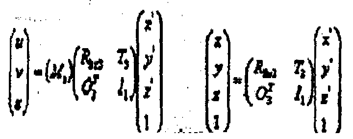
où
pour ainsi orienter lesdites images de tomosynthèse reconstruites à des directions sélectionnées définies par un choix de ladite matrice 4x4. - Système de rayons X comprenant :une unité d'acquisition de données (50) qui utilise des rayons X émanant d'une origine (18) et un récepteur de rayons X (12) pour obtenir des mesures de rayons X pour une pluralité d'images de tomosynthèse de projection du sein prises à des angles respectifs d'un faisceau à rayons X (16) relativement au sein (10) ;une unité de prétraitement (52) configurée pour recevoir lesdites mesures de rayons X en provenance du récepteur de rayons X et pour les soumettre à des opérations informatiques de prétraitement et ainsi créer lesdites images de tomosynthèse de projection ;une unité de reconstruction d'image de tomosynthèse (54) configurée pour recevoir lesdites images de tomosynthèse de projection et pour les soumettre à un traitement informatique afin de former des images de tomosynthèse reconstruites qui représentent des tranches de sein respectives ; etune unité d'affichage qui affiche sélectivement une ou plusieurs des images de tranche de tomosynthèse reconstruites, ladite unité d'affichage comprenant une interface utilisateur commandant l'affichage desdites imagesdans lequelchaque image de tomosynthèse reconstruite comprend un ensemble de valeurs de pixels en 2D représentant des voxels respectifs du sein, lesdites valeurs de pixel se rapportant à des positions de pixel respectives dans l'image de tomosynthèse reconstruite, caractérisé en ce queladite formation des images de tomosynthèse reconstruites comprend l'application d'une matrice de géométrie transformant lesdites positions de pixel en un système de coordonnées dans lequel un objet dans le sein apparaît au même endroit dans chaque image de tranche de tomosynthèse reconstruite dans laquelle l'objet apparaît, tel que dans le cliché mammaire du sein classique.
- Système conformément à la revendication 10, dans lequel ladite unité de reconstruction d'image comprend un algorithme de reconstruction de faisceau conique mis en oeuvre par ordinateur créant directement les images de tomosynthèse reconstruites.
- Système conformément à la revendication 10, dans lequel ladite unité de reconstruction d'image comprend un algorithme créant des images de tomosynthèse préliminaires qui représentent des tranches de sein mais dans lesquelles des structures anatomiques qui sont à des hauteurs différentes dans le sein mais se chevauchent dans l'image de cliché mammaire apparaissent à des positions géométriquement non concordantes, et utilisant les images de tomosynthèse préliminaires afin de créer lesdites images de tomosynthèse reconstruites.
- Système conformément à la revendication 12, dans lequel ladite unité de reconstruction comprend un algorithme mettant en conformité les images de tomosynthése préliminaires avec un système de coordonnées d'image préliminaire différent d'un système de coordonnées auquel une image de cliché mammaire classique est conforme, et traitant les images de tomosynthèse préliminaires pour obtenir lesdites images de tomosynthèse reconstruites.
- Système conformément à la revendication 13, dans lequel ladite unité de reconstruction forme les images de tomosynthèse préliminaires au niveau d'un espacement de pixels qui sont différentes les unes des autres et forme lesdites images de tomosynthése reconstruites dans un espacement de pixel qui sont les mêmes pour une pluralité d'images de tomosynthèse reconstruites.
- Système conformément à la revendication 14 comprenant la création d'une image de cliché mammaire, dans lequel ladite unité de reconstruction forme les images de tomosynthèse reconstruites au niveau d'un espacement de pixels qui est conforme à celui de l'image de cliché mammaire.
- Système conformément à la revendication 10 comprenant la création d'une image de cliché mammaire, dans lequel ladite unité de reconstruction forme les images de tomosynthèse reconstruites au niveau d'un espacement de pixel qui est conforme à celui de l'image de cliché mammaire.
- Système conformément à la revendication 10, dans lequel ladite unité de reconstruction comprend un algorithme filtrant les images de tomosynthèse de projection pour créer des images filtrées, et la rétroprojection des images filtrées pour créer lesdites images de tomosynthèse reconstruites, pour l'une desdites tranches à la fois, à l'aide d'une matrice de géométrie comme suit,

où Mi est une matrice de géométrie 3x4 pour une rétroprojection i, (x,y ,z) est un emplacement d'un pixel d'image dans l'une desdites images de tomosynthèse reconstruites, et (dx, dy) est un emplacement d'un élément ou d'une zone de détecteur par rayons X pour une ligne qui relie un point focal duquel le rayon X émane et le pixel d'image. - Système conformément à la revendication 17, dans lequel ledit algorithme comprend en outre l'application d'une opération de multiplication de matrice 4x4 à ladite matrice de géométrie 3x4 Mi conformément à

où
pour ainsi orienter lesdites images de tomosynthèse reconstruites à des directions sélectionnées définies par un choix de ladite matrice 4x4.
Priority Applications (1)
| Application Number | Priority Date | Filing Date | Title |
|---|---|---|---|
| EP13157683.7A EP2602743B1 (fr) | 2004-11-15 | 2005-11-15 | Affichage et génération de géométrie de mise en correspondance de mammographies et d'images de tomosynthèse |
Applications Claiming Priority (2)
| Application Number | Priority Date | Filing Date | Title |
|---|---|---|---|
| US62851604P | 2004-11-15 | 2004-11-15 | |
| PCT/US2005/041941 WO2006055830A2 (fr) | 2004-11-15 | 2005-11-15 | Generation et affichage geometriques de mise en correspondance de cliches mammaires et d'images de tomosynthese |
Related Child Applications (1)
| Application Number | Title | Priority Date | Filing Date |
|---|---|---|---|
| EP13157683.7A Division EP2602743B1 (fr) | 2004-11-15 | 2005-11-15 | Affichage et génération de géométrie de mise en correspondance de mammographies et d'images de tomosynthèse |
Publications (3)
| Publication Number | Publication Date |
|---|---|
| EP1815388A2 EP1815388A2 (fr) | 2007-08-08 |
| EP1815388A4 EP1815388A4 (fr) | 2011-06-08 |
| EP1815388B1 true EP1815388B1 (fr) | 2013-03-06 |
Family
ID=36407794
Family Applications (2)
| Application Number | Title | Priority Date | Filing Date |
|---|---|---|---|
| EP13157683.7A Expired - Lifetime EP2602743B1 (fr) | 2004-11-15 | 2005-11-15 | Affichage et génération de géométrie de mise en correspondance de mammographies et d'images de tomosynthèse |
| EP05824734A Expired - Lifetime EP1815388B1 (fr) | 2004-11-15 | 2005-11-15 | Generation et affichage geometriques de mise en correspondance de cliches mammaires et d'images de tomosynthese |
Family Applications Before (1)
| Application Number | Title | Priority Date | Filing Date |
|---|---|---|---|
| EP13157683.7A Expired - Lifetime EP2602743B1 (fr) | 2004-11-15 | 2005-11-15 | Affichage et génération de géométrie de mise en correspondance de mammographies et d'images de tomosynthèse |
Country Status (3)
| Country | Link |
|---|---|
| US (7) | US7702142B2 (fr) |
| EP (2) | EP2602743B1 (fr) |
| WO (1) | WO2006055830A2 (fr) |
Families Citing this family (134)
| Publication number | Priority date | Publication date | Assignee | Title |
|---|---|---|---|---|
| US7123684B2 (en) * | 2002-11-27 | 2006-10-17 | Hologic, Inc. | Full field mammography with tissue exposure control, tomosynthesis, and dynamic field of view processing |
| US8571289B2 (en) | 2002-11-27 | 2013-10-29 | Hologic, Inc. | System and method for generating a 2D image from a tomosynthesis data set |
| US7616801B2 (en) | 2002-11-27 | 2009-11-10 | Hologic, Inc. | Image handling and display in x-ray mammography and tomosynthesis |
| US7760924B2 (en) * | 2002-11-27 | 2010-07-20 | Hologic, Inc. | System and method for generating a 2D image from a tomosynthesis data set |
| US10638994B2 (en) | 2002-11-27 | 2020-05-05 | Hologic, Inc. | X-ray mammography with tomosynthesis |
| US7577282B2 (en) | 2002-11-27 | 2009-08-18 | Hologic, Inc. | Image handling and display in X-ray mammography and tomosynthesis |
| US8565372B2 (en) | 2003-11-26 | 2013-10-22 | Hologic, Inc | System and method for low dose tomosynthesis |
| US7662082B2 (en) | 2004-11-05 | 2010-02-16 | Theragenics Corporation | Expandable brachytherapy device |
| EP2602743B1 (fr) | 2004-11-15 | 2014-11-05 | Hologic, Inc. | Affichage et génération de géométrie de mise en correspondance de mammographies et d'images de tomosynthèse |
| EP3106094B1 (fr) | 2004-11-26 | 2021-09-08 | Hologic, Inc. | Systeme radiographique multimode integrant mammographie/tomosynthese |
| US10008184B2 (en) | 2005-11-10 | 2018-06-26 | Hologic, Inc. | System and method for generating a 2D image using mammography and/or tomosynthesis image data |
| WO2013078476A1 (fr) | 2011-11-27 | 2013-05-30 | Hologic, Inc. | Système et procédé pour générer une image 2d en utilisant des données d'images de mammographie et/ou de tomosynthèse |
| US7465268B2 (en) | 2005-11-18 | 2008-12-16 | Senorx, Inc. | Methods for asymmetrical irradiation of a body cavity |
| WO2007095330A2 (fr) | 2006-02-15 | 2007-08-23 | Hologic Inc | Biopsie mammaire et localisation a l'aiguille a l'aide de systemes de tomosynthese |
| SE0702061L (sv) * | 2007-09-17 | 2009-03-18 | Xcounter Ab | Metod för att skapa, visa och analysera röntgenbilder och anordning för att implementera metoden |
| US7630533B2 (en) | 2007-09-20 | 2009-12-08 | Hologic, Inc. | Breast tomosynthesis with display of highlighted suspected calcifications |
| US7929743B2 (en) * | 2007-10-02 | 2011-04-19 | Hologic, Inc. | Displaying breast tomosynthesis computer-aided detection results |
| US7792245B2 (en) | 2008-06-24 | 2010-09-07 | Hologic, Inc. | Breast tomosynthesis system with shifting face shield |
| US7991106B2 (en) | 2008-08-29 | 2011-08-02 | Hologic, Inc. | Multi-mode tomosynthesis/mammography gain calibration and image correction using gain map information from selected projection angles |
| JP2010068929A (ja) * | 2008-09-17 | 2010-04-02 | Fujifilm Corp | 乳房x線透過平面画像断層画像撮影装置 |
| JP2010104771A (ja) * | 2008-09-30 | 2010-05-13 | Fujifilm Corp | 放射線画像診断システム |
| US8515005B2 (en) | 2009-11-23 | 2013-08-20 | Hologic Inc. | Tomosynthesis with shifting focal spot and oscillating collimator blades |
| AU2009316352A1 (en) * | 2008-11-24 | 2010-05-27 | Hologic Inc. | Method and system for controlling x-ray focal spot characteristics for tomosynthesis and mammography imaging |
| SE533704C2 (sv) | 2008-12-05 | 2010-12-07 | Flatfrog Lab Ab | Pekkänslig apparat och förfarande för drivning av densamma |
| US9579524B2 (en) | 2009-02-11 | 2017-02-28 | Hologic, Inc. | Flexible multi-lumen brachytherapy device |
| US9248311B2 (en) | 2009-02-11 | 2016-02-02 | Hologic, Inc. | System and method for modifying a flexibility of a brachythereapy catheter |
| US10207126B2 (en) | 2009-05-11 | 2019-02-19 | Cytyc Corporation | Lumen visualization and identification system for multi-lumen balloon catheter |
| CN102481146B (zh) | 2009-10-08 | 2016-08-17 | 霍罗吉克公司 | 乳房的穿刺活检系统及其使用方法 |
| FR2954556B1 (fr) * | 2009-12-22 | 2017-07-28 | Gen Electric | Procede de traitement d'acquisitions de tomosynthese pour obtenir une representation du contenu d'un organe |
| KR101689866B1 (ko) | 2010-07-29 | 2016-12-27 | 삼성전자주식회사 | 영상 처리 방법 및 장치와 이를 채용한 의료영상시스템 |
| US9352172B2 (en) | 2010-09-30 | 2016-05-31 | Hologic, Inc. | Using a guide member to facilitate brachytherapy device swap |
| CA2813591C (fr) | 2010-10-05 | 2020-09-22 | Hologic, Inc. | Imagerie du sein par rayon x en position debout avec un mode de tomodensitometrie, de multiples modes de tomosynthese et un mode de mammographie |
| US9411444B2 (en) * | 2010-10-11 | 2016-08-09 | Flatfrog Laboratories Ab | Touch determination by tomographic reconstruction |
| US20120133600A1 (en) | 2010-11-26 | 2012-05-31 | Hologic, Inc. | User interface for medical image review workstation |
| JP2014504918A (ja) | 2010-12-14 | 2014-02-27 | ホロジック, インコーポレイテッド | 画像診断に使用するために複数の異なる画像処理システムからの3次元画像データを重ね合わせるシステムおよび方法 |
| US10342992B2 (en) | 2011-01-06 | 2019-07-09 | Hologic, Inc. | Orienting a brachytherapy applicator |
| DE102011003137A1 (de) * | 2011-01-25 | 2012-07-26 | Siemens Aktiengesellschaft | Bildgebungsverfahren mit einer verbesserten Darstellung eines Gewebebereichs |
| WO2012106204A1 (fr) | 2011-01-31 | 2012-08-09 | University Of Massachusetts | Imagerie par la tomosynthèse |
| JP6057922B2 (ja) | 2011-03-08 | 2017-01-11 | ホロジック, インコーポレイテッドHologic, Inc. | スクリーニング、診断及び生検のための二重エネルギー及び/又は造影強化乳房撮像のためのシステム及び方法 |
| US11259759B2 (en) | 2011-11-18 | 2022-03-01 | Hologic Inc. | X-ray mammography and/or breast tomosynthesis using a compression paddle |
| US9332947B2 (en) | 2011-11-18 | 2016-05-10 | Hologic, Inc. | X-ray mammography and/or breast tomosynthesis using a compression paddle with an inflatable jacket with dual bottom layer joined at a seam enhancing imaging and improving patient comfort |
| US9782135B2 (en) | 2011-11-18 | 2017-10-10 | Hologic, Inc. | X-ray mammography and/or breast tomosynthesis using a compression paddle |
| WO2013123091A1 (fr) | 2012-02-13 | 2013-08-22 | Hologic, Inc. | Système et procédé pour naviguer dans une pile de tomosynthèse par utilisation de données d'images synthétisées |
| JP6251164B2 (ja) * | 2012-03-19 | 2017-12-20 | 京セラ株式会社 | 移動通信システム、移動通信方法、無線基地局及びプロセッサ |
| US10168835B2 (en) | 2012-05-23 | 2019-01-01 | Flatfrog Laboratories Ab | Spatial resolution in touch displays |
| US9076246B2 (en) * | 2012-08-09 | 2015-07-07 | Hologic, Inc. | System and method of overlaying images of different modalities |
| WO2014059366A1 (fr) * | 2012-10-12 | 2014-04-17 | Klanian Kelly L | Appareil et procédé pour l'immobilisation du sein |
| FR2997284B1 (fr) | 2012-10-30 | 2016-06-17 | Gen Electric | Procede d'obtention d'images de tomosynthese |
| US8983156B2 (en) | 2012-11-23 | 2015-03-17 | Icad, Inc. | System and method for improving workflow efficiences in reading tomosynthesis medical image data |
| US9940738B2 (en) | 2013-01-10 | 2018-04-10 | Hologic, Inc. | System and method for reducing data transmission volume in tomosynthesis |
| WO2014151646A1 (fr) | 2013-03-15 | 2014-09-25 | Hologic Inc. | Biopsie guidée par tomosynthèse dans une position couchée |
| US10624598B2 (en) | 2013-03-15 | 2020-04-21 | Hologic, Inc. | System and method for navigating a tomosynthesis stack including automatic focusing |
| US9646376B2 (en) | 2013-03-15 | 2017-05-09 | Hologic, Inc. | System and method for reviewing and analyzing cytological specimens |
| US10019113B2 (en) | 2013-04-11 | 2018-07-10 | Flatfrog Laboratories Ab | Tomographic processing for touch detection |
| JP6368779B2 (ja) | 2013-06-28 | 2018-08-01 | コーニンクレッカ フィリップス エヌ ヴェKoninklijke Philips N.V. | トモシンセシスデータからエッジ保存合成マンモグラムを生成するための方法 |
| US9874978B2 (en) | 2013-07-12 | 2018-01-23 | Flatfrog Laboratories Ab | Partial detect mode |
| ES2954561T3 (es) | 2013-10-09 | 2023-11-23 | Hologic Inc | Tomosíntesis de mama por rayos X que potencia la resolución espacial, incluso en la dirección de grosor de una mama aplanada |
| EP3646798B1 (fr) | 2013-10-24 | 2023-09-27 | Hologic, Inc. | Système et procédé de navigation pour une biopise du sein guidée par rayons x |
| WO2015063188A1 (fr) | 2013-10-30 | 2015-05-07 | Koninklijke Philips N.V. | Procédé et dispositif pour afficher des images médicales |
| US10126882B2 (en) | 2014-01-16 | 2018-11-13 | Flatfrog Laboratories Ab | TIR-based optical touch systems of projection-type |
| WO2015108479A1 (fr) | 2014-01-16 | 2015-07-23 | Flatfrog Laboratories Ab | Couplage de lumière dans les systèmes tactiles optiques basés sur la tir |
| US9613440B2 (en) * | 2014-02-12 | 2017-04-04 | General Electric Company | Digital breast Tomosynthesis reconstruction using adaptive voxel grid |
| US10111631B2 (en) | 2014-02-28 | 2018-10-30 | Hologic, Inc. | System and method for generating and displaying tomosynthesis image slabs |
| US10161886B2 (en) | 2014-06-27 | 2018-12-25 | Flatfrog Laboratories Ab | Detection of surface contamination |
| JP6126058B2 (ja) * | 2014-09-30 | 2017-05-10 | 富士フイルム株式会社 | 画像表示装置、画像処理装置、放射線画像撮影システム、断層画像表示方法、及び断層画像表示プログラム。 |
| CN107209608A (zh) | 2015-01-28 | 2017-09-26 | 平蛙实验室股份公司 | 动态触摸隔离帧 |
| US10318074B2 (en) | 2015-01-30 | 2019-06-11 | Flatfrog Laboratories Ab | Touch-sensing OLED display with tilted emitters |
| EP3256936A4 (fr) | 2015-02-09 | 2018-10-17 | FlatFrog Laboratories AB | Système tactile optique comprenant des moyens de projection et de détection de faisceaux de lumière au-dessus et à l'intérieur d'un panneau de transmission |
| WO2016140612A1 (fr) | 2015-03-02 | 2016-09-09 | Flatfrog Laboratories Ab | Composant optique pour couplage lumineux |
| WO2016142492A1 (fr) | 2015-03-10 | 2016-09-15 | Koninklijke Philips N.V. | Extraction de structures correspondantes dans des paires d'images médicales |
| US9984478B2 (en) * | 2015-07-28 | 2018-05-29 | PME IP Pty Ltd | Apparatus and method for visualizing digital breast tomosynthesis and other volumetric images |
| US10775937B2 (en) | 2015-12-09 | 2020-09-15 | Flatfrog Laboratories Ab | Stylus identification |
| WO2017185028A1 (fr) | 2016-04-22 | 2017-10-26 | Hologic, Inc. | Tomosynthèse avec système radiographique à point focal de décalage utilisant un réseau adressable |
| WO2017205386A1 (fr) | 2016-05-27 | 2017-11-30 | Hologic, Inc. | Détection de tumeur interne et de surface synchronisée |
| DE102016217776A1 (de) * | 2016-09-16 | 2018-03-22 | Siemens Healthcare Gmbh | Simultane Abbildung von funktionalen und morphologischen Röntgenbilddaten einer Brust |
| WO2018081569A1 (fr) * | 2016-10-27 | 2018-05-03 | Artemiadis Panagiotis | Systèmes et procédés pour une interface cérébrale hybride pour des essaims robotiques à l'aide de signaux eeg et d'un dispositif d'entrée |
| US10888292B2 (en) | 2016-11-08 | 2021-01-12 | Hologic, Inc. | Imaging with curved compression elements |
| US10096106B2 (en) * | 2016-11-10 | 2018-10-09 | General Electric Company | Combined medical imaging |
| CN110100226A (zh) | 2016-11-24 | 2019-08-06 | 平蛙实验室股份公司 | 触摸信号的自动优化 |
| US11650672B2 (en) * | 2016-11-25 | 2023-05-16 | Hologic, Inc. | Healthcare information manipulation and visualization controllers |
| EP3667475B1 (fr) | 2016-12-07 | 2022-09-07 | FlatFrog Laboratories AB | Dispositif tactile curbe |
| CN116679845A (zh) | 2017-02-06 | 2023-09-01 | 平蛙实验室股份公司 | 触摸感测装置 |
| WO2018174786A1 (fr) | 2017-03-22 | 2018-09-27 | Flatfrog Laboratories | Différenciation de stylo pour écrans tactiles |
| WO2018182476A1 (fr) | 2017-03-28 | 2018-10-04 | Flatfrog Laboratories Ab | Appareil de détection tactile et son procédé d'assemblage |
| CN110621233B (zh) | 2017-03-30 | 2023-12-12 | 豪洛捷公司 | 用于处理乳房组织图像数据的方法 |
| CN110662489B (zh) | 2017-03-30 | 2024-08-02 | 豪洛捷公司 | 用于靶向对象增强以生成合成乳房组织图像的系统和方法 |
| EP3600047A1 (fr) | 2017-03-30 | 2020-02-05 | Hologic, Inc. | Système et procédé de synthèse et de représentation d'image de caractéristique multiniveau hiérarchique |
| JP6949535B2 (ja) * | 2017-04-13 | 2021-10-13 | キヤノン株式会社 | 情報処理装置、情報処理システム、情報処理方法及びプログラム |
| JP7039179B2 (ja) | 2017-04-13 | 2022-03-22 | キヤノン株式会社 | 情報処理装置、情報処理システム、情報処理方法及びプログラム |
| WO2018236565A1 (fr) | 2017-06-20 | 2018-12-27 | Hologic, Inc. | Procédé et système d'imagerie médicale à auto-apprentissage dynamique |
| CN110996801B (zh) | 2017-08-11 | 2023-10-03 | 豪洛捷公司 | 具有接近角部的乳房压迫板 |
| DE202018006903U1 (de) | 2017-08-16 | 2024-07-29 | Hologic Inc. | Techniken zur Patientenbewegungsartefaktkompensation bei Brustbildgebung |
| EP3449835B1 (fr) | 2017-08-22 | 2023-01-11 | Hologic, Inc. | Système et méthode de tomographie assistée par ordinateur pour imager de multiples cibles anatomiques |
| EP3676694A4 (fr) | 2017-09-01 | 2021-06-09 | FlatFrog Laboratories AB | Composant optique amélioré |
| US11567610B2 (en) | 2018-03-05 | 2023-01-31 | Flatfrog Laboratories Ab | Detection line broadening |
| US12121304B2 (en) | 2018-05-04 | 2024-10-22 | Hologic, Inc. | Introducer and localization wire visualization |
| EP3787520B1 (fr) | 2018-05-04 | 2024-09-25 | Hologic, Inc. | Visualisation d'aiguille de biopsie |
| CN108852392A (zh) * | 2018-05-21 | 2018-11-23 | 苏州达影医疗设备有限公司 | 锥形光束影像重建方法、影像系统及摄影系统 |
| US11090017B2 (en) | 2018-09-13 | 2021-08-17 | Hologic, Inc. | Generating synthesized projection images for 3D breast tomosynthesis or multi-mode x-ray breast imaging |
| CN112804944B (zh) | 2018-09-24 | 2024-07-23 | 豪洛捷公司 | 乳房映射和异常定位 |
| JP7482860B2 (ja) | 2018-09-28 | 2024-05-14 | ホロジック, インコーポレイテッド | 高密度要素抑制による合成乳房組織画像発生のためのシステムおよび方法 |
| WO2020080992A1 (fr) | 2018-10-20 | 2020-04-23 | Flatfrog Laboratories Ab | Cadre de dispositif tactile et outil associé |
| US12170140B2 (en) | 2018-11-25 | 2024-12-17 | Hologic, Inc. | Customizable multimodality image hanging protocols |
| WO2020153890A1 (fr) | 2019-01-25 | 2020-07-30 | Flatfrog Laboratories Ab | Terminal de visioconférence son procédé de fonctionnement |
| WO2020205610A1 (fr) | 2019-03-29 | 2020-10-08 | Hologic, Inc. | Génération de rapport d'images numériques déclenchée par une capture d'écran |
| US11883206B2 (en) | 2019-07-29 | 2024-01-30 | Hologic, Inc. | Personalized breast imaging system |
| EP4035176A1 (fr) | 2019-09-27 | 2022-08-03 | Hologic, Inc. | Système d'intelligence artificielle (ai) permettant de prédire le temps de lecture et la complexité de lecture pour examiner des images de sein en 2d/3d |
| CN114846512A (zh) | 2019-10-25 | 2022-08-02 | 永康智能股份有限公司 | 用于分析三维图像数据的系统和方法 |
| US12056316B2 (en) | 2019-11-25 | 2024-08-06 | Flatfrog Laboratories Ab | Touch-sensing apparatus |
| EP3832689A3 (fr) | 2019-12-05 | 2021-08-11 | Hologic, Inc. | Systèmes et procédés pour améliorer la durée de vie d'un tube à rayons x |
| US12367574B2 (en) | 2019-12-23 | 2025-07-22 | DeepHealth, Inc. | Systems and methods for analyzing two-dimensional and three-dimensional image data |
| WO2021150449A1 (fr) | 2020-01-24 | 2021-07-29 | Hologic, Inc. | Palpateur de compression de sein en mousse déplaçable horizontalement |
| JP7270782B2 (ja) | 2020-02-04 | 2023-05-10 | 富士フイルム株式会社 | 画像設定装置、画像設定装置の作動方法および画像設定プログラム |
| JP7321299B2 (ja) | 2020-02-04 | 2023-08-04 | 富士フイルム株式会社 | 画像設定装置、画像設定装置の作動方法および画像設定プログラム |
| EP4100822A4 (fr) | 2020-02-08 | 2024-02-28 | FlatFrog Laboratories AB | Appareil tactile à interactions à faible latence |
| CN121614054A (zh) | 2020-02-10 | 2026-03-06 | 平蛙实验室股份公司 | 触摸感测设备和制造触摸感测设备的框架元件的方法 |
| EP4119055B1 (fr) | 2020-03-13 | 2024-10-30 | FUJIFILM Corporation | Dispositif et programme de génération d'image, dispositif et programme d'apprentissage, et dispositif et programme de traitement d'image |
| WO2021186957A1 (fr) | 2020-03-18 | 2021-09-23 | 富士フイルム株式会社 | Dispositif de traitement d'image, procédé et programme |
| WO2021186956A1 (fr) | 2020-03-18 | 2021-09-23 | 富士フイルム株式会社 | Dispositif de traitement d'image, procédé et programme |
| US11481038B2 (en) | 2020-03-27 | 2022-10-25 | Hologic, Inc. | Gesture recognition in controlling medical hardware or software |
| AU2021241536A1 (en) | 2020-03-27 | 2022-08-18 | Hologic, Inc. | Systems and methods for identifying regions of interest in multiple imaging modalities |
| US11471118B2 (en) | 2020-03-27 | 2022-10-18 | Hologic, Inc. | System and method for tracking x-ray tube focal spot position |
| JP2023519878A (ja) | 2020-03-27 | 2023-05-15 | ホロジック, インコーポレイテッド | 複数のイメージングモダリティにおける関心領域を相関させるためのシステムおよび方法 |
| DE102021210289A1 (de) * | 2020-09-30 | 2022-03-31 | Siemens Healthcare Gmbh | Verfahren zum Erzeugen von Ergebnisschichtbildern mit zumindest teilweise verschiedener Schichtdicke |
| US20220164951A1 (en) | 2020-11-20 | 2022-05-26 | Hologic, Inc. | Systems and methods for using ai to identify regions of interest in medical images |
| AU2022268266A1 (en) | 2021-04-26 | 2023-11-23 | Hologic, Inc. | Systems and methods for measuring thickness of foam compressive elements |
| US11786191B2 (en) | 2021-05-17 | 2023-10-17 | Hologic, Inc. | Contrast-enhanced tomosynthesis with a copper filter |
| US12288275B2 (en) * | 2021-07-28 | 2025-04-29 | GE Precision Healthcare LLC | Methods and systems for breast tomosynthesis |
| US12186119B2 (en) | 2021-10-05 | 2025-01-07 | Hologic, Inc. | Interactive model interface for image selection in medical imaging systems |
| US12254586B2 (en) | 2021-10-25 | 2025-03-18 | Hologic, Inc. | Auto-focus tool for multimodality image review |
| IL313196A (en) | 2021-11-29 | 2024-07-01 | Hologic Inc | Systems and methods for correlating objects of interest |
| US12414217B2 (en) | 2022-02-07 | 2025-09-09 | Hologic, Inc. | Systems and methods for adaptively controlling filament current in an X-ray tube |
| DE102023205095A1 (de) | 2023-05-31 | 2024-12-05 | Siemens Healthineers Ag | Kombination einer 2D-Röntgenbildaufnahme mit einer Tomosynthese-Bildaufnahme |
| WO2025212901A1 (fr) | 2024-04-03 | 2025-10-09 | Hologic, Inc. | Réduction de tailles de fichier de tomosynthèse |
Family Cites Families (140)
| Publication number | Priority date | Publication date | Assignee | Title |
|---|---|---|---|---|
| JP4054402B2 (ja) | 1997-04-25 | 2008-02-27 | 株式会社東芝 | X線断層撮影装置 |
| US3502878A (en) | 1967-09-22 | 1970-03-24 | Us Health Education & Welfare | Automatic x-ray apparatus for limiting the field size of a projected x-ray beam in response to film size and to source-to-film distance |
| US3863073A (en) | 1973-04-26 | 1975-01-28 | Machlett Lab Inc | Automatic system for precise collimation of radiation |
| US3971950A (en) | 1975-04-14 | 1976-07-27 | Xerox Corporation | Independent compression and positioning device for use in mammography |
| US4160906A (en) | 1977-06-23 | 1979-07-10 | General Electric Company | Anatomically coordinated user dominated programmer for diagnostic x-ray apparatus |
| DE2838901C2 (de) | 1978-09-06 | 1986-11-06 | Siemens AG, 1000 Berlin und 8000 München | Katapultrasterlade |
| FR2512024A1 (fr) | 1981-08-27 | 1983-03-04 | Adir | Ethers tricycliques, leur preparation et les compositions pharmaceutiques les contenant |
| FR2549248B1 (fr) | 1983-06-24 | 1986-01-31 | Thomson Csf | Porte-cassette escamotable pour appareil d'examen radiologique et radiographique |
| DE3339775A1 (de) | 1983-11-03 | 1985-05-15 | Siemens AG, 1000 Berlin und 8000 München | Roentgendiagnostikgeraet mit strahlenfiltern |
| JPS60129034A (ja) | 1983-12-16 | 1985-07-10 | 横河メディカルシステム株式会社 | X線断層撮像装置の操作卓 |
| US4706269A (en) | 1985-03-11 | 1987-11-10 | Reina Leo J | Anti-scatter grid structure |
| US4773087A (en) | 1986-04-14 | 1988-09-20 | University Of Rochester | Quality of shadowgraphic x-ray images |
| USRE33634E (en) | 1986-09-23 | 1991-07-09 | Method and structure for optimizing radiographic quality by controlling X-ray tube voltage, current focal spot size and exposure time | |
| US4821727A (en) | 1986-10-30 | 1989-04-18 | Elscint Ltd. | Mammographic biopsy needle holder system |
| US4819258A (en) | 1986-11-28 | 1989-04-04 | Bennett X-Ray Corp. | Auto-setting of KV in an x-ray machine after selection of technic factors |
| US5051904A (en) | 1988-03-24 | 1991-09-24 | Olganix Corporation | Computerized dynamic tomography system |
| DK654488A (da) | 1988-11-23 | 1990-05-24 | Nordisk Roentgen Tech App | Roentgenapparat |
| FR2645006A1 (fr) | 1989-03-29 | 1990-10-05 | Gen Electric Cgr | Mammographe equipe d'un dispositif de vue stereotaxiques integre et procede d'utilisation d'un tel mammographe |
| FR2646340A1 (fr) | 1989-04-28 | 1990-11-02 | Gen Electric Cgr | Porte-cassette adaptable en dimension et en position pour mammographie |
| EP0406455B1 (fr) | 1989-07-03 | 1994-09-21 | Siemens Aktiengesellschaft | Appareil diagnostique à rayons X pour les mammographies |
| CA2014918A1 (fr) | 1989-09-06 | 1991-03-06 | James A. Mcfaul | Systeme de mammographie par balyage avec meilleure visualisation de la ligne cutanee |
| US4969174A (en) | 1989-09-06 | 1990-11-06 | General Electric Company | Scanning mammography system with reduced scatter radiation |
| US5240011A (en) | 1991-11-27 | 1993-08-31 | Fischer Imaging Corporation | Motorized biopsy needle positioner |
| US5415169A (en) | 1989-11-21 | 1995-05-16 | Fischer Imaging Corporation | Motorized mammographic biopsy apparatus |
| US5078142A (en) | 1989-11-21 | 1992-01-07 | Fischer Imaging Corporation | Precision mammographic needle biopsy system |
| US5199056A (en) | 1989-11-28 | 1993-03-30 | Darrah Carol J | Mammography compression paddle |
| US5481623A (en) | 1990-04-19 | 1996-01-02 | Fuji Photo Film Co., Ltd. | Apparatus for determining an image position on imaging media |
| US5163075A (en) | 1991-08-08 | 1992-11-10 | Eastman Kodak Company | Contrast enhancement of electrographic imaging |
| US5594769A (en) | 1991-11-27 | 1997-01-14 | Thermotrex Corporation | Method and apparatus for obtaining stereotactic mammographic guided needle breast biopsies |
| US5289520A (en) | 1991-11-27 | 1994-02-22 | Lorad Corporation | Stereotactic mammography imaging system with prone position examination table and CCD camera |
| US5359637A (en) | 1992-04-28 | 1994-10-25 | Wake Forest University | Self-calibrated tomosynthetic, radiographic-imaging system, method, and device |
| US5596200A (en) | 1992-10-14 | 1997-01-21 | Primex | Low dose mammography system |
| FR2703237B1 (fr) | 1993-03-29 | 1995-05-19 | Ge Medical Syst Sa | Mammographe équipé d'un dispositif de prises en vues stéréotaxiques à détecteur numérique et procédé d'utilisation d'un tel mammographe . |
| US5365562A (en) | 1993-09-20 | 1994-11-15 | Fischer Imaging Corporation | Digital imaging apparatus |
| US6075879A (en) | 1993-09-29 | 2000-06-13 | R2 Technology, Inc. | Method and system for computer-aided lesion detection using information from multiple images |
| US5526394A (en) | 1993-11-26 | 1996-06-11 | Fischer Imaging Corporation | Digital scan mammography apparatus |
| US5452367A (en) | 1993-11-29 | 1995-09-19 | Arch Development Corporation | Automated method and system for the segmentation of medical images |
| CA2113752C (fr) | 1994-01-19 | 1999-03-02 | Stephen Michael Rooks | Systeme d'inspection pour l'imagerie de coupe |
| DE4414689C2 (de) | 1994-04-26 | 1996-08-29 | Siemens Ag | Röntgendiagnostikeinrichtung |
| US5553111A (en) | 1994-10-26 | 1996-09-03 | The General Hospital Corporation | Apparatus and method for improved tissue imaging |
| US5506877A (en) | 1994-11-23 | 1996-04-09 | The General Hospital Corporation | Mammography breast compression device and method |
| US5657362A (en) | 1995-02-24 | 1997-08-12 | Arch Development Corporation | Automated method and system for computerized detection of masses and parenchymal distortions in medical images |
| US6216540B1 (en) | 1995-06-06 | 2001-04-17 | Robert S. Nelson | High resolution device and method for imaging concealed objects within an obscuring medium |
| US5818898A (en) | 1995-11-07 | 1998-10-06 | Kabushiki Kaisha Toshiba | X-ray imaging apparatus using X-ray planar detector |
| US5627869A (en) | 1995-11-22 | 1997-05-06 | Thermotrex Corporation | Mammography apparatus with proportional collimation |
| FI955636A0 (fi) | 1995-11-23 | 1995-11-23 | Planmed Oy | Foerfarande och system foer styrning av funktionerna av en mammografiaanordning |
| US5769086A (en) | 1995-12-06 | 1998-06-23 | Biopsys Medical, Inc. | Control system and method for automated biopsy device |
| DE19619915A1 (de) | 1996-05-17 | 1997-11-20 | Siemens Ag | Verfahren zur Erstellung von Tomosyntheseaufnahmen |
| DE19619924A1 (de) | 1996-05-17 | 1997-11-20 | Siemens Ag | Verfahren zur Erstellung von Tomosyntheseaufnahmen |
| DE19619913C2 (de) | 1996-05-17 | 2001-03-15 | Sirona Dental Systems Gmbh | Röntgendiagnostikgerät für Tomosynthese |
| DE19619925C2 (de) | 1996-05-17 | 1999-09-09 | Sirona Dental Systems Gmbh | Röntgendiagnostikgerät für Tomosynthese |
| JP4458553B2 (ja) * | 1996-07-23 | 2010-04-28 | ザ ジェネラル ホスピタル コーポレイション | 胸部撮像のための断層合成装置 |
| US5986662A (en) | 1996-10-16 | 1999-11-16 | Vital Images, Inc. | Advanced diagnostic viewer employing automated protocol selection for volume-rendered imaging |
| US6137527A (en) | 1996-12-23 | 2000-10-24 | General Electric Company | System and method for prompt-radiology image screening service via satellite |
| US5999639A (en) | 1997-09-04 | 1999-12-07 | Qualia Computing, Inc. | Method and system for automated detection of clustered microcalcifications from digital mammograms |
| US6442288B1 (en) | 1997-12-17 | 2002-08-27 | Siemens Aktiengesellschaft | Method for reconstructing a three-dimensional image of an object scanned in the context of a tomosynthesis, and apparatus for tomosynthesis |
| JP3554172B2 (ja) | 1998-01-09 | 2004-08-18 | キヤノン株式会社 | 放射線撮影装置 |
| US6175117B1 (en) | 1998-01-23 | 2001-01-16 | Quanta Vision, Inc. | Tissue analysis apparatus |
| US6289235B1 (en) | 1998-03-05 | 2001-09-11 | Wake Forest University | Method and system for creating three-dimensional images using tomosynthetic computed tomography |
| US6081577A (en) * | 1998-07-24 | 2000-06-27 | Wake Forest University | Method and system for creating task-dependent three-dimensional images |
| US6375352B1 (en) | 1999-10-01 | 2002-04-23 | General Electric Company | Apparatus and method for obtaining x-ray tomosynthesis data for mammography |
| US6141398A (en) * | 1998-08-25 | 2000-10-31 | General Electric Company | Protocol driven image reconstruction, display, and processing in a multislice imaging system |
| WO2000051484A2 (fr) | 1998-11-25 | 2000-09-08 | Fischer Imaging Corporation | Systeme d'interface utilisateur pour imageur mammographique |
| FR2786388B1 (fr) | 1998-11-27 | 2001-02-16 | Ge Medical Syst Sa | Procede de detection d'un tissu de nature determinee en radiologie numerique et son utilisation pour le reglage des parametres d'exposition |
| US6149301A (en) | 1998-12-30 | 2000-11-21 | General Electric Company | X-ray target centering apparatus for radiographic imaging system |
| US6233473B1 (en) | 1999-02-16 | 2001-05-15 | Hologic, Inc. | Determining body composition using fan beam dual-energy x-ray absorptiometry |
| US6272207B1 (en) | 1999-02-18 | 2001-08-07 | Creatv Microtech, Inc. | Method and apparatus for obtaining high-resolution digital X-ray and gamma ray images |
| US6256370B1 (en) | 2000-01-24 | 2001-07-03 | General Electric Company | Method and apparatus for performing tomosynthesis |
| US6689142B1 (en) | 1999-04-26 | 2004-02-10 | Scimed Life Systems, Inc. | Apparatus and methods for guiding a needle |
| US6292530B1 (en) | 1999-04-29 | 2001-09-18 | General Electric Company | Method and apparatus for reconstructing image data acquired by a tomosynthesis x-ray imaging system |
| DE19922346C2 (de) | 1999-05-14 | 2003-06-18 | Siemens Ag | Röntgendiagnostikeinrichtung für Tomosynthese oder Schichtung |
| US6243441B1 (en) | 1999-07-13 | 2001-06-05 | Edge Medical Devices | Active matrix detector for X-ray imaging |
| US6633674B1 (en) * | 1999-11-24 | 2003-10-14 | General Electric Company | Picture archiving and communication system employing improved data compression |
| US6645520B2 (en) | 1999-12-16 | 2003-11-11 | Dermatrends, Inc. | Transdermal administration of nonsteroidal anti-inflammatory drugs using hydroxide-releasing agents as permeation enhancers |
| FR2803069B1 (fr) | 1999-12-28 | 2002-12-13 | Ge Medical Syst Sa | Procede et systeme de compensation de l'epaisseur d'un organe |
| US6411836B1 (en) * | 1999-12-30 | 2002-06-25 | General Electric Company | Method and apparatus for user preferences configuring in an image handling system |
| US6744848B2 (en) | 2000-02-11 | 2004-06-01 | Brandeis University | Method and system for low-dose three-dimensional imaging of a scene |
| GB0006598D0 (en) * | 2000-03-17 | 2000-05-10 | Isis Innovation | Three-dimensional reconstructions from images |
| US6327336B1 (en) | 2000-06-05 | 2001-12-04 | Direct Radiography Corp. | Radiogram showing location of automatic exposure control sensor |
| JP2002052018A (ja) * | 2000-08-11 | 2002-02-19 | Canon Inc | 画像表示装置、画像表示方法および記憶媒体 |
| JP2004512081A (ja) | 2000-10-20 | 2004-04-22 | コーニンクレッカ フィリップス エレクトロニクス エヌ ヴィ | 限られた角度範囲での断層写真合成法 |
| WO2002069808A2 (fr) | 2000-11-06 | 2002-09-12 | Suros Surgical Systems, Inc. | Appareil de biopsie |
| US6758824B1 (en) | 2000-11-06 | 2004-07-06 | Suros Surgical Systems, Inc. | Biopsy apparatus |
| US6501819B2 (en) | 2000-12-18 | 2002-12-31 | Ge Medical Systems Global Technology Company, Llc | Medical diagnostic method and apparatus to control dual energy exposure techniques based on image information |
| EP1346322A1 (fr) | 2000-12-22 | 2003-09-24 | Koninklijke Philips Electronics N.V. | Visualisation st r oscopique d'une r gion entre deux plans de d coupe |
| US6620111B2 (en) | 2001-04-20 | 2003-09-16 | Ethicon Endo-Surgery, Inc. | Surgical biopsy device having automatic rotation of the probe for taking multiple samples |
| US6965793B2 (en) | 2001-06-28 | 2005-11-15 | Chemimage Corporation | Method for Raman chemical imaging of endogenous chemicals to reveal tissue lesion boundaries in tissue |
| US6611575B1 (en) | 2001-07-27 | 2003-08-26 | General Electric Company | Method and system for high resolution 3D visualization of mammography images |
| AU2002332758A1 (en) | 2001-08-31 | 2003-03-18 | Analogic Corporation | Image positioning method and system for tomosynthesis in a digital x-ray radiography system |
| EP1444873A2 (fr) | 2001-10-19 | 2004-08-11 | Hologic, Inc. | Systeme et procede de mammographie utilisant des palpateurs de compression excentres, la collimation automatique et une grille anti-diffusion retractable |
| US6626849B2 (en) | 2001-11-01 | 2003-09-30 | Ethicon Endo-Surgery, Inc. | MRI compatible surgical biopsy device |
| US6751285B2 (en) | 2001-11-21 | 2004-06-15 | General Electric Company | Dose management system for mammographic tomosynthesis |
| US6978040B2 (en) | 2001-12-19 | 2005-12-20 | Canon Kabushiki Kaisha | Optical recovery of radiographic geometry |
| US6647092B2 (en) | 2002-01-18 | 2003-11-11 | General Electric Company | Radiation imaging system and method of collimation |
| US20030149364A1 (en) * | 2002-02-01 | 2003-08-07 | Ajay Kapur | Methods, system and apparatus for digital imaging |
| SE524458C2 (sv) | 2002-03-01 | 2004-08-10 | Mamea Imaging Ab | Skyddsanordning vid en röntgenapparat |
| US6878115B2 (en) * | 2002-03-28 | 2005-04-12 | Ultrasound Detection Systems, Llc | Three-dimensional ultrasound computed tomography imaging system |
| US6882700B2 (en) | 2002-04-15 | 2005-04-19 | General Electric Company | Tomosynthesis X-ray mammogram system and method with automatic drive system |
| US20030194050A1 (en) | 2002-04-15 | 2003-10-16 | General Electric Company | Multi modality X-ray and nuclear medicine mammography imaging system and method |
| US7783089B2 (en) * | 2002-04-15 | 2010-08-24 | General Electric Company | Method and apparatus for providing mammographic image metrics to a clinician |
| US7218766B2 (en) * | 2002-04-15 | 2007-05-15 | General Electric Company | Computer aided detection (CAD) for 3D digital mammography |
| US7139000B2 (en) * | 2002-05-13 | 2006-11-21 | Ge Medical Systems Global Technology Company, Llc | Method, system and computer product for displaying axial images |
| US7295691B2 (en) | 2002-05-15 | 2007-11-13 | Ge Medical Systems Global Technology Company, Llc | Computer aided diagnosis of an image set |
| US6748044B2 (en) | 2002-09-13 | 2004-06-08 | Ge Medical Systems Global Technology Company, Llc | Computer assisted analysis of tomographic mammography data |
| US6940943B2 (en) | 2002-10-07 | 2005-09-06 | General Electric Company | Continuous scan tomosynthesis system and method |
| US6970531B2 (en) | 2002-10-07 | 2005-11-29 | General Electric Company | Continuous scan RAD tomosynthesis system and method |
| US20040088009A1 (en) * | 2002-10-31 | 2004-05-06 | Degroot Paul J. | Auxilary central nervous system pre-pulse for shock pain inhibition |
| US7577282B2 (en) | 2002-11-27 | 2009-08-18 | Hologic, Inc. | Image handling and display in X-ray mammography and tomosynthesis |
| US8571289B2 (en) * | 2002-11-27 | 2013-10-29 | Hologic, Inc. | System and method for generating a 2D image from a tomosynthesis data set |
| US6597762B1 (en) * | 2002-11-27 | 2003-07-22 | Ge Medical Systems Global Technology Co., Llc | Method and apparatus of lesion detection and validation based on multiple reviews of a CT image |
| US7616801B2 (en) | 2002-11-27 | 2009-11-10 | Hologic, Inc. | Image handling and display in x-ray mammography and tomosynthesis |
| US7831296B2 (en) * | 2002-11-27 | 2010-11-09 | Hologic, Inc. | X-ray mammography with tomosynthesis |
| US7123684B2 (en) | 2002-11-27 | 2006-10-17 | Hologic, Inc. | Full field mammography with tissue exposure control, tomosynthesis, and dynamic field of view processing |
| US7110490B2 (en) * | 2002-12-10 | 2006-09-19 | General Electric Company | Full field digital tomosynthesis method and apparatus |
| US7356113B2 (en) | 2003-02-12 | 2008-04-08 | Brandeis University | Tomosynthesis imaging system and method |
| JP4497837B2 (ja) | 2003-05-12 | 2010-07-07 | キヤノン株式会社 | 放射線画像撮影装置 |
| US6962223B2 (en) | 2003-06-26 | 2005-11-08 | George Edmond Berbari | Flywheel-driven vehicle |
| US6885724B2 (en) | 2003-08-22 | 2005-04-26 | Ge Medical Systems Global Technology Company, Llc | Radiographic tomosynthesis image acquisition utilizing asymmetric geometry |
| US20050089205A1 (en) * | 2003-10-23 | 2005-04-28 | Ajay Kapur | Systems and methods for viewing an abnormality in different kinds of images |
| DE10353611B4 (de) | 2003-11-17 | 2013-01-17 | Siemens Aktiengesellschaft | Röntgendiagnostikgerät für Mammographieuntersuchungen |
| EP1694210B1 (fr) | 2003-11-26 | 2012-03-28 | Koninklijke Philips Electronics N.V. | Optimisation du deroulement des operations pour un environnement d'imagerie a haut debit |
| US8768026B2 (en) | 2003-11-26 | 2014-07-01 | Hologic, Inc. | X-ray imaging with x-ray markers that provide adjunct information but preserve image quality |
| US20050135555A1 (en) * | 2003-12-23 | 2005-06-23 | Claus Bernhard Erich H. | Method and system for simultaneously viewing rendered volumes |
| US7653229B2 (en) * | 2003-12-23 | 2010-01-26 | General Electric Company | Methods and apparatus for reconstruction of volume data from projection data |
| US7142633B2 (en) * | 2004-03-31 | 2006-11-28 | General Electric Company | Enhanced X-ray imaging system and method |
| JP4691552B2 (ja) | 2004-05-14 | 2011-06-01 | コーニンクレッカ フィリップス エレクトロニクス エヌ ヴィ | 乳癌診断システム及び方法 |
| GB0411402D0 (en) | 2004-05-21 | 2004-06-23 | Tissuomics Ltd | Penetrating radiation measurements |
| US7323692B2 (en) * | 2004-08-10 | 2008-01-29 | Research Foundation Of State University Of New York | Flat-panel detector with avalanche gain |
| US7725153B2 (en) | 2004-10-04 | 2010-05-25 | Hologic, Inc. | Estimating visceral fat by dual-energy x-ray absorptiometry |
| EP2602743B1 (fr) | 2004-11-15 | 2014-11-05 | Hologic, Inc. | Affichage et génération de géométrie de mise en correspondance de mammographies et d'images de tomosynthèse |
| EP3106094B1 (fr) | 2004-11-26 | 2021-09-08 | Hologic, Inc. | Systeme radiographique multimode integrant mammographie/tomosynthese |
| US7245694B2 (en) | 2005-08-15 | 2007-07-17 | Hologic, Inc. | X-ray mammography/tomosynthesis of patient's breast |
| DE202005013910U1 (de) | 2005-09-02 | 2005-11-24 | Siemens Ag | Mammographiegerät mit Gesichtsschild |
| WO2007095330A2 (fr) | 2006-02-15 | 2007-08-23 | Hologic Inc | Biopsie mammaire et localisation a l'aiguille a l'aide de systemes de tomosynthese |
| US20070223651A1 (en) | 2006-03-21 | 2007-09-27 | Wagenaar Douglas J | Dual modality mammography device |
| US7489761B2 (en) | 2006-03-27 | 2009-02-10 | Hologic, Inc. | Breast compression for digital mammography, tomosynthesis and other modalities |
| US20090080602A1 (en) | 2006-08-03 | 2009-03-26 | Kenneth Brooks | Dedicated breast radiation imaging/therapy system |
| US7630533B2 (en) | 2007-09-20 | 2009-12-08 | Hologic, Inc. | Breast tomosynthesis with display of highlighted suspected calcifications |
| US7991106B2 (en) | 2008-08-29 | 2011-08-02 | Hologic, Inc. | Multi-mode tomosynthesis/mammography gain calibration and image correction using gain map information from selected projection angles |
| US8170320B2 (en) | 2009-03-03 | 2012-05-01 | Hologic, Inc. | Mammography/tomosynthesis systems and methods automatically deriving breast characteristics from breast x-ray images and automatically adjusting image processing parameters accordingly |
-
2005
- 2005-11-15 EP EP13157683.7A patent/EP2602743B1/fr not_active Expired - Lifetime
- 2005-11-15 US US11/667,650 patent/US7702142B2/en not_active Expired - Lifetime
- 2005-11-15 EP EP05824734A patent/EP1815388B1/fr not_active Expired - Lifetime
- 2005-11-15 WO PCT/US2005/041941 patent/WO2006055830A2/fr not_active Ceased
-
2009
- 2009-08-04 US US12/535,343 patent/US8155421B2/en not_active Expired - Lifetime
-
2012
- 2012-03-13 US US13/418,851 patent/US8712127B2/en not_active Expired - Lifetime
-
2014
- 2014-04-28 US US14/263,216 patent/US9084579B2/en not_active Expired - Lifetime
-
2015
- 2015-06-19 US US14/744,930 patent/US9811758B2/en not_active Expired - Lifetime
-
2017
- 2017-11-06 US US15/804,915 patent/US10248882B2/en not_active Expired - Lifetime
-
2019
- 2019-03-29 US US16/369,176 patent/US10679095B2/en not_active Expired - Lifetime
Also Published As
| Publication number | Publication date |
|---|---|
| US20100195882A1 (en) | 2010-08-05 |
| EP2602743B1 (fr) | 2014-11-05 |
| WO2006055830A3 (fr) | 2008-12-11 |
| US10679095B2 (en) | 2020-06-09 |
| EP1815388A2 (fr) | 2007-08-08 |
| US9811758B2 (en) | 2017-11-07 |
| EP2602743A3 (fr) | 2013-07-10 |
| US9084579B2 (en) | 2015-07-21 |
| US20120195484A1 (en) | 2012-08-02 |
| US20140232752A1 (en) | 2014-08-21 |
| EP1815388A4 (fr) | 2011-06-08 |
| US10248882B2 (en) | 2019-04-02 |
| US7702142B2 (en) | 2010-04-20 |
| US20080130979A1 (en) | 2008-06-05 |
| WO2006055830A2 (fr) | 2006-05-26 |
| US8712127B2 (en) | 2014-04-29 |
| US20150317538A1 (en) | 2015-11-05 |
| US8155421B2 (en) | 2012-04-10 |
| US20180137385A1 (en) | 2018-05-17 |
| US20190325255A1 (en) | 2019-10-24 |
| EP2602743A2 (fr) | 2013-06-12 |
Similar Documents
| Publication | Publication Date | Title |
|---|---|---|
| US10679095B2 (en) | Matching geometry generation and display of mammograms and tomosynthesis images | |
| US10413263B2 (en) | System and method for generating a 2D image from a tomosynthesis data set | |
| US9836872B2 (en) | Methods for generation of edge=preserving synthetic mammograms from tomosynthesis data | |
| US10448911B2 (en) | Method and device for displaying medical images | |
| CA2702782C (fr) | Systeme et procede pour genere une image 20 a partir d'un ensemble de donnees de tomosynthese | |
| JP4965433B2 (ja) | トランケートされた投影と事前に取得した3次元ct画像を用いる円錐ビームct装置 | |
| EP3654292B1 (fr) | Procédé d'affichage d'image ultrasonore tridimensionnelle | |
| US10973485B1 (en) | Enhanced volume viewing | |
| US20090087046A1 (en) | Digital blink comparator apparatus and software and methods for operation |
Legal Events
| Date | Code | Title | Description |
|---|---|---|---|
| PUAI | Public reference made under article 153(3) epc to a published international application that has entered the european phase |
Free format text: ORIGINAL CODE: 0009012 |
|
| 17P | Request for examination filed |
Effective date: 20070614 |
|
| AK | Designated contracting states |
Kind code of ref document: A2 Designated state(s): AT BE BG CH CY CZ DE DK EE ES FI FR GB GR HU IE IS IT LI LT LU LV MC NL PL PT RO SE SI SK TR |
|
| AX | Request for extension of the european patent |
Extension state: AL BA HR MK YU |
|
| DAX | Request for extension of the european patent (deleted) | ||
| R17D | Deferred search report published (corrected) |
Effective date: 20081211 |
|
| RBV | Designated contracting states (corrected) |
Designated state(s): DE FR GB |
|
| A4 | Supplementary search report drawn up and despatched |
Effective date: 20110509 |
|
| RIC1 | Information provided on ipc code assigned before grant |
Ipc: G06T 3/00 20060101ALI20110502BHEP Ipc: G06K 9/00 20060101AFI20061031BHEP |
|
| REG | Reference to a national code |
Ref country code: DE Ref legal event code: R079 Ref document number: 602005038487 Country of ref document: DE Free format text: PREVIOUS MAIN CLASS: G06K0009000000 Ipc: G06T0007000000 |
|
| RIC1 | Information provided on ipc code assigned before grant |
Ipc: G06T 11/00 20060101ALI20120423BHEP Ipc: G06T 3/00 20060101ALI20120423BHEP Ipc: G06T 7/00 20060101AFI20120423BHEP |
|
| GRAP | Despatch of communication of intention to grant a patent |
Free format text: ORIGINAL CODE: EPIDOSNIGR1 |
|
| GRAS | Grant fee paid |
Free format text: ORIGINAL CODE: EPIDOSNIGR3 |
|
| GRAA | (expected) grant |
Free format text: ORIGINAL CODE: 0009210 |
|
| AK | Designated contracting states |
Kind code of ref document: B1 Designated state(s): DE FR GB |
|
| REG | Reference to a national code |
Ref country code: GB Ref legal event code: FG4D |
|
| REG | Reference to a national code |
Ref country code: DE Ref legal event code: R096 Ref document number: 602005038487 Country of ref document: DE Effective date: 20130502 |
|
| PLBE | No opposition filed within time limit |
Free format text: ORIGINAL CODE: 0009261 |
|
| STAA | Information on the status of an ep patent application or granted ep patent |
Free format text: STATUS: NO OPPOSITION FILED WITHIN TIME LIMIT |
|
| 26N | No opposition filed |
Effective date: 20131209 |
|
| REG | Reference to a national code |
Ref country code: DE Ref legal event code: R097 Ref document number: 602005038487 Country of ref document: DE Effective date: 20131209 |
|
| GBPC | Gb: european patent ceased through non-payment of renewal fee |
Effective date: 20131115 |
|
| PG25 | Lapsed in a contracting state [announced via postgrant information from national office to epo] |
Ref country code: GB Free format text: LAPSE BECAUSE OF NON-PAYMENT OF DUE FEES Effective date: 20131115 |
|
| REG | Reference to a national code |
Ref country code: FR Ref legal event code: PLFP Year of fee payment: 11 |
|
| REG | Reference to a national code |
Ref country code: FR Ref legal event code: PLFP Year of fee payment: 12 |
|
| REG | Reference to a national code |
Ref country code: FR Ref legal event code: PLFP Year of fee payment: 13 |
|
| P01 | Opt-out of the competence of the unified patent court (upc) registered |
Effective date: 20230526 |
|
| PGFP | Annual fee paid to national office [announced via postgrant information from national office to epo] |
Ref country code: DE Payment date: 20241127 Year of fee payment: 20 |
|
| PGFP | Annual fee paid to national office [announced via postgrant information from national office to epo] |
Ref country code: FR Payment date: 20241126 Year of fee payment: 20 |
|
| REG | Reference to a national code |
Ref country code: DE Ref legal event code: R071 Ref document number: 602005038487 Country of ref document: DE |