EP1832299B1 - Mittel zur behandlung einer herzkrankheit mit map-kinase-tnni3k - Google Patents
Mittel zur behandlung einer herzkrankheit mit map-kinase-tnni3k Download PDFInfo
- Publication number
- EP1832299B1 EP1832299B1 EP05809227A EP05809227A EP1832299B1 EP 1832299 B1 EP1832299 B1 EP 1832299B1 EP 05809227 A EP05809227 A EP 05809227A EP 05809227 A EP05809227 A EP 05809227A EP 1832299 B1 EP1832299 B1 EP 1832299B1
- Authority
- EP
- European Patent Office
- Prior art keywords
- myocardial
- cells
- tnni3k
- cell
- map kinase
- Prior art date
- Legal status (The legal status is an assumption and is not a legal conclusion. Google has not performed a legal analysis and makes no representation as to the accuracy of the status listed.)
- Expired - Lifetime
Links
Images


Classifications
-
- A—HUMAN NECESSITIES
- A61—MEDICAL OR VETERINARY SCIENCE; HYGIENE
- A61K—PREPARATIONS FOR MEDICAL, DENTAL OR TOILETRY PURPOSES
- A61K35/00—Medicinal preparations containing materials or reaction products thereof with undetermined constitution
- A61K35/12—Materials from mammals; Compositions comprising non-specified tissues or cells; Compositions comprising non-embryonic stem cells; Genetically modified cells
- A61K35/34—Muscles; Smooth muscle cells; Heart; Cardiac stem cells; Myoblasts; Myocytes; Cardiomyocytes
-
- A—HUMAN NECESSITIES
- A61—MEDICAL OR VETERINARY SCIENCE; HYGIENE
- A61P—SPECIFIC THERAPEUTIC ACTIVITY OF CHEMICAL COMPOUNDS OR MEDICINAL PREPARATIONS
- A61P9/00—Drugs for disorders of the cardiovascular system
-
- A—HUMAN NECESSITIES
- A61—MEDICAL OR VETERINARY SCIENCE; HYGIENE
- A61P—SPECIFIC THERAPEUTIC ACTIVITY OF CHEMICAL COMPOUNDS OR MEDICINAL PREPARATIONS
- A61P9/00—Drugs for disorders of the cardiovascular system
- A61P9/10—Drugs for disorders of the cardiovascular system for treating ischaemic or atherosclerotic diseases, e.g. antianginal drugs, coronary vasodilators, drugs for myocardial infarction, retinopathy, cerebrovascula insufficiency, renal arteriosclerosis
Definitions
- the present invention relates to gene therapy technology for heart diseases such as myocardial ischemia or myocardial infarction.
- a pluripotent cell line P19CL6 is an internationally effective experimental system for analyzing in vitro the differentiation and development of cardiac muscle.
- the cell was established by Dr. Akemi Ohkubo at Kumamoto University and then developed as a study model for cardiac muscle by Dr. Kazunari Komuro and Dr. Kohshiro Monzen ( Habara-Ohkubo A. Differentiation of beating cardiac muscle cells from a derivative of P19 embryonal carcinoma cells.
- Bone morphogenetic proteins induce cardiomyocyte differentiation through the mitogen-activated protein kinase kinase kinase TAK1 and cardiac transcription factors Csx/Nkx-2.5 and GATA-4. Mol. Cell. Biol. ⁇ 1999; 19: 7096-7105 ).
- the present inventors have observed autonomous action potentials and Ca, Na, and K currents corresponding to differentiation stages in the P19CL6-derived myocardial cells.
- TNNI3K is one of the MAP kinases that are specifically and continuously expressed in cardiac muscle, from the newborn to the adult stage.
- the TNNI3K molecule is characterized by having an amino acid sequence that has homology of approximately 50% with that of ILK (integrin-linked kinase) that is MAPKKK.
- the amino acid sequence of TNNI3K contains 835 amino acids and has a molecular weight of 93 kD ( Zhao, Yong; Meng, Xian-Min; Wei, Ying-Jie; Zhao, Xiu-Wen; Liu, Dong-Qing; Cao, Hui-Qing; Liew, Choong-Chin; Ding, Jin-Feng.
- TNNI3K has been assumed to specifically bind to myocardial troponin I so as to regulate myocardial contractions or cardiac functions, but this has not been verified.
- CARK TNNI3K molecules that may modulate cell behaviour and act as targets and therapeutic agents for controlling cardiac cell proliferation, differentiation, hypertrophy and migration.
- CARK nucleic acid and/or protein expression or activity can be used in diagnostic and prognostic assays to detect and/or to treat cardiovascular disorders, e.g. coronary artery disease, ischemia reperfusion and myocardial infarction.
- Heart disease such as myocardial ischemia or myocardial infarction is common disease in industrialized nations. There have been few ischemic heart disease cases in Japan compared with the number of such cases in countries in Europe and the United States. With the increase in the elderly population in Japan, the number of patients with ischemic heart disease is increasing. Now ischemic heart disease is one of the three major causes of death in Japan. Treatment for heart disease such as myocardial infarction, and particularly, therapeutic methods using molecules or genes involved in cardiac functions, are currently attracting attention. Although such methods represent very important issues, existing therapeutic methods for such heart disease are insufficient at the present stage. Hence, the development of a novel therapeutic method therefor is desired. Specifically, an object to be achieved by the present invention is to provide a therapeutic means for heart disease such as myocardial ischemia or myocardial infarction.
- TNNI3K is a novel MAP kinase that is specifically expressed in cardiac muscle. It has been demonstrated that expression of TNN13K at high levels in myocardial progenitor cells promotes myocardial cell differentiation. Furthermore, it has been confirmed that implantation of myocardial progenitor cells expressing TNN13K at high 2 levels in a rat infarcted myocardial location has effects of promoting regeneration of the affected myocardial cells, decreasing area of infarction, and improving left ventricular remodeling. Based on the above results, it has been revealed that MAP kinase TNNI3K can be used as a target for treating heart disease such as myocardial ischemia or myocardial infarction. The present invention has been completed based on these findings.
- the present invention provides a pharmaceutical composition for the treatment of myocardial ischemia or myocardial infarction, which comprises a myocardial progenitor cell expressing MAP kinase TNNI3K at a high level as an active ingredient, wherein said myocardial progenitor cell is a myocardial progenitor cell into which an expression vector for expressing MAP kinase TNNI3K at a high level is introduced, with the provision that human embryonic cells are excluded.
- the myocardial progenitor cell is an undifferentiated myocardial cell.
- the myocardial progenitor cell is an undifferentiated P19CL6 cell.
- the pharmaceutical composition is for administration to a myocardial location with ischemia or infarction.
- treatment according to the present invention involves implanting myocardial progenitor cells caused to express TNNI3K into a location affected with myocardial ischemia or myocardial infarction. Furthermore, measurement of TNNI3K expression levels can also be used as a means for determining therapeutic effects on and prognosis of heart disease such as myocardial ischemia or myocardial infarction.
- Myocardial progenitor cells expressing MAP kinase TNNI3K at high levels can be prepared by introducing an expression vector expressing MAP kinase TNNI3K at a high level into myocardial progenitor cells, for example.
- Such an expression vector expressing MAP kinase TNNI3K at a high level can be constructed by incorporating a gene encoding MAP kinase TNNI3K into an appropriate expression vector.
- the gene encoding MAP kinase TNNI3K is known and is described in the Journal of Molecular Medicine (Heidelberg, Germany) (2003), 81(5), 297-304 (Gen Bank accession number AF116826), for example.
- the expression vector to be used in the present invention is not particularly limited, as long as it enables expression of MAP kinase TNNI3K in myocardial progenitor cells.
- Expression vectors for general animal cells can be used. Specific examples of expression vectors for animal cells include pcDNA6, pcDM8 (Funakoshi Corporation), pcDNAI/AmP (Invitrogen), and pREP4 (Invitrogen).
- An expression vector generally contains a promoter. Any promoter can be used herein, as long as expression can be performed in animal cells. Examples of such promoter include a Cytomegalovirus (human CMV) IE (immediate early) gene promoter, an SV40 early promoter, a retrovirus promoter, a metallothionein promoter, a heat shock promoter, and an SR ⁇ promoter. In addition, a human CMV IE gene enhancer can also be used with a promoter.
- CMV Cytomegalovirus
- an electroporation method As a method for introducing an expression vector into myocardial progenitor cells, an electroporation method, a calcium phosphate method, a lipofection method, or the like can be used.
- Cells obtained by the above method through introduction of an expression vector expressing MAP kinase TNNI3K at a high level into myocardial progenitor cells can generally express MAP kinase TNNI3K at high levels.
- the expression levels of MAP kinase TNNI3K in the cells can be confirmed by a method such as Northern blot or RT-PCR at the mRNA level or by a method such as Western blot at the protein level.
- myocardial progenitor cells are preferably undifferentiated myocardial cells.
- undifferentiated myocardial cells cells derived from mammals (e.g., human, rat, mouse, guinea pig, rabbit, sheep, pig, cattle, horse, cat, dog, and monkey) can be used.
- undifferentiated P19CL6 cells can be used.
- P19CL6 cells can be obtained from the RIKEN Bioresource Center. [URL: http://ww2.brc.riken.jp/lab/cell/search.php] with registration No. RCB1539 (Cell name: P19,CL6), for example.
- the therapeutic agent for heart diseases of the present invention can be safely used as a therapeutic agent for various types of heart diseases (e.g., cardiac ischemia, myocardial infarction, congestive cardiomyopathy, hypertrophic obstructive cardiomyopathy, hypertrophic non-obstructive cardiomyopathy, idiopathic cardiomyopathy, and chronic heart failure).
- the therapeutic agent for heart diseases of the present invention can be used as an agent for cardiac myogenesis, a heart activator, an agent for regulating pulsation, an agent for enhancing cardiac functions, a heart regeneration agent, or the like.
- the therapeutic agent for heart diseases of the present invention can be prepared by suspending myocardial progenitor cells expressing MAP kinase TNNI3K at high levels according to a known method.
- the thus obtained therapeutic agent for heart diseases can be administered to a mammal (e.g., human, rat, mouse, guinea pig, rabbit, sheep, pig, cattle, horse, cat, dog, and monkey), for example, via insertion using a catheter, direct injection into the heart, intravenous injection, or intravenous infusion.
- Dose and administration frequency are not particularly limited, as long as the effects of the present invention can be obtained.
- the dose represented by the number of cells per administration for an adult generally ranges from approximately 10 4 to 10 8 cells, and preferably ranges from approximately 10 5 to 10 7 cells. Any administration frequency can be employed, as long as it is 1 instance or more. In general, administration frequency ranges from 1 to 10 instances and preferably approximately ranges from 1 to 5 instances.
- Undifferentiated P19CL6 cells were put into bacteria culture dishes via "hand drop” and then cultured in the presence of 1 % dimethyl sulfoxide. After approximately 4 days of culture, medium was exchanged every 2 days and then the cells were further cultured. A phenomenon appears such that the thus cultured cells form masses that pulsate autonomously. Specifically, 4 ⁇ 10 5 cells were put into a 60-mm bacteria culture dish via "hand drop” (in the presence of 1% dimethyl sulfoxide). Autonomously pulsating myocardial cell populations were formed by adhesion culture. The time at which autonomous pulsation appeared and autonomous pulsation frequency were noted.
- TNNI3K gene pcDNA6-FLAG/TNNI3K of a TNNI3K molecule which is a novel MAP kinase, was introduced, so that a cell line expressing TNNI3K at a high level was prepared by the following method.
- TNNI3K gene pcDNA6-FLAG/TNNI3K was introduced into P19CL6 cells using an electroporation method (electropoie 2000, Invitrogen), thereby preparing a blasticidin (Invitrogen) (500 ⁇ g/ml) resistant stable cell line expressing TNNI3K at a high level.
- TNNI3K expression levels in a TNNI3K cell group and a Flag-only cell group composed of undifferentiated cells and differentiated and mature cells (on day 16) were confirmed using a Western blot method. Hence, cell groups expressing TNNI3K at high levels were obtained.
- the number of pulsating myocardial cell populations, the time at which pulsation appeared, and pulsation frequency in culture dishes were observed. The results were compared with those in the Flag-only group and in cell groups expressing TNNI3K at high levels.
- Spontaneous pulsation of single myocardial cells was observed via a whole-cell patch-clamp method using an EPC-7 patch clamp amp (List Electronic).
- a patch electrode was prepared using a PB-7 puller (Narishige Corp. LTD) and had resistance between 2 and 6 M ⁇ .
- Voltage-clamp pulse generation and data acquisition were controlled and analyzed using a PC computer provided with pCLAM software (Axon Instrument). Recording was performed at 3 Hz, followed by filtration. Whole-cell leakage and currents were subtracted online using a P/4 procedure. The spontaneous action potential of myocardial cells during the differentiation period was recorded using current clamp technology at room temperature.
- ⁇ -actininin protein expression was compared by Western blotting ( ⁇ -actinin expression is a first index for indicating differentiation into myocardial cells). Specifically, a test was conducted as follows.
- differentiated cells derived from the heart were stained with a monoclonal antibody (Sigma) against ⁇ -actinin, followed by analysis using a confocal microscope. Specifically, cells were harvested, fixed in a 35 mm dish using acetone/methanol (1:1) for 2 minutes, washed, and then subjected to blocking using 3% BSA/PBS for 15 minutes. Subsequently, a primary antibody (anti- ⁇ -actinin antibody diluted with 0.2% Triton X-100/PBS) was added for 1 hour. Next, a secondary antibody (FITC-labeled goat anti-mouse IgG) diluted with 0.2 % BSA/PBS was added.
- a monoclonal antibody Sigma
- ⁇ -actininin diluted with 0.2% Triton X-100/PBS
- Proteins were prepared from the myocardial cells (at different differentiation stages of the Flag-only group and of the groups expressing TNNI3K at high levels). These cells were harvested, cultured for 24 hours, harvested, and then lysed. Proteins were then separated by 15% polyacrylamide gel electrophoresis for 2 hours and then transferred to nitrocellulose membranes by electroblot for 3 hours. The membranes were subjected to blocking with skim milk overnight, washed, and then incubated with anti-pcDNA-flag/TNNI3K (fowl anti-human, 1:1000), anti- ⁇ -actinin (rabbit anti-mouse, 1:800), or anti-Bax (rabbit anti-mouse, 1:500). Subsequently, the membranes were washed and then incubated with an HRP-labeled secondary antibody (1:3000 to 1:5000) at room temperature for 1 hour. Specific proteins were detected using ECL (Amersham).
- the spontaneous action potential was recorded.
- single myocardial cells loaded with Fura-2/AM
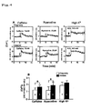
- the effect of caffeine, ryanodine, or a high-K + solution on intracellular Ca 2+ concentration was studied.
- single myocardial cells were isolated using collagenase (1.5 mg/ml), placed on a mount coverslip dish (35 mm in diameter) coated with poly-L-lysine, and then incubated with 3.5 ⁇ M fura-2/AM (DOJINDO LABORATORIES) at 37°C for 45 minutes. Subsequently, the cells were washed 4 times with medium.
- Micro fluorescence intensity measurement was performed at room temperature in physiological saline (140 mM NaCl, 5 mM KCI, 1.5 mM CaCl 2 , 1 mM MgCl 2 , 10 mM Hepes, 10 mM glucose, and pH 7.4). Images of the cells with an image ratio of 340 nm/380 nm were collected using an image processor system (ACOUS cosmos, Hamamatsu Photonics K.K.) and then stored on an analysis MO disk. Moreover, a Ca 2+ response in the presence of a high K + solution (142 mM), caffeine (10 mM), or ryanodine (2.6 ⁇ M) was also examined.
- physiological saline 140 mM NaCl, 5 mM KCI, 1.5 mM CaCl 2 , 1 mM MgCl 2 , 10 mM Hepes, 10 mM glucose, and pH 7.4
- annexin-V positive cells were treated with heat at 45°C for 90 minutes, cultured, and then stained with an annexin-V antibody. The percentage of annexin-V positive cells was calculated by a FACScan method.
- Bax protein expression was analyzed using the Western blot method. Specifically, cells were irradiated with UV for 20 seconds, cultured for 24 hours, harvested, lysed, and then Western-blotted.
- the left anterior descending coronary artery (LAD) of each rat male Spague-Dawley rat (body weight ranging from 275 g to 320 g and 8 to 10 weeks old) was ligated, thereby establishing a myocardial infarction model.
- P19CL6-derived myocardial cells (4 ⁇ 10 6 cells/site, x6 positions) at an early phase of differentiation (day 4) which express TNNI3K at high levels were implanted into locations with myocardial infarction.
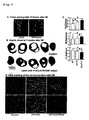
- Myocardial infarction sizes (including infracted myocardial thickness, the ratio of infracted myocardial thickness to non-infarcted myocardial thickness, left ventricular diameter, and enlargement index) and conditions of cell death of myocardial cells (necrosis or apoptosis) were observed. The results were compared with those in the case of the heart into which medium alone had been introduced or into which P19CL6-derived myocardial cells at an early phase of differentiation (day 4) into which no TNNI3K had been introduced had been implanted.
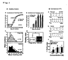
- TNNI3K promotes the formation of pulsating cell groups at an early development phase of myocardial cells and promotes increased pulsation frequency (Fig. 1 A)
- Pulsation frequency resulting from the spontaneous action potential of single myocardial cells was increased in a manner that depended on epinephrine concentrations (10 -8 , 10 -7 , and 10 -6 M).
- TNNI3K significantly enhanced the sensitivity of the myocardial cells to administration of epinephrine at low concentrations. These results demonstrate that TNNI3K promotes the development of autonomic modulation of such as sympathetic nerve at early development and differentiation phases of myocardial cells ( Fig. 1B ).
- TNNI3K increases the number of positive cells immuno-stained with an ⁇ -actinin antibody and the expression level of ⁇ -actinin protein
- TNNI3K When TNNI3K was compared with Flag-only, there were no differences in terms of size and shape of myocardial cells that had been formed by differentiation of progenitor cells. However, in terms of the number of positive cells immuno-stained with the ⁇ -actinin antibody and the expression level of ⁇ -actinin protein, the number and the level were significantly higher in the case of the cells expressing TNNI3K at high levels than in the case of Flag-only ( Fig. 2A , B, and C).
- TNNI3K significantly suppresses the occurrence of apoptosis at an early differentiation phase of P19CL6-derived myocardial cells
- Bax expression analysis Cells were irradiated with UV for 20 seconds and then cultured for 24 hours. Bax expression level was then analyzed by the Western blot method. As a result, compared with the Flag-only group, Bax protein expression level at an early differentiation phase was significantly decreased in the case of cells expressing TNNI3K at high levels ( Fig. 3C ).
- TNNI3K improves left ventricular remodeling in a myocardial infarction model in an in vivo experiment
- P19CL6 cells at an early differentiation phase which express TNNI3K at high levels were injected.
- more decreased myocardial infarction sizes and more suppressed apoptosis at infracted locations were observed, as compared with the case where only P19CL6 cells were injected ( Fig. 5 ).
- the TNNI3K gene is introduced to achieve increased TNNI3K protein levels, regeneration of pathological cardiac muscles, and cardiomuscular functional recovery.
- novel therapeutic strategies against cardiomyopathy and heart malfunction can be provided.
- implantation of myocardial progenitor cells expressing MAP kinase TNNI3K at high levels into a location affected with myocardial ischemia or local myocardial infarction makes it possible to promote myocardial cell regeneration, reduce the infracted area, and improve left ventricular remodeling.
- the technology of the present invention is characterized by simple procedures, high specificity, and significant therapeutic effects.
Landscapes
- Health & Medical Sciences (AREA)
- Life Sciences & Earth Sciences (AREA)
- Engineering & Computer Science (AREA)
- Chemical & Material Sciences (AREA)
- General Health & Medical Sciences (AREA)
- Cardiology (AREA)
- Developmental Biology & Embryology (AREA)
- Cell Biology (AREA)
- Veterinary Medicine (AREA)
- Public Health (AREA)
- Medicinal Chemistry (AREA)
- Animal Behavior & Ethology (AREA)
- Pharmacology & Pharmacy (AREA)
- Vascular Medicine (AREA)
- Bioinformatics & Cheminformatics (AREA)
- Biomedical Technology (AREA)
- Zoology (AREA)
- General Chemical & Material Sciences (AREA)
- Chemical Kinetics & Catalysis (AREA)
- Epidemiology (AREA)
- Heart & Thoracic Surgery (AREA)
- Organic Chemistry (AREA)
- Biotechnology (AREA)
- Immunology (AREA)
- Virology (AREA)
- Nuclear Medicine, Radiotherapy & Molecular Imaging (AREA)
- Urology & Nephrology (AREA)
- Medicines That Contain Protein Lipid Enzymes And Other Medicines (AREA)
- Measuring Or Testing Involving Enzymes Or Micro-Organisms (AREA)
- Medicines Containing Material From Animals Or Micro-Organisms (AREA)
- Materials For Medical Uses (AREA)
Claims (4)
- Eine pharmazeutische Zusammensetzung zur Verwendung in der Behandlung von Myokardischämie oder Myokardinfarkt, enthaltend eine Myokardvorläuferzelle als Wirkstoff, die MAP-Kinase TNNI3K auf hohem Niveau exprimiert, wobei die Myokardvorläuferzelle eine Myokardvorläuferzelle ist, in die ein Expressionsvektor zur Expression der MAP-Kinase TNNI3K auf hohem Niveau eingeführt wurde, mit der Maßgabe, dass menschliche embryonale Zellen ausgeschlossen sind.
- Die pharmazeutische Zusammensetzung gemäß Anspruch 1, wobei die Myokardvorläuferzelle eine undifferenzierte Myokardzelle ist.
- Die pharmazeutische Zusammensetzung gemäß Anspruch 1 oder 2, wobei die Myokardvorläuferzelle eine undifferenzierte P19CL6-Zelle ist.
- Die pharmazeutische Zusammensetzung gemäß irgend einem der Ansprüche 1 bis 3 zur Verabreichung in einen von Ischämie oder Infarkt betroffenen Bereich des Myokards.
Applications Claiming Priority (2)
| Application Number | Priority Date | Filing Date | Title |
|---|---|---|---|
| JP2004338918 | 2004-11-24 | ||
| PCT/JP2005/021535 WO2006057278A1 (ja) | 2004-11-24 | 2005-11-24 | Mapキナーゼtnni3kを用いた心疾患治療剤 |
Publications (3)
| Publication Number | Publication Date |
|---|---|
| EP1832299A1 EP1832299A1 (de) | 2007-09-12 |
| EP1832299A4 EP1832299A4 (de) | 2009-03-11 |
| EP1832299B1 true EP1832299B1 (de) | 2011-07-20 |
Family
ID=36498014
Family Applications (1)
| Application Number | Title | Priority Date | Filing Date |
|---|---|---|---|
| EP05809227A Expired - Lifetime EP1832299B1 (de) | 2004-11-24 | 2005-11-24 | Mittel zur behandlung einer herzkrankheit mit map-kinase-tnni3k |
Country Status (4)
| Country | Link |
|---|---|
| US (1) | US20090022692A1 (de) |
| EP (1) | EP1832299B1 (de) |
| JP (1) | JP5124812B2 (de) |
| WO (1) | WO2006057278A1 (de) |
Families Citing this family (3)
| Publication number | Priority date | Publication date | Assignee | Title |
|---|---|---|---|---|
| EP2347262A4 (de) * | 2008-10-31 | 2012-06-20 | Univ Duke | Verfahren zum schutz gegen herzinsuffizienz |
| WO2011025798A1 (en) * | 2009-08-28 | 2011-03-03 | Glaxosmithkline Llc | Compounds and methods |
| CN104165995B (zh) * | 2014-08-11 | 2016-06-08 | 上海应用技术学院 | 一种血中tnni3k的浓度的检测试剂盒及其制备方法和应用 |
Family Cites Families (3)
| Publication number | Priority date | Publication date | Assignee | Title |
|---|---|---|---|---|
| US6261818B1 (en) * | 1998-12-11 | 2001-07-17 | Millennium Pharmaceuticals, Inc. | CARK protein and nucleic acid molecules and uses therefor |
| US6660490B2 (en) * | 1998-12-11 | 2003-12-09 | Millennnium Pharmaceuticals, Inc. | CARK protein and nucleic acid molecules and uses therefor |
| JP2003259863A (ja) * | 2002-03-07 | 2003-09-16 | Takeda Chem Ind Ltd | 心筋様細胞分化能を有する細胞株 |
-
2005
- 2005-11-24 JP JP2006547812A patent/JP5124812B2/ja not_active Expired - Fee Related
- 2005-11-24 EP EP05809227A patent/EP1832299B1/de not_active Expired - Lifetime
- 2005-11-24 WO PCT/JP2005/021535 patent/WO2006057278A1/ja not_active Ceased
- 2005-11-24 US US11/720,041 patent/US20090022692A1/en not_active Abandoned
Also Published As
| Publication number | Publication date |
|---|---|
| JP5124812B2 (ja) | 2013-01-23 |
| EP1832299A1 (de) | 2007-09-12 |
| WO2006057278A1 (ja) | 2006-06-01 |
| US20090022692A1 (en) | 2009-01-22 |
| JPWO2006057278A1 (ja) | 2008-06-05 |
| EP1832299A4 (de) | 2009-03-11 |
Similar Documents
| Publication | Publication Date | Title |
|---|---|---|
| Zhou et al. | miR‐21 promotes cardiac fibroblast‐to‐myofibroblast transformation and myocardial fibrosis by targeting Jagged1 | |
| Song et al. | Transfection of mesenchymal stem cells with the FGF-2 gene improves their survival under hypoxic conditions | |
| US7794702B2 (en) | Mesenchymal stem cells as a vehicle for ion channel transfer in syncytial structures | |
| Katsuragi et al. | Periostin as a novel factor responsible for ventricular dilation | |
| ES2251773T3 (es) | Regeneracion del musculo cardiaco usando celulas precursoras mesenquimatosas. | |
| Rentschler et al. | Myocardial Notch signaling reprograms cardiomyocytes to a conduction-like phenotype | |
| Lu et al. | Triad3A attenuates pathological cardiac hypertrophy involving the augmentation of ubiquitination-mediated degradation of TLR4 and TLR9 | |
| Craft et al. | Evolution of the marrow adipose tissue microenvironment | |
| Qin et al. | Kindlin-2 mediates mechanotransduction in bone by regulating expression of Sclerostin in osteocytes | |
| US20050002914A1 (en) | Mesenchymal stem cells as a vehicle for ion channel transfer in syncytial structures | |
| Ke et al. | Netrin-1 overexpression in bone marrow mesenchymal stem cells promotes functional recovery in a rat model of peripheral nerve injury | |
| Liu et al. | Type I collagen inhibits adipogenic differentiation via YAP activation in vitro | |
| JP2009507494A (ja) | キメラのhcnチャネル | |
| Leontyev et al. | Transplantation of engineered heart tissue as a biological cardiac assist device for treatment of dilated cardiomyopathy | |
| Chen et al. | MNK2-eIF4E axis promotes cardiac repair in the infarcted mouse heart by activating cyclin D1 | |
| JP2003502065A (ja) | 高められた増殖能を持つ心筋細胞、およびその製造および使用法 | |
| JP2019177224A (ja) | 骨移植片の骨形成潜在能の増強 | |
| US20090022692A1 (en) | Therapeutic agent for heart disease using map kinase tnni3k | |
| Shin et al. | Delta-like 1 homolog (DLK1) inhibits proliferation and myotube formation of avian QM7 myoblasts | |
| Spadaccio et al. | In situ electrostimulation drives a regenerative shift in the zone of infarcted myocardium | |
| JP2009544285A (ja) | 心律動障害を治療するための後期継代間葉系幹細胞(msc)の使用 | |
| Gustafsson et al. | Insulin-like growth factor type 1 upregulates uncoupling protein 3 | |
| Shilo et al. | Tuftelin's involvement in embryonic development | |
| US20210095049A1 (en) | Method of inhibiting ectopic calcification | |
| CA2660661A1 (en) | Side population cells in cardiac repair |
Legal Events
| Date | Code | Title | Description |
|---|---|---|---|
| PUAI | Public reference made under article 153(3) epc to a published international application that has entered the european phase |
Free format text: ORIGINAL CODE: 0009012 |
|
| 17P | Request for examination filed |
Effective date: 20070621 |
|
| AK | Designated contracting states |
Kind code of ref document: A1 Designated state(s): CH DE FR GB LI SE |
|
| DAX | Request for extension of the european patent (deleted) | ||
| RBV | Designated contracting states (corrected) |
Designated state(s): CH DE FR GB LI SE |
|
| A4 | Supplementary search report drawn up and despatched |
Effective date: 20090205 |
|
| 17Q | First examination report despatched |
Effective date: 20090519 |
|
| GRAP | Despatch of communication of intention to grant a patent |
Free format text: ORIGINAL CODE: EPIDOSNIGR1 |
|
| RIC1 | Information provided on ipc code assigned before grant |
Ipc: C12N 9/12 20060101ALI20110126BHEP Ipc: A61K 35/34 20060101AFI20110126BHEP |
|
| RIN1 | Information on inventor provided before grant (corrected) |
Inventor name: MENG, XIAN-MIN,PEKING UNION MEDICAL COLLEGE Inventor name: DING, JIN-FENG,PEKING UNION MEDICAL COLLEGE Inventor name: YORINAKA, HOICHI,KUMAMOTO UNIVERSITY |
|
| GRAS | Grant fee paid |
Free format text: ORIGINAL CODE: EPIDOSNIGR3 |
|
| GRAA | (expected) grant |
Free format text: ORIGINAL CODE: 0009210 |
|
| AK | Designated contracting states |
Kind code of ref document: B1 Designated state(s): CH DE FR GB LI SE |
|
| REG | Reference to a national code |
Ref country code: GB Ref legal event code: FG4D |
|
| REG | Reference to a national code |
Ref country code: CH Ref legal event code: EP |
|
| REG | Reference to a national code |
Ref country code: SE Ref legal event code: TRGR |
|
| REG | Reference to a national code |
Ref country code: DE Ref legal event code: R096 Ref document number: 602005029122 Country of ref document: DE Effective date: 20110915 |
|
| REG | Reference to a national code |
Ref country code: CH Ref legal event code: NV Representative=s name: ISLER & PEDRAZZINI AG |
|
| PLBE | No opposition filed within time limit |
Free format text: ORIGINAL CODE: 0009261 |
|
| STAA | Information on the status of an ep patent application or granted ep patent |
Free format text: STATUS: NO OPPOSITION FILED WITHIN TIME LIMIT |
|
| 26N | No opposition filed |
Effective date: 20120423 |
|
| REG | Reference to a national code |
Ref country code: DE Ref legal event code: R097 Ref document number: 602005029122 Country of ref document: DE Effective date: 20120423 |
|
| PGFP | Annual fee paid to national office [announced via postgrant information from national office to epo] |
Ref country code: DE Payment date: 20131120 Year of fee payment: 9 Ref country code: CH Payment date: 20131126 Year of fee payment: 9 Ref country code: GB Payment date: 20131120 Year of fee payment: 9 Ref country code: SE Payment date: 20131126 Year of fee payment: 9 |
|
| PGFP | Annual fee paid to national office [announced via postgrant information from national office to epo] |
Ref country code: FR Payment date: 20131125 Year of fee payment: 9 |
|
| REG | Reference to a national code |
Ref country code: DE Ref legal event code: R082 Ref document number: 602005029122 Country of ref document: DE Representative=s name: GODEMEYER BLUM LENZE PARTNERSCHAFT, PATENTANWA, DE Ref country code: DE Ref legal event code: R082 Ref document number: 602005029122 Country of ref document: DE Representative=s name: GODEMEYER BLUM LENZE PATENTANWAELTE, PARTNERSC, DE |
|
| REG | Reference to a national code |
Ref country code: DE Ref legal event code: R119 Ref document number: 602005029122 Country of ref document: DE |
|
| REG | Reference to a national code |
Ref country code: SE Ref legal event code: EUG Ref country code: CH Ref legal event code: PL |
|
| GBPC | Gb: european patent ceased through non-payment of renewal fee |
Effective date: 20141124 |
|
| PG25 | Lapsed in a contracting state [announced via postgrant information from national office to epo] |
Ref country code: SE Free format text: LAPSE BECAUSE OF NON-PAYMENT OF DUE FEES Effective date: 20141125 Ref country code: LI Free format text: LAPSE BECAUSE OF NON-PAYMENT OF DUE FEES Effective date: 20141130 Ref country code: CH Free format text: LAPSE BECAUSE OF NON-PAYMENT OF DUE FEES Effective date: 20141130 |
|
| REG | Reference to a national code |
Ref country code: FR Ref legal event code: ST Effective date: 20150731 |
|
| PG25 | Lapsed in a contracting state [announced via postgrant information from national office to epo] |
Ref country code: DE Free format text: LAPSE BECAUSE OF NON-PAYMENT OF DUE FEES Effective date: 20150602 Ref country code: GB Free format text: LAPSE BECAUSE OF NON-PAYMENT OF DUE FEES Effective date: 20141124 |
|
| PG25 | Lapsed in a contracting state [announced via postgrant information from national office to epo] |
Ref country code: FR Free format text: LAPSE BECAUSE OF NON-PAYMENT OF DUE FEES Effective date: 20141201 |