EP1840571A2 - Procédé et appareil pour la mesure d'un échantillon hématologique - Google Patents
Procédé et appareil pour la mesure d'un échantillon hématologique Download PDFInfo
- Publication number
- EP1840571A2 EP1840571A2 EP07005912A EP07005912A EP1840571A2 EP 1840571 A2 EP1840571 A2 EP 1840571A2 EP 07005912 A EP07005912 A EP 07005912A EP 07005912 A EP07005912 A EP 07005912A EP 1840571 A2 EP1840571 A2 EP 1840571A2
- Authority
- EP
- European Patent Office
- Prior art keywords
- scattered light
- cell
- cell group
- information
- myeloblast
- Prior art date
- Legal status (The legal status is an assumption and is not a legal conclusion. Google has not performed a legal analysis and makes no representation as to the accuracy of the status listed.)
- Granted
Links
- 0 CC(C)(*=CC)C1=*(*)c2ccccc2*1 Chemical compound CC(C)(*=CC)C1=*(*)c2ccccc2*1 0.000 description 3
Images






Classifications
-
- G—PHYSICS
- G01—MEASURING; TESTING
- G01N—INVESTIGATING OR ANALYSING MATERIALS BY DETERMINING THEIR CHEMICAL OR PHYSICAL PROPERTIES
- G01N15/00—Investigating characteristics of particles; Investigating permeability, pore-volume or surface-area of porous materials
- G01N15/10—Investigating individual particles
- G01N15/14—Optical investigation techniques, e.g. flow cytometry
- G01N15/1456—Optical investigation techniques, e.g. flow cytometry without spatial resolution of the texture or inner structure of the particle, e.g. processing of pulse signals
- G01N15/1459—Optical investigation techniques, e.g. flow cytometry without spatial resolution of the texture or inner structure of the particle, e.g. processing of pulse signals the analysis being performed on a sample stream
-
- G—PHYSICS
- G01—MEASURING; TESTING
- G01N—INVESTIGATING OR ANALYSING MATERIALS BY DETERMINING THEIR CHEMICAL OR PHYSICAL PROPERTIES
- G01N15/00—Investigating characteristics of particles; Investigating permeability, pore-volume or surface-area of porous materials
- G01N15/10—Investigating individual particles
- G01N2015/1006—Investigating individual particles for cytology
-
- G—PHYSICS
- G01—MEASURING; TESTING
- G01N—INVESTIGATING OR ANALYSING MATERIALS BY DETERMINING THEIR CHEMICAL OR PHYSICAL PROPERTIES
- G01N15/00—Investigating characteristics of particles; Investigating permeability, pore-volume or surface-area of porous materials
- G01N15/10—Investigating individual particles
- G01N15/14—Optical investigation techniques, e.g. flow cytometry
- G01N2015/1402—Data analysis by thresholding or gating operations performed on the acquired signals or stored data
-
- G—PHYSICS
- G01—MEASURING; TESTING
- G01N—INVESTIGATING OR ANALYSING MATERIALS BY DETERMINING THEIR CHEMICAL OR PHYSICAL PROPERTIES
- G01N15/00—Investigating characteristics of particles; Investigating permeability, pore-volume or surface-area of porous materials
- G01N15/10—Investigating individual particles
- G01N15/14—Optical investigation techniques, e.g. flow cytometry
- G01N2015/1477—Multiparameters
-
- Y—GENERAL TAGGING OF NEW TECHNOLOGICAL DEVELOPMENTS; GENERAL TAGGING OF CROSS-SECTIONAL TECHNOLOGIES SPANNING OVER SEVERAL SECTIONS OF THE IPC; TECHNICAL SUBJECTS COVERED BY FORMER USPC CROSS-REFERENCE ART COLLECTIONS [XRACs] AND DIGESTS
- Y10—TECHNICAL SUBJECTS COVERED BY FORMER USPC
- Y10T—TECHNICAL SUBJECTS COVERED BY FORMER US CLASSIFICATION
- Y10T436/00—Chemistry: analytical and immunological testing
- Y10T436/10—Composition for standardization, calibration, simulation, stabilization, preparation or preservation; processes of use in preparation for chemical testing
-
- Y—GENERAL TAGGING OF NEW TECHNOLOGICAL DEVELOPMENTS; GENERAL TAGGING OF CROSS-SECTIONAL TECHNOLOGIES SPANNING OVER SEVERAL SECTIONS OF THE IPC; TECHNICAL SUBJECTS COVERED BY FORMER USPC CROSS-REFERENCE ART COLLECTIONS [XRACs] AND DIGESTS
- Y10—TECHNICAL SUBJECTS COVERED BY FORMER USPC
- Y10T—TECHNICAL SUBJECTS COVERED BY FORMER US CLASSIFICATION
- Y10T436/00—Chemistry: analytical and immunological testing
- Y10T436/10—Composition for standardization, calibration, simulation, stabilization, preparation or preservation; processes of use in preparation for chemical testing
- Y10T436/101666—Particle count or volume standard or control [e.g., platelet count standards, etc.]
-
- Y—GENERAL TAGGING OF NEW TECHNOLOGICAL DEVELOPMENTS; GENERAL TAGGING OF CROSS-SECTIONAL TECHNOLOGIES SPANNING OVER SEVERAL SECTIONS OF THE IPC; TECHNICAL SUBJECTS COVERED BY FORMER USPC CROSS-REFERENCE ART COLLECTIONS [XRACs] AND DIGESTS
- Y10—TECHNICAL SUBJECTS COVERED BY FORMER USPC
- Y10T—TECHNICAL SUBJECTS COVERED BY FORMER US CLASSIFICATION
- Y10T436/00—Chemistry: analytical and immunological testing
- Y10T436/25—Chemistry: analytical and immunological testing including sample preparation
-
- Y—GENERAL TAGGING OF NEW TECHNOLOGICAL DEVELOPMENTS; GENERAL TAGGING OF CROSS-SECTIONAL TECHNOLOGIES SPANNING OVER SEVERAL SECTIONS OF THE IPC; TECHNICAL SUBJECTS COVERED BY FORMER USPC CROSS-REFERENCE ART COLLECTIONS [XRACs] AND DIGESTS
- Y10—TECHNICAL SUBJECTS COVERED BY FORMER USPC
- Y10T—TECHNICAL SUBJECTS COVERED BY FORMER US CLASSIFICATION
- Y10T436/00—Chemistry: analytical and immunological testing
- Y10T436/25—Chemistry: analytical and immunological testing including sample preparation
- Y10T436/25125—Digestion or removing interfering materials
Definitions
- the present invention relates to a method and an apparatus for measuring a hematological sample, more particularly, a method and an apparatus for measuring a hematological sample which can count immature leukocyte, particularly, myeloblast by discriminating it from other hemocyte at a high precision.
- a cell component of blood is roughly classified into platelet, leukocyte and erythrocyte and, further, leukocyte is classified into granulocyte (eosinophil, neurtrophil, and basophil), monocyte and lymphocyte. These cells are produced in bone marrow, are differentiated and matured from an immature cell, and migrated to peripheral blood.
- immature leukocyte which has not been matured to leukocyte contained in normal blood, such as granulocyte, monocyte and lymphocyte (hereinafter, these are referred to as "mature leukocyte” in some cases) does not appear in peripheral blood, but in peripheral blood of a patient with leukemia, cancer bone marrow metastasis or serious infectious disease, immature leukocyte such as myeloblast appears in some cases. Particularly, detection of myeloblast is important for diagnosing the aforementioned diseases.
- Flowcytometry is a method of introducing a sample obtained by fluorescently staining a cell membrane or a nucleus into a flow cell, irradiating a cell passing through a flow cell with excitation light of a dye, obtaining information regarding scattered light or fluorescent light emitted from individual cells passing through a flow cell, and measuring a kind and the number of cells from the information.
- a hemocyte in blood is classified as in the following (1) to (3).
- leukocyte discriminated in (2) is developed into a two-dimensional scattergram using a fluorescent intensity and a side scattered light intensity as two axes, and leukocyte is sub-classified into lymphocyte, monocyte, granulocyte, myeloblast, and granulocyte immature-cytes.
- myeloblast in (3), myeloblast can be classified, but when a sample containing a small amount of platelet aggregation is measured, there is a possibility that platelet aggregation appears in a region of leukocyte and an erythrocyte ghost in (1), and appears in a region of myeloblast in (3). For this reason, development of the technique for measuring myeloblast at a better precision is desired.
- the present invention was done in view of the aforementioned circumstances, and an object thereof is to provide a measuring method an a measuring apparatus which can more precisely classify and count myeloblast without influence of other components in a sample including platelet aggregation, in measurement of a blood sample with flowcytometry.
- the present inventors found out that, based on two kinds of scattered light information having different angles and fluorescent information obtained by applying a hematological sample which has been more strongly stained than immature leukocyte by giving damage to a cell membrane of mature leukocyte to flowcytometry, influence of platelet aggregation is excluded by specifying a cell group containing myeloblast based on the above two kinds of scattered light information, and myeloblast is discriminated from mature leukocyte by specifying a cell group containing myeloblast specified based on one kind of scattered light information and fluorescent information and, thereby, myeloblast can be measured, being discriminated from other hemocyte cells, which resulted in completion of the present invention.
- the first measuring method comprises a sample treatment step of giving damage to a cell membrane of erythrocyte and mature leukocyte contained in a hematological sample, constricting a hemocyte in which a cell membrane is damaged, and dyeing this with a fluorescent dye which can stain a nucleic acid; an obtaining step of obtaining first scattered light information generated by irradiating the treated sample with light, second scattered light information based on scattered light having a different angle from that of first scattered light and fluorescent information; a first specification step of specifying a first cell group containing myeloblast based on the first scattered light information and the second scattered light information; a second specification step of specifying a second cell group containing myeloblast based on the first scattered light information and the fluorescent information; and a counting step of counting a cell belonging to both of the first and second cell groups as myeloblast.
- a cell belonging to both of the first and second cell groups is discriminated by a scattergram using the fluorescent information and the second scattered light information as two axes, and a cell appearing in the scattergram may be counted as myeloblast.
- the second specification step in the first measuring method may be a step of specifying a second cell group among the first cell group based on first scattered light information and fluorescent information of the first cell group, or the first specification step in the first measuring method may be a step of specifying the first cell group among the second cell group based on first scattered light information and second scattered light information of the second cell group (second measuring method).
- the first specification step is a step of setting a myeloblast appearance candidate region in a first scattergram using the first scatted light information and the second scattered light information as two axes, and specifying a cell group appearing in the candidate region as the first cell group
- the second specification step is a step of setting a myeloblast appearance candidate region in a second scattergram using the first scattered light information and the fluorescent information as two axes, and specifying a cell group appearing in the candidate region as the second cell group.
- the third measuring method comprises a sample treatment step of giving damage to a cell membrane of erythrocyte and mature leukocyte contained in a hematological sample, constricting a hemocyte in which a cell membrane is damaged, and dyeing this with a fluorescent dye which can stained a nucleic acid; an obtaining step of obtaining first scattered light information generated by irradiating the treated sample with excitation light of the fluorescent dye, second scattered light information based on scattered light having a different angle from that of first scattered light, and fluorescent information; a specification step of specifying a first cell group containing myeloblast based on the first scattered light information and the second scattered light information and, at the same time, specifying a second cell group containing myeloblast based on the first scattered light information and the fluorescent information; and a counting step of counting a cell belonging to both of the first and second cell groups as myelob
- a fourth measuring method of the method for measuring a hematological sample of the present invention is a method which is applied to the case where a third cell group containing total leukocyte is specified in place of a second cell group containing myeloblast, as a cell group to be specified based on the first scattered light information and the fluorescent information.
- the fourth measuring method comprises a sample treatment step of giving damage to a cell membrane of erythrocyte and mature leukocyte contained in a hematological sample, constricting a hemocyte in which a cell membrane is damaged, and dyeing this with a fluorescent dye which can stain a nucleic acid; an obtaining step of obtaining first scattered light information generated by irradiating the treated sample with excitation light of the fluorescent coloring matter, second scattered light information based on scattered light having a different angle from that of first scattered light, and fluorescent information; a first specification step of specifying a first cell group containing myeloblast based on the first scattered light information and the second scattered light information; a third specification step of specifying a cell population other than a ghost population as a third cell group based on the first scattered light information and the fluorescent information; a fourth specification step of specifying a fourth cell group which does not substantially contain at least mature leukocyte, among the third cell group, based on second scattered light information and fluorescent information of the third cell group; and
- the first scattered light information is a forward scattered light intensity
- the second scattered information is a side scattered light intensity
- the fluorescent information is a fluorescent intensity
- the treatment step is performed by mixing a surfactant which gives damage to a cell membrane of erythrocyte and mature leukocyte, a solubilizer which constricts a damaged hemocyte, a fluorescent dye which stains a nucleic acid, and a hematological sample.
- the method for discriminating myeloblast and platelet aggregation is a method of discriminating myeloblast and platelet aggregation contained in a hematological cell, and comprises a step of giving damage to a cell membrane of erythrocyte and mature leukocyte contained in the sample, and constricting a hemocyte in which a cell membrane is damaged; a step of obtaining forward scattered light information and side scattered light information generated by irradiating the treated sample with light; and a step of discriminating the myeloblast and the platelet aggregation based on the forward scattered light information and the side scattered light information.
- the apparatus for measuring a hematological sample of the present invention comprises a sample treatment part for giving damage to a cell membrane of erythrocyte and mature leukocyte contained in a hematological sample, constricting a hemocyte in which a cell membrane is damaged, and dyeing this with a fluorescent dye which can stain a nucleic acid; an obtainer for obtaining first scattered light information generated by irradiating the treated sample with excitation light of the fluorescent dye, second scattered light information based on scattered light having a different angle from that of first scattered light, and florescent information; a first classifier for specifying a first cell group containing myeloblast based on the first scattered light information and the second scattered light information; a second classifier for specifying a second cell group containing myeloblast based on the first scattered light information and the fluorescent information; and a calculator for counting a cell belonging to both of the first and second cell groups as myeloblast.
- the first cell group is a cell group appearing in a myeloblast appearance candidate region set in a first scattergram using the first scattered light information and the second scattered light information as two axes
- the second cell group is a cell group appearing in a myeloblast appearance candidate region set in second scattergram using the first scattered light information and the fluorescent information as two axes.
- the calculator discriminates a cell belonging to both of the first and second cell groups by a third scattergram using the fluorescent information and the second scattered light information as two axes, and counts a cell appearing in the third scattergram as myeloblast.
- the measuring apparatus of the present invention may be further provided with a display part for displaying at least one of a scattergram in which a cell belonging to both of the first and second cell groups appears, and result of counting of the myeloblast, and may be further provided with a light source for irradiating the excitation light.
- the "immature leukocyte” referred in the present invention refers to an immature cell which is usually present in bone marrow, and is not present in a peripheral blood liquid, such as myeloblast, promyelocyte, myelocyte, and postmyelocyte. Promyelocyte, myelocyte and postmyelocyte are collectively referred to as immature granulocyte in some cases.
- a leukocytic hematopoietic precursor cell such as marrow stem cell, neutrophil/macrophage colony forming cell, eosinophils colony forming cell and the like, which are at a differentiation stage before a blast cell, is included in the scope of immature leukocyte of the present invention.
- the "mature leukocyte” referred in the present invention refers to mature lymphocyte, monocyte, granulocyte (neutrophil, eosinophils, basophil).
- platelet aggregation refers to aggregation of two or more platelets.
- the "damage of cell membrane” referred in the present invention refers to opening of such a pore in a cell membrane that a particular substance can pass therethrough.
- the "hematological sample” referred in the present invention mainly refers to a peripheral blood liquid and, additionally, a sample containing a bone marrow liquid, or a blood component recovered by apheresis or the like, or a biological sample containing a leukocyte component such as an abdominal cavity liquid, a urine sample or the like is also a suitable sample.
- the method for measuring a hematological sample of the present invention upon discrimination of a cell group using fluorescent information and two kinds of scattered light information obtained by measuring a blood sample by flowcytometry, can specify a cell group containing myeloblast by excluding influence of platelet aggregation, the method can precisely classify and count myeloblast by appropriately combining with a particular method which can discriminate a cell group containing myeloblast and a cell group of mature leukocyte.
- the apparatus can measure myeloblast at a high precision.
- the method for measuring a hematological sample of the present invention comprises a sample treatment step of giving damage to a cell membrane of erythrocyte and mature leukocyte contained in a hematological sample, constricting a hemocyte in which a cell membrane is damaged, and dyeing this with a fluorescent dye which can stain a nucleic acid; an obtaining step of obtaining first scattered light information generated by irradiating the treated sample with excitation light of the fluorescent dye, second scattered light information based on scattered light having a different angle from that of first scattered light, and fluorescent information; a step of specifying a cell population containing myeloblast by excluding influence of platelet aggregation, and discriminating from other hemocyte cell, using the obtained fluorescent information and two kinds of scattered light information; and a counting step of counting a cell population of specified myeloblast as myeloblast.
- the sample treatment step in the measuring method of the present invention is performed by mixing a hematological sample and a treating reagent described below.
- the treating reagent is a reagent which can give damage to a cell membrane of erythrocyte and mature leukocyte contained in a hematological sample, constricting a hemocyte in which a cell membrane is damaged, and stain a nucleic acid.
- the reagent includes a surfactant which gives damage to a cell membrane of erythrocyte and mature leukocyte contained in a sample, and a fluorescent dye which fluorescently stains a nucleic acid.
- the surfactant used in the present invention gives damage to a cell membrane of erythrocyte and mature leukocyte. Although action of mechanism is not clear, it is thought that, by extracting (pulling out) a part of a particular cell membrane liquid constituent component, a pore through which a particular substance can pass is opened (referred to damage) in a cell membrane. And, a fluorescent dye can penetrate into the interior of a cell damaged with the surfactant, and stain a nucleic acid.
- the surfactant gives damage also to immature leukocyte, but needs a time to damage a cell as compared with erythrocyte and mature leukocyte. For this reason, within a predetermined time after a hematological sample and a treating reagent are mixed, the state is realized where damaged mature leukocyte is more easily dyed than undamaged myeloblast and immature granulocyte. As a result, since myeloblast and immature granulocyte are hardly stained with a fluorescent dye which stains a nucleus as compared with mature leukocyte which undergoes damage and whose nucleus is stained, a fluorescent intensity of the myeloblast and immature granulocyte becomes lower than that of mature leukocyte.
- the surfactant used in the present invention examples include a polyoxyethylene nonionic surfactant.
- the surfactant is a nonionic surfactant represented by R 1 R 2 (CH 2 CH 2 O) nH.
- R 1 is an alkyl group, an alkenyl group or an alkynyl group of a carbon number of 9 to 25, R 2 is -O- or -COO- or and n is an integer of 10 to 40.
- Examples of the alkyl group of a carbon number of 9 to 25 include nonyl, decyl, undecyl, dodecyl, tridecyl, tetradecyl and the like.
- Examples of the alkenyl group of a carbon number of 9 to 25 include dodecenyl, tetradecenyl and the like.
- Examples of the alkynyl group of a carbon number of 9 to 25 include dodecynyl, undecynyl, dodecynyl and the like.
- polyoxyethylene(20)lauryl ether examples include polyoxyethylene(20)lauryl ether, polyoxyethylene(15)oleyl ether, and polyoxyethylene(16)oleyl ether.
- the surfactant can be used in a form of an aqueous solution.
- a concentration of the polyoxyethylene nonionic surfactant in water is different depending on a kind of a surfactant to be used, and the polyoxyethylene (16) oleyl ether can be used in a range of 5 to 50g/l (preferably, 15 to 35g/l).
- the polyoxyethylene nonionic surfactant when a carbon number of a hydrophobic group is the same, as a value of n becomes smaller, a force of damaging a cell is stronger and, as a value of n becomes greater, the force is weakened.
- n when the value of n is the same, as a carbon number of a hydrophobic group becomes smaller, a force of damaging a cell becomes stronger. In view of that point, and using the value as a measure, a required concentration of the surfactant may be appropriately obtained by an experiment.
- the fluorescent dye used in the present invention is a dye which fluorescently stains a nucleic acid.
- a fluorescent dye can pass through a cell membrane, and stain a nucleus.
- ethidium bromide propidium iodide, and ethidium-acridine heterodimer
- ethidium azide ethidium homodimer-1, ethidium homodimer-2, ethidium monoazide
- TOTO-1, TO-PRO-1, TOTO-3, TO-PRO-3 sold from Molecular Probe, and the like.
- a dye suitable on the case where He-Ne, or a red semiconductor laser is used as a light source a dye represented by the following (1) formula can be used.
- R 3 is a hydrogen atom, a lower alkyl group or a lower alkoxy group
- R 4 is a hydrogen atom, an acyl group, or a lower alkyl group
- R 5 is a hydrogen atom or an optionally substituted lower alkyl group
- R 6 is a hydrogen atom or a lower alkyl group
- R 7 is a hydrogen atom, a lower alkyl group or a lower alkoxy group
- Z is a sulfur atom, an oxygen atom, or a carbon atom substituted with a lower alkyl group
- m is 1 or 2
- X is an anion.
- the lower alkyl in R 3 , R 4 , R 5 , R 6 and R 7 means a straight or branched alkyl group of a carbon number of 1 to 6, and examples include methyl, ethyl, propyl, butyl, isobutyl, sec-butyl, tert-butyl, pentyl and hexyl groups. Inter alia, a methyl group and an ethyl group are preferable.
- the lower alkoxy group in R 3 and R 7 means an alkoxy group of a carbon number of 1 to 6, and examples include methoxy, ethoxy, and propoxy groups. Inter alia, a methoxy group and an ethoxy group are preferable.
- R 3 and R 7 are preferably a hydrogen atom.
- the acyl group in R 4 is preferably an acyl derivatized from aliphatic carboxylic acid, and examples include acetyl, propionyl and the like. Inter alia, an acetyl group is preferable.
- the optionally substituted lower alkyl group in R 5 means a lower alkyl group optionally substituted with 1 to 3 hydroxy groups, halogen atoms (fluorine, chlorine, bromine or iodine atom) and the like. Inter alia, a methyl group and an ethyl group substituted with one hydroxy group are preferable.
- the lower alkyl group in Z is preferably such that Z is a sulfur atom.
- anion in X- examples include a halogen ion (fluorine, chlorine, bromine or iodine ion), a halogenated boron ion (BF 4 -, BCl 4 -, BBr 4 - etc.), a phosphorus compound ion, a halogen oxygen acid ion, a fluorosulfate ion, a methylsulfate ion, a tetraphenylboron compound ion having a halogen or an alkyl group having a halogen as a substituent on an aromatic ring, and the like.
- a bromine ion or BF 4 - is preferable.
- the following dye A, dye B and dye C are preferable.
- a dye concentration can be appropriately determined depending on a dye to be used.
- a concentration in a reagent is preferably 0.01 to 500ppm, more preferably 0.1 to 200 ppm.
- a cell membrane is damaged with the aforementioned surfactant, and a part in a cell is discharged to the outside of a cell, thereby, cell constriction occurs, or cell constriction occurs due to a change in the surface state of a cell membrane, but it is preferable that a solubilizer for sufficiently constricting a damaged cell is contained in order to render a hemocyte other than leukocyte, particularly, erythrocyte a ghost population which does not provide useful scattered light information and fluorescent information.
- solubilizer specifically, one or more selected from a sarcosine derivative represented by the following (2) or a salt thereof: (wherein R 10 is an alkyl group of a carbon number of 10 to 22, and p is an integer of 1 to 5) a cholic acid derivative represented by the following (3) formula; (wherein R 11 is a hydrogen atom or a hydroxy group) and methylglucan amide represented by the following (4) formula; (wherein q is 5 to 7) can be used.
- a sarcosine derivative represented by the following (2) or a salt thereof: wherein R 10 is an alkyl group of a carbon number of 10 to 22, and p is an integer of 1 to 5
- R 11 is a hydrogen atom or a hydroxy group
- methylglucan amide represented by the following (4) formula
- alkyl group of a carbon number of 10 to 22 examples include decyl, dodecyl, tetradecyl, oleyl and the like.
- Specific examples include sodium N-lauroylsarcosinate, lauroylmethyl ⁇ -alanine sodium, lauroylsarcosine, CHAPS (3-[3-cholamidopripyl]dimethylammonio)-1-propane sulfonate), CHAPSO (3-[3-cholamidoproypl]dimethylammonio)-2-hydroxy-1-propane sulfonate), MEGA8 (octanoyl-N-methylglucamide), MEGA9 (nonanoyl-N-methylglucamide), MEGA10 (decanoyl-N-methylglucamide) and the like.
- a concentration of the solubilizer is 0.2 to 2.0g/l in a sarcosinic acid derivative or a salt thereof, 0.1 to 0.5g/l in a cholic acid derivative, and 1.0 to 8.0g/l in methylglucaneamide.
- solubilizer in addition to the aforementioned solubilizers, n-octyl ⁇ -glucoside, sucrose monocaprate, N-formylmethylleucylalanine and the like may be used, and are preferably used at a concentration of 0.01 to 50.g/l.
- a pH adjusting agent and a buffer for adjusting a pH of a reagent are contained and, furthermore, if necessary, it is preferable that an osmotic pressure adjusting agent is contained.
- a Good buffer such as HEPES and the like, and a phosphate buffer are used.
- a pH adjusting agent sodium hydroxide is used.
- a sugar, an amino acid, an organic solvent, and sodium chloride are used.
- sugar glucose, xylitol, mannitol, arabinose, and ribitol can be used.
- amino acid alanine, proline, glycine, and valine can be used.
- organic solvent ethylene glycol, and glycerin can be used.
- a concentration of the osmotic pressure adjusting agent in a reagent is appropriately adjusted by a molecular weight and, for example, in the case of use of xylitol, the concentration is preferably 10 to 75g/l, in the case of use of glycerin, the concentration is preferably 5 to 45g/l and, in the case of use of glycine, the concentration is preferably 5 to 45g/l.
- a pH is 5.0 to 9.0, and an osmotic pressure is 150 to 600mOsm/kg.
- the treating reagent may be a one-pack reagent containing a surfactant, a dye, and other component, or a staining solution containing a fluorescent dye, and a solution (hemolyzing agent) containing the aforementioned surfactant may be accommodated in separated containers to obtain a reagent kit containing them.
- a solution (hemolyzing agent) containing the aforementioned surfactant may be accommodated in separated containers to obtain a reagent kit containing them.
- a water-soluble organic solvent such as ethylene glycol and the like as a solvent for a fluorescent dye.
- a measuring sample By mixing a solution containing the treatment reagent and a hematological sample, a measuring sample can be prepared.
- a solution (hemolyzing agent) containing a surfactant and a hematological sample may be mixed and, then, a staining solution may be mixed to obtain a measuring sample.
- a ratio of sample: treating reagent is 1:10 to 1:1000, and a reaction temperature is 20 to 40°C, and a reaction time is 3 seconds to 5 minutes, more preferably 4 seconds to 1 minute.
- erythrocyte and mature leukocyte are constricted to a small size.
- erythrocyte has no nucleus, hemolysis and cell constriction due to cell membrane damage progress, and since staining with a fluorescent dye is not performed, any of forward scattered light, side scattered light and fluorescent light is decreased, and this can be handled as a ghost population.
- membrane damage with a treating reagent needs a time like immature leukocyte, and a dye does not penetrate into a platelet to stain platelet itself, but in the case of platelet aggregation body, a dye is physically adsorbed on a surrounding of an aggregate body, and the body is stained.
- the prepared measuring sample is measured with a flowcytometer to obtain scattered light information and fluorescent information of a cell and platelet contained in a sample.
- obtaining of information is performed by introducing a measuring sample into a flow cell of a flowcytometer, and irradiating a sample flowing in the flow cell with excitation light which can excite the fluorescent dye. Thereby, scattered light information and fluorescent information emitted from a cell passing through a flow cell can be obtained.
- Irradiation light includes light having a wavelength which can excite a fluorescent dye used. Depending on a wavelength of light of a light source which is mounted on a flowcytometer used, a fluorescent dye which can be excited by the light may be selected.
- scattered light information two kinds of scattered lights having different angles are obtained. Specifically, a combination of forward scattered light and side scattered light is preferably used.
- forward scattered light is thought to be information reflecting a size of a cell. That is, when a cell is large, forward scattered light is great.
- Side scattered light is scattered light having an angle which is different by 80 to 100 degree, preferably 85 to 95 degree relative to forward scattered light. From side scattered light, cell internal information, particularly, information of a nucleus can be obtained. When a granule is contained, or a shape of a nucleus is irregular, side scattered light tends to increase.
- the measuring method of the present invention can be classified into the following first measuring method to fourth measuring method from a viewpoint of a specification step. This will be explained sequentially below.
- a first cell group containing myeloblast is specified based on the obtained first scattered light information and second scattered light information (first specification step)
- a second cell group containing myeloblast is specified based on the obtained first scattered light information and the fluorescent information (second specification step)
- a cell belonging to both of the first and second cell groups is counted as myeloblast.
- a first cell group is specified based on the obtained two kinds of scattered light information, preferably a forward scattered light intensity and a side scattered light intensity. It is preferable that a first cell group is specified by setting a region thought that myeloblast appears therein, in a scattergram using these two kinds of scattered light information as two axes (first scattergram). In addition, since an extent of staining and a size of myeloblast are different depending on a specimen, it is preferable that a region in which a first cell group appears, is set every specimen.
- a center of this population is specified. From a center of a cell population of myeloblast to a part in which a cell of a myeloblast cell population appears until other cell population appearance region, can be determined as a boundary of a first cell group appearance region.
- a center of each population is specified and, at the same time, its appearance region is specified.
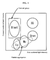
- the measuring sample used in the present invention generally becomes the state in Fig.1.
- Fig.1 "Blast” is a myeloblast appearance region
- IG is a immature granulocyte appearance region
- Gram is a granulocyte appearance region
- Ly is a lymphocyte appearance region
- Mo is a monocyte appearance region.
- myeloblast Since myeloblast is weak in a side scattered light intensity, it appears in a region on a relatively left side in a scattergram. On the other hand, since granulocyte, and immature granulocyte appear on a right side where side scattered light is relatively great, due to irregularity of a form of their nucleus, these populations appear in a region remote from a myeloblast appearance region. Therefore, as an appearance region of a first cell group, a region indicated with a chain line, surrounding a Blast part may be set.
- a first cell group is discriminated from platelet aggregation and other hemocyte component, and it is also thought that a first cell group can be counted as myeloblast.
- lymphocyte having a great size is contained in some cases, and a part of a lymphocyte appearance region is overlapped with a first cell group appearance region, though it is a rare case.
- a second cell group is specified based on the obtained first scattered light information and fluorescent information, preferably forward scattered light information and fluorescent information.
- a second cell group is a cell population contained in a region thought that myeloblast appears in a scattergram (second scatter gram) using a forward scattered light intensity and a fluorescent intensity as two axes.
- a center of this population is specified. From a center of a cell population of myeloblast, to a part where a cell of a myeloblast cell population appears, until an appearance region of a cell population of mature leukocyte, can be determined as a boundary of a second cell group appearance region.
- a center of each population is specified and, at the same time, its appearance region is specified.
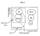
- a second cell group in a scattergram using a fluorescent intensity as an abscissa, and a forward scattered light intensity as an ordinate, a second cell group becomes a population contained in a region surrounded with a chain line, of a left region where a fluorescent intensity is low.
- "Blast” is a myeloblast appearance region
- "IG” is a immature granulocyte appearance region
- "Gran” is a granulocyte appearance region
- “Ly” is a lymphocyte appearance region
- “Mo” is a monocyte appearance region.
- the platelet aggregation Since platelet aggregation is stained to the extent where it is physically adsorbed, the platelet aggregation appears in a left region. In addition, since from a size smaller than that of myeloblast and a size larger than that of myeloblast can be present depending on an extent of aggregation, the platelet aggregation appears in a region extending longitudinally, from a small forward scattered light intensity to a large one, as shown in Fig.2.
- the above-specified cell contained in a first cell group and contained in a second cell group is counted as myeloblast.
- Lymphocyte which may be contained in a first cell group is not contained in a second cell group.
- platelet aggregation, and immature leukocyte which may be contained in a second cell group are not contained in a first cell group. Therefore, a cell contained in a first cell group and contained in a second cell group does not include lymphocyte, platelet aggregation, and immature leukocyte, and it can be said that the cell substantially contains only myeloblast. Therefore, by counting a cell contained in a first cell group and contained in a second cell group as myeloblast, myeloblast can be precisely counted.
- a cell contained in the first cell group and the second cell group may be discriminated by a scattergram (third scattergram) using fluorescent information and second scattered light information as two axes.
- a cell appearing in this third scattergram is substantially a population of only myeloblast.
- First scattered light information is preferably a forward scattered light intensity
- second scattered light information is preferably a side scattered light intensity.
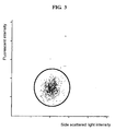
- a scattergram third scatter gram
- a scattergram as shown in Fig.3 is obtained. Since in this scattergram, a cell contained in a first cell group and contained in a second cell group, that is, substantially only myeloblast appears, a cell which appears here may be counted as myeloblast.
- a first cell group (or a second cell group) may be specified first and, then, a second cell group (or a first cell group) may be specified among the first specified first cell group (or second cell group).
- a second cell group containing myeloblast is specified based on the obtained first scattered light information and fluorescent information (second specification step), then, based on first scattered light information and the second light information of the specified second cell group, a first cell group is specified among the second cell group (when a second measuring method is particularly referred, this specification step is referred to as "first' specification step", and a first cell group specified by the first' specified step is referred to as "first' cell group”), and this first' cell group is counted as myeloblast.
- a first cell group containing myeloblast is specified based on the obtained first scattered light information and second scattered light information (first specification step), then, a second cell group is specified among the first cell group based on first scattered light information and fluorescent information of the specified first cell group (when a second measuring method is particularly referred, this specification step is referred to as "second' specification step", and a second cell group specified by a second' specification step is referred to as "second' cell group”), and this second' cell group is counted as myeloblast.
- a first specification step and a first cell group specified therein, and a second specification step and a second cell group specified therein are specified based on scattered light information and fluorescent information obtained in each obtaining step, and a specific procedure thereof is the same as that of the first measuring method, therefore, explanation is omitted.
- a second' specification step specifies a population containing myeloblast (specification of second' cell population) among the first cell group based on first scattered light information (preferably, forward scattered light intensity) and fluorescent information (preferably, fluorescent intensity) of the first specified first cell group.
- the thus specified second' cell group corresponds to a cell contained in a first cell group and contained in a second cell group in the first measuring method and the second measuring method.
- a scattergram using first scattered light information and second scattered light information as axes is produced, and all components having a shape (cell, platelet etc.) are discriminate by this scattergram.
- a candidate region for myeloblast is set in this first scattergram, and a first cell group appearing in this region is specified.
- a scattergram (second' scattergram) using first scattered light information and fluorescent information as two axes is produced, and only a first cell group is discriminated by this scattergram.
- a cell population contained in a region thought that myeloblast appears in a second' scattergram is a second' cell group.
- a second' cell group is a myeloblast population not substantially containing other component such as platelet aggregation, and lymphocyte. Therefore, a cell contained in a second' cell group may be counted as myeloblast.
- a first' specification step first specifies a second cell group by a second specification step, and specifies a population containing myeloblast (specification of first' cell population) among the second cell group based on first scattered light information (preferably, forward scattered light intensity) and second scattered information (preferably, side scattered light intensity) of the specified second cell group.
- first scattered light information preferably, forward scattered light intensity
- second scattered information preferably, side scattered light intensity
- Specification of the first' cell population is specifically performed as follows: First, a scattergram (second scatter gram) using the obtained first scattered light information (preferably, forward scattered light intensity) and fluorescent information (preferably, fluorescent intensity) as two axes is produced, and all components having a shape (cell, platelet etc.) are discriminated by this scattergram. In this second scattergram, a candidate region of a second cell group containing myeloblast is specified. Then, a scattergram (first' scatter gram) using first scattered light information and second scattered light information as two axes is produced, and only the above specified second cell group is discriminated by this scattergram. A cell population contained in a region thought that myeloblast appears in a first' scattergram is a first' cell group.
- first scattered light information preferably, forward scattered light intensity
- fluorescent information preferably, fluorescent intensity
- a first' cell group does not substantially contain other component such as platelet aggregation, lymphocyte, and immature leukocyte. Therefore, a cell contained in a first' cell group may be counted as myeloblast.
- first and second measuring methods upon specification of a first cell population (or a first' cell population) and a second cell population (or a second' cell population), in any specification step, after respective cell populations were separately specified based on two kinds of information, a cell population contained in a first cell population and contained in a second cell population was specified, and this was counted as myeloblast.
- a first cell population and a second cell population may be specified in a single step, a cell contained in both of a first cell population and a second cell population may be determined, and this may be counted as myeloblast.
- a first cell group containing myeloblast is specified based on the obtained first scattered light information and second scattered light information and, at the same time, a second cell group containing myeloblast is specified based on the obtained first scattered light information and fluorescent information, and a cell contained in a first cell group and contained in the second cell group is counted as myeloblast.
- a sample is discriminated by a three-dimensional scattergram using first scattered light information, second scattered light information, and fluorescent information as three axes
- a first cell group containing myeloblast is specified based on first scattered light information (preferably, forward scattered light intensity) and second scattered light information (preferably, side scattered light intensity) and, at the same time, a second cell group containing myeloblast is specified based on first scattered light information (preferably, forward scattered light intensity) and fluorescent information (preferably, fluorescent intensity).
- a method of specifying a first cell group and a method of specifying a second cell group thereupon can be performed as in the first measuring method.
- specification of a first cell group and specification of a second cell group can be performed simultaneously, and, further, a cell contained in a first cell group and contained in a second cell group can be specified.
- a cell contained in a first cell group and contained in a second cell group does not substantially contain other cell component such as platelet aggregation, lymphocyte and the like. Therefore, when a cell contained in a first cell group and contained in a second cell group is counted as myeloblast in a three-dimensional scattergram, myeloblast can be counted at a high precision.
- a fourth measuring method is a method applied to the case where, as a cell group to be specified based on the obtained first scattered light information and fluorescent information, a third cell group containing a cell other than a ghost population (substantially total leukocyte, since in the measuring sample used in the present information, erythrocyte becomes a ghost population due to cell constriction) is specified in place of a second cell group containing myeloblast.
- a first cell group containing myeloblast is specified based on the obtained first scattered light information and second scattered light information (first specification step)
- a third cell group containing total leukocyte is specified based on the obtained first scattered light information and fluorescent information (third specification step)
- a fourth cell group not substantially containing at least mature leukocyte among the third cell group based on second scattered light information and fluorescent information of the third cell group (fourth specified step)
- a cell contained in the first cell group and contained in the fourth cell group is counted.
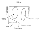
- a third cell group is a cell population appearing in a region except for a region where an erythrocyte ghost population appears in a scattergram using a forward scattered light intensity and a fluorescent intensity as two axes, that is, a population of substantially total leukocyte.
- a cell population appearing in a region surrounded with a chain line as shown in Fig.4 when a scattergram using a fluorescent intensity as an abscissa, and a forward scattered light intensity as an ordinate is drawn becomes a third cell group.
- a third cell group appearance region also contains platelet aggregation as shown in Fig.2.
- a fourth cell group is specified based on second scattered light information and fluorescent information of a third cell group, preferably side scattered light information and fluorescent information.
- a third cell group is discriminated by a scattergram using a fluorescent intensity and a side scattered light intensity as two axes, since a region where mature leukocyte appears, and a region where immature leukocyte appears are clearly discriminated, when a region thought that myeloblast is contained is specified in a scattergram using a fluorescent intensity and a side scattered light intensity as two axes, an appearance region of a cell population containing immature leukocyte, further, a population containing myeloblast can be specified.
- a myeloblast candidate region herein is performed every specimen like specification of a first cell group or a second cell group.
- a region may be set in advance from data accumulated regarding various sizes of myeloblast and mature leukocyte, depending on a dye used, and a concentration.
- a third cell group is discriminated by a scattergram using a side scattered light intensity as an abscissa, and a fluorescent intensity as an ordinate, as shown in Fig. 5, mature leukocyte (lymphocyte, monocyte, and granulocyte) appears upwardly due to a stained nucleus, and can be clearly discriminated from immature leukocyte whose nucleus is not substantially stained.
- Mature leukocyte appears as a lymphocyte population (Ly), a monocyte population (Mo) and a granulocyte population (Gran) in an order from a left side, depending on a nucleus form or the like. Platelet aggregation appears downwardly from a left side to a right side.
- a cell population in a region surrounded with a chain line containing myeloblast, situated at a position of a relatively low to intermediate fluorescent intensity, in a left region where a side scattered light intensity is low may be specified as a fourth cell group. Since myeloblast and immature granulocyte are different in a nuclear form and complexity, both can be discriminated by a magnitude of a side scattered light intensity, but when gating for clearly discriminating both of them is difficult, a cell population contained in a immature granulocyte appearance region, containing both of myeloblast and immature granulocyte may be adopted as a fourth cell group. (4-3) Specification and counting of myeloblast
- the above-specified cell contained in a first cell group and contained in a fourth cell group is counted. Since a first cell group does not contain platelet aggregation, and a fourth cell group does not contain mature leukocyte, a cell contained in a first cell group and contained in a fourth cell group is a population of myeloblast or immature leukocyte, which does not substantially contain platelet aggregation, and mature leukocyte. That is, even when lymphocyte is contained in a first cell group, since lymphocyte is excluded from a cell contained in a first cell group and contained in a fourth cell group, myeloblast or immature leukocyte can be substantially counted at a high precision.
- a method for discriminating myeloblast and platelet aggregation contained in a hematological sample of the present invention is a method for discriminating a cell population containing myeloblast, and platelet aggregation in the measuring method of the present invention, by a step of a sample treatment step; a step of obtaining forward scattered light information and side scattered light information produced by irradiating the treated sample with light; and a step of specifying a first cell group containing myeloblast based on the forward scattered light information and the side scattered light information.
- the measuring apparatus of the present invention comprises a sample treatment part for giving damage to a cell membrane of erythrocyte and mature leukocyte contained in a hematological sample, constricting a hemocyte in which a cell membrane is damaged, and dyeing this with a fluorescent dye which can stain a nucleic acid; an obtainer for obtaining first scattered light information generated by irradiating the treated sample with excitation light of the fluorescent dye, second scattered light information based on scattered light having a different angle from that of first scattered light, and fluorescent information; a first classifier for specifying a first cell group containing myeloblast based on the first scattered light information and the second scattered light information; a second classifier for specifying a second cell group containing myeloblast based on the first scattered light information and the fluorescent information; and a calculator for counting a cell belonging to both of the first and second cell groups as myeloblast.
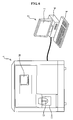
- the measuring apparatus of the present invention has, as its appearance, for example, appearance shown in Fig.6.
- the apparatus shown in Fig.6 has a measuring part 1, an analyzing part 2, and a signal cable 3 connecting the analyzing part 2 and the measuring part 1.
- the measuring part 1 contains the a sample treatment part and the an obtainer of the apparatus of the present invention
- the analyzing part 2 contains the a first classifier, the a second classifier, and the a calculator of the present invention.
- the measuring part 1 is provided with a liquid crystal touch panel 10 for performing various setting inputs by an operator, a start switch 11 for initiating measurement action, and a probe 12 for sucking a specimen.
- a flowcytometer as shown in Fig.7 is mounted in the interior of the measuring part 1.
- the analyzing part 2 is provided with a control part 6 for processing of information sent from the measuring part 1 and for controlling action of the measuring part 1, an output part 7 which is a display part for displaying various measurement results, and a keyboard 8 for inputting specimen information by an operator.
- a beam emitted from a light source (e.g. red semiconductor laser: wavelength 633nm) 21 irradiates an orifice part of a sheath flow cell 23 via a collimating lens 22.
- a light source e.g. red semiconductor laser: wavelength 633nm
- Forward scattered light radiated from a hemocyte which is discharged from a nozzle 20 and passes through an orifice part is introduced into a forward scattered light detector (photodiode) 26 via a condensing lens 24 and a pinhole plate 25.
- side scattered light radiated from a hemocyte passing through an orifice part is introduced into a side scattered light detector (photomultiplier tube) 29 via a condensing lens 27 and a dichroic mirror 28.
- side fluorescent light radiated from a hemocyte passing through an orifice part is introduced into a side fluorescent light detector (photomultiplier tube) 31 via a condensing lens 27, a dichroic mirror 28, a filter 28' and a pinhole plate 30.
- a forward scattered light signal outputted from the forward scattered light detector 26, a side scattered light signal outputted from the side scattered light detector 29, and a side fluorescent signal outputted from the side fluorescent light detector 31 are amplified with amplifiers 32, 33, 34, respectively, and are inputted into the control part 6.
- the light source 21 irradiates an orifice part of a flow cell into which the prepared measuring sample has been introduced, with light which can excite a dye used in treatment of a sample, and is selected depending on a fluorescent dye which stains a hemocyte in a sample. Therefore, depending on a kind of a fluorescent dye used, in addition to the red semiconductor laser, for example, an argon laser, a He-Ne laser, and a blue semiconductor laser may be used.
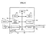
- Fig. 8 is a block view showing a construction of the control part 6.
- This control part 6 is composed mainly of CPU6a, ROM6b, RAM6c, a hard disc 6d, an input and output interface 6e, an image outputting interface 6f, and a communication interface 6g, and the CPU6a, the ROM6b, the RAM6c, the hard disc 6d, the input and output interface 6e, the output interface 6f, and the communication interface 6g are connected with a bus 6h so that data can be communicated.
- CPU6a can execute a computer program stored in ROM6b and a hard disc 6d, and a computer program read out into RAM6c. Therefore, the measuring part 1 specifically executes operation processing of a set program, specifies a cell group such as a first cell group and a second cell group and, further, counts myeloblast, based on a forward scattered light signal, a side scattered light signal, and a fluorescent signal, which are inputted from a flowcytometer.
- ROM6b stores a computer program to be executed by CPU6a, and data used for executing the computer program.
- RAM6c is used in reading out of computer programs stored in ROM6b and a hard disc 6d, and a working region of CPU6a when a computer program is executed.
- the hard disc 6d stores a computer program to be executed by CPU6a, and data used in executing the computer program. This computer program plays the function of analyzing optical information, and outputting the analysis result.
- a keyboard 8 is connected to an input and output interface 6e.
- the keyboard 8 which is an input part is provided for operation on an output screen.
- An output interface 6f is connected to an output part 7 consisted of a liquid crystal display.
- the output part 7 is provided for outputting and displaying the analysis result obtained in the controlling part 6.
- a communication interface 6g is connected to the measuring part 1, and plays the 'function for receiving optical information.
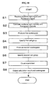
- Fig. 9 is a flow chart showing information processing corresponding to the first measuring method of the present invention.
- the control part 6 adopts the second measuring method, first, the part receives a forward scattered light signal, a side scattered light signal and a fluorescent signal detected in the measuring part 1, via a signal cable 3 (step S1). The control part 6 analyzes these signals, and calculates respective signal intensities (step S2). Then, using a forward scattered light intensity and a side scattered light intensity of a formed element (cell, platelet etc.) in a sample, a first scattergram using a forward scattered light intensity and a side scattered light intensity as two axes is produces (step S3).
- a myeloblast candidate region thought that myeloblast appears therein is set, and a first cell group contained in this region is specified (step S4).
- a second scattergram using a forward scattered light intensity and a fluorescent intensity as two axes is produced (step S5).
- a myeloblast candidate region thought that myeloblast appears therein is set, and a second cell group contained in this region is specified (step S6).
- step S7 using a fluorescent intensity and a side scattered light intensity of a cell contained in a first cell group and contained in a second cell group, a third scattergram using a fluorescent intensity and a side scattered light intensity as two axes is produced (step S7).
- a cell appearing in this third scattergram (cell contained in first cell group and contained in second cell group) is counted as myeloblast (step S8), and this counting result and the third scattergram are outputted in an output part 7 (step S9).
- Fig.10 is a flow chart showing information processing corresponding to a method of first specifying a first cell group among the second measuring method of the present invention.
- the control part 6 adopts such the second measuring method, first, a forward scattered light signal, a side scattered light signal and a fluorescent signal detected in the measuring part 1 are received via a signal cable 3 (step S1).
- the control part 6 analyzes these signals, and calculates respective signal intensities (step S2).
- step S3 using a forward scattered light intensity and a side scattered light intensity of a formed element (cell, platelet etc.) in a sample, a first scattergram using a forward scattered light intensity and a side scattered light intensity as two axes is produced (step S3) .
- a myeloblast candidate region thought that myeloblast appears therein is set, and a first cell group contained in this region is specified (step S4).
- a scattergram (second' scattergram) using a forward scattered light intensity and a fluorescent intensity as two axes is produced (step S5).
- a myeloblast candidate region thought that myeloblast appears therein is set, and a second' cell group contained in this candidate region is specified (step S6). This second' cell group is counted as myeloblast (step S7), and this counting result and the second' scattergram are outputted in an output part 7 (step S8).
- Fig.11 is a flow chart showing information processing corresponding to a method of first specifying a second cell group among the second measuring method of the present invention.
- the control part 6 adopts such the second measuring method, first, a forward scattered light signal, a side scattered light signal and a fluorescent signal detected in the measuring part 1 are received via a signal cable 3 (step S1).
- the control part 6 analyzes these signals, and calculates respective signal intensities (step S2).
- step S3 using a forward scattered light intensity and a fluorescent signal of a formed element (cell, platelet etc.) in a sample, a second scattergram using a forward scattered light intensity and a fluorescent intensity as two axes is produced (step S3).
- a myeloblast candidate region thought that myeloblast appears therein is set, and a second cell group contained in this region is specified (step S4). Then, using a forward scattered light intensity and a side scattered light intensity of a second cell group, a scattergram (first' scattergram) using a forward scattered light intensity and a side scattered light intensity as two axes is produced (step S5).
- a myeloblast candidate region thought that myeloblast appears therein is set, and a first' cell group contained in this candidate region is specified (step S6). This first' cell group is counted as myeloblast (step S7), and this counting result and the first' scattergram are outputted in an output part 7 (step S8).
- Fig.12 is a flow chart showing information processing corresponding to the third measuring method of the present invention.
- the control part 6 adopts such the third measuring method, first, a forward scattered light signal, a side scattered light signal and a fluorescent signal detected in the measuring part 1 are received via a signal cable 3 (step S1). The control part 6 analyzes these signals, and calculates respective signal intensities (step S2). Then, a three-dimensional scattergram using a forward scattered light intensity, a side scattered light intensity and a fluorescent intensity of a formed element (cell, platelet etc.) in a sample is produced (step S3).
- a first cell group based on a forward scattered light intensity and a side scattered light intensity, and a second cell group based on a forward scattered light intensity and a fluorescent intensity are specified (step S4).
- the result of counting of myeloblast, and the three-dimensional scattergram are outputted in an output part 7 (step S8).
- control part 6 outputs and displays the result of counting of myeloblast, and a scattergram used in counting on a display of an output part 7, but may output any of them.
- a scattergram is outputted, not only a scattergram which is finally used in counting myeloblast, but also all of produced scattergrams may be outputted.
- the measuring apparatuses of the aforementioned embodiments adopts the first to third measuring methods of the present invention, but when a fourth measuring method is adopted, it is preferable that the apparatus further comprises third specification means for specifying a fourth cell group not containing at least mature leukocyte based on the obtained fluorescent information and second scattered light information, and the a calculator is means for counting, as myeloblast, a cell belonging to both of the first and second cell groups and contained in a cell group specified by third specifying means.
- a second classifier as a second cell group specified based on first scattered light information and fluorescent information, a population of total leukocyte containing myeloblast (corresponding to third cell group) is specified and, in third specification means, in order to specify a fourth cell group, a scattergram using second scattered light information (side scattered light intensity) and fluorescent information (fluorescent intensity) as two axes is produced. And, a cell contained in a first cell group, contained in a second cell group (herein, substantially corresponding to a third cell group), and contained in a fourth cell group is counted, and the calculation result is outputted in an output part 7.
- the "counting result of myeloblast" referred in the above embodiment includes the number of myeloblast in a sample, the number of myeloblast per unit volume (concentration), and a ratio of myeloblast relative to total leukocyte, and the control part 6 can output at least one of them.
- sample a As a hematological sample, a sample containing a large amount of myeloblast (sample a), and a sample containing myeloblast and immature leukocyte (sample b) were used. Samples a and b contain platelet aggregation.
- a hemolyzing agent of the following composition containing the following components was prepared.
- Glycine 19.5g/l
- Polyoxyethylene(16)oleyl ether 25.0g/l
- Sodium N-lauroylsarcosinate 0.75g/l
- HEPES 10mM
- Purified water 11 Sodium hydroxide amount to adjust a pH at 7.0
- a fluorescent dye A represented by the following formula was dissolved in ethylene glycol.
- the hemolyzing agent and the hematological sample were mixed, and this was mixed with the staining solution so that a final concentration of a fluorescent dye became 20ppm, to prepare a measuring sample.
- a measuring sample was prepared.
- Xylitol 37.0g/l Polyoxyethylene(16)oleyl ether 25.0g/l Sodium N-lauroylsarcosinate 0.75g/l HEPES 10mM
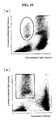
- the hematological sample a and the treating reagent B were mixed, and reacted at 33°C for 5 seconds to prepare a measuring sample, which was introduced into a flow cell, and forward scattered light, side scattered light and fluorescent light were measured with a flowcytometer (light source: red semiconductor: wavelength: 633nm). Measurement results are shown in a first scattergram (Fig.13(a)) using a forward scattered light intensity and a side scattered light intensity as two axes, and a second scattergram (Fig.13(b)) using a fluorescent intensity and a forward scattered light intensity as two axes.
- an appearance region of a first cell group is a region surrounded with a solid line.
- an appearance region of a second cell group is a region surrounded with a solid line.
- a cell population containing lymphocyte was spread and a part of thereof was overlapped with a first cell group.
- FIG. 14 is a third scattergram using a side scattered light intensity and a fluorescent intensity as two axes, and a cell population contained in both of a first cell group and a second cell group appeared concentratedly in a part surrounded with a solid line.
- the number of cells in a region surrounded with a solid line in Fig. 14 was counted, and a ratio relative to the number of total leukocyte was calculated, and was found to be 58.5%.
- the hematological sample a was observed with a microscope, and myeloblast was counted. A ratio of myeloblast relative to the number of total leukocyte in the field was obtained, and was found to be 42.0%.
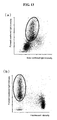
- a measuring sample prepared by treating the hematological sample b with the treating reagent A at 33°C for 5 minutes was introduced into a flow cell, and forward scattered light, side scattered light, and fluorescent light were measured with a flowcytometer (light source: red semiconductor, wavelength: 633nm).
- the measurement results are shown in a first scattergram (Fig.15 (a)) using a forward scattered light intensity and a side scattered light intensity as two axes, and a second scattergram (Fig.15(b)) using a fluorescent intensity and a forward scattered light intensity as two axes.
- an appearance region of a first cell group is a region surrounded with a solid line.
- an appearance region of a second cell group is a region surrounded with a solid line.
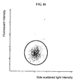
- a cell population contained in both of a first cell group and a second cell group is shown in Fig.16.
- Fig. 16 is a third scattergram using a side scattered light intensity and a fluorescent intensity as two axes, and a cell population contained in both of a first cell group and a second cell group appeared concentratedly in a part surrounded with a solid line.
- the number of cells in a region surrounded with a solid line in Fig.16 was counted, and a ratio relative to the number of total leukocyte was calculated, and found to be 2.4%.
- the hematological sample a was observed with a microscope, and myeloblast was counted.
- a ratio of myeloblast relative to total leukocyte was 2.5%.
- myeloblast can be detected at the same extent of precision as that of microscope observation and, particularly, myeloblast can be detected at a high precision, distinguishing also from immature granulocyte.
Landscapes
- Chemical & Material Sciences (AREA)
- Dispersion Chemistry (AREA)
- Physics & Mathematics (AREA)
- Health & Medical Sciences (AREA)
- Life Sciences & Earth Sciences (AREA)
- Analytical Chemistry (AREA)
- Biochemistry (AREA)
- General Health & Medical Sciences (AREA)
- General Physics & Mathematics (AREA)
- Immunology (AREA)
- Pathology (AREA)
- Investigating Or Analysing Biological Materials (AREA)
- Investigating, Analyzing Materials By Fluorescence Or Luminescence (AREA)
- Investigating Or Analysing Materials By Optical Means (AREA)
Applications Claiming Priority (1)
| Application Number | Priority Date | Filing Date | Title |
|---|---|---|---|
| JP2006092480A JP4976038B2 (ja) | 2006-03-29 | 2006-03-29 | 血液学的試料の測定方法 |
Publications (3)
| Publication Number | Publication Date |
|---|---|
| EP1840571A2 true EP1840571A2 (fr) | 2007-10-03 |
| EP1840571A3 EP1840571A3 (fr) | 2009-09-09 |
| EP1840571B1 EP1840571B1 (fr) | 2020-02-26 |
Family
ID=38324015
Family Applications (1)
| Application Number | Title | Priority Date | Filing Date |
|---|---|---|---|
| EP07005912.6A Active EP1840571B1 (fr) | 2006-03-29 | 2007-03-22 | appareil et procédé pour la mesure d'un échantillon hématologique |
Country Status (4)
| Country | Link |
|---|---|
| US (1) | US7892841B2 (fr) |
| EP (1) | EP1840571B1 (fr) |
| JP (1) | JP4976038B2 (fr) |
| CN (1) | CN101046439B (fr) |
Cited By (1)
| Publication number | Priority date | Publication date | Assignee | Title |
|---|---|---|---|---|
| EP2703813A4 (fr) * | 2011-04-28 | 2014-11-05 | Sysmex Corp | Analyseur sanguin, procédé d'analyse de sang et programme informatique |
Families Citing this family (43)
| Publication number | Priority date | Publication date | Assignee | Title |
|---|---|---|---|---|
| WO2009001869A1 (fr) * | 2007-06-25 | 2008-12-31 | Sysmex Corporation | Réactif et trousse de réactifs pour analyse de leucocyte primitif |
| CN101424614B (zh) * | 2007-10-29 | 2011-04-13 | 深圳迈瑞生物医疗电子股份有限公司 | 嗜碱性粒细胞分析试剂及测定方法 |
| US8603773B2 (en) * | 2008-09-19 | 2013-12-10 | Beckman Coulter | Method and system for analyzing a blood sample |
| JP5288970B2 (ja) | 2008-09-26 | 2013-09-11 | シスメックス株式会社 | 血液試料希釈用試薬を用いた平均赤血球容積の測定方法 |
| JP5670052B2 (ja) * | 2009-03-26 | 2015-02-18 | シスメックス株式会社 | 血液分析装置、血液分析方法及びコンピュータプログラム |
| JP5452058B2 (ja) * | 2009-03-31 | 2014-03-26 | シスメックス株式会社 | 血液分析装置 |
| CN102472702B (zh) * | 2009-07-07 | 2014-05-21 | 索尼公司 | 一种检测样品中的颗粒的方法 |
| JP5530691B2 (ja) * | 2009-09-25 | 2014-06-25 | シスメックス株式会社 | 血球計数装置、診断支援装置、診断支援方法及びコンピュータプログラム |
| JP5533055B2 (ja) | 2010-03-10 | 2014-06-25 | ソニー株式会社 | 光学的測定装置及び光学的測定方法 |
| EP2520926B1 (fr) | 2011-05-05 | 2022-06-15 | Sysmex Corporation | Analyseur de sang, procédé d'analyse de sang et produit de programme informatique |
| CN102331397A (zh) * | 2011-07-08 | 2012-01-25 | 无锡荣兴科技有限公司 | 一种用于血液细胞统计分析的光电传感器 |
| EP2607899B1 (fr) | 2011-12-21 | 2016-08-10 | Beckman Coulter, Inc. | Procédé d'étiquetage des cibles intracellulaires et extracellulaires de leucocytes |
| ES2895391T3 (es) * | 2011-12-27 | 2022-02-21 | Abbott Lab | Análisis celular de fluidos corporales |
| JP6208473B2 (ja) * | 2012-06-20 | 2017-10-04 | アークレイ株式会社 | 血液成分を含む試料の処理方法 |
| JP5461642B2 (ja) * | 2012-09-06 | 2014-04-02 | シスメックス株式会社 | 血液試料の平均赤血球容積を測定するための血液試料希釈用試薬 |
| JP6001425B2 (ja) * | 2012-11-26 | 2016-10-05 | シスメックス株式会社 | 血球分析方法、血球分析装置およびプログラム |
| CN103063634A (zh) * | 2012-12-25 | 2013-04-24 | 华南师范大学 | 虾类血细胞一氧化氮含量的流式细胞术检测方法 |
| US9176112B2 (en) * | 2012-12-31 | 2015-11-03 | Beckman Coulter, Inc. | Systems and methods for platelet count with clump adjustment |
| CN103175817B (zh) * | 2013-03-18 | 2015-02-04 | 苏州大学附属第一医院 | 一种检测抗肿瘤药物抑制肿瘤细胞和血小板粘附的活性的方法 |
| JP6100658B2 (ja) * | 2013-03-29 | 2017-03-22 | シスメックス株式会社 | 血球分析装置および血球分析方法 |
| CN103234892A (zh) * | 2013-04-11 | 2013-08-07 | 广州南沙珠江啤酒有限公司 | 酵母浓度的流式细胞术检测方法 |
| CN112485167A (zh) * | 2013-05-31 | 2021-03-12 | 深圳迈瑞生物医疗电子股份有限公司 | 粒子分析仪的光学系统 |
| JP6092132B2 (ja) * | 2014-01-29 | 2017-03-08 | シスメックス株式会社 | 血球分析装置 |
| JP6238856B2 (ja) * | 2014-08-25 | 2017-11-29 | シスメックス株式会社 | 尿中異型細胞の分析方法、尿分析装置および体液中異型細胞の分析方法 |
| CN106687810B (zh) * | 2014-12-31 | 2019-10-22 | 深圳迈瑞生物医疗电子股份有限公司 | 一种非诊断目的的有核红细胞报警方法、装置及流式细胞分析仪 |
| EP3943912B1 (fr) * | 2015-02-12 | 2025-08-20 | Shenzhen Mindray Bio-Medical Electronics Co., Ltd. | Analyseur cellulaire et procédé et dispositif de tri de particules |
| WO2017011626A1 (fr) * | 2015-07-15 | 2017-01-19 | Becton, Dickinson And Company | Système et procédé pour régler des mesures de cytométrie |
| JP6680492B2 (ja) * | 2015-09-11 | 2020-04-15 | シスメックス株式会社 | 細胞分析装置および細胞分析方法 |
| WO2018148942A1 (fr) * | 2017-02-17 | 2018-08-23 | 深圳迈瑞生物医疗电子股份有限公司 | Procédé d'analyse de cellules sanguines et instrument d'analyse de cellules sanguines |
| WO2018232589A1 (fr) * | 2017-06-20 | 2018-12-27 | 深圳迈瑞生物医疗电子股份有限公司 | Procédé et dispositif d'identification d'agrégation plaquettaire et analyseur de cellules |
| WO2019047042A1 (fr) * | 2017-09-05 | 2019-03-14 | 深圳迈瑞生物医疗电子股份有限公司 | Procédé d'alarme d'échantillon à agrégation de plaquettes, analyseur de cellules sanguines et support d'informations |
| CN108387553B (zh) * | 2018-02-09 | 2021-04-13 | 重庆东渝中能实业有限公司 | 针对白细胞与血小板共存全息图的分块重建与分类计数方法 |
| WO2019206311A1 (fr) | 2018-04-28 | 2019-10-31 | 深圳迈瑞生物医疗电子股份有限公司 | Système d'analyse de sang, analyseur de sang, procédé d'analyse de sang et support de stockage |
| CN116609243A (zh) * | 2018-04-28 | 2023-08-18 | 深圳迈瑞生物医疗电子股份有限公司 | 血液分析方法、血液分析系统及存储介质 |
| JP7291337B2 (ja) * | 2018-11-14 | 2023-06-15 | 学校法人順天堂 | 骨髄液分析方法、試料分析装置及びコンピュータプログラム |
| CN114222907B (zh) * | 2019-08-06 | 2024-10-25 | 贝克曼库尔特有限公司 | 检测和报道中性粒细胞亚群 |
| JP7660123B2 (ja) | 2019-12-27 | 2025-04-10 | ベックマン コールター, インコーポレイテッド | サンプル調製器具 |
| WO2021207896A1 (fr) * | 2020-04-13 | 2021-10-21 | 深圳迈瑞生物医疗电子股份有限公司 | Dispositif et procédé d'analyse d'échantillon |
| CN112195177B (zh) * | 2020-10-28 | 2021-08-06 | 上海慕柏生物医学科技有限公司 | 一种核酸提取方法及试剂盒 |
| CN114544467B (zh) * | 2022-02-24 | 2024-05-24 | 安徽华好阳光乳业有限公司 | 一种生乳中体细胞的测定方法 |
| WO2024123776A1 (fr) | 2022-12-05 | 2024-06-13 | Beckman Coulter, Inc. | Système de flux d'hématologie à canaux multiples |
| EP4492037A1 (fr) * | 2023-07-10 | 2025-01-15 | Sysmex Corporation | Procédé de détection d'agrégation plaquettaire |
| WO2025145095A1 (fr) | 2023-12-29 | 2025-07-03 | Beckman Coulter, Inc. | Manipulation d'échantillon personnalisée dans une analyse biologique |
Family Cites Families (19)
| Publication number | Priority date | Publication date | Assignee | Title |
|---|---|---|---|---|
| JP3048260B2 (ja) * | 1991-07-29 | 2000-06-05 | シスメックス株式会社 | 白血球分類計数用試料調製方法 |
| DE4219811A1 (de) | 1992-06-17 | 1993-12-23 | Solvay Fluor & Derivate | Herstellung von Difluormethylethern und -estern |
| JP3345135B2 (ja) * | 1992-11-19 | 2002-11-18 | シスメックス株式会社 | 血液分析方法 |
| JP3301646B2 (ja) * | 1993-03-19 | 2002-07-15 | シスメックス株式会社 | 幼若細胞測定用試薬 |
| JP3467310B2 (ja) * | 1994-04-21 | 2003-11-17 | シスメックス株式会社 | 白血球分析用試薬及び白血球の分類方法 |
| US5905031A (en) * | 1995-05-18 | 1999-05-18 | Coulter International Corp. | Identification of blast cells in a leukocyte cell preparation |
| JP4042925B2 (ja) * | 1996-11-20 | 2008-02-06 | シスメックス株式会社 | 幼若白血球の分類計数法 |
| US5830701A (en) * | 1997-03-28 | 1998-11-03 | Tao Medical Electronics Co., Ltd. | Method of detecting hematopoietic progenitor cells |
| JP3830613B2 (ja) * | 1997-04-18 | 2006-10-04 | シスメックス株式会社 | 血液中の白血球及びヘモグロビン濃度測定試薬 |
| JP3783808B2 (ja) * | 1997-05-19 | 2006-06-07 | シスメックス株式会社 | 白血球分類計数用試薬 |
| US6900023B1 (en) * | 1999-09-02 | 2005-05-31 | Sysmex Corporation | Method for classifying and counting leukocytes |
| FR2821428B1 (fr) | 2001-02-23 | 2004-08-06 | Abx Sa | Reactif et procede pour l'identification et le comptage de cellules biologiques |
| US6916658B2 (en) * | 2001-07-27 | 2005-07-12 | Beckman Coulter, Inc. | Method for measurement of immature granulocytes |
| JP4212827B2 (ja) * | 2002-05-16 | 2009-01-21 | シスメックス株式会社 | 骨髄液有核細胞自動分析方法 |
| US7625730B2 (en) | 2002-06-24 | 2009-12-01 | Sysmex Corporation | Method for classifying and counting leukocytes |
| WO2005085842A2 (fr) * | 2004-03-09 | 2005-09-15 | Universite Libre De Bruxelles | Procede de detection simultanee de populations de plusieurs entites biologiques differentes par cytometrie de flux, dispositif et programme informatique a cet effet |
| JP4417143B2 (ja) * | 2004-03-11 | 2010-02-17 | シスメックス株式会社 | 試料分析装置、プログラムおよびそのプログラムを記録した記録媒体 |
| US7625712B2 (en) * | 2004-05-21 | 2009-12-01 | Beckman Coulter, Inc. | Method for a fully automated monoclonal antibody-based extended differential |
| US7625757B2 (en) * | 2006-01-27 | 2009-12-01 | Sysmex Corporation | Reagent for immature leukocyte analysis and reagent kit |
-
2006
- 2006-03-29 JP JP2006092480A patent/JP4976038B2/ja not_active Expired - Fee Related
-
2007
- 2007-03-22 EP EP07005912.6A patent/EP1840571B1/fr active Active
- 2007-03-28 US US11/727,806 patent/US7892841B2/en active Active
- 2007-03-29 CN CN200710086991XA patent/CN101046439B/zh active Active
Non-Patent Citations (1)
| Title |
|---|
| None |
Cited By (2)
| Publication number | Priority date | Publication date | Assignee | Title |
|---|---|---|---|---|
| EP2703813A4 (fr) * | 2011-04-28 | 2014-11-05 | Sysmex Corp | Analyseur sanguin, procédé d'analyse de sang et programme informatique |
| EP3159692A1 (fr) * | 2011-04-28 | 2017-04-26 | Sysmex Corporation | Analyseur de sang, procédé d'analyse de sang et programme informatique |
Also Published As
| Publication number | Publication date |
|---|---|
| JP4976038B2 (ja) | 2012-07-18 |
| US7892841B2 (en) | 2011-02-22 |
| EP1840571A3 (fr) | 2009-09-09 |
| CN101046439B (zh) | 2011-01-19 |
| CN101046439A (zh) | 2007-10-03 |
| EP1840571B1 (fr) | 2020-02-26 |
| US20070231913A1 (en) | 2007-10-04 |
| JP2007263894A (ja) | 2007-10-11 |
Similar Documents
| Publication | Publication Date | Title |
|---|---|---|
| EP1840571B1 (fr) | appareil et procédé pour la mesure d'un échantillon hématologique | |
| EP1930723B1 (fr) | Procédé pour la mesure d'un échantillon biologique et appareil de mesure correspondant | |
| JP4324552B2 (ja) | 白血球の分類計数方法 | |
| EP1813942B1 (fr) | Réactif pour analyse de leucocytes immatures et kit de réactif | |
| EP2520926B1 (fr) | Analyseur de sang, procédé d'analyse de sang et produit de programme informatique | |
| US20250237644A1 (en) | Bone marrow fluid analysis method, sample analyzer, and non-transitory storage medium | |
| JP4981907B2 (ja) | 幼若白血球の分析用試薬及び分析用試薬キット | |
| US20030219850A1 (en) | Method for automatically analyzing nucleated bone marrow cells | |
| US20050002826A1 (en) | Apparatus and method for measuring immature platelets | |
| WO2012147451A1 (fr) | Analyseur sanguin, procédé d'analyse de sang et programme informatique | |
| CN101086473A (zh) | 试样分析用试剂、试样分析用试剂盒及试样分析方法 | |
| WO2011140042A1 (fr) | Procédé d'analyse hématologique | |
| EP3249384B1 (fr) | Procédé d'analyse d'échantillons et analyseur d'échantillons | |
| JP5214603B2 (ja) | 幼若白血球の分析用試薬及び分析用試薬キット | |
| JP5416232B2 (ja) | 血液学的試料の測定装置 | |
| JP4806342B2 (ja) | 幼若白血球分析用試薬及び試薬キット | |
| JP2012088336A (ja) | 骨髄芽球と血小板凝集との弁別方法及び弁別装置 | |
| JP4354982B2 (ja) | 白血球の分類計数方法 | |
| WO2026070927A1 (fr) | Dispositif de mesure, réactif et procédé d'analyse |
Legal Events
| Date | Code | Title | Description |
|---|---|---|---|
| PUAI | Public reference made under article 153(3) epc to a published international application that has entered the european phase |
Free format text: ORIGINAL CODE: 0009012 |
|
| AK | Designated contracting states |
Kind code of ref document: A2 Designated state(s): AT BE BG CH CY CZ DE DK EE ES FI FR GB GR HU IE IS IT LI LT LU LV MC MT NL PL PT RO SE SI SK TR |
|
| AX | Request for extension of the european patent |
Extension state: AL BA HR MK YU |
|
| PUAL | Search report despatched |
Free format text: ORIGINAL CODE: 0009013 |
|
| AK | Designated contracting states |
Kind code of ref document: A3 Designated state(s): AT BE BG CH CY CZ DE DK EE ES FI FR GB GR HU IE IS IT LI LT LU LV MC MT NL PL PT RO SE SI SK TR |
|
| AX | Request for extension of the european patent |
Extension state: AL BA HR MK RS |
|
| 17P | Request for examination filed |
Effective date: 20100309 |
|
| AKX | Designation fees paid |
Designated state(s): AT BE BG CH CY CZ DE DK EE ES FI FR GB GR HU IE IS IT LI LT LU LV MC MT NL PL PT RO SE SI SK TR |
|
| 17Q | First examination report despatched |
Effective date: 20100713 |
|
| STAA | Information on the status of an ep patent application or granted ep patent |
Free format text: STATUS: EXAMINATION IS IN PROGRESS |
|
| REG | Reference to a national code |
Ref country code: DE Ref legal event code: R079 Ref document number: 602007059889 Country of ref document: DE Free format text: PREVIOUS MAIN CLASS: G01N0033520000 Ipc: G01N0015140000 |
|
| RIC1 | Information provided on ipc code assigned before grant |
Ipc: G01N 33/52 20060101ALI20190726BHEP Ipc: G01N 15/14 20060101AFI20190726BHEP Ipc: G01N 21/47 20060101ALI20190726BHEP Ipc: G01N 21/64 20060101ALI20190726BHEP Ipc: G01N 15/10 20060101ALI20190726BHEP |
|
| GRAP | Despatch of communication of intention to grant a patent |
Free format text: ORIGINAL CODE: EPIDOSNIGR1 |
|
| STAA | Information on the status of an ep patent application or granted ep patent |
Free format text: STATUS: GRANT OF PATENT IS INTENDED |
|
| INTG | Intention to grant announced |
Effective date: 20191024 |
|
| GRAS | Grant fee paid |
Free format text: ORIGINAL CODE: EPIDOSNIGR3 |
|
| GRAA | (expected) grant |
Free format text: ORIGINAL CODE: 0009210 |
|
| STAA | Information on the status of an ep patent application or granted ep patent |
Free format text: STATUS: THE PATENT HAS BEEN GRANTED |
|
| REG | Reference to a national code |
Ref country code: DE Ref legal event code: R081 Ref document number: 602007059889 Country of ref document: DE Owner name: SYSMEX CORPORATION, KOBE-SHI, JP Free format text: FORMER OWNER: SYSMEX CORP., KOBE, HYOGO, JP |
|
| AK | Designated contracting states |
Kind code of ref document: B1 Designated state(s): AT BE BG CH CY CZ DE DK EE ES FI FR GB GR HU IE IS IT LI LT LU LV MC MT NL PL PT RO SE SI SK TR |
|
| REG | Reference to a national code |
Ref country code: GB Ref legal event code: FG4D |
|
| REG | Reference to a national code |
Ref country code: CH Ref legal event code: EP |
|
| REG | Reference to a national code |
Ref country code: DE Ref legal event code: R096 Ref document number: 602007059889 Country of ref document: DE |
|
| REG | Reference to a national code |
Ref country code: AT Ref legal event code: REF Ref document number: 1238246 Country of ref document: AT Kind code of ref document: T Effective date: 20200315 |
|
| REG | Reference to a national code |
Ref country code: IE Ref legal event code: FG4D |
|
| PG25 | Lapsed in a contracting state [announced via postgrant information from national office to epo] |
Ref country code: FI Free format text: LAPSE BECAUSE OF FAILURE TO SUBMIT A TRANSLATION OF THE DESCRIPTION OR TO PAY THE FEE WITHIN THE PRESCRIBED TIME-LIMIT Effective date: 20200226 |
|
| REG | Reference to a national code |
Ref country code: NL Ref legal event code: MP Effective date: 20200226 |
|
| REG | Reference to a national code |
Ref country code: LT Ref legal event code: MG4D |
|
| PG25 | Lapsed in a contracting state [announced via postgrant information from national office to epo] |
Ref country code: SE Free format text: LAPSE BECAUSE OF FAILURE TO SUBMIT A TRANSLATION OF THE DESCRIPTION OR TO PAY THE FEE WITHIN THE PRESCRIBED TIME-LIMIT Effective date: 20200226 Ref country code: LV Free format text: LAPSE BECAUSE OF FAILURE TO SUBMIT A TRANSLATION OF THE DESCRIPTION OR TO PAY THE FEE WITHIN THE PRESCRIBED TIME-LIMIT Effective date: 20200226 Ref country code: GR Free format text: LAPSE BECAUSE OF FAILURE TO SUBMIT A TRANSLATION OF THE DESCRIPTION OR TO PAY THE FEE WITHIN THE PRESCRIBED TIME-LIMIT Effective date: 20200527 Ref country code: IS Free format text: LAPSE BECAUSE OF FAILURE TO SUBMIT A TRANSLATION OF THE DESCRIPTION OR TO PAY THE FEE WITHIN THE PRESCRIBED TIME-LIMIT Effective date: 20200626 Ref country code: BG Free format text: LAPSE BECAUSE OF FAILURE TO SUBMIT A TRANSLATION OF THE DESCRIPTION OR TO PAY THE FEE WITHIN THE PRESCRIBED TIME-LIMIT Effective date: 20200526 |
|
| PG25 | Lapsed in a contracting state [announced via postgrant information from national office to epo] |
Ref country code: NL Free format text: LAPSE BECAUSE OF FAILURE TO SUBMIT A TRANSLATION OF THE DESCRIPTION OR TO PAY THE FEE WITHIN THE PRESCRIBED TIME-LIMIT Effective date: 20200226 |
|
| PG25 | Lapsed in a contracting state [announced via postgrant information from national office to epo] |
Ref country code: DK Free format text: LAPSE BECAUSE OF FAILURE TO SUBMIT A TRANSLATION OF THE DESCRIPTION OR TO PAY THE FEE WITHIN THE PRESCRIBED TIME-LIMIT Effective date: 20200226 Ref country code: EE Free format text: LAPSE BECAUSE OF FAILURE TO SUBMIT A TRANSLATION OF THE DESCRIPTION OR TO PAY THE FEE WITHIN THE PRESCRIBED TIME-LIMIT Effective date: 20200226 Ref country code: RO Free format text: LAPSE BECAUSE OF FAILURE TO SUBMIT A TRANSLATION OF THE DESCRIPTION OR TO PAY THE FEE WITHIN THE PRESCRIBED TIME-LIMIT Effective date: 20200226 Ref country code: SK Free format text: LAPSE BECAUSE OF FAILURE TO SUBMIT A TRANSLATION OF THE DESCRIPTION OR TO PAY THE FEE WITHIN THE PRESCRIBED TIME-LIMIT Effective date: 20200226 Ref country code: PT Free format text: LAPSE BECAUSE OF FAILURE TO SUBMIT A TRANSLATION OF THE DESCRIPTION OR TO PAY THE FEE WITHIN THE PRESCRIBED TIME-LIMIT Effective date: 20200719 Ref country code: ES Free format text: LAPSE BECAUSE OF FAILURE TO SUBMIT A TRANSLATION OF THE DESCRIPTION OR TO PAY THE FEE WITHIN THE PRESCRIBED TIME-LIMIT Effective date: 20200226 Ref country code: CZ Free format text: LAPSE BECAUSE OF FAILURE TO SUBMIT A TRANSLATION OF THE DESCRIPTION OR TO PAY THE FEE WITHIN THE PRESCRIBED TIME-LIMIT Effective date: 20200226 Ref country code: LT Free format text: LAPSE BECAUSE OF FAILURE TO SUBMIT A TRANSLATION OF THE DESCRIPTION OR TO PAY THE FEE WITHIN THE PRESCRIBED TIME-LIMIT Effective date: 20200226 |
|
| REG | Reference to a national code |
Ref country code: CH Ref legal event code: PL |
|
| REG | Reference to a national code |
Ref country code: AT Ref legal event code: MK05 Ref document number: 1238246 Country of ref document: AT Kind code of ref document: T Effective date: 20200226 |
|
| REG | Reference to a national code |
Ref country code: DE Ref legal event code: R097 Ref document number: 602007059889 Country of ref document: DE |
|
| PG25 | Lapsed in a contracting state [announced via postgrant information from national office to epo] |
Ref country code: MC Free format text: LAPSE BECAUSE OF FAILURE TO SUBMIT A TRANSLATION OF THE DESCRIPTION OR TO PAY THE FEE WITHIN THE PRESCRIBED TIME-LIMIT Effective date: 20200226 |
|
| REG | Reference to a national code |
Ref country code: BE Ref legal event code: MM Effective date: 20200331 |
|
| PG25 | Lapsed in a contracting state [announced via postgrant information from national office to epo] |
Ref country code: LU Free format text: LAPSE BECAUSE OF NON-PAYMENT OF DUE FEES Effective date: 20200322 |
|
| PLBE | No opposition filed within time limit |
Free format text: ORIGINAL CODE: 0009261 |
|
| STAA | Information on the status of an ep patent application or granted ep patent |
Free format text: STATUS: NO OPPOSITION FILED WITHIN TIME LIMIT |
|
| PG25 | Lapsed in a contracting state [announced via postgrant information from national office to epo] |
Ref country code: IT Free format text: LAPSE BECAUSE OF FAILURE TO SUBMIT A TRANSLATION OF THE DESCRIPTION OR TO PAY THE FEE WITHIN THE PRESCRIBED TIME-LIMIT Effective date: 20200226 Ref country code: AT Free format text: LAPSE BECAUSE OF FAILURE TO SUBMIT A TRANSLATION OF THE DESCRIPTION OR TO PAY THE FEE WITHIN THE PRESCRIBED TIME-LIMIT Effective date: 20200226 Ref country code: IE Free format text: LAPSE BECAUSE OF NON-PAYMENT OF DUE FEES Effective date: 20200322 Ref country code: CH Free format text: LAPSE BECAUSE OF NON-PAYMENT OF DUE FEES Effective date: 20200331 Ref country code: LI Free format text: LAPSE BECAUSE OF NON-PAYMENT OF DUE FEES Effective date: 20200331 |
|
| 26N | No opposition filed |
Effective date: 20201127 |
|
| PG25 | Lapsed in a contracting state [announced via postgrant information from national office to epo] |
Ref country code: BE Free format text: LAPSE BECAUSE OF NON-PAYMENT OF DUE FEES Effective date: 20200331 Ref country code: PL Free format text: LAPSE BECAUSE OF FAILURE TO SUBMIT A TRANSLATION OF THE DESCRIPTION OR TO PAY THE FEE WITHIN THE PRESCRIBED TIME-LIMIT Effective date: 20200226 Ref country code: SI Free format text: LAPSE BECAUSE OF FAILURE TO SUBMIT A TRANSLATION OF THE DESCRIPTION OR TO PAY THE FEE WITHIN THE PRESCRIBED TIME-LIMIT Effective date: 20200226 |
|
| PG25 | Lapsed in a contracting state [announced via postgrant information from national office to epo] |
Ref country code: TR Free format text: LAPSE BECAUSE OF FAILURE TO SUBMIT A TRANSLATION OF THE DESCRIPTION OR TO PAY THE FEE WITHIN THE PRESCRIBED TIME-LIMIT Effective date: 20200226 Ref country code: MT Free format text: LAPSE BECAUSE OF FAILURE TO SUBMIT A TRANSLATION OF THE DESCRIPTION OR TO PAY THE FEE WITHIN THE PRESCRIBED TIME-LIMIT Effective date: 20200226 Ref country code: CY Free format text: LAPSE BECAUSE OF FAILURE TO SUBMIT A TRANSLATION OF THE DESCRIPTION OR TO PAY THE FEE WITHIN THE PRESCRIBED TIME-LIMIT Effective date: 20200226 |
|
| PGFP | Annual fee paid to national office [announced via postgrant information from national office to epo] |
Ref country code: FR Payment date: 20230208 Year of fee payment: 17 |
|
| PGFP | Annual fee paid to national office [announced via postgrant information from national office to epo] |
Ref country code: DE Payment date: 20240130 Year of fee payment: 18 Ref country code: GB Payment date: 20240201 Year of fee payment: 18 |
|
| PG25 | Lapsed in a contracting state [announced via postgrant information from national office to epo] |
Ref country code: FR Free format text: LAPSE BECAUSE OF NON-PAYMENT OF DUE FEES Effective date: 20240331 |
|
| PG25 | Lapsed in a contracting state [announced via postgrant information from national office to epo] |
Ref country code: FR Free format text: LAPSE BECAUSE OF NON-PAYMENT OF DUE FEES Effective date: 20240331 |
|
| REG | Reference to a national code |
Ref country code: DE Ref legal event code: R119 Ref document number: 602007059889 Country of ref document: DE |
|
| GBPC | Gb: european patent ceased through non-payment of renewal fee |
Effective date: 20250322 |
|
| PG25 | Lapsed in a contracting state [announced via postgrant information from national office to epo] |
Ref country code: DE Free format text: LAPSE BECAUSE OF NON-PAYMENT OF DUE FEES Effective date: 20251001 |
|
| PG25 | Lapsed in a contracting state [announced via postgrant information from national office to epo] |
Ref country code: GB Free format text: LAPSE BECAUSE OF NON-PAYMENT OF DUE FEES Effective date: 20250322 |