EP1865320A2 - Zellantagonist - Google Patents
Zellantagonist Download PDFInfo
- Publication number
- EP1865320A2 EP1865320A2 EP07252322A EP07252322A EP1865320A2 EP 1865320 A2 EP1865320 A2 EP 1865320A2 EP 07252322 A EP07252322 A EP 07252322A EP 07252322 A EP07252322 A EP 07252322A EP 1865320 A2 EP1865320 A2 EP 1865320A2
- Authority
- EP
- European Patent Office
- Prior art keywords
- gpcr
- cells
- receptor
- distribution
- candidate compound
- Prior art date
- Legal status (The legal status is an assumption and is not a legal conclusion. Google has not performed a legal analysis and makes no representation as to the accuracy of the status listed.)
- Withdrawn
Links
Images








Classifications
-
- A—HUMAN NECESSITIES
- A61—MEDICAL OR VETERINARY SCIENCE; HYGIENE
- A61K—PREPARATIONS FOR MEDICAL, DENTAL OR TOILETRY PURPOSES
- A61K31/00—Medicinal preparations containing organic active ingredients
- A61K31/33—Heterocyclic compounds
- A61K31/395—Heterocyclic compounds having nitrogen as a ring hetero atom, e.g. guanethidine or rifamycins
- A61K31/40—Heterocyclic compounds having nitrogen as a ring hetero atom, e.g. guanethidine or rifamycins having five-membered rings with one nitrogen as the only ring hetero atom, e.g. sulpiride, succinimide, tolmetin, buflomedil
-
- G—PHYSICS
- G01—MEASURING; TESTING
- G01N—INVESTIGATING OR ANALYSING MATERIALS BY DETERMINING THEIR CHEMICAL OR PHYSICAL PROPERTIES
- G01N33/00—Investigating or analysing materials by specific methods not covered by groups G01N1/00 - G01N31/00
- G01N33/48—Biological material, e.g. blood, urine; Haemocytometers
- G01N33/50—Chemical analysis of biological material, e.g. blood, urine; Testing involving biospecific ligand binding methods; Immunological testing
- G01N33/5005—Chemical analysis of biological material, e.g. blood, urine; Testing involving biospecific ligand binding methods; Immunological testing involving human or animal cells
- G01N33/5008—Chemical analysis of biological material, e.g. blood, urine; Testing involving biospecific ligand binding methods; Immunological testing involving human or animal cells for testing or evaluating the effect of chemical or biological compounds, e.g. drugs, cosmetics
- G01N33/502—Chemical analysis of biological material, e.g. blood, urine; Testing involving biospecific ligand binding methods; Immunological testing involving human or animal cells for testing or evaluating the effect of chemical or biological compounds, e.g. drugs, cosmetics for testing non-proliferative effects
- G01N33/5035—Chemical analysis of biological material, e.g. blood, urine; Testing involving biospecific ligand binding methods; Immunological testing involving human or animal cells for testing or evaluating the effect of chemical or biological compounds, e.g. drugs, cosmetics for testing non-proliferative effects on sub-cellular localization
-
- A—HUMAN NECESSITIES
- A61—MEDICAL OR VETERINARY SCIENCE; HYGIENE
- A61K—PREPARATIONS FOR MEDICAL, DENTAL OR TOILETRY PURPOSES
- A61K35/00—Medicinal preparations containing materials or reaction products thereof with undetermined constitution
- A61K35/12—Materials from mammals; Compositions comprising non-specified tissues or cells; Compositions comprising non-embryonic stem cells; Genetically modified cells
-
- A—HUMAN NECESSITIES
- A61—MEDICAL OR VETERINARY SCIENCE; HYGIENE
- A61P—SPECIFIC THERAPEUTIC ACTIVITY OF CHEMICAL COMPOUNDS OR MEDICINAL PREPARATIONS
- A61P9/00—Drugs for disorders of the cardiovascular system
-
- C—CHEMISTRY; METALLURGY
- C07—ORGANIC CHEMISTRY
- C07K—PEPTIDES
- C07K14/00—Peptides having more than 20 amino acids; Gastrins; Somatostatins; Melanotropins; Derivatives thereof
- C07K14/435—Peptides having more than 20 amino acids; Gastrins; Somatostatins; Melanotropins; Derivatives thereof from animals; from humans
- C07K14/705—Receptors; Cell surface antigens; Cell surface determinants
-
- G—PHYSICS
- G01—MEASURING; TESTING
- G01N—INVESTIGATING OR ANALYSING MATERIALS BY DETERMINING THEIR CHEMICAL OR PHYSICAL PROPERTIES
- G01N33/00—Investigating or analysing materials by specific methods not covered by groups G01N1/00 - G01N31/00
- G01N33/48—Biological material, e.g. blood, urine; Haemocytometers
- G01N33/50—Chemical analysis of biological material, e.g. blood, urine; Testing involving biospecific ligand binding methods; Immunological testing
- G01N33/5005—Chemical analysis of biological material, e.g. blood, urine; Testing involving biospecific ligand binding methods; Immunological testing involving human or animal cells
- G01N33/5008—Chemical analysis of biological material, e.g. blood, urine; Testing involving biospecific ligand binding methods; Immunological testing involving human or animal cells for testing or evaluating the effect of chemical or biological compounds, e.g. drugs, cosmetics
-
- G—PHYSICS
- G01—MEASURING; TESTING
- G01N—INVESTIGATING OR ANALYSING MATERIALS BY DETERMINING THEIR CHEMICAL OR PHYSICAL PROPERTIES
- G01N33/00—Investigating or analysing materials by specific methods not covered by groups G01N1/00 - G01N31/00
- G01N33/48—Biological material, e.g. blood, urine; Haemocytometers
- G01N33/50—Chemical analysis of biological material, e.g. blood, urine; Testing involving biospecific ligand binding methods; Immunological testing
- G01N33/53—Immunoassay; Biospecific binding assay; Materials therefor
- G01N33/566—Immunoassay; Biospecific binding assay; Materials therefor using specific carrier or receptor proteins as ligand binding reagents where possible specific carrier or receptor proteins are classified with their target compounds
-
- G—PHYSICS
- G01—MEASURING; TESTING
- G01N—INVESTIGATING OR ANALYSING MATERIALS BY DETERMINING THEIR CHEMICAL OR PHYSICAL PROPERTIES
- G01N2333/00—Assays involving biological materials from specific organisms or of a specific nature
- G01N2333/435—Assays involving biological materials from specific organisms or of a specific nature from animals; from humans
- G01N2333/575—Hormones
- G01N2333/5754—Endothelin, vasoactive intestinal contractor [VIC]
-
- G—PHYSICS
- G01—MEASURING; TESTING
- G01N—INVESTIGATING OR ANALYSING MATERIALS BY DETERMINING THEIR CHEMICAL OR PHYSICAL PROPERTIES
- G01N2333/00—Assays involving biological materials from specific organisms or of a specific nature
- G01N2333/435—Assays involving biological materials from specific organisms or of a specific nature from animals; from humans
- G01N2333/705—Assays involving receptors, cell surface antigens or cell surface determinants
- G01N2333/70571—Assays involving receptors, cell surface antigens or cell surface determinants for neuromediators, e.g. serotonin receptor, dopamine receptor
-
- G—PHYSICS
- G01—MEASURING; TESTING
- G01N—INVESTIGATING OR ANALYSING MATERIALS BY DETERMINING THEIR CHEMICAL OR PHYSICAL PROPERTIES
- G01N2333/00—Assays involving biological materials from specific organisms or of a specific nature
- G01N2333/435—Assays involving biological materials from specific organisms or of a specific nature from animals; from humans
- G01N2333/705—Assays involving receptors, cell surface antigens or cell surface determinants
- G01N2333/72—Assays involving receptors, cell surface antigens or cell surface determinants for hormones
- G01N2333/726—G protein coupled receptor, e.g. TSHR-thyrotropin-receptor, LH/hCG receptor, FSH
-
- G—PHYSICS
- G01—MEASURING; TESTING
- G01N—INVESTIGATING OR ANALYSING MATERIALS BY DETERMINING THEIR CHEMICAL OR PHYSICAL PROPERTIES
- G01N2800/00—Detection or diagnosis of diseases
- G01N2800/12—Pulmonary diseases
-
- G—PHYSICS
- G01—MEASURING; TESTING
- G01N—INVESTIGATING OR ANALYSING MATERIALS BY DETERMINING THEIR CHEMICAL OR PHYSICAL PROPERTIES
- G01N2800/00—Detection or diagnosis of diseases
- G01N2800/32—Cardiovascular disorders
Definitions
- the present invention provides a new approach for the treatment of G-protein coupled receptor based disease.
- This approach comprises altering the physiology of G-protein coupled receptor based responses by exerting chemical control over the cellular distribution of the receptor.
- This approach allows a method for phenotypic screening for candidate compounds which disrupt GPCR distribution signalling activity and are therefore candidates for the treatment of GPCR-related conditions.
- Compounds that create an altered distribution of GPCR upon exposure to the cell are identified.
- the method uses these molecules to alter physiology through their effect on receptor distribution and reduction to practice is demonstrated in an ex-vivo model of g-protein coupled receptor activity.
- GPCRs cell surface G-protein coupled receptors
- This signaling is of critical importance for physiological homeostasis in systems as diverse as blood pressure regulation, odor recognition, and visual perception 1 .
- More than half of the available prescription drugs target G-protein coupled receptors and most can be described as agonists or antagonists of G protein coupled receptor activity.
- drug design efforts are aimed at the manipulation of the interaction(s) between a receptor and its endogenous ligands, whether to repress this through chemical antagonism or stimulate receptor activity through the use of artificial agonists.
- G-protein coupled receptors are transmembrane proteins with a ligand-binding site exposed on the outside surface of the cell and an effector site that extends into the cytosol.
- ligand (agonist) binding Upon ligand (agonist) binding, conformational changes in heptahelical receptors recruit specific heterotrimeric G-proteins 2 , enable their dissociation into G ⁇ and G ⁇ subunits and results in the activation of the G ⁇ subunit.
- This activates a series of class specific downstream effectors, such as phospholipase C- ⁇ for the Gq class and adenyl cyclase for Gs, which then produce changes in the levels of second messengers such as intracellular Ca++ and cyclic AMP (CAMP) 3 .
- CAMP cyclic AMP
- Agonist induced internalization is a property of many GPCRs, such as the ⁇ -adrenergic, endothelin A, and ⁇ opioid receptors 4 . Internalization controls signaling for it desensitizes cellular responses as receptors are removed from the cell surface. It is also implicated in hypertension and drug tolerance. It demonstrates the importance of cellular location in receptor function.
- GPCRs G-protein receptor kinases
- Arrestins promote the internalization of the receptor into plasma membrane derived vesicles, either caveolae or clathrin-coated vesicles, which pinch off into the cell interior to deliver the receptor into the endosomal organelles.
- GPCRs Once internalized, GPCRs have several endocytic itineraries and may be transported for degradation, rapidly recycled to the cell surface, or resensitize and recycle from the pericentriolar recycling compartment to the cell surface and enable another cycle of signaling.
- Blood pressure is under the control of the coordinated action of biochemical stimuli that mediate vascular smooth muscle tone, and a prime stimulus are the endothelin hormones.
- Endothelins are the most potent vasoconstrictors yet identified and are implicated in blood pressure regulation, cardiac development and cancer 8-12 . These hormones exert their physiological effect through the activation of the endothelin A and endothelin B GPCRs, which differ in terms of their G-protein coupling and trafficking. Agonist binding to the endothelin A receptor acts via Gq signaling to promote vascular constriction through second messengers. The endothelin A receptor (ETAR) undergoes agonist induced internalization and mediates vasoconstriction via activation of both Gq and Gs. In contrast, the endothelin B receptor (ETBR) mediates vasodilation and is constitutively internalized 13-15 . Clinically, medications targeted at the endothelin system antagonize agonist activity and disrupt endothelin: receptor interactions (bosentan: 16 ).
- standard agonists and antagonists that target GPCRs have a number of disadvantages. For example, they must be administered in high doses in order to reach the required concentration. In addition, higher doses are required as patients develop drug resistance especially where they are administered long term for the treatment of chronic pain and blood pressure control, etc.
- Treating GPCR related diseases remains a challenge, and this has been approached through the introduction of drugs acting as agonists and antagonists. More than a quarter of the leading 100 drugs on the market are GPCR related and have a combined value of more than 30 billion dollars annually. In a broader sense, receptors serve as the drug target for approximately half of known drugs. Given the importance of this set of pharmaceutical targets, new strategies for drug identification may provide new drugs for GPCR diseases.
- the present invention provides a new approach for the treatment of G-protein coupled receptor based disease.
- This approach comprises altering the physiology of G-protein coupled receptor based responses by exerting chemical control over the cellular distribution of the receptor.
- Compounds that create an altered distribution of GPCR upon exposure to the cell are identified. The ability of these molecules to alter physiology through their effect on receptor distribution is demonstrated in an ex-vivo model of G-protein coupled receptor activity.
- cellular antagonism This approach is referred to a “cellular antagonism” and the candidate compounds used within it are referred to as “cellular antagonists” with “cellular antagonism” being the intervention in the function of a GPCR by modulating its location or signalling properties within the cell.
- cellular antagonists are required in much lower concentrations to modify GPCR function compared to conventional GPCR modulating compounds.
- the present invention provides a method for identifying a compound that modifies the cellular distribution of a GPCR in a population of cells comprising:
- G-protein coupled receptors are characterised as 7 transmembrane receptors.
- the GPCR for use in the method of the present invention is one that undergoes agonist-induced receptor internalization.
- Such GPCRs includes muscarinic receptors among others.
- the GPCR is a ⁇ -adrenergic, endothelin A , kappa opioid receptor or ⁇ opioid receptor.
- the term GPCR includes both naturally occuring GPCRs as well as GPCRs that have been modified.
- GPCRs are generally found at the cell surface. However, following receptor activation through ligand binding, many receptors are internalised and then recycled to the cell surface through the intracellular endosome pathway or routed for transcellular transport or destruction in degradative lysosomal compartments. Accordingly, in the method of the present invention, "altered distribution” or “altered cellular distribution” of GPCR in cells is measured by reference to cellular distribution of GPCR, following receptor activation, in a control assay cell which has not been treated with a candidate compound.
- GPCR location can be detected through antibody-based recognition of GPCR and visualisation of an antibody labelled GPCR.
- the GPCR may also express a detectable tag.
- the cellular distribution of GPCR may be determined by detecting a labelled GPCR in treated and untreated cells.
- labelled GPCR is meant a GPCR which has been modify to comprise a detectable label or marker. Suitable labels and markers for use in such protein location studies will be familiar to those skilled in the art and include labels and markers as described herein.
- distribution of the GPCR is determined by microscopy.
- Suitable microscopic methods include optical microscopy, confocal microscopy or automated microscopic screening methods.
- Optical microscopy is well suited to the study of GPCR activation and trafficking, and automated optical screening of cell based GPCR activation assays (high content screening 19,20 ) provides the means to analyze these pathways on a larger scale than previously possible 21 .
- automated microscopic screening enables the tracing of endocytosis of activated receptors and currently serves for the screening of novel drugs.
- a candidate compound is one that modifies GPCR cellular distribution is given by an altered cellular distribution of GPCR in a cell treated with a candidate compound when compared to a cell which has not been treated with a candidate compound.
- Methods for obtaining data images of treated and control cells and performing a comparison of distribution are described herein.
- a comparison may be made between levels of labelled GPCR at the cell surface, in the early endosomes or in the recycling endosomes.
- the distribution of GPCR is compared by measuring labelled GPCR at the cell surface, or in peripheral endosomes or the peri-centriolar recycling endosome.
- a candidate compound that modifies the cellular distribution of a GPCR is one that gives relative numbers of labelled endosomes outside a threshold of +/two to three times the standard deviation of the untreated but ligand treated control.
- the candidate compound molecules are identified by the phenotype of ability to alter cellular distribution of the GPCR, and then confirmed by physiological testing, a definition of the 'target' of the molecules is not required.
- the method of the present invention further comprises comparing the effect of the candidate compound on the distribution of said labelled GPCR with its effect on house keeping endocytosis.
- the marker for internalisation is a labelled compound such as labelled dextran.
- the labelled dextran is a BODIPYTM-labelled dextran such as the labelled dextran described herein.
- the cells which are used for the assay in accordance with the invention and those for measuring house keeping endocytosis are the same.
- the determination of GPCR location and marker internalisation are carried out simultaneously.
- the label on the GPCR and the label on the marker are suitably detectably different. Accordingly, there is provided a method in accordance with the invention wherein the determination of the effect of the candidate compound on GPCR distribution and on labelled dextran distribution is determined simultaneously.
- GPCR ligands are those compounds which bind to and activate a GPCR. Suitable ligands for a particular GPCR will be known to those skilled in the art and include agonists of GPCRs. For example, where the GPCR is endothelin A receptor, suitable ligands include endothelin-1.
- an increase in GPCR at the cell surface is indicative of a candidate compound for treating a GPCR related disease such as a disease related to aortal superconstriction.
- a GPCR related disease such as a disease related to aortal superconstriction.
- an increase in intracellular GPCR is indicative of a candidate compound for slowed aortal vasoconstriction.
- candidate compounds which have been selected according to the method of the invention can be used in additional assays to confirm their activity.
- a method of the invention further comprises taking the candidate compound identified from steps a) to c) in accordance with the first aspect of the invention and determining its effect in an assay for vasoconstriction.
- Suitable assays for vasoconstriction include an ex-vivo rat thoracic aorta constriction-relaxation model.
- a method of modulating GPCR distribution in a cell comprising administering an inhibitor of PKC or PKA.
- Candidate compounds identified in accordance the present invention are candidates for use in the treatment of GPCR related diseases.
- a large number of GPCR related diseases are familiar to those skilled in the art and suitable diseases are described herein.
- the invention provides a method of treating a GPCR related disease comprising administering a compound that modifies the cellular distribution of a GPCR.
- a compound that modifies cellular distribution of a GPCR for use in the treatment of a GPCR related disease.
- Suitable compounds have been identified herein and include the protein kinase C inhibitors RO 31 8220, Gö 6976, palmitoyl DL carnitine chloride, protein kinase A inhibitors KT 5720, H-89 and erbstatin A.
- the invention provides a use of a compound that modifies the cellular distribution of a GPCR in the preparation of a medicament for use in the treatment of a GPCR related disease.
- the compound is selected from the protein kinase C inhibitors RO 31 8220, Gö 6976, palmitoyl DL carnitine chloride, protein kinase A inhibitors KT 5720, H-89 and erbstatin A.
- the GPCR related disease may be endothelin related pulmonary arterial hypertension. Suitable the GPCR related disease is related to impaired vascular tone and can be examined in the rat thoracic aorta vasoconstriction model.
- C- ETAR and C- HK are the relative values for the negative controls for the ETAR assay ( ⁇ ) and the housekeeping assay ( ⁇ ), respectively. Hits are defined as falling outside ⁇ 3 standard deviations from the mean for the ETAR (---) or housekeeping assay (---).
- the red lines connecting the relative values of the two complementary assays indicate the specificity of the compound for the ETAR or the housekeeping assay. The greater this line grows the greater the difference between the assay results and the higher the specificity of the compound.
- Confocal image pairs are of endothelin-1 stimulated ETAR-GFP cells (upper panels) or fluorescent dextran internalization (lower panels). All compounds were used at 10 ⁇ M. Scale bar 20 ⁇ m.
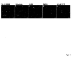
- Endothelin A receptor internalization was imaged after stimulation with 40 nM Endothelin and a 10 min internalization of red fluorescent dextran on a confocal microscope in presence of the following compounds: ( a ) 10 ⁇ M Ro 31-2880. ( b ) 10 ⁇ M Erbstatin A. ( c ) 10 ⁇ M H-89 ( d ) DMSO (e) 40 nM endothelin-1 alone. Confocal images,are from a 1 ⁇ m Z section. The endothelin A receptor is shown in green, the dextran in red. Scale bar 5 ⁇ m.
- HEK293T cells expressing a human kappa Opioid receptor fusion to the green fluorescent protein were treated with 10 ⁇ M Gö 6976, 10 ⁇ M Erbstatin A, 10 ⁇ M H-89, 10 ⁇ M KT-5720 with 300 nM agonist, agonist alone or DMSO. Scale bar 20 ⁇ m.
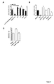
- ETAR and housekeeping internalization were measured for a set of PKA inhibitors ( a ), and ETAR internalization was measured after ( b ) KT5720, H89 or forskolin and ( c ) the number of endosomes /cell was quantified after agonist stimulation.
- ETAR and housekeeping internalization were measured for a set of tyrosine inhibitors ( a ), and ETAR internalization was measured after ( b ) single and combinatorial treatment with 100nM phorbol ester, RO 31 8220, erbstatin A and H-89.
- cAMP production requires sequential G-protein activation.
- Cells were treated with 10 ⁇ M RO 31 8220, H-89, KT 5720 Forskolin or Erbstatin A, then stimulated with 40nM ET-1 and cAMP was measured over time by indirect ELISA using standard concentration cAMP curves.
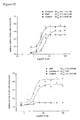
- Endothelin receptor internalization was measured after treatment with a range of compound concentrations from 0.1 to 50 ⁇ M. Relative internalization of the ETAR is plotted against compound concentration for Ro 31-8220, the erbstatin Analogue, H-89 and GW 5074. Results are the mean of four experiments, showing the standard deviation, where each experimental point comprises five pairs of images giving an n (cell) of > 4000 per compound concentration. Dotted lines indicate the ⁇ 3 SD threshold for receptor internalization in control cells.
- GPCR-related disease includes any disease or disorder associated with aberrant GPCR signaling, including, but not limited to, neuropsychiatric disorders such as, for example, schizophrenia, bipolar disorders and depression; cardiopulmonary disorders such as, for example, cardiachypertrophy, hypertension, thrombosis and arrhythmia; inflammation, cystic fibrosis and ocular disorders. Without limitation as to mechanism, GPCR-related diseases are generally associated with decreased GPCR-signaling.
- GPCR ligand and "GPCR agonist” includes any molecule or agent which binds to a GPCR and elicits a response.
- GPCR antagonist includes any molecule or agent which binds to a GPCR but which does not elicit a response.
- Cellular antagonism is defined as intervening in the function of a GPCR by modulating its location, or signaling properties within a cell using a small molecule modulator (compound).
- cellular antagonism acts at the level of the receptor.
- altered cellular distribution includes an alteration in a detectable distribution compared to a distribution in a control cell.
- Candidate compound means any solution, compound, or other substance (including, but not limited to, small molecules such as deoxyribonucleotide and ribonucleotide molecules as well as peptides, proteins, and nucleic acids) to be screened according to the methods described herein for altered GPCR cellular distribution.
- the invention provides methods (also referred to herein as “screening assays”) for identifying candidate or test compounds or agents comprising therapeutic moieties (e. g. peptides, peptidomimetics, peptoids, polynucleotides, small molecules or other drugs) which alter the cellular distribution of a GPCR.
- therapeutic moieties e. g. peptides, peptidomimetics, peptoids, polynucleotides, small molecules or other drugs
- test compounds of the present invention are generally either small molecules or bioactive agents.
- the test compound is a small molecule.
- the test compound is a bioactive agent.
- Bioactive agents include, but are not limited to, naturally-occurring or synthetic compounds or molecules ("biomolecules") having bioactivity in mammals, as well as proteins, peptides, oligopeptides, polysaccharides, nucleotides and polynucleotides.
- the candidate compounds of the present invention may be obtained from any available source, including systematic libraries of natural and/or synthetic compounds.
- the candidate compound to be tested may be administered to the cell in several ways. For example, it may be added directly to the cell culture medium or injected into the cell. Alternatively, in the case of polypeptide agents, the cell may be transfected with a nucleic acid construct, which directs expression of the polypeptide in the cell. Preferably, the expression of the polypeptide is under the control of an inducible promoter.
- Cells useful for assays and methods in accordance with the present invention include eukaryotic and prokaryotic cells, including, but not limited to, bacterial cells, yeast cells, fungal cells, insect cells, nematode cells, plant cells, and animal cells.
- Suitable animal cells include, but are not limited to, HEK cells, HeLa cells, COS cells, U20S cells,CHO-K1 cells, and various primary mammalian cells.
- Cells useful in the present invention may stably or transiently express the labelled GPCRs used in the methods described herein.
- Methods of expressing genes using non-mammalian viruses e. g. , baculoviruses described in U. S. Patent Nos. 4,745, 051 ; 4,879, 236 ; 5,348, 886 ; 5,731, 182 ; 5,871, 986 ; 6,281, 009 ; and 6,238, 914 ; may be used in the present methods.
- Labels or marker molecules that may be used to conjugate with the GPCR include, but are not limited to, molecules that are detectable by spectroscopic, photochemical, radioactivity, biochemical, immunochemical, colorimetric, electrical, or optical means, including, but not limited to, bioluminescence, phosphorescence, and fluorescence. These labels should be biologically compatible molecules and should not compromise the ability of the GPCR to interact with its ligand or with the GPCR signalling system or receptor internalisation system, and the interaction of the ligand with the GPCR must not compromise the ability of the label to be detected.
- Labels include radioisotopes, epitope tags, affinity labels, enzymes, fluorescent groups, chemiluminescent groups, and the like. Labels include molecules that are directly or indirectly detected as a function of their interaction with other molecule (s) as well as molecules detected as a function of their location or translocation. In some embodiments, the labels are optically detectable marker molecules, including optically detectable proteins, such that they may be excited chemically, mechanically, electrically, or radioactively to emit fluorescence, phosphorescence, or bioluminescence.
- Optically detectable marker molecules include, for example, beta-galactosidase, firefly luciferase, bacterial luciferase, fluorescein, Texas Red, horseradish peroxidase, alkaline phosphatase, and rhodamine-conjugated antibody.
- the optically detectable labels or marker molecules are inherently fluorescent molecules, such as fluorescent proteins, including, for example, Green Fluorescent Protein (GFP).
- fluorescent proteins including, for example, Green Fluorescent Protein (GFP).
- the label may be conjugated to the GPCR by methods as described in U. S. Patent Nos. 5,891, 646 and 6,110, 693 .
- the label may be conjugated to the GPCR at the front-end, at the back-end, or in the middle.
- the labels are molecules that are capable of being synthesized in the cell.
- the cell can be transfected with DNA so that the conjugate of label and a GPCR is produced within the cell.
- cells may be genetically engineered to express the conjugate of GPCR and a label by molecular biological techniques standard in the genetic engineering art. Suitable methods are described herein with particular reference to GFP tagged GPCRs.
- any optical method may be used where a change in the fluorescence, bioluminescence, or phosphorescence may be measured due to a redistribution orreorientation of emitted light.
- Such methods include, for example, polarization microscopy, bioluminescence resonance energy transfer (BRET), fluorescence resonance energy transfer (FRET), evanescent wave excitation microscopy, and standard or confocal microscopy.
- expression refers to the transcription of a genes DNA template to produce the corresponding mRNA and translation of this mRNA to produce the corresponding gene product (i.e., a peptide, polypeptide, or protein).
- agonists capable of activating or increasing the signalling from a GPCR are referred to as agonists.
- Agents capable of reducing, inhibiting or blocking the activity of a GPCR are referred to as antagonists.
- agents that alter the cellular distribution of a GPCR will typically be formulated into a pharmaceutical composition.
- agents described herein can be administered alone, they will generally be administered in admixture with a pharmaceutical carrier, excipient or diluent selected with regard to the intended route of administration and standard pharmaceutical practice.
- the agents may be admixed with any suitable binder(s), lubricant(s), suspending agent(s), coating agent(s), or solubilising agent(s).
- Tablets or capsules of the agents may be administered singly or two or more at a time, as appropriate. It is also possible to administer the agents in sustained release formulations.
- the present invention also provides a method of treating GPCR related disease in a subject comprising administering to said subject an effective amount of a candidate compound identified in accordance with the invention.
- the pharmaceutical compositions - which may be for human or animal usage - will comprise any one or more of a pharmaceutically acceptable diluent, carrier, excipient or adjuvant.
- a pharmaceutically acceptable diluent carrier, excipient or adjuvant.
- the choice of pharmaceutical carrier, excipient or diluent can be selected with regard to the intended route of administration and standard pharmaceutical practice.
- the pharmaceutical compositions may comprise as - or in addition to - the carrier, excipient or diluent any suitable binder(s), lubricant(s), suspending agent(s), coating agent(s), solubilising agent(s).
- the agent may be derived from a prodrug.
- prodrugs include certain protected group(s) which may not possess pharmacological activity as such, but may, in certain instances, be administered (such as orally or parenterally) and thereafter metabolised in the body to form an agent that is pharmacologically active.
- the agent may be administered as a pharmaceutically acceptable salt.
- a pharmaceutically acceptable salt may be readily prepared by using a desired acid or base, as appropriate.
- the salt may precipitate from solution and be collected by filtration or may be recovered by evaporation of the solvent.
- the term "administered” includes delivery by viral or non-viral techniques.
- Viral delivery mechanisms include but are not limited to adenoviral vectors, adeno-associated viral (AAV) vectos, herpes viral vectors, retroviral vectors, lentiviral vectors, and baculoviral vectors.
- Non-viral delivery mechanisms include lipid mediated transfection, liposomes, immunoliposomes, lipofectin, cationic facial amphiphiles (CFAs) and combinations thereof.
- the components may be administered alone but will generally be administered as a pharmaceutical composition - e.g. when the components are in admixture with a suitable pharmaceutical excipient, diluent or carrier selected with regard to the intended route of administration and standard pharmaceutical practice.
- the components can be administered in the form of tablets, capsules, ovules, elixirs, solutions or suspensions, which may contain flavouring or colouring agents, for immediate-, delayed-, modified-, sustained-, pulsed- or controlled-release applications.
- the tablet may contain excipients such as microcrystalline cellulose, lactose, sodium citrate, calcium carbonate, dibasic calcium phosphate and glycine, disintegrants such as starch (preferably corn, potato or tapioca starch), sodium starch glycollate, croscarmellose sodium and certain complex silicates, and granulation binders such as polyvinylpyrrolidone, hydroxypropylmethylcellulose (HPMC), hydroxypropylcellulose (HPC), sucrose, gelatin and acacia. Additionally, lubricating agents such as magnesium stearate, stearic acid, glyceryl behenate and talc may be included.
- excipients such as microcrystalline cellulose, lactose, sodium citrate, calcium carbonate, dibasic calcium phosphate and glycine
- disintegrants such as starch (preferably corn, potato or tapioca starch), sodium starch glycollate, croscarmellose sodium and certain complex silicates
- Solid compositions of a similar type may also be employed as fillers in gelatin capsules.
- Preferred excipients in this regard include lactose, starch, a cellulose, milk sugar or high molecular weight polyethylene glycols.
- the agent may be combined with various sweetening or flavouring agents, colouring matter or dyes, with emulsifying and/or suspending agents and with diluents such as water, ethanol, propylene glycol and glycerin, and combinations thereof.
- the routes for administration may include, but are not limited to, one or more of oral (e.g. as a tablet, capsule, or as an ingestable solution), topical, mucosal (e.g. as a nasal spray or aerosol for inhalation), nasal, parenteral (e.g. by an injectable form), gastrointestinal, intraspinal, intraperitoneal, intramuscular, intravenous, intrauterine, intraocular, intradermal, intracranial, intratracheal, intravaginal, intracerebroventricular, intracerebral, subcutaneous, ophthalmic (including intravitreal or intracameral), transdermal, rectal, buccal, vaginal, epidural, sublingual.
- oral e.g. as a tablet, capsule, or as an ingestable solution
- mucosal e.g. as a nasal spray or aerosol for inhalation
- nasal parenteral (e.g. by an injectable form)
- gastrointestinal intraspinal, intraperitoneal
- administration may be by inhalation.
- nebulisers for liquid formulations including jet nebulisers and ultrasonic nebulisers are useful for such administration.
- Liquid formulations can be directly nebulised and lyophilised powder can be nebulised after reconstitution.
- the agents are conveniently delivered in the form of an aerosol spray presentation from pressurised packs or nebulisers.
- the agents may also be delivered as powders which may be formulated and the powder composition may be inhaled with the aid of an insufflation powder inhaler device.
- a physician will determine the actual dosage which will be most suitable for an individual subject.
- the specific dose level and frequency of dosage for any particular patient may be varied and will depend upon a variety of factors including the activity of the specific compound employed, the metabolic stability and length of action of that compound, the age, body weight, general health, sex, diet, mode and time of administration, rate of excretion, drug combination, the severity of the particular condition, and the individual undergoing therapy.
- the component(s) may be formulated into a pharmaceutical composition, such as by mixing with one or more of a suitable carrier, diluent or excipient, by using techniques that are known in the art.
- the materials for use in the present invention are ideally suited for the preparation of kits.
- kit may comprise containers, each with one or more of the various reagents (optionally in concentrated form) utilised in the methods, including, a cell that expresses or is capable of expressing a labelled GPCR.
- the kit optionally further comprises one or more controls.
- a set of instructions will also typically be included.
- HeLa cells were obtained from the German Collection of Microorganisms and Cell Cultures (Braunschweig, Germany). HeLa cells were cultivated in phenol red free Dulbecco's modified eagles medium (Invitrogen; Carlsbad, USA) supplemented with 10% foetal calf serum (Biochrom; Berlin, Germany) and 1% penicillin streptomycin (Invitrogen; Carlsbad, USA). HEK 293 cells were cultivated in Dulbecco's modified eagle's medium/F12 (Invitrogen; Carlsbad, USA) supplemented with 10% foetal calf serum, 1% penicillin streptomycin and 1% genetecin.
- a human endothelin A receptor cDNA clone in pCDNA3.1(-) was excised via Kpn I/ Hind III and fused to the egfp coding sequence of the pEGFP-N1 vector (Clontech, Palo Alto USA) by splice overlap PCR using specific primers.
- the amplified fragment was digested via Kpn I/ Eco RV and ligated in the pCDNA3.1(-) vector digested with the same combination of restriction enzymes.
- the resulting pETAR-EGFP DNA was cloned, checked by sequencing and used for transfection of HEK 293 cells using standard protocols.
- a recombinant clone was obtained through several cycles of selection and limiting dilutions. For screening, cells were passed onto coverslip bottomed 96 well plates (Greiner, Longwood, USA) at a density of 4 ⁇ 10 3 cells/well for Hela and 5 ⁇ 10 3 cells/well for HEK293 cells, 48 hours in advance.
- a human kappa opioid receptor cDNA clone in pcDNA3.1(+) (invitrogen) was amplified by splice overlap PCR using specific primers. The amplified fragment was digested via Nhe I/ KpnI and ligated in the pEGFP-N3 vector (Clontech) digested with the same combination of restriction enzymes. The resulting pKappa-EGFP DNA was cloned, checked by sequencing and used for transfection of HEK 293 cells using standard protocols.
- the endothelin receptor translocation assay and the housekeeping endocytosis assay were screened on the Opera ultra-High throughput confocal screening system (Evotec Technologies, Hamburg; Germany).
- the Opera is a fully automated 4 color laser excitation confocal system (405, 488, 532, 637 nm) based on an inverted microscope architecture to image cells cultivated in coverslip bottomed microtitre plates. Images were acquired with 0.7 NA 20x water immersion or 40x 0.X NA water immersion lenses (Olympus instruments, Japan) at room temperature with confocality generated by a nipkow disc system and image acquisition with 3 parallel integrated 16 bit CCD cameras. Images were corrected for optical vignetting using the Opera acquisition software.
- Image analysis used custom written scripts within the Opera software. Images were exported as 16 bit *.TIFF files and scaled with Metamorph (Universal Imaging, West Chester PA) before export to Adobe Photoshop. Coverslip grown cells were imaged using a Leica TCS SP confocal with a 40 x oil objective using 488 nm and 568 nm excitation and 510-530 and 590-620 nm emission filter settings, respectively. Images were uniformly scaled, processed and overlaid using Metamorph (Universal Imaging, West Chester PA).
- HEK screens serum (HEK screens) or serum free (Hela screens) buffered media at the working concentration (0.1 nM to 10 ⁇ M) just prior to screening.
- HEK screens serum free
- Hela screens serum free buffered media
- recombinant HEK cells were plated in 96 well plates and incubated overnight in DMEM/F12 containing 1% FBS. The plates were washed and the cells incubated for 120 min under tissue culture conditions with the compounds in 1% serum medium. The compound solutions were then supplemented with 40 nM endothelin-1 and 10 ⁇ M Syto60. Cells were incubated for 120 min at 22°C. Plates were imaged by the Opera using 488/633 nm excitation and 510 nm (50 nm bandpass) or 680 (50 nm bandpass) filters in parrallel. Typically, 5 image pairs were acquired per well.
- HeLa cells in 96 well plates were washed and incubated for 120 min with the compounds in serum free buffered medium under tissue culture conditions. The medium was then replaced with serum-free buffered medium supplemented with 1 mg/ml BODIPY-FL-Dextran and 10 ⁇ M Syto60. Cells were incubated for 20 min at 37°C, then placed on a cooled block and washed extensively with ice cold phosphate buffered saline 1 % w/v bovine serum albumin (Serva; Heidelberg, Germany). Plates were imaged by the Opera using 488/633 nm excitation and 510 nm (50 nm bandpass) or 680 (50 nm bandpass) filters.
- GPCR translocation was evaluated using scripts within the Acapella script software environment of the Opera.
- the script measured translocation of the ETAR-EGFP fusion protein from the plasma membrane into recycling endosomes.
- the script passed the acceptance criteria of measuring the EC 50 of endothelin stimulation as 3.4 nM, with a Z' factor of >0.7 (Fig. 1c).
- Curve fitting was performed with Graphpad Prism 4.0 for OS X.
- Housekeeping endocytosis was measured using custom-written scripts in Metamorph. Endocytosis was expressed as relative number of endosomes per cell, average endosomal pixel intensity and integrated total fluorescence intensity per cell. This assay had a Z' factor of 0.68.
- the strip was mounted on a Grass force displacement transducer at a tension of 2 grams and placed in a 25 ml chamber maintained at 34°C. Following mounting of aorta strip on transducer, noradrenalin was added to a final concentration of 5 ⁇ M and increase of tension-constriction was recorded for 20 min. Subsequently, the noradrenalin was washed with 16 chamber volumes (400ml) until tension returned to 2 grams. Each Drug (H89, Erbstatin A and Go6976) was added to a final concentration of 10 ⁇ M and incubated for 30 min subsequent to which ET was added to a final concentration of 40nM and further incubated for another 30 minutes. Tension was recorded throughout the experiment. To enable comparative study between preparations data is shown relative to noradrenalin constriction. Each experiment was repeated at least 3 times.
- vasoactive agonist endothelin activates the coupling of the endothelin A receptor to Gq and Gs, causing vasoconstriction and also induces rapid internalization of the receptor.
- a high content screen of agonist-induced internalization to screen a palette of established kinase and phosphatase inhibitors.
- the involvement of kinases in the endocytic pathway has been described 22 .
- Our rationale was to examine the sensitivity of both GPCR internalization to kinase inhibitors, and exclude those acting indirectly on the receptor by disrupting housekeeping endocytosis. To enable the correlation between compounds identified in the cell based assay and their physiological impact, we examined their effect in an ex vivo vasoconstriction model.
- G-protein coupled receptors are internalized via membrane traffic upon agonist exposure.
- Agonist induced Endothelin A receptor internalization was imaged in a HEK293 cell line stably expressing a fusion protein between the ETAR and the enhanced green fluorescent protein, while housekeeping endocytosis was resolved by imaging fluid phase fluorescent marker internalization.
- the ETAR fusion protein was localized to the plasma membrane, with no detectable internal labeling (Fig. 1a) and showed agonist dependent internalization into an intracellular peri-centriolar recycling endosome (Fig. 1b), as confirmed by co localization with red fluorescent transferrin (data not shown,).
- Computational image analysis automatically determined the fraction of cells with the receptor in the pericentriolar recycling endosome (Fig. 1e).
- Computational analysis was applied to cells treated with a titration of endothelin-1 and data fitting determined an EC 50 of 3.4 nM endothelin-1 for agonist dependent ETAR activation (Fig. le), in agreement with reported measurements 23-25 , a Z' factor > 0.7 26 and validated the assay as a measure of GPCR activation and internalization.
- the housekeeping endocytosis assay measured internalization of a green fluorescent 40 kDa BODIPY-FL dextran.
- Dextrans are ideal inert markers for labeling endosomes and macropinosomes via fluid phase endocytosis because of their high solubility 27 .
- Endosomes labeled with fluorescent dextran were detected in living cells by automated confocal imaging after 20 minutes of internalization at 37°C (Fig. 1d). Dextran uptake was temperature dependent, arrested at 4°C (Fig. 1c), and required a source of energy in the medium (data not shown).
- Image analysis determined the relative number of endosomes per cell, the average pixel intensity of each endosome and the integrated intensity per cell.
- the analysis was validated on cells following internalization of fluorescent dextran for 20 min at 4°C or 37°C with a Z' factor >0.68 26 .
- This assay measured housekeeping endocytic processes that contribute to ETAR endocytosis but that are not receptor specific- such as caveolar endocytosis, clathrin mediated endocytosis and macro-pinocytosis.
- ETAR expressing cells were treated with 10 ⁇ M compounds prior to and throughout agonist stimulation and then analyzed by automated imaging. No internalization was observed in unstimulated cells treated with DMSO alone (Fig. 1a). No changes in receptor distribution were detected after compound treatment prior to stimulation with agonist (data not shown).
- ETAR arrest at the cell surface Ro 31 8220, Palmitoyl DL carnitine chloride and staurosporine
- ETAR arrest in peripheral early endosomes erbstatin analogue
- ETAR accumulation in the recycling endosome H-89
- Fig. 2 The compounds were classified according to the phenotype identified by visual analysis. (A complete list of tested compounds is provided in supplementary data 1).
- ETAR GFP expressing cells by internalization of red fluorescent dextran
- Fig 3 The protein kinase C inhibitor Ro 31-8220 arrested ETAR at the plasma membrane but housekeeping endocytosis was unaffected (Fig. 3a).
- the protein kinase A inhibitor H-89 increased delivery of the receptor to the recycling endosome but left housekeeping endocytosis unaltered (Fig. 3c).
- inhibition of protein kinase A and C second messenger activated kinases potently altered ETAR GPCR internalization but left housekeeping endocytosis unaffected.
- kinase inhibitors affecting ETAR endocytosis may have effects on related receptors or related receptors in other cell lines.
- the kinase inhibitors that disrupted agonist induced ETAR endocytosis were assayed on agonist-induced endocytosis of the Gq/11 coupled M1 muscarinic acetylcholine receptor (M1AR).
- Agonist-induced M1AR endocytosis was unaffected by 10 ⁇ M RO 31 8220, 10 ⁇ M Erbstatin A or 10 ⁇ M H-89 in comparison to the untreated agonist stimulated positive control (Fig 4A). This demonstrated that the inhibitor targets are not part of the machinery required for the endocytosis of a similar Gq coupled GPCR.
- PKC inhibition blocks ETAR internalization while erbstatin or PKA inhibition may affect the GPCR endocytic machinery common to the ETAR and kOPr.
- Erbstatin can be defined as an inhibitor of transport to the peri-centriolar endosomes, and PKA inhibitors as affecting GPCR recycling, but protein kinase C inhibition was only effective on the ETAR.
- the PKC delta-isoform specific inhibitor rottlerin had no effect on either assay, indicating that a subset of PKC isoforms control ETAR endocytosis.
- the PKC inhibitor GF109203x blocked both ETAR and housekeeping endocytosis, in contrast HDBM ether and H7 had no significant effect on the assays.
- hypericine stimulated housekeeping endocytosis 0.2 fold compared to control but had a marginal effect on ETAR internalization.
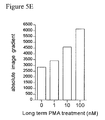
- Phorbol ester stimulation of PKC activity increased ET-1 induced ETAR endocytosis by >50% (Fig. 5b), with no internalization in the absence of stimulation.
- the phorbol ester stimulation of ETAR internalization was reduced to the negative control level when cells were simultaneously treated with PMA and RO 31 8220 (Fig. 5b). This indicated that the PKC isoform(s) sensitive to Ro 31 8220 were activated by phorbol ester and were required for receptor internalization.
- ETAR internalization was also inhibited by Gö 6976 (1 ⁇ M), a highly specific alpha and beta PKC isoform inhibitor 32 (Fig 5C), indicating the involvement of PKC a and b in the internalization of ETAR.
- the protein kinase A inhibitor H-89 caused accumulation of the ETAR in the recycling endosome after stimulation. This corresponded to a 1.25 fold stimulation in ETAR internalization compared to control (Fig 6a). Stimulation of cAMP production with 10 ⁇ M forskolin blocked the effect of H-89 (Fig. 6b). Treatment of cells with 10 ⁇ M KT5720, a similar PKA inhibitor, stimulated ETAR sequestration >1.5 fold and the GPCR accumulated in the recycling endosome (Fig 6b).
- the visual screens identified erbstatin A as acting on the ETAR endocytic trafficking pathway.
- a panel of tyrosine kinase inhibitors were screened (Fig. 7) and of these only erbstatin A arrested ETAR in endosomes but had no effect on housekeeping endocytosis.
- erbstatin blocked receptor internalization to the recycling endosome (Fig 7b).
- Erbstatin has one known target kinase- the EGF receptor tyrosine kinase- and it has been shown that the ETAR trans-activates the EGFR ( 34, 35 ).
- ETAR endocytosis was not affected by EGFR activation and this had no effect on the inhibition of ETAR transport by erbstatin A (not shown). It is likely that EGFR is not the erbstatin A sensitive kinase required for ETAR exit from the early endosome to the recycling endosome.
- GPCRs can couple to multiple G-proteins and switching between G-proteins is generally mediated by protein phosphorylation 36-38 .
- H-89 markedly increased cAMP over control (Fig. 8b).
- Gq coupling and Protein kinase C activation are a requirement for ETAR coupling to Gs, and in the presence of PKC inhibitors, agonist mediated ETAR activation does not lead to CAMP production. Restriction of ETAR to the recycling endosome with H-89 increased intracellular cAMP, indicating that internalization is required for cAMP production and recycling to the PM may terminate Gs coupling,
- Endothelin A receptor cellular antagonism modulates vasoconstriction
- GPCR related physiology can be modulated by compounds that function as 'cellular antagonists' of receptor function and are a novel approach to drug development for GPCRs.
- the present data demonstrates a drug discovery approach relying on the identification of compounds perturbing the subcellular pathway of G-protein coupled receptors.
- High content screening of a cell based assay of agonist induced receptor internalization readily identified compounds that arrested the endothelin A receptor at the cell surface or in endosomes.
- An important aspect of using cell based assays to select drugs that cause phenotypic disruption of receptor trafficking is to exclude effects on the pathways underlying receptor trafficking. Compounds were excluded that had effects on ETAR internalization and also disrupted endocytosis - as assessed by measured alterations in the uptake of inert fluid phase dextran. To further demonstrate the potential for introducing specificity at the level of the receptor affected by compounds, we screened compounds affecting ETAR but not endocytosis on the agonist induced internalization of the muscarinic acetylcholine receptor 1 and the kappa opioid receptor.
- Protein kinase C has been implicated in a variety of GPCR endocytosis mechanisms, but most significantly in the heterologous (receptor independent) desensitization of signaling for the 5HTA receptor.
- the sensitivity to Gö6976, which is highly specific for the alpha and beta isoforms 32 is consistent with the involvement of PKC-beta in ETAR internalization.
- the actual substrate for PKC phosphorylation is unclear, while there is evidence for direct receptor phosphorylation from in vivo labeling 42 and there are 7 consensus PKC phosphorylation sites in the cytoplasmic tail of the ETAR.
Landscapes
- Health & Medical Sciences (AREA)
- Life Sciences & Earth Sciences (AREA)
- Engineering & Computer Science (AREA)
- Immunology (AREA)
- Chemical & Material Sciences (AREA)
- Biomedical Technology (AREA)
- Molecular Biology (AREA)
- Hematology (AREA)
- Urology & Nephrology (AREA)
- Medicinal Chemistry (AREA)
- General Health & Medical Sciences (AREA)
- Cell Biology (AREA)
- Biotechnology (AREA)
- Biochemistry (AREA)
- Food Science & Technology (AREA)
- Physics & Mathematics (AREA)
- Analytical Chemistry (AREA)
- Microbiology (AREA)
- General Physics & Mathematics (AREA)
- Pathology (AREA)
- Toxicology (AREA)
- Bioinformatics & Cheminformatics (AREA)
- Tropical Medicine & Parasitology (AREA)
- Organic Chemistry (AREA)
- Veterinary Medicine (AREA)
- Public Health (AREA)
- Animal Behavior & Ethology (AREA)
- Pharmacology & Pharmacy (AREA)
- Zoology (AREA)
- Epidemiology (AREA)
- Genetics & Genomics (AREA)
- Virology (AREA)
- Developmental Biology & Embryology (AREA)
- Proteomics, Peptides & Aminoacids (AREA)
- Gastroenterology & Hepatology (AREA)
- Biophysics (AREA)
- Cardiology (AREA)
- Nuclear Medicine, Radiotherapy & Molecular Imaging (AREA)
- Heart & Thoracic Surgery (AREA)
- Chemical Kinetics & Catalysis (AREA)
Applications Claiming Priority (2)
| Application Number | Priority Date | Filing Date | Title |
|---|---|---|---|
| GB0611451A GB2439377A (en) | 2006-06-09 | 2006-06-09 | Methods of identifying modulators of and modulating GPCR cellular distribution and medical uses thereof |
| US81697106P | 2006-06-27 | 2006-06-27 |
Publications (2)
| Publication Number | Publication Date |
|---|---|
| EP1865320A2 true EP1865320A2 (de) | 2007-12-12 |
| EP1865320A3 EP1865320A3 (de) | 2008-02-20 |
Family
ID=36745616
Family Applications (1)
| Application Number | Title | Priority Date | Filing Date |
|---|---|---|---|
| EP07252322A Withdrawn EP1865320A3 (de) | 2006-06-09 | 2007-06-08 | Zellantagonist |
Country Status (3)
| Country | Link |
|---|---|
| US (1) | US20080138834A1 (de) |
| EP (1) | EP1865320A3 (de) |
| GB (1) | GB2439377A (de) |
Cited By (1)
| Publication number | Priority date | Publication date | Assignee | Title |
|---|---|---|---|---|
| WO2015074792A1 (en) * | 2013-11-25 | 2015-05-28 | Ontochem Gmbh | Method for diagnosing g-protein coupled receptor-related diseases |
Families Citing this family (1)
| Publication number | Priority date | Publication date | Assignee | Title |
|---|---|---|---|---|
| CN114038498B (zh) * | 2022-01-06 | 2022-03-18 | 北京晶泰科技有限公司 | Gpcr的热稳定性突变预测方法、结构筛选方法及其装置 |
Family Cites Families (8)
| Publication number | Priority date | Publication date | Assignee | Title |
|---|---|---|---|---|
| US6159700A (en) * | 1997-01-27 | 2000-12-12 | Smithkline Beecham Corporation | Method of finding agonist and antagonist to human and rat GPR14 |
| US7541151B2 (en) * | 1997-06-05 | 2009-06-02 | Duke University | Single-cell biosensor for the measurement of GPCR ligands in a test sample |
| US6639051B2 (en) * | 1997-10-20 | 2003-10-28 | Curis, Inc. | Regulation of epithelial tissue by hedgehog-like polypeptides, and formulations and uses related thereto |
| FR2788602B1 (fr) * | 1999-01-20 | 2002-06-07 | Inst Nat Sante Rech Med | Procede de criblage utile pour identifier des ligands potentiels pour un recepteur capable de s'internaliser |
| US6696478B2 (en) * | 1999-08-09 | 2004-02-24 | Zhimin Lu | Cancer treatment with Gö6976 and its related compounds |
| US20050136431A1 (en) * | 2000-11-03 | 2005-06-23 | Oakley Robert H. | Methods of screening compositions for G protein-coupled receptor desensitization inhibitory activity |
| US7018812B2 (en) * | 2000-11-03 | 2006-03-28 | Duke University | Modified G-protein coupled receptors |
| US6664266B2 (en) * | 2002-03-14 | 2003-12-16 | Children's Medical Center Corporation | Axon regeneration with PKC inhibitiors |
-
2006
- 2006-06-09 GB GB0611451A patent/GB2439377A/en not_active Withdrawn
-
2007
- 2007-06-07 US US11/810,884 patent/US20080138834A1/en not_active Abandoned
- 2007-06-08 EP EP07252322A patent/EP1865320A3/de not_active Withdrawn
Cited By (1)
| Publication number | Priority date | Publication date | Assignee | Title |
|---|---|---|---|---|
| WO2015074792A1 (en) * | 2013-11-25 | 2015-05-28 | Ontochem Gmbh | Method for diagnosing g-protein coupled receptor-related diseases |
Also Published As
| Publication number | Publication date |
|---|---|
| US20080138834A1 (en) | 2008-06-12 |
| GB0611451D0 (en) | 2006-07-19 |
| GB2439377A (en) | 2007-12-27 |
| EP1865320A3 (de) | 2008-02-20 |
Similar Documents
| Publication | Publication Date | Title |
|---|---|---|
| Drube et al. | GPCR kinase knockout cells reveal the impact of individual GRKs on arrestin binding and GPCR regulation | |
| Kara et al. | A phosphorylation cluster of five serine and threonine residues in the C-terminus of the follicle-stimulating hormone receptor is important for desensitization but not for β-arrestin-mediated ERK activation | |
| Iturrioz et al. | Identification and pharmacological properties of E339–3D6, the first nonpeptidic apelin receptor agonist | |
| Shenoy et al. | β-Arrestin-dependent, G protein-independent ERK1/2 activation by the β2 adrenergic receptor | |
| Vrecl et al. | Agonist-induced endocytosis and recycling of the gonadotropin-releasing hormone receptor: effect of β-arrestin on internalization kinetics | |
| Ferguson et al. | Molecular mechanisms of G protein-coupled receptor desensitization and resensitization | |
| Ge et al. | Dual modulation of formyl peptide receptor 2 by aspirin‐triggered lipoxin contributes to its anti‐inflammatory activity | |
| Milasta et al. | The sustainability of interactions between the orexin-1 receptor and β-arrestin-2 is defined by a single C-terminal cluster of hydroxy amino acids and modulates the kinetics of ERK MAPK regulation | |
| Wacker et al. | Disease-causing mutation in GPR54 reveals the importance of the second intracellular loop for class A G-protein-coupled receptor function | |
| US7897386B2 (en) | Cell-based assays for G-protein-coupled receptor-mediated activities | |
| Li et al. | Rab26 modulates the cell surface transport of α2-adrenergic receptors from the Golgi | |
| Kim et al. | Distinct regulation of internalization and mitogen-activated protein kinase activation by two isoforms of the dopamine D2 receptor | |
| Pula et al. | Agonist‐independent internalization of metabotropic glutamate receptor 1a is arrestin‐and clathrin‐dependent and is suppressed by receptor inverse agonists | |
| Milligan et al. | G protein-coupled receptor fusion proteins in drug discovery | |
| Echeverría et al. | The regulator of G protein signaling homologous domain of G protein-coupled receptor kinase 2 mediates short-term desensitization of β3-adrenergic receptor | |
| WO2008021552A9 (en) | Biased ligands and methods of identifying same | |
| US7604959B2 (en) | Cell-based assays employing voltage and calcium dyes | |
| Verkaar et al. | G protein-independent cell-based assays for drug discovery on seven-transmembrane receptors | |
| EP1865320A2 (de) | Zellantagonist | |
| Li et al. | Identification and characterization of distinct C-terminal domains of the human hydroxycarboxylic acid receptor-2 that are essential for receptor export, constitutive activity, desensitization, and internalization | |
| Eglen | An overview of high throughput screening at G protein coupled receptors | |
| Huang et al. | Identification of distinct c-terminal domains of the Bombyx adipokinetic hormone receptor that are essential for receptor export, phosphorylation and internalization | |
| Lecat et al. | Contribution of a tyrosine-based motif to cellular trafficking of wild-type and truncated NPY Y1 receptors | |
| Janezic | Elucidating the role of unique structural features of the α1D-adrenergic receptor: A tale of two tails | |
| EP1682577B1 (de) | Assays auf zellbasis unter anwendung von spannung und calciumfarbstoffen |
Legal Events
| Date | Code | Title | Description |
|---|---|---|---|
| PUAI | Public reference made under article 153(3) epc to a published international application that has entered the european phase |
Free format text: ORIGINAL CODE: 0009012 |
|
| AK | Designated contracting states |
Kind code of ref document: A2 Designated state(s): AT BE BG CH CY CZ DE DK EE ES FI FR GB GR HU IE IS IT LI LT LU LV MC MT NL PL PT RO SE SI SK TR |
|
| AX | Request for extension of the european patent |
Extension state: AL BA HR MK YU |
|
| RIN1 | Information on inventor provided before grant (corrected) |
Inventor name: KWON, YONGJUN Inventor name: ALTSCHULER, YORAM Inventor name: EMANS, NEIL |
|
| PUAL | Search report despatched |
Free format text: ORIGINAL CODE: 0009013 |
|
| AK | Designated contracting states |
Kind code of ref document: A3 Designated state(s): AT BE BG CH CY CZ DE DK EE ES FI FR GB GR HU IE IS IT LI LT LU LV MC MT NL PL PT RO SE SI SK TR |
|
| AX | Request for extension of the european patent |
Extension state: AL BA HR MK YU |
|
| AKX | Designation fees paid | ||
| STAA | Information on the status of an ep patent application or granted ep patent |
Free format text: STATUS: THE APPLICATION IS DEEMED TO BE WITHDRAWN |
|
| 18D | Application deemed to be withdrawn |
Effective date: 20080821 |
|
| REG | Reference to a national code |
Ref country code: DE Ref legal event code: 8566 |