EP1868005A2 - Imagerie terahertz - Google Patents
Imagerie terahertz Download PDFInfo
- Publication number
- EP1868005A2 EP1868005A2 EP07104990A EP07104990A EP1868005A2 EP 1868005 A2 EP1868005 A2 EP 1868005A2 EP 07104990 A EP07104990 A EP 07104990A EP 07104990 A EP07104990 A EP 07104990A EP 1868005 A2 EP1868005 A2 EP 1868005A2
- Authority
- EP
- European Patent Office
- Prior art keywords
- terahertz
- information
- examining
- body structure
- radiation
- Prior art date
- Legal status (The legal status is an assumption and is not a legal conclusion. Google has not performed a legal analysis and makes no representation as to the accuracy of the status listed.)
- Withdrawn
Links
Images

Classifications
-
- A—HUMAN NECESSITIES
- A61—MEDICAL OR VETERINARY SCIENCE; HYGIENE
- A61B—DIAGNOSIS; SURGERY; IDENTIFICATION
- A61B5/00—Measuring for diagnostic purposes; Identification of persons
- A61B5/05—Detecting, measuring or recording for diagnosis by means of electric currents or magnetic fields; Measuring using microwaves or radio waves
- A61B5/0507—Detecting, measuring or recording for diagnosis by means of electric currents or magnetic fields; Measuring using microwaves or radio waves using microwaves or terahertz waves
-
- G—PHYSICS
- G01—MEASURING; TESTING
- G01V—GEOPHYSICS; GRAVITATIONAL MEASUREMENTS; DETECTING MASSES OR OBJECTS; TAGS
- G01V8/00—Prospecting or detecting by optical means
- G01V8/005—Prospecting or detecting by optical means operating with millimetre waves, e.g. measuring the black losey radiation
-
- A—HUMAN NECESSITIES
- A61—MEDICAL OR VETERINARY SCIENCE; HYGIENE
- A61B—DIAGNOSIS; SURGERY; IDENTIFICATION
- A61B5/00—Measuring for diagnostic purposes; Identification of persons
- A61B5/0059—Measuring for diagnostic purposes; Identification of persons using light, e.g. diagnosis by transillumination, diascopy, fluorescence
- A61B5/0082—Measuring for diagnostic purposes; Identification of persons using light, e.g. diagnosis by transillumination, diascopy, fluorescence adapted for particular medical purposes
- A61B5/0088—Measuring for diagnostic purposes; Identification of persons using light, e.g. diagnosis by transillumination, diascopy, fluorescence adapted for particular medical purposes for oral or dental tissue
Definitions
- the present invention relates to a method and a device for examining a body, in particular an imaging method or an imaging device used in the medical field, the body being examined by means of radiation in the terahertz frequency range in order to obtain information about the body, in particular about its shape, To get kind or position.
- Known medical imaging systems can be deleterious to health, such as x-ray systems, being inaccurate, such as ultrasound systems, can not satisfactorily render soft tissues, such as computed tomography systems, or can not render bones sufficiently well, such as magnetic resonance systems.
- the radiation emitted or reflected by the body or body part or the body structure is detected in the terahertz frequency range with a sensor or a detector, wherein under the terahertz frequency range used Radiation with a frequency between 0.1 and 30 THz is to be understood.
- the radiation emitted by the body in the terahertz frequency range can be understood to mean both a self-emission of the body in the terahertz frequency range and a transmission or transmission of the body with radiation in the terahertz frequency range.
- the emitted or reflected radiation of the body or body part or the body structure can be detected with a terahertz sensor and evaluated or processed by a computing unit to obtain information about the body or the body structure.
- the radiation detected by the terArtertz sensor may be evaluated such that information about the nature, nature, composition, material, shape, structure, state, temperature, or position of the surface or interior of the body or body part or body structure.
- a registration process in particular an automatic registration process, of the body or the body part or the body structure can be performed by, for example, locating landmark points of the body and assigning the landmarks to room positions, or taking pictures with a camera Stirred through various known positions and the recordings are further processed until a registration of the body or the body part or the body structure is done.
- the information about the body determined by the Teraheriz radiation can also be combined with other information.
- multiple exposures of the body or body part may be performed by means of terahertz radiation, where the terahertz radiation used may have the same frequency or the same frequency range, or a different frequency or a different frequency range, such as the same body or body structures can be taken from different positions and combined with each other, or different parts of a body, such as the surface or the interior of a body, can be recorded and combined by means of different frequencies or frequency ranges.
- the radiation can not or only slightly penetrate into the surface of the body and is eg reflected on the surface or near the surface or can penetrate deep into the body or penetrate the body or, for example, also penetrate clothing.
- the information about the body or the body structure or the body part ascertained by means of terahertz radiation can also be combined by means of an image fusion process with information about the same body or the same body part or the same body part, for example with a further imaging Methods such as an X-ray method, a magnetic resonance method, a computed tomography method, an ultrasound method, a positron emission tomography method (PET method) or a single photon emission computed tomography method (SPECT method) are determined.
- PET method positron emission tomography method
- SPECT method single photon emission computed tomography method
- the information about the body determined from the detected terahertz radiation is combined with two- or three-dimensional information about the body, which was determined for example by means of terahertz radiation or another imaging method.
- the terahertz sensor may detect, for example, radiation emitted by the body part or body structure in the terahertz frequency peak.
- the examined body can also be irradiated or irradiated with terahertz radiation, for example, such that the radiation reflected on the body, for example the radiation reflected on the surface of the body or near the surface of the body, is detected by a terahertz sensor, in particular a terahertz sensor.
- Sensor field which is preferably arranged around the body or the body part is detected.
- Terahertz radiation can also be detected with the terahertz sensor, which is transmitted or transmitted through the body, wherein the sensor is preferably arranged opposite the radiation source.
- a different amount of terahertz radiation is transmitted through the body, or of reflects or emits the body, or the terahertz radiation is attenuated or absorbed to different degrees so that information about the surface or interior of the body can be obtained from the detected terahertz radiation.
- an instrument such as a microscope or an endoscope, on which, for example, active or passive markers are arranged, can be navigated.
- characteristics about the body or body part such as the nature, shape, structure, condition or position of the body or body
- Surface of the body can be determined by the body from a preferably known distance is irradiated with terahertz radiation and the radiation reflected at the body or the surface of the body with a sensor detected or detected, wherein the running time of the terahertz radiation or the Terahertz signal can be closed for example on the shape or the position or the distance of the body or the surface
- the terahertz radiation detected by the sensor or the terahertz signal can be evaluated such that the spectrum or a frequency-domain representation of the radiation or the signal is determined, for example by means of a Fourier transformation, and from the spectral properties on properties of the body or of the body Part of the body which has emitted or reflected or transmitted the radiation is closed.
- the determined spectral representation of the detected terahertz radiation can be compared, for example, with spectra of known materials or shapes or states or temperatures, it being possible to determine from the comparison the properties of the body or body part examined,
- characteristic frequencies of the detected terahertz signal such as frequencies of maximum or minimum absorption, reflection or transmission with characteristic frequencies, such as resonance frequencies or maximum or minimum absorption, transmission or reflection frequencies of known bodies or body structures, for example, previously determined or stored and comparing the frequencies to determine the nature, type, composition, material, shape, structure, state, temperature or position of the body or the surface or interior of the body.
- a body or body parts such as a head, a face, an arm, a jaw, a dentition or a hand can be examined and body structures, such as tissue, bones or structures, vessels, ligaments, tendons, teeth or skin of a patient.
- body structures such as tissue, bones or structures, vessels, ligaments, tendons, teeth or skin of a patient.
- body structures such as tissue, bones or structures, vessels, ligaments, tendons, teeth or skin of a patient.
- tumors on the exposed brain can be detected, for example, by detecting terahertz radiation emitted by the brain during a surgery using a terahertz spectral sensor to obtain detailed information about the nature of the tissue.
- terahertz radiation can be used to detect, for example, the thickness of the enamel, the internal condition of the teeth or the shape of the teeth, or caries or paradontosis can be detected.
- Terahertz radiation in a frequency range between 0.1 and 5 THz is preferably used to examine the body or the body structure, wherein the spectral ranges between 0.1 and 0.6 THz and between 0.5 and 2 THz are preferably used.
- terahertz radiation in a range around 1.6 THz or in a range around 2.5 THz or in a wide broadband range around 3 THz can also be used.
- the invention further relates to a computer program which, when loaded into a computer or running on a computer, performs a method as described above. Furthermore, the invention relates to a program storage medium or a computer program product with such a program.
- the device according to the invention for the examination of a body comprises a terahertz sensor or a terahertz detector, in particular a terahertz camera, which has a thermal radiation reflected by a body or a body structure or transmitted through a body or a body structure or emitted by a body or a body structure. Can detect radiation.
- the device further comprises a computing unit, such as a tracking system, which is wired or wirelessly connected to the terahertz sensor, whereby the detected terahertz radiation or the acquired data can be transmitted to the arithmetic unit, in which the detected terahertz radiation evaluated or further processed.
- the detected terahertz radiation can be evaluated in order to obtain information about the body or the body structure.
- the terahertz signal detected by means of the terahertz sensor can be analyzed, such as transformed into the frequency domain by means of a Fourier transmission so that, for example, from the spectrum of the detected terahertz signal characteristic frequencies, such as absorption or resonance frequencies, are determined can be.
- the device according to the invention can also comprise a terahertz radiation source, in particular terahertz lamps, which can emit terahertz radiation in order to irradiate or irradiate the body or the body structure with terahertz radiation, the terahertz radiation source having the terahertz sensor or can be connected to the arithmetic unit.
- the radiation emitted by the terahertz radiation source can be reflected, for example, on the body structure to be examined or the body part to be examined, or can be transmitted through the body or the body structure, so that the reflected or transmitted radiation can be detected by the terahertz sensor.
- the terahertz radiation source may be arranged at a known location, or may be provided with markers, so that the position of the terahertz radiation source can be determined, for example, by a tracking system.
- the terahertz radiation source is preferably opposite to the terahertz sensor, so that emitted by the terahertz radiation source Radiation, the body or the body structure at least partially penetrates and can be detected on the opposite side of the terahertz sensor.
- the terahertz radiation source may, for example, be located at a known or detectable location, wherein the terahertz sensor, for example as a sensor array, may be disposed about the body or body structure, to those on the body detect at least partially reflected terahertz radiation.
- the terahertz sensor for example as a sensor array
- the apparatus of the present invention may also comprise a database which is preferably connected to a computing unit and in which characteristic information of a plurality of bodies or body structures may be stored.
- characteristic frequencies such as absorption or resonance frequencies, of certain body parts or body structures, such as tissue, bones, vessels, ligaments, tendons, teeth or skin may be stored in the database, or it may be characteristic spectral fingerprints of a body or a body structure Be stored in a plurality of body parts or body structures.
- the arithmetic unit can compare the information about the body or the body structure obtained by means of the detected terahertz radiation with the information stored in the database, in particular a comparison of the characteristic frequencies or the spectrum or frequency range, and conclusions can be drawn from the comparison made Texture, type, composition, material, shape, structure, condition, temperature, or position of the surface or interior of the body.
- characteristic frequencies of the detected terahertz signal or the spectrum of the detected terahertz signal are compared with the stored information and infered, for example, from the similarity of characteristic frequencies or spectra that a particular material or structure or temperature of the body is present.
- the device according to the invention can also have a data output device, in particular a screen, which can display or fused or registered the ascertained information about the body as numerical values or as an image Can represent body structures.
- a eingabevoiraum be connected to the arithmetic unit or the database, in particular a keyboard or a scanner such as an X-ray device, a computed tomography, a nuclear magnetic resonance tomograph, an ultrasound tomograph, a positron emission tomograph or a single photon emission tomography tomograph.
- information can be entered into the arithmetic unit or into the database, so that the information can be stored in the database or can be compared or processed in the arithmetic unit with, for example, the information obtained using terahertz radiation.
- the terahertz radiation source or the terahertz sensor comprises an electronic or optical terahertz oscillator, such as a titanium: sapphire laser.
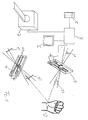
- FIG. 1 shows an embodiment of the present invention, wherein a hand 2 is irradiated or irradiated as body 2 or body part by means of a tera-ray radiation source 4.
- the terahertz radiation source 4 preferably comprises a mode-locked titanium: sapphire laser, which can emit pulses 20 of only a few tens of long-duration. With these optical pulses 20 can be switched photoconductive dipole antennas 40, which preferably consist of gallium arsenide, on which two metal strip lines have been vapor-deposited.
- the short laser pulses 20 generate charge carriers between the leads which are accelerated by an electric field applied to the dipole antennas 40, resulting in a short current pulse which generates a terahertz pulse 30 radiated into the space.
- the terahertz pulse hits in the present embodiment of the invention on a hand 2 and is at the Surface or reflected from structures near the surface, wherein the reflected terahertz pulse 32 is detected by a terahertz sensor 1 and terahertz detector.
- the terahertz sensor 1 may, as in the present embodiment, have a similar structure to the terahertz radiation source 4, wherein no external field needs to be applied, or may be configured differently, such as a purely electronic sensor.
- a switching laser pulse 21 generates in the terahertz sensor 1 free charge carriers, which move in the electric field of the incoming or detected by the hand 2 reflected terahertz wave 32, whereby there is a small current flow, which can be amplified and registered.
- the generated current flow can be transmitted to a computing unit 3, such as a computer, where it can be further processed or evaluated.
- a computing unit 3 such as a computer
- the temporal progression of the detected terahertz radiation 32 can be determined from the generated current and, for example, the spectrum or the frequency domain representation of the detected terahertz pulse 32 in the arithmetic unit 3 can be determined by means of a Fourier transfusion.
- the determined information may preferably be output on the data output device 6, such as a screen, and compared with time and frequency domain representations of known bodies or body structures stored in the database 5.
- the detected terahertz pulse 32 may include information about the skin or the surface or tissue of the hand 2 and may be compared by the computing unit 3 with known tissue information stored in the database 5.
- a computed tomograph 7 information about the body 2, in particular the hand 2, can be acquired, transmitted to the arithmetic unit 3 and stored in the database 5 as reference data for comparison with the acquired information.
- the method of determining the information about the body part by means of reflection shown in FIG. 1 it is also possible to carry out an examination by transmission by means of the radiation source 4 and the terahertz sensor 1 or by emission by means of the terahertz sensor 1.
- another input device such as a keyboard, an X-ray device such as a C-arm, an ultrasound tomograph, a magnetic resonance tomograph, a positron emission tomograph or a SPECT tomograph can be used for information on a Body part or body 2 to gain and the information in the arithmetic unit 3 to evaluate or store in the database 5 as reference information.
- FIG. 2 shows a detected terahertz pulse in the time domain 10 and in the frequency domain 11.
- the frequency domain representation 11 of the detected terahertz signal can be compared by an arithmetic unit with terahertz signals of known body parts or body structures, such as skin or tissue, to draw conclusions about this examined body part or the examined body structure, such as the examined tissue to draw.
- the entire frequency domain representation 11 can be compared with stored frequency domain representations, and by finding the most similar frequency response, one can infer the nature of the tissue being examined, e.g. healthy from diseased tissue, such as skin cancer, can be differentiated.
- characteristic frequencies such as the frequencies of maximum absorption or minimum reflection which are to be recognized in frequency curve 11
- frequencies of different body parts or body structures or tissue types stored in the database. From the greatest similarity of the frequencies of maximum absorption or minimum reflection, e.g. on properties such as the nature or composition or condition of the examined skin or the examined tissue are closed, which in particular healthy and diseased tissue can be distinguished and recognized.
Landscapes
- Health & Medical Sciences (AREA)
- Life Sciences & Earth Sciences (AREA)
- Physics & Mathematics (AREA)
- Medical Informatics (AREA)
- Surgery (AREA)
- Biophysics (AREA)
- Pathology (AREA)
- Engineering & Computer Science (AREA)
- Biomedical Technology (AREA)
- Heart & Thoracic Surgery (AREA)
- Nuclear Medicine, Radiotherapy & Molecular Imaging (AREA)
- Molecular Biology (AREA)
- Radiology & Medical Imaging (AREA)
- Animal Behavior & Ethology (AREA)
- General Health & Medical Sciences (AREA)
- Public Health (AREA)
- Veterinary Medicine (AREA)
- General Life Sciences & Earth Sciences (AREA)
- General Physics & Mathematics (AREA)
- Geophysics (AREA)
- Investigating Or Analysing Materials By Optical Means (AREA)
Priority Applications (1)
| Application Number | Priority Date | Filing Date | Title |
|---|---|---|---|
| EP07104990A EP1868005A3 (fr) | 2006-05-24 | 2007-03-27 | Imagerie terahertz |
Applications Claiming Priority (2)
| Application Number | Priority Date | Filing Date | Title |
|---|---|---|---|
| EP06010634 | 2006-05-24 | ||
| EP07104990A EP1868005A3 (fr) | 2006-05-24 | 2007-03-27 | Imagerie terahertz |
Publications (2)
| Publication Number | Publication Date |
|---|---|
| EP1868005A2 true EP1868005A2 (fr) | 2007-12-19 |
| EP1868005A3 EP1868005A3 (fr) | 2011-08-17 |
Family
ID=38687441
Family Applications (1)
| Application Number | Title | Priority Date | Filing Date |
|---|---|---|---|
| EP07104990A Withdrawn EP1868005A3 (fr) | 2006-05-24 | 2007-03-27 | Imagerie terahertz |
Country Status (1)
| Country | Link |
|---|---|
| EP (1) | EP1868005A3 (fr) |
Cited By (10)
| Publication number | Priority date | Publication date | Assignee | Title |
|---|---|---|---|---|
| WO2008030427A3 (fr) * | 2006-09-06 | 2008-07-03 | Intellectual Ventures Holding | Spectroscopie passive de substances in vivo |
| US7750299B2 (en) | 2006-09-06 | 2010-07-06 | Donald Martin Monro | Active biometric spectroscopy |
| US7786907B2 (en) | 2008-10-06 | 2010-08-31 | Donald Martin Monro | Combinatorial coding/decoding with specified occurrences for electrical computers and digital data processing systems |
| US7786903B2 (en) | 2008-10-06 | 2010-08-31 | Donald Martin Monro | Combinatorial coding/decoding with specified occurrences for electrical computers and digital data processing systems |
| US7791513B2 (en) | 2008-10-06 | 2010-09-07 | Donald Martin Monro | Adaptive combinatorial coding/decoding with specified occurrences for electrical computers and digital data processing systems |
| US7864086B2 (en) | 2008-10-06 | 2011-01-04 | Donald Martin Monro | Mode switched adaptive combinatorial coding/decoding for electrical computers and digital data processing systems |
| WO2011107575A1 (fr) * | 2010-03-04 | 2011-09-09 | Siemens Aktiengesellschaft | Dispositif médical d'examen et/ou de traitement |
| WO2012140587A3 (fr) * | 2011-04-15 | 2013-06-27 | Ariel-University Research And Development Company, Ltd. | Détecteur passif d'ondes millimétriques |
| WO2013137031A1 (fr) * | 2012-03-14 | 2013-09-19 | Canon Kabushiki Kaisha | Appareil et procédé de calcul d'un emplacement d'un tissu anormal dans un objet et procédé de formation d'une image d'un objet, en utilisant une onde électromagnétique dans une bande térahertz |
| DE102012209422A1 (de) * | 2012-06-04 | 2013-12-05 | Siemens Aktiengesellschaft | Röntgeneinrichtung mit Terahertz-Messeinrichtung und Verfahren zur Steuerung des Rekonstruktions- und/oder Auswertebetriebs einer Röntgeneinrichtung |
Family Cites Families (5)
| Publication number | Priority date | Publication date | Assignee | Title |
|---|---|---|---|---|
| US5789750A (en) * | 1996-09-09 | 1998-08-04 | Lucent Technologies Inc. | Optical system employing terahertz radiation |
| US6957099B1 (en) * | 1999-02-23 | 2005-10-18 | Teraview Limited | Method and apparatus for terahertz imaging |
| JP2005261826A (ja) * | 2004-03-22 | 2005-09-29 | Pentax Corp | 内視鏡システム |
| US20050231416A1 (en) * | 2004-04-14 | 2005-10-20 | Rowe Richard L | Relational millimeter-wave interrogating |
| US7386150B2 (en) * | 2004-11-12 | 2008-06-10 | Safeview, Inc. | Active subject imaging with body identification |
-
2007
- 2007-03-27 EP EP07104990A patent/EP1868005A3/fr not_active Withdrawn
Cited By (12)
| Publication number | Priority date | Publication date | Assignee | Title |
|---|---|---|---|---|
| WO2008030427A3 (fr) * | 2006-09-06 | 2008-07-03 | Intellectual Ventures Holding | Spectroscopie passive de substances in vivo |
| US7750299B2 (en) | 2006-09-06 | 2010-07-06 | Donald Martin Monro | Active biometric spectroscopy |
| US7786907B2 (en) | 2008-10-06 | 2010-08-31 | Donald Martin Monro | Combinatorial coding/decoding with specified occurrences for electrical computers and digital data processing systems |
| US7786903B2 (en) | 2008-10-06 | 2010-08-31 | Donald Martin Monro | Combinatorial coding/decoding with specified occurrences for electrical computers and digital data processing systems |
| US7791513B2 (en) | 2008-10-06 | 2010-09-07 | Donald Martin Monro | Adaptive combinatorial coding/decoding with specified occurrences for electrical computers and digital data processing systems |
| US7864086B2 (en) | 2008-10-06 | 2011-01-04 | Donald Martin Monro | Mode switched adaptive combinatorial coding/decoding for electrical computers and digital data processing systems |
| WO2011107575A1 (fr) * | 2010-03-04 | 2011-09-09 | Siemens Aktiengesellschaft | Dispositif médical d'examen et/ou de traitement |
| WO2012140587A3 (fr) * | 2011-04-15 | 2013-06-27 | Ariel-University Research And Development Company, Ltd. | Détecteur passif d'ondes millimétriques |
| US9207317B2 (en) | 2011-04-15 | 2015-12-08 | Ariel-University Research And Development Company Ltd. | Passive millimeter-wave detector |
| WO2013137031A1 (fr) * | 2012-03-14 | 2013-09-19 | Canon Kabushiki Kaisha | Appareil et procédé de calcul d'un emplacement d'un tissu anormal dans un objet et procédé de formation d'une image d'un objet, en utilisant une onde électromagnétique dans une bande térahertz |
| DE102012209422A1 (de) * | 2012-06-04 | 2013-12-05 | Siemens Aktiengesellschaft | Röntgeneinrichtung mit Terahertz-Messeinrichtung und Verfahren zur Steuerung des Rekonstruktions- und/oder Auswertebetriebs einer Röntgeneinrichtung |
| DE102012209422B4 (de) | 2012-06-04 | 2019-04-25 | Siemens Healthcare Gmbh | Röntgeneinrichtung mit Terahertz-Messeinrichtung und Verfahren dazu |
Also Published As
| Publication number | Publication date |
|---|---|
| EP1868005A3 (fr) | 2011-08-17 |
Similar Documents
| Publication | Publication Date | Title |
|---|---|---|
| EP1868005A2 (fr) | Imagerie terahertz | |
| DE69329886T2 (de) | Verfahren zur Bestimmung der Lage eines Organs | |
| Webb et al. | Measurements of the relationship between CT Hounsfield units and acoustic velocity and how it changes with photon energy and reconstruction method | |
| DE69431741T2 (de) | Vorrichtung zur medizinischen Behandlung mit Ultraschall | |
| EP0234198B1 (fr) | Procédé et appareil pour déterminer sans contact la distribution de la température dans un objet à examiner | |
| DE69332531T2 (de) | Gerät zur medizinischen Ultraschall-Behandlung unter Verwendung von Computer-Tomographie | |
| DE102005061557B3 (de) | Bildgebungsgerät sowie Verfahren zum Betrieb eines Bildgebungsgerätes | |
| US20080319321A1 (en) | Terahertz imaging | |
| EP2230641B1 (fr) | Procédé de détermination de la position d'une structure dans un corps | |
| DE102008006711A1 (de) | Medizinische Diagnose- oder Therapieeinheit und Verfahren zur Verbesserung von Untersuchungs- bzw. Behandlungsabläufen mit einer medizinischen Diagnose- oder Therapieeinheit | |
| DE102010010192A1 (de) | Medizinische Untersuchungs- und/oder Behandlungsvorrichtung | |
| DE19915689A1 (de) | Vorrichtung und Verfahren zum Lokalisieren elektrisch aktiver Orte innerhalb eines Lebewesens | |
| DE102008032996B4 (de) | Verfahren zur Bestimmung einer Schwächungskarte | |
| CN110074864B (zh) | 颅脑血肿引流的规划系统及方法 | |
| DE102014217283B4 (de) | Überwachung einer Strahlentherapie eines Patienten mittels einer MR-Fingerprinting-Methode | |
| CN107865658A (zh) | 用于修正合成电子密度图的方法和设备 | |
| DE102006026490A1 (de) | Radiotherapievorrichtung | |
| DE102009049519A1 (de) | Computertomograph mit Abstandssensor und Verfahren zur Abstandsmessung in einem Computertomographen | |
| EP3441936A1 (fr) | Procédé d'évaluation de données d'image d'un patient soumis à une intervention chirurgicale peu invasive, dispositif d'évaluation, programme informatique et support de données lisible par voie électronique | |
| Coila et al. | A regularization approach for ultrasonic attenuation imaging | |
| Rohrbach et al. | The early phases of bone healing can be differentiated in a rat osteotomy model by focused transverse-transmission ultrasound | |
| CA2865830A1 (fr) | Procede de reconstruction optique par deconvolution cinetique | |
| DE102008041941A1 (de) | Stabilisierung von bildgebenden Verfahren in der medizinischen Diagnostik | |
| DE102011076882B4 (de) | Verfahren zur Steuerung eines medizinischen Gerätes, Einrichtung mit einem medizinischen Gerät und Datenträger | |
| Kumabe et al. | Ultrasound frequency-based monitoring for bone healing |
Legal Events
| Date | Code | Title | Description |
|---|---|---|---|
| PUAI | Public reference made under article 153(3) epc to a published international application that has entered the european phase |
Free format text: ORIGINAL CODE: 0009012 |
|
| 17P | Request for examination filed |
Effective date: 20070327 |
|
| AK | Designated contracting states |
Kind code of ref document: A2 Designated state(s): AT BE BG CH CY CZ DE DK EE ES FI FR GB GR HU IE IS IT LI LT LU LV MC MT NL PL PT RO SE SI SK TR |
|
| AX | Request for extension of the european patent |
Extension state: AL BA HR MK YU |
|
| PUAL | Search report despatched |
Free format text: ORIGINAL CODE: 0009013 |
|
| AK | Designated contracting states |
Kind code of ref document: A3 Designated state(s): AT BE BG CH CY CZ DE DK EE ES FI FR GB GR HU IE IS IT LI LT LU LV MC MT NL PL PT RO SE SI SK TR |
|
| AX | Request for extension of the european patent |
Extension state: AL BA HR MK RS |
|
| RIC1 | Information provided on ipc code assigned before grant |
Ipc: A61B 5/05 20060101ALI20110711BHEP Ipc: G01V 8/00 20060101AFI20110711BHEP |
|
| AKX | Designation fees paid |
Designated state(s): DE FR GB IT |
|
| RAP1 | Party data changed (applicant data changed or rights of an application transferred) |
Owner name: BRAINLAB AG |
|
| 17Q | First examination report despatched |
Effective date: 20141010 |
|
| STAA | Information on the status of an ep patent application or granted ep patent |
Free format text: STATUS: THE APPLICATION IS DEEMED TO BE WITHDRAWN |
|
| RAP1 | Party data changed (applicant data changed or rights of an application transferred) |
Owner name: BRAINLAB AG |
|
| 18D | Application deemed to be withdrawn |
Effective date: 20161001 |