EP1875855A1 - Bildbearbeitungsvorrichtung, bildbearbeitungsverfahren und bildbearbeitungsprogramm - Google Patents
Bildbearbeitungsvorrichtung, bildbearbeitungsverfahren und bildbearbeitungsprogramm Download PDFInfo
- Publication number
- EP1875855A1 EP1875855A1 EP06729058A EP06729058A EP1875855A1 EP 1875855 A1 EP1875855 A1 EP 1875855A1 EP 06729058 A EP06729058 A EP 06729058A EP 06729058 A EP06729058 A EP 06729058A EP 1875855 A1 EP1875855 A1 EP 1875855A1
- Authority
- EP
- European Patent Office
- Prior art keywords
- area
- hemorrhage
- areas
- candidate
- edge
- Prior art date
- Legal status (The legal status is an assumption and is not a legal conclusion. Google has not performed a legal analysis and makes no representation as to the accuracy of the status listed.)
- Granted
Links
Images


Classifications
-
- A—HUMAN NECESSITIES
- A61—MEDICAL OR VETERINARY SCIENCE; HYGIENE
- A61B—DIAGNOSIS; SURGERY; IDENTIFICATION
- A61B1/00—Instruments for performing medical examinations of the interior of cavities or tubes of the body by visual or photographical inspection, e.g. endoscopes; Illuminating arrangements therefor
-
- G—PHYSICS
- G06—COMPUTING OR CALCULATING; COUNTING
- G06T—IMAGE DATA PROCESSING OR GENERATION, IN GENERAL
- G06T7/00—Image analysis
- G06T7/0002—Inspection of images, e.g. flaw detection
- G06T7/0012—Biomedical image inspection
-
- A—HUMAN NECESSITIES
- A61—MEDICAL OR VETERINARY SCIENCE; HYGIENE
- A61B—DIAGNOSIS; SURGERY; IDENTIFICATION
- A61B5/00—Measuring for diagnostic purposes; Identification of persons
-
- A—HUMAN NECESSITIES
- A61—MEDICAL OR VETERINARY SCIENCE; HYGIENE
- A61B—DIAGNOSIS; SURGERY; IDENTIFICATION
- A61B5/00—Measuring for diagnostic purposes; Identification of persons
- A61B5/07—Endoradiosondes
-
- G—PHYSICS
- G06—COMPUTING OR CALCULATING; COUNTING
- G06T—IMAGE DATA PROCESSING OR GENERATION, IN GENERAL
- G06T1/00—General purpose image data processing
-
- G—PHYSICS
- G06—COMPUTING OR CALCULATING; COUNTING
- G06T—IMAGE DATA PROCESSING OR GENERATION, IN GENERAL
- G06T7/00—Image analysis
- G06T7/10—Segmentation; Edge detection
- G06T7/12—Edge-based segmentation
-
- G—PHYSICS
- G06—COMPUTING OR CALCULATING; COUNTING
- G06T—IMAGE DATA PROCESSING OR GENERATION, IN GENERAL
- G06T7/00—Image analysis
- G06T7/40—Analysis of texture
- G06T7/41—Analysis of texture based on statistical description of texture
- G06T7/44—Analysis of texture based on statistical description of texture using image operators, e.g. filters, edge density metrics or local histograms
-
- G—PHYSICS
- G06—COMPUTING OR CALCULATING; COUNTING
- G06V—IMAGE OR VIDEO RECOGNITION OR UNDERSTANDING
- G06V10/00—Arrangements for image or video recognition or understanding
- G06V10/40—Extraction of image or video features
- G06V10/44—Local feature extraction by analysis of parts of the pattern, e.g. by detecting edges, contours, loops, corners, strokes or intersections; Connectivity analysis, e.g. of connected components
- G06V10/457—Local feature extraction by analysis of parts of the pattern, e.g. by detecting edges, contours, loops, corners, strokes or intersections; Connectivity analysis, e.g. of connected components by analysing connectivity, e.g. edge linking, connected component analysis or slices
-
- G—PHYSICS
- G06—COMPUTING OR CALCULATING; COUNTING
- G06T—IMAGE DATA PROCESSING OR GENERATION, IN GENERAL
- G06T2207/00—Indexing scheme for image analysis or image enhancement
- G06T2207/10—Image acquisition modality
- G06T2207/10068—Endoscopic image
-
- G—PHYSICS
- G06—COMPUTING OR CALCULATING; COUNTING
- G06T—IMAGE DATA PROCESSING OR GENERATION, IN GENERAL
- G06T2207/00—Indexing scheme for image analysis or image enhancement
- G06T2207/20—Special algorithmic details
- G06T2207/20021—Dividing image into blocks, subimages or windows
-
- G—PHYSICS
- G06—COMPUTING OR CALCULATING; COUNTING
- G06T—IMAGE DATA PROCESSING OR GENERATION, IN GENERAL
- G06T2207/00—Indexing scheme for image analysis or image enhancement
- G06T2207/30—Subject of image; Context of image processing
- G06T2207/30004—Biomedical image processing
- G06T2207/30028—Colon; Small intestine
-
- G—PHYSICS
- G06—COMPUTING OR CALCULATING; COUNTING
- G06V—IMAGE OR VIDEO RECOGNITION OR UNDERSTANDING
- G06V2201/00—Indexing scheme relating to image or video recognition or understanding
- G06V2201/03—Recognition of patterns in medical or anatomical images
Definitions
- the present invention aims to provide an image processing apparatus, an image processing method, and an image processing program capable of detecting a hemorrhage area by using an amount of change in an image signal or a color signal in an outline part of a bloody area.
- a bleeding part area where bleeding from mucous membrane actually occurs, a reddened part area where the surface of mucous membrane is reddened by hyperemia, and the like are given.
- the case of detecting, for example, the outline part of a bleeding part area (hereinafter referred to as a bleeding part edge) will be described.
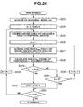
- FIG. 6 is a flowchart illustrating the procedure for the shape edge extraction processing at step S20 in FIG. 3.
- a divided area is a bleeding part edge is determined with the use of a color edge feature quantity B i calculated based on change of two or more color signals among the R signal, the G signal and the B signal, it is possible to relatively determine and detect whether or not the divided area is a bleeding part edge. Furthermore, in this embodiment, since it is possible to extract bleeding part edges with various sizes, such as a bleeding part with a large area and a bleeding part where an edge is extracted being divided, the precision of detecting a bleeding part edge is further enhanced.
- the entire configuration of the image processing apparatus 1 is the same as that in the third embodiment except that the content of processing performed by an image processing program 71 is different. Therefore, only characteristic operations will be described here. The same components will be given the same reference numerals, and description thereof will be omitted.
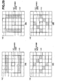
- the CPU 21 sets the direction in which the R change is the largest among the first to eighth R changes calculated at step S304, as a candidate area feature quantity D i' . For example, if the fourth R change is the largest, then the candidate area feature quantity D i' is the diagonally lower-right direction as shown in FIG. 7(d).
- the image processing apparatus 1 of this embodiment it is possible to relatively determine and detect whether or not a bleeding part edge candidate area is a bleeding part edge by using an edge feature quantity which is the amount of change in color signals in a bleeding part edge.
- the object of this embodiment is to provide an image processing apparatus, an image processing method and an image processing program capable of relatively determining whether or not an area is a hemorrhage edge with the use of the amount of change in color signals on the outline part of a hemorrhage area (hereinafter referred to as a hemorrhage edge) and detecting a hemorrhage area surrounded by the hemorrhage edge.
- the CPU 21 By the image processing program 128 which is stored in the memory 22 being executed, the CPU 21 operates.
- the CPU 21 performs image analysis processing for image data acquired from the endoscope filing apparatus 4. Analysis data 29 acquired or generated by each processing by the CPU 21 is stored in the memory 22.
- This analysis data 29 includes an original image 31 which is the image data acquired from the endoscope filing apparatus 4. Furthermore, the analysis data 29 includes an edge image 132 generated by various processings to be described later, a bleeding part candidate image 133 and a bleeding part image 134.
- the CPU 21 executes the processings at steps S421 to S423 as evaluation area setting means, and sets peripheral areas 143 as evaluation target areas, for the i-th divided area 141 as a hemorrhage evaluation area.
- the CPU 21 determines whether or not the 1-th peripheral area 143 is a bleeding part edge. Specifically, if the color edge feature quantity B1 1 >Thr4 is satisfied, then the CPU 21 determines that the 1-th peripheral area 143 is a bleeding part edge.
- the bleeding part edge determination at step S447 is tentative determination. The result of the determination at this step is not adopted until it is determined by the processing to be described later that the k-th divided area 141 is a bleeding part area.
- the entire configuration of the image processing apparatus 1 is the same as that of the fifth embodiment except that the content of the processing performed by an image processing program 81 is different from that of the image processing program 128 in the fifth embodiment. Therefore, only characteristic operations will be described here.
- the same components will be given the same reference numerals, and description thereof will be omitted.
- the case of detecting, for example, a bleeding part will be described, similarly to the fifth embodiment.
- step S524 the CPU 21 initializes m which indicates the number identifying the peripheral area 143 to be analyzed and a counter Cnt3 for counting the number of bleeding part edge candidate areas in the peripheral areas 143, to 1 and 0, respectively.
- n indicates the number identifying the divided areas 141 in the m-th peripheral area 143, to 1.
- the number n identifying the divided area 141 takes an integer value equal to or above 1 and equal to or below Z.
- the CPU 21 determines whether or not the m-th peripheral area 143 is a bleeding part edge candidate. Specifically, as for the angle ⁇ imn calculated at step S528, the CPU 21 determines whether or not ⁇ imn ⁇ Thr6 is satisfied.
- bleeding part determination is performed based on the color edge feature quantity of the bleeding part edge candidates, similarly to the fifth embodiment.
Landscapes
- Engineering & Computer Science (AREA)
- Physics & Mathematics (AREA)
- Health & Medical Sciences (AREA)
- Life Sciences & Earth Sciences (AREA)
- General Physics & Mathematics (AREA)
- Theoretical Computer Science (AREA)
- Computer Vision & Pattern Recognition (AREA)
- General Health & Medical Sciences (AREA)
- Medical Informatics (AREA)
- Surgery (AREA)
- Nuclear Medicine, Radiotherapy & Molecular Imaging (AREA)
- Radiology & Medical Imaging (AREA)
- Public Health (AREA)
- Animal Behavior & Ethology (AREA)
- Biophysics (AREA)
- Pathology (AREA)
- Biomedical Technology (AREA)
- Heart & Thoracic Surgery (AREA)
- Molecular Biology (AREA)
- Veterinary Medicine (AREA)
- Probability & Statistics with Applications (AREA)
- Quality & Reliability (AREA)
- Multimedia (AREA)
- Optics & Photonics (AREA)
- Image Analysis (AREA)
- Image Processing (AREA)
Priority Applications (1)
| Application Number | Priority Date | Filing Date | Title |
|---|---|---|---|
| EP10006302A EP2224400B1 (de) | 2005-04-27 | 2006-03-14 | Bildverarbeitungsvorrichtung, Bildverarbeitungsverfahren und Bildverarbeitungsprogramm |
Applications Claiming Priority (3)
| Application Number | Priority Date | Filing Date | Title |
|---|---|---|---|
| JP2005130229A JP4832794B2 (ja) | 2005-04-27 | 2005-04-27 | 画像処理装置及び画像処理プログラム |
| JP2005130231A JP4855709B2 (ja) | 2005-04-27 | 2005-04-27 | 画像処理装置、画像処理方法、及び画像処理プログラム |
| PCT/JP2006/305024 WO2006117932A1 (ja) | 2005-04-27 | 2006-03-14 | 画像処理装置、画像処理方法、及び画像処理プログラム |
Related Child Applications (1)
| Application Number | Title | Priority Date | Filing Date |
|---|---|---|---|
| EP10006302.3 Division-Into | 2010-06-17 |
Publications (3)
| Publication Number | Publication Date |
|---|---|
| EP1875855A1 true EP1875855A1 (de) | 2008-01-09 |
| EP1875855A4 EP1875855A4 (de) | 2010-10-13 |
| EP1875855B1 EP1875855B1 (de) | 2012-05-02 |
Family
ID=37307737
Family Applications (2)
| Application Number | Title | Priority Date | Filing Date |
|---|---|---|---|
| EP06729058A Expired - Lifetime EP1875855B1 (de) | 2005-04-27 | 2006-03-14 | Bildbearbeitungsvorrichtung, bildbearbeitungsverfahren und bildbearbeitungsprogramm |
| EP10006302A Expired - Lifetime EP2224400B1 (de) | 2005-04-27 | 2006-03-14 | Bildverarbeitungsvorrichtung, Bildverarbeitungsverfahren und Bildverarbeitungsprogramm |
Family Applications After (1)
| Application Number | Title | Priority Date | Filing Date |
|---|---|---|---|
| EP10006302A Expired - Lifetime EP2224400B1 (de) | 2005-04-27 | 2006-03-14 | Bildverarbeitungsvorrichtung, Bildverarbeitungsverfahren und Bildverarbeitungsprogramm |
Country Status (4)
| Country | Link |
|---|---|
| US (2) | US7907775B2 (de) |
| EP (2) | EP1875855B1 (de) |
| KR (1) | KR100943367B1 (de) |
| WO (1) | WO2006117932A1 (de) |
Cited By (5)
| Publication number | Priority date | Publication date | Assignee | Title |
|---|---|---|---|---|
| CN101971210A (zh) * | 2008-03-12 | 2011-02-09 | 皇家飞利浦电子股份有限公司 | 实时数字图像处理体系结构 |
| EP2085019A4 (de) * | 2006-10-11 | 2011-11-30 | Olympus Corp | Bildbearbeitungsvorrichtung, bildbearbeitungsverfahren und bildbearbeitungsprogramm |
| EP2156782A4 (de) * | 2007-06-14 | 2012-05-09 | Olympus Corp | Bildverarbeitungsvorrichtung, bildverarbeitungsprogramm und bildverarbeitungsverfahren |
| CN103945755A (zh) * | 2011-11-25 | 2014-07-23 | 奥林巴斯株式会社 | 图像处理装置、图像处理方法和图像处理程序 |
| EP2499956A4 (de) * | 2009-11-13 | 2015-03-04 | Olympus Corp | Bildverarbeitungsvorrichtung, elektronische vorrichtung, endoskopsystem und programm dafür |
Families Citing this family (10)
| Publication number | Priority date | Publication date | Assignee | Title |
|---|---|---|---|---|
| JP5117353B2 (ja) * | 2008-11-07 | 2013-01-16 | オリンパス株式会社 | 画像処理装置、画像処理プログラムおよび画像処理方法 |
| JP5336939B2 (ja) * | 2009-06-15 | 2013-11-06 | キヤノン株式会社 | 画像処理装置、画像処理方法、及びプログラム |
| JP5290915B2 (ja) * | 2009-09-03 | 2013-09-18 | キヤノン株式会社 | 画像処理装置、画像処理方法及びプログラム |
| JP5460507B2 (ja) * | 2009-09-24 | 2014-04-02 | 富士フイルム株式会社 | 内視鏡装置の作動方法及び内視鏡装置 |
| JP5460506B2 (ja) | 2009-09-24 | 2014-04-02 | 富士フイルム株式会社 | 内視鏡装置の作動方法及び内視鏡装置 |
| TWI432168B (zh) * | 2009-12-31 | 2014-04-01 | Univ Nat Yunlin Sci & Tech | 內視鏡導航方法以及內視鏡導航系統 |
| WO2011145377A1 (ja) * | 2010-05-17 | 2011-11-24 | コニカミノルタエムジー株式会社 | 放射線画像処理装置 |
| JP5269921B2 (ja) * | 2011-01-24 | 2013-08-21 | 富士フイルム株式会社 | 電子内視鏡システム及び電子内視鏡システムの作動方法 |
| WO2013140667A1 (ja) * | 2012-03-21 | 2013-09-26 | オリンパスメディカルシステムズ株式会社 | 画像処理装置 |
| US9633276B2 (en) * | 2014-07-14 | 2017-04-25 | Sony Corporation | Blood detection system with real-time capability and method of operation thereof |
Family Cites Families (11)
| Publication number | Priority date | Publication date | Assignee | Title |
|---|---|---|---|---|
| JPH05210736A (ja) | 1992-01-31 | 1993-08-20 | Olympus Optical Co Ltd | 画像輪郭抽出方法 |
| JPH0660182A (ja) | 1992-08-04 | 1994-03-04 | Komatsu Ltd | テクスチャ解析を用いた領域分割方法及び装置 |
| JPH0737056A (ja) | 1993-07-19 | 1995-02-07 | Toshiba Corp | 医用診断支援装置 |
| US6621924B1 (en) * | 1999-02-26 | 2003-09-16 | Sony Corporation | Contour extraction apparatus, a method thereof, and a program recording medium |
| EP1339017A4 (de) * | 2000-12-01 | 2007-08-29 | Japan Science & Tech Corp | Nuklearbereichserkennungsverfahren und nukleargenealogie-erzeugungsverfahren |
| ATE509328T1 (de) * | 2001-03-14 | 2011-05-15 | Given Imaging Ltd | Verfahren und system zum erkennen colorimetrischer anomalien |
| CN101288582A (zh) * | 2003-04-25 | 2008-10-22 | 奥林巴斯株式会社 | 图像显示装置和图像显示方法 |
| JP4493386B2 (ja) * | 2003-04-25 | 2010-06-30 | オリンパス株式会社 | 画像表示装置、画像表示方法および画像表示プログラム |
| US20040254478A1 (en) * | 2003-05-22 | 2004-12-16 | De Josselin De Jong Elbert | Fluorescence filter for tissue examination and imaging |
| JP4287240B2 (ja) | 2003-10-24 | 2009-07-01 | 日本無線株式会社 | ベースバンド信号復調装置 |
| JP2005130229A (ja) | 2003-10-24 | 2005-05-19 | Fuji Photo Film Co Ltd | 撮像装置 |
-
2006
- 2006-03-14 EP EP06729058A patent/EP1875855B1/de not_active Expired - Lifetime
- 2006-03-14 EP EP10006302A patent/EP2224400B1/de not_active Expired - Lifetime
- 2006-03-14 KR KR1020077024775A patent/KR100943367B1/ko not_active Expired - Fee Related
- 2006-03-14 WO PCT/JP2006/305024 patent/WO2006117932A1/ja not_active Ceased
- 2006-03-14 US US11/630,934 patent/US7907775B2/en active Active
-
2010
- 2010-08-19 US US12/859,628 patent/US8204287B2/en not_active Expired - Fee Related
Cited By (11)
| Publication number | Priority date | Publication date | Assignee | Title |
|---|---|---|---|---|
| EP2085019A4 (de) * | 2006-10-11 | 2011-11-30 | Olympus Corp | Bildbearbeitungsvorrichtung, bildbearbeitungsverfahren und bildbearbeitungsprogramm |
| US8594396B2 (en) | 2006-10-11 | 2013-11-26 | Olympus Corporation | Image processing apparatus, image processing method, and computer program product |
| US8917920B2 (en) | 2006-10-11 | 2014-12-23 | Olympus Corporation | Image processing apparatus, image processing method, and computer program product |
| EP2156782A4 (de) * | 2007-06-14 | 2012-05-09 | Olympus Corp | Bildverarbeitungsvorrichtung, bildverarbeitungsprogramm und bildverarbeitungsverfahren |
| US8989459B2 (en) | 2007-06-14 | 2015-03-24 | Olympus Corporation | Image processing apparatus, image processing program product, and image processing method |
| CN101971210A (zh) * | 2008-03-12 | 2011-02-09 | 皇家飞利浦电子股份有限公司 | 实时数字图像处理体系结构 |
| CN105913386A (zh) * | 2008-03-12 | 2016-08-31 | 皇家飞利浦电子股份有限公司 | 实时数字图像处理体系结构 |
| CN105913386B (zh) * | 2008-03-12 | 2020-03-10 | 皇家飞利浦电子股份有限公司 | 实时数字图像处理体系结构 |
| EP2499956A4 (de) * | 2009-11-13 | 2015-03-04 | Olympus Corp | Bildverarbeitungsvorrichtung, elektronische vorrichtung, endoskopsystem und programm dafür |
| US9503692B2 (en) | 2009-11-13 | 2016-11-22 | Olympus Corporation | Image processing device, electronic apparatus, endoscope system, information storage device, and method of controlling image processing device |
| CN103945755A (zh) * | 2011-11-25 | 2014-07-23 | 奥林巴斯株式会社 | 图像处理装置、图像处理方法和图像处理程序 |
Also Published As
| Publication number | Publication date |
|---|---|
| US8204287B2 (en) | 2012-06-19 |
| KR100943367B1 (ko) | 2010-02-18 |
| US20100316273A1 (en) | 2010-12-16 |
| EP1875855B1 (de) | 2012-05-02 |
| EP2224400B1 (de) | 2012-03-07 |
| US7907775B2 (en) | 2011-03-15 |
| KR20080002893A (ko) | 2008-01-04 |
| EP2224400A2 (de) | 2010-09-01 |
| EP1875855A4 (de) | 2010-10-13 |
| US20090196495A1 (en) | 2009-08-06 |
| EP2224400A3 (de) | 2010-10-13 |
| WO2006117932A1 (ja) | 2006-11-09 |
Similar Documents
| Publication | Publication Date | Title |
|---|---|---|
| US8204287B2 (en) | Image processing apparatus, image processing method and image processing program | |
| JP5800468B2 (ja) | 画像処理装置、画像処理方法、および画像処理プログラム | |
| Oh et al. | Informative frame classification for endoscopy video | |
| JP5106928B2 (ja) | 画像処理装置および画像処理プログラム | |
| JP4767591B2 (ja) | 内視鏡診断支援方法、内視鏡診断支援装置および内視鏡診断支援プログラム | |
| US8743189B2 (en) | Image processing apparatus, image processing method, and computer-readable recording medium storing image processing program | |
| JP5374135B2 (ja) | 画像処理装置、画像処理装置の作動方法および画像処理プログラム | |
| US8515141B2 (en) | Medical image processing apparatus and method for detecting locally protruding lesion | |
| EP2679142B1 (de) | Vorrichtung zur verarbeitung medizinischer bilder und verfahren zur verarbeitung medizinischer bilder | |
| EP2474949B1 (de) | Bildverarbeitungsvorrichtung, Bildverarbeitungsverfahren und Bildverarbeitungsprogramm | |
| US8457376B2 (en) | Image processing apparatus, image processing method, and computer-readable recording medium | |
| CN102056530A (zh) | 图像处理装置、图像处理程序以及图像处理方法 | |
| WO2015141302A1 (ja) | 画像処理装置、画像処理方法、及び画像処理プログラム | |
| CN104203075B (zh) | 医疗用图像处理装置 | |
| KR101875004B1 (ko) | 캡슐 내시경 영상을 이용한 자동 출혈 감지 방법 및 컴퓨터 프로그램 | |
| JP6552601B2 (ja) | 画像処理装置、画像処理装置の作動方法および画像処理プログラム | |
| CN101166456B (zh) | 图像处理装置 | |
| KR20160118037A (ko) | 의료 영상으로부터 병변의 위치를 자동으로 감지하는 장치 및 그 방법 | |
| Ghosh et al. | Block based histogram feature extraction method for bleeding detection in wireless capsule endoscopy | |
| JP4855709B2 (ja) | 画像処理装置、画像処理方法、及び画像処理プログラム | |
| Cui et al. | Detection of lymphangiectasia disease from wireless capsule endoscopy images with adaptive threshold | |
| KR20240079994A (ko) | 민감도 설정이 가능한 인공지능 기반의 병변 검출 방법 및 장치 | |
| Coelho et al. | A colour rotation based detector for bleeding gastric pathologies | |
| JP2011120731A (ja) | 内視鏡装置 |
Legal Events
| Date | Code | Title | Description |
|---|---|---|---|
| PUAI | Public reference made under article 153(3) epc to a published international application that has entered the european phase |
Free format text: ORIGINAL CODE: 0009012 |
|
| 17P | Request for examination filed |
Effective date: 20071018 |
|
| AK | Designated contracting states |
Kind code of ref document: A1 Designated state(s): DE FR GB |
|
| DAX | Request for extension of the european patent (deleted) | ||
| RBV | Designated contracting states (corrected) |
Designated state(s): DE FR GB |
|
| A4 | Supplementary search report drawn up and despatched |
Effective date: 20100913 |
|
| REG | Reference to a national code |
Ref country code: DE Ref legal event code: R079 Ref document number: 602006029247 Country of ref document: DE Free format text: PREVIOUS MAIN CLASS: A61B0001040000 Ipc: G06T0007000000 |
|
| GRAP | Despatch of communication of intention to grant a patent |
Free format text: ORIGINAL CODE: EPIDOSNIGR1 |
|
| RIC1 | Information provided on ipc code assigned before grant |
Ipc: G06K 9/46 20060101ALI20110824BHEP Ipc: G06T 7/00 20060101AFI20110824BHEP |
|
| GRAS | Grant fee paid |
Free format text: ORIGINAL CODE: EPIDOSNIGR3 |
|
| RTI1 | Title (correction) |
Free format text: IMAGE PROCESSING APPARATUS, IMAGE PROCESSING METHOD, AND IMAGE PROCESSING PROGRAM |
|
| GRAA | (expected) grant |
Free format text: ORIGINAL CODE: 0009210 |
|
| AK | Designated contracting states |
Kind code of ref document: B1 Designated state(s): DE FR GB |
|
| REG | Reference to a national code |
Ref country code: GB Ref legal event code: FG4D |
|
| REG | Reference to a national code |
Ref country code: DE Ref legal event code: R096 Ref document number: 602006029247 Country of ref document: DE Effective date: 20120712 |
|
| PLBE | No opposition filed within time limit |
Free format text: ORIGINAL CODE: 0009261 |
|
| STAA | Information on the status of an ep patent application or granted ep patent |
Free format text: STATUS: NO OPPOSITION FILED WITHIN TIME LIMIT |
|
| 26N | No opposition filed |
Effective date: 20130205 |
|
| REG | Reference to a national code |
Ref country code: DE Ref legal event code: R097 Ref document number: 602006029247 Country of ref document: DE Effective date: 20130205 |
|
| GBPC | Gb: european patent ceased through non-payment of renewal fee |
Effective date: 20130314 |
|
| REG | Reference to a national code |
Ref country code: FR Ref legal event code: ST Effective date: 20131129 |
|
| PG25 | Lapsed in a contracting state [announced via postgrant information from national office to epo] |
Ref country code: FR Free format text: LAPSE BECAUSE OF NON-PAYMENT OF DUE FEES Effective date: 20130402 Ref country code: GB Free format text: LAPSE BECAUSE OF NON-PAYMENT OF DUE FEES Effective date: 20130314 |
|
| REG | Reference to a national code |
Ref country code: DE Ref legal event code: R082 Ref document number: 602006029247 Country of ref document: DE Representative=s name: WUESTHOFF & WUESTHOFF, PATENTANWAELTE PARTG MB, DE Ref country code: DE Ref legal event code: R081 Ref document number: 602006029247 Country of ref document: DE Owner name: OLYMPUS CORPORATION, JP Free format text: FORMER OWNER: OLYMPUS MEDICAL SYSTEMS CORP., TOKIO/TOKYO, JP |
|
| PGFP | Annual fee paid to national office [announced via postgrant information from national office to epo] |
Ref country code: DE Payment date: 20160308 Year of fee payment: 11 |
|
| REG | Reference to a national code |
Ref country code: DE Ref legal event code: R119 Ref document number: 602006029247 Country of ref document: DE |
|
| PG25 | Lapsed in a contracting state [announced via postgrant information from national office to epo] |
Ref country code: DE Free format text: LAPSE BECAUSE OF NON-PAYMENT OF DUE FEES Effective date: 20171003 |