EP1900807A2 - Verfahren zur Separierung von Mikroorganismen über nichtplanare Festsubstrate und Vorrichtung zur Separierung von Mikroorganismen - Google Patents
Verfahren zur Separierung von Mikroorganismen über nichtplanare Festsubstrate und Vorrichtung zur Separierung von Mikroorganismen Download PDFInfo
- Publication number
- EP1900807A2 EP1900807A2 EP07114680A EP07114680A EP1900807A2 EP 1900807 A2 EP1900807 A2 EP 1900807A2 EP 07114680 A EP07114680 A EP 07114680A EP 07114680 A EP07114680 A EP 07114680A EP 1900807 A2 EP1900807 A2 EP 1900807A2
- Authority
- EP
- European Patent Office
- Prior art keywords
- solid substrate
- nonplanar
- sample
- microorganism
- pillar
- Prior art date
- Legal status (The legal status is an assumption and is not a legal conclusion. Google has not performed a legal analysis and makes no representation as to the accuracy of the status listed.)
- Granted
Links
Images


Classifications
-
- C—CHEMISTRY; METALLURGY
- C12—BIOCHEMISTRY; BEER; SPIRITS; WINE; VINEGAR; MICROBIOLOGY; ENZYMOLOGY; MUTATION OR GENETIC ENGINEERING
- C12N—MICROORGANISMS OR ENZYMES; COMPOSITIONS THEREOF; PROPAGATING, PRESERVING, OR MAINTAINING MICROORGANISMS; MUTATION OR GENETIC ENGINEERING; CULTURE MEDIA
- C12N15/00—Mutation or genetic engineering; DNA or RNA concerning genetic engineering, vectors, e.g. plasmids, or their isolation, preparation or purification; Use of hosts therefor
- C12N15/09—Recombinant DNA-technology
- C12N15/10—Processes for the isolation, preparation or purification of DNA or RNA
- C12N15/1003—Extracting or separating nucleic acids from biological samples, e.g. pure separation or isolation methods; Conditions, buffers or apparatuses therefor
- C12N15/1006—Extracting or separating nucleic acids from biological samples, e.g. pure separation or isolation methods; Conditions, buffers or apparatuses therefor by means of a solid support carrier, e.g. particles, polymers
-
- B—PERFORMING OPERATIONS; TRANSPORTING
- B01—PHYSICAL OR CHEMICAL PROCESSES OR APPARATUS IN GENERAL
- B01L—CHEMICAL OR PHYSICAL LABORATORY APPARATUS FOR GENERAL USE
- B01L7/00—Heating or cooling apparatus; Heat insulating devices
- B01L7/52—Heating or cooling apparatus; Heat insulating devices with provision for submitting samples to a predetermined sequence of different temperatures, e.g. for treating nucleic acid samples
-
- C—CHEMISTRY; METALLURGY
- C12—BIOCHEMISTRY; BEER; SPIRITS; WINE; VINEGAR; MICROBIOLOGY; ENZYMOLOGY; MUTATION OR GENETIC ENGINEERING
- C12Q—MEASURING OR TESTING PROCESSES INVOLVING ENZYMES, NUCLEIC ACIDS OR MICROORGANISMS; COMPOSITIONS OR TEST PAPERS THEREFOR; PROCESSES OF PREPARING SUCH COMPOSITIONS; CONDITION-RESPONSIVE CONTROL IN MICROBIOLOGICAL OR ENZYMOLOGICAL PROCESSES
- C12Q1/00—Measuring or testing processes involving enzymes, nucleic acids or microorganisms; Compositions therefor; Processes of preparing such compositions
- C12Q1/68—Measuring or testing processes involving enzymes, nucleic acids or microorganisms; Compositions therefor; Processes of preparing such compositions involving nucleic acids
- C12Q1/6806—Preparing nucleic acids for analysis, e.g. for polymerase chain reaction [PCR] assay
-
- B—PERFORMING OPERATIONS; TRANSPORTING
- B01—PHYSICAL OR CHEMICAL PROCESSES OR APPARATUS IN GENERAL
- B01L—CHEMICAL OR PHYSICAL LABORATORY APPARATUS FOR GENERAL USE
- B01L2300/00—Additional constructional details
- B01L2300/06—Auxiliary integrated devices, integrated components
- B01L2300/0627—Sensor or part of a sensor is integrated
- B01L2300/0636—Integrated biosensor, microarrays
-
- C—CHEMISTRY; METALLURGY
- C12—BIOCHEMISTRY; BEER; SPIRITS; WINE; VINEGAR; MICROBIOLOGY; ENZYMOLOGY; MUTATION OR GENETIC ENGINEERING
- C12N—MICROORGANISMS OR ENZYMES; COMPOSITIONS THEREOF; PROPAGATING, PRESERVING, OR MAINTAINING MICROORGANISMS; MUTATION OR GENETIC ENGINEERING; CULTURE MEDIA
- C12N2533/00—Supports or coatings for cell culture, characterised by material
- C12N2533/30—Synthetic polymers
Definitions
- the present invention relates to a method of separating a microorganism using a nonplanar solid substrate and a device for the separation of a microorganism.
- a microorganism is separated form the microorganism-containing media by centrifugation and filtration.
- the cell is specifically bound to a substrate containing a recipient or ligand, which selectively binds the specific cell, and then, the bound specific cell is concentrated or separated.
- a sample that contains the specific cell is contacted with a substrate.
- An antibody that binds to a specific protein of the cell is bound to the substrate, so that the specific cell is bound to the antibody and unbound cells can be washed off, which is an affinity chromatography technique.
- a cell or "a microorganism” refers to at least one cell or microorganism.
- Korean Laid-open Patent Publication No.2006-0068979 discloses a device for separating a cell using an ultrasound field and a traveling wave dielectrophoresis.
- the device includes a piezoelectric transducer which is connected to ends of an upper glass substrate.
- the transducer converts an external electric input into a mechanical vibration to apply the converted mechanical vibration to the upper glass substrate.
- the device further comprises a cell separation device in which "n" number of electrodes are disposed on a lower substrate parallel to the upper glass substrate. Each electrode is perpendicular to a lengthwise direction of the piezoelectric transducer, and the "n" number of electrodes are disposed at constant intervals in the lengthwise direction of the piezoelectric transducer.
- the present invention provides a method of separating a microorganism using a nonplanar solid substrate.
- the present invention also provides a device for the separation of a microorganism including a nonplanar solid substrate.
- a method of separating a microorganism according to an embodiment of the present invention includes contacting a microorganism-containing sample with a nonplanar solid substrate in a liquid medium having a pH of 3.0 to 6.0. By the contacting, the microorganism is bound to the solid substrate.
- a microorganism such as bacteria
- a microorganism can be present in the liquid medium or can be bound to the solid substrate.
- the location of microorganism may be determined by a difference in the surface tensions of the liquid medium and the microorganism.
- the microorganism when the liquid medium has a greater surface tension than the microorganism, the microorganism may be easily bound to a solid substrate having low surface tension, that is, to a hydrophobic solid substrate; when the liquid medium has a lower surface tension than the microorganism, the microorganism may be easily bound to a solid substrate having high surface tension, that is, to a hydrophilic solid substrate; and when a liquid medium and a microorganism have nearly the same surface tension, the surface tension does not affect the binding of a microorganism to the solid substrate and other interaction factors, such as electrostatic interaction, may affect such binding (see Applied and Environmental Microbiology, July 1983, p.90-97 ).
- it is known that microorganisms can be bound to a solid substrate by electrostatic attraction as well as by a thermodynamic approach based on the surface tension. However, such bindings occur very slowly and the bound quantity is very small.
- Inventors of the present invention made efforts to solve these problems of the conventional techniques and found that a microorganism can be separated in a high yield by contacting a nonplanar solid substrate with a microorganism-containing sample in the pH of 3.0 to 6.0.
- the high yield may result from the increased surface area of the solid substrate.
- the cell membrane of a microorganism is denatured and thus the solubility of a microorganism in a liquid medium is lowered and more microorganisms tend to bind to the solid surface.
- the present invention is not limited to such a mechanism.
- the sample can be any sample containing a microorganism.
- the sample can be a biological sample containing a microorganism, a clinical sample containing a microorganism, or a lab sample containing a microorganism.
- the biological sample refers to a sample that includes or is formed from a cell or tissue, such as a cell or biological liquid separated from an individual.
- the individual can be an animal including humans.
- the biological sample can be saliva, sputum, blood, blood cell (for example, red blood cell or white blood cell), amniotic fluid, serum, semen, bone marrow, tissue or micro needle biopsy sample, urine, peritoneum fluid, pleura fluid, or cell cultures.
- the biological sample can be a tissue section, such as a frozen section taken for a histological object.
- the biological sample is a clinical sample derived from a human patient. More preferably, the biological sample is blood, urine, saliva, or sputum.
- a microorganism that is to be separated can be a bacterial cell, fungus, or a virus.
- the biological sample can be diluted in a liquid medium.
- the liquid medium can be a solution or buffer that buffers the liquid medium at a low pH.
- the buffer can be a phosphate buffer, such as sodium phosphate of pH 3.0 to 6.0, or an acetate buffer, such as sodium acetate of pH 3.0 to 6.0.
- the degree of dilution is not limited, and the biological sample can be diluted in the liquid medium in a range of 1:1 to 1:1,000, and preferably, 1:1 to 1:10.
- the liquid medium may have a salt concentration of 10 mM to 500 mM, and preferably, of 50 mM to 300 mM. That is, the sample is contacting the nonplanar solid substrate in a liquid medium having an acetate or phosphate ion concentration of 10 mM to 500 mM, preferably of 50 mM to 300 mM.
- the above described preferred pH-range and salt concentrations refer to the pH and salt concentrations of the liquid medium containing the cell-containing sample during the contacting step.
- the solid substrate has a nonplanar shape so that the surface area of the solid substrate can be increased compared to a plane.
- the solid substrate may have a corrugated surface.
- the corrugated surface refers to an uneven surface having grooves and ridges.
- the corrugated surface can be a surface with a plurality of pillars or a sieve-shaped surface with a plurality of pores.
- the corrugated surface may have other shapes.
- the nonplanar solid substrate may have various shapes.
- the nonplanar solid substrate can be selected from a solid substrate having a surface with a plurality of pillars, a bead-shaped solid substrate, and a sieve-shaped solid substrate having a plurality of pores at its surface.
- the solid substrate can be a single solid substrate or a combination of solid substrates, such as a solid substrate assembly which fills a tube or container.
- the solid substrate may form an inner wall of a microchannel or microchamber of a microfluidic device. Accordingly, the method according to an embodiment of the present invention can be used in a fluidic device or microfluidic device having at least one inlet and outlet connected through a channel or microchannel.
- microfluidic device incorporates the concept of a microfluidic device that comprises microfluidic elements such as, e.g., microfluidic channels (also called microchannels or microscale channels).
- microfluidic refers to a device component, e.g., chamber, channel, reservoir, or the like, that includes at lest one cross-sectional dimension, such as depth, width, length, diameter, etc. of from about 0.1 micrometer to about 1000 micrometer.
- microchamber and “microchannel” refer to a channel and a chamber that includes at lest one cross-sectional dimension, such as depth, width, and diameter of from about 0.1 micrometer to about 1000 micrometer, respectively.
- the nonplanar solid substrate may have a surface having a plurality of pillars.
- a method of forming pillars on a solid substrate is well known in the art.
- micro pillars can be formed in a high density using a photolithography process used in a semiconductor manufacturing process.
- the pillars may have an aspect ratio, the height of the pillar : the length of the cross section of 1:1-20:1, but the aspect ratio is not limited thereto.
- the term "aspect ratio" used herein refers to a ratio of a height of a pillar to the length of a cross section of a pillar.
- the length of a cross section of the pillar refers to a diameter when the shape of the cross section is a circle and it refers to an average value of the length of each side when the shape of the cross section is a rectangle.
- a ratio of the pillar height to the distance between adjacent pillars may be in the range of 1:1 to 25:1.
- the distance between adjacent pillars may be in the range of 5 ⁇ m to 100 ⁇ m.
- the nonplanar solid substrate or at least one defined surface region thereof may have a water contact angle of 70° to 95° and thus be hydrophobic.
- a nonplanar solid substrate may have a surface having a plurality of pillars, the surface comprising the pillar structure may be hydrophobic.
- the hydrophobic property of the solid substrate having a water contact angle of 70° to 95° can be obtained by coating any of octadecyldimethyl(3-trimethoxysilyl propyl)ammonium (OTC) or tridecafluorotetrahydrooctyltrimethoxysilane (DFS) or CF 3 (CF 2 ) 3 CH 2 CH 2 Sl(OCH 3 ) 3 , CF 3 (CF 2 ) 5 CH 2 CH 2 Sl(OCH 3 ) 3 , CF 3 (CF 2 ) 7 CH 2 CH 2 Sl(OCH 3 ) 3 , CF 3 (CF 2 ) 9 CH 2 CH 2 SI(OCH 3 ) 3 , (CF 3 ) 2 CF(CF 2 ) 4 CH 2 CH 2 SI(OCH 3 ) 3 , (CF 3 ) 2 CF(CF 2 ) 6 CH 2 CH 2 Sl(OCH 3 ) 3 , (CF 3 ) 2 CF(CF 2 ) 8
- water contact angle refers to water contact angle measured by a Kruss Drop Shape Analysis System type DSA 10 Mk2.
- a droplet of 1.5 ⁇ l deionized water is automatically placed on the sample.
- the droplet was monitored every 0.2 seconds for a period of 10 seconds by a CCD-camera and analyzed by Drop Shape Analysis software (DSA version 1.7, Kruss).
- the complete profile of the droplet was fitted by the tangent method to a general conic section equation. The angles were determined both at the right and left side. An average value is calculated for each drop and a total of five drops per sample are measured. The average of the five drops is taken the contact angle.
- the nonplanar solid substrate may have at least one amine-based functional group at its surface or surface regions thereof.
- a nonplanar solid substrate may have a surface having a plurality of pillars, the surface comprising the pillar structure may have at least one amine-based functional group.
- the surface with the amine-based functional group may be obtained by coating polyethyleneiminetrimethoxysilane (PEIM), aminopropyltriethoxysilane, N-(3-trimethoxysily)-propyl)ethylenediamine, and/or N-trimethoxysilylpropy-N,N,N-chloride trimethylammonium onto a solid substrate or defined regions thereof.
- PEIM polyethyleneiminetrimethoxysilane
- aminopropyltriethoxysilane aminopropyltriethoxysilane
- N-(3-trimethoxysily)-propyl)ethylenediamine and/or N-trimethoxysilylpropy-N,N
- the coated surface can be obtained by self-assembled molecule (SAM) coating polyethyleneiminetrimethoxysilane (PEIM) on a SiO 2 layer of the solid substrate.
- SAM self-assembled molecule
- PEIM polyethyleneiminetrimethoxysilane
- the amine-based functional group is positively charged at a pH of 3.0-6.0.
- the nonplanar solid substrate can be a substrate formed of any material that has or is provided with a coating having a water contact angel in the range described above and/or that has at least one amine-based functional group at its surface.
- the solid substrate can be formed of glass, silicon wafer, plastic, or the like, but is not limited thereto.
- a solid substrate having the surface having the water contact angle of 70° to 95° and/or a surface having at least one amine-based functional group is contacted with a sample containing a microorganism in a liquid medium, the microorganism is assumed to be bound to the solid substrate.
- the present invention is not limited to such a specific mechanism.
- the method may further include, after the contacting process, washing the nonplanar solid substrate and removing unbound matters, i.e. other materials excluding the target microorganism, which are not bound to the solid substrate.
- any solution that does not release the target microorganism bound to the solid substrate from the solid substrate and does removes impurities, which may adversely affect subsequent processes can be used.
- an acetate buffer or phosphate buffer which is used as a binding buffer can be used as the washing solution.
- the washing solution may have the pH of 3.0 to 6.0.
- separation of a microorganism intends to include concentrating a microorganism in the sample as well as purely separating the microorganism.
- the concentrated microorganism bound to the nonplanar solid substrate according to an embodiment of the present invention can be consecutively used in an additional process, such as DNA separation, without separating the microorganism from the solid substrate.
- the concentrated microorganism bound to the solid substrate can be eluted from the nonplanar solid substrate, and then, the eluted microorganism is used in an additional process.
- the method according to an embodiment of the present invention may further include eluting of the bound microorganism, after the contacting and/or the washing.
- an eluting solution used can be any solution known in the art suitable for releasing the microorganism from the nonplanar solid substrate.
- the eluting solution can be water or tris buffer.
- the pH of the eluting solution may be 6.0 or higher.
- a device for separating a microorganism includes a container which includes a nonplanar solid substrate, a sample inlet, and a buffer solution storage unit.
- the solid substrate has a nonplanar surface having a greater surface area in relation to a planar surface.
- the nonplanar solid substrate may have a corrugated surface.
- the corrugated surface refers to an uneven surface having grooves and ridges.
- the corrugated surface can be a surface with a plurality of pillars or a sieve or matrix-shaped surface with a plurality of pores.
- the corrugated surface may have other shapes.
- the nonplanar solid substrate may have various shapes.
- the nonplanar solid substrate can be selected from a solid substrate having a surface with a plurality of pillars, a bead-shaped solid substrate, and a sieve-shaped solid substrate having a plurality of pores at its surface.
- the solid substrate can be a single solid substrate or a combination of solid substrates, such as a solid substrate assembly which fills a tube or container.
- the nonplanar solid substrate may form an inner wall of a microchannel or microchamber of a microfluidic device.
- the device can be a fluidic device or microfluidic device having at least one inlet and outlet connected through a channel or microchannel.
- the nonplanar solid substrate may have a surface having a plurality of pillars.
- a method of forming pillars on a solid substrate is well known in the art.
- micro pillars can be formed in a high density using a photolithography process used in a semiconductor manufacturing process.
- the pillars may have an aspect ratio, the height of the pillar : the length of the cross section of 1:1-20:1, but the aspect ratio is not limited thereto.
- the term "aspect ratio" used herein refers to a ratio of a height of a pillar to the length of a cross section of a pillar.
- the length of a cross section of the pillar refers to a diameter when the shape of the cross section is a circle and it refers to an average value of the length of each side when the shape of the cross section is a rectangle.
- a ratio of the pillar height to the distance between adjacent pillars may be in the range of 1:1 to 25:1.
- the distance between adjacent pillars may be in the range of 5 ⁇ m to 100 ⁇ m.
- the nonplanar solid substrate may be hydrophobic, having a water contact angle of 70° to 95°.
- the hydrophobic property of the solid substrate having a water contact angle of 70° to 95° may be provided by coating a surface of the non-planar solid support with a compound selected from the group consisting of octadecyldimethyl (3-trimethoxysilyl propyl)ammonium (OTC), tridecafluorotetrahydrooctyltrimethoxysilane (DFS), CF 3 (CF 2 ) 3 CH 2 CH 2 SI(OCH 3 ) 3 , CF 3 (CF 2 ) 5 CH 2 CH 2 SI(OCH 3 ) 3 , CF 3 (CF 2 ) 7 CH 2 CH 2 SI(OCH 3 ) 3 , CF 3 (CF 2 ) 9 CH 2 CH 2 SI(OCH 3 ) 3 , (CF 3 ) 2 CF(CF 2 ) 4 CH 2 CH 2 SI(OCH 3 ) 3 , (CF
- the nonplanar solid substrate may have at least one amine-based functional group at its surface.
- the substrate or regions thereof may be coated with a compound selected from the group consisting of polyethyleneiminetrimethoxysilane (PEIM), aminopropyltriethoxysilane, N-(3-trimethoxysily)-propyl)ethylenediamine, and N-trimethoxysilylpropy-N,N,N-chloride trimethylammonium to provide a surface having the at least one amine-based functional group.
- PEIM polyethyleneiminetrimethoxysilane
- aminopropyltriethoxysilane aminopropyltriethoxysilane
- N-(3-trimethoxysily)-propyl)ethylenediamine N-trimethoxysilylpropy-N,N,N-chloride trimethylammonium
- a SiO2 layer of a solid support may be coated with an SAM of PEIM.
- the nonplanar solid substrate can be a substrate formed of any material or that is coated with a material that has the water contact angel in the range described above an/or that has at least one amine-based functional group at its surface.
- the solid substrate can be formed of glass, can be a silicon wafer, plastic, or the like, but is not limited thereto.
- a solid substrate having a surface having the water contact angle of 70° to 95° and/or a surface having at least one amine-based functional group is contacted with a sample containing a microorganism in a liquid medium, the microorganism is assumed to be bound to the solid substrate.
- the present invention is not limited to such a specific mechanism.
- the container comprising the solid substrate may have various shapes, and can be a chamber, a channel, or a column.
- the device may include a column filled with bead-shaped solid substrates.
- the container may include a microorganism sample inlet and a buffer storage unit.
- the buffer storage unit may contain a buffer which adjusts the liquid media to a pH of 3.0 to 6.0 in the contacting step and/or dilutes the sample.
- the buffer can be an acetate buffer or a phosphate buffer.
- the sample inlet and the buffer storage unit are in fluid communication with the inside of the container.
- a bacteria-containing sample was flowed through a fluidic device comprising an inlet, an outlet, and a chamber having a pillar array on a 10 mm x 23 mm silicon chip substrate so that the bacterial cell was bound to the nonplanar solid substrate.
- the cell number (N in ) in the bacteria-containing sample introduced into the chamber and the cell number (N out ) of the sample discharged from the chamber were determined by colony counting.
- the bacteria capturing efficiency (%), (N in - N out )/N in )x100 was determined with respect to the buffer and the respective buffer salt concentration used.
- the distance between adjacent pillars was 12 ⁇ m
- the height of each pillar was 100 ⁇ m
- the sectional surface of each pillar was a regular square with each side of 25 ⁇ m.
- the pillar array had a surface coated with OTC having a water contact angle of 70° to 95°, more specifically, 80°.
- the bacteria sample used was prepared by suspending an E.coli sample of 1.0 OD 600 in a LB medium using 1x PBS of pH 7.0 and then diluting the suspended sample with respective buffers to 100 times. As a result, a sample of 0.01 OD 600 was obtained.
- the sample was adjusted to a pH of 4.0 by using 100 mM sodium phosphate buffer (pH 4.0), 100 mM sodium acetate buffer (pH 4.0), 100 mM sodium citrate buffer (pH 4.0), and 300 mM sodium phosphate buffer (pH 4.0). 200 ⁇ l of each of the adjusted samples was conveyed at a flow rate of 300 ⁇ l/minute from the inlet through the chamber to the outlet. This experiment was repeated three times.
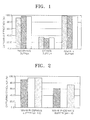
- FIG. 1 is a bar chart illustrating the effects of the buffer on the capturing efficiency of binding E.coli to the solid substrate having a surface with a pillar array of a fluidic device. As illustrated in FIG. 1, among the buffers having a low pH and similar salt concentrations, the sodium phosphate buffer and the sodium acetate buffer showed excellent capturing efficiency > 90 %.
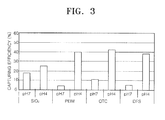
- FIG. 2 is a bar chart illustrating the effects of the salt concentration of the buffer on the capturing efficiency for binding E.coli to a solid substrate having a surface with a pillar array of a fluidic device.
- a real sample such as a biological sample, requires a buffer solution having a buffering capacity to maintain the pH of the liquid medium in the range of 3.0 to 6.0.
- the salt concentration required to obtain high capturing efficiency of the cell to the solid substrate may be in the range of 10 mM to 500 mM, more preferably, 50 mM to 300 mM (not shown in data.)
- Example 2 Capturing of Bacterial Cell in Blood using a Solid Substrate having a Pillar Array of a Fluidic Device
- a bacteria-containing sample was flowed through a fluidic device comprising an inlet, an outlet, and a chamber having a pillar array on a 10 mm x 23 mm silicon chip substrate so that the bacterial cell was bound to the nonplanar solid substrate.
- the cell number (N in ) in the bacteria-containing sample introduced into the chamber and the cell number (N out ) of the sample discharged from the chamber were determined by colony counting. Then, the bacteria capturing efficiency (%), (N in - N out )/N in )x100, was determined with respect to the surface material of the solid substrate.
- the distance between adjacent pillars was 12 ⁇ m
- the height of each pillar was 100 ⁇ m
- the sectional surface of each pillar was a regular square with each side of 25 ⁇ m.
- a pillar array having a SiO 2 layer at its surface, a pillar array having a SiO 2 layer coated with PEIM at its surface, a pillar array having a SiO 2 layer coated with OTC at its surface, and a pillar array having a SiO 2 layer coated with DFS at its surface were prepared.
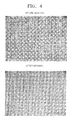
- FIG. 3 is a bar chart illustrating the effects of the buffer pH and of the surface material of the pillar array on the capturing efficiency for binding E.coli contained in blood to a solid substrate having a surface with a pillar array of a fluidic device.
- a sample comprising a liquid media having a low pH showed a higher capturing efficiency to the solid substrate than a sample in a liquid media with a pH of about 7.0.
- the effects of surface material of the solid substrate on the binding efficiency were lower than the effect of the buffer pH.
- a relatively hydrophobic surface having a relatively high electrostatic properties or a low surface tension such as PEIM, OTC and DFS, showed a higher capturing efficiency than the SiO 2 surface having a low electrostatic properties and a high surface tension.
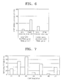
- FIG. 4 depicts optical microscopic images of a surface with a pillar array of a solid substrate of a fluidic device.
- the upper image shows the surface after a blood sample (pH 5.2) diluted in acetate buffer was contacted with the solid substrate.
- the lower image shows the result after washing the nonplanar solid substrate with 100 mM sodium acetate (pH 4.0) consecutively to the binding.
- E.coli cell was not eluted in the washing step.
- removal of an animal cell such as a red blood cell, which can act as a PCR inhibitor during the washing process, can be identified with amicroscopy.
- impurity materials shown between retangular structures is considered to animal cell such as red blood cell and white blood cell, and/or proteins.
- FIG 4B. shows such materials except bacteria can be removed by a washing process. The method of present invention is directed to capture target bacterial cell after removing the materials.
- FIG 4. shows that the removal of animal cells such as red blood cell and/or white blood cell, but not bacterial cell, can be identified with a microscopy.
- Example 3 Binding of Bacterial Cell contained in Urine to a Planar Solid Substrate
- 25.4 mm x 25.4 mm silicon chips containing 24 unit cells respectively including a SiO 2 layer, a SiO 2 layer coated with PEIM, a SiO 2 layer coated with OTC, and a SiO 2 layer coated with DFS were contacted with a sample including a bacterial cell so that the bacterial cell was bound to the solid substrate and washed.
- the binding of the bacterial cell was identified using a microscope.
- the diluted urine sample was added to the substrate covered with lid in the form of a patch and then incubated for 5 minutes. Then, the liquid media was removed from the substrate and a washing solution was added to the substrate. The substrate was incubated for 5 min in the washing solution for washing the planar solid substrate.
- the sodium acetate buffer was used as the liquid medium in the binding step
- the sample was washed using the sodium acetate buffer for 5 minutes
- the sodium phosphate buffer was used as the liquid medium in the binding step
- FIG. 5 shows the effects of the pH of the diluted urine sample on the capturing efficiency for binding E.coli contained in the sample to a solid substrate.
- the pH of the diluted urine sample significantly affected the binding of E.coli to the solid substrate.
- the number of E.coli cells attached to the solid substate having a SiO2 layer, a SiO2 layer coated with PEIM, a Si02 layer coated with OTC, or a SiO2 layer coated with DFS was about 97 cells, 95 cells, 56 cells, and 46 cells, respectively per 25.4 mm x 25.4 mm (24 unit cells) silicon chips at pH 5.6 (upper panel), however, no E.coli cells were remain attached to the same solid substrate at pH 6.4 (lower panel). The number of E.coli cells attached to the solid substrate was determined by counting on the microscopy.
- Example 4 Capturing of Bacterial Cell in Urine using a Solid Substrate having a Surface with a Pillar Array
- a bacteria-containing sample was flowed through a fluidic device comprising an inlet, an outlet, and a chamber having a pillar array on a 10 mm x 23 mm silicon chip substrate so that the bacterial cell was bound to the nonplanar solid substrate.
- the cell number (N in ) in the bacteria-containing sample introduced into the chamber and the cell number (N out ) of the sample discharged from the chamber were determined by colony counting. Then, the bacteria capturing efficiency (%), (N in - N out )/N in )x100, was determined with respect to the surface material of the solid substrate.
- the distance between adjacent pillars was 12 ⁇ m
- the height of each pillar was 100 ⁇ m
- the sectional surface of each pillar was a regular square with each side of 25 ⁇ m.
- a pillar array having a SiO 2 layer at its surface and a pillar array having a SiO 2 layer coated with PEIM at its surface were prepared.
- FIG. 6 is a bar chart illustrating the effects of pH and the surface material on the capturing efficiency for binding E.coli contained in a diluted urine sample to a solid substrate having a surface with a pillar array.
- the cell capturing efficiency measured according to the current example is shown in Table 1: Table 1 Diluted Buffer and Final pH Acetate Buffer (pH3) Final pH 4.7 Phosphate Buffer (pH3) Final pH 6.1 Surface Characteristics (Water Contact Angle: CA) SiO 2 ( ⁇ 10°) PEIM (Approximately 35°) SiO 2 ( ⁇ 10°) PEIM (Approximately 35°) Capturing Efficiency (%) 4 30 11 0
- a bacteria-containing sample was flowed through a fluidic device comprising an inlet, an outlet, and a chamber having a pillar array on a 10 mm x 23 mm silicon chip substrate so that the bacterial cell was bound to the nonplanar solid substrate.
- the cell number (N in ) in the sample introduced into the chamber and the cell number (N out ) of the sample discharged from the chamber were determined by colony counting. Then, the cell capturing efficiency (%), (N in - N out )/N in )x100, was determined with respect to urine used.
- the distance between adjacent pillars was 12 ⁇ m
- the height of each pillar was 100 ⁇ m
- the sectional surface of each pillar was a regular square with each side of 25 ⁇ m.
- a pillar array had a SiO 2 layer coated with PEIM at its surface.
- 200 ⁇ l of the diluted urine sample was conveyed at a flow rate of 200 ⁇ l/minute from the inlet through the chamber to the outlet. Then, 300 ⁇ l of 100 mM sodium acetate (pH 4.0) was conveyed at a flow rate of 200 ⁇ l/minute from the inlet through the chamber to the outlet, to wash the sample.
- the capturing efficiency was calculated by determining the cell number (N in ) in the urine sample introduced into the chamber and the cell number (N out ) of the urine sample discharged from the chamber by colony counting.
- FIG. 7 is a bar chart illustrating the results of separating cells from a urine sample containing the cell using the solid substrate having a surface with a pillar array of a fluidic device with respect to urine variation.
- the cell capturing efficiency was significantly changed according to a kind of the urine. This may result from the fact that the salt concentration of urine varies according to the body conditions and the alimentation of the person, which provides the urine, and other components.
- the present invention is not limited to such a specific mechanism.
- it can be assumed that the pH and the conductivity of the urine are not directly related to a change in the capturing efficiency (not shown in data).
- a bacteria-containing sample was flowed through a fluidic device comprising an inlet, an outlet, and a chamber having a pillar array on a 10 mm x 23 mm silicon chip substrate so that the bacterial cell was bound to the nonplanar solid substrate.
- the cell number (N in ) in the sample introduced into the chamber and the cell number (N out ) of the sample discharged from the chamber were determined by colony counting. Then, the cell capturing efficiency (%), (N in - N out )/N in )x100, was determined with respect to the dilution rate of the urine.
- the distance between adjacent pillars was 12 ⁇ m
- the height of each pillar was 100 ⁇ m
- the sectional surface of each pillar was a regular square with each side of 25 ⁇ m.
- a pillar array having a SiO 2 layer coated with PEIM at its surface and a pillar array having a SiO 2 layer coated with OTC were prepared.
- Cell samples of three dilution ratios were prepared, namely, a1 ⁇ 2 diluted sample of pH 3.97, which was prepared by mixing urine containing E.coli of 0.01 OD 600 1:1 with 100 mM sodium acetate buffer (pH 3.0) (hereinafter, referred to as 1/2 diluted sample), a 1/5 diluted sample of pH 4.05, which was prepared by mixing urine containing E.coli of 0.01 OD 600 - 1:4 with 100 mM sodium acetate buffer (pH 4.0) (hereinafter, referred to as 1/5 diluted sample, and a 1/7 diluted sample of pH 4.05, which was prepared by mixing urine containing E.coli of 0.01 OD 600 - 1:6 with 100 mM sodium acetate buffer (pH 4.0) in 1:6 (hereinafter, referred to as 1/7 diluted sample).
- a1 ⁇ 2 diluted sample of pH 3.97 which was prepared by mixing urine containing E.coli of 0.01 OD 600 1:1 with 100 mM sodium acetate buffer
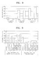
- FIG. 8 is a bar chart illustrating the results of concentrating E.coli from a urine sample containing E.coli using the solid substrate having a surface with a pillar array with respect to ratio of urine dilution. As illustrated in FIG. 8, the higher the dilution ratio of the urine sample was, the higher the cell capturing efficiency. In the washing process, the cells were not eluted.
- Example 7 Capturing E.coli in a Mimic Urine Solution using a Solid Substrate having a Surface with a Pillar Array: Detecting of Factor that inhibits Cell Capturing
- a bacteria-containing sample was flowed through a fluidic device comprising an inlet, an outlet, and a chamber having a pillar array on a 10 mm x 23 mm silicon chip substrate so that the bacterial cell was bound to the nonplanar solid substrate.
- the cell number (N in ) in the sample introduced into the chamber and the cell number (N out ) of the sample discharged from the chamber were determined by colony counting. Then, the cell capturing efficiency (%), (N in - N out )/N in )x100, was determined.
- the distance between adjacent pillars was 12 ⁇ m
- the height of each pillar was 100 ⁇ m
- the sectional surface of each pillar was a regular square with each side of 25 ⁇ m.
- a pillar array having a SiO 2 layer coated with OTC at its surface and a pillar array having a SiO 2 layer coated with SAM were prepared.
- a solution containing E.coli of 0.01 OD 600 were used as a cell sample.
- the solution was either based on a sodium acetate buffer or on dialyzed urine as the liquid medium.
- the solutions based on dialyzed urine were obtained by mixing dialyzed urine 1:1 with the respective 2x sodium acetate buffer-based solution.
- the solutions based on dialyzed urine have a final pH of 3.97.
- FIG. 9 is a bar chart illustrating the results of concentrating the cell in the urine sample using the solid substrate having a surface with a pillar array. As illustrated in FIG. 9, the capturing efficiency was lowered when the urine sample contained salt and other materials, such as creatine.
- Example 8 Capturing E.coli in Urine using a Solid Substrate having a Surface with a Pillar Array: Capturing efficiency with respect to the Dilution Rate of Urine and the Flow rate
- a bacteria-containing sample was flowed through a fluidic device comprising an inlet, an outlet, and a chamber having a pillar array on a 10 mm x 23 mm silicon chip substrate so that the bacterial cell was bound to the nonplanar solid substrate.
- the cell number (N in ) in the sample introduced into the chamber and the cell number (N out ) of the sample discharged from the chamber were determined by colony counting. Then, the cell capturing efficiency (%), (N in - N out )/N in )x100, was determined.
- the distance between adjacent pillars was 12 ⁇ m
- the height of each pillar was 100 ⁇ m
- the sectional surface of each pillar was a regular square with each side of 25 ⁇ m.
- a pillar array having a SiO 2 layer coated with PEIM by SAM coating process at its surface prepared.
- the samples were prepared by mixing 100mM sodium acetate buffer with urine in dilution rates of 1 ⁇ 2, 1/3, 1 ⁇ 4, 1/5 and 1/7 until the final volume of the mixture was 1 ml. Then, 10 ⁇ l of 1.0 OD 600 E.coli was added to the diluted sample.
- FIG. 10 is a bar chart illustrating the results of concentrating E.coli contained in the respective urine sample using the solid substrate having a surface with a pillar array with respect to the urine dilution ratio and the flow rate of conveying the sample through the chamber. As illustrated in FIG. 10, the more diluted the urine sample was, the higher the cell capturing efficiency . Further, if the flow rate increased, the cell capturing efficiency decreased.
- a microorganism such as a bacterial cell, fungus cell, or virus, present in a biological sample, can be efficiently separated.
- a microorganism such as a bacterial cell, fungus cell, or virus, present in a biological sample, can be efficiently separated.
Landscapes
- Chemical & Material Sciences (AREA)
- Health & Medical Sciences (AREA)
- Life Sciences & Earth Sciences (AREA)
- Engineering & Computer Science (AREA)
- Organic Chemistry (AREA)
- Genetics & Genomics (AREA)
- Zoology (AREA)
- Wood Science & Technology (AREA)
- Bioinformatics & Cheminformatics (AREA)
- Biomedical Technology (AREA)
- General Engineering & Computer Science (AREA)
- Biotechnology (AREA)
- Molecular Biology (AREA)
- Analytical Chemistry (AREA)
- General Health & Medical Sciences (AREA)
- Biochemistry (AREA)
- Chemical Kinetics & Catalysis (AREA)
- Microbiology (AREA)
- Physics & Mathematics (AREA)
- Biophysics (AREA)
- Proteomics, Peptides & Aminoacids (AREA)
- Crystallography & Structural Chemistry (AREA)
- Plant Pathology (AREA)
- Immunology (AREA)
- Clinical Laboratory Science (AREA)
- Apparatus Associated With Microorganisms And Enzymes (AREA)
- Measuring Or Testing Involving Enzymes Or Micro-Organisms (AREA)
- Micro-Organisms Or Cultivation Processes Thereof (AREA)
Applications Claiming Priority (4)
| Application Number | Priority Date | Filing Date | Title |
|---|---|---|---|
| KR1020060079054A KR100813264B1 (ko) | 2006-08-21 | 2006-08-21 | 비평면 형상의 고체 지지체를 이용하여 미생물로부터핵산을 증폭하는 방법 |
| KR1020060079055A KR100813265B1 (ko) | 2006-08-21 | 2006-08-21 | 비평면 형상의 고체 지지체를 이용하여 미생물로부터핵산을 증폭하는 방법 |
| KR1020060079056A KR100837401B1 (ko) | 2006-08-21 | 2006-08-21 | 비평면 형상의 고체 지지체를 이용하여 미생물로부터핵산을 분리하는 방법,상기 분리된 핵산을 주형으로 하여핵산을 증폭하는 방법 및 상기 고체 지지체를 포함하는핵산 분리 및 증폭 장치 |
| KR1020060079053A KR100917465B1 (ko) | 2006-08-21 | 2006-08-21 | 비평면 형상의 고체 지지체를 이용하여 미생물을 분리하는방법 및 미생물분리 장치 |
Publications (3)
| Publication Number | Publication Date |
|---|---|
| EP1900807A2 true EP1900807A2 (de) | 2008-03-19 |
| EP1900807A3 EP1900807A3 (de) | 2008-04-02 |
| EP1900807B1 EP1900807B1 (de) | 2014-02-26 |
Family
ID=38980858
Family Applications (3)
| Application Number | Title | Priority Date | Filing Date |
|---|---|---|---|
| EP07114679A Not-in-force EP1892295B1 (de) | 2006-08-21 | 2007-08-21 | Verfahren und Vorrichtung zur Isolierung und Verstärkung von Nukleinsäure aus Zellen von Mikroorganismen mittels eines nonplanaren festen Substrats |
| EP07114683A Active EP1972688B1 (de) | 2006-08-21 | 2007-08-21 | Verfahren zur Verstärkung von Nukleinsäure aus Zellen von Mikroorganismen mittels eines nonplanaren festen Substrats |
| EP07114680.7A Not-in-force EP1900807B1 (de) | 2006-08-21 | 2007-08-21 | Verfahren zur Separierung von Mikroorganismen über nichtplanare Festsubstrate und Vorrichtung zur Separierung von Mikroorganismen |
Family Applications Before (2)
| Application Number | Title | Priority Date | Filing Date |
|---|---|---|---|
| EP07114679A Not-in-force EP1892295B1 (de) | 2006-08-21 | 2007-08-21 | Verfahren und Vorrichtung zur Isolierung und Verstärkung von Nukleinsäure aus Zellen von Mikroorganismen mittels eines nonplanaren festen Substrats |
| EP07114683A Active EP1972688B1 (de) | 2006-08-21 | 2007-08-21 | Verfahren zur Verstärkung von Nukleinsäure aus Zellen von Mikroorganismen mittels eines nonplanaren festen Substrats |
Country Status (4)
| Country | Link |
|---|---|
| US (3) | US20080044884A1 (de) |
| EP (3) | EP1892295B1 (de) |
| JP (1) | JP2008048735A (de) |
| DE (1) | DE602007006908D1 (de) |
Families Citing this family (17)
| Publication number | Priority date | Publication date | Assignee | Title |
|---|---|---|---|---|
| EP1712284B1 (de) * | 2005-04-15 | 2012-10-10 | Samsung Electronics Co., Ltd. | Methode zur Zelltrennung mit Benutzung hydrophober fester Träger |
| US20080044884A1 (en) * | 2006-08-21 | 2008-02-21 | Samsung Electronics Co., Ltd. | Method and device for separating cells from a sample using a nonplanar solid substrate |
| US8158411B2 (en) | 2006-08-21 | 2012-04-17 | Samsung Electronics Co., Ltd. | Method of separating microorganism using nonplanar solid substrate and device for separating microorganism using the same |
| KR101569832B1 (ko) * | 2008-11-19 | 2015-11-18 | 삼성전자주식회사 | 시료 중의 게놈 dna 및 플라스미드 dna를 서로 분리하는 방법 및 그를 위한 키트 |
| WO2010110843A1 (en) | 2009-03-25 | 2010-09-30 | Eastman Kodak Company | Droplet generator |
| JP5622189B2 (ja) * | 2010-01-28 | 2014-11-12 | 国立大学法人東京工業大学 | 単一細胞分離用プレート |
| GB201010237D0 (en) | 2010-06-18 | 2010-07-21 | Lgc Ltd | Methods and apparatuses |
| WO2012112151A1 (en) * | 2011-02-16 | 2012-08-23 | Empire Technology Development Llc | Tissue engineering supports and methods therefor |
| US9670540B2 (en) | 2011-07-21 | 2017-06-06 | Cornell University | Methods and devices for DNA sequencing and molecular diagnostics |
| US10137673B2 (en) * | 2013-12-31 | 2018-11-27 | Canon U.S. Life Sciences, Inc. | Methods and systems for continuous flow cell lysis in a microfluidic device |
| KR102207922B1 (ko) | 2014-03-06 | 2021-01-26 | 삼성전자주식회사 | 반코마이신 저항성 엔테로코커스 특이적인 프라이머 세트, 그를 포함하는 조성물 및 시료 중 반코마이신 저항성 엔테로코커스 속 미생물을 검출하는 방법 |
| KR102287811B1 (ko) | 2014-10-31 | 2021-08-09 | 삼성전자주식회사 | 2개 표면을 결합시키는 방법 및 그에 의하여 제조된 구조체, 및 상기 구조체를 포함하는 미세유동 장치 |
| EP3427672B1 (de) * | 2017-05-08 | 2020-03-04 | Tcm Biosciences Inc. | Vorrichtung zum sammeln von proben wie z. b. scheidenausfluss, einschliesslich gebärmutterhalszellen |
| GB201805479D0 (en) * | 2018-04-03 | 2018-05-16 | Momentum Bioscience Ltd | Microorganism separation and detection |
| EP3999624B1 (de) | 2019-07-16 | 2026-03-25 | Meliolabs Inc. | Verfahren und vorrichtungen für einzellenbasierte digitale hochauflösende schmelzanalyse |
| US11481230B2 (en) * | 2020-12-17 | 2022-10-25 | Oracle International Corporation | Techniques for modifying a compute instance |
| KR102591870B1 (ko) * | 2022-06-09 | 2023-10-20 | 성균관대학교산학협력단 | 병렬식 액적 생성 장치 |
Citations (5)
| Publication number | Priority date | Publication date | Assignee | Title |
|---|---|---|---|---|
| WO1998051693A1 (en) | 1997-05-13 | 1998-11-19 | Genpoint As | Solid-phase nucleic acid isolation |
| US20020042125A1 (en) | 1997-08-13 | 2002-04-11 | Cepheid | Method for separating analyte from a sample |
| WO2005093065A1 (en) | 2004-03-16 | 2005-10-06 | Roche Diagnostics Gmbh | Improved method of isolating nucleic acids |
| EP1712284A1 (de) | 2005-04-15 | 2006-10-18 | Samsung Electronics Co., Ltd. | Methode zur Zelltrennung mit Benutzung hydrophober fester Träger |
| EP1726641A1 (de) | 2005-05-24 | 2006-11-29 | Samsung Electronics Co., Ltd. | Phasetrennung- verwendendes Zellentrennverfahren und Kit |
Family Cites Families (29)
| Publication number | Priority date | Publication date | Assignee | Title |
|---|---|---|---|---|
| JPS5928484A (ja) | 1982-08-11 | 1984-02-15 | Mitsui Toatsu Chem Inc | L−アミノ酸の単離方法 |
| EP0208647A3 (de) | 1985-07-09 | 1989-03-08 | Protein Foods Group Inc. | Immobilisierungsträger für chemische und physikalische Verfahren und Methoden zu ihrer Herstellung |
| JPH0623758B2 (ja) * | 1987-04-09 | 1994-03-30 | テルモ株式会社 | Bリンパ球分離材、分離方法および分離器 |
| JP2852307B2 (ja) * | 1989-02-09 | 1999-02-03 | 吉富製薬株式会社 | ウイルスの除去された蛋白質製剤の製造法 |
| US5234809A (en) * | 1989-03-23 | 1993-08-10 | Akzo N.V. | Process for isolating nucleic acid |
| US5085779A (en) * | 1989-06-07 | 1992-02-04 | J. T. Baker, Inc. | Polyethyleneimine matrixes for affinity chromatography |
| GB9003253D0 (en) * | 1990-02-13 | 1990-04-11 | Amersham Int Plc | Precipitating polymers |
| IES940182A2 (en) | 1994-03-01 | 1995-11-29 | Teagasc Agric Food Dev Authori | "Rapid detection of bacteria in liquid cultures" |
| US5705628A (en) | 1994-09-20 | 1998-01-06 | Whitehead Institute For Biomedical Research | DNA purification and isolation using magnetic particles |
| US5707799A (en) * | 1994-09-30 | 1998-01-13 | Abbott Laboratories | Devices and methods utilizing arrays of structures for analyte capture |
| EP1003908B1 (de) * | 1997-04-16 | 2006-12-06 | Applera Corporation | Nukleinsäuresammlung |
| EP1003759A2 (de) * | 1997-08-13 | 2000-05-31 | Cepheid | Mikrostrukturen für die manipulation von flüssigen proben |
| BR9815569A (pt) | 1997-12-06 | 2001-10-09 | Dna Res Instr Ltd | Método para extração de biomoléculas de material biológico |
| ES2262506T3 (es) * | 1999-03-17 | 2006-12-01 | Jimro Co., Ltd. | Aferesis leucocitaria sanguinea para el tratamiento del vih. |
| JP2001098007A (ja) | 1999-09-29 | 2001-04-10 | Kawamura Inst Of Chem Res | 親水性成形物およびその製造法 |
| GB0013658D0 (en) * | 2000-06-05 | 2000-07-26 | Dynal Asa | Nucleic acid isolation |
| AU2002324289A1 (en) | 2001-07-24 | 2003-02-17 | Societe D'amenagement Urbain Et Rural | Automated process for detecting pathogenic organisms in water |
| EP2359689B1 (de) * | 2002-09-27 | 2015-08-26 | The General Hospital Corporation | Mikrofluid-Vorrichtung zur Zelltrennung, sowie Verwendung derselben |
| JP5441290B2 (ja) * | 2002-10-04 | 2014-03-12 | ジーイー・ヘルスケア・バイオサイエンス・コーポレイション | 核酸精製システムの媒体上に核酸を保存するツールとして、化学物質を用いる方法および材料 |
| WO2004051231A1 (ja) * | 2002-11-29 | 2004-06-17 | Nec Corporation | 分離装置および分離方法 |
| SE0400073D0 (sv) | 2003-04-04 | 2004-01-14 | Appear Sweden Hb | Antibacterial material |
| KR100953612B1 (ko) | 2003-06-02 | 2010-04-20 | 삼성에스디아이 주식회사 | 생체물질 고정용 기판 및 이의 제조방법 |
| JP4202391B2 (ja) * | 2004-03-17 | 2008-12-24 | パナソニック株式会社 | バイオチップの製造方法、プローブ溶液、および、バイオチップ |
| JP2005295969A (ja) * | 2004-04-16 | 2005-10-27 | Hitachi Medical Corp | 細胞分離装置 |
| KR100594408B1 (ko) | 2004-12-17 | 2006-06-30 | 한국과학기술연구원 | 초음파장 및 진행파 유전영동을 이용한 세포 분리 장치 |
| KR100647335B1 (ko) | 2005-04-15 | 2006-11-23 | 삼성전자주식회사 | 소수성 고체 지지체를 이용한 세포 분리 방법 |
| US8158411B2 (en) * | 2006-08-21 | 2012-04-17 | Samsung Electronics Co., Ltd. | Method of separating microorganism using nonplanar solid substrate and device for separating microorganism using the same |
| US20080044884A1 (en) * | 2006-08-21 | 2008-02-21 | Samsung Electronics Co., Ltd. | Method and device for separating cells from a sample using a nonplanar solid substrate |
| KR100862660B1 (ko) * | 2006-09-25 | 2008-10-10 | 삼성전자주식회사 | 단일 비드에 의한 핵산의 분리 및 정제 방법 및 장치 |
-
2007
- 2007-08-20 US US11/841,089 patent/US20080044884A1/en not_active Abandoned
- 2007-08-20 US US11/841,107 patent/US7919278B2/en active Active
- 2007-08-20 US US11/841,069 patent/US20080070282A1/en not_active Abandoned
- 2007-08-20 JP JP2007213969A patent/JP2008048735A/ja active Pending
- 2007-08-21 EP EP07114679A patent/EP1892295B1/de not_active Not-in-force
- 2007-08-21 EP EP07114683A patent/EP1972688B1/de active Active
- 2007-08-21 DE DE602007006908T patent/DE602007006908D1/de active Active
- 2007-08-21 EP EP07114680.7A patent/EP1900807B1/de not_active Not-in-force
Patent Citations (5)
| Publication number | Priority date | Publication date | Assignee | Title |
|---|---|---|---|---|
| WO1998051693A1 (en) | 1997-05-13 | 1998-11-19 | Genpoint As | Solid-phase nucleic acid isolation |
| US20020042125A1 (en) | 1997-08-13 | 2002-04-11 | Cepheid | Method for separating analyte from a sample |
| WO2005093065A1 (en) | 2004-03-16 | 2005-10-06 | Roche Diagnostics Gmbh | Improved method of isolating nucleic acids |
| EP1712284A1 (de) | 2005-04-15 | 2006-10-18 | Samsung Electronics Co., Ltd. | Methode zur Zelltrennung mit Benutzung hydrophober fester Träger |
| EP1726641A1 (de) | 2005-05-24 | 2006-11-29 | Samsung Electronics Co., Ltd. | Phasetrennung- verwendendes Zellentrennverfahren und Kit |
Also Published As
| Publication number | Publication date |
|---|---|
| US7919278B2 (en) | 2011-04-05 |
| JP2008048735A (ja) | 2008-03-06 |
| EP1972688B1 (de) | 2010-06-02 |
| US20080044884A1 (en) | 2008-02-21 |
| US20080070282A1 (en) | 2008-03-20 |
| EP1892295B1 (de) | 2012-07-25 |
| EP1900807A3 (de) | 2008-04-02 |
| EP1892295A3 (de) | 2008-03-26 |
| US20080044864A1 (en) | 2008-02-21 |
| EP1892295A2 (de) | 2008-02-27 |
| EP1972688A1 (de) | 2008-09-24 |
| DE602007006908D1 (de) | 2010-07-15 |
| EP1900807B1 (de) | 2014-02-26 |
Similar Documents
| Publication | Publication Date | Title |
|---|---|---|
| EP1900807B1 (de) | Verfahren zur Separierung von Mikroorganismen über nichtplanare Festsubstrate und Vorrichtung zur Separierung von Mikroorganismen | |
| EP2215103B1 (de) | Verfahren zur aufreinigung von nukleinsäuren | |
| DE60123480T2 (de) | Behandlung von hydrophoben oder hydrophilen oberflächen mit polymeren | |
| KR100785010B1 (ko) | 수소 결합을 이용하여 고체 지지체의 친수성 표면 상에서핵산 정제 방법 및 장치 | |
| CN110891664B (zh) | 用于多孔材料中双水相分离的相分离行为改性剂 | |
| TW201518498A (zh) | 用於分離或富集化細胞的方法及組合物 | |
| JP2004042012A (ja) | 分離装置、分析システム、分離方法および分離装置の製造方法 | |
| US20210220827A1 (en) | Systems and methods for nucleic acid purification using flow cells with actuated surface-attached structures | |
| US8557564B2 (en) | Method of separating microorganism using nonplanar solid substrate and device for separating microorganism using the same | |
| EP1712284B1 (de) | Methode zur Zelltrennung mit Benutzung hydrophober fester Träger | |
| KR100647335B1 (ko) | 소수성 고체 지지체를 이용한 세포 분리 방법 | |
| EP1900808B1 (de) | Verfahren und Vorrichtung zur Separierung von Mikroorganismen von Proben durch Elektrodialyse und Mittel zur Separierung von Mikroorganismen | |
| KR100917465B1 (ko) | 비평면 형상의 고체 지지체를 이용하여 미생물을 분리하는방법 및 미생물분리 장치 | |
| KR100785023B1 (ko) | 전기투석 및 미생물 포획수단을 이용하여 시료로부터미생물을 분리하는 방법 및 그를 위한 미생물분리 장치 | |
| KR100813268B1 (ko) | 이온교환반응 및 미생물 포획 수단을 이용하여 시료로부터미생물을 분리하는 방법, 미생물 시료의 전처리용 용기 및미생물분리 장치 | |
| KR100837401B1 (ko) | 비평면 형상의 고체 지지체를 이용하여 미생물로부터핵산을 분리하는 방법,상기 분리된 핵산을 주형으로 하여핵산을 증폭하는 방법 및 상기 고체 지지체를 포함하는핵산 분리 및 증폭 장치 | |
| KR100813265B1 (ko) | 비평면 형상의 고체 지지체를 이용하여 미생물로부터핵산을 증폭하는 방법 | |
| US20240316549A1 (en) | Systems and methods for nucleic acid purification using flow cells with actuated surface-attached structures | |
| KR101206039B1 (ko) | 비평면 형상의 고체 지지체를 이용하여 미생물로부터핵산을 분리하는 방법,상기 분리된 핵산을 주형으로 하여핵산을 증폭하는 방법 및 상기 고체 지지체를 포함하는핵산 분리 및 증폭 장치 | |
| KR100813264B1 (ko) | 비평면 형상의 고체 지지체를 이용하여 미생물로부터핵산을 증폭하는 방법 |
Legal Events
| Date | Code | Title | Description |
|---|---|---|---|
| PUAI | Public reference made under article 153(3) epc to a published international application that has entered the european phase |
Free format text: ORIGINAL CODE: 0009012 |
|
| PUAL | Search report despatched |
Free format text: ORIGINAL CODE: 0009013 |
|
| AK | Designated contracting states |
Kind code of ref document: A2 Designated state(s): AT BE BG CH CY CZ DE DK EE ES FI FR GB GR HU IE IS IT LI LT LU LV MC MT NL PL PT RO SE SI SK TR |
|
| AX | Request for extension of the european patent |
Extension state: AL BA HR MK YU |
|
| AK | Designated contracting states |
Kind code of ref document: A3 Designated state(s): AT BE BG CH CY CZ DE DK EE ES FI FR GB GR HU IE IS IT LI LT LU LV MC MT NL PL PT RO SE SI SK TR |
|
| AX | Request for extension of the european patent |
Extension state: AL BA HR MK YU |
|
| RIC1 | Information provided on ipc code assigned before grant |
Ipc: B01L 3/00 20060101ALI20080226BHEP Ipc: C12N 1/02 20060101AFI20080226BHEP Ipc: C12M 1/26 20060101ALI20080226BHEP |
|
| 17P | Request for examination filed |
Effective date: 20080724 |
|
| R17P | Request for examination filed (corrected) |
Effective date: 20080724 |
|
| 17Q | First examination report despatched |
Effective date: 20080911 |
|
| AKX | Designation fees paid |
Designated state(s): DE FR GB |
|
| RAP1 | Party data changed (applicant data changed or rights of an application transferred) |
Owner name: SAMSUNG ELECTRONICS CO., LTD. |
|
| GRAP | Despatch of communication of intention to grant a patent |
Free format text: ORIGINAL CODE: EPIDOSNIGR1 |
|
| INTG | Intention to grant announced |
Effective date: 20130819 |
|
| RIN1 | Information on inventor provided before grant (corrected) |
Inventor name: HAN, JUNG-IM Inventor name: JEONG, SUNG-YOUNG Inventor name: HWANG, KYU-YOUN Inventor name: KIM, JOON-HO Inventor name: LEE, HUN-JOO |
|
| GRAS | Grant fee paid |
Free format text: ORIGINAL CODE: EPIDOSNIGR3 |
|
| GRAA | (expected) grant |
Free format text: ORIGINAL CODE: 0009210 |
|
| AK | Designated contracting states |
Kind code of ref document: B1 Designated state(s): DE FR GB |
|
| REG | Reference to a national code |
Ref country code: GB Ref legal event code: FG4D |
|
| REG | Reference to a national code |
Ref country code: DE Ref legal event code: R096 Ref document number: 602007035182 Country of ref document: DE Effective date: 20140410 |
|
| REG | Reference to a national code |
Ref country code: DE Ref legal event code: R097 Ref document number: 602007035182 Country of ref document: DE |
|
| PLBE | No opposition filed within time limit |
Free format text: ORIGINAL CODE: 0009261 |
|
| STAA | Information on the status of an ep patent application or granted ep patent |
Free format text: STATUS: NO OPPOSITION FILED WITHIN TIME LIMIT |
|
| 26N | No opposition filed |
Effective date: 20141127 |
|
| REG | Reference to a national code |
Ref country code: DE Ref legal event code: R097 Ref document number: 602007035182 Country of ref document: DE Effective date: 20141127 |
|
| REG | Reference to a national code |
Ref country code: FR Ref legal event code: PLFP Year of fee payment: 10 |
|
| PGFP | Annual fee paid to national office [announced via postgrant information from national office to epo] |
Ref country code: GB Payment date: 20160817 Year of fee payment: 10 Ref country code: DE Payment date: 20160816 Year of fee payment: 10 |
|
| PGFP | Annual fee paid to national office [announced via postgrant information from national office to epo] |
Ref country code: FR Payment date: 20160729 Year of fee payment: 10 |
|
| REG | Reference to a national code |
Ref country code: DE Ref legal event code: R119 Ref document number: 602007035182 Country of ref document: DE |
|
| GBPC | Gb: european patent ceased through non-payment of renewal fee |
Effective date: 20170821 |
|
| REG | Reference to a national code |
Ref country code: FR Ref legal event code: ST Effective date: 20180430 |
|
| PG25 | Lapsed in a contracting state [announced via postgrant information from national office to epo] |
Ref country code: GB Free format text: LAPSE BECAUSE OF NON-PAYMENT OF DUE FEES Effective date: 20170821 Ref country code: DE Free format text: LAPSE BECAUSE OF NON-PAYMENT OF DUE FEES Effective date: 20180301 |
|
| PG25 | Lapsed in a contracting state [announced via postgrant information from national office to epo] |
Ref country code: FR Free format text: LAPSE BECAUSE OF NON-PAYMENT OF DUE FEES Effective date: 20170831 |