EP1903503A2 - Procédé, système et produit de programme informatique pour fournir des protocoles, configurables par l'utilisateur et standardisés, de visualisation anatomique de données volumétriques - Google Patents
Procédé, système et produit de programme informatique pour fournir des protocoles, configurables par l'utilisateur et standardisés, de visualisation anatomique de données volumétriques Download PDFInfo
- Publication number
- EP1903503A2 EP1903503A2 EP07116999A EP07116999A EP1903503A2 EP 1903503 A2 EP1903503 A2 EP 1903503A2 EP 07116999 A EP07116999 A EP 07116999A EP 07116999 A EP07116999 A EP 07116999A EP 1903503 A2 EP1903503 A2 EP 1903503A2
- Authority
- EP
- European Patent Office
- Prior art keywords
- voxel data
- views
- reference set
- view
- viewing
- Prior art date
- Legal status (The legal status is an assumption and is not a legal conclusion. Google has not performed a legal analysis and makes no representation as to the accuracy of the status listed.)
- Withdrawn
Links
Images


Classifications
-
- G—PHYSICS
- G06—COMPUTING OR CALCULATING; COUNTING
- G06T—IMAGE DATA PROCESSING OR GENERATION, IN GENERAL
- G06T15/00—Three-dimensional [3D] image rendering
- G06T15/08—Volume rendering
-
- G—PHYSICS
- G06—COMPUTING OR CALCULATING; COUNTING
- G06T—IMAGE DATA PROCESSING OR GENERATION, IN GENERAL
- G06T7/00—Image analysis
- G06T7/30—Determination of transform parameters for the alignment of images, i.e. image registration
- G06T7/38—Registration of image sequences
-
- G—PHYSICS
- G06—COMPUTING OR CALCULATING; COUNTING
- G06T—IMAGE DATA PROCESSING OR GENERATION, IN GENERAL
- G06T2207/00—Indexing scheme for image analysis or image enhancement
- G06T2207/30—Subject of image; Context of image processing
- G06T2207/30004—Biomedical image processing
Definitions
- the present invention relates generally to computer-generated images and, more particularly, to a method for storing and comparing views of three-dimensional images and sequences of three-dimensional images.
- Physicians are faced with an increased need to more efficiently interpret large volumetric image datasets produced by medical imaging scanners utilizing technologies such as computed tomography (CT) or magnetic resonance imaging (MRI). These imaging scanners typically produce a series of cross-sectional images or slices in each patient session for viewing by the physician. Sequences of two-dimensional slices may be combined to produce information about a volume corresponding to an area of the patient's anatomy. Whereas, in the past, viewing the slice images of the image volume individually, side-by-side, was sufficient, the increased number of slices routinely acquired with modern imagers - often approaching 1,000 slices/exam - renders such methods impractical. As well, the increased number of possible diagnostic purposes for which such volumetric data is useful further compounds the problem. Physicians are therefore increasingly using 3D rendering methods, such as pixel averaging, maximum intensity projection (MIP), and color/opacity compositing for examining the volume contents.
- 3D rendering methods such as pixel averaging, maximum intensity projection (MIP), and color/opacity
- a method of automatically generating a set of views for a new set of voxel data according to a viewing protocol comprises a) storing a reference set of views for a reference set of voxel data according to the viewing protocol, wherein each view in the reference set of views is defined by a set of geometrical parameters defined with respect to a reference coordinate system of the reference set of voxels; b) defining a registration mapping from the reference set of voxel data to the new set of voxel data; and, c) for each view in the reference set of views, using the registration mapping to map each view in the reference set of views to the new set of voxel data to provide the set of views for the new set of voxel data according to the viewing protocol.
- a system for automatically generating a set of views for a new set of voxel data according to a viewing protocol comprises a memory for storing the new set of voxel data and the viewing protocol; and means for performing the steps of a) storing a reference set of views for a reference set of voxel data according to the viewing protocol, wherein each view in the reference set of views is defined by a set of geometrical parameters defined with respect to a reference coordinate system of the reference set of voxels; b) defining a registration mapping from the reference set of voxel data to the new set of voxel data; and, c) for each view in the reference set of views, using the registration mapping to map each view in the reference set of views to the new set of voxel data to provide the set of views for the new set of voxel data according to the viewing protocol.
- a computer program product for use on a computer.
- the computer program product comprises a recording medium; and, means recorded on the recording medium for instructing the computer system to perform the steps of: a) storing a reference set of views for a reference set of voxel data according to the viewing protocol, wherein each view in the reference set of views is defined by a set of geometrical parameters defined with respect to a reference coordinate system of the reference set of voxels; b) defining a registration mapping from the reference set of voxel data to the new set of voxel data; and, c) for each view in the reference set of views, using the registration mapping to map each view in the reference set of views to the new set of voxel data to provide the set of views for the new set of voxel data according to the viewing protocol.
- FIG. 1 provides examples of slab geometries and their respective sampling grids.
- FIG. 2 is a flowchart illustrating a method of creating and saving a 3D viewing protocol in accordance with an aspect of an embodiment of the invention.
- FIG. 3 is a flowchart illustrating a method of applying a viewing protocol using volume-to-volume registration in accordance with a further aspect of an embodiment of the invention.
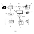
- FIG. 4 is a block diagram illustrating a computer system in accordance with an embodiment of the invention.
- FIG. 5 is a flowchart illustrating a method of applying an atlas-based viewing protocol using volume-to-volume registration in accordance with a further aspect of an embodiment of the invention.
- Examples of 3D rendering geometries are illustrated in FIG. 1. These include: i) a planar slab 10, in which an extruded plane of a given thickness (typically 0-10mm) is generated; ii) a curved slab, in which an extruded curve of a given thickness is generated); and iii) simulated camera views, in which a virtual camera is placed at a given orientation and location (inside or outside the data volume) and an image is computed using orthonormal or perspective projection geometry.
- the extrusion may be of a fixed curve along a linear path 20 (2D), a fixed curve along a curved path 30 (2.5D), or a variable curve along a curved path 40, 50 (3D).
- the view may optionally contain cutting planes and masks to help uncover anatomical regions that would otherwise be hidden from view of the virtual camera.
- a patient is scanned using a medical imaging scanner 201, such as a CT or MRI system.
- the medical imaging scanner produces a sequence of two-dimensional image slices.
- the sequence of 2D image slices is combined to synthesize image data in a third-dimension, to produce three-dimensional volume data.
- the medical imaging scanner may provide complete 3D volume data P.
- the volume data is represented as a set of volume cells, or voxels, representing data values at grid locations in three-dimensional space.
- Voxels in the data volume are geometrically associated with a reference coordinate system (RCS) P, chosen for convenience.
- RCS reference coordinate system
- each voxel intensity value may have associated Cartesian coordinates (x P , y P , z P ).
- a suitable substitute coordinate system may be used, such as, e.g., a cylindrical coordinate system.
- a rendering algorithm 203 controlled by a set of rendering parameters 204 prescribing the rendering operation, generates each view.
- the parameters may include the rendering type (e.g., average, MIP, compositing), the geometry type (e.g., flat slab, curved slab, or simulated camera), the geometrical coordinates and angles associated with the given geometry type, and additional optional parameters.
- Optional parameters may include, for example, color, contrast, transparency, volumetric masks or thresholding ranges to highlight specific features in a volume.
- Some of the parameters may be set to change automatically over a given range to create animation effects.
- the rendering algorithm may animate the output image to produce a rotating effect or to progressively unmask or cut away anatomical elements.
- the rendering techniques describe here, and their controlling parameters, are well known in the art and are embodied in commercial visualization workstations marketed by CT and MRI scanner manufacturers, such as Philips (www.medical.philips.com), GE (www.gehealthcare.com), Siemens (www.medical.siemens.com), as well by dedicated workstation developers such as Vital Images (www.vitalimages.com) and TeraRecon (www.terarecon.com).
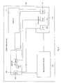
- the user may modify some or all of the rendering parameters until the views in the collage best show the anatomy of interest.
- the user then saves in step 206 the collage description 207 to a persistent storage medium, such as a computer hard drive.
- the collage description 207 contains the layout of the views 211 within the collage viewing area 210, and, for each view, the parameters 204 that specify the rendering operation for generating that view.
- the user may further change the layout of the views 211 in collage viewing area 210, adjust its contents by changing rendering parameters 204, and again saving in step 206 the new collage description 207 to the persistent storage medium to create a saved protocol 208, consisting of multiple collage descriptions 207. This process may be repeated as many times as necessary.
- the result is a sequence of collages forming a streamlined review protocol 208 preferred by the user when reviewing the type of anatomy shown in the data volume P.
- the data volume P from which the collages were generated is saved in step 220, forming a protocol consisting of a data volume P and a sequence of collage descriptions 207, each containing views described by rendering parameters 204 relating to data volume P.
- the use of data volume P is described in greater detail below, with regard to FIG. 3.
- saved protocols may be generated for various classes of patients to facilitate easier comparison. For example, a clinician may save a protocol for male patients, female patients, children or for specific medical conditions.
- the user may create a sequence of collages, each presenting a region of possible abnormalities: left carotids, right carotids, middle cerebral artery, Circle of Willis, etc.
- the views of the region are optimized for a quick detection of blood flow abnormalities.
- the left carotid collage may present a simulated camera view of the neck from the left side, a curved slab of 5mm thickness through the middle of the carotid, and a small 1 mm flat slab view programmed to scroll automatically along the length of the carotid, perpendicular to its centerline, at 1 mm increments.
- the viewing protocol created for volume P may subsequently be used with other data volumes, such as a current data volume C, acquired from another patient or from the same patient at a different time.
- the particular profile used can be selected from among many different protocols.
- the selection of protocol P from amongst the set of available protocols can be done either manually by selection from a descriptive list, or automatically, by the computer executing programmed decision rules that select the most suitable protocol to apply based on the identity and/or profession of the user (eg, radiologist or surgeon) and/or organizational affiliation of the user (eg, department A or department B) and/or the contents of the data and its associated descriptors (eg, anatomical region, clinical indications for the study) and/or the nature of the patient (male or female, adult or child, physiological condition).
- FIG. 3 there is shown a flowchart illustrating the steps involved in a method according to an aspect of an embodiment of the invention.
- the method involves applying a saved viewing protocol 208 and rendering parameters 204 using volume-to-volume registration obtained in step 301.
- the method involves automatic data-volume to data-volume registration, in which two or more data volumes are correlated.
- Many volume to volume registration algorithms are currently known in the art, as surveyed in the book " Handbook or Biomedical Image Analysis Volume III: Registration Methods", edited by Suri, Wilson, Laxminarayan, 2005 , the contents of which are included here by reference.
- the registration of the two volumes generates a mapping T ( x ) of any coordinate in RCS P to a corresponding anatomical location in RCS C, or a P ⁇ C coordinate mapping 302.
- T ( x ) T global x + T local x
- the global transform is computed first, using an affine transformation algorithm, to compensate for pose differences between data volumes by maximization of normalized mutual information.
- the local transform is computed to facilitate compensation for local deformations, using a spline-based approach.
- Deformation is modeled as free-form deformation using B-splines, which can be written as the 3D tensor product of 1D cubic B-splines:
- ⁇ denotes the control points which parameterize the transform.
- the optimal transform is found by minimizing a cost function associated with both the global and local transform parameters.
- the cost function can comprise two components representing competing goals: maximizing voxel value correlation in the regions around each of the control points and maintaining smoothness and regularity in the transform.
- Control points may be defined in volume P in a suitable arrangement, for example, on a regular grid of a desired resolution (e.g., 1 cm spacing).
- the geometrical parameters of each view in P are mapped 305 to coordinate space C.
- a parameter describes an angle or a directional unit vector, it is converted into two sets of coordinates (i.e., the base and tip of a vector), which are then mapped from P to C to provide the equivalent vector in C.
- flat slab viewing geometry is defined by a rectangular section of a plane representing one boundary of the slab, and a thickness over which sampled values would be processed to generate the output image.
- the rectangular section may be further defined by the P coordinates of its corners.
- a curve may be specified by a sequence of equidistant positions along the curve, each position having a specific coordinate in volume P. The requirement that the positions be equidistant may be specified in the curve descriptor or implicitly assumed by the program reading the descriptor from file.
- a processing step 306 may be applied following the mapping to compute parameters that observe the geometrical constraints, yet remain as close as possible to the transformed views.
- locations on the planar rectangular section in volume P do not form a planar rectangular section in volume C.
- a set of new corner coordinates may be produced by sampling the section in volume C in a rectangular grid, transforming each grid location to volume P, computing the closest plane in volume P using bilinear regression, then selecting a rectangular section of the plane whose corners are the closest to the corners of the transformed grid. The thickness of the slab, in mm, remains unchanged.
- the mapped curve locations in volume C are not equidistant. Therefore, the mapped curve in volume C may be re-interpolated using a spline algorithm and re-sampled at new equidistant positions.
- Geometrical constraints may also apply to the spatial relationship between multiple views in the same collage, such as relative angles and distances. Such constraints are best handled in a combined manner that best reflects the intended use of the views.
- the collage contains 3 views of mutually-perpendicular flat slabs intersecting at a location of interest, for example at the center of a lesion.
- the intersection point and the ends of the two orientation unit vector are mapped to the C coordinate system in step 305.
- the vectors may not be of unit length and may not be perpendicular to each other.
- the constraints can be reapplied to the two vectors using well known linear algebra techniques, for example by using 3 cross products to generate a new set of mutually-perpendicular vectors similar to the original ones, which are then normalized to a unit length.
- Non-geometrical rendering parameters such as color, contrast and transparency may be passed on to the rendering step unmodified, or, optionally, they may be recomputed based on the contents of data volume C.
- a thresholding value for hiding voxels in a 3D projection may be required to allow visualization of bright objects. This value may be specified either as a fixed value, in which case it is simply passed on unmodified, or it may be specified as some function of the data value histogram (e.g., at the 80 th percentile), in which case it is recomputed.
- a collage of multiple views in the protocol is presented to the user by computing new rendering parameters 307 for each of the views, as described above, and rendering them 308 in the collage viewing area 210 according to the collage's layout description.
- the user can step through the collages stored in the protocol to obtain a complete presentation of the anatomy of the given body region.
- the data volume of interest can be registered to an atlas volume, in which data regions have been labeled or tagged.
- the atlas can serve, for example, as a model for segmenting volume P into anatomically meaningful regions.
- the atlas volume itself may serve as a reference volume, eliminating the need for saving dataset C itself as an additional reference for the viewing protocol.
- FIG. 5 An alternative atlas-based method for saving a protocol is described in figure 5.
- the same reference numerals, with 200 added, are used to designate steps of the atlas-based method of Figure 5 relative to analogous steps of the method of Figure 3.
- the description of Figure 3 is not repeated in relation to Figure 5.
- Data volume P is registered to atlas volume A.
- mapping P->A is then used to map the relevant rendering parameters, established in step 509, to the atlas coordinate space.
- Constraints are then applied to preserve the viewing parameters' integrity, as described above, and the views are finally saved, in step 508, with reference to the atlas volume A, which is already in the database.
- the reference data volume A may be advantageously generated as a composite of individual datasets, obtained from multiple people.
- the compositing process averages the anatomical variations in a population to create a more typical arrangement of anatomy, enabling the registration of new datasets to the reference dataset to be more robust than in the case the reference dataset came from a single individual with some unusual anatomical features.
- Another advantage of compositing multiple datasets with partial spatial overlap is increased anatomical coverage, enabling the support region for registration to be potentially larger than in the case of a single dataset. This advantage applies not only when the composited datasets were obtained from different persons, but also when they were obtained from the same person at different times.
- a variety of methods for compositing individual datasets into a single population-averaged dataset are known in the art. For example, see “ A Population-Average, Landmark- and Surface-based (PALS) atlas of human cerebral cortex”, Van Essen, Neuroimage. 2005 Nov 15;28(3):635-62 , and “Groupwise Construction of Appearance Models using Piece-wise Affine Deformations", Cootes et al, Proc. of the British Machine Vision Conference, Sept 2005 .
- PALS Landmark- and Surface-based
- the computer system 400 can comprise a display 410, memory 412, processor 414 and input/output device 416.
- all of these components may be provided on a single workstation.
- some of the elements may be located offsite.
- the display 410 and input/output device 416 can be provided in a workstation computer used by a user, such as a medical professional, while some components at least of the memory 412 and processor 414 are provided offsite.
- these components of the memory 412 and processor 414 could, for example, be remotely accessed by the user via the workstation and the internet.
- memory 412 is operable to store a viewing protocol module (VPM) 418.
- This viewing protocol module (VPM) 418 can comprise a single reference set of views or a plurality of reference sets of views. Each reference set of views i) is for a corresponding reference set of voxel data; ii) can comprise at least two views, and iii) defines a particular protocol.
- a user or the system can choose between multiple viewing protocols.
- a user for example, could choose between viewing protocols using the input/output device 416 and a protocol selection module (PSM) 420.
- PSM protocol selection module
- protocol selection module (PSM) 420 can be operable to automatically select a particular viewing protocol from among the plurality of viewing protocols based on factors, such as, for example, the identity of the user, the profession of the user, an organizational affiliation of the user, and a content of a new set of voxel data being considered.
- the plurality of reference sets of voxel data, on which corresponding reference sets of views are defined to provide the plurality of viewing protocols, may comprise distinct reference sets of voxel data for each of the following groups:
- the reference set of views for a reference set of voxel data according to a particular viewing protocol are selected by a user, using the input/output device 416, when viewing different views on the display 410.
- the particular views selected are then stored in the reference set of views for that protocol.
- the views may be obtained from an anatomical atlas (AA) 421, also stored in memory 412, or from a person being scanned.
- AA anatomical atlas
- Each view in each reference set of views can be defined by a set of geometrical parameters defined with respect to a reference coordinate system of the reference set of voxels, and by a set of non-geometrical parameters defined with respect to the reference set of voxels.
- the set of non-geometrical parameters can comprise attributes such as contrast, transparency and color.
- the system 400 is ready to pre-process a set of views for a new set of voxel data, based on a particular viewing protocol selected by the user via the protocol selection module (PSM) 420, or by the protocol selection module (PSM) 420 itself.
- the viewing protocol module (VPM) 418 comprises but a single reference set of views for a reference set of voxel data to define only a single viewing protocol, no selection of a particular viewing protocol is required.
- a registration mapping module (RMM) 422 determines a registration mapping from the reference set of voxel data to the new set of voxel data.
- this registration mapping is a volume-to-volume registration mapping.
- the registration mapping module (RMM) 422 can map each view in the reference set of views to the new set of voxel data by, for each view in the reference set of views, 1) mapping a geometrical parameter in the set of geometrical parameters to a corresponding geometrical parameter to provide a new set of geometrical parameters defined with respect to a new reference coordinate system of the new set of voxel data; and 2) modifying each non-geometrical parameter defined with respect to the reference set of voxel data based on the new set of voxel data.
- CM correction module
- GC geometrical constraints
- the set of geometrical constraints can comprise: 1) a shape constraint for preserving a view shape in the new set of views for the new set of voxel data; 2) an angular constraint for preserving a viewing angle in the new set of views for the new set of voxel data; 3) a spacing constraint for preserving internal relative spacing in the new set of views for the new set of voxel data; 4) an angular view-to-view constraint for preserving an angular relationship between different views in the new set of views for the new set of voxel data; and 5) a spacing view-to-view constraint for preserving spacing relationships between different views in the new set of views for the new set of voxel data.
- the view correction module (CM) 426 is operable to generate a corrected set of views according to the viewing protocol selected.
Landscapes
- Engineering & Computer Science (AREA)
- Physics & Mathematics (AREA)
- General Physics & Mathematics (AREA)
- Theoretical Computer Science (AREA)
- Computer Vision & Pattern Recognition (AREA)
- Computer Graphics (AREA)
- Image Generation (AREA)
- Measuring And Recording Apparatus For Diagnosis (AREA)
- Processing Or Creating Images (AREA)
Applications Claiming Priority (1)
| Application Number | Priority Date | Filing Date | Title |
|---|---|---|---|
| US11/534,345 US20080074422A1 (en) | 2006-09-22 | 2006-09-22 | Method, system and computer program product for providing user-customizable standardized anatomical viewing protocols for volumetric data |
Publications (2)
| Publication Number | Publication Date |
|---|---|
| EP1903503A2 true EP1903503A2 (fr) | 2008-03-26 |
| EP1903503A3 EP1903503A3 (fr) | 2011-06-15 |
Family
ID=38834474
Family Applications (1)
| Application Number | Title | Priority Date | Filing Date |
|---|---|---|---|
| EP07116999A Withdrawn EP1903503A3 (fr) | 2006-09-22 | 2007-09-21 | Procédé, système et produit de programme informatique pour fournir des protocoles, configurables par l'utilisateur et standardisés, de visualisation anatomique de données volumétriques |
Country Status (2)
| Country | Link |
|---|---|
| US (1) | US20080074422A1 (fr) |
| EP (1) | EP1903503A3 (fr) |
Cited By (1)
| Publication number | Priority date | Publication date | Assignee | Title |
|---|---|---|---|---|
| WO2013105042A3 (fr) * | 2012-01-10 | 2014-10-30 | Koninklijke Philips N.V. | Appareil de traitement d'images |
Families Citing this family (5)
| Publication number | Priority date | Publication date | Assignee | Title |
|---|---|---|---|---|
| JP2007275312A (ja) * | 2006-04-06 | 2007-10-25 | Terarikon Inc | 解析プロトコルに基づいた前処理装置を備える三次元画像表示装置 |
| US8435033B2 (en) | 2010-07-19 | 2013-05-07 | Rainbow Medical Ltd. | Dental navigation techniques |
| KR101570856B1 (ko) * | 2014-05-16 | 2015-11-24 | 큐렉소 주식회사 | 조직 위치 검출 방법 및 이를 이용하는 장치 |
| US10163262B2 (en) | 2015-06-19 | 2018-12-25 | Covidien Lp | Systems and methods for navigating through airways in a virtual bronchoscopy view |
| JP6921711B2 (ja) * | 2017-10-31 | 2021-08-18 | キヤノン株式会社 | 画像処理装置、画像処理方法、及びプログラム |
Family Cites Families (7)
| Publication number | Priority date | Publication date | Assignee | Title |
|---|---|---|---|---|
| US5986662A (en) * | 1996-10-16 | 1999-11-16 | Vital Images, Inc. | Advanced diagnostic viewer employing automated protocol selection for volume-rendered imaging |
| US6603494B1 (en) * | 1998-11-25 | 2003-08-05 | Ge Medical Systems Global Technology Company, Llc | Multiple modality interface for imaging systems including remote services over a network |
| US6697067B1 (en) * | 1999-09-28 | 2004-02-24 | Cedera Software Corp. | Method and system for storing information regarding a selected view of a three dimensional image generated from a multi-frame object |
| AU2001239926A1 (en) * | 2000-02-25 | 2001-09-03 | The Research Foundation Of State University Of New York | Apparatus and method for volume processing and rendering |
| US6909794B2 (en) * | 2000-11-22 | 2005-06-21 | R2 Technology, Inc. | Automated registration of 3-D medical scans of similar anatomical structures |
| GB2382509B (en) * | 2001-11-23 | 2003-10-08 | Voxar Ltd | Handling of image data created by manipulation of image data sets |
| EP1851725A1 (fr) * | 2005-02-08 | 2007-11-07 | Philips Intellectual Property & Standards GmbH | Protocoles de visualisation d'images medicales |
-
2006
- 2006-09-22 US US11/534,345 patent/US20080074422A1/en not_active Abandoned
-
2007
- 2007-09-21 EP EP07116999A patent/EP1903503A3/fr not_active Withdrawn
Cited By (1)
| Publication number | Priority date | Publication date | Assignee | Title |
|---|---|---|---|---|
| WO2013105042A3 (fr) * | 2012-01-10 | 2014-10-30 | Koninklijke Philips N.V. | Appareil de traitement d'images |
Also Published As
| Publication number | Publication date |
|---|---|
| US20080074422A1 (en) | 2008-03-27 |
| EP1903503A3 (fr) | 2011-06-15 |
Similar Documents
| Publication | Publication Date | Title |
|---|---|---|
| CN112529834B (zh) | 病理图像模式在3d图像数据中的空间分布 | |
| Udupa | Three-dimensional visualization and analysis methodologies: a current perspective | |
| US7408546B2 (en) | System and method for displaying and comparing 3D models (“3D matching”) | |
| JP7324268B2 (ja) | 複雑なデータのリアルタイムレンダリングのためのシステムおよび方法 | |
| Robb et al. | Interactive display and analysis of 3-D medical images | |
| Stytz et al. | Three-dimensional medical imaging: algorithms and computer systems | |
| CN101796544B (zh) | 体素数据的可视化方法和系统 | |
| US8698795B2 (en) | Interactive image segmentation | |
| US7505037B2 (en) | Direct volume rendering of 4D deformable volume images | |
| US9280815B2 (en) | Comparison workflow automation by registration | |
| US7397475B2 (en) | Interactive atlas extracted from volume data | |
| Burns et al. | Feature emphasis and contextual cutaways for multimodal medical visualization. | |
| JP2003503136A (ja) | 器官の特性を測定する方法およびシステム | |
| EP2476102B1 (fr) | Améliorations apportées à une reconstruction planaire curviligne | |
| EP1903503A2 (fr) | Procédé, système et produit de programme informatique pour fournir des protocoles, configurables par l'utilisateur et standardisés, de visualisation anatomique de données volumétriques | |
| Bullitt et al. | Volume rendering of segmented image objects | |
| CN110807770A (zh) | 医学影像的处理、识别、显示方法及存储介质 | |
| Udupa | 3D imaging: principles and approaches | |
| CN112541882A (zh) | 医学体积渲染中的隐式表面着色 | |
| Hachaj et al. | Visualization of perfusion abnormalities with GPU-based volume rendering | |
| CN1870054B (zh) | 处理医学图像数据的方法和系统 | |
| Higuera et al. | Automatic adjustment of bidimensional transfer functions for direct volume visualization of intracranial aneurysms | |
| AU2007247081A1 (en) | A method, a system, a computer program product and a user interface for segmenting image sets | |
| Muraki et al. | A survey of medical applications of 3D image analysis and computer graphics | |
| Rhee et al. | Scan-based volume animation driven by locally adaptive articulated registrations |
Legal Events
| Date | Code | Title | Description |
|---|---|---|---|
| PUAI | Public reference made under article 153(3) epc to a published international application that has entered the european phase |
Free format text: ORIGINAL CODE: 0009012 |
|
| AK | Designated contracting states |
Kind code of ref document: A2 Designated state(s): AT BE BG CH CY CZ DE DK EE ES FI FR GB GR HU IE IS IT LI LT LU LV MC MT NL PL PT RO SE SI SK TR |
|
| AX | Request for extension of the european patent |
Extension state: AL BA HR MK YU |
|
| PUAL | Search report despatched |
Free format text: ORIGINAL CODE: 0009013 |
|
| AK | Designated contracting states |
Kind code of ref document: A3 Designated state(s): AT BE BG CH CY CZ DE DK EE ES FI FR GB GR HU IE IS IT LI LT LU LV MC MT NL PL PT RO SE SI SK TR |
|
| AX | Request for extension of the european patent |
Extension state: AL BA HR MK RS |
|
| AKY | No designation fees paid | ||
| REG | Reference to a national code |
Ref country code: DE Ref legal event code: R108 Effective date: 20120222 |
|
| STAA | Information on the status of an ep patent application or granted ep patent |
Free format text: STATUS: THE APPLICATION IS DEEMED TO BE WITHDRAWN |
|
| 18D | Application deemed to be withdrawn |
Effective date: 20111216 |