EP1905845A2 - Verfahren zur Erkennung mehrerer Myelome und Verfahren zu deren Hemmung - Google Patents
Verfahren zur Erkennung mehrerer Myelome und Verfahren zu deren Hemmung Download PDFInfo
- Publication number
- EP1905845A2 EP1905845A2 EP07018522A EP07018522A EP1905845A2 EP 1905845 A2 EP1905845 A2 EP 1905845A2 EP 07018522 A EP07018522 A EP 07018522A EP 07018522 A EP07018522 A EP 07018522A EP 1905845 A2 EP1905845 A2 EP 1905845A2
- Authority
- EP
- European Patent Office
- Prior art keywords
- gene
- pou2af1
- cell
- tnfrsf17
- ppp2r1b
- Prior art date
- Legal status (The legal status is an assumption and is not a legal conclusion. Google has not performed a legal analysis and makes no representation as to the accuracy of the status listed.)
- Withdrawn
Links
Images



Classifications
-
- C—CHEMISTRY; METALLURGY
- C12—BIOCHEMISTRY; BEER; SPIRITS; WINE; VINEGAR; MICROBIOLOGY; ENZYMOLOGY; MUTATION OR GENETIC ENGINEERING
- C12Q—MEASURING OR TESTING PROCESSES INVOLVING ENZYMES, NUCLEIC ACIDS OR MICROORGANISMS; COMPOSITIONS OR TEST PAPERS THEREFOR; PROCESSES OF PREPARING SUCH COMPOSITIONS; CONDITION-RESPONSIVE CONTROL IN MICROBIOLOGICAL OR ENZYMOLOGICAL PROCESSES
- C12Q1/00—Measuring or testing processes involving enzymes, nucleic acids or microorganisms; Compositions therefor; Processes of preparing such compositions
- C12Q1/68—Measuring or testing processes involving enzymes, nucleic acids or microorganisms; Compositions therefor; Processes of preparing such compositions involving nucleic acids
- C12Q1/6876—Nucleic acid products used in the analysis of nucleic acids, e.g. primers or probes
- C12Q1/6883—Nucleic acid products used in the analysis of nucleic acids, e.g. primers or probes for diseases caused by alterations of genetic material
- C12Q1/6886—Nucleic acid products used in the analysis of nucleic acids, e.g. primers or probes for diseases caused by alterations of genetic material for cancer
-
- A—HUMAN NECESSITIES
- A61—MEDICAL OR VETERINARY SCIENCE; HYGIENE
- A61P—SPECIFIC THERAPEUTIC ACTIVITY OF CHEMICAL COMPOUNDS OR MEDICINAL PREPARATIONS
- A61P19/00—Drugs for skeletal disorders
-
- A—HUMAN NECESSITIES
- A61—MEDICAL OR VETERINARY SCIENCE; HYGIENE
- A61P—SPECIFIC THERAPEUTIC ACTIVITY OF CHEMICAL COMPOUNDS OR MEDICINAL PREPARATIONS
- A61P35/00—Antineoplastic agents
-
- C—CHEMISTRY; METALLURGY
- C12—BIOCHEMISTRY; BEER; SPIRITS; WINE; VINEGAR; MICROBIOLOGY; ENZYMOLOGY; MUTATION OR GENETIC ENGINEERING
- C12Q—MEASURING OR TESTING PROCESSES INVOLVING ENZYMES, NUCLEIC ACIDS OR MICROORGANISMS; COMPOSITIONS OR TEST PAPERS THEREFOR; PROCESSES OF PREPARING SUCH COMPOSITIONS; CONDITION-RESPONSIVE CONTROL IN MICROBIOLOGICAL OR ENZYMOLOGICAL PROCESSES
- C12Q2600/00—Oligonucleotides characterized by their use
- C12Q2600/158—Expression markers
Definitions
- the present invention relates to a method for detecting cancer by detecting genome amplification in the human chromosome 11q23.1 region for the purpose of diagnosing multiple myeloma at early stage by observing its genotype.
- the present invention also relates to a method for inhibiting tumor growth based on findings concerning the correlation between the POUR domain, class 2, associating factor 1 (POU2AF1) gene and multiple myeloma.
- POU2AF1 associating factor 1
- MM Multiple myeloma
- MM is caused by abnormal growth of plasma cells originating from B cells.
- MM has a poor long-term prognosis (MM is reported to have an average survival duration of about 3 years after initiation of treatment. There is a chance of about 10% of surviving five years, and of about 3% to 5% of surviving 10 or more years).
- Multiple myeloma is described in Bhawna Sirohi et al., Lancet, 363, 875-87, 2004 .
- association of cancer genes with this disease such as the association of c-my and Bcl-1/2, point mutation of N-ras and K-ras, and abnormality of translocation of several IgH genes have been reported up to the present.
- the DNA microarray-based comparative genomic hybridization (CGH) technique i.e., the array CGH technique, is a simple, rapid, and the best method for analyzing genetic abnormalities caused by amplification and deletion of many genes in genomic DNA.
- CGH comparative genomic hybridization
- the present inventors also succeeded in discovering that amplification of the POU2AF1 gene, i.e., increase in the POU2AF1 protein, remarkably accelerated multiple myeloma cell growth and that inhibition of the POU2AF1 gene transcript remarkably lowered multiple myeloma cell growth. This has led to the completion of the present invention.
- the present invention provides a method for detecting cancer which comprises detecting and/or typing tumorigenesis of a specimen by employing gene amplification in the chromosome 11 q23 region of the specimen as an indicator.
- the gene is any of the RDX, FDX1, ARHGAP20, POU2AF1, LAYN, SNF1LK2, PPP2R1B, or ALG9 genes.
- an indicator for amplification is at least 1.32 times higher than that of a normal specimen.
- the specimen is of multiple myeloma.
- the cancer is multiple myeloma.
- the present invention provides a method for detecting multiple myeloma which comprises detecting and/or typing the multiple myeloma tumorigenesis of a specimen by employing amplification of the POU2AF1 gene as an indicator.
- gene amplification is detected via a DNA chip technique, Southern blotting, Northern blotting, PCR, real-time RT-PCR, FISH method, CGH method, gene amplification, RFLP detection, nucleotide sequencing, or the array CGH method.
- Another aspect of the present invention provides a method for detecting cancer which comprises detecting and/or typing tumorigenesis of a specimen using, as an indicator, the elevated expression level of any of the RDX gene, the FDX1 gene, the ARHGAP20 gene, the POU2AF1 gene, the LAYN gene, the SNF1LK2 gene, the PPP2R1B gene, or the ALG9 gene of the specimen.
- Another aspect of the present invention provides a method for inhibiting cell growth which comprises introducing in vitro siRNA, shRNA, an antisense oligonucleotide, or a loss-of-function gene of a gene selected from among the RDX gene, the FDX1 gene, the ARHGAP20 gene, the POU2AF1 gene, the TNFRSF17 gene, the LAYN gene, the SNF1LK2 gene, the PPP2R1B gene, and the ALG9 gene into a tumor cell.
- Another aspect of the present invention provides a method for inhibiting cell growth which comprises introducing in vitro a loss-of-function protein of a gene selected from among the RDX gene, the FDX1 gene, the ARHGAP20 gene, the POU2AF1 gene, the TNFRSF17 gene, the LAYN gene, the SNF1LK2 gene, the PPP2R1B gene, and the ALG9 gene into a tumor cell.
- a gene selected from among the RDX gene, the FDX1 gene, the ARHGAP20 gene, the POU2AF1 gene, the TNFRSF17 gene, the LAYN gene, the SNF1LK2 gene, the PPP2R1B gene, and the ALG9 gene into a tumor cell.
- Another aspect of the present invention provides a cell growth inhibitor which comprises siRNA, shRNA, antisense oligonucleotide, or a loss-of-function gene of a gene selected from among the RDX gene, the FDX1 gene, the ARHGAP20 gene, the POU2AF1 gene, the TNFRSF17 gene, the LAYN gene, the SNF1LK2 gene, the PPP2R1B gene, and the ALG9 gene.
- Another aspect of the present invention provides a cell growth inhibitor which comprises a loss-of-function protein of a gene selected from among the RDX gene, the FDX1 gene, the ARHGAP20 gene, the POU2AF1 gene, the TNFRSF17 gene, the LAYN gene, the SNF1LK2 gene, the PPP2R1B gene, and the ALG9 gene.
- Another aspect of the present invention provides a method for activating cell growth which comprises introducing in vitro a gene selected from among the RDX gene, the FDX1 gene, the ARHGAP20 gene, the POU2AF1 gene, the TNFRSF17 gene, the LAYN gene, the SNF 1 LK2 gene, the PPP2R1B gene, and the ALG9 gene into a cell.
- Another aspect of the present invention provides a cell growth activator which comprises a gene selected from among the RDX gene, the FDX1 gene, the ARHGAP20 gene, the FOU2AF1 gene, the TNFRSF17 gene, the LAYN gene, the SNF1LK2 gene, the PPP2R1B gene, and the ALG9 gene.
- gene amplification in the chromosome 11 q23 region of a specimen is employed as an indicator to detect and/or type the tumorigenesis of the specimen.
- a transcript of the human POU2AF1 gene (also known as OBF-1 (OCT binding factor 1), BOB-1 (B-cell-specific coactivator OBF-1), or OCA-B) is already known.
- This gene is the gene in the 11q23.1 chromosome ( Junker, S. et al., Genomics 33: 143-145, 1996 ).
- the nucleotide sequence of the human POU2AF1 gene is registered in the National Center for Biotechnology Information (NCBI) database as NM_006235, and the amino acid sequence of the human POU2AF1 protein is registered in the same database as NP_006226.
- NCBI National Center for Biotechnology Information
- the nucleotide sequence of the human POU2AF1 gene is as shown in SEQ ID NO: 1
- the POU2AF1 protein encodes a region comprising nucleotides 524 to 1294 of the nucleotide sequence as shown in SEQ ID NO: 1
- the amino acid sequence thereof is as shown in SEQ ID NO: 2.
- POU2AF1 gene refers to a human-derived gene that is identified by the aforementioned nucleotide sequence
- POU2AF1 protein refers to a protein that is encoded by the POU2AF1 gene and identified by the aforementioned amino acid sequence.
- the protein encoded by the human POU2AF1 gene is known to have the POU domain (the POU domain protein has in its sequence a common DNA-binding sequence containing a POU specific domain and a POU homeodomain and functions as a transcription regulator), and it is known to bind to the transcription factor OCT1 or OCT2, and enhance its activity.
- this human POU2AF1 gene is an important cancer gene associated with development of human multiple myeloma was previously unknown.
- genes that are present in the chromosome 11 q23 region the RDX gene, the FDX1 gene, the ARHGAP20 gene, the LAYN gene, the SNF1LK2 gene, the PPP2R1B gene, and the ALG9 gene are known.
- NEB1 National Center for Biotechnology Information
- an embodiment of the method for detecting cancer of the present invention is characterized in that amplification of the human POU2AF1 gene in the multiple myeloma cell is detected.
- the target multiple myeloma cell for detecting amplification of the human POU2AF1 gene is preferably a viable cell recovered from a specimen donor.
- the cell specimen may be a bone marrow cell or blood cell of a healthy volunteer, or the tumor cell of a patient with multiple myeloma.
- a main target cell can be a lesion cell in which an indication of tumorigenesis is observed in bone marrow as a result of the test, or a multiple myeloma cell which is determined to be multiple myeloma wherein the degree of progress or recovery should be determined.
- amplification of the human POU2AF1 gene is observed in a "multiple myeloma cell that is determined to be multiple myeloma and regarding which a method of typing MM should be determined," this also indicates that the tumor cell should be typed and an adequate full-fledged treatment (e.g., a full-fledged chemotherapy) should be provided.
- a multiple myeloma cell recovered as a specimen can be subjected to necessary processing, such as preparation of DNA or RNA from the recovered cell, and the resultant can be the target of the detection method.
- amplification of the human POU2AF1 gene in the multiple myeloma cell can be detected to detect and/or type tumorigenesis of the cell, as described above.
- gene amplification in the chromosome 11 q23 region of the specimen is employed as an indicator to detect and/or type tumorigenesis of the specimen.
- Types of cancer to be detected and/or typed are not particularly limited as long as gene amplification in the chromosome 11 q23 region is observed.
- cancer examples include, but are not limited to, malignant melanoma, malignant lymphoma, lung cancer, esophageal cancer, gastric cancer, large bowel cancer, rectal cancer, colonic cancer, ureteral tumor, gallbladder cancer, bile duct cancer, biliary tract cancer, mammary cancer, liver cancer, pancreatic cancer, testicular tumor, maxillary cancer, lingual cancer, labial cancer, oral cavity cancer, pharyngeal cancer, laryngeal cancer, ovarian cancer, uterine cancer, prostate cancer, thyroid gland cancer, brain tumor, Kaposi's sarcoma, angioma, leukemia, polycythemia vera, neuroblastoma, retinoblastoma, myeloma, bladder tumor, sarcoma, osteosarcoma, myosarcoma, skin cancer, basal cell cancer, skin appendage carcinoma, metastatic skin cancer, and cutaneous melanoma.
- genes in the chromosome 11 q23 region that are used as indicators for amplification in the method of the present invention include the RDX gene, the FDX1 gene, the ARHGAP20 gene, the POU2AF1 gene, the LAYN gene, the SNF1LK2 gene, the PPP2R1B gene, and the ALG9 gene.
- genes in the chromosome 11 q23 region include the RDX gene, the FDX1 gene, the ARHGAP20 gene, the POU2AF1 gene, the LAYN gene, the SNF1LK2 gene, the PPP2R1B gene, and the ALG9 gene.
- Examples of representative methods for directly detecting amplification of genes in the chromosome 11 q23 region, such as the human POU2AF1 gene include the comparative genomic hybridization (CGH) technique and the fluorescence in situ hybridization (FISH) technique.
- CGH comparative genomic hybridization
- FISH fluorescence in situ hybridization
- bacterial artificial chromosome (BAC) DNA, yeast artificial chromosome (YAC) DNA, or P1-derived artificial chromosome (PAC) DNA (and hereafter, it may be referred to as BAC DNA or the like) having the human POU2AF1 gene is labeled, and the amplified portion of the human POU2AF1 gene can be detected via FISH.
- BAC DNA having the human POU2AF1 gene include RP11-262A12, RP11-686G14, and RP11-792P2,
- This embodiment of the method is preferably and practically carried out with the use of a substrate on which genomic DNA was immobilized.
- BAC DNA or the like that is commonly obtained is too small to mass-produce substrates on which genomic DNA was immobilized and to put the same to practical use. Accordingly, such DNA must be obtained as a gene amplification product (and this process of gene amplification is also referred to as "infinuitazatlon").
- BAC DNA or the like is first digested with a four-nucleotide recognition enzyme, such as RsaI, DpnI, or HaeIII, and an adaptor is added thereto to perform ligation.
- An adaptor is an oligonucleotide of 10 to 30 nucleotides, and preferably 15 to 25 nucleotides, and two strands each have a complementary sequence.
- an oligonucleotide at the 3'-end which is to be blunt-ended, should be phosphorylated.
- a primer having the same sequence as an oligonucleotide of the adaptor may be used to amplify DNA of interest by polymerase chain reaction (PCR) for infinitization.
- PCR polymerase chain reaction
- an aminated oligonucleotide of 50 to 70 nucleotides that is peculiar to BAC DNA or the like can be used as a detection probe.
- the BAC DNA or the like that has been infinitized in such a manner can be immobilized on a substrate, and preferably a solid substrate, to produce a substrate of interest on which DNA has been immobilized.
- a solid substrate is preferably made of glass.
- a solid substrate such as a glass substrate is preferably coated with poly-L-lysine, aminosilane, gold/aluminum or the like via adhesion.
- the infinitized DNA is spotted on a substrate at concentration of preferably 10 pg/ ⁇ l to 5 ⁇ g/ ⁇ l, and more preferably 1 ng/ ⁇ l to 200 ng/ ⁇ l.
- the amount to be spotted is preferably 1 nl to 1 ⁇ l, and more preferably 10 nl to 100 nl.
- the size and the configuration of each spot to be immobilized on a substrate are not particularly limited.
- the diameter of a spot can be 0.01 to 1 mm, and the aerial configuration can have a circular to oval shape.
- the thickness of a dry spot is not particularly limited, and it is 1 to 100 ⁇ m.
- the number of spots is not particularly limited, and 10 to 50,000, and more preferably 100 to 5,000 spots are used per substrate.
- Each DNA is spotted singly to in quadruplicate, and spotting in duplicate or in triplicate is preferable.
- Dry spots can be prepared by, for example, using a spotter, adding the infinitized BAC DNA or the like dropwise onto a substrate to form a plurality of spots, and drying the spots.
- An inkjet printer, pin array printer, or Bubble Jet (R) printer may be used as a spotter, with the use of an inkjet printer being preferable.
- An example is GENESHOT (NGK Insulators, Ltd., Nagoya, Japan).
- the BAC DNA or the like that had been infinitized in the above-described manner can be immobilized on a substrate, preferably on a solid substrate, to produce a substrate on which DNA has been immobilized.
- Southern blotting may be performed as a means for directly detecting amplification of the human POU2AF1 gene.
- Southern blotting is a technique wherein genomic DNA is separated from a specimen and immobilized, the resultant is subjected to hybridization to the human POU2AF1 gene, and the presence of the gene in the specimen is then detected by detecting hybridization.
- PCR can also be employed as a means for directly detecting amplification of the human POU2AF1 gene. Genomic DNA is separated from the analyte, amplified using a primer that is capable of amplifying all or part of the gene, and quantified. Thus, amplification can be detected.
- amplification of the human POU2AF1 gene may be detected by Northern blotting or real-time RT-PCR.
- Northern blotting is a technique wherein mRNA obtained from a specimen is separated and immobilized, the resultant is subjected to hybridization to the human POU2AF1 gene, and the presence of mRNA in the gene of the specimen is then detected by detecting hybridization.
- Real-time RT-PCR is a technique wherein amplification of the target gene resulting from reverse transcription and PCR is assayed with the elapse of time (real time). According to this technique, template mRNA can be quantified based on the rate of amplification.
- Such quantification is performed using a fluorescent dye, and two types of techniques are known; i.e., a method involving the use of a dye (e.g., SYBR green) that emits fluorescence via intercalation in a double-stranded DNA-specific manner; and a method involving the use of a probe comprising an oligonucleotide specific to the DNA sequence to be amplified and a fluorescent dye bound thereto.
- a dye e.g., SYBR green
- a probe comprising an oligonucleotide specific to the DNA sequence to be amplified and a fluorescent dye bound thereto.
- the elevated expression level of a gene selected from among the RDX gene, the FDX1 gene, the ARHGAP20 gene, the POU2AF1 gene, the LAYN gene, the SNF1LK2 gene, the PPP2R1B gene, and the ALG9 gene of the specimen is employed as an indicator to detect and/or type tumorigenesis of the specimen.
- cancer can be detected.
- an antibody against the POU2AF1 protein or a fragment thereof is used to analyze the POU2AF1 protein level in the specimen sample, and the elevated expression level of the POU2AF1 gene is employed as an indicator to detect and/or type tumorigenesis of the specimen.
- cancer can be detected.
- the expression of genes other than the POU2AF1 gene can also be analyzed in the same manner with the use of an antibody against a protein encoded by such gene or a fragment thereof.
- detection and/or typing in which the elevated expression level of the POU2AF1 gene is employed as an indicator is described as an example.
- POU2AF1 An antibody against the POU2AF1 protein that can be employed in the method of the present invention (hereafter referred to as a "POU2AF1" antibody) can be prepared in accordance with a conventional technique using all or part of the POU2AF1 protein as an antigen.
- Part of the POU2AF1 protein refers to a polypeptide comprising at least 6 continuous amino acid residues, preferably at least about 8 to 10 amino acid residues, and more preferably at least about 11 to 20 amino acid residues of the amino acid sequence of the POU2AF1 protein as shown in SEQ ID NO: 2. All or part of the antigen POU2AF1 protein may be prepared via a biological method or chemical syntheses.
- a polyclonal antibody can be prepared by, for example, thoroughly immunizing an animal such as a mouse, guinea pig, or rabbit by inoculating such animal with the above antigen several times subcutaneously, intramuscularly, intraperitoneally, or intravenously, sampling blood from the immunized animal, and separating blood serum therefrom.
- a monoclonal antibody can be prepared by, for example, preparing a hybridoma obtained by cell fusion between the spleen cell of the mouse immunized with the above antigen and a commercialized mouse myeloma cell, culturing the hybridoma, and obtaining a monoclonal antibody from the culture supernatant of the hybridoma or ascites of a mouse to which the hybridoma has been administered.
- the expression level of the FOU2AF1 protein in the specimen sample can be determined.
- Immunological assay techniques such as immunoblotting, enzyme immunoassay (EIA), radioimmunoassay (RIA), a fluorescence antibody technique, or immunocyte staining or Western blotting can be employed, for example.
- a fragment of the POU2AF1 protein antibody refers to a single-stranded antibody fragment (scFv) of such antibody, for example.
- a specimen sample that can be used include a bone marrow sample, tissue slice, blood, lymph, sputum, lung lavage, urine, stool, and tissue culture supernatant obtained from a subject suspected of having a tumor.
- the present invention provides a method for inhibiting cell growth which comprises introducing in vitro the siRNA, shRNA, antisense oligonucleotide, or a loss-of-function gene of a gene selected from among the RDX gene, the FDX1 gene, the ARHGAP20 gene, the POU2AF1 gene, the TNFRSF17 gene, the LAYN gene, the SNF1LK2 gene, the PPP2R1B gene, and the ALG9 gene into a tumor cell, and a cell growth inhibitor which comprises the aforementioned siRNA, shRNA, antisense oligonucleotide, or loss-of-function gene.
- the present invention provides a method for inhibiting cell growth which comprises introducing in vitro a loss-of-function protein of a gene selected from among the RDX gene, the FDX1 gene, the ARHGAP20 gene, the POU2AF1 gene, the TNFRSF17 gene, the LAYN gene, the SNF1LK2 gene, the PPP2R1B gene, and the ALG9 gene into a tumor cell, and a cell growth inhibitor which comprises the aforementioned loss-of-function protein.
- siRNA is double-stranded RNA comprising about 20 (e.g., about 21 to 23 nucleotides) or fewer nucleotides. Such siRNA may be expressed in the cell to inhibit expression of the target gene of siRNA (the RDX gene, the FDX1 gene, the ARHGAP20 gene, the POU2AF1 gene, the LAYN gene, the SNF1LK2 gene, the PPP2R1B gene, and the ALG9 gene in the present invention).
- the target gene of siRNA the RDX gene, the FDX1 gene, the ARHGAP20 gene, the POU2AF1 gene, the LAYN gene, the SNF1LK2 gene, the PPP2R1B gene, and the ALG9 gene in the present invention.
- siRNA of any configuration may be used as long as it can induce RNAi.
- siRNA used herein is an abbreviation of "short interfering RNA,” which is 10-bp or longer short double-stranded RNA that has been chemically or biochemically synthesized, that has been synthesized in vivo, or that is an in vivo degradation product of double-stranded RNA comprising at least about 40 nucleotides.
- siRNA has a 5'-phosphoric acid or 3'-OH structure, and the 3'-end thereof protrudes by about 2 nucleotides.
- a protein specific to this siRNA is bound thereto in order to form an RNA-induced-silencing-complex (RISC).
- RISC RNA-induced-silencing-complex
- the siRNA sequence is preferably 100% consistent with the mRNA sequence, which is the target of cleavage. If nucleotides other than those at the center of siRNA are not consistent, however, RNAi-induced cleavage activity often partially remains. Accordingly, 100% consistency is not always required.
- a homologous region between the nucleotide sequence of siRNA and the nucleotide sequence of the RDX gene, the FDX1 gene, the ARHGAP20 gene, the POU2AF1 gene, the LAYN gene, the SNF1LK2 gene, the PPP2R1B gene, and the ALG9 gene e, the expression of which should be inhibited does not contain a translation initiation region of such gene.
- a homologous sequence is preferably 20, and more preferably 70, nucleotides away from the translation initiation region of the gene.
- An example of a homologous sequence is a sequence around the 3'-end of the gene.
- shRNA short hairpin RNA having a short hairpin structure and a protrusion at the 3' end
- shRNA short hairpin RNA
- shRNAi short hairpin RNA
- shRNA is a single-stranded RNA molecule of about 20-bp or longer (typically 21 to 23 nucleotides, for example) that has a double-strand structure therein and a hairpin-like structure due to a nucleotide sequence that is a partial palindrome.
- shRNA is introduced into the cell, it is degraded into a length of about 20 nucleotides (typically, for example, 21 to 23 nucleotides) in the cell, and it is capable of inducing RNAi, as in the case of siRNA. Since shRNA induces RNAi, as in the case of siRNA as described above, shRNA can be effectively used in the present invention.
- shRNA has a 3'-protruding end.
- the length of the double-stranded portion is not particularly limited, and it is preferably about 10 nucleotides or longer, and more preferably about 20 nucleotides or longer.
- the 3'-protruding end is preferably DNA, more preferably DNA of at least 2 nucleotides, and still more preferably DNA of 2 to 4 nucleotides.
- siRNA or shRNA can be used as a factor that can inhibit expression of the RDX gene, the FDX1 gene, the ARHGAP20 gene, the POU2AF1 gene, the LAYN gene, the SNF1LK2 gene, the PPP2R1B gene, and the ALG9 gene via RNAi, as described above.
- advantages of siRNA include: (1) even when it is introduced into a cell, RNA is not incorporated into a chromosome of a normal cell, variation that is inherited by the progeny does not occur, and thus the treatment is highly safe; and (2) short double-stranded RNA is relatively easily chemically synthesized, and a double-strand is more stable.
- An advantage of shRNA is, for example, the fact that when treatment is carried out by inhibiting gene expression for a long period of time, a vector that transcribes shRNA in the cell may be prepared and introduced into the cell.
- siRNA or shRNA that can inhibit expression of the RDX gene, the FDX1 gene, the ARHGAP20 gene, the POU2AF1 gene, the LAYN gene, the SNF1LK2 gene, the PPP2R1B gene, and the ALG9 gene via RNAi used in the present invention may be artificially and chemically synthesized.
- a DNA sequence of a sense strand and that of an antisense strand are invertedly ligated in order to form hairpin DNA, and RNA is synthesized in vitro from such DNA with the use of T7 RNA polymerase.
- siRNA or shRNA can be prepared.
- RNA When siRNA or shRNA is synthesized in vitro, antisense and sense RNA can be synthesized from template DNA using T7 RNA polymerase and a T7 promoter. After such RNAs are annealed in vitro, they are introduced into cells, RNAi is induced, and expression of the target genes is inhibited. Such RNA can be introduced into the cell by the calcium phosphate method or with the use of various transfection reagents (e.g., oligofectamine, lipofectamine, or lipofection), for example.
- various transfection reagents e.g., oligofectamine, lipofectamine, or lipofection
- the aforementioned siRNA or shRNA is useful as a cell growth inhibitor.
- the cell growth inhibitor of the present invention can be administered via oral administration, parenteral administration (e.g., intravenous, intramuscular, subcutaneous, intracutaneous, transmucosal, intrarectal, or intravaginal administration, topical administration to a lesion, and skin administration), or direct administration to a lesion.
- parenteral administration e.g., intravenous, intramuscular, subcutaneous, intracutaneous, transmucosal, intrarectal, or intravaginal administration, topical administration to a lesion, and skin administration
- a pharmacologically acceptable additive can be incorporated according to need.
- a pharmacologically acceptable additive include, but are not limited to, an antioxidant, a preservative, a coloring agent, a flavoring agent, a diluent, an emulsifier, a suspending agent, a solvent, a filler, an extender, a buffer, a delivery vehicle, an attenuant, a carrier, an excipient, and/or a pharmacological adjuvant.
- the dosage form of the agent of the present invention is not particularly limited. Examples thereof include a solution, an injection, and a controlled-release agent.
- An aqueous or non-aqueous solvent may be used to formulate the agent of the present invention as the aforementioned pharmaceutical preparation.
- siRNA and shRNA which are active ingredients of the cell growth inhibitor of the present invention, can be administered in the form of non-viral or viral vectors.
- a non-viral vector for example, a method wherein a nucleic acid molecule is introduced using a liposome (e.g., the liposome, HVJ-liposome, cationic liposome, lipofection, or lipofectamine method), microinjection, or a method wherein a nucleic acid molecule is introduced into the cell with a carrier (metal particles) by the gene gun method may be employed.
- a viral vector such as a recombinant adenovirus or retroviral vector
- DNA that expresses siRNA or shRNA is introduced into a DNA or RNA of a virus such as detoxicated retrovirus, adenovirus, adeno-associated virus, herpes virus, vaccinia virus, poxvirus, poliovirus, Sindbis virus, hemagglutinating virus of Japan, or SV40, and the cell or tissue is infected with the recombinant virus.
- a gene can be introduced into the cell or tissue.
- a person skilled in the art can determine the dose of the cell growth inhibitor of the present invention in accordance with the purpose of use, the severity of the disease, the age, the body weight, sex, or disease history of the patient, or the type of siRNA or shRNA used as an active ingredient.
- the dose of siRNA or shRNA is not particularly limited. For example, it is about 0.1 ng to about 100 mg/kg/day, and preferably about 1 ng to about 10 mg/kg/day.
- the effect of RNAi is observed 1 to 3 days after administration. Accordingly, administration once every day to once every 3 days is preferable.
- administration can be carried out about once a week.
- an antisense oligonucleotide can be used as a cell growth inhibitor.
- the antisense oligonucleotide used in the present invention is a nucleotide that is complementary to or hybridizes to a sequence comprising 5 to 100 continuous nucleotides in the DNA sequence of the RDX gene, the FDX1 gene, the ARHGAP20 gene, the POU2AF1 gene, the LAYN gene, the SNF1LK2 gene, the PPP2R1B gene, and the ALG9 gene. It may be DNA or RNA, and it may be modified as long as no interference with the functions thereof takes place.
- antisense oligonuc1eotjde refers not only to a sequence comprising nucleotides complementary to nucleotides constituting a given region of DNA or mRNA but also a sequence comprising several mismatches, as long as DNA or mRNA can stably hybridize to an oligonucleotide.
- the antisense oligonucleotide may be modified. By providing adequate modification, such antisense oligonucleotide becomes less likely to be decomposed in vivo, and it can more stably inhibit the target gene.
- modified antisense oligonucleotides include those of S-oligo (phosphorothioate), C-5 thiazole, D-oligo (phosphodiester), M-oligo (methylphosphonate), peptide nucleic acid, phosphodiester bond, C-5 propinyl pyrimidine, 2-0-propyl ribose, and 2'-methvxyethoxy ribose types.
- the antisense oligonucleotide at least some oxygen atoms that constitute a phosphoric acid group may be substituted with sulfur atoms and modified.
- Such antisense oligonucleotide is excellent particularly in nuclease resistance, water solubility, and affinity with RNA.
- An example of the antisense oligonucleotide in which at least some oxygen atoms that constitute a phosphoric acid group may be substituted with sulfur atoms and modified is an S-oligo type oligonucleotide.
- the number of nucleotides of the antisense oligonucleotide is preferably 50 or less, and more preferably 25 or less. If the number of nucleotides becomes excessively large, oligonucleotide synthesis becomes laborious and incurs an elevated cost, which in turn lowers the yield. Further, the number of nucleotides of the antisense oligonucleotide is preferably 5 or more, and more preferably 9 or more. When the number of nucleotides is 4 or less, specificity for the target gene is disadvantageously lowered.
- An antisense oligonucleotide (or a derivative thereof) can be synthesized in accordance with a common technique.
- an antisense oligonucleotide can be easily synthesized using a commercially available DNA synthesizer (e.g., a DNA synthesizer manufactured by Applied Biosystems).
- An antisense oligonucleotide can be synthesized by, for example, a solid-phase synthesis method using phosphoroamidite or a solid-phase synthesis method using hydrogen phosphonate.
- an antisense oligonucleotide is used as a cell growth inhibitor in the present invention
- such cell growth inhibitor is administered in the form of a pharmaceutical composition
- a pharmaceutical additive e.g., a carrier or excipient
- An antisense oligonucleotide can be administered to mammalians including humans as a medicine.
- the route of antisense oligonucleotide administration is not particularly limited, and it may be oral or parenteral administration (e.g., intramuscular, intravenous, subcutaneous, or intraperitoneal administration, application to the mucosa of the nasal cavity or the like, or inhalation administration).
- the dosage form of an antisense oligonucleotide is not particularly limited.
- dosage forms for oral administration include tablets, capsules, subtle granules, powder, granules, liquid drugs, and syrups.
- dosage forms for parenteral administration include injections, drops, suppositories, inhalants, transmucosal absorbents, transdermal absorbents, nasal drops, and ear drops.
- Persons skilled in the art can adequately select the form of a drug containing an antisense oligonucleotide, a pharmaceutical additive to be used, a method for preparing a drug, and the like.
- the dose of an antisense oligonucleotide can be adequately determined by comprehensively taking factors such as the sex, age, or body weight of a patient, severity of condition, purpose of administration such as preventive or therapeutic administration, or the occurrence of other complications into consideration.
- the dose is generally 0.1 ⁇ g/kg body-weight/day to 100 mg/kg body-weight/day, and preferably 0.1 ⁇ g/kg body-weight/day to 10 mg/kg body-weight/day.
- a loss-of-function gene of the RDX gene, the FDX1 gene, the ARHGAP20 gene, the POU2AF1 gene, the LAYN gene, the SNF1LK2 gene, the PPP2R1B gene, and the ALG9 gene can be used as a cell growth inhibitor.
- the term "loss-of-function gene” refers to a gene into which a variation has been introduced so as to eliminate functions of the gene.
- the term refers to a gene that translates a protein that lacks its inherent functions and is generally referred to as mutein, such as a gene that lacks at least 1 constitutive amino acid of the amino acid sequence prepared from the gene of interest, a gene in which at least 1 constitutive amino acid of the gene of interest has been substituted with another amino acid, or a gene in which at least 1 amino acid added to the gene of interest.
- mutein such as a gene that lacks at least 1 constitutive amino acid of the amino acid sequence prepared from the gene of interest, a gene in which at least 1 constitutive amino acid of the gene of interest has been substituted with another amino acid, or a gene in which at least 1 amino acid added to the gene of interest.
- the aforementioned gene as an active ingredient may be mixed with a base material that is commonly used for a gene therapeutic agent to prepare a cell growth inhibitor.
- a base material that is commonly used for a gene therapeutic agent.
- a base material that is commonly used for an injection can be used.
- distilled water a salt solution of sodium chloride or a salt solution mixture of sodium chloride and an inorganic salt, a solution of mannitol, lactose, dextran, or glucose, an amino acid solution of glycine or arginine, or a mixed solution of an organic acid solution or salt solution and a glucose solution can be used.
- an adjuvant such as an osmotic regulator, a pH adjuster, vegetable oil, or a surfactant may be added to such base material to obtain an injection in the form of a solution, suspension, or dispersion.
- Such injection can also be prepared as a preparation to be dissolved before use via pulverization or lyophilization.
- the dosage form of the loss-of-function gene may be systemic administration, such as general intravenous or intraarterial administration, or topical administration, such as topical injection or oral administration. Further, administration can be carried out in combination with catheterization, gene introduction, surgery, or the like.
- the administration route of the loss-of-function gene varies in accordance with the age, sex, condition of a patient, route of administration, the number of times of administration, or dosage form.
- the dose is about 1 ⁇ g/kg body-weight to 1,000 mg/kg body-weight, and preferably 10 ⁇ g/kg body-weight to 100 mg/kg body-weight, per day per adult in terms of the weight of a recombinant gene.
- the number of times of administration is not particularly limited.
- the gene therapeutic agents of the present invention described above can also be prepared by adding genes to a liposome suspension prepared in accordance with a conventional technique, lyophilizing the mixture, and thawing the mixture.
- Liposomes can be prepared by, for example, thin-film agitation, ultrasonication, reversed-phase evaporation, or a surfactant-removing method.
- a liposome suspension is first subjected to ultrasonication, and the genes are then added thereto, from the viewpoint of improving the efficiency for sealing genes.
- the liposomes comprising genes sealed therein can be intravenously administered in that state or in the form of a suspension in water or physiological saline.
- a loss-of-function protein of a gene selected from among the RDX gene, the FDX1 gene, the ARHGAP20 gene, the POU2AF1 gene, the TNFRSF17 gene, the LAYN gene, the SNF1LK2 gene, the PPP2R1B gene, and the ALG9 gene can be used as a cell growth inhibitor (i.e., a protein preparation).
- a loss-of-function protein of the POU2AF1 gene when used as a cell growth inhibitor, for example, a loss-of-function protein of POU2AF1 as an active ingredient or a loss-of-function homologous protein thereof can be administered in the form of a pharmaceutical composition containing a pharmaceutical additive (e.g., a carrier or excipient).
- a pharmaceutical additive e.g., a carrier or excipient
- preparations for oral administration include tablets, capsules, subtle granules, powder, granules, liquid drugs, and syrups.
- preparations for parenteral administration include injections, drops, suppositories, inhalants, transmucosal absorbents, and transdermal absorbents.
- the route of such protein preparation administration is not particularly limited, and examples thereof include oral administration and parenteral administration (e.g., intramuscular administration, intravenous administration, subcutaneous administration, intraperitoneal administration, application to the mucosa of the nasal cavity or the like, or inhalation administration).
- oral administration and parenteral administration e.g., intramuscular administration, intravenous administration, subcutaneous administration, intraperitoneal administration, application to the mucosa of the nasal cavity or the like, or inhalation administration.
- the dose of the protein preparation varies in accordance with age, sex, condition of a patient, route of administration, the number of times of administration, or dosage form.
- the dose is about 0.001 ⁇ g/kg body-weight to 1,000 ⁇ g/kg body-weight, and preferably about 0.001 ⁇ g/kg body-weight to 100 ⁇ g/kg body-weight, per day per adult.
- the number of times of administration is not particularly limited.
- the cell growth inhibitor of the present invention can be used for inhibiting tumors by administering an effective amount thereof to mammalians including humans.
- the antitumorigenic agent can be used for preventing and/or treating tumors by administering a preventively and/or therapeutically effective amount thereof to mammalians including humans.
- the present invention further provides a method for activating cell growth which comprises introducing in vitro a gene selected from among the RDX gene, the FDX1 gene, the ARHGAP20 gene, the POU2AF1 gene, the TNFRSF17 gene, the LAYN gene, the SNFILK2 gene, the PPP2RIB gene, and the ALG9 gene into a cell, and a cell growth activator comprising such gene.
- cDNA obtained from a cultured cell in accordance with a technique known in the art or cDNA enzymatically synthesized via PCR based on the nucleotide sequence as shown in SEQ ID NO: 1 of the description of the present application may be used,
- DNA having the nucleotide sequence as shown in SEQ ID NO: 1 is obtained via PCR, PCR is carried out using a human chromosome DNA or cDNA library as a template and a primer designed to be capable of amplifying the nucleotide sequence as shown in SEQ ID NO: 1.
- the PCR-amplified DNA, fragment can be cloned into an adequate vector that is capable of amplification in an E. coli host or the like.
- a gene selected from among the RDX gene, the FDX1 gene, the ARHGAP20 gene, the POU2AF1 gene, the TNFRSF17 gene, the LAYN gene, the SNF1LK2 gene, the PPP2R1B gene, and the ALG9 gene may be incorporated into a vector and may then be used in the form of a recombinant vector.
- a viral vector or an expression vector for an animal cell may be used, and the use of a viral vector is preferable.
- examples of viral vectors include retrovirus vector, adenovirus vector, adeno-associated virus vector, baculovirus vector, vaccinia virus vector, and lentiviral vector.
- Use of a retroviral vector is particularly preferable for the following reasons. That is, after a host cell is infected with a retroviral vector, a virus genome is incorporated into a host cell chromosome, and a foreign gene incorporated into the vector may be expressed stably for a long period of time.
- Examples of an expression vector for an animal cell include pCXN2 ( Gene, 108, 193-200, 1991 ), PAGE207 ( JP Patent Publication (Kokai) No. 6-46841 A (1994 )), and a modified form of either thereof.
- the aforementioned recombinant vector can be produced by introducing a vector into an adequate host for transformation and culturing the resulting transformant.
- a recombinant vector is a viral vector
- a host cell into which such vector is to be introduced is an animal cell that is capable of virus production. Examples thereof include COS-7 cell, CHO cell, BALB/3T3 cell, and HeLa cell.
- host cells for a retroviral vector include ⁇ CRE, ⁇ CRIP, and MLV.
- An example of a host cell for an adenoviral vector and an adeno-associated virus is the 293 cell obtained from the human embryonic kidney cell.
- a viral vector can be introduced into an animal cell by the calcium phosphate method, for example.
- the E . coli K12 strain, HB101 strain, or DH5 ⁇ strain can be used as a host cell into which such vector is to be introduced.
- a method of E. coli transformation is known in the art.
- the obtained transformants are cultured in a medium under culture conditions that are suitable for each transformant.
- E . coli transformants can be cultured in a liquid medium (pH: about 5 to 8) containing a carbon source, a nitrogen source, inorganic matter, and other substances that are necessary for growth.
- culture is conducted at 15°C to 43°C for about 8 to 24 hours.
- a recombinant vector of interest can be obtained by a common DNA isolation/purification technique after the completion of culture.
- Animal cell transformants can be cultured in medium such as a 199 medium, MEM medium, or DMEM medium containing about 5% to 20% fetal bovine serum, for example.
- the pH level of a medium is preferably about 6 to 8.
- culture is conducted at about 30°C to 40°C for about 18 to 60 hours.
- virus particles containing recombinant vectors of interest are released in a culture supernatant.
- the recombinant vectors of interest can be obtained by concentrating and purifying the virus particles by cesium chloride centrifugation, polyethylene glycol precipitation, filter-concentration, or the like.
- the cell growth activator of the present invention can be produced by mixing the aforementioned gene or a homologous gene thereof as an active ingredient with a base material that is usually used for a gene therapeutic agent.
- a base material that is usually used for a gene therapeutic agent.
- homologous gene refers to: a gene having a nucleotide sequence derived from the nucleotide sequence of a given gene by deletion, addition, or substitution of 1 to several nucleotides; or a gene having a nucleotide sequence hybridizing under stringent conditions to the nucleotide sequence of a given gene.
- the term “homologous gene” preferably refers to a gene having a nucleotide sequence encoding a protein having tumorigenic activity.
- a homologous gene of a given gene includes a fragment of such gene.
- nucleotide sequence derived from the nucleotide sequence of a given gene by deletion, addition, or substitution of 1 to several nucleotides the range of "1 to several” is not particularly limited. For example, it refers to 1 to 60, preferably 1 to 30, more preferably 1 to 20, further preferably 1 to 10, and particularly preferably 1 to 5 nucleotides.
- the "gene having a nucleotide sequence derived from the nucleotide sequence of a given gene by deletion, addition, or substitution of 1 to several nucleotides” can be prepared by any techniques known in the art, such as chemical synthesis, genetic engineering, or mutagenesis. Specifically, the aforementioned given gene is used, mutation is introduced into DNA of such gene, and the nucleotide sequence of interest can be obtained. For example, the aforementioned given gene can be subjected to a method wherein the gene is contacted with a mutagenic agent, a method wherein the gene is irradiated with ultraviolet rays, or genetic engineering.
- a genetic engineering technique i.e., site-directed mutagenesis
- site-directed mutagenesis is a useful technique since this technique enables introduction of a specific mutation into a specific site.
- Such technique can be implemented in accordance with a method described in, for example, Molecular Cloning: A laboratory Manual, 2nd Ed., Cold Spring Harbor Laboratory, Cold Spring Harbor, NY., 1989 , or Current Protocols in Molecular Biology, Supplement 1 to 38, John Wiley &Sons, 1987-1997 .
- a nucleotide sequence hybridizing under stringent conditions refers to a nucleotide sequence of DNA obtained by colony hybridization, plaque hybridization, or Southern hybridization using DNA as a probe.
- An example thereof is DNA that can be identified by performing hybridization using a filter on which colony- or plaque-derived DNA or a fragment thereof is immobilized, in the presence of 0.7 to 1.0 M NaCl at 65°C, and washing the filter with a 0.1 to 2 ⁇ SSC solution (1 ⁇ SSC solution comprises 150 mM sodium chloride and 15 mM sodium citrate) at 65°C.
- Hybridization can be performed in accordance with a method described in, for example, Molecular Cloning: A laboratory Manual, 2nd Ed., Cold Spring Harbor Laboratory, Cold Spring Harbor, NY., 1989 .
- DNA hybridizing under stringent conditions is DNA having homology of a given level or higher to the nucleotide sequence of DNA that is used as a probe.
- DNA having 70% or higher, preferably 80% or higher, more preferably 90% or higher, further preferably 93% or higher, and particularly preferably 95% or higher homology may be used.
- nucleotide sequence hybridizing under stringent conditions to the nucleotide sequence of a given gene can be obtained under certain hybridization conditions by colony hybridization, plaque hybridization, or Southern hybridization, as described above.
- a base material that is usually used for injections can be used.
- distilled water a salt solution of sodium chloride or a salt solution mixture of sodium chloride and an inorganic salt, a solution of mannitol, lactose, dextran, or glucose, an amino acid solution of glycine or arginine, or a mixed solution of an organic acid solution or salt solution with a glucose solution can be used.
- an adjuvant such as an osmotic regulator, a pH adjuster, vegetable oil, or a surfactant may be added to such base material to obtain an injection in the form of a solution, suspension, or dispersion.
- an adjuvant such as an osmotic regulator, a pH adjuster, vegetable oil, or a surfactant may be added to such base material to obtain an injection in the form of a solution, suspension, or dispersion.
- Such injection can also be prepared as a preparation to be dissolved before use via pulverization or lyophilization.
- the administration route of the cell growth activator of the present invention may be systemic administration, such as general intravenous or intraarterial administration, or topical administration, such as topical injection or oral administration. Further, administration of the cell growth activator can be carried out in combination with catheterization, gene introduction, surgery, or the like.
- the dose of the cell growth activator of the present invention varies in accordance with the age, sex, condition of a patient, route of administration, the number of times of administration, or dosage form.
- the dose is about 1 ⁇ g/kg body-weight to 1,000 mg/kg body-weight, and preferably about 10 ⁇ g/kg body-weight to 100 mg/kg body-weight, per day per adult in terms of the weight of a recombinant gene.
- the number of times of administration is not particularly limited.
- 28 types of multiple myeloma-derived cell lines which were used include AMO1, KMM1, KM-1, KM-4, KM-5, KM-6, KM-7, KM-11, KMS-5, KMS-11, KMS-18, KMS-20, KMS-26, KMS-27, KMS-34, KMS-12BM, KMS-12PE, KMS-21BM, KMS-21PE, KMS-28BM, KMS-28PE, HS, ILKM-10, ILKM-12, ILKM-13, MOLP-2, MOLP-6, OPM-2, which were established from a clinical sample. Also, one type of lymphocyte-derived cell line obtained from healthy volunteers, which was transformed with Epstein-Barr virus was used.
- Example 1 Amplification and deletion of human gene region in multiple myeloma cells
- genomic DNA prepared from 28 types of multiple myeloma cells were subjected to CGH array analysis using a CGH array of the MGC Cancer Array-800.
- Cy5-labeled lymphocyte-derived genomic DNA obtained from a healthy volunteer was used.
- Cy3-labeled genomic DNA prepared from the aforementioned multiple myeloma cell was used.
- DpnII-digested genomic DNA (0.5 ⁇ g) was labeled using the BioPrime Array CGH Genomic Labeling System (Invitrogen) in the presence of 0.6 mM dATP, 0.6 mM dTTP, 0.6 mM dGTP, 0.3 mM dCTP and 0.3 mM Cy3-dCTP (multiple myeloma cell), or 0.3 mM Cy5-dCTP (normal cell). Cy3- and Cy5-labeled dCTP was obtained from Amersham Biosciences.
- Both labeled genomic DNA samples were precipitated with the addition of ethanol in the presence of Cot-1 DNA (Invitrogen), and the resultant was dissolved in 120 ⁇ l of a hybridization mixture (50% formamide, 10% dextran sulfate, 2x SSC (1x SSC: 150 mM NaCl/15 mM sodium citrate), and 4% sodium dodecyl sulfate, pH 7.0).
- a hybridization mixture 50% formamide, 10% dextran sulfate, 2x SSC (1x SSC: 150 mM NaCl/15 mM sodium citrate), and 4% sodium dodecyl sulfate, pH 7.0.
- the resultant was incubated at 37°C for 30 minutes, applied to the CGH array mounted in a hybridization chamber, and then incubated for 48 to 72 hours while shaking at 37°C at 3 rpm (rounds per minute).
- the CGH array was washed in a 50% formamide/2x SSC (pH 7.0) solution at 50°C for 15 minutes and then washed in 2x SSC/0.1% SDS at 50°C for 15 minutes. After air-drying, the CGH array was applied to the GenePix 4000B scanner (Axon Instruments, CA, U.S.A.) to monitor fluorescence derived from Cy3 and Cy5. The results were analyzed using the GenePix Pro 6.0 imaging software (Axon Instruments, CA, U.S.A.).
- the average fluorescence intensity derived from Cy3 and the average fluorescence intensity derived from Cy5 were adjusted to the same value, and the quantitative change in array CGH of each gene was detected by determining the Cy3:Cy5 ratio.
- the ratio is 1:1.
- the gene exhibiting a ratio that deviates by 0.4 or more in both positive and negative directions in terms of the log value base of 2 was designated as having an abnormality (i.e., amplification or deletion).
- KMS-28BM KMS-28PE PGR RP11-111A13 11q22.1 Chr11:101517505-101688954 4 KMS-20, KMS-28BM, KMS-28PE, MOLP-6 YAP1 RP11-864G5 11q22.1 Chr11:101638105-101818088 3 KMS-20.
- the chromosome 11q23 region observed by the array CGH technique in Example 1 was subjected to FISH analysis in accordance with a conventional technique using BAC clones of RP11-792P2 ( Inoue J, Otsuki T, Hirasawa A, et al., Am J Pathol.; 165: 71-81, 2004 ) (Fig. 2).
- FISH signals arrows
- strong amplification signals (wedge-shaped) comprising multiple copies of genes were observed in both cell lines. This indicates that gene amplification actually took place.
- Example 3 Narrowing-down of amplified region by FISH analysis in AMO1 and MOLP-2 cells, and expression analysis of constitutive gene
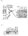
- FISH analysis was carried out using 15 types of HAC clones substantially typified by RP11-792P2. Also, the copy number of the corresponding genomic DNA region was schematically expressed based on fluorescent signal intensities (Fig. 3, left).
- the vertical axis represents the BAC clone IDs in the 11q23 region and relative positions thereof
- the homogeneously stained regions with the highest luminescence i.e., the homogenously staining regions (HSRs)
- HSRs homogenously staining regions
- the AMO1, RDX, ARHGAP20, POU2AF1, SNF1LK2, PPP2R1B, and ALG9 genes located in the determined 11q23 region were subjected to PCR for the purpose of simple quantification.
- GAPDH the expression level of which was known to be less likely to be influenced by cell species and conditions.
- Total RNA was recovered from cells at the logarithmic growth phase, and cDNA was prepared in accordance with a conventional technique. Primers and conditions specific to each gene were determined in advance, followed by PCR and electrophoresis in 3% agarose gel.
- the gel image was obtained using an LAS-3000 (Fuji Photo Film Co., Ltd.) and the obtained image was analyzed using Multi Gauge software (Fuji Photo Film Co., Ltd.) (Fig. 3, right).
- Multi Gauge software Feji Photo Film Co., Ltd.
- the 11q23 regions of which had been amplified, KMM1 and KM-5, which did not exhibit amplification in this region were used.
- LCL lymphocyte clones obtained from a healthy volunteer was used as control. Table 2 shows the summary of the results of quantification.
- Table 2 Comparison of expression analysis of genes in the 11 q23 region in the cells wherein the genome DNA of the 11 q23 region was amplified and in the cells wherein the genome DNA of the 11q23 region was not amplified Known gene Amplified cells
- Non-amplified cells Fold increase (/reference value) a AMO1 MOLP-2 KMM1 KM-5
- the expression level of the POU2AF1 gene was found to be elevated to a level 4.5 to 5.0 times higher than the average expression level in the cells (KMM1, KM-5) in which gene amplification was not observed.
- the lower part of the figure shows the copy number of the POU2AF1 gene in genomic DNA determined via FISH.
- the AMO1 and the MOLP-2 cells in which amplification of the POU2AF1 genes were observed in their genomic DNAs exhibited higher expression levels of POU2AF1 proteins than other cells.
- siRNAs of POU2AF1 were designed as POU2AF1-A: CACCUUACACCGAGUAUGU (SEQ ID NO: 3) and POU2AF1-B:GGUUCUGUGUCUGCAGU (SEQ ID NO: 4), and were purchased (Japan Bioservice).
- POU2AF1-A CACCUUACACCGAGUAUGU
- POU2AF1-B GGUUCUGUGUCUGCAGU
- SEQ ID NO: 4 purchased (Japan Bioservice).
- 200 nM of each primer was transduced with the aid of Nucleofector (Germany), and the efficiency thereof was analyzed by Western blotting by monitoring via fluorescence-labeled pmaxGFP analysis in the same manner as in Example 4 (Fig. 5A).
- An anti- ⁇ -actin antibody was used as a control.
- siRNA of POU2AF1 was transduced into 1 x 10 4 AMO1 and KMS-21BM cells, the resultants were sowed on a 96-well plate, and the viable cell count was determined by water-soluble tetrazolium salt (WST) assay (Cell counting kit-8; Dojindo) with the elapse of time (Figs. 5B and 5C). The experiment was carried out twice in triplicate, and an asterisk is used for results that were determined to be significant (p ⁇ 0.05) via the unpaired Student's t test. In both cells, cell growth was inhibited by introducing siRNA of POU2AF1.
- WST water-soluble tetrazolium salt
- Example 6 Establishment of POU2AF1 gene-expressing cell line in KMS-11 cells, distribution thereof, and observation of influence on viable cell count
- An expression vector (pCMV-Tag3-p34-POU2AF1) was constructed so as to express the full-length POU2AF1 gene (p34) wherein Myc was bound to the N-terminus.
- the resulting vector was introduced into the KMS-11 cell in which the expression level of endogenous POU2AF 1 gene was low using Nucleofector, and drug selection was carried out for 3 weeks with G418 (50 ⁇ g/ml).
- the occurrence of POU2AF1 gene expression was detected using a solution of disrupted cells via Western blotting using an anti-Myc antibody (Fig. 6A).
- pCMV-3B that did not contain any foreign gene was introduced into the KMS-11 cell, and the resultant was designated as a "mock control.”
- the recombinant POU2AF1 proteins were detected in cells into which the gene had been introduced.
- the transformed cells were cultured on a glass slide with a Shandon cytospin (Thermo), centrifuged, immobilized with 10% formamide and stained with an anti-Myc polyclonal antibody (Cell Signaling Technology) overnight. After the reaction, the antibody was stained with the Alexa-488-labeled sheep-derived anti-rabbit antibody (Molecular Probes), and the stained antibody was observed under a fluorescence microscope.
- Example 7 TNFRSF17 as a POU2AF1 gene-regulating gene
- TNFRSF17 was found to be the only gene that would exhibit the same behavior as the POU2AF1 gene in the multiple myeloma cells (data is not shown).
- gene expression behavior of TNFRSF 17 was thoroughly analyzed (Fig. 7).
- 200 nM each of siRNA of POU2AF1-A and POU2AF1-B were transduced into 2 x 10 6 AMO1 and KMS-21BM cells (only POU2AF1-B regarding the KMS-21BM cells) using Nucleofector, and expression of the TNFRSF17 gene was analyzed by real-time RT-PCR 48 hours thereafter (Fig. 7A). In both cells, the expression levels of the TNFRSF17 genes were found to become lower with the knocking down of the POU2AF1 gene.
- Example 6 the KMS-11 cell line expressing the POU2AF1 gene was prepared with the mock control.
- the TNFRSF17 gene expression behaviors in those cells were compared by real-time RT-PCR (Fig. 7B).
- Fig. 7B the expression level of the TNFRSF17 gene was found to increase in a cell in which the POU2AF1 gene was forced to express.
- a transcription-factor-binding site in the 5' region of the human TNFRSF17 gene in genomic DNA was searched by using the database (RecGroupScan; http://wwwmgs.bionet.nsc.ru/mgs/programs/yura/RecGropScanStart.html).
- the cells were subjected to immunoprecipitation using an anti-POU2AF1 antibody (Santa Cruz Biotechnology), and 1/100 of the resultant was used as a template for PCR.
- the "input" indicates a chromatin material before immunoprecipitation, and (-) indicates a negative control without any immunoprecipitation antibody.
- the octamer site bound to the POU2AF1 protein was detected in the POU2AF1 gene-expressing cell and in the AMO1 cell that would express the endogenous POU2AF1 gene.
- Example 8 Promoter-reporter assay in combination with the POU2AF1 expression vector using an octamer-site containing region upstream of the TNFRSF17 gene
- a 307-bp DNA region containing the octamer site upstream of the TNFRSF17 gene was cloned by PCR, a region containing an octamer site of a wild type (wt; TTTAGCAT) and a region containing an octamer site of a mutant (Mut; TTCCGCAT) were introduced into pGL3-promoter vectors (Promega), and the resultants were designated as a pGL3-TNFRSF17-wt octamer and a pGL3-TNFRSF17-Mut octamer, respectively.
- the two types of luelforuse-promoter-reporter vectors were transduced into the KMS-11 cell together with p34 or p35 of pCMV-Tag3-POU2AF1 and internal reference vectors, phRL-TK (Promega), for correcting transduction efficiency using the lipofectamine 2000 (Invitrogen). Luminescence was assayed using the dual-luciferase reporter assay system (Promega) 48 hours thereafter. The activity of the cell into which pGL3-TNFRSF17-wt octamer and pCMV-Tag3 (mock) had been transduced was designated as 1, and relative luciferase activity was shown (Fig. 8).
- the POU2AF1 and TNFRSF17 gene expression levels in the multiple myeloma cell lines and the primary cells established from the multiple myeloma clinical sample were measured by quantitative PCR, and the determined values were represented in terms of the ratio relative to ⁇ -actin and ⁇ 2-microglobulin ( ⁇ 2M) (Fig. 9A: cell line; B: primary cell).
- Expression of the POU2AF1 gene was highly correlated with that of the TNFRSF17 gene both in the cell lines and in the primary cells, and both values were significant p values (significance determined by an unpaired Student's t test of p ⁇ 0.05).
- TNFRSF17 gene expression was found to be positively correlated with POU2AF1 gene expression not only in the cell line but also in a clinical sample.
- the AMO1 cells in which the expression level of the POU2AF1 gene was high and the KMS-21BM cells in which expression of the POU2AF1 gene was observed were used.
- a nonspecific sequence was employed as control siRNA, and siRNA of the FOU2AF1 gene and the TNFRSF17 gene (200 nM each, M-011217-00, Dharmacon, Inc.) were transduced.
- POU2AF1 and TNFRSF17 gene expression was assayed by quantitative PCR using ⁇ -actin as an indicator 48 hours thereafter (Fig. 10A: POU2AF1; Fig. 10 B: TNFRSF17).
- POU2AF1 gene expression was inhibited by inhibiting the POU2AF1 gene by siRNA; however, POU2AF1 gene expression was not inhibited by siRNA of the TNFRSF17 gene, and TNFRSF17 gene expression was inhibited by inhibiting the POU2AF 1 gene by siRNA. This indicates that the POU2AF1 gene inhibits TNFRSF17 gene expression at a site upstream of the TNFRSF17 gene.
- the viable cell counts in the AMO1 and the KMS-21BM cells after the inhibition of the TNFRSF17 genes by siRNA were determined by the WST technique (Fig. 10C).
- TNFRSF17 gene expression is positively regulated by POU2AF1 gene expression, and the effects thereof are exhibited as the cell growth effects.
- the present invention enables typing of tumor and accurate understanding of an indication of tumorigenesis and the extent of recovery from tumor of a multiple myeloma cell specimen. Also, inhibition of the POU2AF1 gene transcript amplified in the multiple myeloma cell enables inhibition of growth of multiple myeloma cells. Specifically, the present invention provides an inhibitor for transcription of the POU2AF1 gene, the tumorigenic functions of which have been newly found, or an antitumorigenic agent comprising a loss-of-function phenotype of the POU2AF1 protein encoded by such gene. Such agents are very useful from the clinical point of view in terms of improvement in treatment or prognosis of tumors based on tumor individuality, and also from the viewpoint of basic research concerning tumors. Further, assay of the expression level of messenger RNA of the POU2AF1 gene, determination of the amount of such gene in genomic DNA, or assay of the POU2AF1 protein level enables genetic typing of tumor of a patient with multiple myeloma.
Landscapes
- Health & Medical Sciences (AREA)
- Chemical & Material Sciences (AREA)
- Life Sciences & Earth Sciences (AREA)
- Organic Chemistry (AREA)
- Proteomics, Peptides & Aminoacids (AREA)
- Engineering & Computer Science (AREA)
- Pathology (AREA)
- Immunology (AREA)
- Zoology (AREA)
- General Health & Medical Sciences (AREA)
- Wood Science & Technology (AREA)
- Analytical Chemistry (AREA)
- Genetics & Genomics (AREA)
- Bioinformatics & Cheminformatics (AREA)
- Biophysics (AREA)
- Molecular Biology (AREA)
- Medicinal Chemistry (AREA)
- Hospice & Palliative Care (AREA)
- Veterinary Medicine (AREA)
- General Chemical & Material Sciences (AREA)
- Physics & Mathematics (AREA)
- Pharmacology & Pharmacy (AREA)
- Chemical Kinetics & Catalysis (AREA)
- Biotechnology (AREA)
- Microbiology (AREA)
- Oncology (AREA)
- Public Health (AREA)
- Animal Behavior & Ethology (AREA)
- Biochemistry (AREA)
- Nuclear Medicine, Radiotherapy & Molecular Imaging (AREA)
- General Engineering & Computer Science (AREA)
- Physical Education & Sports Medicine (AREA)
- Measuring Or Testing Involving Enzymes Or Micro-Organisms (AREA)
- Medicines That Contain Protein Lipid Enzymes And Other Medicines (AREA)
- Pharmaceuticals Containing Other Organic And Inorganic Compounds (AREA)
Applications Claiming Priority (1)
| Application Number | Priority Date | Filing Date | Title |
|---|---|---|---|
| JP2006255155A JP2008072939A (ja) | 2006-09-21 | 2006-09-21 | 多発性骨髄腫の検出方法および抑制方法 |
Publications (2)
| Publication Number | Publication Date |
|---|---|
| EP1905845A2 true EP1905845A2 (de) | 2008-04-02 |
| EP1905845A3 EP1905845A3 (de) | 2008-09-17 |
Family
ID=38924734
Family Applications (1)
| Application Number | Title | Priority Date | Filing Date |
|---|---|---|---|
| EP20070018522 Withdrawn EP1905845A3 (de) | 2006-09-21 | 2007-09-20 | Verfahren zur Erkennung mehrerer Myelome und Verfahren zu deren Hemmung |
Country Status (4)
| Country | Link |
|---|---|
| US (1) | US20080182250A1 (de) |
| EP (1) | EP1905845A3 (de) |
| JP (1) | JP2008072939A (de) |
| CN (1) | CN101392285A (de) |
Cited By (1)
| Publication number | Priority date | Publication date | Assignee | Title |
|---|---|---|---|---|
| EP2810655A4 (de) * | 2012-01-27 | 2015-10-28 | Univ Osaka | Neuartige antitumormittel und screeningverfahren dafür |
Families Citing this family (5)
| Publication number | Priority date | Publication date | Assignee | Title |
|---|---|---|---|---|
| JP5331404B2 (ja) * | 2008-08-01 | 2013-10-30 | 国立大学法人 東京医科歯科大学 | 先天性異常症の染色体欠失の検出方法 |
| EP4092045A1 (de) | 2016-05-16 | 2022-11-23 | Checkmab S.R.L. | Selektiv deregulierte markermolekülen in tumor-infiltrierenden regulierende t-zellen |
| CN108179181B (zh) * | 2017-12-29 | 2020-12-15 | 北京泱深生物信息技术有限公司 | Rdx基因在临床用药中的应用 |
| US12150973B2 (en) | 2018-05-03 | 2024-11-26 | University Of Utah Research Foundation | OCA-B peptide conjugates and methods of treatment |
| CN110197701B (zh) * | 2019-04-22 | 2021-08-10 | 福建医科大学附属第一医院 | 一种新型多发性骨髓瘤诺模图构建方法 |
Family Cites Families (3)
| Publication number | Priority date | Publication date | Assignee | Title |
|---|---|---|---|---|
| WO1995032284A1 (en) * | 1994-05-24 | 1995-11-30 | Ciba-Geigy Ag | Factor interacting with nuclear proteins |
| US7935679B2 (en) * | 2001-11-07 | 2011-05-03 | Board Of Trustees Of The University Of Arkansas | Gene expression profiling based identification of CKS1B as a potential therapeutic target in multiple myeloma |
| WO2005005980A2 (en) * | 2003-07-14 | 2005-01-20 | Imed Ab | Methods for tumor evaluation, identification of anti-tumor agents and treatment |
-
2006
- 2006-09-21 JP JP2006255155A patent/JP2008072939A/ja not_active Abandoned
-
2007
- 2007-09-20 EP EP20070018522 patent/EP1905845A3/de not_active Withdrawn
- 2007-09-21 CN CNA2007101406506A patent/CN101392285A/zh active Pending
- 2007-09-21 US US11/902,504 patent/US20080182250A1/en not_active Abandoned
Cited By (1)
| Publication number | Priority date | Publication date | Assignee | Title |
|---|---|---|---|---|
| EP2810655A4 (de) * | 2012-01-27 | 2015-10-28 | Univ Osaka | Neuartige antitumormittel und screeningverfahren dafür |
Also Published As
| Publication number | Publication date |
|---|---|
| CN101392285A (zh) | 2009-03-25 |
| EP1905845A3 (de) | 2008-09-17 |
| US20080182250A1 (en) | 2008-07-31 |
| JP2008072939A (ja) | 2008-04-03 |
Similar Documents
| Publication | Publication Date | Title |
|---|---|---|
| EP1837405B1 (de) | Verfahren zur Erkennung von anaplastischem Schilddrüsenkrebs, und Verfahren zur seiner Unterdrückung | |
| EP2740742B1 (de) | Fusionsgen des kif5b-gens und ret-gens sowie verfahren zur bestimmung der wirksamkeit einer auf das fusionsgen abzielenden krebsbehandlung | |
| US8044193B2 (en) | Short interfering RNAs targeted to T-LAK cell-originated protein kinase (TOPK) polynucleotide | |
| DK2456889T3 (en) | Markers of endometrial cancer | |
| JP2005304497A (ja) | 特定の癌関連遺伝子を用いる癌の検出方法及び癌の抑制方法 | |
| EP1997911A2 (de) | Verfahren zur Erkennung von oralem Plattenepithelkarzinom und Verfahren zur Unterdrückung desselben | |
| KR20150122731A (ko) | 암 전이의 예후 및 치료 방법 | |
| KR20130115250A (ko) | 암에 대한 분자적 진단 테스트 | |
| EP1997910B1 (de) | Verfahren zur Erkennung von Ovarialkarzinom und Verfahren zur Unterdrückung desselben | |
| EP1905845A2 (de) | Verfahren zur Erkennung mehrerer Myelome und Verfahren zu deren Hemmung | |
| US20120207755A1 (en) | Cancer | |
| CN106701986A (zh) | 分子标志物在胃癌诊治中的应用 | |
| US20090011424A1 (en) | Cancer-suppressing agents | |
| EP2253720B1 (de) | Verfahren zur Erkennung eines Ösophagus-Karzinoms und Mittel zur Ösophagus-Karzinomunterdrückung | |
| US9229003B2 (en) | Method for detecting thyroid carcinoma | |
| JP5645089B2 (ja) | 卵巣癌の検出方法、及び抑制方法 | |
| JP5704545B2 (ja) | 甲状腺癌の検出方法 | |
| WO2008075713A1 (ja) | 癌診断用マーカーならびに治療の標的分子 | |
| CN105861736B (zh) | miRNA在子宫内膜癌诊疗中的应用 |
Legal Events
| Date | Code | Title | Description |
|---|---|---|---|
| PUAI | Public reference made under article 153(3) epc to a published international application that has entered the european phase |
Free format text: ORIGINAL CODE: 0009012 |
|
| AK | Designated contracting states |
Kind code of ref document: A2 Designated state(s): AT BE BG CH CY CZ DE DK EE ES FI FR GB GR HU IE IS IT LI LT LU LV MC MT NL PL PT RO SE SI SK TR |
|
| AX | Request for extension of the european patent |
Extension state: AL BA HR MK YU |
|
| PUAL | Search report despatched |
Free format text: ORIGINAL CODE: 0009013 |
|
| AK | Designated contracting states |
Kind code of ref document: A3 Designated state(s): AT BE BG CH CY CZ DE DK EE ES FI FR GB GR HU IE IS IT LI LT LU LV MC MT NL PL PT RO SE SI SK TR |
|
| AX | Request for extension of the european patent |
Extension state: AL BA HR MK RS |
|
| 17P | Request for examination filed |
Effective date: 20090317 |
|
| AKX | Designation fees paid |
Designated state(s): AT BE BG CH CY CZ DE DK EE ES FI FR GB GR HU IE IS IT LI LT LU LV MC MT NL PL PT RO SE SI SK TR |
|
| 17Q | First examination report despatched |
Effective date: 20091105 |
|
| STAA | Information on the status of an ep patent application or granted ep patent |
Free format text: STATUS: THE APPLICATION IS DEEMED TO BE WITHDRAWN |
|
| 18D | Application deemed to be withdrawn |
Effective date: 20100316 |