EP1910575B1 - Procede de detection par immunohistochimie - Google Patents
Procede de detection par immunohistochimie Download PDFInfo
- Publication number
- EP1910575B1 EP1910575B1 EP06809193.3A EP06809193A EP1910575B1 EP 1910575 B1 EP1910575 B1 EP 1910575B1 EP 06809193 A EP06809193 A EP 06809193A EP 1910575 B1 EP1910575 B1 EP 1910575B1
- Authority
- EP
- European Patent Office
- Prior art keywords
- nucleic acid
- pna
- minutes
- acid analog
- dako
- Prior art date
- Legal status (The legal status is an assumption and is not a legal conclusion. Google has not performed a legal analysis and makes no representation as to the accuracy of the status listed.)
- Not-in-force
Links
- 0 *c1c(*C=N2)c2ncn1 Chemical compound *c1c(*C=N2)c2ncn1 0.000 description 5
Images











Classifications
-
- C—CHEMISTRY; METALLURGY
- C12—BIOCHEMISTRY; BEER; SPIRITS; WINE; VINEGAR; MICROBIOLOGY; ENZYMOLOGY; MUTATION OR GENETIC ENGINEERING
- C12Q—MEASURING OR TESTING PROCESSES INVOLVING ENZYMES, NUCLEIC ACIDS OR MICROORGANISMS; COMPOSITIONS OR TEST PAPERS THEREFOR; PROCESSES OF PREPARING SUCH COMPOSITIONS; CONDITION-RESPONSIVE CONTROL IN MICROBIOLOGICAL OR ENZYMOLOGICAL PROCESSES
- C12Q1/00—Measuring or testing processes involving enzymes, nucleic acids or microorganisms; Compositions therefor; Processes of preparing such compositions
- C12Q1/68—Measuring or testing processes involving enzymes, nucleic acids or microorganisms; Compositions therefor; Processes of preparing such compositions involving nucleic acids
- C12Q1/6804—Nucleic acid analysis using immunogens
-
- A—HUMAN NECESSITIES
- A61—MEDICAL OR VETERINARY SCIENCE; HYGIENE
- A61K—PREPARATIONS FOR MEDICAL, DENTAL OR TOILETRY PURPOSES
- A61K49/00—Preparations for testing in vivo
- A61K49/001—Preparation for luminescence or biological staining
- A61K49/0013—Luminescence
- A61K49/0017—Fluorescence in vivo
- A61K49/0019—Fluorescence in vivo characterised by the fluorescent group, e.g. oligomeric, polymeric or dendritic molecules
- A61K49/0021—Fluorescence in vivo characterised by the fluorescent group, e.g. oligomeric, polymeric or dendritic molecules the fluorescent group being a small organic molecule
- A61K49/0041—Xanthene dyes, used in vivo, e.g. administered to a mice, e.g. rhodamines, rose Bengal
-
- A—HUMAN NECESSITIES
- A61—MEDICAL OR VETERINARY SCIENCE; HYGIENE
- A61K—PREPARATIONS FOR MEDICAL, DENTAL OR TOILETRY PURPOSES
- A61K47/00—Medicinal preparations characterised by the non-active ingredients used, e.g. carriers or inert additives; Targeting or modifying agents chemically bound to the active ingredient
- A61K47/50—Medicinal preparations characterised by the non-active ingredients used, e.g. carriers or inert additives; Targeting or modifying agents chemically bound to the active ingredient the non-active ingredient being chemically bound to the active ingredient, e.g. polymer-drug conjugates
- A61K47/51—Medicinal preparations characterised by the non-active ingredients used, e.g. carriers or inert additives; Targeting or modifying agents chemically bound to the active ingredient the non-active ingredient being chemically bound to the active ingredient, e.g. polymer-drug conjugates the non-active ingredient being a modifying agent
- A61K47/56—Medicinal preparations characterised by the non-active ingredients used, e.g. carriers or inert additives; Targeting or modifying agents chemically bound to the active ingredient the non-active ingredient being chemically bound to the active ingredient, e.g. polymer-drug conjugates the non-active ingredient being a modifying agent the modifying agent being an organic macromolecular compound, e.g. an oligomeric, polymeric or dendrimeric molecule
- A61K47/61—Medicinal preparations characterised by the non-active ingredients used, e.g. carriers or inert additives; Targeting or modifying agents chemically bound to the active ingredient the non-active ingredient being chemically bound to the active ingredient, e.g. polymer-drug conjugates the non-active ingredient being a modifying agent the modifying agent being an organic macromolecular compound, e.g. an oligomeric, polymeric or dendrimeric molecule the organic macromolecular compound being a polysaccharide or a derivative thereof
-
- A—HUMAN NECESSITIES
- A61—MEDICAL OR VETERINARY SCIENCE; HYGIENE
- A61K—PREPARATIONS FOR MEDICAL, DENTAL OR TOILETRY PURPOSES
- A61K47/00—Medicinal preparations characterised by the non-active ingredients used, e.g. carriers or inert additives; Targeting or modifying agents chemically bound to the active ingredient
- A61K47/50—Medicinal preparations characterised by the non-active ingredients used, e.g. carriers or inert additives; Targeting or modifying agents chemically bound to the active ingredient the non-active ingredient being chemically bound to the active ingredient, e.g. polymer-drug conjugates
- A61K47/51—Medicinal preparations characterised by the non-active ingredients used, e.g. carriers or inert additives; Targeting or modifying agents chemically bound to the active ingredient the non-active ingredient being chemically bound to the active ingredient, e.g. polymer-drug conjugates the non-active ingredient being a modifying agent
- A61K47/68—Medicinal preparations characterised by the non-active ingredients used, e.g. carriers or inert additives; Targeting or modifying agents chemically bound to the active ingredient the non-active ingredient being chemically bound to the active ingredient, e.g. polymer-drug conjugates the non-active ingredient being a modifying agent the modifying agent being an antibody, an immunoglobulin or a fragment thereof, e.g. an Fc-fragment
- A61K47/6835—Medicinal preparations characterised by the non-active ingredients used, e.g. carriers or inert additives; Targeting or modifying agents chemically bound to the active ingredient the non-active ingredient being chemically bound to the active ingredient, e.g. polymer-drug conjugates the non-active ingredient being a modifying agent the modifying agent being an antibody, an immunoglobulin or a fragment thereof, e.g. an Fc-fragment the modifying agent being an antibody or an immunoglobulin bearing at least one antigen-binding site
- A61K47/6843—Medicinal preparations characterised by the non-active ingredients used, e.g. carriers or inert additives; Targeting or modifying agents chemically bound to the active ingredient the non-active ingredient being chemically bound to the active ingredient, e.g. polymer-drug conjugates the non-active ingredient being a modifying agent the modifying agent being an antibody, an immunoglobulin or a fragment thereof, e.g. an Fc-fragment the modifying agent being an antibody or an immunoglobulin bearing at least one antigen-binding site the antibody targeting a material from animals or humans
-
- A—HUMAN NECESSITIES
- A61—MEDICAL OR VETERINARY SCIENCE; HYGIENE
- A61K—PREPARATIONS FOR MEDICAL, DENTAL OR TOILETRY PURPOSES
- A61K47/00—Medicinal preparations characterised by the non-active ingredients used, e.g. carriers or inert additives; Targeting or modifying agents chemically bound to the active ingredient
- A61K47/50—Medicinal preparations characterised by the non-active ingredients used, e.g. carriers or inert additives; Targeting or modifying agents chemically bound to the active ingredient the non-active ingredient being chemically bound to the active ingredient, e.g. polymer-drug conjugates
- A61K47/51—Medicinal preparations characterised by the non-active ingredients used, e.g. carriers or inert additives; Targeting or modifying agents chemically bound to the active ingredient the non-active ingredient being chemically bound to the active ingredient, e.g. polymer-drug conjugates the non-active ingredient being a modifying agent
- A61K47/68—Medicinal preparations characterised by the non-active ingredients used, e.g. carriers or inert additives; Targeting or modifying agents chemically bound to the active ingredient the non-active ingredient being chemically bound to the active ingredient, e.g. polymer-drug conjugates the non-active ingredient being a modifying agent the modifying agent being an antibody, an immunoglobulin or a fragment thereof, e.g. an Fc-fragment
- A61K47/6889—Conjugates wherein the antibody being the modifying agent and wherein the linker, binder or spacer confers particular properties to the conjugates, e.g. peptidic enzyme-labile linkers or acid-labile linkers, providing for an acid-labile immuno conjugate wherein the drug may be released from its antibody conjugated part in an acidic, e.g. tumoural or environment
-
- A—HUMAN NECESSITIES
- A61—MEDICAL OR VETERINARY SCIENCE; HYGIENE
- A61K—PREPARATIONS FOR MEDICAL, DENTAL OR TOILETRY PURPOSES
- A61K49/00—Preparations for testing in vivo
- A61K49/001—Preparation for luminescence or biological staining
- A61K49/0013—Luminescence
- A61K49/0017—Fluorescence in vivo
- A61K49/0019—Fluorescence in vivo characterised by the fluorescent group, e.g. oligomeric, polymeric or dendritic molecules
- A61K49/0021—Fluorescence in vivo characterised by the fluorescent group, e.g. oligomeric, polymeric or dendritic molecules the fluorescent group being a small organic molecule
- A61K49/0041—Xanthene dyes, used in vivo, e.g. administered to a mice, e.g. rhodamines, rose Bengal
- A61K49/0043—Fluorescein, used in vivo
-
- A—HUMAN NECESSITIES
- A61—MEDICAL OR VETERINARY SCIENCE; HYGIENE
- A61K—PREPARATIONS FOR MEDICAL, DENTAL OR TOILETRY PURPOSES
- A61K49/00—Preparations for testing in vivo
- A61K49/001—Preparation for luminescence or biological staining
- A61K49/0013—Luminescence
- A61K49/0017—Fluorescence in vivo
- A61K49/005—Fluorescence in vivo characterised by the carrier molecule carrying the fluorescent agent
- A61K49/0056—Peptides, proteins, polyamino acids
-
- C—CHEMISTRY; METALLURGY
- C07—ORGANIC CHEMISTRY
- C07C—ACYCLIC OR CARBOCYCLIC COMPOUNDS
- C07C231/00—Preparation of carboxylic acid amides
- C07C231/02—Preparation of carboxylic acid amides from carboxylic acids or from esters, anhydrides, or halides thereof by reaction with ammonia or amines
-
- C—CHEMISTRY; METALLURGY
- C07—ORGANIC CHEMISTRY
- C07C—ACYCLIC OR CARBOCYCLIC COMPOUNDS
- C07C233/00—Carboxylic acid amides
- C07C233/01—Carboxylic acid amides having carbon atoms of carboxamide groups bound to hydrogen atoms or to acyclic carbon atoms
- C07C233/34—Carboxylic acid amides having carbon atoms of carboxamide groups bound to hydrogen atoms or to acyclic carbon atoms having the nitrogen atom of at least one of the carboxamide groups bound to a carbon atom of a hydrocarbon radical substituted by amino groups
- C07C233/35—Carboxylic acid amides having carbon atoms of carboxamide groups bound to hydrogen atoms or to acyclic carbon atoms having the nitrogen atom of at least one of the carboxamide groups bound to a carbon atom of a hydrocarbon radical substituted by amino groups with the substituted hydrocarbon radical bound to the nitrogen atom of the carboxamide group by an acyclic carbon atom
- C07C233/36—Carboxylic acid amides having carbon atoms of carboxamide groups bound to hydrogen atoms or to acyclic carbon atoms having the nitrogen atom of at least one of the carboxamide groups bound to a carbon atom of a hydrocarbon radical substituted by amino groups with the substituted hydrocarbon radical bound to the nitrogen atom of the carboxamide group by an acyclic carbon atom having the carbon atom of the carboxamide group bound to a hydrogen atom or to a carbon atom of an acyclic saturated carbon skeleton
-
- C—CHEMISTRY; METALLURGY
- C07—ORGANIC CHEMISTRY
- C07H—SUGARS; DERIVATIVES THEREOF; NUCLEOSIDES; NUCLEOTIDES; NUCLEIC ACIDS
- C07H19/00—Compounds containing a hetero ring sharing one ring hetero atom with a saccharide radical; Nucleosides; Mononucleotides; Anhydro-derivatives thereof
- C07H19/02—Compounds containing a hetero ring sharing one ring hetero atom with a saccharide radical; Nucleosides; Mononucleotides; Anhydro-derivatives thereof sharing nitrogen
- C07H19/04—Heterocyclic radicals containing only nitrogen atoms as ring hetero atom
- C07H19/06—Pyrimidine radicals
-
- C—CHEMISTRY; METALLURGY
- C07—ORGANIC CHEMISTRY
- C07H—SUGARS; DERIVATIVES THEREOF; NUCLEOSIDES; NUCLEOTIDES; NUCLEIC ACIDS
- C07H19/00—Compounds containing a hetero ring sharing one ring hetero atom with a saccharide radical; Nucleosides; Mononucleotides; Anhydro-derivatives thereof
- C07H19/02—Compounds containing a hetero ring sharing one ring hetero atom with a saccharide radical; Nucleosides; Mononucleotides; Anhydro-derivatives thereof sharing nitrogen
- C07H19/04—Heterocyclic radicals containing only nitrogen atoms as ring hetero atom
- C07H19/06—Pyrimidine radicals
- C07H19/10—Pyrimidine radicals with the saccharide radical esterified by phosphoric or polyphosphoric acids
-
- C—CHEMISTRY; METALLURGY
- C07—ORGANIC CHEMISTRY
- C07H—SUGARS; DERIVATIVES THEREOF; NUCLEOSIDES; NUCLEOTIDES; NUCLEIC ACIDS
- C07H19/00—Compounds containing a hetero ring sharing one ring hetero atom with a saccharide radical; Nucleosides; Mononucleotides; Anhydro-derivatives thereof
- C07H19/02—Compounds containing a hetero ring sharing one ring hetero atom with a saccharide radical; Nucleosides; Mononucleotides; Anhydro-derivatives thereof sharing nitrogen
- C07H19/04—Heterocyclic radicals containing only nitrogen atoms as ring hetero atom
- C07H19/16—Purine radicals
-
- C—CHEMISTRY; METALLURGY
- C07—ORGANIC CHEMISTRY
- C07H—SUGARS; DERIVATIVES THEREOF; NUCLEOSIDES; NUCLEOTIDES; NUCLEIC ACIDS
- C07H19/00—Compounds containing a hetero ring sharing one ring hetero atom with a saccharide radical; Nucleosides; Mononucleotides; Anhydro-derivatives thereof
- C07H19/02—Compounds containing a hetero ring sharing one ring hetero atom with a saccharide radical; Nucleosides; Mononucleotides; Anhydro-derivatives thereof sharing nitrogen
- C07H19/04—Heterocyclic radicals containing only nitrogen atoms as ring hetero atom
- C07H19/16—Purine radicals
- C07H19/20—Purine radicals with the saccharide radical esterified by phosphoric or polyphosphoric acids
-
- C—CHEMISTRY; METALLURGY
- C07—ORGANIC CHEMISTRY
- C07H—SUGARS; DERIVATIVES THEREOF; NUCLEOSIDES; NUCLEOTIDES; NUCLEIC ACIDS
- C07H21/00—Compounds containing two or more mononucleotide units having separate phosphate or polyphosphate groups linked by saccharide radicals of nucleoside groups, e.g. nucleic acids
-
- C—CHEMISTRY; METALLURGY
- C08—ORGANIC MACROMOLECULAR COMPOUNDS; THEIR PREPARATION OR CHEMICAL WORKING-UP; COMPOSITIONS BASED THEREON
- C08G—MACROMOLECULAR COMPOUNDS OBTAINED OTHERWISE THAN BY REACTIONS ONLY INVOLVING UNSATURATED CARBON-TO-CARBON BONDS
- C08G69/00—Macromolecular compounds obtained by reactions forming a carboxylic amide link in the main chain of the macromolecule
- C08G69/02—Polyamides derived from amino-carboxylic acids or from polyamines and polycarboxylic acids
-
- C—CHEMISTRY; METALLURGY
- C12—BIOCHEMISTRY; BEER; SPIRITS; WINE; VINEGAR; MICROBIOLOGY; ENZYMOLOGY; MUTATION OR GENETIC ENGINEERING
- C12Q—MEASURING OR TESTING PROCESSES INVOLVING ENZYMES, NUCLEIC ACIDS OR MICROORGANISMS; COMPOSITIONS OR TEST PAPERS THEREFOR; PROCESSES OF PREPARING SUCH COMPOSITIONS; CONDITION-RESPONSIVE CONTROL IN MICROBIOLOGICAL OR ENZYMOLOGICAL PROCESSES
- C12Q1/00—Measuring or testing processes involving enzymes, nucleic acids or microorganisms; Compositions therefor; Processes of preparing such compositions
- C12Q1/68—Measuring or testing processes involving enzymes, nucleic acids or microorganisms; Compositions therefor; Processes of preparing such compositions involving nucleic acids
- C12Q1/6876—Nucleic acid products used in the analysis of nucleic acids, e.g. primers or probes
-
- G—PHYSICS
- G01—MEASURING; TESTING
- G01N—INVESTIGATING OR ANALYSING MATERIALS BY DETERMINING THEIR CHEMICAL OR PHYSICAL PROPERTIES
- G01N33/00—Investigating or analysing materials by specific methods not covered by groups G01N1/00 - G01N31/00
- G01N33/48—Biological material, e.g. blood, urine; Haemocytometers
- G01N33/50—Chemical analysis of biological material, e.g. blood, urine; Testing involving biospecific ligand binding methods; Immunological testing
- G01N33/53—Immunoassay; Biospecific binding assay; Materials therefor
- G01N33/5306—Improving reaction conditions, e.g. reduction of non-specific binding, promotion of specific binding
Definitions
- the invention provides compositions and methods for the detection of targets in a sample, including an immunohistochemistry (IHC) sample.
- Probes and detectable labels may be provided in multiple layers in order to increase the flexibility of a detection system, and to enhance the signal from a target.
- the compositions and methods are compatible with a variety of detection systems, and with many Different types of targets, probes, and detectable labels.
- Detection of a target in a sample may ordinarily be achieved by contacting the target with a probe that specifically recognizes it.
- the probe may be linked, either directly or indirectly to a detectable label, such as a fluorophore or radioactive tag, which provides a signal representing the target.
- the label's signal may be enhanced by increasing the number of detectable labels used to detect each target, or by instrumentation that may amplify the signal.
- a multiple-antibody system may amplify the detection signal.
- the target may first be bound by a primary antibody, which, in turn, is capable of binding many secondary antibodies or even tertiary antibodies, which, in their turn, bind to the probe. This method, thus, increases the number of probes that recognize each antigen target by adding extra layers of molecular interactions between the probe and target.
- the secondary antibody technique is widely used, but may be limited due to, for example i) the availability of the secondary antibodies; ii) unwanted cross reactivity between closely related species, e.g. rat, mouse; iii) the size of antibodies reduces the penetration of the reagents. Furthermore, conjugation of antibodies to other antibodies, enzymes, color labels, etc. is somewhat unique for every antibody due to biological variations. Fab or Fab2 fragments of IgG have been used to overcome the size and non-specific binding problems, but such secondary antibody based visualization systems still remain limited to staining of one, two or three different targets. Amplification of the signal from individual targets is both laborious and complex due to the above-mentioned technical limitations.
- the instant invention uses the flexibility of nucleic acid hybridization to provide a general set of compositions and methods that may be used to detect one or many targets in a sample and amplify their signals.
- the instant methods and compositions may also provide for increased flexibility as compared to, for example, capture assays or sandwich assays, such as those described in United States Patent 4,868,105 , for example, that rely on one type of binding interaction such as a hapten-protein or primary antibody-secondary antibody interaction.
- capture assays or sandwich assays such as those described in United States Patent 4,868,105 , for example, that rely on one type of binding interaction such as a hapten-protein or primary antibody-secondary antibody interaction.
- the ability to design a variety of nucleic acid analog hybridization pairs, for instance, may dramatically increase the ways in which the probe and detectable label may interact.
- compositions and methods may separate the probe and the detectable label such that they may be comprised on different units within a detection system. See, for example, Figure 1 a and 1 c.
- the units may then interact through specific hybridization of nucleic acid analog segments.
- the units may be designed such that a given probe may interact with a variety of.different detectable labels, depending upon the needs of the assay.
- the units may also be designed to include multiple interacting nucleic acid analog segments, either to increase the affinity between the units, or to amplify a signal.
- adaptor units may be included that provide, for example, one or more additional molecular layers between the probe and the detectable label. See Figure 1 b.
- the adaptor units may allow for even greater mixing and matching of probes and labels as well as additional amplification of a signal.
- the components of the instant compositions may also be designed to be of similar chemical compositions so that they may be prepared simply using standardized conjugation schemes.
- the methods and compositions of the invention may also be used to detect more than one target within a sample. For instance, multiple targets in a sample are not always expressed in equal amounts. Thus, there may be a differential need for amplification.
- the instant invention is also useful in normalizing the detection of two or more targets in a system.
- the system may also be designed such that one unit specifically hybridizes to more than one other unit.
- the various units of the invention may be designed such that one unit hybridizes to several other units, for example, by providing multiple nucleic acid analog segments on the same unit.
- certain nucleic acid analogs may allow for degenerate hybridization schemes such that one nucleic acid analog segment may specifically hybridize to more than one other nucleic acid analog segment, creating a "master key” unit. (See the international application entitled "New Nucleic Acid Base Pairs" submitted herewith, for an example of such segments.) This unique feature increases the flexibility of the instant compositions and methods even further.
- the instant compositions and methods are compatible with a large variety of samples and are adaptable to a large number of targets, probes, and detectable labels.
- the present invention is useful in immunohistochemistry applications (IHC), but can be applied to other detection methods as well.
- Other detection that may be compatible with this invention include, for example, immunocytochemistry (ICC), in situ hybridization (ISH), flow cytometry, enzyme immuno-assays (EIA), enzyme linked immuno-assays (ELISA), blotting methods (e.g. Western, Southern, and Northern), labeling inside electrophoresis systems or on surfaces or arrays, and precipitation, among others.
- detection formats for example, are useful in research as well as in diagnosing diseases or conditions. Further, if multiple targets are detected, such systems may be useful in analyzing expression patterns of genes or levels of proteins within a sample.
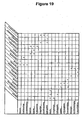
- Figure 19 shows interactions between each of the 18 bases shown in Figure 1 : 3 refers to three hydrogen bonds being present between the bases; 2 refers to two hydrogen bonds being present between the bases; 1 is the presence of one hydrogen bond; and X is a repulsion or no H bonding between the pairs. There are 3 three bond base pairs, 12 two bond base pairs, and 2 single bond base pairs. As may be seen from the figure and the text below, these pairing schemes may be used to expand the normal genetic code and thus may allow nucleic acid analog segments to specifically hybridize to more than one other nucleic acid analog segment within the instant recognition, adaptor, and detection units of the invention.
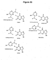
- Figure 20 depicts yet other non-natural bases and base-pairings that may be used in the inventive compositions and methods.
- Sample refers to any composition potentially containing a target.
- Target refers to any substance present in a sample that is capable of detection.
- a recognition unit is a substance that recognizes at least one target in a sample.
- the recognition unit comprises a probe , which, as defined herein, comprises any substance that is capable of recognizing a target.
- a recognition unit also comprises at least one nucleic acid analog segment.
- the recognition unit may also comprise at least one polymer and/or at least one linker.
- recognize, recognition, or recognizing , etc. mean an event in which one substance, such as a probe or recognition unit comprising a probe, directly or indirectly interacts with a target in any way such that the interaction with the target may be detected by a detection unit.
- a probe may react with a target, or directly bind to a target, or indirectly react with or bind to a target by directly binding to another substance that in turn directly binds to or reacts with a target.
- a detection unit refers to a substance comprising at least one detectable label, and capable of binding directly to a recognition unit or indirectly to a recognition unit through an optional adaptor unit.
- a detection unit also comprises at least one nucleic acid analog segment.
- the detection unit may also comprise at least one polymer and/or at least one linker.
- an adaptor unit means a substance that is capable of linking a recognition unit to a detection unit.
- an adaptor unit comprises at least two nucleic acid analog segments.
- the adaptor unit may also comprise at least one polymer and/or at least one linker.
- Non-limiting examples of the formation of hydrogen bonds between the segments include the formation of Watson-Crick, wobble, and Hoogsteen base-pair geometries, such as to form double strands.
- a primary binding agent as used herein refers to a substance that binds directly to a target in a sample.
- a secondary binding agent refers to a substance which binds directly to a primary binding agent.
- a tertiary binding agent refers to a substance which specifically binds a secondary binding agent.
- Antibody as used herein, means an immunoglobulin or a fragment thereof, and encompasses any polypeptide comprising an antigen-binding site regardless of the source, method of production, and other characteristics.
- An antigen refers to any substance recognized by an antibody.
- a detectable label is any molecule or functional group that allows for the detection of the presence of the target in the sample.
- base and nucleobase refer to any purine-like or pyrimidine-like molecule that may be comprised in a nucleic acid segment or nucleic acid analog segment.
- a nucleic acid segment refers to a nucleobase sequence comprising any oligomer, polymer, or polymer segment, having a backbone formed solely from RNA or DNA nucleosides and comprising only the bases adenine (A), cytosine (C), guanine (G), thymine (T), and uracil (U), wherein an oligomer means a sequence of two or more nucleobases.
- a non-natural base means any nucleobase other than: Adenine, A; Guanine, G; Urasil, U; Thymine, T; Cytosine, C.
- a non-natural backbone unit includes any type of backbone unit to which a nucleobase may be attached that is not a ribose-phosphate (RNA) or a deoxyribose-phosphate (DNA) backbone unit.
- RNA ribose-phosphate
- DNA deoxyribose-phosphate
- nucleic acid analog segment means any oligomer, polymer, or polymer segment, comprising at least one monomer that comprises a non-natural base and/or a non-natural backbone unit.
- compositions useful for detecting at least one target in a sample are provided.
- the invention provides for a composition comprising a recognition unit and a detection unit wherein:
- Figure 1a and 1c illustrate exemplary recognition units and detection units, while other non-limiting examples are provided in Figures 2-17 and elsewhere in the application as a whole.
- the nucleic acid analog segments present on the recognition, detection, and optional adaptor units may comprise at least one non-natural base and/or a non-natural backbone unit within the segment as a whole.
- non-natural units thus include, but are not limited to, for example, PNA's or phosphorothioate or 2'O-methyl nucleosides comprising the one of the natural bases A, C,G, T, or U, and, for example, natural RNA or DNA nucleosides comprising non-natural base such as 4-thio-Uracil or Inosine.
- Non-natural bases may include, for example, purine-like and pyrimidine-like molecules, such as those that may interact using Watson-Crick-type, wobble, or Hoogsteen-type pairing interactions. Examples include generally any nucleobase referred to elsewhere as “non-natural” or as an “analog.”
- Examples include: halogen-substituted bases, alkyl-substituted bases, hydroxy-substituted bases, and thiol-substituted bases, as well as 5-propynyl-uracil, 2-thio-5-propynyl-uracil, 5-methylcytosine, isoguanine, isocytosine, pseudoisocytosine, 4-thiouracil, 2-thiouracil and 2-thiothymine, inosine, 2-aminopurine, N9-(2-amino-6-chloropurine), N9-(2,6-diaminopurine), hypoxanthine, N9-(7-deaza-guanine), N9-(7-deaza-8-aza-guanine) and N8-(7-deaza-8-aza-adenine).
- bases in which one amino group with a hydrogen is substituted with a halogen small “h” below
- bases in which one amino group with a hydrogen is substituted with a halogen small “h” below
- bases in which one amino group with a hydrogen is substituted with a halogen small “h” below
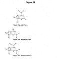
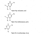
- R2 R3 R4 R5 R6 A H or CH 3 H H isoA H or CH 3 H H D H or CH 3 H G H or CH 3 H Gs H or CH 3 H I H or CH 3 H U H or CH 3 U2s H or CH 3 U4s H or CH 3 C H or CH 3 H Py-2o H or CH 3 H or CH 3 Cs H or CH 3 H isoG H or CH 3 H isoGs H or CH 3 H Pu-2o H or CH 3 H isoC H H or CH 3 isoCs H H or CH 3 Py-4o H or CH 3 H or CH 3 Base (Symbol) R2 R3 R4 R5 R6 A H or CH 3 H CH 3 isoA H or CH 3 H CH 3 D H or CH 3 CH 3 G H or CH 3 CH 3 Gs H or CH 3 CH 3 I H or CH 3 CH 3 U H or CH 3 U2s
- one or more of the H or CH 3 are independently substituted with a halogen such as Cl or F.
- a halogen such as Cl or F.
- R 1 or "BB" in the structures of Figures 18-20 may serve as a point of attachment to a backbone group, such as PNA, DNA, RNA, etc.
- the following types of base pairs are used: one or more of Us:A, T:D, C:G, and P:Gs.
- T:A and P:G are used. Still other examples are illustrated in Figures 2(A) and 2(B) of Buchardt et al. ( US 6,357,163 ).
- Nucleic acid analog segments also include any oligomer, polymer, or polymer segment, comprising at least one monomer with a non-natural backbone unit: in other words, any backbone unit that is not a phosphoribo (RNA) or a phosphodeoxyribo (DNA) unit.
- non-natural backbone units include, but are not limited to, for example PNA's or phosphorothioate or 2'O-methyl backbones.
- one or more phosphate oxygens may be replaced by another molecule, such as sulfur.
- a different sugar or a sugar analog may be used, for example, one in which a sugar oxygen is replaced by hydrogen or an amine, or an O-methyl.
- nucleic acid analog segments comprise synthetic molecules that can bind to a nucleic acid or nucleic acid analog.
- a nucleic acid analog may be comprised of peptide nucleic acids (PNAs), locked nucleic acids (LNAs), or any derivatized form of a nucleic acid.
- PNAs peptide nucleic acids
- LNAs locked nucleic acids
- Such backbone units may be attached to any base, including the natural bases A, C, G, T, and U, and non-natural bases.
- peptide nucleic acid or "PNA” means any oligomer or polymer comprising at least one or more PNA subunits (residues), including, but not limited to, any of the oligomer or polymer segments referred to or claimed as peptide nucleic acids in United States Patent Nos.
- PNA also applies to any oligomer or polymer segment comprising one or more subunits of the nucleic acid mimics described in the following publications: Lagriffoul et al., Bioorganic & Medicinal Chemistry Letters, 4: 1081-1082 (1994 ); Petersen et al., Bioorganic & Medicinal Chemistry Letters, 6: 793-796 (1996 ); Diderichsen et al., Tett. Lett. 37: 475-478 (1996 ); Fujii et al., Bioorg. Med. Chem. Lett. 7: 637-627 (1997 ); Jordan et al., Bioorg. Med. Chem. Lett.
- locked nucleic acid or "LNA” means an oligomer or polymer comprising at least one or more LNA subunits.
- LNA subunit means a ribonucleotide containing a methylene bridge that connects the 2'-oxygen of the ribose with the 4'-carbon. See generally, Kurreck, Eur. J. Biochem., 270:1628-44 (2003 ).
- Nucleic acid segments may be synthesized chemically or produced recombinantly in cells (see e.g. Sambrook et al. (1989) Molecular Cloning: A Laboratory Manual, 2nd ed. Cold Spring Harbor Press ). Methods of making PNAs and LNAs are also known in the art (see e.g. Nielson, 2001, Current Opinion in Biotechnology 12:16 ; Sorenson et al. 2003, Chem. Commun. 7(17):2130 ).
- one or more of the recognition, adaptor, and detection units according to the invention comprise more than one nucleic acid analog segment.
- the two segments may have the same or different sequences.
- nucleic acid analog segments may serve to link a recognition unit to a detector unit.
- Different nucleic acid analog segments may hybridize, for instance, using Watson-Crick-type, wobble, or Hoogsteen-type base-pairing. Accordingly, the nucleic acid analog segments comprise sequences which allow for hybridization to take place at a desired stringency.
- the nucleic acid analog segments may pair specifically with more that one other nucleic acid analog segment, thereby providing degeneracy to the recognition, detection and/or adaptor units. See, for example, the International Application submitted herewith entitled “New Nucleic Acid Base Pairs,” and see the examples below.
- nucleic acid analog segment may function as a "master-key" with the ability to hybridize to many partners, where each partner may also hybridize to separate nucleic acid analog segments.
- a very versatile and flexible detection system may be constructed in some embodiments that allows the user to choose between visualizing several targets via different detectable labels and detection units, or via only one detectable label and detection unit.
- a detectable label according to the invention may include any molecule which may be detected directly or indirectly so as to reveal the presence of a target in the sample.
- a direct detectable label is used.
- Direct detectable labels may be detected per se without the need for additional molecules. Examples include fluorescent dyes, radioactive substances, and metal particles.
- indirect detectable labels are used, which require the employment of one or more additional molecules. Examples include enzymes that affect a color change in a suitable substrate, as well as any molecule that may be specifically recognized by another substance carrying a label or react with a substance carrying a label.
- Other examples of indirect detectable labels thus include antibodies, antigens, nucleic acids and nucleic acid analogs, ligands, substrates, and haptens.
- detectable labels examples include fluorophores, chromophores, electrochemiluminescent labels, bioluminescent labels, polymers, polymer particles, bead or other solid surfaces, gold or other metal particles or heavy atoms, spin labels, radioisotopes, enzyme substrates, haptens, antigens, Quantum Dots, aminohexyl, pyrene, nucleic acids or nucleic acid analogs, or proteins, such as receptors, peptide ligands or substrates, enzymes, and antibodies (including antibody fragments).
- Some detectable labels according to this invention comprise “color labels,” in which the target is detected by the presence of a color, or a change in color in the sample.
- color labels are chromophores, fluorophores, chemiluminescent compounds, electrochemiluminescent labels, bioluminescent labels, and enzymes that catalyze a color change in a substrate.
- more than one type of color may be used, for instance, by attaching distinguishable color labels to a single detection unit or by using more than one detection unit, each carrying a different and distinguishable color label.
- Fluorophores as described herein are molecules that emit detectable electro-magnetic radiation upon excitation with electro-magnetic radiation at one or more wavelengths.
- a large variety of fluorophores are known in the art and are developed by chemists for use as detectable molecular labels and can be conjugated to the linkers of the present invention.
- fluorescein or its derivatives such as fluorescein-5-isothiocyanate (FITC), 5-(and 6)-carboxyfluorescein, 5- or 6-carboxyfluorescein, 6-(fluorescein)-5-(and 6)-carboxamido hexanoic acid, fluorescein isothiocyanate, rhodamine or its derivatives such as tetramethylrhodamine and tetramethylrhodamine-5-(and-6)-isothiocyanate (TRITC).
- FITC fluorescein-5-isothiocyanate
- 5- or 6-carboxyfluorescein 5- or 6-carboxyfluorescein
- 6-(fluorescein)-5-(and 6)-carboxamido hexanoic acid fluorescein isothiocyanate
- rhodamine or its derivatives such as tetramethylrhodamine and tetramethylrhodamine-5-(
- fluorophores that could be conjugated to the instant linkers include: coumarin dyes such as (diethyl-amino)coumarin or 7-amino-4-methylcoumarin-3-acetic acid, succinimidyl ester (AMCA); sulforhodamine 101 sulfonyl chloride (TexasRedTM or TexasRedTM sulfonyl chloride; 5-(and-6)-carboxyrhodamine 101, succinimidyl ester, also known as 5-(and-6)-carboxy-X-rhodamine, succinimidyl ester (CXR); lissamine or lissamine derivatives such as lissamine rhodamine B sulfonyl Chloride (LisR); 5-(and-6)-carboxyfluorescein, succinimidyl ester (CFI); fluorescein-5-isothiocyanate (FITC); 7-diethylaminocoumarin
- fluorescent proteins such as green fluorescent protein and its analogs or derivatives, fluorescent amino acids such as tyrosine and tryptophan and their analogs, fluorescent nucleosides, and other fluorescent molecules such as Cy2, Cy3, Cy 3.5, Cy5, Cy5.5, Cy 7, IR dyes, Dyomics dyes, phycoerythrine, Oregon green 488, pacific blue, rhodamine green, and Alexa dyes.
- fluorescent labels which may be used in the invention include and conjugates of R-phycoerythrin or allophycoerythrin, inorganic fluorescent labels such as particles based on semiconductor material like coated CdSe nanocrystallites.
- polymer particles labels which may be used in the invention include micro particles, beads, or latex particles of polystyrene, PMMA or silica, which can be embedded with fluorescent dyes, or polymer micelles or capsules which contain dyes, enzymes or substrates.
- metal particles which may be used in the invention include gold particles and coated gold particles, which can be converted by silver stains.
- haptens that may be conjugated in some embodiments are fluorophores, myc, nitrotyrosine, biotin, avidin, strepavidin, 2,4-dinitrophenyl, digoxigenin, bromodeoxy uridine, sulfonate, acetylaminoflurene, mercury trintrophonol, and estradiol.
- Examples of enzymes which may be used in the invention comprise horse radish peroxidase (HRP), alkaline phosphatase (AP), beta-galactosidase (GAL), glucose-6-phosphate dehydrogenase, beta-N-acetylglucosaminidase, ß-glucuronidase, invertase, Xanthine Oxidase, firefly luciferase and glucose oxidase (GO).
- HRP horse radish peroxidase
- AP alkaline phosphatase
- GAL beta-galactosidase
- glucose-6-phosphate dehydrogenase beta-N-acetylglucosaminidase
- ß-glucuronidase invertase
- Xanthine Oxidase firefly luciferase
- glucose oxidase GO
- HRP horse radish peroxidase
- DAB 3,3'-diaminobenzidine
- AEC Benzidine dihydrochloride
- HRR Hanker-Yates reagent
- IB Indophane blue
- TMB tetramethylbenzidine
- CN 4-chloro-1-naphtol
- ⁇ -NP ⁇ -naphtol pyronin
- OD o-dianisidine
- BCIP 5-bromo-4-chloro-3-indolylphosphate
- NBT Nitro blue tetrazolium
- TNBT tetranitro blue tetrazolium
- Examples of commonly used substrates for Alkaline Phosphatase include Naphthol-AS-B1-phosphate/fast red TR (NABP/FR), Naphthol-AS-MX-phosphate/fast red TR (NAMP/FR), Naphthol-AS-B1-phosphate/fast red TR (NABP/FR), Naphthol-AS-MX-phosphate/fast red TR (NAMP/FR), Naphthol-AS-B1-phosphate/new fuschin (NABP/NF), bromochloro.indolyl phosphate/nitroblue tetrazolium (BCIP/NBT), 5-Bromo-4-chloro-3-indolyl-b(beta) -d (delta)-galactopyranoside (BCIG).
- BCIP/NBT bromochloro.indolyl phosphate/nitroblue tetrazolium
- BCIG 5-Bromo-4-chloro-3-in
- luminescent labels which may be used in the invention include luminol, isoluminol, acridinium esters, 1,2-dioxetanes and pyridopyridazines.
- electrochemiluminescent labels include ruthenium derivatives.
- radioactive labels examples include radioactive isotopes of iodide, cobalt, selenium, hydrogen, carbon, sulfur and phosphorous.
- the detection unit may comprise from 1 up to 500 detectable label molecules.
- the detectable label is an enzyme, which may be conjugated to a polymer, such that the number of enzyme molecules conjugated to each polymer molecule is, for instance, 1 to 200, 2 to 50, or 2 to 25.
- the detectable label is a gold particle, a radioactive isotope, or a color label, e.g. a low molecular weight fluorochrome, and the number of detectable labels conjugated to each polymer molecule is, for instance, 1 to 500, or for instance, 2 to 200.
- the detectable label is a protein fluorochrome and the number of detectable labels conjugated to each polymer molecule is 1-50, 2-20. In some embodiments, the number of detectable label molecules conjugated to each polymer is 1-200, 2-50, 2-25, or is 10-20, 5-10, or 1-5.
- the detectable label can be detected by numerous methods, including, for example, reflectance, transmittance, light scatter, optical rotation, and fluorescence or combinations hereof in the case of optical labels or by film, scintillation counting, or phosphorimaging in the case of radioactive labels. See, e.g., Larsson, 1988, Immunocytochemistry: Theory and Practice, (CRC Press, Boca Raton, FL ); Methods in Molecular Biology, vol. 80 1998, John D. Pound (ed.) (Humaria Press, Totowa, NJ ). In some embodiments, more than one detectable label is employed.
- the different colors may have different, distinguishable colors. In some embodiments both colors can be detected simultaneously, such as by fusion or juxtaposition of the signals, signal enhancement or quenching, or detection of multiple colors in the sample.
- the exact choice of detectable label or combinations of detectable labels may be based on personal preferences in combinations with restrictions of the sample type, sample preparation method, detection method and equipment, and optional contrasting labels used in the sample.
- the instant invention is further compatible with a variety of types of probes, including molecules capable of recognizing a target in a sample either directly or indirectly through another binding agent.
- recognition takes place through binding, while in other embodiments, recognition takes place through a chemical reaction or another change in the sample caused by the presence of the target and probe.
- the recognition may be direct, such as through non-covalent or covalent binding or reaction between the probe and the target. In other embodiments, the recognition may be indirect, such as through a primary binding agent, or higher level binding agent.
- the probe may bind to a primary binding agent, which in turn binds to a target, or a probe may bind to a secondary binding agent, which binds to a primary binding agent, which binds to a target, and so on.
- the probe comprises a nucleic acid segment, nucleic acid analog segment, protein (including, for instance, an antibody, receptor protein, or enzyme), ligand, receptor, substrate, or hapten.
- the invention comprises at least one binding agent, such as a primary binding agent which directly binds to a target in a sample.
- a primary binding agent which directly binds to a target in a sample.
- the probe may directly bind to one or more of the binding agents rather than to the target itself.
- the primary binding agent is an antibody, i.e., a primary antibody.
- the primary binding agent is a nucleic acid segment or nucleic acid analog segment.
- the primary binding agent is a receptor, hapten, substrate, or a ligand.
- the secondary binding agent may be any molecule that binds the primary binding agent.
- the primary binding agent is a primary antibody.
- the secondary binding agent may comprise e.g. a secondary antibody, a Fc receptor or C1q, a protein from the classical pathway of the complement cascade.
- the secondary binding agent may be e.g an anti-hapten antibody, an MHC molecule, such as an MHC class I and MHC class II and non conventional MHC, a molecule having a specific binding partner, such as molecules involved in cellular signaling pathways or molecules having leucine zipper domains, e.g., fos/ jun, myc, GCN4, molecules having SH1 or SH2 domains, such as Src or Grb-2.
- MHC molecule such as an MHC class I and MHC class II and non conventional MHC
- a molecule having a specific binding partner such as molecules involved in cellular signaling pathways or molecules having leucine zipper domains, e.g., fos/ jun, myc, GCN4, molecules having SH1 or SH2 domains, such as Src or Grb-2.
- a secondary binding agent may also be comprised of a chimeric or a fusion protein, i.e., a protein engineered to combine the features of two or more specific binding partners.
- the secondary binding agent may also comprise a hapten, such as fluorophores, myc, nitrotyrosine, biotin, avidin, strepavidin, 2,4-dinitrophenyl, digoxigenin, bromodeoxy uridine, sulfonate, acetylaminoflurene, mercury-trintrophonol, and estradiol.
- the secondary binding agent may comprise a nucleic acid molecule that specifically hybridizes to a complementary nucleic acid molecule of the primary binding agent.
- inventions may comprise a tertiary binding agent that binds the secondary binding agent.
- the tertiary binding agent may comprise, for example, a tertiary antibody or a nucleic acid molecule or any of the specific binding partners described above for the secondary binding agent, so long as it specifically binds the secondary binding agent.
- Certain embodiments of the invention may further comprise additional forth, fifth, or even higher order, binding agents similar to the binding agents described above.
- Antibodies may be used as detectable labels, binding agents, or probes, for example, in various embodiments of this invention. Some embodiments may comprise, for example, polyclonal, monoclonal, monospecific, polyspecific, humanized, single-chain, chimeric, synthetic, recombinant, hybrid, mutated, and CDR-grafted antibodies.
- Various techniques for producing antibodies and preparing recombinant antibody molecules are known in the art and have been described, see, e.g., Kohler and Milstein, (1975) Nature 256:495 ; Harlow and Lane, Antibodies: a Laboratory Manual, (1988) (Cold Spring Harbor Press, Cold Spring Harbor, NY ).
- Antibodies used in the invention may be derived from any mammal species, e.g., rat, mouse, goat, guinea pig, donkey, rabbit, horse, lama, camel, or any avian species e.g., chicken, duck.
- the origin of the antibody is defined by the genomic sequence irrespective of the method of production.
- the antibody may be of any isotype, e.g., IgG, IgM, IgA, IgD, IgE or any subclass, e.g., IgG1, IgG2, IgG3, IgG4.
- antibodies produced recombinantly, or by other means, for use in the invention include any antibody fragment which can still bind antigen, e.g. an Fab, an F(ab) 2 , Fv, scFv.
- the antibody, including an antibody fragment may be recombinantly engineered to include a hapten, e.g, a peptide.
- the hapten may be a myc tag (se figure 1N ). Inclusion of a hapten in an antibody or antibody fragment facilitates subsequent binding of a binding agent, probe, or label
- Certain embodiments employ a primary antibody containing an antigen binding region which can specifically bind to an antigen target in a sample, such as an IHC sample.
- a primary antibody may act as either a primary binding agent or a probe in such embodiments, by directly recognizing the antigen target.
- Some embodiments further employ a secondary antibody containing an antigen binding region which specifically binds to the primary antibody, e.g., the constant region of the primary antibody.
- the secondary antibody is conjugated to a polymer.
- the polymer is conjugated with 2-20 secondary antibodies.
- the polymer is conjugated with 2-10 secondary antibodies.
- the polymer is conjugated with 1-5 tertiary antibodies, such as 1, 2, 3, 4, or 5.
- the secondary antibody acts as a secondary binding agent, while in other such embodiments, the secondary antibody acts as a probe, recognizing the target antigen indirectly through a primary antibody.
- a tertiary antibody containing an antigen binding region which specifically binds to the secondary antibody e.g., a constant region of the secondary antibody, or a hapten linked to the secondary antibody or a polymer conjugated to the secondary antibody.
- the tertiary antibody is conjugated to a polymer.
- the polymer is conjugated with 1-20 tertiary antibodies.
- the polymer is conjugated with 1-5 tertiary antibodies, such as 1,2, 3, 4, or 5.
- the tertiary antibody acts as a tertiary binding agent, while in other such embodiments, the tertiary antibody acts as a probe, recognizing the target antigen indirectly through a primary and a secondary antibody.
- the chosen hybridization conditions are "stringent conditions," meaning herein conditions for hybridization and washes under which nucleotide sequences that are significantly complementary to each other remain bound to each other.
- the conditions are such that sequences at least 70%, at least 80%, at least 85-90% complementary remain bound to each other. The percent complementary is determined as described in Altschul et al. (1997) Nucleic Acids Res. 25:3389-3402 .
- the chosen hybridization conditions are "high stringency conditions.”
- An example of high stringency hybridization conditions is hybridization in 4X sodium chloride/sodium citrate (SSC) at 65-70°C or hybridization in 4X SSC plus 50% formamide at 42-50°C, followed by one or more washes in 1X SSC, at 65-70°C.
- additional reagents may be added to hybridization and/or wash buffers, e.g., blocking agents (BSA or salmon sperm DNA), detergents (SDS), chelating agents (EDTA), Ficoll, PVP, etc.
- the chosen conditions are “moderately stringent conditions.”
- Moderate stringency includes conditions that can be readily determined by those having ordinary skill in the art based on, for example, the length of the nucleic acid analog segment. Exemplified conditions are set forth by Sambrook et al. Molecular Cloning: A Laboratory Manual, 2d ed. Vol. 1, pp.
- Low stringency conditions may include, as used herein, conditions that can be readily determined by those having ordinary skill in the art based on, for example, the length of the nucleic acid analog segment. Low stringency may include, for example, pretreating the segment for 6 hours at 40°C in a solution containing 35% formamide, 5 x SSC, 50 mM Tris-HCl (pH 7.5), 5 mM EDTA, 0.1% PVP, 0.1 % Ficoll, 1% BSA, and 500 ⁇ g/ml denatured salmon sperm DNA.
- Hybridizations are carried out in the same solution with the following modifications: 0.02% PVP, 0.02% Ficoll, 0.2% BSA, 100 ⁇ g/ml salmon sperm DNA, 10% W/V dextran sulfate, and 5-20x10 6 CPM probe is used. Samples are incubated in hybridization mixture for 18-20 hours at 40°C, and then washed for 1.5 h at 55°C in a solution containing 2 x SSC, 25 mM Tris-HCl (pH 7.4), 5 mM EDTA, and 0.1% SDS. The wash solution is replaced with fresh solution and incubated an additional 1.5 h at 60°C.
- nucleic acid segment or nucleic acid analog segment may also be used as a binding agent, a probe, or a detection label.
- One or more of the recognition unit, detection unit, and adaptor unit may also comprise at least one polymer.
- a "polymer,” as used herein, may be any molecule that facilitates covalent or non-covalent attachment of one or more other components of a recognition unit, detection unit, and/or adaptor unit. For instance, the polymer may facilitate the attachment of one or more probes, nucleic acid analog segments, and or detectable labels.
- the polymer may be a soluble molecule or an insoluble molecule and may have any shape including a linear polymer, branched polymer, bead or other globular shaped polymer.
- suitable polymers include polysaccharides such as dextrans, carboxy methyl dextran, dextran polyaldehyde, carboxymethyl dextran lactone, and cyclodextrins; pullulans, schizophyllan, scleroglucan, xanthan, gellan, O-ethylamino guaran, chitins and chitosans such.as 6-O-carboxymethyl chitin and N-carboxymethyl chitosan; derivatized cellolosics such as carboxymethyl cellulose, carboxymethyl hydroxyethyl cellulose, hydroxyethyl cellulose, 6-amino-6-deoxy cellulose and O-ethylamine cellulose; hydroxylated starch, hydroxypropyl starch, hydroxyethyl starch, carrageenans, alginates, and agarose; synthetic polysaccharides such as ficoll and carboxymethylated ficoll; vinyl polymers including poly(
- Properties of the polymer can be varied, depending on the desired application, to optimize performance. Examples of parameters that may be considered in the choice of a polymer include the length of the polymer and branching of the polymer. Furthermore, the polymer may carry various substituents. The substituents may be chemically protected and/or activated, allowing the polymer to be derivatized further.
- the recognition units, detection units, and adaptor units of the present invention may also comprise one or more linkers.
- a "linker,” as used herein, is a molecule that may help to join other atoms, molecules, or functional groups together through chemical bonds. In the instant applications for example, a linker may serve to join various components of each of the units together, such as probes, nucleic acid analog segments, polymers, and detectable labels.
- the linker also is of sufficient length or size such that the various parts, though chemically attached together, nonetheless remain separated from each other in space, thus minimizing steric clashes.
- a linker on a recognition unit may serve to join a probe to a nucleic acid analog segment, while separating them sufficiently to avoid steric clashes.
- a linker may also serve to separate a polymer from another component of one of the units such as a nucleic acid analog segment, to separate two or more nucleic acid analog segments, or to separate a detectable label from a nucleic acid analog segment, or to separate multiple probes or multiple detectable labels.
- Linkers may also increase the solubility of the conjugates and may prevent unwanted interactions by shielding the components and may thereby confer a general and significant lower non-specific background for the visualization system.
- Reducing the steric hindrance between the various components of the different units of the composition may also improve detection efficiency. For example, certain detection labels show reduced signals when in close proximity to other detection labels. Fluorescent labels, for instance, may become quenched if present in close proximity. Further, reducing steric hindrance increases the binding affinity of the various components for their intended binding partners and decreases the level of the background and the risk of false positive signals.
- linkers examples include 6-amino-hexanoic acid, succimidyl 4-(N-malemidomethyl) cylohexane-1-carboxylate (SMCC), homobifunctional linkers such as divinyl sulfone (DVS), glutaric dialdehyde, hexane di-isocyanate, dimethylapimidate, 1,5-difluoro-2,4-dinitrobenzene, heterobifunctional linkers like e.g. N-gamma-maleimidobytyroloxy succinimide ester (GMBS), and zero length linkers such as 1-ethyl-3-(3-dimethylaminopropyl)cabodiimide
- PEG polyethylene glycol
- the present invention may also use a long uncharged linker comprising at least two units of the Formula I.
- R 1 and R 2 may comprise either NH or 0, while R 3 may comprise methyl, ethyl, propyl, CH 2 -O-CH 2 , and (CH 2 -O-CH 2 ) 2 .
- the linker comprises at least two units of the Formula I wherein R 1 and R 2 are both NH and R 3 is CH 2 -O-CH 2 . See the examples that follow and the accompanying International Application entitled "MONOMERIC AND POLYMERIC LINKERS USEFUL FOR CONJUGATING BIOLOGICAL MOLECULES AND OTHER SUBSTANCES" for further description of this linker.
- the recognition units, detection units, and adaptor units according to the invention may comprise, for example, molecules in which the various components such as probes, detection labels, nucleic acid analog segments, linkers, and polymers are covalently attached to form conjugates.
- conjugates conjugation
- the terms "conjugate, conjugation” and the like refer to the formation of covalent attachments between various substances, either directly without intervening bonds, or indirectly through at least one intervening bond.
- the components of each unit may be attached through stable, non-covalent interactions such as base-pairing, adsorption, intercalation, and similar hydrogen bonding, van der Waals, or hydrophobic interactions, that are sufficiently stable under conditions of use.
- conjugates comprising a linker or polymer according to this invention may be formed by covalently coupling amino groups to conjugated double bonds on a polymer or linker.
- the polymer is activated with divinylsulfone and mixed with a probe, nucleic acid analog segment, and/or detectable label to form a polymer conjugate.
- aldehydes may be used to activate a polymeric backbone. For instance, dextrans may then be mixed with the binding agent and an optional detectable label.
- Yet another method of preparing polymeric conjugates is by using so called chemo-selective schemes for coupling the components together, e.g., enzymes or other molecules can be derivatized with thiol-reactive maleimide groups before being conjugated to a thiol-modified polymeric carrier or backbone.
- no exogenous polymeric backbone is required for attachment of a probe, detectable label, and/or nucleic acid analog segment to one of the instant units.
- the components themselves may be activated for conjugation or may be self-polymerizable.
- a vinyl group may be used to activate the components for conjugation.
- Polymerization then occurs by addition of a radical, which results in polymerization of the vinyl groups to form a polymeric conjugate.
- the conjugate thus will contain a poly vinyl backbone or blocks of poly vinyl.
- active esters of acrylic acid can be used to activate proteins and other molecules. Generation of free radicals can polymerize the derivatized molecules. Small molecule linkers with more than one vinyl group can be further added to help form a polymeric conjugate.
- the components may be organized in the unit with the help of one or more linkers, as described above.
- linkers are known in the art and available commercially, as described, and the linkers may be activated for attachment to other components of the units according to the invention according to methods available from commercial suppliers or in the literature. See the examples that follow, and the International Application entitled “MONOMERIC AND POLYMERIC LINKERS USEFUL FOR CONJUGATING BIOLOGICAL MOLECULES AND OTHER SUBSTANCES,” for examples.
- the detection and/or recognition units may comprise more than one probe, detectable label, nucleic acid analog segment, polymer, or linker.
- the detection unit may comprise more than one probe, detectable label, nucleic acid analog segment, polymer, or linker.
- detectable label may be attached to the detection unit via a polymer.
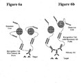
- a recognition unit may comprise a probe that directly binds to the target, as shown in Figure 4 , for example.
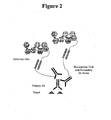
- the probe may recognize the target by binding to it indirectly, as shown in Figure 2 , in which the probe is a secondary binding agent (e.g. a secondary antibody) that binds to a primary binding agent (e.g. a primary antibody) that in turn directly binds to the target.
- a secondary binding agent e.g. a secondary antibody
- a primary binding agent e.g. a primary antibody
- Such a system may allow for an enhancement of the signal, if, for example, each primary binding agent may be bound to more than one secondary binding agent acting as a probe. If more than one probe recognizes a target, more than one detection unit may become associated with a target, for example. Compare, for example, Figure 2 to Figure 4 .
- the adaptor units may create a third "layer" to the detection system, which may merely connect different recognition and detection units together, while in other embodiments, one or more adaptor units may also serve to further enhance the signal obtained from the target.
- the adaptor unit may be structured such that it can specifically hybridize to more than one detection unit, thus increasing the number of detection units ultimately joined to a target. See, for example, Figures 2 and 3 , Figures 4 and 5 , and Figures 7a and 7b . This is useful, for instance, when the amounts of targets are low or when a strong detection signal is desired.
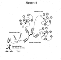
- more than one adaptor unit may be used in order to create yet additional layers allowing for further adaptability and amplification. See, for example, Figures 9 , 10 , 11 , and 12 , each of which use two adaptor units.
- nucleic acid analog segments may increase the flexibility of the detection methods as a whole.
- flexibility may be generated, such as including multiple nucleic acid analog segments within the recognition unit, either close together as shown in Figure 8a , or joined by different linkers, as shown in Figure 8b .
- multiple probes may also be used, such that a recognition unit binds to more than one type of target.
- thermodynamics and kinetics of the interactions of the different units may also be controlled through the number and sequence of the nucleic acid analog segments as well as through the concentrations of the different units. For instance, the binding strength between the different units may be increased using multiple nucleic acid analog segments.
- the invention may also be employed to detect more than one target in a sample.
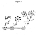
- different targets in the same sample may be detected with different sets of recognition and detection units and different detectable labels. See Figure 13 .
- cross-reactivity may be avoided using different sets of base-paired nucleic acid analog segments within each of the sets of recognition and detection units. Because of the lack of cross-reactivity that the different specific hybridizations allow, the instant invention allows for methods in which all of the recognition units and detection units are added to the sample at the same time.
- a flexible system may be generated in which the same set of reagents may be used to detect more than one type of target.
- three different targets are detected using the same detection unit and adaptor unit, but different recognition units.
- many different targets in the same sample such as nucleic acid segments, may be detected using the same detection unit and adaptor unit, but different recognition units each carrying a different probe.
- the method comprises steps of adding a mixture of primary binding agents or compositions comprising a mixture of primary binding agents and steps of adding compositions comprising a mixture of detectable labels.
- compositions comprising such mixtures may be designed for multi-purpose use, wherein not all binding agents binds to a target or another binding agent. Multi-purpose compositions may for example be useful in methods for analysis of different samples expressing only few of several targets.
- one or more probes or binding agents or external components present in a sample may be sterically hindered.
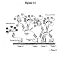
- Figure 17 left panel, illustrates that the ability of a primary antibody to bind to a target may be reduced by steric hindrance from a conjugated nucleic acid analog segment. Such engineered steric hindrance may reduce non-specific binding or cross-reactivity, thus increasing the sensitivity of a system.
- Methods according to the invention may also be used for or in conjunction with methods of diagnosing at least one disease or condition.
- the instant invention can be applied to a variety of targets. Any target which can be recognized by a suitable recognition unit and probe is compatible with the instant invention.
- the recognition may be direct, while in other embodiments, the recognition may be indirect, via another binding agent, such as at least one primary, secondary, or higher order binding agent.
- the target comprises a protein, such as a glycoprotein or lipoprotein, phosphoprotein, methylated protein, or a protein fragment, a peptide, or a polypeptide.
- the target comprises a nucleic acid segment.
- the target comprises a nucleic acid analog segment.
- the target may comprise a lipid; a glyco-lipid; a sugar; a polysaccharide; a starch; a salt; an ion; or one of a variety of other organic and inorganic substances; any of which may be free in solution or bound to another substance.
- the target may be expressed on the surface of the sample, e.g., such as on a membrane or interface.
- the target may be contained in the interior of the sample.
- an interior target may comprise a target located within the cell membrane, periplasmic space, cytoplasm, or nucleus, or within an intracellular compartment or organelle.
- Targets may also include viral particles, or portions thereof, e.g., a nucleic acid segment or a protein.
- the viral particle may be a free viral particle, i.e., not associated with any other molecule, or it may be associated with any sample described above.
- the target may be an antigen or an antibody.
- the instant invention is compatible with many known detection formats and their associated samples.
- the invention may be used in connection with immunoassays, protein detection assays, or nucleic acid hybridization assays such as: immunohistochemistry (IHC), immunocytochemistry (ICC), in situ hybridization (ISH), flow cytometry, enzyme immuno-assays (EIA), enzyme linked immuno-assays (ELISA), blotting methods (e.g. Western, Southern, and Northern), labeling inside electrophoresis systems or on surfaces or arrays, and precipitation, among others. All of those detection assays are useful in research as well as in the detection and diagnosis of a variety of diseases and conditions, for example.
- IHC specifically provides a method of detecting targets in a sample or tissue specimen in situ (see Mokry 1996, ACTA MEDICA 39:129 ).
- the overall cellular integrity of the sample is maintained in IHC, thus allowing detection of both the presence and location of the targets of interest.
- a sample is fixed with formalin, embedded in paraffin and cut into sections for staining and subsequent inspection by light microscopy.
- Current methods of IHC use either direct labeling or secondary antibody-based or hapten-based labeling.
- IHC systems examples include, for example, EnvisionTM (DakoCytomation), Powervision® (Immunovision, Springdale, AZ), the NBATM kit (Zymed Laboratories Inc., South San Francisco, CA), HistoFine® (Nichirei Corp, Tokyo, Japan).
- EnvisionTM Deoxychrome
- Powervision® Immunovision, Springdale, AZ
- the NBATM kit Zamed Laboratories Inc., South San Francisco, CA
- HistoFine® Neichirei Corp, Tokyo, Japan.
- the present invention may allow for enhancement of signal or increased flexibility in IHC detection platforms.
- IHC, ISH and cytological techniques may be performed in a matrix of tissue, cell and proteins which may be partly cross-linked and very inhomogeneous in nature. Diffusion rates increase with increasing concentrations and increasing temperature, but decrease with molecular weight and molecular size. Therefore, the physical size of the components is of great importance. For instance, large molecules can be excluded from diffusing into parts of the sample whereas small sized components more easily may diffuse in and out of the different compartments of the sample.
- the units of the invention may be designed to be of small size and, for example, smaller than an antibody or biotin-streptavidine complex, in order to improve target recognition and detection.
- Samples may comprise solid and liquid solutions, for example, containing targets in a buffer. Samples may also be derived from living matter taken from any living organism, e.g., an animal, such as mammals (e.g. humans), plants, fungi, archaea, or bacteria. Thus, samples may comprise eukaryotic cells, archaeal cells, or prokaryotic cells. Samples may comprise a cell sample, such as a cell smear or colony, or a tissue specimen derived from a living organism, such as a tissue sample from an organ. They may also comprise a biological fluid, such as an animal-derived fluid, e.g.
- Samples may also comprise other naturally-obtained samples such as soil or water samples, and synthetically derived samples such as chemical or industrial products or solutions, food products, and buffers.
- tissue or cell samples according to the invention may be prepared by a variety of methods known to those of ordinary skill in the art, depending on the type of sample and the assay format.
- tissue or cell samples may be fresh or preserved, and may be, for example, in liquid solution, flash-frozen or lyophilized, smeared or dried, embedded, or fixed on slides or other supports.
- samples may be prepared and stained using a free-floating technique. In this method a tissue section is brought into contact with different reagents and wash buffers in suspension or freely floating in appropriate containers, for example micro centrifuge tubes, before being mounted on slides for further treatment and examination.
- a tissue section may be mounted on a slide or other support after an incubation with immuno-specific reagents. The remains of the staining process are then conducted after mounting.
- samples may be comprised in a tissue section mounted on a suitable solid support.
- sections comprising samples may be mounted on a glass slide or other planar support, to highlight by selective staining certain morphological indicators of disease states or detection of detectable targets.
- a sample may be taken from an individual, fixed and exposed to, for example, antibodies which specifically bind to the detectable target of interest.
- Sample processing steps may include, for example, antigen retrieval, exposure to a primary antibody, washing, exposure to a secondary antibody (optionally coupled to a suitable detectable label), washing, and exposure to a tertiary antibody linked to a detectable label. Washing steps may be performed with any suitable buffer or solvent, e.g., phosphate-buffered saline, TRIS-buffered saline, distilled water.
- the wash buffer may optionally contain a detergent, e.g., TWEEN® 20 or NP-40.
- IHC samples may include, for instance: (a) preparations comprising un-fixed fresh tissues and/or cells or solution samples (b) fixed and embedded tissue specimens, such as archived material; and (c) frozen tissues or cells.
- an IHC staining procedure may comprise steps such as: cutting and trimming tissue, fixation, dehydration, paraffin infiltration, cutting in thin sections, mounting onto glass slides, baking, deparaffination, rehydration, antigen retrieval, blocking steps, applying primary antibody, washing, applying secondary antibody - enzyme conjugate, washing, applying a tertiary antibody conjugated to a polymer and linked with an enzyme, applying a chromogen substrate, washing, counter staining, applying a cover slip and microscopic examination.
- ISH samples may be taken from an individual and fixed before being exposed to a nucleic acid or nucleic acid analog probe on a recognition unit.
- the nucleic acid in the sample may first be denatured to expose the target binding sites.
- Various counter-stains or paints may further be used in order to locate nucleic acid molecules or chromosomes within an ISH sample.
- the approximate amount of a target in a sample is also determined.
- a control target within the sample may be assayed as well as an experimental target.
- a nucleic acid target for example, a chromosomal paint or counter-stain may be used.
- the intensity of a contrasting label for the plasmid or chromosome or a neutral locus thereon may be compared to the intensity of the target locus.
- the intensity of the label from the sample may also be compared to that of a known standard or control sample.
- Estimating the amount of a detectable target in a sample is helpful, for instance, in a variety of diagnostic tests, and the estimate may be used to plan a course of treatment for a suspected disease or condition.
- Several commercial densitometry software programs and related instruments are available to quantitate the intensity of a stained target in a sample, such as those available from Fuji Film, Applied Biosystems, and Molecular Dynamics.
- tissue or cell samples may be fixed or embedded.
- Fixatives may be needed, for example, to preserve cells and tissues in a reproducible and life-like manner. Fixatives may also stabilize cells and tissues, thereby protecting them from the rigors of processing and staining techniques. For example, samples comprising tissue blocks, sections, or smears may be immersed in a fixative fluid, or in the case of smears, dried.
- FFPE paraffin embedding
- Any suitable fixing agent may be used. Examples include ethanol, acetic acid, picric acid, 2-propanol, 3,3'-diaminobenzidine tetrahydrochloride dihydrate, acetoin (mixture of monomer) and dimer, acrolein, crotonaldehyde (cis+trans), formaldehyde, glutaraldehyde, glyoxal, potassium dichromate, potassium permanganate, osmium tetroxide, paraformaldehyde, mercuric chloride, tolylene-2,4-diisocyanate, trichloroacetic acid, tungstic acid.
- Fresh biopsy specimens, cytological preparations (including touch preparations and blood smears), frozen sections, and tissues for IHC analysis may be fixed in organic solvents, including ethanol, acetic acid, methanol and/or acetone.
- antigen retrieval may be used (and which is also known in the art as target retrieval, epitope retrieval, target unmasking, or antigen unmasking). See, e.g., Shi et al., J Histochem Cytochem, 45(3): 327 (1997 ).
- Antigen retrieval encompasses a variety of methods including enzymatic digestion with proteolytic enzymes, such as e.g. proteinase, pronase, pepsin, papain, trypsin or neuraminidase. Some embodiments may use heat, e.g. "heat-induced epitope retrieval" or HIER.
- Heating may involve a microwave irradiation, or a water bath, a steamer, a regular oven, an autoclave, or a pressure cooker in an appropriately pH stabilized buffer, usually containing EDTA, EGTA, Tris-HCl, citrate, urea, glycin-HCl or boric acid.
- an appropriately pH stabilized buffer usually containing EDTA, EGTA, Tris-HCl, citrate, urea, glycin-HCl or boric acid.
- One may add detergents to the HIER buffer to increase the epitope retrieval, or to the dilution media and/or rinsing buffers to lower non-specific binding.
- combinations of different antigen retrieval methods may be used.
- the antigen retrieval buffer may be aqueous, but may also contain other solvents, including solvents with a boiling point above that of water such as e.g glycerol. This allows for treatment of the tissue at more than 100 °C at normal pressure.
- the signal-to-noise ratio may be increased by different physical methods, including application of vacuum, or ultrasound, or freezing and thawing tissue samples before or during incubation of the reagents.
- treatments may be performed to reduce non-specific binding.
- carrier proteins, carrier nucleic acid molecules, salts, or detergents may reduce or prevent non-specific binding.
- Non-specific binding sites may be blocked in some embodiments with inert proteins like, HSA, BSA, ovalbumin, with fetal calf serum or other sera, or with detergents like TWEEN®20, TRITON® X-100, Saponin, BRIJ®, or PLURONICS®.
- non-specific binding sites may be blocked with unlabeled competitors for the recognition event between the target and the probe.
- non-specific binding may be reduced by adding unlabeled competitor nucleic acids or nucleic acid analogs such as digested, total human DNA or salmon sperm DNA, or unlabeled versions of the binding agent.
- repetitive sequences may be blocked, for example, using nucleic acids or nucleic acid analogs that specifically recognize those sequences, or sequences derived from a total DNA preparation. Salt, buffer, and temperature conditions may also be modified so as to reduce non-specific binding.
- Cross reactivity of different components of the detection methods may be avoided, for example, by using antibodies derived from different species.
- combinations of e.g. secondary antibodies against primary antibodies and haptens may also be used to avoid unwanted cross reactivity.
- unwanted cross-reactivity or non-specific binding may be reduced or eliminated by designing sterically hindered recognition units, adaptor units, or detection units.
- a recognition unit may be designed such that the nucleic acid analog segments provide a certain degree of steric hindrance near the probe.
- one may remove endogenous biotin binding sites or endogenous enzyme activity (for example phosphatase, catalase or peroxidase) as a step in the staining procedure. Endogenous biotin and peroxidase activity may be removed by treatment with peroxides, while endogenous phosphatase activity may be removed by treatment with levamisole. Heating may destroy endogenous phosphatase and esterase activity.
- the invention also provides a kit comprising one or more compositions according to the invention.
- the kit may optionally comprise one or more binding agents, and suitable reagents for, for instance: antigen retrieval, sample dilution, reagent dilution, blocking of non-specific binding, blocking of endogenous enzyme activity, or blocking of repetitive sequences.
- the kit may optionally also comprise at least one container, instructions for use, and reference targets or samples.
- Instruments may be used in various embodiments of the invention. Instruments, capable of performing steps of staining, are useful for carrying out both single as well as multi-staining procedures, and, in particular, useful for detection of multiple targets that frequently requires balancing of the signals emanating from the different detectable labels.
- the DCM phases were pooled and washed with 10 mL NaCitrate/NaOH - mixture.
- the washed DCM phases were evaporated under reduced pressure and resulted in 17.2 g of crude solid product.
- This crude solid product was recrystallized with ethylacetate giving a yellow powder.
- the yield for this step was 11.45 g (63 %).
- the yellow foam (8.0 g) was hydrolyzed by reflux overnight in 11 mL DIPEA, 22 mL water, and 22 mL dioxane. The solvent was evaporated and the oil was dehydrated by evaporation from toluene leaving an orange foam. The yield for this step was 100 %.
- Step 1 In dry equipment 9.2 g of solid Na in small pieces was dissolved in 400 mL ethanol (99.9%), with stirring. Hydroxypyrimidine hydrochlorid, 26.5 g, was added, and the mixture was stirred for 10 minutes at 50°C. Then 24.4 mL Ethyl bromoacetate (98%) was added and the mixture stirred at 50°C for 1 hour. The reaction was followed using Thin Layer Chromatography (TLC).
- TLC Thin Layer Chromatography
- the ethanol was evaporated leaving a white compound, which was dissolved in 70 mL of water and extracted with 20 mL DCM. Another 30 mL of water was added to the water phase, which was extracted with 3x100 mL DCM.
- the DCM-phase from the first extraction contains a lot of product, but also some impurities, wherefore this phase was extracted twice with water. These two water phases then were back extracted with DCM.
- Step 3 Pyrimidinone acetic acid 11.1 g and triethylamine 12.5 mL were dissolved in N,N-dimethylformamide (DMF) 24 ml, HBTU 26.2 g was added plus 6 mL extra DMF. After 2 minutes a solution of PNA-Backbone ethylester 14.7g dissolved in 15 mL DMF was added. The reaction mixture was stirred at room temperature and followed using TLC. After 11 ⁇ 2 hour precipitate had formed. This was filtered off.
- DMF N,N-dimethylformamide
- Step 5 To make a test on the P-monomer 3 consecutive P's were coupled to Boc-L300-Lys(Fmoc) -resin, following normal PNA standard procedure. The product was cleaved from the resin and precipitated also following standard procedures: HPPP-L300-Lys(Fmoc). Maldi-Tof on the crude product: 6000 (calc. 6000) showing only minor impurities.
- Example 18a an MBHA-resin was loaded with Boc-Lys(Dde)-OH.
- amino acids were coupled according to PNA solid phase procedure provided in Example 18d yielding Boc-L 90 -Lys(Fmoc)-L 30 -Lys(Dde).
- the Boc and Fmoc protections groups where removed and the amino groups marked with flourescein using the procedure in Example 18e.
- the Dde protection group was removed and 0.4 M cysteine was added according to the procedure in Example 18b.
- the PNA was cleaved from the resin, precipitated with ether and purified on HPLC according to Example 18d.
- the product was found to have a molecular weight of 3062 using MALDI-TOF mass spectrometry; the calculated molecular weight is 3061.
- Example 8 Synthesis of a conjugate made from sequence AA from Example 5, DexVS70, and Flu(10)
- the above four compounds were mixed. The mixture was placed in a water bath at 30° C for 16 hours. The mixture was added to 50 nmol of freeze-dried PNA (sequence AA from Example 5). The mixture was placed in a water bath at 30° C for 30 minutes. The conjugating reaction was quenched with 50 ⁇ L of 500 mM cysteine for 30 minutes at 30° C. Purification of the product was performed using FPLC: column SUPERDEX® - 200, buffer 10 mM Hepes 100 mM NaCl, method 7 bank 2, Loop 1 mL. Two fractions were collected: one with the product and one with the residue.
- the conjugation ratio of Flu 2 to DexVS70 was 9.4.
- the conjugation ratio of PNA (sequence AA) to DexVS70 was 1.2.
- Dextran (molecular weight 70 kDa) was activated with divinylsulphone to a degree of 92 reactive groups/dextran polymer, and is designated DexVS70.
- the antibody Anti-Human-BCL2 is designated AHB.
- 105 ⁇ L DexVS70 7.5 nmol 800 ⁇ L AHB conc. (2.9 g/L) 15.1 nmol 25 ⁇ L 4 M NaCl 32 ⁇ L 0.8 M NaHCO 3 (pH 9.5)
- the above four compounds were mixed and placed in a water bath at 30° C for 65 minutes. From this mixture, 875 ⁇ L was taken out and added to the indicated number of nmol of PNA in the table below; before the addition the PNA had been dissolved in the ⁇ L of H 2 O indicated in the table below. Then the number of ⁇ Ls of 0.8 M NaHCO 3 (pH 9.5) was added according to the table below. The resulting mixture was placed in a water bath at 30° C for 70 minutes. Quenching was performed by adding 6 mg of solid cysteine (0.05 M) to the mixture and letting it stand for 30 minutes at 30° C.
- the precipitate was washed with ether and was then dissolved in 100 ⁇ L TFA.
- the precipitate was washed with ether and then dried using N 2 gas.
- the above five components are mixed together placed in a water bath at 30° C for 16 hours. Five hundred microliters of this mixture are added to 50 nmol PNA, which is previously dissolved in 40 ⁇ L H 2 O. Then, 10 ⁇ L of 0.8 M NaHCO 3 (pH 9.5) is added. The mixture is then placed in a water bath at 30° C for 2 hours. Quenching is performed by adding 55 ⁇ L of 110 mM cysteine and letting the resultant mixture set for 30 minutes at 30° C.
- the above five components are mixed and placed in a water bath at 30° C for 40 minutes. Two hundred and ninety ⁇ L is taken out of the mixture and added to 50 nmol of PNA, which is previously dissolved in 40 ⁇ L of H 2 O. Then, 10 ⁇ L of 0.8 M NaHCO 3 (pH 9.5) is added and the mixture placed in a water bath at 30° C for 1 hour. Quenching is performed by adding 34 ⁇ L of 500 mM cysteine and letting the resultant mixture set for 30 minutes at 30° C.
- Dextran (molecular weight 70 kDa) is activated with divinylsulfone to a degree of 92 reactive groups/dextran polymer.
- PNA1 100 nmol
- DexVS70 10 nmol
- To this mixture 12.5 ⁇ L of PNA2 (12.5 nmol) dissolved in H 2 O is added, and then 30 ⁇ L of NaHCO 3 (pH 9.5) is added and the solution mixed.
- the resultant mixture is placed in a water bath at 30° C for 35 minutes. Quenching was performed by adding 18.3 ⁇ L of 500 mM cysteine in Hepes and letting this mixture set for 30 minutes at 30° C.
- 6-Benzyloxypurine Sodiumhydride (60 % Dispersion in mineral oil;3,23g;80 mmol) was slowly added to benzyl alcohol (30 ml;34,7 mmol). After the addition of more benzyl alcohol (10 ml) and 6-chloropurine (5,36 g;). The reaction mixture was heated to 100 °C for 4 hours. When the reaction mixture has reached room temperature, water (1ml) was slowly added. 6-Benzyloxypurine was precipitated by the addition of acetic acid (4,6 ml) and diethylether (550 ml). The precipitate was separated by filtration (11,72 g). Re-crystallization from ether gave (4,78 g; 65,4 %).
- 6-Benzyloxypurine (4,18 g; 18,5 mmol) was added to a suspension of potassium carbonate (3,1 g; 22,4 mmol) in DMF (100 ml). After 15 min., bromoacetic acid methyl ester (1,93 ml; 20,4 mmol) was added. The reaction was monitored by TLC in butanol:acetic acid:water 4:1:1. Upon completion, the reaction mixture was partitioned between water (600 ml) and ethyl acetate (600 ml). The organic phase was dried over magnesium sulfate and evaporated to a volume of ⁇ 10 ml and precipitated with pet.
- Boc-PNA-Diaminopurine-(N6-Z)-monomer was prepared according to Gerald Haaima, Henrik F. Hansen, Leif Christensen, Otto Dahl and Peter E. Nielsen; Nucleic Acids Research, 1997, Vol 25, Issue 22 4639-4643 .
- Boc-PNA-2-Thiouracil-(S-4-MeOBz)-monomer was prepared according to Jesper Lohse, Otto Dahl and Peter E. Nielsen; Proceedings of the National Academy of Science of the United States of America, 1999, Vol 96, Issue 21,11804-11808 .
- Boc-PNA-Adenine-(Z)-monomer was from PE Biosystems catalog GEN063011.
- Boc-PNA-Cytosine-(Z)-monomer was from PE Biosystems cat. GEN063013.
- Boc-PNA-Guanine-(Z)-monomer was from PE Biosystems cat. GEN063012.
- Boc-PNA-Thymine-monomer was from PE Biosystems cat. GEN063010.
- IsoAdenine (2-aminopurine) may be prepared as a PNA-monomer by 9-N alkylation with methylbromoacetate, protection of the amino group with benzylchloroformate, hydrolysis of the methyl ester, carbodiimide mediate coupling to methyl-(2-Boc-aminoethyl)-glycinate, and finally hydrolysis of the methyl ester.
- 4-thiouracil may be prepared as a PNA-monomer by S-protection with 4-methoxy-benzylchloride, 1-N alkylation with methylbromoacetate, hydrolysis of the methyl ester, carbodiimide mediate coupling to methyl-(2-Boc-aminoethyl)-glycinate, and finally hydrolysis of the methyl ester.
- Thiocytosine may be prepared as a PNA monomer by treating the Boc-PNA-cytosine(Z)-monomer methyl ester with Lawessons reagent, followed by hydrolysis of the methyl ester.
- halogenated bases are commercially available, and may be converted to PNA monomers analogously to the non-halogenated bases. These include the guanine analog 8-bromo-guanine, the adenine analogs 8-bromo-adenine and 2-fluoro-adenine, the isoadenine analog 2-amino-6-chloro-purine, the 4-thiouracil analog 5-fluoro-4-thio-uracil, and the 2-thiouracil analog 5-chloro-2-thiouracil.
- Boc-PNA-Uracil monomers were first described in "Uracil og 5-bromouracil I PNA," a bachelor project by Kristine Kils ⁇ Jensen, K ⁇ benhavns Universitet 1992.
- Tonsil tissue samples were fixed in neutral buffered formalin, NBF (10 mM NaH 2 PO 4 / Na 2 HPO 4 , pH 7.0), 145 mM NaCl; and 4% formaldehyde (all obtained from Merck, Whitehouse Station, NJ). The samples were incubated overnight in a ventilated laboratory hood at room temperature.
- NBF neutral buffered formalin
- the tissue samples were placed in a marked plastic histocapsule (Sakura, Japan). Dehydration was performed by sequential incubation in 70% ethanol twice for 45 min, 96% ethanol twice for 45 min, 99% ethanol twice for 45 min, and xylene twice for 45 min. The samples were subsequently transferred to melted paraffin (melting point 56-58°C) (Merck, Whitehouse Station, NJ) and incubated overnight (12-16 hours) at 60°C. The paraffin-infiltrated samples were transferred to fresh warm paraffin and incubated for an additional 60 min prior to paraffin embedding in a cast (Sekura, Japan). The samples were cooled to form the final paraffin blocks. The marked paraffin blocks containing the embedded tissue samples were stored at room temperature in the dark.
- paraffin blocks were cut and optionally also mounted in a microtome (0355 model RM2065, Feather S35 knives, set at 5.0 micrometer; Leica, Bannockburn, IL). The first few millimeters were cut and discarded. Paraffin sections 4-6 micrometers thick were then cut and collected at room temperature. The sections were gently stretched on a 45-60°C hot water bath before being mounted onto marked microscope glass slides (SUPERFROST ® Plus; Fisher, Medford, MA), two tissue sections per slide. The slides were then dried and baked in an oven at 60°C.
- TBST Tris-buffered saline with TWEEN®
- TBST comprises 50 mM Tris adjusted to pH 7.6 with HCl; 150 mM NaCl; 0.05 % TWEEN®20.
- the slides were deparaffinated by subsequently incubation in xylene twice for 5 min ⁇ 2 min, 96% ethanol twice for 2min +/- 30 sec and 70% ethanol twice for 2 min +/- 30 sec.
- the slides were immersed in deionized water and left for 1 to 5 min.
- Antigens in the sample were retrieved by immersing the slides in a container containing Antigen Retrieval Solution, pH 6.0 (DakoCytomation code No. K5204 Vial 7 or optional code No. K5205 Vial 7).
- the container was closed with a perforated lid and placed in the middle of a microwave oven and left boiling for 10 min. The container was removed from the oven and allowed to cool at room temperature for 20 min. The samples were rinsed in deionized water.
- Antigens in the sample were retrieved by immersing the slides in a beaker containing Antigen Retrieval Solution, pH 6.0 (DakoCytomation code No. K5204 Vial 7 or optional code No. K5205 Vial 7). The samples were incubated for 40 min in a water bath at 95-100°C. The beaker was removed from the water bath and allowed to cool at room temperature for 20 min. The samples were rinsed in deionized water.
- TBST Tris-buffered saline with TWEEN ®
- Monoclonal Mouse Anti-Human Cytokeratin (DakoCytomation code No. M3515) diluted 1:300, 1:900 and 1:1600; monoclonal Mouse Anti-Human CD20cy (DakoCytomation code No. M0755) diluted 1:2000, 1:8000 and 1:14000; and monoclonal Mouse Anti-Human Ki-67 Antigen (DakoCytomation code No. M7240) diluted 1:400, 1:1200 and 1:2400 were used.
- the antibodies were diluted in ChemMateTM Antibody Diluent (DakoCytomation code No. S2022), applied on the tissue samples, and incubated for 30 min in a humid chamber at ambient temperature. The slides were individually rinsed and washed in TBST for 5 min.
- Antibody / Dextran / PNA1 conjugate recognition unit is also called "PNA1 conjugate” in the examples that follow.
- the PNA1 conjugate comprises 70,000 molecular weight dextran.
- Table 5 summarizes PNA1 conjugates based on a secondary antibody: goat anti-mouse Ig, called herein GAM (DakoCytomation code No. Z0420).
- Table 6 summarizes PNA1 conjugates based on a primary antibody: mouse anti-human BCL2 oncoprotein, such as Clone 124 (DakoCytomation code No. M0887). The primary antibody was protein A-purified prior to conjugation.
- the conjugates were diluted in BBA (50 mM Tris adjusted to pH 7.6 with HCl; 150 mM NaCl; 2% BSA; 0.02% bronidox; 2.44 mM 4-aminoantipyrin) and were applied on the tissue sample in a range of dilutions, then incubated for 30 min in a humid chamber at ambient temperature. The slides were individually rinsed and washed in TBST for 5 min. Table 5.
- PNA1 conjugates for direct recognition of targets anti - BCL2 / Dextran / PNA1 Conjugate No. Sequence ⁇ M Dex Ab / Dex PNA1 / Dex D14128 U s GU s DPP TTG D 0.8 1.1 5.6 D14126 U s GU s DPP TTG D 1.0 1.2 2.9 D14122 U s GU s DPP TTG D 1.1 1.6 9.5
- the letters A, C, G, U, and T stand for the natural bases adenine, cytosine, guanine, uracil, and thymine.
- P stands for pyrimidinone
- D stands for 2,6-diaminopurine
- U stands for 2-thiouracil.
- the samples were washed in deionized water for 30 sec. Then, 1 % glutardialdehyde (Merck Art. No. 820603), called herein GA, diluted in 22 mM calcium phosphate buffer, pH 7.2, was applied, and the samples were incubated for 10 min in a humid chamber at ambient temperature. The samples were washed in deionized water for 30 sec and in TBST for 5 min.
- PNA 1 -PNA 2 / Dextran conjugate is also called "PNA 1 -PNA 2 " in the following examples.
- Table 7 summarizes the compositions of PNA 1 -PNA 2 conjugates.
- PNA 1 is complementary to the PNA1 conjugate
- PNA 2 is complementary to the PNA2 conjugates D14079 and D13155 described in step 13 below.
- the sequence of PNA 1 is CU s G s G s DD TU s D GsDC and the sequence of PNA 2 is U s GU s DPP TTG D, in which U s stands for 2-thio-uracil, G s stands for 2-amino-6-thioxopurine, D stands for diaminopurine, and P stands for pyrimidinone.
- Horse Radish Peroxidase (HRP) / Dextran / PNA2 conjugates are also called "PNA2 conjugate" in the examples that follow, and are listed in table 8.
- the PNA2 conjugates comprise 70.000 Da molecular weight dextran.
- the conjugates diluted in BBA were applied to the tissue samples in a range of dilutions, and samples were incubated for 30 min in a humid chamber at ambient temperature. The samples were individually rinsed and washed twice in TBST for 5 min. Table 8.
- PNA2 conjugates HRP / Dextran / PNA2 Conjugate No.
- the diaminobenzidine chromogenic substrate solution, DAB+ (DakoCytomation code No. K3468) was applied on the tissue samples, and the samples were incubated for 10 min in a humid chamber at ambient temperature. The samples were washed with deionized water for 5 min.
- tissue samples were immersed in Mayers Hematoxylin (Bie & Berntsen Code No. LAB00254) for 3 min, rinsed in tap water for 5 min, and finally rinsed with deionized water.
- the tissue staining was examined in a bright field microscope at 10x, 20x or 40x magnification. Both the specific and the non-specific staining intensity were described with a score-system using the range 0 to 3+ with 0.5+ score interval.
- ChemMateTM EnVisionTM Detection kit Rabbit/Mouse (DakoCytomation code No. K5007 bottle A) was used as a reference, and was included in all experiments for testing in parallel with the PNA conjugates. K5007 was used according to manufacturer's instructions.
- the antibodies were used in the following dilutions: M3515 at 1:900, M0755 at 1:8000, and M7240 at 1:1200.
- the staining intensity of the K5007 reference using the primary antibody M3515 diluted 1:900 was set to 2+ in order to compare and assess the staining result of the PNA conjugate tested. If the reference deviated more than ⁇ 0.5, the test was repeated.
- the various visualization system combinations of the invention were tested on routine tissue samples.
- the staining performance was compared with a reference visualization system, using EnVisionTM and a very dilute antibody from DakoCytomation.
- the practical dynamic range of quantitative IHC may be narrow, and e.g. strongly stained (+3) tissues are not easy to compare with respect to intensity. Therefore, on purpose, the staining intensity of the reference system was adjusted to be approximately +2. This was done in order to better monitor and compare differences in staining intensity with the system of the invention.
- the conjugates to be tested were diluted to the final concentrations 0.05 ⁇ M and 0.2 ⁇ M.
- two PNA2 conjugates were used in the final concentration 0.05 ⁇ M.
- the first reference, for example, PNA2, D13108 was known to give non-specific nuclear staining, and so was used as a positive control.
- the second reference, for example, PNA2, D13155 was known not to give any non-specific nuclear staining, and was used as a negative control.
- 250 ⁇ L of each reagent was applied unless otherwise specified.
- Protocol for test of a PNA pair with one antibody Protocol for test of a PNA pair with one antibody.
- Tonsil tissues were taken through the steps 1-4, 6-8, 10, 11, and 13-17 above. Step 11 was left out for the tonsils not fixed with 1% GA. In general, 250 ⁇ L of each reagent was applied unless otherwise specified.
- Tonsil tissues were taken through the steps 1-4, 6-8, and 10-17 above. Step 11 was left out for the tonsils not fixed with 1 % GA. In general, 250 ⁇ L of each reagent was applied unless otherwise specified.
- Tonsil tissues were taken through the steps 1-4, 6, 7, 9 -11, and 13-17 above. Step 11 was left out for the tonsils not fixed with 1 % GA.
- Conjugates comprising example PNA segments were tested for their ability to specifically hybridize according to the invention. Tonsil tissues were taken through the steps 1-4, 6-8, 10 and 13-17 above. K5007 was included as a reference to secure the level of the staining. The concentration of the conjugates was 0.08 ⁇ M for PNA1 and 0.05 ⁇ M for PNA2.
- the PNA pair D13102-D13106 was used as a starting point for further investigation of introducing base substitutions in either PNA1 or PNA2 conjugates. Tonsil tissues were taken through the steps 1-4, 6-8, 10 and 13-17. Each of the three different PNA1 conjugates was tested with each of the three different PNA2 conjugates. The concentration of the conjugates used was 0.08 ⁇ M for PNA1 and 0.05 ⁇ M for PNA2. Table 10.
- PNA2 D13106 D13148 D13155 PNA1: TCA AGG TAC A TCA AG s G s TAC A TCD DG s G s TAC A D13102 2.5 0 2 TGT ACC TTG A D13150 2.5 0.5 3 TGT APP TTG A D13171 3 2.5 3 U s GU s DPP TTG D
- Table 10 shows the effect of base substitutions on the specific binding between paired PNA variants. No non-specific binding was observed. D13102 tested with D13106 gave a specific staining of 2.5+. Replacement of 2 G's with 2 G s 's (D13148) resulted in the abolishment of specific staining, but by introducing 2 D's instead of 2 A's (D13155) achieved a specific staining of 2+. When the 2 C's in D13102 were replaced with 2 P's (D13150) and tested with D13106, the specific staining was unchanged at 2.5+, despite the lower number of hydrogen bonds as compared to the PNA-pair D13102-D13106.
- Test of D13150 with D13148 resulted in a reduced specific staining of 0.5+, whereas specific staining to 3+ was observed for the D13150-D13155 pair.
- the replacement in D13150 of 2 A's with 2 D's and of 2 T's with 2 U s 's resulted in improved specific binding compared to D13106.
- This modified PNA1 was now able to bind specifically to D13148 with a score of 2.5+, and also bound to D13155.
- the two PNA-pairs D13150-D13155 and D13161-D13159, were tested for cross-reactivity. Tonsil tissues were taken through the steps 1-4, 6-8, 10, and 13-17. The concentration of conjugates used was 0.16 ⁇ M for PNA1 and 0.1 ⁇ M for PNA2.
- PNA2 conjugates Two PNA pairs, D14083-D14079 and D14096-D14089, were tested at different PNA2 concentrations for the purpose of determining the optimal concentration of PNA2 conjugates.
- concentrations used were 0.08 ⁇ M for PNA1 conjugates and 0.025; 0.05; 0.1 and 0.2 ⁇ M for PNA2 conjugates.
- Tonsil tissues were taken through the steps 1-4, 6-8, 10, and 13-17.
- the optimal concentration of the PNA2 conjugate was 100 nM. See Table 13 below, Table 13: Determination of PNA2 conjugate concentration.
- PNA1 1% GA fixation of PNA1 PNA2 Specific staining 0 . 025 ⁇ M 0.05 ⁇ M 0.1 ⁇ M 0.2 ⁇ M D14083 - D14079 1.5 2 3 2.5 D14096 - D14089 0.5 1 2 1.5
- Tonsil tissues were taken through the steps 1-4, 6-8, 10, 11, and 13-17. Step 11 was omitted for tissues not fixed with 1 % GA.
- This example shows the results of using a 3-layer PNA system employing a PNA 1 -PNA 2 / Dextran conjugate adaptor unit to link the PNA1 and PNA2 conjugates together.
- Tonsil tissues were taken through the steps 1-4, 6-8, and 10-17. Step 11 was omitted for tissues not to be fixed with 1% GA.
- the concentration of the conjugates used was 0.08 ⁇ M for PNA1, 0.1 ⁇ M (calculated based on PNA 1 ) for PNA 1 -PNA 2 and 0.05 ⁇ M for PNA2.
- Table 15 shows that a 3-layer system resulted in a stronger specific staining intensity in comparison with a 2-layer system. No non-specific staining was observed.
- Table 15 Improvement of staining intensity by using 3 layers PNA1 1% GA fixation of PNA1 PNA-PNA PNA2 Specific staining intensity D14096 + D14104 D14079 2 D14096 - D14104 D14079 2 D14096 - - D14089 1
- Table 16 Improvement of specific staining in a 3 -l ayer system by fixation PNA1 1% GA fixation of PNA1 PNA-PNA PNA2 Specific staining intensity D14102 + D14119 D14079 3 D14102 + D14106 D14079 3 D14102 + D14104 D14079 3 D14102 - D14119 D14079 2 D14102 - D14106 D14079 2 D14102 - D14104 D14079 2
- Example 28 Test of 3-layer PNA systems using different PNA 1 -PNA 2 concentrations in the presence or absence of fixation
- Tonsil tissues were taken through the steps 1-4, 6-8, and 10-17 above. Step 11 was left out for the tonsil tissues, which were not going to be fixed with 1% GA.
- the concentration of the conjugates in the table was 0.08 ⁇ M for PNA1, 0.025, 0.05, 0.1 and 0.2 ⁇ M for PNA 1 -PNA 2 (based on [PNA 1 ]), and 0.05 ⁇ M for PNA2.
- a 2-layer PNA test system was employed to study the effect of using different concentrations of GA. Tonsil tissues were taken through the steps 1-4, 6-8, 10, 11 and 13-17. In step 11, the concentration of GA used was 0.1%, 0.3% and 1.0% respectively.
- Example 30 Comparison of a PNA-based detection system with an EnVisionTM based detection system
- Tonsil tissues were taken through the steps 1-4, 6, 7, 9-10, and 13-17.
- the concentration of the conjugates was 0.08 ⁇ M for PNA1, D12102 and 0.05 ⁇ M for PNA2, D12094.
- a negative Ig control, mouse IgG1 (DakoCytomation code No. X0931) diluted 1:300 in S2022 was included in the protocol for PNA conjugates.
- Example 31 Recognition of a conjugated primary antibody by another detection system.
- Tonsil tissues were taken through the steps 1-4, 6, 7, 10, 11, 1S and 14-17 above. 20 ⁇ L of PNA1, respectively D14122 and D14128 (0.08 and 0.3 ⁇ M) were applied. Slides were cover slipped during incubation with PNA1. Then 200 ⁇ L PNA2, D14079 (0.1 ⁇ M) was applied. Samples were incubated with K5007 GaM:HRP complex for 30 min in parallel with PNA2 conjugates in step 13.
- tonsil tissues were taken through steps 1-4 and 6-8 using uncomplexed anti-BCL2, M0887, diluted 1:100 to a concentration of 0.015 ⁇ M in S2022 as primary antibody, and visualized by incubation with K5007 GaM:HRP complex for 30 min. as an alternative to the PNA2 conjugates in step 13. These slides were then taken through steps 14-17.
- Table 20 summarizes the staining results.
- PNA1 PNA conjugates with multiple PNAs, here illustrated by PNA1
- the PNAs remained accessible for hybridization to complementary PNAs comprised in components further comprising dextran and enzymes.
- Conjugates comprising more PNA did not necessarily show improved specific staining. Instead, the amount of staining peaked and then fell as the PNA to dextran ratio increased. For example, samples incubated with D14126 scored 1.0+, those with D14128 scored 2.5+, and those with D14122 scored 2.0+.
- D14128 with a PNA:Dextran ratio of about six, gave a stronger signal than both D14122 with a PNA:Dex ratio of about nine as well as D14126 with a PNA:Dex ratio of about three.
- PNA1 the PNAs remained accessible for hybridization to complementary PNAs comprised in components further comprising dextran and enzymes.
- Tonsil tissues were taken through the steps 1-5, 7, 13 and 14-17 above.
- the PNA2 conjugates tested are listed in Table 21.
- Replacing 2 D's in D12120 with 2 A's (D13106) reduced the non-specific staining from 3+ to 1+ (at 0,05 ⁇ M PNA2).
- 2 G's in D13106 were replaced with 2 G s 's (D13148)
- the non-specific staining was reduced from 1+ to 0.
- Reintroducing 2 D's in D13155 instead of 2 A's (D13148) increased the non-specific staining from 1.5+ to 2.0+ (at 0.2 ⁇ M PNA).
- Both D12120 and D13155 had 2 D's in the sequence, but the non-specific staining in D13155 did not reach the same level as in D12120, probably due to the 2 G s 's in D13155.
- the same effect of non-natural bases was seen when comparing D12120 with D14133: the non-specific staining in D14133 did not reach the same level as in D12120, this time probably due to 2 inosines (I)'s in D14133.
- the replacement of 2 G's in D13122 with 2 G s 's (D13159) reduced the non-specific staining from 3+ to 0 (at a PNA concentration of 0.2 ⁇ M).
- the non-specific background staining generated by the PNA2 conjugates could be subtly fine tuned, and ultimately completely eliminated, by base substitution using both natural as well as non-natural bases.
- the level of background correlated directly to the DNA/RNA affinity of the PNAs of the conjugates. This was surprising, as the conjugates have a molecular weight around 500 kDa, and the changes in the PNAs necessary to bring about strongly reduced background in some cases were as little as one D to A substitution (thus removing one potentially hydrogen bonding amino group) or one G to G s substitution (introducing a single carbonyl to thiocarbonyl change).
- Table 21 Non-specific staining intensities of due to PNA2 conjugates.
- Tonsil tissues were taken through the steps 1-4, 6-8, 10, and 13 (PNA2 was conjugated with fluorescein) and 16 (the slides were mounted with Vectashield containing DAPI).
- concentration used for the conjugates was 0.08 ⁇ M for PNA1, D13171 and 0.02, 0.05, 0.1, and 0.2 ⁇ M for PNA2, D14008 (TCD DG s G s TAC A).
- the slides were evaluated using a fluorescence microscope at 40x and 100x magnifications.
- a multi-block containing tissue from mammalian carcinoma, kidney, colon and two tonsils was taken through the steps 3-4, 6-8, 10, 13-17.
- step 8 ten different primary antibodies and a negative control mouse IgG1 were used.
- the concentration of the conjugates used was 0.08 ⁇ M for PNA1, D12102 and 0.05 ⁇ M for PNA2, D12094.
- Visualization of primary reagents by the K5007 detection kit was performed in parallel according to the manufacturer's instructions.
- Two primary antibodies targeting membranous targets were used: Monoclonal Mouse Anti-Human CD20cy (DakoCytomation code No.
- Three primary antibodies targeting nuclear targets were used: Monoclonal Mouse Anti-Human Estrogen Receptor (DakoCytomation code No. M7047), Monoclonal Mouse Anti-Human Ki-67 Antigen (DakoCytomation code No. M7240) and Monoclonal Mouse Anti-Human p27 (DakoCytomation code No. M7203).
- the primary antibodies, all products from DakoCytomation, were diluted in S2022 as indicated in Table 22.
- the mixture was placed in a water bath at 40°C for 3 hours. Quenching was performed by adding 30.6 ⁇ L of 0.1 M ethanolamine and letting the mixture stand for 30 minutes in water bath at 40°C.
- the product was purified on FPLC with: Column Superdex-200, buffer: 2mM HEPES, pH 7.2; 0.1M NaCl; 5mM MgCl 2 ; 0.1 mM ZnCl 2 . Two fractions were collected, one with the product and one with the residue.
- SMCC Succinimidyl -4(N-maleimidomethyl) cyclohexan-1-carboxylate molw. 334.33.
- PNA Acetyl- L 30 -GTP-TAA-TTP-PAG-L 150 -Lys(Cys)
- the product was purified on FPLC: Column SUPERDEX®-75, Buffer 0.01 M Hepes 0.1 M NaCl pH 7.2. The fraction with the product was collected. Relative absorbance between PNA ( ⁇ 260 nm ) and antibody ( ⁇ 278 nm ) was used to calculate the average conjugation ratio of PNA and antibody. PNA / CD45: 5.2. Yield 39 % based on antibody.
- the product was purified on FPLC: Column Superdex-75, Buffer 0.01 M Hepes 0.1 M NaCl pH 7.2. The fraction with the product was collected. Relative absorbance between PNA ( ⁇ 260 nm ) and antibody ( ⁇ 278 nm ) was used to calculate the average conjugation ratio of PNA and antibody. PNA / CD45: 3.4. Yield 55 % based on antibody.
- Antibody GAM (goat-anti-mouse) dialysed overnight against 0.1 M NaCl.
- SMCC Succinimidyl -4(N-maleimidomethyl) cyclohexan-1-carboxylate molw. 334.33.
- Flu-Link Flu-L 90 -Lys(Flu)-L 30 -Lys(Cys)
- the product was purified on FPLC: Column SUPERDEX®-75, Buffer 0.01 M Hepes 0.1 M NaCl pH 7.2. The fraction with the product was collected. Relative absorbance between Flu-Link ( ⁇ 498 nm ) and antibody ( ⁇ 273 nm ) was used to calculate the average conjugation ratio of PNA and antibody. Flu-Link / GAM: 7.1. Yield 55 % based on antibody.
- Antibody GAM dialysed overnight against 0.1 M NaCl.
- SMCC Succinimidyl -4(N-maleimidomethyl) cyclohexan-1-carboxylate molw. 334.33.
- Flu-Link Flu-L 90 -Lys(Flu)-L 30 -Lys(Cys)
- the product was purified on FPLC: Column SUPERDEX®-75, Buffer 0.01 M Hepes 0.1 M NaCl pH 7.2. The fractions with the product from each purification were collected. Relative absorbance between Flu-Link ( ⁇ 498 nm ) and antibody ( ⁇ 278 nm ) was used to calculate the average conjugation ratio of PNA and antibody. 30 minutes conjugation Flu-Link / GAM: 7.0 Yield 55 % based on antibody. 60 minutes conjugation Flu-Link / GAM: 6.9 Yield 52 % based on antibody. The above results show that 30 minutes conjugation between GAM / SMCC and Flu-Link is sufficient.
- Goat-anti-mouse secondary antibody conjugated with dextran and a first PNA sequence was diluted to final concentration of 0.08 ⁇ M (based on dextran) in BP-HEPES-buffer (1.5% BSA, 3% PEG, 0.15M NaCl, 10mM HEPES, pH 7.2) and was applied to the section. Following 10 minutes incubation at room temperature (RT), the section was washed 5 minutes using 10x diluted S3006 (Dako). The sections were rinsed in deionized water. Following 10 minutes incubation in 0.5% glutaraldehyde at RT, the sections were rinsed in deionized water and washed 5 minutes using 10x diluted S3006 (Dako).
- PNA2-dex-PNA3 218-057
- BP-HEPES-buffer 1.5% BSA, 3% PEG, 0.15M NaCl, 10mM HEPES, pH 7.2
- a conjugate of a PNA4, complementary to PNA3 above, dextran, and the detectable label alkaline phosphatase was diluted to final concentration of 0.05 ⁇ M (dextran) in BAP-HEPES-buffer (1.5% BSA, 3% PEG, 0.15M NaCl, 0.05% 4-aminoantipyrin, 10mM HEPES, pH 7.2), and was applied. Following 10 minutes incubation at RT, the sections were washed 5 minutes using 10x diluted S3006 (Dako).
- Permanent Red working solution an aqueous Tris buffer with naphthol-phosphate and a diazonium dye; K0640 Dako
- K0640 Dako a diazonium dye
- PNA2a (GaR-dex-Alexander (209-127)) was diluted to final concentration of 0.08 ⁇ M (dextran) in BP-HEPES-buffer (1.5% BSA, 3% PEG, 0.15M NaCl, 10 mM HEPES, pH 7.2) and was applied. Following 10 minutes incubation at RT the sections were washed 5 minutes using 10x diluted S3006 (Dako). The sections were rinsed in deionized water. Following 10 minutes incubation in 0.5% glutaraldehyde at RT the sections were rinsed in deionized water and washed 5 minutes using 10x diluted S3006 (Dako).
- PNA2b-dex-AP (209-177) a complementary PNA coupled to dextran and detectable label alkaline phosphatase (AP) (PNA2b-dex-AP (209-177) was diluted to final concentration of 0.05 ⁇ M (dextran) in BAP-HEPES-buffer (1.5% BSA, 3% PEG, 0.15M NaCl, 0.05% 4-aminoantipyrin, 10mM HEPES, pH 7.2) and was applied. Following 10 minutes incubation at RT the section was washed 5 minutes using 10x diluted S3006 (Dako).
- BAP-HEPES-buffer (1.5% BSA, 3% PEG, 0.15M NaCl, 0.05% 4-aminoantipyrin, 10mM HEPES, pH 7.2
- the specimen Before conducting the detection experiment on formalin-fixed, paraffin-embedded (FPPE) tissue sections, the specimen should be deparaffinized (dewaxed), rehydrated, and blocked for endogenous peroxidase activity. Some specimens should be subjected to target retrieval using heat or enzyme digestion. Following target retrieval, the specimens should be rinsed gently with wash buffer.
- FPPE paraffin-embedded
- a mouse primary antibody was used as a primary binding agent for a specific target in a tissue sample. That antibody was then recognized by a goat-anti-mouse-dextran-PNA conjugate recognition unit.
- a different primary antibody a rabbit antibody, was used as a primary binding agent for a different target in the sample. That antibody was recognized by a goat-anti-rabbit-dextran-PNA recognition unit.
- PNA-dextran-HRP humanse-radish peroxidase
- PNA-dextran-AP alkaline phosphatase
- Goat-anti-mouse-dextran-PNA1 (GaM-dex-PNA1) and goat-anti-rabbit-dextran-PNA3 (GaR-dex-PNA3) were both diluted to a final concentration of 0.08 ⁇ M (dextran) in BP-HEPES-buffer (1.5% BSA, 3% PEG, 0.15M NaCl, 10mM HEPES, pH 7.2). The two conjugates were applied simultaneously on the sections. Following 10 minutes incubation at room temperature (RT), the sections were washed 5 minutes using 10x diluted S3006 (Dako). The sections were rinsed in deionized water.
- Detection method 1 Permanent Red working solution was applied. Following 10 minutes incubation the sections were washed 5 minutes using 10x diluted S3006 (Dako). Then DAB+ working solution was applied and following 10 minutes incubation the sections were washed 5 minutes using deionized water. Finally the sections were counter stained 5 minutes using haematoxylin S3301 (Dako), rinsed in deionzed water, washed 3 minutes in wash buffer, and mounted in Faramount S3025 (Dako).
- Detection method 2 DAB+ working solution was applied. Following 10 minutes incubation the sections were washed 5 minutes using 10x diluted S3006 (Dako). Then Permanent Red working solution was applied and following 10 minutes incubation the sections were washed 5 minutes using deionized water. Finally the sections were counter stained 5 minutes using haematoxylin S3301 (Dako), rinsed in deionzed water, washed 3 minutes in wash buffer, and mounted in Faramount S3025 (Dako).
- the order of detection affects the staining result. If detection method 1 is used then Permanent Red dominates. If detection method 2 is used then DAB+ dominates.
- Detection method 1 Permanent Red working solution was applied. Following 10 minutes incubation the sections were washed 5 minutes using 10x diluted S3006 (Dako). Then DAB+ working solution was applied and following 10 minutes incubation the sections were washed 5 minutes using deionized water. Finally the sections were counter stained 5 minutes using haematoxylin S3301 (Dako), rinsed in deionzed water, washed 3 minutes in wash buffer, and mounted in Faramount S3025 (Dako).
- Detection method 2 DAB+ working solution was applied. Following 10 minutes incubation the sections were washed 5 minutes using 10x diluted S3006 (Dako). Then Permanent Red working solution was applied and following 10 minutes incubation the sections were washed 5 minutes using deionized water. Finally the sections were counter stained 5 minutes using haematoxylin S3301 (Dako), rinsed in deionzed water, washed 3 minutes in wash buffer, and mounted in Faramount S3025 (Dako).
- Example 40 Further 2 and 3 Layer Systems for Detection of Multiple Targets
- a mouse antibody primary binding agent was recognized by a GaM-dex-PNA1 and a rabbit antibody primary binding agent was recognized by GaR-dex-PNA2.
- One reaction was detected by a PNA-dex-Enzyme1 conjugate and the other by a PNA-dex-PNA adaptor unit and then a PNA-dex-Enzyme2 conjugate.
- PNA1 recognizes PNA2 while PNA3 recognizes PNA4.
- the enzymes used were HRP and AP, bringing along respectively a brown and red end-product within the same tissue section.
- the PNA-dex-PNA adaptor unit adds a third layer to the detection system.
- GaM-dex-PNA1 (218-117) and GaR-dex-PNA3 (209-127) were both diluted to final concentration of 0.08 ⁇ M (dextran) in BP-HEPES-buffer (1.5% BSA, 3% PEG, 0.15M NaCl, 10mM HEPES, pH 7.2).
- the two conjugates were applied simultaneously on the sections. Following 10 minutes incubation at RT the sections were washed 5 minutes using 10x diluted S3006 (Dako). The sections were rinsed in deionized water. Following 10 minutes incubation in 0.5% glutaraldehyde at RT the sections were rinsed in deionized water and washed 5 minutes using 10x diluted S3006 (Dako).
- PNA4-dex-HRP (218-021) was diluted to final concentration of 0.05 ⁇ M (dextran) in BAP-HEPES-buffer (1.5% BSA, 3% PEG, 0.15M NaCl, 0.05% 4-aminoantipyrin, 10mM HEPES, pH 7.2) and applied on the sections. Following 10 minutes incubation at RT the sections were washed 5 minutes using 10x diluted S3006 (Dako).
- PNA2-dex-PNA3 (218-057) amplification unit was diluted to final concentration of 0.05 ⁇ M (dextran) in BP-HEPES-buffer (1.5% BSA, 3% PEG, 0.15M NaCl, 10mM HEPES, pH 7.2) and applied on the sections. Following 10 minutes incubation at RT the sections were washed 5 minutes using 10x diluted S3006 (Dako).
- PNA4-dex-AP (209-177) was diluted to final concentration of 0.05 ⁇ M/dex in BAP-HEPES-buffer (1.5% BSA, 3% PEG, 0.15M NaCl, 0.05% 4-aminoantipyrin, 10mM HEPES, pH 7.2) and applied on the sections. Following 10 minutes incubation at RT the sections were washed 5 minutes using 10x diluted S3006 (Dako). Permanent Red working solution (K0640 Dako) and DAB+ working solution (K3468 Dako) were prepared.
- Detection method 1 Permanent Red working solution was applied. Following 10 minutes incubation the sections were washed 5 minutes using 10x diluted S3006 (Dako). Then DAB+ working solution was applied and following 10 minutes incubation the sections were washed 5 minutes using deionized water. Finally the sections were counter stained 5 minutes using haematoxylin S3301 (Dako), rinsed in deionzed water, washed 3 minutes in wash buffer, and mounted in Faramount S3025 (Dako).
- Detection method 2 DAB+ working solution was applied. Following 10 minutes incubation the sections were washed 5 minutes using 10x diluted S3006 (Dako). Then Permanent Red working solution was applied and following 10 minutes incubation the sections were washed 5 minutes using deionized water. Finally the sections were counter stained 5 minutes using haematoxylin S3301 (Dako), rinsed in deionzed water, washed 3 minutes in wash buffer, and mounted in Faramount S3025 (Dako).
- GaM-dex-PNA1 (218-117) and GaR-dex-PNA3 (209-127) were both diluted to final concentration of 0.08 ⁇ M (dextran) in BP-HEPES-buffer (1.5% BSA, 3% PEG, 0.15M NaCl, 10mM HEPES, pH 7.2).
- the two conjugates were applied simultaneously on the sections. Following 10 minutes incubation at RT the sections were washed 5 minutes using 10x diluted S3006 (Dako). The sections were rinsed in deionized water. Following 10 minutes incubation in 0.5% glutaraldehyde at RT the sections were rinsed in deionized water and washed 5 minutes using 10x diluted S3006 (Dako).
- PNA4-dex-AP (209-177) was diluted to final concentration of 0.05 ⁇ M (dextran) in BAP-HEPES-buffer (1.5% BSA, 3% PEG, 0.15M NaCl, 0:05% 4-aminoantipyrin, 10mM HEPES, pH 7.2) and applied on the sections. Following 10 minutes incubation at RT the sections were washed 5 minutes using 10x diluted S3006 (Dako).
- PNA2-dex-PNA3 (218-057) was diluted to final concentration of 0.05 ⁇ M (dextran) in BP-HEPES-buffer (1.5% BSA, 3% PEG, 0.15M NaCl, 10mM HEPES, pH 7.2) and applied on the sections. Following 10 minutes incubation at RT the sections were washed 5 minutes using 10x diluted S3006 (Dako).
- PNA4-dex-HRP (218-021) was diluted to final concentration of 0.05 ⁇ M (dextran) in BAP-HEPES-buffer (1.5% BSA, 3% PEG, 0.15M NaCl, 0.05% 4-aminoantipyrin, 10mM HEPES, pH 7.2) and applied on the sections. Following 10 minutes incubation at RT the sections were washed 5 minutes using 10x diluted S3006 (Dako).
- Permanent Red working solution K0640 Dako
- DAB+ working solution K3468 Dako
- Detection method 1 Permanent Red working solution was applied. Following 10 minutes incubation the sections were washed 5 minutes using 10x diluted S3006 (Dako). Then DAB+ working solution was applied and following 10 minutes incubation the sections were washed 5 minutes using deionized water. Finally the sections were counter stained 5 minutes using haematoxylin S3301 (Dako), rinsed in deionized water, washed 3 minutes in wash buffer, and mounted in Faramount S3025 (Dako).
- Detection method 2 DAB+ working solution was applied. Following 10 minutes incubation the sections were washed 5 minutes using 10x diluted S3006 (Dako). Then Permanent Red working solution was applied and following 10 minutes incubation the sections were washed 5 minutes using deionized water. Finally the sections were counter stained 5 minutes using haematoxylin S3301 (Dako), rinsed in deionzed water, washed 3 minutes in wash buffer, and mounted in Faramount S3025 (Dako).
- This example presents a 2-layer detection of two targets in which mouse-Ab-dex-PNA is recognized by PNA-dex-Enzyme1 and rabbit-Ab-dex-PNA is recognized by PNA-dex-Enzyme2.
- the enzymes are HRP and AP bringing along respectively a brown and red end-product within the same tissue section.
- PNA1 and 2 specifically hybridize, as do PNA3 and 4.
- CD3-dex-PNA1 D16043
- MIB-1-dex-PNA2 (218-097) were both diluted to final concentration of 0.1 ⁇ M (dextran) in BP-HEPES-buffer (1.5% BSA, 3% PEG, 0.15M NaCl, 10mM HEPES, pH 7.2).
- the two conjugates were applied simultaneously on the sections. Following 10 minutes incubation at RT the sections were washed 5 minutes using 10x diluted S3006 (Dako).
- PNA2-dex-HRP (209-141) and PNA4-dex-AP (209-177) were diluted to final concentration of 0.2 ⁇ M (dextran)and 0.05 ⁇ M (dextran) in BAP-HEPES-buffer (1.5% BSA, 3% PEG, 0.15M NaCl, 0.05% 4-aminoantipyrin, 10mM HEPES, pH 7.2), respectively.
- BAP-HEPES-buffer (1.5% BSA, 3% PEG, 0.15M NaCl, 0.05% 4-aminoantipyrin, 10mM HEPES, pH 7.2
- the two conjugates were applied simultaneously on the sections. Following 10 minutes incubation at RT the sections were washed 5 minutes using 10x diluted S3006 (Dako). Permanent Red working solution (K0640 Dako) and DAB+ working solution (K3468 Dako) were prepared.
- Detection method 1 Permanent Red working solution was applied. Following 10 minutes incubation the sections were washed 5 minutes using 10x diluted S3006 (Dako). Then DAB+ working solution was applied and following 10 minutes incubation the sections were washed 5 minutes using deionized water. Finally the sections were counter stained 5 minutes using haematoxylin S3301 (Dako), rinsed in deionzed water, washed 3 minutes in wash buffer, and mounted in Faramount S3025 (Dako).
- Detection method 2 DAB+ working solution was applied. Following 10 minutes incubation the sections were washed 5 minutes using 10x diluted S3006 (Dako). Then Permanent Red working solution was applied and following 10 minutes incubation the sections were washed 5 minutes using deionized water. Finally the sections were counter stained 5 minutes using haematoxylin S3301 (Dako), rinsed in deionzed water, washed 3 minutes in wash buffer, and mounted in Faramount S3025 (Dako).
- Example 42 3-layer detection system for detecting MIB-1 primary mouse antibody
- Aim To show that the MIB-1 primary mouse antibody can be detected in a 3-layer system.
- PNA2-dex-PNA3 was diluted to a final concentration of 0.05 ⁇ M (dextran) in BP-HEPES-buffer (1.5% BSA, 3% PEG, 0.15M NaCl, 10mM HEPES, pH 7.2) and was applied. (PNA2 hybridizes to PNA1 while PNA3 hybridizes to PNA4.) Following 10 minutes incubation at RT the section was washed 5 minutes using 10x diluted S3006 (Dako).
- PNA4-dex-HRP was diluted to final concentration of 0.05 ⁇ M (dextran) in BAP-HEPES-buffer (1.5% BSA, 3% PEG, 0.15M NaCl, 0.05% 4-aminoantipyrin, 10mM HEPES, pH 7.2) and was applied. Following 10 minutes incubation at RT the sections were washed 5 minutes using 10x diluted S3006 (Dako).
- Example 43 2-layer IHC detection test comparing orientation of PNA hybridization.
- PNA-dex-HRP (209-141 or 218-121) was diluted to final concentration of 0.05 ⁇ M/dex in BAP-HEPES-buffer (1.5% BSA, 3% PEG, 0.15M NaCl, 0.05% 4-aminoantipyrin, 10mM HEPES, pH 7.2) and was applied. Following 10 minutes incubation at RT the sections were washed 5 minutes using 10x diluted S3006 (Dako).
- the brown end product of the HRP reaction visualize the specific cytokeratin staining of epithelia cells and show that performance depend on which one of the PNA sequences from a PNA pair were used when composing the recognition and detection unit.
- Example 44 2-layer IHC testing of recognition units with different linker length.
- GaM-dex-PNA1 (209-033, 209-029, or 195-147) was diluted to a final concentration of 0.08 ⁇ M/dex in BP-HEPES-buffer (1.5% BSA, 3% PEG, 0.15M NaCl, 10mM HEPES, pH 7.2) and was applied. Following 10 minutes incubation at RT the section was washed 5 minutes using 10x diluted S3006 (Dako). The sections were rinsed in deionized water. Following 10 minutes incubation in 0.5% glutaraldehyde at RT the sections were rinsed in deionized water and washed 5 minutes using 10x diluted S3006 (Dako).
- PNA2-dex-HRP (209-041) was diluted to final concentration of 0.05 ⁇ M/dex in BAP-HEPES-buffer (1.5% BSA, 3% PEG, 0.15M NaCl, 0.05% 4-aminoantipyrin, 10mM HEPES, pH 7.2) and was applied. Following 10 minutes incubation at RT the sections were washed 5 minutes using 10x diluted S3006 (Dako).
- the brown end product of the HRP reaction visualize the specific cytokeratin staining of epithelia cells and show no significant difference in performance when comparing recognition units having different linker length.
- Example 45 2-layer IHC testing of recognition units with different dextran size.
- GaM-dex-PNA1 (195-147 or 195-151) was diluted to final concentration of 0.08 ⁇ M/dex in BP-HEPES-buffer (1.5% BSA, 3% PEG, 0.15M NaCl, 10mM HEPES, pH 7.2) and was applied. Following 10 minutes incubation at RT the section was washed 5 minutes using 10x diluted S3006 (Dako). The sections were rinsed in deionized water. Following 10 minutes incubation in 0.5% glutaraldehyde at RT the sections were rinsed in deionized water and washed 5 minutes using 10x diluted S3006 (Dako).
- PNA2-dex-HRP (209-041) diluted to final concentration of 0.05 ⁇ M/dex in BAP-HEPES-buffer (1.5% BSA, 3% PEG, 0.15M NaCl, 0.05% 4-aminoantipyrin, 10mM HEPES, pH 7.2) was applied. Following 10 minutes incubation at RT the sections were washed 5 minutes using 10x diluted S3006 (Dako).
- Example 46 2-layer IHC testing of recognition units with different number of linker-PNA attached.
- the brown end product of the HRP reaction visualize the specific cytokeratin staining of epithelia cells and show no significant difference in performance when comparing detection units having different number of PNA/dex.
- Example 47 2-layer IHC testing comparing recognition and detection unitshaving "linker-PNA” or “linker-PNA-linker tail” attached.
- PNA-dex-HRP (218-021 or D16076) diluted to final concentration of 0.05 ⁇ M/dex in BAP-HEPES-buffer (1.5% BSA, 3% PEG, 0.15M NaCl, 0.05% 4-aminoantipyrin, 10mM HEPES, pH 7.2) was applied. Following 10 minutes incubation at RT the sections were washed 5 minutes using 10x diluted S3006 (Dako).
- the brown end product of the HRP reaction visualize the specific cytokeratin staining of epithelia cells and show that performance depend on the presence of a "linker tail" on the PNA sequence.
- a "linker-tail" on the PNA sequence may influence both specific score and background score.
- Example 48 2-layer IHC testing comparing recognition and detection units having "linker-PNA” or “linker-PNA-charge” attached.
- GaM-dex-PNA (D15078 or 209-149) diluted to final concentration of 0.08 ⁇ M/dex in BP-HEPES-buffer (1.5% BSA, 3% PEG, 0.15M NaCl, 10mM HEPES, pH 7.2) was applied. Following 10 minutes incubation at RT the section was washed 5 minutes using 10x diluted S3006 (Dako). The sections were rinsed in deionized water. Following 10 minutes incubation in 0.5% glutaraldehyde at RT the sections were rinsed in deionized water and washed 5 minutes using 10x diluted S3006 (Dako).
- PNA-dex-HRP (D15069 or 209-157) diluted to final concentration of 0.05 ⁇ M/dex in BAP-HEPES-buffer (1.5% BSA, 3% PEG, 0.15M NaCl, 0.05% 4-aminoantipyrin, 10mM HEPES, pH 7.2) was applied. Following 10 minutes incubation at RT the sections were washed 5 minutes using 10x diluted S3006 (Dako).
- the brown end product of the HRP reaction visualize the specific cytokeratin staining of epithelia cells and show that performance depend on the presence of charge on the PNA sequence. Charge on the PNA sequences within a PNA pair may influence both specific score and background score.
- Example 49 Method of synthesizing mono and 2,4-diamino-pyrimidine-5-yl PNA monomers.
- 2,4-diamino-pyrimidine-5-yl may be introduced into DNA-oligomers by methods known in the art (e.g. S.A. Benner et al., Nucleic Acid Research 24(7): 1308-1313 (1996 )).
- a corresponding PNA oligomer is prepared by chlorinating pyrimidine-5-acetic acid to yield 2-chloro-pyrimidine-5-acetic acid, 4-chloro-pyrimidine-5-acetic acid, and 2,4-dichloro-pyrimidine-5-acetic acid. Separation of isomers, followed by high temperature and pressure treatment with ammonia, gives the three corresponding amino-pyrimidine derivatives (see Figure 20 ).
- the amino-pyrimidine derivatives are separated and amino-protected, then coupled to a protected PNA backbone ester. Ester hydrolysis results in PNA monomers for production of PNA oligomers containing 2-amino; 4-amino; and/or 2,4-diamino pyrimidine-5-yl bases.
- Example 50 Synthesis of xanthine and thio-xanthine-coupled PNA monomers.
- Xanthine, 2-thio-xanthine, and 6-thio-xanthine are commercially available, for instance, from ScienceLab.com. Further, S.A. Benner et al., Nucleic Acid Research 24(7): 1308-1313 (1996 ) teaches the preparation of a xanthosine-DNA monomer, including a less acidic and preferable 7-deaza analog, and notes the preferred protection of both oxygens during solid phase synthesis.
- Xanthine PNA-monomers as well as 2-thio and 6-thio xanthine monomers, are prepared by:
Landscapes
- Health & Medical Sciences (AREA)
- Chemical & Material Sciences (AREA)
- Life Sciences & Earth Sciences (AREA)
- Organic Chemistry (AREA)
- Engineering & Computer Science (AREA)
- General Health & Medical Sciences (AREA)
- Molecular Biology (AREA)
- Biochemistry (AREA)
- Biotechnology (AREA)
- Genetics & Genomics (AREA)
- Public Health (AREA)
- Veterinary Medicine (AREA)
- Epidemiology (AREA)
- Animal Behavior & Ethology (AREA)
- Immunology (AREA)
- Bioinformatics & Cheminformatics (AREA)
- Medicinal Chemistry (AREA)
- Biomedical Technology (AREA)
- Proteomics, Peptides & Aminoacids (AREA)
- Zoology (AREA)
- Pharmacology & Pharmacy (AREA)
- Analytical Chemistry (AREA)
- Wood Science & Technology (AREA)
- Chemical Kinetics & Catalysis (AREA)
- Physics & Mathematics (AREA)
- Microbiology (AREA)
- Urology & Nephrology (AREA)
- Biophysics (AREA)
- General Engineering & Computer Science (AREA)
- Pathology (AREA)
- Hematology (AREA)
- Polymers & Plastics (AREA)
- Food Science & Technology (AREA)
- General Physics & Mathematics (AREA)
- Cell Biology (AREA)
- Measuring Or Testing Involving Enzymes Or Micro-Organisms (AREA)
- Peptides Or Proteins (AREA)
- Pharmaceuticals Containing Other Organic And Inorganic Compounds (AREA)
- Addition Polymer Or Copolymer, Post-Treatments, Or Chemical Modifications (AREA)
Claims (16)
- Composition comprenant au moins une unité de reconnaissance et au moins une unité de détection, dans laquellea) chaque unité comprend indépendamment au moins un segment d'analogue d'acide nucléique ;b) au moins un segment d'analogue d'acide nucléique de l'unité de reconnaissance s'hybride de façon spécifique à au moins un segment d'analogue d'acide nucléique de l'unité de détection ;c) l'unité de reconnaissance comprend en outre au moins une sonde qui reconnaît au moins une cible dans un échantillon ;d) l'unité de détection comprend en outre au moins un marqueur détectable ; ladite composition étant caractérisée en ce que(i) au moins un desdits segments d'analogue d'acide nucléique comprend de la 6-thioguanine (Gs), et(ii) au moins un segment d'analogue d'acide nucléique de l'unité de reconnaissance s'hybride de façon spécifique au segment d'analogue d'acide nucléique, au moins au nombre de un, de l'unité de détection par un appariement des bases qui comprend une ou plusieurs paires de base 6-thioguanine:2-oxopyrimidine (Gs:Py-2o), et(iii) où les segments d'analogue d'acide nucléique comprennent un squelette d'acide peptidonucléique (APN).
- Composition selon la revendication 1, dans lequel le segment d'analogue d'acide nucléique, au moins au nombre de un, de l'unité de reconnaissance s'hybride de façon spécifique au segment d'analogue d'acide nucléique, au moins au nombre de un, de l'unité de détection par un appariement des bases qui comprend plus d'une paire de bases 6-thioguanine:2-oxopyrimidine (Gs:Py-2o).
- Composition selon la revendication 1 ou 2, dans laquelle au moins une unité comprend en outre au moins un polymère.
- Composition selon la revendication 3, dans laquelle le polymère est le dextrane.
- Composition selon l'une quelconque des revendications 1 à 4, dans laquelle au moins une unité comprend en outre au moins un segment de liaison.
- Composition selon la revendication 5, dans laquelle le segment de liaison est une molécule comprenant au moins deux motifs selon la formule I :

- Procédé de détection d'une cible dans un échantillon, comprenant :a) la mise en contact d'un échantillon comprenant éventuellement la cible avec au moins une unité de reconnaissance comprenant au moins une sonde, de façon telle que la sonde reconnaît la cible pour former un premier complexe, où l'unité de reconnaissance comprend en outre au moins un segment d'analogue d'acide nucléique ;b) la mise en contact du premier complexe de (a) avec au moins une unité de détection comprenant au moins un segment d'analogue d'acide nucléique et au moins un marqueur détectable,
où le segment d'analogue d'acide nucléique, au moins au nombre de un, de l'unité de reconnaissance s'hybride de façon spécifique à au moins un segment d'analogue d'acide nucléique de l'unité de détection pour former un second complexe,c) la détection du second complexe de (b) ; etd) la comparaison optionnelle du signal de la cible de l'échantillon avec le signal d'une cible de référence ou d'un échantillon de référence,ledit procédé étant caractérisé en ce que(i) au moins un desdits segments d'analogue d'acide nucléique comprend de la 6-thioguanine (Gs), et(ii) au moins un segment d'analogue d'acide nucléique de l'unité de reconnaissance s'hybride de façon spécifique au segment d'analogue d'acide nucléique, au moins au nombre de un, de l'unité de détection par un appariement des bases qui comprend une ou plusieurs paires de bases 6-thioguanine:2-oxopyrimidine (Gs:Py-2o),(iii) où les segments d'analogue d'acide nucléique comprennent un squelette d'acide peptidonucléique (APN). - Procédé de la revendication 7, dans lequel le segment d'analogue d'acide nucléique, au moins au nombre de un, de l'unité de reconnaissance s'hybride de façon spécifique au segment d'analogue d'acide nucléique, au moins au nombre de un, de l'unité de détection par un appariement des bases qui comprend plus d'une paire de bases 6-thioguanine:2-oxopyrimidine (Gs:Py-2o).
- Procédé selon les revendications 7 ou 8, dans lequel au moins une unité comprend en outre au moins un polymère.
- Procédé selon la revendication 9, dans lequel le polymère est le dextrane.
- Procédé selon l'une quelconque des revendications 7 à 10, dans lequel au moins une unité comprend en outre au moins un segment de liaison.
- Procédé selon la revendication 11, dans lequel le segment de liaison est une molécule comprenant au moins deux unités selon la formule I :
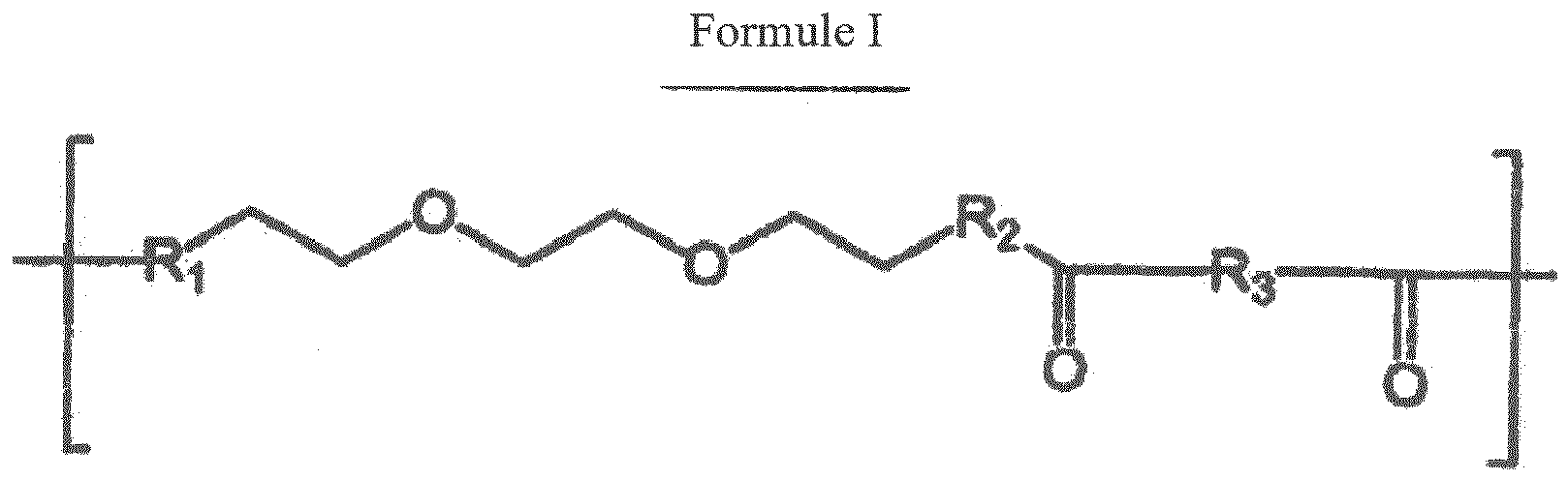
- Procédé selon l'une quelconque des revendications 7 à 12, dans lequel la cible comprend une protéine, un acide nucléique ou un segment d'analogue d'acide nucléique.
- Procédé selon l'une quelconque des revendications 7 à 13, dans lequel la sonde comprend un segment d'analogue d'acide nucléique, un segment d'analogue d'acide nucléique, une protéine, un récepteur, un substrat ou un haptène.
- Procédé selon l'une quelconque des revendications 7 à 14, dans lequel l'échantillon comprend un échantillon cellulaire ou un prélèvement tissulaire dérivé d'un organisme vivant.
- Trousse comprenant une composition selon l'une quelconque des revendications 1 à 6.
Applications Claiming Priority (4)
| Application Number | Priority Date | Filing Date | Title |
|---|---|---|---|
| US69540905P | 2005-07-01 | 2005-07-01 | |
| US69541005P | 2005-07-01 | 2005-07-01 | |
| US69540805P | 2005-07-01 | 2005-07-01 | |
| PCT/IB2006/003130 WO2007023390A2 (fr) | 2005-07-01 | 2006-06-30 | Procede de detection par immunohistochimie |
Publications (2)
| Publication Number | Publication Date |
|---|---|
| EP1910575A2 EP1910575A2 (fr) | 2008-04-16 |
| EP1910575B1 true EP1910575B1 (fr) | 2014-12-10 |
Family
ID=37708983
Family Applications (4)
| Application Number | Title | Priority Date | Filing Date |
|---|---|---|---|
| EP20060808856 Not-in-force EP1907592B1 (fr) | 2005-07-01 | 2006-06-30 | Molecules de liaison monomeriques et polymeriques utilisees dans la conjugaison de molecules biologiques et d'autres substances |
| EP06842206.2A Not-in-force EP1919930B1 (fr) | 2005-07-01 | 2006-06-30 | Paires de bases d'acides nucléiques |
| EP20060820855 Withdrawn EP1920072A2 (fr) | 2005-07-01 | 2006-06-30 | Procede de detection d'hybridation in situ |
| EP06809193.3A Not-in-force EP1910575B1 (fr) | 2005-07-01 | 2006-06-30 | Procede de detection par immunohistochimie |
Family Applications Before (3)
| Application Number | Title | Priority Date | Filing Date |
|---|---|---|---|
| EP20060808856 Not-in-force EP1907592B1 (fr) | 2005-07-01 | 2006-06-30 | Molecules de liaison monomeriques et polymeriques utilisees dans la conjugaison de molecules biologiques et d'autres substances |
| EP06842206.2A Not-in-force EP1919930B1 (fr) | 2005-07-01 | 2006-06-30 | Paires de bases d'acides nucléiques |
| EP20060820855 Withdrawn EP1920072A2 (fr) | 2005-07-01 | 2006-06-30 | Procede de detection d'hybridation in situ |
Country Status (6)
| Country | Link |
|---|---|
| US (6) | US8481697B2 (fr) |
| EP (4) | EP1907592B1 (fr) |
| AT (1) | ATE501277T1 (fr) |
| DE (1) | DE602006020577D1 (fr) |
| DK (1) | DK1907592T3 (fr) |
| WO (4) | WO2007023390A2 (fr) |
Families Citing this family (118)
| Publication number | Priority date | Publication date | Assignee | Title |
|---|---|---|---|---|
| DE602006020577D1 (de) | 2005-07-01 | 2011-04-21 | Dako Denmark As | Monomere und polymere linker zur konjugierung von biologischen molekülen und anderen stoffen |
| GB0517097D0 (en) | 2005-08-19 | 2005-09-28 | Solexa Ltd | Modified nucleosides and nucleotides and uses thereof |
| WO2008116468A2 (fr) | 2007-03-26 | 2008-10-02 | Dako Denmark A/S | Complexes peptidiques du cmh et leurs utilisations dans des maladies infectieuses |
| EP3620465B1 (fr) | 2007-07-03 | 2025-02-19 | Dako Denmark A/S | Procédés améliorés pour produire, marquer et utiliser des multimères du cmh |
| ES2624450T3 (es) | 2007-09-18 | 2017-07-14 | Dako Denmark A/S | Método rápido y sensible para la detección de dianas biológicas |
| US10611818B2 (en) | 2007-09-27 | 2020-04-07 | Agilent Technologies, Inc. | MHC multimers in tuberculosis diagnostics, vaccine and therapeutics |
| DK2254592T3 (da) | 2008-02-28 | 2019-09-09 | Dako Denmark As | MHC-multimerer til Borrelia-diagnostik og sygdom |
| US7968702B2 (en) * | 2008-03-13 | 2011-06-28 | Pacific Biosciences Of California, Inc. | Labeled reactants and their uses |
| WO2010009735A2 (fr) | 2008-07-23 | 2010-01-28 | Dako Denmark A/S | Analyse et réparation combinatoires |
| WO2010037402A1 (fr) | 2008-10-02 | 2010-04-08 | Dako Denmark A/S | Vaccins moléculaires contre les maladies infectieuses |
| US11992518B2 (en) | 2008-10-02 | 2024-05-28 | Agilent Technologies, Inc. | Molecular vaccines for infectious disease |
| CN111808084A (zh) | 2008-11-10 | 2020-10-23 | 阿布特斯生物制药公司 | 用于递送治疗剂的新型脂质和组合物 |
| EP2398908B1 (fr) | 2009-02-19 | 2013-08-21 | Dako Denmark A/S | Procédés et composés pour la détection de cibles moléculaires |
| WO2011028952A1 (fr) | 2009-09-02 | 2011-03-10 | Xencor, Inc. | Compositions et procédés pour une co-liaison bivalente et monovalente simultanée d'antigènes |
| CN104714007B (zh) | 2009-10-20 | 2017-04-12 | 丹麦达科有限公司 | 单个靶实体的免疫化学检测 |
| EP2539355B1 (fr) * | 2010-02-26 | 2016-10-05 | Ventana Medical Systems, Inc. | Hybridation in-situ avec des sondes polytag |
| AU2011283694B2 (en) | 2010-07-29 | 2017-04-13 | Xencor, Inc. | Antibodies with modified isoelectric points |
| CN103201627B (zh) | 2010-11-08 | 2016-10-12 | 丹麦达科有限公司 | 组织学样品中单个靶标分子的定量 |
| US20120258870A1 (en) | 2010-11-22 | 2012-10-11 | The University Of Chicago | Methods, Systems, and/or Use of Oligonucleotide Conjugates to Develop Panels for Use in Assays and Detections |
| CN103328979B (zh) | 2010-12-06 | 2015-10-07 | 丹麦达科有限公司 | 组合的组织学染色 |
| EP2699691B1 (fr) | 2011-04-19 | 2017-10-18 | Dako Denmark A/S | Nouvelle méthode d'amplification de signal médiée par l'enzyme |
| EP2705160B1 (fr) * | 2011-05-06 | 2018-08-22 | Qiagen GmbH | Nucléotides et oligonucléotides comportant un marqueur liée à un agent de liaison |
| EP2755026B1 (fr) * | 2011-09-09 | 2021-10-27 | Konica Minolta, Inc. | Procédé de teinture d'étoffe |
| US12466897B2 (en) | 2011-10-10 | 2025-11-11 | Xencor, Inc. | Heterodimeric human IgG1 polypeptides with isoelectric point modifications |
| US10851178B2 (en) | 2011-10-10 | 2020-12-01 | Xencor, Inc. | Heterodimeric human IgG1 polypeptides with isoelectric point modifications |
| EP3604555B1 (fr) | 2011-10-14 | 2024-12-25 | President and Fellows of Harvard College | Séquençage par assemblage de structure |
| EP2773390B1 (fr) * | 2011-11-03 | 2020-12-30 | Tripath Imaging, Inc. | Procede et compositions pour la preparation d`echantillons pour la coloration immunologique |
| US10648970B2 (en) * | 2011-11-08 | 2020-05-12 | Agilent Technologies, Inc. | Method for evaluation of target in histological sample |
| CN103917529B (zh) | 2011-11-11 | 2016-08-17 | 辉瑞大药厂 | 2-硫代嘧啶酮类 |
| EP4249605B1 (fr) | 2011-12-22 | 2024-08-28 | President And Fellows Of Harvard College | Procédés de détection d'analyte |
| ES2991004T3 (es) | 2011-12-22 | 2024-12-02 | Harvard College | Métodos para la detección de analitos |
| ES2678211T3 (es) | 2012-03-27 | 2018-08-09 | Ventana Medical Systems, Inc. | Conjugados de señalización y procedimientos de uso |
| WO2013177046A1 (fr) | 2012-05-21 | 2013-11-28 | Solulink, Inc. | Procédés et/ou utilisation de conjugués oligonucléotidiques pour la suppression d'arrière-plan dû à l'hybridation croisée |
| US9914967B2 (en) | 2012-06-05 | 2018-03-13 | President And Fellows Of Harvard College | Spatial sequencing of nucleic acids using DNA origami probes |
| US10487155B2 (en) | 2013-01-14 | 2019-11-26 | Xencor, Inc. | Heterodimeric proteins |
| KR102211837B1 (ko) * | 2013-01-14 | 2021-02-03 | 젠코어 인코포레이티드 | 신규한 이형이량체 단백질 |
| US10131710B2 (en) | 2013-01-14 | 2018-11-20 | Xencor, Inc. | Optimized antibody variable regions |
| US9701759B2 (en) | 2013-01-14 | 2017-07-11 | Xencor, Inc. | Heterodimeric proteins |
| US11053316B2 (en) | 2013-01-14 | 2021-07-06 | Xencor, Inc. | Optimized antibody variable regions |
| US10968276B2 (en) | 2013-03-12 | 2021-04-06 | Xencor, Inc. | Optimized anti-CD3 variable regions |
| US9605084B2 (en) | 2013-03-15 | 2017-03-28 | Xencor, Inc. | Heterodimeric proteins |
| WO2014113510A1 (fr) | 2013-01-15 | 2014-07-24 | Xencor, Inc. | Elimination rapide de complexes antigéniques à l'aide de nouveaux anticorps |
| EP2971184B1 (fr) | 2013-03-12 | 2019-04-17 | President and Fellows of Harvard College | Procédé de génération d'une matrice tridimensionnelle contenant des acides nucléiques |
| US10858417B2 (en) | 2013-03-15 | 2020-12-08 | Xencor, Inc. | Heterodimeric proteins |
| US10106624B2 (en) | 2013-03-15 | 2018-10-23 | Xencor, Inc. | Heterodimeric proteins |
| JP6594855B2 (ja) * | 2013-03-15 | 2019-10-23 | ゼンコア インコーポレイテッド | ヘテロ二量体タンパク質 |
| EP2970486B1 (fr) | 2013-03-15 | 2018-05-16 | Xencor, Inc. | Modulation de cellules t avec des anticorps bispecifiques et des fusions fc |
| US10519242B2 (en) | 2013-03-15 | 2019-12-31 | Xencor, Inc. | Targeting regulatory T cells with heterodimeric proteins |
| CA2907493A1 (fr) | 2013-04-30 | 2014-11-13 | California Institute Of Technology | Marquage multiplex de molecules par marquage par code-barres a hybridation sequentielle |
| WO2015074001A1 (fr) | 2013-11-17 | 2015-05-21 | Quantum-Si Incorporated | Système optique et puce d'analyse pour sonder, détecter et analyser des molécules |
| EP3699195A3 (fr) | 2014-03-28 | 2020-11-04 | Xencor, Inc. | Anticorps bispécifiques se liant à cd38 et cd3 |
| CN103969436B (zh) * | 2014-05-08 | 2015-09-16 | 东南大学 | 一种超灵敏碱性磷酸酶检测的新方法 |
| WO2015188839A2 (fr) * | 2014-06-13 | 2015-12-17 | Immudex Aps | Détection générale et isolement de cellules spécifiques par liaison de molécules marquées |
| WO2016007839A1 (fr) | 2014-07-11 | 2016-01-14 | President And Fellows Of Harvard College | Méthodes de marquage et de détection à haut rendement de caractéristiques biologiques in situ à l'aide de microscopie |
| EP4328322A3 (fr) | 2014-07-30 | 2024-05-22 | President and Fellows of Harvard College | Construction de bibliothèque de sondes |
| US10174363B2 (en) | 2015-05-20 | 2019-01-08 | Quantum-Si Incorporated | Methods for nucleic acid sequencing |
| EP4421188A3 (fr) | 2014-08-08 | 2024-10-23 | Quantum-Si Incorporated | Dispositif intégré avec source de lumière externe pour sonder, détecter et analyser des molécules |
| CA2957543A1 (fr) | 2014-08-08 | 2016-02-11 | Quantum-Si Incorporated | Systeme optique et puce d'analyse pour sonder, detecter, et analyser des molecules |
| EP3471402B1 (fr) | 2014-08-08 | 2023-05-31 | Quantum-Si Incorporated | Dispositif intégré pour le groupement temporel de photons reçus |
| WO2016057842A2 (fr) * | 2014-10-08 | 2016-04-14 | Aratome, LLC | Imagerie haute résolution de protéines tissulaires |
| EP3223907A2 (fr) | 2014-11-26 | 2017-10-04 | Xencor, Inc. | Anticorps hétérodimériques se liant aux antigènes cd3 et cd38 |
| PE20171324A1 (es) | 2014-11-26 | 2017-09-11 | Xencor Inc | Anticuerpos heterodimericos que se unen a cd3 y a antigenos tumorales |
| US10259887B2 (en) | 2014-11-26 | 2019-04-16 | Xencor, Inc. | Heterodimeric antibodies that bind CD3 and tumor antigens |
| WO2016105450A2 (fr) | 2014-12-22 | 2016-06-30 | Xencor, Inc. | Anticorps trispécifiques |
| US10227411B2 (en) | 2015-03-05 | 2019-03-12 | Xencor, Inc. | Modulation of T cells with bispecific antibodies and FC fusions |
| US10036066B2 (en) | 2015-03-17 | 2018-07-31 | Dako Denmark A/S | In situ hybridization detection method |
| AU2016257179A1 (en) | 2015-05-05 | 2017-11-02 | Pfizer Inc. | 2-thiopyrimidinones |
| JP6841609B2 (ja) | 2015-07-10 | 2021-03-10 | 3スキャン インコーポレイテッド | 組織学的染色の空間的多重化 |
| AU2016349288A1 (en) | 2015-11-03 | 2018-05-31 | President And Fellows Of Harvard College | Method and apparatus for volumetric imaging of a three-dimensional nucleic acid containing matrix |
| CA3007030A1 (fr) | 2015-12-07 | 2017-06-15 | Xencor, Inc. | Anticorps heterodimees se liant a cd3 et psma |
| EP4336184A3 (fr) | 2015-12-18 | 2024-06-05 | Agilent Technologies, Inc. (A Delaware Corporation) | Substrats de peroxydase à fraction chromogène |
| KR20180111999A (ko) | 2016-02-17 | 2018-10-11 | 테서렉트 헬스, 인코포레이티드 | 수명 촬상 및 검출 응용을 위한 센서 및 디바이스 |
| WO2017160662A1 (fr) * | 2016-03-12 | 2017-09-21 | The Regents Of The University Of California | Vecteurs biodégradables pour une administration efficace d'arn |
| EP4613756A3 (fr) | 2016-04-25 | 2025-11-12 | President And Fellows Of Harvard College | Procedes de reaction en chaine d'hybridation pour detection moleculaire in situ |
| JP7010854B2 (ja) | 2016-06-14 | 2022-01-26 | ゼンコア インコーポレイテッド | 二重特異性チェックポイント阻害剤抗体 |
| CN109715663B (zh) | 2016-06-28 | 2022-11-25 | Xencor股份有限公司 | 结合生长抑素受体2的异源二聚抗体 |
| US10793632B2 (en) | 2016-08-30 | 2020-10-06 | Xencor, Inc. | Bispecific immunomodulatory antibodies that bind costimulatory and checkpoint receptors |
| CN118853848A (zh) | 2016-08-31 | 2024-10-29 | 哈佛学院董事及会员团体 | 将生物分子的检测组合到使用荧光原位测序的单个试验的方法 |
| MY203000A (en) | 2016-10-14 | 2024-06-01 | Xencor Inc | Il15/il15r� heterodimeric fc-fusion proteins |
| EP4556575A3 (fr) | 2016-11-21 | 2025-10-08 | Bruker Spatial Biology, Inc. | Procédé de séquençage d'acides nucléiques |
| CN106674134B (zh) * | 2016-11-30 | 2019-01-01 | 辽宁大学 | 一种新型荧光探针及其制备方法和在检测6-硫代鸟嘌呤中的应用 |
| DK3555313T3 (da) | 2016-12-16 | 2021-08-09 | Aratome Llc | Molekylær detektering ved hjælp af ligeringsamplifikation |
| EP3554556B1 (fr) * | 2016-12-19 | 2022-03-09 | Ventana Medical Systems, Inc. | Conjugués de peptides et d'acides nucléiques |
| CA3047826A1 (fr) | 2016-12-22 | 2018-06-28 | Quantum-Si Incorporated | Photodetecteur integre a pixel de compartimentage direct |
| US20210389313A1 (en) * | 2017-01-26 | 2021-12-16 | Confer Health, Inc. | Magnetic particle-based immunoassay and methods of using the same |
| US11788123B2 (en) | 2017-05-26 | 2023-10-17 | President And Fellows Of Harvard College | Systems and methods for high-throughput image-based screening |
| WO2019006472A1 (fr) | 2017-06-30 | 2019-01-03 | Xencor, Inc. | Protéines de fusion fc hétérodimères ciblées contenant il-15/il-15ra et domaines de liaison à l'antigène |
| BR112020000826A2 (pt) | 2017-07-24 | 2020-07-21 | Quantum-Si Incorporated | instrumento bio-optoeletrônico maciçamente paralelo portátil |
| WO2019023257A1 (fr) | 2017-07-24 | 2019-01-31 | Quantum-Si Incorporated | Compositions de réactifs marqués à haute intensité et méthodes de séquençage |
| JP2021502100A (ja) | 2017-11-08 | 2021-01-28 | ゼンコア インコーポレイテッド | 新規抗pd−1配列を用いた二重特異性および単一特異性抗体 |
| US10981992B2 (en) | 2017-11-08 | 2021-04-20 | Xencor, Inc. | Bispecific immunomodulatory antibodies that bind costimulatory and checkpoint receptors |
| JP7727385B2 (ja) * | 2017-12-18 | 2025-08-21 | ヴェンタナ メディカル システムズ, インク. | ペプチド核酸コンジュゲート |
| IL275426B2 (en) | 2017-12-19 | 2025-03-01 | Xencor Inc | Engineered il-2 fc fusion proteins |
| CA3096052A1 (fr) | 2018-04-04 | 2019-10-10 | Xencor, Inc. | Anticorps heterodimeres qui se lient a la proteine d'activation des fibroblastes |
| SG11202010163QA (en) | 2018-04-18 | 2020-11-27 | Xencor Inc | Pd-1 targeted heterodimeric fusion proteins containing il-15/il-15ra fc-fusion proteins and pd-1 antigen binding domains and uses thereof |
| EP3781598A1 (fr) | 2018-04-18 | 2021-02-24 | Xencor, Inc. | Protéines de fusion hétérodimères ciblant tim-3 contenant des protéines de fusion fc d'il-15/il-15ra et domaines de liaison à l'antigène de tim-3 |
| EP3794146B1 (fr) | 2018-05-14 | 2025-12-10 | Bruker Spatial Biology, Inc. | Méthode d'identifcation d'une séquence nucléotide predeterminée |
| CN119364214A (zh) | 2018-06-22 | 2025-01-24 | 宽腾矽公司 | 具有不同检测时间的电荷存储仓的集成光电检测器 |
| CA3105715A1 (fr) | 2018-07-13 | 2020-01-16 | Quantum-Si Incorporated | Indicateurs bioconjugables et methodes d'utilisation |
| KR20210063379A (ko) * | 2018-09-24 | 2021-06-01 | 브리스톨-마이어스 스큅 컴퍼니 | 바코딩된 펩티드-mhc 복합체 및 그의 용도 |
| AU2019355971B2 (en) | 2018-10-03 | 2025-05-08 | Xencor, Inc. | IL-12 heterodimeric Fc-fusion proteins |
| WO2020076976A1 (fr) | 2018-10-10 | 2020-04-16 | Readcoor, Inc. | Indexation moléculaire spatiale tridimensionnelle |
| JP7307793B2 (ja) * | 2018-11-16 | 2023-07-12 | エフ. ホフマン-ラ ロシュ アーゲー | 結合対のメンバーを有するストレプトアビジン被覆固相 |
| EP3894583A4 (fr) | 2018-12-13 | 2022-11-09 | President and Fellows of Harvard College | Systèmes et procédés d'amplification pour merfish et d'autres applications |
| WO2020127222A2 (fr) | 2018-12-17 | 2020-06-25 | Immudex Aps | Panneau comprenant des multimères de borrelia cmh |
| WO2020154546A1 (fr) | 2019-01-23 | 2020-07-30 | Quantum-Si Incorporated | Compositions de réactifs marqués à haute intensité et méthodes de séquençage |
| CA3132185A1 (fr) | 2019-03-01 | 2020-09-10 | Xencor, Inc. | Anticorps heterodimeres qui se lient a enpp3 et cd3 |
| US11287422B2 (en) | 2019-09-23 | 2022-03-29 | Element Biosciences, Inc. | Multivalent binding composition for nucleic acid analysis |
| EP4097251A1 (fr) | 2020-01-29 | 2022-12-07 | 10X Genomics, Inc. | Compositions et procédés pour la détection d'analytes |
| WO2021231976A1 (fr) | 2020-05-14 | 2021-11-18 | Xencor, Inc. | Anticorps hétérodimères qui se lient à l'antigène membranaire spécifique de la prostate (psma) et cd3 |
| US20220049303A1 (en) | 2020-08-17 | 2022-02-17 | Readcoor, Llc | Methods and systems for spatial mapping of genetic variants |
| CA3195446A1 (fr) * | 2020-10-16 | 2022-04-21 | Amelie Eriksson Karlstrom | Sondes pna pour imagerie et therapie preciblees |
| US11739144B2 (en) | 2021-03-09 | 2023-08-29 | Xencor, Inc. | Heterodimeric antibodies that bind CD3 and CLDN6 |
| EP4305065A1 (fr) | 2021-03-10 | 2024-01-17 | Xencor, Inc. | Anticorps hétérodimères qui se lient au cd3 et au gpc3 |
| CN118647732A (zh) * | 2022-01-21 | 2024-09-13 | 领先细胞医疗诊断有限公司 | 用于检测蛋白质和联合检测蛋白质和核酸的方法及其试剂盒 |
| EP4573213A1 (fr) | 2022-08-15 | 2025-06-25 | Element Biosciences, Inc. | Capture sur surface à résolution spatiale d'acides nucléiques |
| US12607551B2 (en) * | 2023-12-22 | 2026-04-21 | Cytek Biosciences, Inc. | Image capture and automated regulation of liquid jet breakoff for cell sorters and flow cytometers |
| US20260092309A1 (en) * | 2024-09-30 | 2026-04-02 | Celnovte Biotechnology, Inc. | Ultra sensitive probes for detecting biomarkers |
Family Cites Families (44)
| Publication number | Priority date | Publication date | Assignee | Title |
|---|---|---|---|---|
| US4447574A (en) * | 1982-06-29 | 1984-05-08 | Standard Oil Company (Indiana) | Injection moldable amide-imide polymers containing calcium moieties |
| US4447474A (en) * | 1982-08-30 | 1984-05-08 | Neefe Charles W | Method of selectively tinting soft contact lenses |
| US5166315A (en) * | 1989-12-20 | 1992-11-24 | Anti-Gene Development Group | Sequence-specific binding polymers for duplex nucleic acids |
| WO1989002921A1 (fr) * | 1987-09-22 | 1989-04-06 | Temple University | Analogues en paires de base adn artificielles |
| US5656731A (en) * | 1987-10-15 | 1997-08-12 | Chiron Corporation | Nucleic acid-amplified immunoassay probes |
| US5359100A (en) | 1987-10-15 | 1994-10-25 | Chiron Corporation | Bifunctional blocked phosphoramidites useful in making nucleic acid mutimers |
| US5641625A (en) | 1992-05-22 | 1997-06-24 | Isis Pharmaceuticals, Inc. | Cleaving double-stranded DNA with peptide nucleic acids |
| US5766855A (en) | 1991-05-24 | 1998-06-16 | Buchardt, Deceased; Ole | Peptide nucleic acids having enhanced binding affinity and sequence specificity |
| DK51092D0 (da) | 1991-05-24 | 1992-04-15 | Ole Buchardt | Oligonucleotid-analoge betegnet pna, monomere synthoner og fremgangsmaade til fremstilling deraf samt anvendelser deraf |
| US5714331A (en) | 1991-05-24 | 1998-02-03 | Buchardt, Deceased; Ole | Peptide nucleic acids having enhanced binding affinity, sequence specificity and solubility |
| US5719262A (en) | 1993-11-22 | 1998-02-17 | Buchardt, Deceased; Ole | Peptide nucleic acids having amino acid side chains |
| US6228982B1 (en) | 1992-05-22 | 2001-05-08 | Benget Norden | Double-stranded peptide nucleic acids |
| US5539082A (en) | 1993-04-26 | 1996-07-23 | Nielsen; Peter E. | Peptide nucleic acids |
| AU4406793A (en) * | 1992-06-04 | 1993-12-30 | Clover Consolidated, Limited | Water-soluble polymeric carriers for drug delivery |
| GB9211979D0 (en) | 1992-06-05 | 1992-07-15 | Buchard Ole | Uses of nucleic acid analogues |
| US5527675A (en) | 1993-08-20 | 1996-06-18 | Millipore Corporation | Method for degradation and sequencing of polymers which sequentially eliminate terminal residues |
| DE4331012A1 (de) | 1993-09-13 | 1995-03-16 | Bayer Ag | Nukleinsäuren-bindende Oligomere mit N-Verzweigung für Therapie und Diagnostik |
| US7375198B2 (en) | 1993-10-26 | 2008-05-20 | Affymetrix, Inc. | Modified nucleic acid probes |
| US5538848A (en) * | 1994-11-16 | 1996-07-23 | Applied Biosystems Division, Perkin-Elmer Corp. | Method for detecting nucleic acid amplification using self-quenching fluorescence probe |
| GB2284209A (en) | 1993-11-25 | 1995-05-31 | Ole Buchardt | Nucleic acid analogue-induced transcription of RNA from a double-stranded DNA template |
| IL112759A0 (en) * | 1994-02-25 | 1995-05-26 | Khepri Pharmaceuticals Inc | Novel cysteine protease inhibitors |
| US5705333A (en) | 1994-08-05 | 1998-01-06 | The Regents Of The University Of California | Peptide-based nucleic acid mimics(PENAMS) |
| US5681702A (en) * | 1994-08-30 | 1997-10-28 | Chiron Corporation | Reduction of nonspecific hybridization by using novel base-pairing schemes |
| US6361951B1 (en) * | 1995-06-27 | 2002-03-26 | The University Of North Carolina At Chapel Hill | Electrochemical detection of nucleic acid hybridization |
| US20040033973A1 (en) | 2002-08-16 | 2004-02-19 | Muthiah Manoharan | Compounds and oligomeric compounds comprising novel nucleobases |
| EP0938588B1 (fr) * | 1996-07-12 | 2003-02-12 | TM Technologies Inc. | Procede d'amplification de signaux |
| EP2319854B1 (fr) | 1997-01-08 | 2016-11-30 | Sigma-Aldrich Co. LLC | Bio-conjugaison de macromolécules |
| US6107470A (en) | 1997-05-29 | 2000-08-22 | Nielsen; Peter E. | Histidine-containing peptide nucleic acids |
| US6294331B1 (en) * | 1997-08-08 | 2001-09-25 | The United States Of America As Represented By The Department Of Health And Human Services | Methods for assessing genetic and phenotypic markers by simultaneous multicolor visualization of chromogenic dyes using brightfield microscopy and spectral imaging |
| US6342381B1 (en) | 1998-02-27 | 2002-01-29 | Buckman Laboratories Internationals, Inc. | Enzyme stabilization with pre-superpolyamide or pre-fiber-forming polyamide oligomers |
| US6287772B1 (en) * | 1998-04-29 | 2001-09-11 | Boston Probes, Inc. | Methods, kits and compositions for detecting and quantitating target sequences |
| WO2000068434A2 (fr) | 1999-05-07 | 2000-11-16 | Yale University | Analyse a etiquetage multiple |
| JP2001252097A (ja) * | 2000-03-10 | 2001-09-18 | Fuji Photo Film Co Ltd | 核酸ハイブリダイゼーションの効率化法 |
| US6451588B1 (en) * | 2000-06-30 | 2002-09-17 | Pe Corporation (Ny) | Multipartite high-affinity nucleic acid probes |
| WO2002063299A1 (fr) | 2001-02-05 | 2002-08-15 | Graffinity Pharmaceuticals Ag | Procede de criblage des faibles affinites |
| US20020132260A1 (en) | 2001-02-22 | 2002-09-19 | Erlander Mark G. | Quantitative immunohistochemistry (QIHC) |
| US20020197630A1 (en) * | 2001-04-12 | 2002-12-26 | Knapp Michael R. | Systems and methods for high throughput genetic analysis |
| JP2005509028A (ja) * | 2001-10-09 | 2005-04-07 | バーテックス ファーマシューティカルズ インコーポレイテッド | アスパラギン酸およびグルタミン酸誘導体ならびにそのジアゾケトン中間体を合成するための方法 |
| WO2003080857A2 (fr) * | 2002-03-21 | 2003-10-02 | Boston Probes,Inc. | Oligomeres pna, ensembles d'oligomeres, procedes et trousses relatifs a la detection de bacillus anthracis |
| CA2480673A1 (fr) | 2002-03-29 | 2003-10-09 | Genentech, Inc. | Procedes et compositions de detection et quantification d'analytes d'acides nucleiques |
| WO2004091542A2 (fr) * | 2003-04-15 | 2004-10-28 | Covx Pharmaceuticals, Inc. | Composes de ciblage d'integrine contenant de l'azote |
| US7256198B2 (en) * | 2004-02-18 | 2007-08-14 | Wyeth | Pyrimidoindolones and methods for using same |
| EP1766089A1 (fr) * | 2004-06-30 | 2007-03-28 | Applera Corporation | Complexes sondes analogiques |
| DE602006020577D1 (de) | 2005-07-01 | 2011-04-21 | Dako Denmark As | Monomere und polymere linker zur konjugierung von biologischen molekülen und anderen stoffen |
-
2006
- 2006-06-30 DE DE200660020577 patent/DE602006020577D1/de active Active
- 2006-06-30 WO PCT/IB2006/003130 patent/WO2007023390A2/fr not_active Ceased
- 2006-06-30 EP EP20060808856 patent/EP1907592B1/fr not_active Not-in-force
- 2006-06-30 AT AT06808856T patent/ATE501277T1/de not_active IP Right Cessation
- 2006-06-30 WO PCT/IB2006/003123 patent/WO2007026252A2/fr not_active Ceased
- 2006-06-30 WO PCT/IB2006/002584 patent/WO2007015168A2/fr not_active Ceased
- 2006-06-30 US US11/993,565 patent/US8481697B2/en not_active Expired - Fee Related
- 2006-06-30 US US11/993,528 patent/US8741566B2/en active Active
- 2006-06-30 US US11/993,571 patent/US9017992B2/en active Active
- 2006-06-30 EP EP06842206.2A patent/EP1919930B1/fr not_active Not-in-force
- 2006-06-30 DK DK06808856T patent/DK1907592T3/da active
- 2006-06-30 EP EP20060820855 patent/EP1920072A2/fr not_active Withdrawn
- 2006-06-30 US US11/993,545 patent/US8546309B2/en active Active
- 2006-06-30 EP EP06809193.3A patent/EP1910575B1/fr not_active Not-in-force
- 2006-06-30 WO PCT/IB2006/003306 patent/WO2007045998A2/fr not_active Ceased
-
2013
- 2013-08-21 US US13/971,996 patent/US9598720B2/en active Active
-
2014
- 2014-04-14 US US14/251,934 patent/US9340824B2/en active Active
Non-Patent Citations (2)
| Title |
|---|
| ISHIKAWA M ET AL: "Synthesis of 3-(2-deoxy-beta-d-ribofuranosyl)pyridin-2-one and 2-amino-6-(N,N-dimethylamino)-9-(2-deoxy-beta-d-ribofuranosyl)purine derivatives for an unnatural base pair", TETRAHEDRON LETTERS, ELSEVIER, AMSTERDAM, NL, vol. 41, no. 20, 1 May 2000 (2000-05-01), pages 3931 - 3934, XP004203291, ISSN: 0040-4039, DOI: 10.1016/S0040-4039(00)00520-7 * |
| RAY A ET AL: "PEPTIDE NUCLEIC ACID (PNA): ITS MEDICAL AND BIOTECHNICAL APPLICATIONS AND PROMISE FOR THE FUTURE", FASEB JOURNAL, FED. OF AMERICAN SOC. FOR EXPERIMENTAL BIOLOGY, US, vol. 14, no. 9, 1 June 2000 (2000-06-01), pages 1041 - 1060, XP001162782, ISSN: 0892-6638 * |
Also Published As
| Publication number | Publication date |
|---|---|
| DE602006020577D1 (de) | 2011-04-21 |
| WO2007045998A2 (fr) | 2007-04-26 |
| WO2007023390A2 (fr) | 2007-03-01 |
| US20100120054A1 (en) | 2010-05-13 |
| US9598720B2 (en) | 2017-03-21 |
| DK1907592T3 (da) | 2011-06-27 |
| EP1920072A2 (fr) | 2008-05-14 |
| US9017992B2 (en) | 2015-04-28 |
| EP1919930B1 (fr) | 2016-09-14 |
| US20100221705A1 (en) | 2010-09-02 |
| US20110177500A1 (en) | 2011-07-21 |
| WO2007015168A2 (fr) | 2007-02-08 |
| EP1907592B1 (fr) | 2011-03-09 |
| WO2007023390A3 (fr) | 2007-08-02 |
| US20100105030A1 (en) | 2010-04-29 |
| US8546309B2 (en) | 2013-10-01 |
| WO2007026252A3 (fr) | 2007-09-13 |
| US20140080145A1 (en) | 2014-03-20 |
| US20140315204A1 (en) | 2014-10-23 |
| US8741566B2 (en) | 2014-06-03 |
| EP1910575A2 (fr) | 2008-04-16 |
| ATE501277T1 (de) | 2011-03-15 |
| WO2007026252A2 (fr) | 2007-03-08 |
| EP1907592A2 (fr) | 2008-04-09 |
| US8481697B2 (en) | 2013-07-09 |
| WO2007045998A3 (fr) | 2008-04-10 |
| WO2007015168A3 (fr) | 2007-08-30 |
| EP1919930A2 (fr) | 2008-05-14 |
| US9340824B2 (en) | 2016-05-17 |
Similar Documents
| Publication | Publication Date | Title |
|---|---|---|
| EP1910575B1 (fr) | Procede de detection par immunohistochimie | |
| US20100105145A1 (en) | Method of simultaneously visualizing multiple biological targets | |
| ES2624450T3 (es) | Método rápido y sensible para la detección de dianas biológicas | |
| JP5721638B2 (ja) | コンジュゲート分子 | |
| JPH03261798A (ja) | 変性ヌクレオチド | |
| EP0185547A2 (fr) | Tests d'hybridation de polynucléotides employant la luminescence catalysée | |
| US10036066B2 (en) | In situ hybridization detection method | |
| US20100151460A1 (en) | Systems and Methods for Biological and Chemical Detection, Comprising Automatic Selection of Reagent Sets | |
| WO2004037989A2 (fr) | Nucleotides modifies et procedes de marquage d'acides nucleiques |
Legal Events
| Date | Code | Title | Description |
|---|---|---|---|
| PUAI | Public reference made under article 153(3) epc to a published international application that has entered the european phase |
Free format text: ORIGINAL CODE: 0009012 |
|
| 17P | Request for examination filed |
Effective date: 20080125 |
|
| AK | Designated contracting states |
Kind code of ref document: A2 Designated state(s): AT BE BG CH CY CZ DE DK EE ES FI FR GB GR HU IE IS IT LI LT LU LV MC NL PL PT RO SE SI SK TR |
|
| RIN1 | Information on inventor provided before grant (corrected) |
Inventor name: LOHSE, JESPER Inventor name: GABS, SUSANNE Inventor name: PETERSEN, KENNETH, HEESCHE Inventor name: WINTHER, LARS |
|
| 17Q | First examination report despatched |
Effective date: 20080626 |
|
| DAX | Request for extension of the european patent (deleted) | ||
| REG | Reference to a national code |
Ref country code: DE Ref legal event code: R079 Ref document number: 602006043976 Country of ref document: DE Free format text: PREVIOUS MAIN CLASS: C12Q0001680000 Ipc: A61K0047480000 |
|
| RIC1 | Information provided on ipc code assigned before grant |
Ipc: A61K 47/48 20060101AFI20130829BHEP Ipc: C08G 69/02 20060101ALI20130829BHEP Ipc: A61K 49/00 20060101ALI20130829BHEP Ipc: C07H 19/06 20060101ALI20130829BHEP Ipc: C07H 21/00 20060101ALI20130829BHEP Ipc: C07H 19/10 20060101ALI20130829BHEP Ipc: C07H 19/16 20060101ALI20130829BHEP Ipc: C07H 19/20 20060101ALI20130829BHEP |
|
| GRAP | Despatch of communication of intention to grant a patent |
Free format text: ORIGINAL CODE: EPIDOSNIGR1 |
|
| INTG | Intention to grant announced |
Effective date: 20131122 |
|
| RIN1 | Information on inventor provided before grant (corrected) |
Inventor name: GABS, SUSANNE Inventor name: LOHSE, JESPER Inventor name: WINTHER, LARS Inventor name: PETERSEN, KENNETH, HEESCHE |
|
| GRAP | Despatch of communication of intention to grant a patent |
Free format text: ORIGINAL CODE: EPIDOSNIGR1 |
|
| GRAS | Grant fee paid |
Free format text: ORIGINAL CODE: EPIDOSNIGR3 |
|
| INTG | Intention to grant announced |
Effective date: 20140513 |
|
| GRAA | (expected) grant |
Free format text: ORIGINAL CODE: 0009210 |
|
| AK | Designated contracting states |
Kind code of ref document: B1 Designated state(s): AT BE BG CH CY CZ DE DK EE ES FI FR GB GR HU IE IS IT LI LT LU LV MC NL PL PT RO SE SI SK TR |
|
| REG | Reference to a national code |
Ref country code: GB Ref legal event code: FG4D |
|
| REG | Reference to a national code |
Ref country code: CH Ref legal event code: EP |
|
| REG | Reference to a national code |
Ref country code: IE Ref legal event code: FG4D |
|
| REG | Reference to a national code |
Ref country code: AT Ref legal event code: REF Ref document number: 700314 Country of ref document: AT Kind code of ref document: T Effective date: 20150115 |
|
| REG | Reference to a national code |
Ref country code: DE Ref legal event code: R096 Ref document number: 602006043976 Country of ref document: DE Effective date: 20150122 |
|
| REG | Reference to a national code |
Ref country code: AT Ref legal event code: MK05 Ref document number: 700314 Country of ref document: AT Kind code of ref document: T Effective date: 20141210 Ref country code: NL Ref legal event code: VDEP Effective date: 20141210 |
|
| REG | Reference to a national code |
Ref country code: NL Ref legal event code: VDEP Effective date: 20141210 |
|
| PG25 | Lapsed in a contracting state [announced via postgrant information from national office to epo] |
Ref country code: LT Free format text: LAPSE BECAUSE OF FAILURE TO SUBMIT A TRANSLATION OF THE DESCRIPTION OR TO PAY THE FEE WITHIN THE PRESCRIBED TIME-LIMIT Effective date: 20141210 Ref country code: ES Free format text: LAPSE BECAUSE OF FAILURE TO SUBMIT A TRANSLATION OF THE DESCRIPTION OR TO PAY THE FEE WITHIN THE PRESCRIBED TIME-LIMIT Effective date: 20141210 Ref country code: FI Free format text: LAPSE BECAUSE OF FAILURE TO SUBMIT A TRANSLATION OF THE DESCRIPTION OR TO PAY THE FEE WITHIN THE PRESCRIBED TIME-LIMIT Effective date: 20141210 |
|
| REG | Reference to a national code |
Ref country code: LT Ref legal event code: MG4D |
|
| PG25 | Lapsed in a contracting state [announced via postgrant information from national office to epo] |
Ref country code: LV Free format text: LAPSE BECAUSE OF FAILURE TO SUBMIT A TRANSLATION OF THE DESCRIPTION OR TO PAY THE FEE WITHIN THE PRESCRIBED TIME-LIMIT Effective date: 20141210 Ref country code: SE Free format text: LAPSE BECAUSE OF FAILURE TO SUBMIT A TRANSLATION OF THE DESCRIPTION OR TO PAY THE FEE WITHIN THE PRESCRIBED TIME-LIMIT Effective date: 20141210 Ref country code: GR Free format text: LAPSE BECAUSE OF FAILURE TO SUBMIT A TRANSLATION OF THE DESCRIPTION OR TO PAY THE FEE WITHIN THE PRESCRIBED TIME-LIMIT Effective date: 20150311 Ref country code: AT Free format text: LAPSE BECAUSE OF FAILURE TO SUBMIT A TRANSLATION OF THE DESCRIPTION OR TO PAY THE FEE WITHIN THE PRESCRIBED TIME-LIMIT Effective date: 20141210 |
|
| PG25 | Lapsed in a contracting state [announced via postgrant information from national office to epo] |
Ref country code: NL Free format text: LAPSE BECAUSE OF FAILURE TO SUBMIT A TRANSLATION OF THE DESCRIPTION OR TO PAY THE FEE WITHIN THE PRESCRIBED TIME-LIMIT Effective date: 20141210 |
|
| PG25 | Lapsed in a contracting state [announced via postgrant information from national office to epo] |
Ref country code: CZ Free format text: LAPSE BECAUSE OF FAILURE TO SUBMIT A TRANSLATION OF THE DESCRIPTION OR TO PAY THE FEE WITHIN THE PRESCRIBED TIME-LIMIT Effective date: 20141210 Ref country code: EE Free format text: LAPSE BECAUSE OF FAILURE TO SUBMIT A TRANSLATION OF THE DESCRIPTION OR TO PAY THE FEE WITHIN THE PRESCRIBED TIME-LIMIT Effective date: 20141210 Ref country code: SK Free format text: LAPSE BECAUSE OF FAILURE TO SUBMIT A TRANSLATION OF THE DESCRIPTION OR TO PAY THE FEE WITHIN THE PRESCRIBED TIME-LIMIT Effective date: 20141210 Ref country code: PT Free format text: LAPSE BECAUSE OF FAILURE TO SUBMIT A TRANSLATION OF THE DESCRIPTION OR TO PAY THE FEE WITHIN THE PRESCRIBED TIME-LIMIT Effective date: 20150410 Ref country code: RO Free format text: LAPSE BECAUSE OF FAILURE TO SUBMIT A TRANSLATION OF THE DESCRIPTION OR TO PAY THE FEE WITHIN THE PRESCRIBED TIME-LIMIT Effective date: 20141210 |
|
| PG25 | Lapsed in a contracting state [announced via postgrant information from national office to epo] |
Ref country code: IS Free format text: LAPSE BECAUSE OF FAILURE TO SUBMIT A TRANSLATION OF THE DESCRIPTION OR TO PAY THE FEE WITHIN THE PRESCRIBED TIME-LIMIT Effective date: 20150410 Ref country code: PL Free format text: LAPSE BECAUSE OF FAILURE TO SUBMIT A TRANSLATION OF THE DESCRIPTION OR TO PAY THE FEE WITHIN THE PRESCRIBED TIME-LIMIT Effective date: 20141210 |
|
| REG | Reference to a national code |
Ref country code: DE Ref legal event code: R097 Ref document number: 602006043976 Country of ref document: DE |
|
| PLBE | No opposition filed within time limit |
Free format text: ORIGINAL CODE: 0009261 |
|
| STAA | Information on the status of an ep patent application or granted ep patent |
Free format text: STATUS: NO OPPOSITION FILED WITHIN TIME LIMIT |
|
| PG25 | Lapsed in a contracting state [announced via postgrant information from national office to epo] |
Ref country code: DK Free format text: LAPSE BECAUSE OF FAILURE TO SUBMIT A TRANSLATION OF THE DESCRIPTION OR TO PAY THE FEE WITHIN THE PRESCRIBED TIME-LIMIT Effective date: 20141210 |
|
| 26N | No opposition filed |
Effective date: 20150911 |
|
| PG25 | Lapsed in a contracting state [announced via postgrant information from national office to epo] |
Ref country code: IT Free format text: LAPSE BECAUSE OF FAILURE TO SUBMIT A TRANSLATION OF THE DESCRIPTION OR TO PAY THE FEE WITHIN THE PRESCRIBED TIME-LIMIT Effective date: 20141210 |
|
| PG25 | Lapsed in a contracting state [announced via postgrant information from national office to epo] |
Ref country code: MC Free format text: LAPSE BECAUSE OF FAILURE TO SUBMIT A TRANSLATION OF THE DESCRIPTION OR TO PAY THE FEE WITHIN THE PRESCRIBED TIME-LIMIT Effective date: 20141210 |
|
| REG | Reference to a national code |
Ref country code: CH Ref legal event code: PL |
|
| PG25 | Lapsed in a contracting state [announced via postgrant information from national office to epo] |
Ref country code: LU Free format text: LAPSE BECAUSE OF FAILURE TO SUBMIT A TRANSLATION OF THE DESCRIPTION OR TO PAY THE FEE WITHIN THE PRESCRIBED TIME-LIMIT Effective date: 20150630 Ref country code: SI Free format text: LAPSE BECAUSE OF FAILURE TO SUBMIT A TRANSLATION OF THE DESCRIPTION OR TO PAY THE FEE WITHIN THE PRESCRIBED TIME-LIMIT Effective date: 20141210 |
|
| REG | Reference to a national code |
Ref country code: IE Ref legal event code: MM4A |
|
| PG25 | Lapsed in a contracting state [announced via postgrant information from national office to epo] |
Ref country code: CH Free format text: LAPSE BECAUSE OF NON-PAYMENT OF DUE FEES Effective date: 20150630 Ref country code: LI Free format text: LAPSE BECAUSE OF NON-PAYMENT OF DUE FEES Effective date: 20150630 Ref country code: IE Free format text: LAPSE BECAUSE OF NON-PAYMENT OF DUE FEES Effective date: 20150630 |
|
| REG | Reference to a national code |
Ref country code: FR Ref legal event code: PLFP Year of fee payment: 11 |
|
| PG25 | Lapsed in a contracting state [announced via postgrant information from national office to epo] |
Ref country code: BE Free format text: LAPSE BECAUSE OF FAILURE TO SUBMIT A TRANSLATION OF THE DESCRIPTION OR TO PAY THE FEE WITHIN THE PRESCRIBED TIME-LIMIT Effective date: 20141210 |
|
| REG | Reference to a national code |
Ref country code: DE Ref legal event code: R079 Ref document number: 602006043976 Country of ref document: DE Free format text: PREVIOUS MAIN CLASS: A61K0047480000 Ipc: A61K0047500000 |
|
| REG | Reference to a national code |
Ref country code: FR Ref legal event code: PLFP Year of fee payment: 12 |
|
| PG25 | Lapsed in a contracting state [announced via postgrant information from national office to epo] |
Ref country code: HU Free format text: LAPSE BECAUSE OF FAILURE TO SUBMIT A TRANSLATION OF THE DESCRIPTION OR TO PAY THE FEE WITHIN THE PRESCRIBED TIME-LIMIT; INVALID AB INITIO Effective date: 20060630 Ref country code: BG Free format text: LAPSE BECAUSE OF FAILURE TO SUBMIT A TRANSLATION OF THE DESCRIPTION OR TO PAY THE FEE WITHIN THE PRESCRIBED TIME-LIMIT Effective date: 20141210 |
|
| PG25 | Lapsed in a contracting state [announced via postgrant information from national office to epo] |
Ref country code: CY Free format text: LAPSE BECAUSE OF FAILURE TO SUBMIT A TRANSLATION OF THE DESCRIPTION OR TO PAY THE FEE WITHIN THE PRESCRIBED TIME-LIMIT Effective date: 20141210 |
|
| PG25 | Lapsed in a contracting state [announced via postgrant information from national office to epo] |
Ref country code: TR Free format text: LAPSE BECAUSE OF FAILURE TO SUBMIT A TRANSLATION OF THE DESCRIPTION OR TO PAY THE FEE WITHIN THE PRESCRIBED TIME-LIMIT Effective date: 20141210 |
|
| REG | Reference to a national code |
Ref country code: FR Ref legal event code: PLFP Year of fee payment: 13 |
|
| PGFP | Annual fee paid to national office [announced via postgrant information from national office to epo] |
Ref country code: DE Payment date: 20180619 Year of fee payment: 13 |
|
| PGFP | Annual fee paid to national office [announced via postgrant information from national office to epo] |
Ref country code: FR Payment date: 20180511 Year of fee payment: 13 |
|
| PGFP | Annual fee paid to national office [announced via postgrant information from national office to epo] |
Ref country code: GB Payment date: 20180627 Year of fee payment: 13 |
|
| REG | Reference to a national code |
Ref country code: DE Ref legal event code: R119 Ref document number: 602006043976 Country of ref document: DE |
|
| GBPC | Gb: european patent ceased through non-payment of renewal fee |
Effective date: 20190630 |
|
| PG25 | Lapsed in a contracting state [announced via postgrant information from national office to epo] |
Ref country code: DE Free format text: LAPSE BECAUSE OF NON-PAYMENT OF DUE FEES Effective date: 20200101 Ref country code: GB Free format text: LAPSE BECAUSE OF NON-PAYMENT OF DUE FEES Effective date: 20190630 |
|
| PG25 | Lapsed in a contracting state [announced via postgrant information from national office to epo] |
Ref country code: FR Free format text: LAPSE BECAUSE OF NON-PAYMENT OF DUE FEES Effective date: 20190630 |
|
| P01 | Opt-out of the competence of the unified patent court (upc) registered |
Effective date: 20230527 |