EP1925662A1 - Composition de Poxvirus Cytomegalovirus recombinant et utilisations - Google Patents
Composition de Poxvirus Cytomegalovirus recombinant et utilisations Download PDFInfo
- Publication number
- EP1925662A1 EP1925662A1 EP07023799A EP07023799A EP1925662A1 EP 1925662 A1 EP1925662 A1 EP 1925662A1 EP 07023799 A EP07023799 A EP 07023799A EP 07023799 A EP07023799 A EP 07023799A EP 1925662 A1 EP1925662 A1 EP 1925662A1
- Authority
- EP
- European Patent Office
- Prior art keywords
- virus
- plasmid
- recombinant
- alvac
- seq
- Prior art date
- Legal status (The legal status is an assumption and is not a legal conclusion. Google has not performed a legal analysis and makes no representation as to the accuracy of the status listed.)
- Withdrawn
Links
- JXLXRYINTLELKI-XOZUOACSSA-N CC(C1)[C@@](C2)(CCCCCC3)C2CC13I Chemical compound CC(C1)[C@@](C2)(CCCCCC3)C2CC13I JXLXRYINTLELKI-XOZUOACSSA-N 0.000 description 1
Images



































Classifications
-
- A—HUMAN NECESSITIES
- A61—MEDICAL OR VETERINARY SCIENCE; HYGIENE
- A61P—SPECIFIC THERAPEUTIC ACTIVITY OF CHEMICAL COMPOUNDS OR MEDICINAL PREPARATIONS
- A61P31/00—Antiinfectives, i.e. antibiotics, antiseptics, chemotherapeutics
- A61P31/12—Antivirals
-
- A—HUMAN NECESSITIES
- A61—MEDICAL OR VETERINARY SCIENCE; HYGIENE
- A61P—SPECIFIC THERAPEUTIC ACTIVITY OF CHEMICAL COMPOUNDS OR MEDICINAL PREPARATIONS
- A61P37/00—Drugs for immunological or allergic disorders
- A61P37/02—Immunomodulators
- A61P37/04—Immunostimulants
-
- C—CHEMISTRY; METALLURGY
- C07—ORGANIC CHEMISTRY
- C07K—PEPTIDES
- C07K14/00—Peptides having more than 20 amino acids; Gastrins; Somatostatins; Melanotropins; Derivatives thereof
- C07K14/005—Peptides having more than 20 amino acids; Gastrins; Somatostatins; Melanotropins; Derivatives thereof from viruses
-
- C—CHEMISTRY; METALLURGY
- C07—ORGANIC CHEMISTRY
- C07K—PEPTIDES
- C07K16/00—Immunoglobulins [IG], e.g. monoclonal or polyclonal antibodies
- C07K16/08—Immunoglobulins [IG], e.g. monoclonal or polyclonal antibodies against material from viruses
- C07K16/081—DNA viruses
- C07K16/085—Orthoherpesviridae (F), e.g. pseudorabies virus or Epstein-Barr virus
- C07K16/089—Cytomegalovirus
-
- A—HUMAN NECESSITIES
- A61—MEDICAL OR VETERINARY SCIENCE; HYGIENE
- A61K—PREPARATIONS FOR MEDICAL, DENTAL OR TOILETRY PURPOSES
- A61K39/00—Medicinal preparations containing antigens or antibodies
-
- C—CHEMISTRY; METALLURGY
- C12—BIOCHEMISTRY; BEER; SPIRITS; WINE; VINEGAR; MICROBIOLOGY; ENZYMOLOGY; MUTATION OR GENETIC ENGINEERING
- C12N—MICROORGANISMS OR ENZYMES; COMPOSITIONS THEREOF; PROPAGATING, PRESERVING, OR MAINTAINING MICROORGANISMS; MUTATION OR GENETIC ENGINEERING; CULTURE MEDIA
- C12N2710/00—MICROORGANISMS OR ENZYMES; COMPOSITIONS THEREOF; PROPAGATING, PRESERVING, OR MAINTAINING MICROORGANISMS; MUTATION OR GENETIC ENGINEERING; CULTURE MEDIA dsDNA viruses
- C12N2710/00011—Details
- C12N2710/16011—Herpesviridae
- C12N2710/16111—Cytomegalovirus, e.g. human herpesvirus 5
- C12N2710/16122—New viral proteins or individual genes, new structural or functional aspects of known viral proteins or genes
-
- C—CHEMISTRY; METALLURGY
- C12—BIOCHEMISTRY; BEER; SPIRITS; WINE; VINEGAR; MICROBIOLOGY; ENZYMOLOGY; MUTATION OR GENETIC ENGINEERING
- C12N—MICROORGANISMS OR ENZYMES; COMPOSITIONS THEREOF; PROPAGATING, PRESERVING, OR MAINTAINING MICROORGANISMS; MUTATION OR GENETIC ENGINEERING; CULTURE MEDIA
- C12N2710/00—MICROORGANISMS OR ENZYMES; COMPOSITIONS THEREOF; PROPAGATING, PRESERVING, OR MAINTAINING MICROORGANISMS; MUTATION OR GENETIC ENGINEERING; CULTURE MEDIA dsDNA viruses
- C12N2710/00011—Details
- C12N2710/24011—Poxviridae
- C12N2710/24022—New viral proteins or individual genes, new structural or functional aspects of known viral proteins or genes
-
- C—CHEMISTRY; METALLURGY
- C12—BIOCHEMISTRY; BEER; SPIRITS; WINE; VINEGAR; MICROBIOLOGY; ENZYMOLOGY; MUTATION OR GENETIC ENGINEERING
- C12N—MICROORGANISMS OR ENZYMES; COMPOSITIONS THEREOF; PROPAGATING, PRESERVING, OR MAINTAINING MICROORGANISMS; MUTATION OR GENETIC ENGINEERING; CULTURE MEDIA
- C12N2710/00—MICROORGANISMS OR ENZYMES; COMPOSITIONS THEREOF; PROPAGATING, PRESERVING, OR MAINTAINING MICROORGANISMS; MUTATION OR GENETIC ENGINEERING; CULTURE MEDIA dsDNA viruses
- C12N2710/00011—Details
- C12N2710/24011—Poxviridae
- C12N2710/24111—Orthopoxvirus, e.g. vaccinia virus, variola
- C12N2710/24122—New viral proteins or individual genes, new structural or functional aspects of known viral proteins or genes
-
- C—CHEMISTRY; METALLURGY
- C12—BIOCHEMISTRY; BEER; SPIRITS; WINE; VINEGAR; MICROBIOLOGY; ENZYMOLOGY; MUTATION OR GENETIC ENGINEERING
- C12N—MICROORGANISMS OR ENZYMES; COMPOSITIONS THEREOF; PROPAGATING, PRESERVING, OR MAINTAINING MICROORGANISMS; MUTATION OR GENETIC ENGINEERING; CULTURE MEDIA
- C12N2760/00—MICROORGANISMS OR ENZYMES; COMPOSITIONS THEREOF; PROPAGATING, PRESERVING, OR MAINTAINING MICROORGANISMS; MUTATION OR GENETIC ENGINEERING; CULTURE MEDIA ssRNA viruses negative-sense
- C12N2760/00011—Details
- C12N2760/20011—Rhabdoviridae
- C12N2760/20111—Lyssavirus, e.g. rabies virus
- C12N2760/20122—New viral proteins or individual genes, new structural or functional aspects of known viral proteins or genes
Definitions
- the present invention relates to a modified poxvirus and to methods of making and using the same; for instance, a vaccinia virus or avipox (e.g. canarypox or fowlpox), e.g., modified recombinant poxvirus-cytomegalovirus (CMV), e.g., human cytomegalovirus (HCMV) such as an attenuated recombinant, especially a NYVAC or ALVAC CMV or HCMV recombinant.
- CMV poxvirus-cytomegalovirus
- HCMV human cytomegalovirus
- the invention relates to improved vectors for the insertion and expression of foreign genes for use as safe immunization vehicles to elicit an immune response against CMV or HCMV virus.
- the invention relates to a recombinant poxvirus, which virus expresses gene products of CMV or HCMV and to immunogenic compositions which induce an immunological response against CMV or HCMV infections when administered to a host, or in vitro (e.g., ex vivo modalities) as well as to the products of expression of the poxvirus which by themselves are useful for eliciting an immune response e.g., raising antibodies, which antibodies are useful against CMV or HCMV infection, in either seropositive or seronegative individuals, or which expression products or antibodies elicited thereby, isolated from an animal or human or cell culture as the case may be, are useful for preparing a diagnostic kit, test or assay for the detection of the virus, or of infected cells, or, of the expression of the antigens or products in other systems.
- an immune response e.g., raising antibodies, which antibodies are useful against CMV or HCMV infection, in either seropositive or seronegative individuals, or which expression products or antibodies elicit
- the isolated expression products are especially useful in kits, tests or assays for detection of antibodies in a system, host, serum or sample, or for generation of antibodies.
- the poxvirus recombinants preferably contain DNA coding for any or all of CMV or HCMVgB, gH, gL, pp150, pp65 and IE1, including recombinants expressing truncated versions of IE1; and, the recombinant poxvirus DNA is useful for probes for CMV or HCMV or for preparing PCR primers for detecting the presence or absence of CMV or HCMV or antigens thereof.
- Vaccinia virus and more recently other poxviruses have been used for the insertion and expression of foreign genes.
- the basic technique of inserting foreign genes into live infectious poxvirus involves recombination between pox DNA sequences flanking a foreign genetic element in a donor plasmid and homologous sequences present in the rescuing poxvirus (Piccini et al., 1987).
- the recombinant poxviruses are constructed in two steps known in the art and analogous to the methods for creating synthetic recombinants of poxviruses such as the vaccinia virus and avipox virus described in U.S. Patent Nos. 4,769,330 , 4,772,848 , 4,603,112 , 5,100,587 , and 5,179,993 , the disclosures of which are incorporated herein by reference.
- the DNA gene sequence to be inserted into the virus is placed into an E . coli plasmid construct into which DNA homologous to a section of DNA of the poxvirus has been inserted.
- the DNA gene sequence to be inserted is ligated to a promoter.
- the promoter-gene linkage is positioned in the plasmid construct so that the promoter-gene linkage is flanked on both ends by DNA homologous to a DNA sequence flanking a region of pox DNA containing a nonessential locus.
- the resulting plasmid construct is then amplified by growth within E. coli bacteria (Clewell, 1972) and isolated (Clewell et al., 1969; Maniatis et al., 1982).
- the isolated plasmid containing the DNA gene sequence to be inserted is transfected into a cell culture, e.g. chick embryo fibroblasts, along with the poxvirus.
- a cell culture e.g. chick embryo fibroblasts
- Recombination between homologous pox DNA in the plasmid and the viral genome respectively gives a poxvirus modified by the presence, in a nonessential region of its genome, of foreign DNA sequences.
- the term "foreign" DNA designates exogenous DNA, particularly DNA from a non-pox source, that codes for gene products not ordinarily produced by the genome into which the exogenous DNA is placed.
- RNA may replace DNA.
- homologous sections of nucleic acid are sections of nucleic acid (DNA or RNA) which have the same sequence of nucleotide bases.
- Genetic recombination may take place naturally during the replication or manufacture of new viral genomes within the infected host cell.
- genetic recombination between viral genes may occur during the viral replication cycle that takes place in a host cell which is co-infected with two or more different viruses or other genetic constructs.
- a section of DNA from a first genome is used interchangeably in constructing the section of the genome of a second co-infecting virus in which the DNA is homologous with that of the first viral genome.
- recombination can also take place between sections of DNA in different genomes that are not perfectly homologous. If one such section is from a first genome homologous with a section of another genome except for the presence within the first section of, for example, a genetic marker or a gene coding for an antigenic determinant inserted into a portion of the homologous DNA, recombination can still take place and the products of that recombination are then detectable by the presence of that genetic marker or gene in the recombinant viral genome. Additional strategies have recently been reported for generating recombinant vaccinia virus.
- Successful expression of the inserted DNA genetic sequence by the modified infectious virus requires two conditions. First, the insertion must be into a nonessential region of the virus in order that the modified virus remain viable.
- the second condition for expression of inserted DNA is the presence of a promoter in the proper relationship to the inserted DNA. The promoter must be placed so that it is located upstream from the DNA sequence to be expressed.
- Vaccinia virus has been used successfully to immunize against smallpox, culminating in the worldwide eradication of smallpox in 1980.
- many strains of vaccinia have arisen. These different strains demonstrate varying immunogenicity and are implicated to varying degrees with potential complications, the most serious of which are post-vaccinial encephalitis and generalized vaccinia (Behbehani, 1983).
- the genetic background of the vaccinia vector has been shown to affect the protective efficacy of the expressed foreign immunogen.
- EBV Epstein Barr Virus
- expression of Epstein Barr Virus (EBV) gp340 in the Wyeth vaccine strain of vaccinia virus did not protect cottontop tamarins against EBV virus induced lymphoma, while expression of the same gene in the WR laboratory strain of vaccinia virus was protective (Morgan et al., 1988).
- a fine balance between the efficacy and the safety of a vaccinia virus-based recombinant vaccine candidate is extremely important.
- the recombinant virus must present the immunogen(s) in a manner that elicits a protective immune response in the vaccinated animal but lacks any significant pathogenic properties. Therefore attenuation of the vector strain would be a highly desirable advance over the current state of technology.
- vaccinia genes have been identified which are non-essential for growth of the virus in tissue culture and whose deletion or inactivation reduces virulence in a variety of animal systems.
- TK thymidine kinase
- herpes simplex virus type 2 intravaginal inoculation of guinea pigs with TK- virus resulted in significantly lower virus titers in the spinal cord than did inoculation with TK + virus (Stanberry et al., 1985). It has been demonstrated that herpesvirus encoded TK activity in vitro was not important for virus growth in actively metabolizing cells, but was required for virus growth in quiescent cells (Jamieson et al., 1974).
- TK - vaccinia Attenuation of TK - vaccinia has been shown in mice inoculated by the intracerebral and intraperitoneal routes (Buller et al., 1985). Attenuation was observed both for the WR neurovirulent laboratory strain and for the Wyeth vaccine strain. In mice inoculated by the intradermal route, TK - recombinant vaccinia generated equivalent anti-vaccinia neutralizing antibodies as compared with the parental TK + vaccinia virus, indicating that in this test system the loss of TK function does not significantly decrease immunogenicity of the vaccinia virus vector.
- mice with TK - and TK + recombinant vaccinia virus Following intranasal inoculation of mice with TK - and TK + recombinant vaccinia virus (WR strain), significantly less dissemination of virus to other locations, including the brain, has been found (Taylor et al., 1991a).
- ribonucleotide reductase Another enzyme involved with nucleotide metabolism is ribonucleotide reductase. Loss of virally encoded ribonucleotide reductase activity in herpes simplex virus (HSV) by deletion of the gene encoding the large subunit was shown to have no effect on viral growth and DNA synthesis in dividing cells in vitro , but severely compromised the ability of the virus to grow on serum starved cells (Goldstein et al., 1988).
- HSV herpes simplex virus
- the vaccinia virus hemagglutinin gene has been mapped and sequenced (Shida, 1986).
- the HA gene of vaccinia virus is nonessential for growth in tissue culture (Ichihashi et al., 1971). Inactivation of the HA gene of vaccinia virus results in reduced neurovirulence in rabbits inoculated by the intracranial route and smaller lesions in rabbits at the site of intradermal inoculation (Shida et al., 1988).
- the HA locus was used for the insertion of foreign genes in the WR strain (Shida et al., 1987), derivatives of the Lister strain (Shida et al., 1988) and the Copenhagen strain (Guo et al., 1989) of vaccinia virus.
- Recombinant HA - vaccinia virus expressing foreign genes have been shown to be immunogenic (Guo et al., 1989; Itamura et al., 1990; Shida et al., 1988; Shida et al., 1987) and protective against challenge by the relevant pathogen (Guo et al., 1989; Shida et al., 1987).
- Cowpox virus (Brighton red strain) produces red (hemorrhagic) pocks on the chorioallantoic membrane of chicken eggs. Spontaneous deletions within the cowpox genome generate mutants which produce white pocks (Pickup et al., 1984).
- the hemorrhagic function ( u ) maps to a 38 kDa protein encoded by an early gene (Pickup et al., 1986). This gene, which has homology to serine protease inhibitors, has been shown to inhibit the host inflammatory response to cowpox virus (Palumbo et al., 1989) and is an inhibitor of blood coagulation.
- the u gene is present in WR strain of vaccinia virus (Kotwal et al., 1989b). Mice inoculated with a WR vaccinia virus recombinant in which the u region has been inactivated by insertion of a foreign gene produce higher antibody levels to the foreign gene product compared to mice inoculated with a similar recombinant vaccinia virus in which the u gene is intact (Zhou et al., 1990). The u region is present in a defective nonfunctional form in Copenhagen strain of vaccinia virus (open reading frames B13 and B14 by the terminology reported in Goebel et al., 1990a,b).
- Cowpox virus is localized in infected cells in cytoplasmic A type inclusion bodies (ATI) (Kato et al., 1959).
- the function of ATI is thought to be the protection of cowpox virus virions during dissemination from animal to animal (Bergoin et al., 1971).
- the ATI region of the cowpox genome encodes a 160 kDa protein which forms the matrix of the ATI bodies (Funahashi et al., 1988; Patel et al., 1987).
- Vaccinia virus though containing a homologous region in its genome, generally does not produce ATI.
- a WR strain of vaccinia virus with a 10 kb spontaneous deletion (Moss et al., 1981; Panicali et al., 1981) was shown to be attenuated by intracranial inoculation in mice (Buller et al., 1985). This deletion was later shown to include 17 potential ORFs (Kotwal et al., 1988b). Specific genes within the deleted region include the virokine NlL and a 35 kDa protein (C3L, by the terminology reported in Goebel et al., 1990a,b). Insertional inactivation of NlL reduces virulence by intracranial inoculation for both normal and nude mice (Kotwal et al., 1989a).
- the 35 kDa protein is secreted like NlL into the medium of vaccinia virus infected cells.
- the protein contains homology to the family of complement control proteins, particularly the complement 4B binding protein (C4bp) (Kotwal et al., 1988a).
- C4bp complement 4B binding protein
- the vaccinia 35 kDa protein binds the fourth component of complement and inhibits the classical complement cascade (Kotwal et al., 1990).
- the vaccinia 35 kDa protein appears to be involved in aiding the virus in evading host defense mechanisms.
- the left end of the vaccinia genome includes two genes which have been identified as host range genes, K1L (Gillard et al., 1986) and C7L (Perkus et al., 1990). Deletion of both of these genes reduces the ability of vaccinia virus to grow on a variety of human cell lines (Perkus et al., 1990).
- avipox viruses Two additional vaccine vector systems involve the use of naturally host-restricted poxviruses, avipox viruses. Both fowlpoxvirus (FPV) and canarypoxvirus (CPV) have been engineered to express foreign gene products.
- Fowlpox virus (FPV) is the prototypic virus of the Avipox genus of the Poxvirus family. The virus causes an economically important disease of poultry which has been well controlled since the 1920's by the use of live attenuated vaccines.
- Replication of the avipox viruses is limited to avian species (Matthews, 1982) and there are no reports in the literature of avipoxvirus causing a productive infection in any non-avian species including man. This host restriction provides an inherent safety barrier to transmission of the virus to other species and makes use of avipoxvirus based vaccine vectors in veterinary and human applications an attractive proposition.
- FPV has been used advantageously as a vector expressing antigens from poultry pathogens.
- the hemagglutinin protein of a virulent avian influenza virus was expressed in an FPV recombinant (Taylor et al., 1988a).
- an immune response was induced which was protective against either a homologous or a heterologous virulent influenza virus challenge (Taylor et al., 1988a).
- FPV recombinants expressing the surface glycoproteins of Newcastle Disease Virus have also been developed (Taylor et al., 1990; Edbauer et al., 1990).
- recombinants derived from these viruses were found to express extrinsic proteins in cells of nonavian origin. Further, such recombinant viruses were shown to elicit immunological responses directed towards the foreign gene product and where appropriate were shown to afford protection from challenge against the corresponding pathogen (Tartaglia et al., 1993a,b; Taylor et al., 1992; 1991b; 1988b).
- HCMV Human cytomegalovirus
- betaherpesviridae subfamily family Herpesviridae
- HCMV is ubiquitous in humans, with usually mild or inapparent acute infection followed by persistence or latency.
- HCMV is a significant cause of morbidity and mortality in infants infected in-utero (Stagno et al., 1983).
- HCMV is the most common infectious complication of organ transplantation (Glenn et al., 1981) and in immunocompromised hosts (Weller et al., 1971).
- CMV retinitis is the leading cause of blindness (Roarty et al., 1993; Gallant et al., 1992; Gross et al., 1990)
- a potential role of HCMV in coronary restinosis has recently been described (Speir et al., 1994).
- the live attenuated Towne strain of HCMV has been shown to protect seronegative renal transplant recipients from severe clinical symptoms of HCMV infection (Plotkin et al., 1976, 1984 and 1989) and to protect initially seronegative healthy individuals from infection and clinical symptoms after subcutaneous challenge with a wild-type strain of HCMV (Plotkin et al., 1989).
- HCMV proteins The role of individual HCMV proteins in protective immunity is unclear.
- Three immunologically distinct families of glycoproteins associated with the HCMV envelope have been described (Gretch et al., 1988b); gCI (gp55 and gp93-130); gCII (gp47-52); and gCIII (gp85-p145).
- Neutralization of HCMV has been demonstrated in vitro with antibodies specific for each of these glycoprotein families (Pachl et al., 1989; Rasmussen et al., 1988; Kari et al., 1986).
- HCMVgB is synthesized as a glycosylated uncleaved precursor of apparent molecular weight 130-140 kDa which is processed by cellular proteinase into N-terminal 90-110 kDa and C-terminal 55-58 kDa products which remain associated in a disulfide linked complex (Britt and Auger, 1986; Britt and Vugler, 1989; Reis et al., 1993).
- Monoclonal antibodies capable of neutralizing HCMV have been obtained from mice immunized with lysates of HCMV infected cells or HCMV virions, these monoclonals were predominantly reactive with the C-terminal 55-58 kDa fragment (Britt, 1984; Kari et al., 1986; Pereira et al., 1984; Rasmussen et al., 1988).
- immunization with biochemically purified gP93 resulted in the development of gp93-specific neutralizing mAbs (Kari et al., 1990).
- HCMV-gB may serve to elicit protective immunity in humans: immunization with the purified gB protein induces neutralizing antibody (Gönczöl et al., 1990) and human anti-gB monoclonal antibodies neutralize the virus (Masuho et al., 1987). Following natural infection neutralizing antibody specific for HCMV-gB is observed. When gB specific antibody is absorbed from human sera, HCMV neutralizing antibody titer is reduced significantly (50-88%, Gönczöl et al., 1991; 0-98% median 48%, Marshall et al., 1992).
- helper T cells have been detected in humans in some studies (Borysiewicz et al., 1988; Liu et al., 1991; Riddell, et al., 1991).
- the gCII glycoproteins are encoded by a gene or genes in the US6 gene family (US6 through US11, Gretch et al., 1988a). These glycoproteins are recognized by human anti-HCMV antibody in sera from convalescent adults. However, sera from congenitally infected infants with persistent infection failed to react with gCII glycoproteins (Kari and Gehrz, 1990), suggesting that gCII may be important to human protective immune responses to HCMV.
- the gP86 component of the gCIII complex is encoded by a gene that is homologous to HSV-I gH (Cranage et al., 1988; Pachl et al., 1989).
- the HCMV gH protein is capable of inducing a neutralizing immune response in humans (10% of HCMV infected individuals have a detectable level of circulating gH specific antibody (Rasmussen et al., 1991) as well as in laboratory animals (Baboonian et al., 1989; Cranage et al., 1988; Ehrlich et al., 1988; Rasmussen et al., 1984).
- Murine gH-specific monoclonal antibodies neutralize virus infectivity in a complement-independent manner (Baboonian et al., 1989; Cranage et al., 1988; Rasmussen et al., 1984) and inhibit viral spread (Pachl et al., 1989) suggesting that gH may be responsible for virus attachment, penetration and or spread.
- gH is found on the surface of HCMV infected cells (Cranage et al., 1988), when expressed by a variety of recombinant systems it is restricted to the endoplasmic reticulum (Spaete et al., 1991). Coexpression of the HCMV UL115 gene product (glycoprotein gL) results in the formation of a stable complex of these two proteins and the transport of gH to the cell surface (Spaete et al., 1993; Kaye et al., 1992).
- HCMV synthesizes a number of matrix tegument phosphoproteins.
- the pp150 phosphoprotein is highly immunogenic apparently more so than any other of the HCMV structural proteins (Jahn et al., 1987).
- a second matrix phosphoprotein, pp65 elicits a variable humoral response in humans (Jahn et al., 1987; Plachter et al., 1990).
- This protein can stimulate lymphoproliferation, IL-2 and interferon production, B-cell stimulation of antibody and natural killer cell activity (Forman et al., 1985). It also serves as a target antigen for HCMV-specific, HLA-restricted cytotoxic T cells (CTLs) (Pande et al., 1991; Gilbert et al., 1993).
- CTLs cytotoxic T cells
- the major capsid protein (UL86) is known to induce specific antibodies during natural infection and has been considered as the CMV-group common antigen (Spaete et al., 1994).
- the tegument phosphoprotein, pp28 (UL99), is also known to elicit persistent antibody responses during a natural infection. Further, this protein has also been implicated as a CTL target immunogen (Charpentier et al., 1986).
- the immune response to the upper tegument phosphoprotein, pp71 (UL82), is not as well characterized as the other tegument phosphoproteins (pp28, pp65), but as a known tegument protein requires further attention.
- non-structural proteins may also be candidates for inclusion in a recombinant subunit vaccine.
- Immunization of mice with a recombinant vaccinia virus expressing murine cytomegalovirus (MCMV) pp89 (functional homolog of HCMV IE 1) induces CD8 + T-cell responses that mediate protective immunity from challenge with MCMV (Jonjic et al., 1988).
- the human CMV major immediate early protein (IE 1) has been shown to be a target for CTLs isolated from HCMV seropositive individuals (Borysiewicz et al., 1988).
- IE 1 is among the initial viral proteins expressed and is necessary for inducing the expression of other CMV genes and initiating the viral life cycle in latently infected cells (Blanton and Tevethia, 1981; Cameron and Preston, 1981; DeMarchi et al., 1980: McDonough and Spector, 1983; Wathen et al., 1981), CTL responses directed against IE 1 may be important for controlling and/or eliminating HCMV infection.
- Recently Gilbert et al., (1993) have suggested that HCMV has evolved a mechanism by which other viral encoded proteins selectively interfere with the presentation of IE-derived peptides in association with Class I major histocompatibility complex (MHC) molecules.
- MHC major histocompatibility complex
- Nonstructural proteins may also be candidates for inclusion in a recombinant subunit HCMV vaccine candidate.
- the immediate early protein, IE2 (UL122), and the regulatory protein UL69 are known to contain human T-helper epitopes (Beninga et al., 1995).
- HCMV vaccine One approach to the development of a subunit HCMV vaccine is the use of live viral vectors to express relevant HCMV gene products.
- a CMV or an HCMV recombinant poxvirus and of compositions and products therefrom particularly NYVAC or ALVAC based CMV or HCMV recombinants and compositions and products therefrom, especially such recombinants containing coding for any or all of HCMVgB, gH, gL, pp150, pp65 and IE1, including recombinants expressing altered or truncated versions of IE1 and/or gB and compositions and products therefrom would be a highly desirable advance over the current state of technology.
- the present invention relates to a modified recombinant virus having inactivated virus-encoded genetic functions so that the recombinant virus has attenuated virulence and enhanced safety.
- the functions can be non-essential, or associated with virulence.
- the virus is advantageously a poxvirus, particularly a vaccinia virus or an avipox virus, such as fowlpox virus or canarypox virus.
- the modified recombinant virus can include, within a non-essential region of the virus genome, a heterologous DNA sequence which encodes an antigen or epitope derived from HCMV, such as any or all of HCMVgB, gH, gL, pp150, pp65, IE1, including altered or truncated versions of IE1, and/or gB.
- a heterologous DNA sequence which encodes an antigen or epitope derived from HCMV, such as any or all of HCMVgB, gH, gL, pp150, pp65, IE1, including altered or truncated versions of IE1, and/or gB.
- the present invention relates to an antigenic, immunological or vaccine composition or a therapeutic composition for inducing an antigenic or immunological response in a host animal inoculated with the composition, said vaccine including a carrier and a modified recombinant virus having inactivated nonessential virus-encoded genetic functions so that the recombinant virus has attenuated virulence and enhanced safety.
- the virus used in the composition according to the present invention is advantageously a poxvirus, particularly a vaccinia virus or an avipox virus, such as fowlpox virus and canarypox virus.
- the modified recombinant virus can include, within a non-essential region of the virus genome, a heterologous DNA sequence which encodes an antigenic protein, e.g., derived from HCMV, such as any or all of HCMVgB, gH, gL, pp150, pp65, IE1, including altered or truncated versions of IE1, and/or gB.
- a heterologous DNA sequence which encodes an antigenic protein e.g., derived from HCMV, such as any or all of HCMVgB, gH, gL, pp150, pp65, IE1, including altered or truncated versions of IE1, and/or gB.
- the present invention relates to an immunogenic composition containing a modified recombinant virus having inactivated nonessential virus-encoded genetic functions so that the recombinant virus has attenuated virulence and enhanced safety.
- the modified recombinant virus includes, within a non-essential region of the virus genome, a heterologous DNA sequence which encodes an antigenic protein (e.g., derived from HCMV, such as any or all of HCMVgB, gH, gL, pp150, pp65, IE1, including altered or truncated versions of IE1, and/or gB) wherein the composition, when administered to a host, is capable of inducing an immunological response specific to the antigen.
- an antigenic protein e.g., derived from HCMV, such as any or all of HCMVgB, gH, gL, pp150, pp65, IE1, including altered or truncated versions of IE1, and/or
- the present invention relates to a method for expressing a gene product in a cell in vitro by introducing into the cell a modified recombinant virus having attenuated virulence and enhanced safety.
- the modified recombinant virus can include, within a nonessential region of the virus genome, a heterologous DNA sequence which encodes an antigenic protein, e.g. derived from HCMV such as any or all of HCMVgB, gH, gL, pp150, pp65, IE1, including altered or truncated versions of IE1, and/or gB.
- the cells can then be reinfused directly into the individual or used to amplify specific reactivities for reinfusion ( Ex vivo therapy).
- the present invention relates to a method for expressing a gene product in a cell cultured in vitro by introducing into the cell a modified recombinant virus having attenuated virulence and enhanced safety.
- the modified recombinant virus can include, within a non-essential region of the virus genome, a heterologous DNA sequence which encodes an antigenic protein, e.g., derived from HCMV such as any or all of HCMVgB, gH, gL, pp150, pp65, IE1, including altered or truncated versions of IE1, and/or gB.
- the product can then be administered to individuals or animals to stimulate an immune response.
- the antibodies raised can be useful in individuals for the prevention or treatment of HCMV and, the antibodies from individuals or animals or the isolated in vitro expression products can be used in diagnostic kits, assays or tests to determine the presence or absence in a sample such as sera of HCMV or antigens therefrom or antibodies thereto (and therefore the absence or presence of the virus or of the products, or of an immune response to the virus or antigens).
- the present invention relates to a modified recombinant virus having nonessential virus-encoded genetic functions inactivated therein so that the virus has attenuated virulence, and wherein the modified recombinant virus further contains DNA from a heterologous source in a nonessential region of the virus genome.
- the DNA can code for HCMV such as any or all of HCMVgB, gH, gL, pp150, pp65, IE1, including altered or truncated versions of IE1, and/or gB.
- the genetic functions are inactivated by deleting an open reading frame encoding a virulence factor or by utilizing naturally host restricted viruses.
- the virus used according to the present invention is advantageously a poxvirus, particularly a vaccinia virus or an avipox virus, such as fowlpox virus or canarypox virus.
- the open reading frame is selected from the group consisting of J2R, B13R + B14R, A26L, A56R, C7L - K1L, and I4L (by the terminology reported in Goebel et al., 1990a,b); and, the combination thereof.
- the open reading frame comprises a thymidine kinase gene, a hemorrhagic region, an A type inclusion body region, a hemagglutinin gene, a host range gene region or a large subunit, ribonucleotide reductase; or, the combination thereof.
- a suitable modified Copenhagen strain of vaccinia virus is identified as NYVAC (Tartaglia et al., 1992), or a vaccinia virus from which has been deleted J2R, B13R+B14R, A26L, A56R, C7L-K11 and I4L or a thymidine kinase gene, a hemorrhagic region, an A type inclusion body region, a hemagglutinin gene, a host range region, and a large subunit, ribonucleotide reductase ( See also U.S. Patent No. 5,364,773 ).
- a suitable poxvirus is an ALVAC or, a canarypox virus (Rentschler vaccine strain) which was attenuated, for instance, through more than 200 serial passages on chick embryo fibroblasts, a master seed therefrom was subjected to four successive plaque purifications under agar from which a plaque clone was amplified through five additional passages.
- the invention in yet a further aspect relates to the product of expression of the inventive recombinant poxvirus and uses therefor, such as to form antigenic, immunological or vaccine compositions for treatment, prevention, diagnosis or testing; and, to DNA from the recombinant poxvirus which is useful in constructing DNA probes and PCR primers.
- deletion loci were also engineered as recipient loci for the insertion of foreign genes.
- NYVAC is a genetically engineered vaccinia virus strain that was generated by the specific deletion of eighteen open reading frames encoding gene products some of which associated with virulence and host range (Tartaglia et al., 1992; Goebel et al., 1990a,b).
- the deletion of host range genes diminishes the ability of the virus to replicate in tissue culture cell derived from certain species such as swine and humans (Tartaglia et al., 1992; Perkus et al., 1990).
- NYVAC was shown to be highly attenuated by a number of criteria including (a) lack of induration or ulceration on rabbit skin, (b) rapid clearance from the site of inoculation, (c) high avirulence by intracranial inoculation into newborn mice when compared with other vaccinia strains including WYETH, and (d) failure to cause death, secondary lesions or disseminated infection when inoculated intraperitoneally in immunocompromised animals (Tartaglia et al., 1992).
- NYVAC based recombinants were effective in protecting mice from rabies challenge (Tartaglia et al., 1992), swine from challenge with Japanese encephalitis virus and pseudorabies virus challenge (Brockmeier et al., 1993; Konishi et al., 1992) and horses from equine influenza virus challenge (Taylor et al., 1993).
- NYVAC is also highly attenuated by a number of criteria including i) decreased virulence after intracerebral inoculation in newborn mice, ii) inocuity in genetically ( nu + / nu + ) or chemically (cyclophosphamide) immunocompromised mice, iii) failure to cause disseminated infection in immunocompromised mice, iv) lack of significant induration and ulceration on rabbit skin, v) rapid clearance from the site of inoculation, and vi) greatly reduced replication competency on a number of tissue culture cell lines including those of human origin. Nevertheless, NYVAC based vectors induce excellent responses to extrinsic immunogens and provided protective immunity.
- TROVAC refers to an attenuated fowlpox that was a plaque-cloned isolate derived from the FP-1 vaccine strain of fowlpoxvirus which is licensed for vaccination of 1 day old chicks.
- ALVAC is an attenuated canarypox virus-based vector that was a plaque-cloned derivative of the licensed canarypox vaccine, Kanapox (Tartaglia et al., 1992).
- Kanapox a plaque-cloned derivative of the licensed canarypox vaccine
- ALVAC has some general properties which are the same as some general properties of Kanapox.
- ALVAC-based recombinant viruses expressing extrinsic immunogens have also been demonstrated efficacious as vaccine vectors (Tartaglia et al., 1993 a,b). For instance, mice immunized with an ALVAC recombinant expressing the rabies virus glycoprotein were protected from lethal challenge with rabies virus (Tartaglia et al., 1992) demonstrating the potential for ALVAC as a vaccine vector.
- ALVAC-based recombinants have also proven efficacious in dogs challenged with canine distemper virus (Taylor et al., 1992) and rabies virus (Perkus et al., 1994), in cats challenged with feline leukemia virus (Tartaglia et al., 1993b), and in horses challenged with equine influenza virus (Taylor et al., 1993).
- This avipox vector is restricted to avian species for productive replication.
- canarypox virus replication is aborted early in the viral replication cycle prior to viral DNA synthesis.
- authentic expression and processing is observed in vitro in mammalian cells and inoculation into numerous mammalian species induces antibody and cellular immune responses to the extrinsic immunogen and provides protection against challenge with the cognate pathogen (Taylor et al., 1992; Taylor et al., 1991b).
- ALVAC-HIV HIV envelope glycoprotein gp160
- vCP125 HIV envelope glycoprotein gp160
- HCMVgB protein can be used in prime/boost protocols with NYVAC or ALVAC-gB recombinants.
- NYVAC, ALVAC and TROVAC have also been recognized as unique among all poxviruses in that the National Institutes of Health ("NIH")(U.S. Public Health Service), Recombinant DNA Advisory Committee, which issues guidelines for the physical containment of genetic material such as viruses and vectors, i.e., guidelines for safety procedures for the use of such viruses and vectors which are based upon the pathogenicity of the particular virus or vector, granted a reduction in physical containment level: from BSL2 to BSL1. No other poxvirus has a BSL1 physical containment level. Even the Copenhagen strain of vaccinia virus - the common smallpox vaccine - has a higher physical containment level; namely, BSL2. Accordingly, the art has recognized that NYVAC, ALVAC and TROVAC have a lower pathogenicity than any other poxvirus.
- NASH National Institutes of Health
- TROVAC Recombinant DNA Advisory Committee
- CMV is a frequent cause of morbidity and mortality in AIDS patients, bone marrow transplant recipients, and patients undergoing immunosuppressive therapies for neoplastic diseases.
- One approach might be the ex vivo stimulation of donor CMV-specific CTLs for the treatment and control of the often fatal pneumonia caused by CMV infection in the bone marrow transplant recipient.
- the treatment and control of CMV infection in man by adoptive transfer of CMV CTL clones has been successfully demonstrated (Riddell et al., 1992). However, in this instance, CMV was used to stimulate and maintain the CMV-specific CTL clones used in this therapeutic protocol.
- CMV CMV
- the use of CMV for the purpose of ex vivo stimulation of CTL clones has its drawbacks, the most obvious being the possibility of introducing additional CMV into an immunosuppressed patient.
- immunotherapeutic agents that provide a safe and acceptable means for stimulating antigen-specific cellular immune effector activities seems to be a major shortcoming in the field of adoptive immunotherapy.
- Protein subunits although potentially safe, are notoriously poor at stimulating CTLs.
- Peptides generally considered safe yet effective at stimulating a CTL response, are highly restrictive in their abilities to stimulate CTL responses.
- Peptides are typically capable of inducing a CTL response to only one CTL epitope of many possible CTL epitopes contained within a single protein.
- peptides typically stimulate CTL responses from only a restricted portion of the population, being restricted to only those individuals expressing a particular allele of the human major histocompatibility complex (MHC).
- MHC human major histocompatibility complex
- Recombinant virus vectors are considered excellent inducers of CTL reactivities since they are capable of expressing the entire antigen, thus not restricted to a single epitope for a single segment of the population.
- most of these virus vectors such as adenovirus, are capable of replication and are not considered safe for use in this type of protocol.
- ALVAC recombinants do not replicate in mammalian cells, yet are capable of stimulating antigen-specific CTL responses, as demonstrated by data contained within this application, ALVAC recombinants represent a uniquely safe and effective method for the ex vivo stimulation of virus-specific CTL clones for utilization in immunotherapeutic applications.
- This invention pertains to NYVAC, ALVAC and vaccinia (WR strain) recombinants containing the HCMV genes encoding for gB, gH, gL, pp150, pp65 and IE 1, including truncated versions thereof, which are further described in the Examples below.
- compositions of the invention such as immunological, antigenic or vaccine compositions or therapeutic compositions can be via a parenteral route (intradermal, intramuscular or subcutaneous). Such an administration enables a systemic immune response.
- compositions containing the poxvirus recombinants of the invention can be prepared in accordance with standard techniques well known to those skilled in the pharmaceutical art. Such compositions can be administered in dosages and by techniques well known to those skilled in the medical arts taking into consideration such factors as the age, sex, weight, and condition of the particular patient, and the route of administration.
- the compositions can be administered alone, or can be co-administered or sequentially administered with compositions of the invention or with other immunological, antigenic or vaccine or therapeutic compositions in seropositive individuals.
- compositions can be administered alone, or can be co-administered or sequentially administered with compositions of the invention or with other antigenic, immunological, vaccine or therapeutic compositions in seronegative individuals.
- Such other compositions can include purified antigens from HCMV or from the expression of such antigens by a recombinant poxvirus or other vector system or, such other compositions can include a recombinant poxvirus which expresses other HCMV antigens or biological response modifiers again taking into consideration such factors as the age, sex, weight, and condition of the particular patient, and, the route of administration.
- compositions of the invention include liquid preparations for orifice, e.g., oral, nasal, anal, vaginal, etc., administration such as suspensions, syrups or elixirs; and, preparations for parenteral, subcutaneous, intradermal, intramuscular or intravenous administration (e.g., injectable administration) such as sterile suspensions or emulsions.
- parenteral, subcutaneous, intradermal, intramuscular or intravenous administration e.g., injectable administration
- sterile suspensions or emulsions e.g., injectable administration
- the recombinant poxvirus may be in admixture with a suitable carrier, diluent, or excipient such as sterile water, physiological saline, glucose or the like.
- the products of expression of the inventive recombinant poxviruses can be used directly to stimulate an immune response in either seronegative or seropositive individuals or in animals.
- the expression products can be used in compositions of the invention instead or in addition to the inventive recombinant poxvirus in the aforementioned compositions.
- the inventive recombinant poxvirus and the expression products therefrom stimulate an immune or antibody response in humans and animals and therefore those products are antigens.
- monoclonal antibodies can be prepared and, those monoclonal antibodies or the antigens, can be employed in well known antibody binding assays, diagnostic kits or tests to determine the presence or absence of particular HCMV antigen(s) and therefore the presence or absence of the virus or expression of the antigen(s) (in HCMV or other systems), or to determine whether an immune response to the virus or antigen(s) has simply been stimulated.
- Those monoclonal antibodies or the antigens can also be employed in immunoadsorption chromatography to recover or isolate HCMV or expression products of the inventive recombinant poxvirus.
- inventive recombinants and compositions have numerous utilities, including:
- the products of expression of the inventive recombinant poxvirus can be used directly to stimulate an immune response in either seronegative or seropositive individuals or in animals.
- the expression products can be used in compositions of the invention instead of or in addition to the inventive recombinant poxvirus.
- the inventive recombinant poxvirus and the expression products therefrom stimulate an immune or antibody response in humans and animals.
- monoclonal antibodies can be prepared and, those monoclonal antibodies or the expression products of the inventive poxvirus and composition can be employed in well known antibody binding assays, diagnostic kits or tests to determine the presence or absence of particular HCMV antigen(s) or antibody(ies) and therefore the presence or absence of the virus, or to determine whether an immune response to the virus or antigen(s) has simply been stimulated.
- Those monoclonal antibodies can also be employed in immunoadsorption chromatography to recover, isolate or detect HCMV or expression products of the inventive recombinant poxvirus.
- Methods for producing monoclonal antibodies and for uses of monoclonal antibodies, and, of uses and methods for HCMV antigens - the expression products of the inventive poxvirus and composition - are well known to those of ordinary skill in the art. They can be used in diagnostic methods, kits, tests or assays, as well as to recover materials by immunoadsorption chromatography or by immunoprecipitation.
- Monoclonal antibodies are immunoglobulins produced by hybridoma cells.
- a monoclonal antibody reacts with a single antigenic determinant and provides greater specificity than a conventional, serum-derived antibody. Furthermore, screening a large number of monoclonal antibodies makes it possible to select an individual antibody with desired specificity, avidity and isotype.
- Hybridoma cell lines provide a constant, inexpensive source of chemically identical antibodies and preparations of such antibodies can be easily standardized. Methods for producing monoclonal antibodies are well known to those of ordinary skill in the art, e.g., Koprowski, H. et al., U.S. Patent No. 4,196,265, issued April 1, 1989 , incorporated herein by reference.
- monoclonal antibodies Uses of monoclonal antibodies are known. One such use is in diagnostic methods, e.g., David, G. and Greene, H. U.S. Patent No. 4,376,110, issued March 8, 1983 ; incorporated herein by reference. Monoclonal antibodies have also been used to recover materials by immunoadsorption chromatography, e.g., Milstein, C. 1980, Scientific American 243:66, 70 , incorporated herein by reference.
- inventive recombinant poxvirus or expression products therefrom can be used to stimulate a response in cells in vitro or ex vivo for subsequent reinfusion into a patient.
- the reinfusion is to stimulate an immune response, e.g., an immunological or antigenic response such as active immunization.
- the reinfusion is to stimulate or boost the immune system against HCMV.
- the inventive recombinant poxvirus has several utilities: In antigenic, immunological or vaccine compositions such as for administration to seronegative individuals. In therapeutic compositions in seropositive individuals in need of therapy to stimulate or boost the immune system against HCMV. In vitro to produce antigens which can be further used in antigenic, immunological or vaccine compositions or in therapeutic compositions.
- antibodies either by direct administration or by administration of an expression product of the inventive recombinant poxvirus
- expression products or antigens which can be further used: in diagnosis, tests or kits to ascertain the presence or absence of antigens in a sample such as sera, for instance, to ascertain the presence or absence of HCMV in a sample such as sera or, to determine whether an immune response has elicited to the virus or, to particular antigen(s); or, in immunoadsorption chromatography, immunoprecipitation and the like.
- the recombinant poxviruses of the invention are useful for generating DNA for probes or for PCR primers which can be used to detect the presence or absence of hybridizable DNA or to amplify DNA, e.g., to detect HCMV in a sample or for amplifying HCMV DNA.
- Plasmids were constructed, screened and grown by standard procedures (Maniatis et al., 1982; Perkus et al., 1985; Piccini et al., 1987). Restriction endonucleases were obtained from Bethesda Research Laboratories, Gaithersburg, MD, New England Biolabs, Beverly, MA; and Boehringer Mannheim Biochemicals, Indianapolis, IN. Klenow fragment of E. coli polymerase was obtained from Boehringer Mannheim Biochemicals. BAL-31 exonuclease and phage T4 DNA ligase were obtained from New England Biolabs. The reagents were used as specified by the various suppliers.
- Synthetic oligodeoxyribonucleotides were prepared on a Biosearch 8750 or Applied Biosystems 380B DNA synthesizer as previously described (Perkus et al., 1989). DNA sequencing was performed by the dideoxy-chain termination method (Sanger et al., 1977) using Sequenase (Tabor et al., 1987) as previously described (Guo et al., 1989).
- PCR polymerase chain reaction
- GeneAmp DNA amplification Reagent Kit Perkin Elmer Cetus, Norwalk, CT
- Excess DNA sequences were deleted from plasmids by restriction endonuclease digestion followed by limited digestion by BAL-31 exonuclease and mutagenesis (Mandecki, 1986) using synthetic oligonucleotides.
- the parental canarypox virus (Rentschler strain) is a vaccinal strain for canaries.
- the vaccine strain was obtained from a wild type isolate and attenuated through more than 200 serial passages on chick embryo fibroblasts.
- a master viral seed was subjected to four successive plaque purifications under agar and one plaque clone was amplified through five additional passages after which the stock virus was used as the parental virus in in vitro recombination tests.
- the plaque purified canarypox isolate is designated ALVAC.
- the strain of fowlpox virus (FPV) designated FP-1 has been described previously (Taylor et al., 1988a). It is an attenuated vaccine strain useful in vaccination of day old chickens.
- the parental virus strain Duvette was obtained in France as a fowlpox scab from a chicken. The virus was attenuated by approximately 50 serial passages in chicken embryonated eggs followed by 25 passages on chicken embryo fibroblast cells. The virus was subjected to four successive plaque purifications. One plaque isolate was further amplified in primary CEF cells and a stock virus, designated as TROVAC, established.
- NYVAC, ALVAC and TROVAC viral vectors and their derivatives were propagated as described previously (Piccini et al., 1987; Taylor et al., 1988a,b). Vero cells and chick embryo fibroblasts (CEF) were propagated as described previously (Taylor et al., 1988a,b).
- plasmid pSD406 contains vaccinia Hin dIII J (pos. 83359 - 88377) cloned into pUC8.
- pSD406 was cut with Hin dIII and Pvu II, and the 1.7 kb fragment from the left side of Hin dIII J cloned into pUC8 cut with Hin dIII/ Sma I, forming pSD447.
- pSD447 contains the entire gene for J2R (pos. 83855 - 84385).
- the initiation codon is contained within an Nla III site and the termination codon is contained within an Ssp I site.
- Direction of transcription is indicated by an arrow in FIG. 1.
- a 0.8 kb Hin dIII/ Eco RI fragment was isolated from pSD447, then digested with Nla III and a 0.5 kb Hin dIII/ Nla III fragment isolated.
- Annealed synthetic oligonucleotides MPSYN43/MPSYN44 (SEQ ID NO:1/SEQ ID NO:2) were ligated with the 0.5 kb Hin dIII/ Nla III fragment into pUC18 vector plasmid cut with Hin dIII/ Eco RI, generating plasmid pSD449.
- pSD447 was cut with Ssp I (partial) within vaccinia sequences and Hin dIII at the pUC/vaccinia junction, and a 2.9 kb vector fragment isolated.
- This vector fragment was ligated with annealed synthetic oligonucleotides MPSYN45/MPSYN46 (SEQ ID NO:3/SEQ ID NO:4) generating pSD459.
- pSD460 was used as donor plasmid for recombination with wild type parental vaccinia virus Copenhagen strain VC-2.
- 32 P labelled probe was synthesized by primer extension using MPSYN45 (SEQ ID NO:3) as template and the complementary 20mer oligonucleotide MPSYN47 (SEQ ID NO:5) (5' TTAGTTAATTAGGCGGCCGC 3') as primer.
- Recombinant virus vP410 was identified by plaque hybridization.
- plasmid pSD419 contains vaccinia Sal I G (pos. 160,744-173,351) cloned into pUC8.
- pSD422 contains the contiguous vaccinia Sal I fragment to the right, Sal I J (pos. 173,351-182,746) cloned into pUC8.
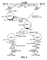
- u , B13R - B14R pos. 172,549 - 173,552
- pSD419 was used as the source for the left flanking arm and pSD422 was used as the source of the right flanking arm.
- the direction of transcription for the u region is indicated by an arrow in FIG. 2.
- sequences to the left of the Nco I site were removed by digestion of pSD419 with Nco I/ Sma I followed by blunt ending with Klenow fragment of E . coli polymerase and ligation generating plasmid pSD476.
- a vaccinia right flanking arm was obtained by digestion of pSD422 with Hpa I at the termination codon of B14R and by digestion with Nru I 0.3 kb to the right. This 0.3 kb fragment was isolated and ligated with a 3.4 kb Hin cII vector fragment isolated from pSD476, generating plasmid pSD477.
- pSD477 contains an initiation codon (underlined) followed by a Bam HI site. To place E.
- Beta-galactosidase in the B13-B14 ( u ) deletion locus under the control of the u promoter a 3.2 kb Bam HI fragment containing the Beta-galactosidase gene (Shapira et al., 1983) was inserted into the Bam HI site of pSD479, generating pSD479BG.
- pSD479BG was used as donor plasmid for recombination with vaccinia virus vP410.
- Recombinant vaccinia virus vP533 was isolated as a blue plaque in the presence of chromogenic substrate X-gal.
- the B13R-B14R region is deleted and is replaced by Beta-galactosidase.
- plasmid pSD486 a derivative of pSD477 containing a polylinker region but no initiation codon at the u deletion junction, was utilized.
- Cla I/ Hpa I vector fragment from pSD477 referred to above was ligated with annealed synthetic oligonucleotides SD42mer/SD40mer (SEQ ID NO:8/SEQ ID NO:9) generating plasmid pSD478.
- the Eco RI site at the pUC/vaccinia junction was destroyed by digestion of pSD478 with Eco RI followed by blunt ending with Klenow fragment of E.
- pSD478E - was digested with Bam HI and HpaI and ligated with annealed synthetic oligonucleotides HEMS/HEM6 (SEQ ID NO:10/SEQ ID NO:11) generating plasmid pSD486.
- pSD486 was used as donor plasmid for recombination with recombinant vaccinia virus vP533, generating vP553, which was isolated as a clear plaque in the presence of X-gal.
- pSD414 contains Sal I B cloned into pUC8.
- pSD414 was cut with Xba I within vaccinia sequences (pos. 137,079) and with Hin dIII at the pUC/vaccinia junction, then blunt ended with Klenow fragment of E. coli polymerase and ligated, resulting in plasmid pSD483.
- pSD483 was cut with Eco RI (pos. 140,665 and at the pUC/vaccinia junction) and ligated, forming plasmid pSD484.
- pSD484 was cut with Nde I (partial) slightly upstream from the A26L ORF (pos. 139,004) and with Hpa I (pos. 137,889) slightly downstream from the A26L ORF.
- the 5.2 kb vector fragment was isolated and ligated with annealed synthetic oligonucleotides ATI3/ATI4 (SEQ ID NO:12/SEQ ID NO:13) reconstructing the region upstream from A26L and replacing the A26L ORF with a short polylinker region containing the restriction sites Bgl II, Eco RI and Hpa I, as indicated above.
- the resulting plasmid was designated pSD485.
- Bgl II and Eco RI sites in the polylinker region of pSD485 are not unique, unwanted Bgl II and Eco RI sites were removed from plasmid pSD483 (described above) by digestion with Bgl II (pos. 140,136) and with Eco RI at the pUC/vaccinia junction, followed by blunt ending with Klenow fragment of E . coli polymerase and ligation.
- the resulting plasmid was designated pSD489.
- plasmid pSD492 was deleted by mutagenesis (Mandecki, 1986) using synthetic oligonucleotide MPSYN177 (SEQ ID NO:14) (5' AAAATGGGCGTGGATTGTTAACTTTATATAACTTATTTTTTGAATATAC 3').
- MPSYN177 SEQ ID NO:14
- vaccinia DNA encompassing positions [137,889 - 138,937], including the entire A26L ORF is deleted.
- vaccinia Sal I G restriction fragment (pos. 160,744-173,351) crosses the Hin dIII A/B junction (pos. 162,539).
- pSD419 contains vaccinia Sal I G cloned into pUC8.
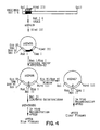
- the direction of transcription for the hemagglutinin (HA) gene is indicated by an arrow in FIG. 4.
- Vaccinia sequences derived from Hin dIII B were removed by digestion of pSD419 with Hin dIII within vaccinia sequences and at the pUC/vaccinia junction followed by ligation.
- the resulting plasmid, pSD456, contains the HA gene, A56R, flanked by 0.4 kb of vaccinia sequences to the left and 0.4 kb of vaccinia sequences to the right.
- A56R coding sequences were removed by cutting pSD456 with Rsa I (partial; pos. 161,090) upstream from A56R coding sequences, and with Eag I (pos. 162,054) near the end of the gene.
- the 3.6 kb Rsa I/ Eaq I vector fragment from pSD456 was isolated and ligated with annealed synthetic oligonucleotides MPSYN59 (SEQ ID NO:15), MPSYN62 (SEQ ID NO:16), MPSYN60 (SEQ ID NO:17), and MPSYN61 (SEQ ID NO:18) reconstructing the DNA sequences upstream from the A56R ORF and replacing the A56R ORF with a polylinker region as indicated above.
- the resulting plasmid is pSD466.
- the vaccinia deletion in pSD466 encompasses positions [161,185-162,053].
- the site of the deletion in pSD466 is indicated by a triangle in FIG. 4.
- Beta-galactosidase sequences were deleted from vP708 using donor plasmid pSD467.
- pSD467 is identical to pSD466, except that Eco RI, Sma I and Bam HI sites were removed from the pUC/vaccinia junction by digestion of pSD466 with Eco RI/ Bam HI followed by blunt ending with Klenow fragment of E. coli polymerase and ligation.
- Recombination between vP708 and pSD467 resulted in recombinant vaccinia deletion mutant, vP723, which was isolated as a clear plaque in the presence of X-gal.
- pSD420 is Sal I H cloned into pUC8.
- pSD435 is Kpn I F cloned into pUC18.
- pSD435 was cut with Sph I and religated, forming pSD451.
- DNA sequences to the left of the Sph I site (pos. 27,416) in Hin dIII M are removed (Perkus et al., 1990).
- pSD409 is Hin dIII M cloned into pUC8.
- E . coli Beta-galactosidase was first inserted into the vaccinia M2L deletion locus (Guo et al., 1990) as follows. To eliminate the Bgl II site in pSD409, the plasmid was cut with Bgl II in vaccinia sequences (pos. 28,212) and with Bam HI at the pUC/vaccinia junction, then ligated to form plasmid pMP409B. pMP409B was cut at the unique Sph I site (pos. 27,416).
- M2L coding sequences were removed by mutagenesis (Guo et al., 1990; Mandecki, 1986) using synthetic oligonucleotide
- the resulting plasmid, pMP409D contains a unique Bgl II site inserted into the M2L deletion locus as indicated above.
- a 3.2 kb Bam HI (partial)/ Bgl II cassette containing the E. coli Beta-galactosidase gene (Shapira et al., 1983) under the control of the 11 kDa promoter (Bertholet et al., 1985) was inserted into pMP409D cut with Bgl II.
- pMP409DBG The resulting plasmid, pMP409DBG (Guo et al., 1990), was used as donor plasmid for recombination with rescuing vaccinia virus vP723.
- a plasmid deleted for vaccinia genes [C7L-K1L] was assembled in pUC8 cut with Sma I, Hin dIII and blunt ended with Klenow fragment of E. coli polymerase.
- the left flanking arm consisting of vaccinia Hin dIII C sequences was obtained by digestion of pSD420 with Xba I (pos. 18,628) followed by blunt ending with Klenow fragment of E. coll polymerase and digestion with Bgl II (pos. 19,706).
- the right flanking arm consisting of vaccinia Hin dIII K sequences was obtained by digestion of pSD451 with Bgl II (pos. 29,062) and Eco RV (pos. 29,778).
- the resulting plasmid, pMP581CK is deleted for vaccinia sequences between the Bgl II site (pos. 19,706) in Hin dIII C and the Bgl II site (pos. 29,062) in Hin dIII K.
- the site of the deletion of vaccinia sequences in plasmid pMP581CK is indicated by a triangle in FIG. 5.
- plasmid pMP581CK was cut at the Nco I sites within vaccinia sequences (pos. 18,811; 19,655), treated with Bal-31 exonuclease and subjected to mutagenesis (Mandecki, 1986) using synthetic oligonucleotide MPSYN233 (SEQ ID NO:20) 5'-TGTCATTTAACACTATACTCATATTAATAAAAATAATATTTATT-3'.
- the resulting plasmid, pMPCSK1 ⁇ is deleted for vaccinia sequences positions 18,805-29,108, encompassing 12 vaccinia open reading frames [C7L - K1L].
- plasmid pSD405 contains vaccinia Hin dIII I (pos. 63,875-70,367) cloned in pUC8.
- pSD405 was digested with Eco RV within vaccinia sequences (pos. 67,933) and with Sma I at the pUC/vaccinia junction, and ligated, forming plasmid pSD518.
- pSD518 was used as the source of all the vaccinia restriction fragments used in the construction of pSD548.
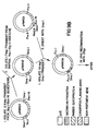
- the vaccinia I4L gene extends from position 67,371-65,059. Direction of transcription for I4L is indicated by an arrow in FIG. 6.
- pSD518 was digested with Bam HI (pos. 65,381) and Hpa I (pos. 67,001) and blunt ended using Klenow fragment of E. coli polymerase. This 4.8 kb vector fragment was ligated with a 3.2 kb Sma I cassette containing the E.
- coli Beta-galactosidase gene (Shapira et al., 1983) under the control of the vaccinia 11 kDa promoter (Bertholet et al., 1985; Perkus et al., 1990), resulting in plasmid pSD524KBG.
- pSD524KBG was used as donor plasmid for recombination with vaccinia virus vP804.
- Recombinant vaccinia virus, vP855, containing Beta-galactosidase in a partial deletion of the I4L gene was isolated as a blue plaque in the presence of X-gal.
- deletion plasmid pSD548 was constructed.
- the left and right vaccinia flanking arms were assembled separately in pUC8 as detailed below and presented schematically in FIG. 6.
- pUC8 was cut with Bam HI/ Eco RI and ligated with annealed synthetic oligonucleotides 518A1/518A2 (SEQ ID NO:21/SEQ ID NO:22) forming plasmid pSD531.
- pSD531 was cut with Rsa I (partial) and Bam HI and a 2.7 kb vector fragment isolated.
- pSD518 was cut with Bgl II (pos. 64,459)/ Rsa I (pos. 64,994) and a 0.5 kb fragment isolated. The two fragments were ligated together, forming pSD537, which contains the complete vaccinia flanking arm left of the I4L coding sequences.
- pUC8 was cut with Bam HI/ Eco RI and ligated with annealed synthetic oligonucleotides 518B1/518B2 (SEQ ID NO:23/SEQ ID NO:24) forming plasmid pSD532.
- pSD532 was cut with Rsa I (partial)/ Eco RI and a 2.7 kb vector fragment isolated.
- pSD518 was cut with Rsa I within vaccinia sequences (pos. 67,436) and Eco RI at the vaccinia/pUC junction, and a 0.6 kb fragment isolated. The two fragments were ligated together, forming pSD538, which contains the complete vaccinia flanking arm to the right of I4L coding sequences.
- the right vaccinia flanking arm was isolated as a 0.6 kb Eco RI/ Bal II fragment from pSD538 and ligated into pSD537 vector plasmid cut with Eco RI/ Bgl II.
- the I4L ORF pos. 65,047-67,386
- a polylinker region which is flanked by 0.6 kb vaccinia DNA to the left and 0.6 kb vaccinia DNA to the right, all in a pUC background.
- the site of deletion within vaccinia sequences is indicated by a triangle in FIG. 6.
- the vaccinia I4L deletion cassette was moved from pSD539 into pRC11, a pUC derivative from which all Beta-galactosidase sequences have been removed and replaced with a polylinker region (Colinas et al., 1990).
- pSD539 was cut with Eco RI/ Pst I and the 1.2 kb fragment isolated. This fragment was ligated into pRC11 cut with Eco RI/ Pst I (2.35 kb), forming pSD548.
- DNA from recombinant vaccinia virus vP866 was analyzed by restriction digests followed by electrophoresis on an agarose gel. The restriction patterns were as expected. Polymerase chain reactions (PCR) (Engelke et al., 1988) using vP866 as template and primers flanking the six deletion loci detailed above produced DNA fragments of the expected sizes. Sequence analysis of the PCR generated fragments around the areas of the deletion junctions confirmed that the junctions were as expected. Recombinant vaccinia virus vP866, containing the six engineered deletions as described above, was designated vaccinia vaccine strain "NYVAC.”
- telomere sequence under the control of the vaccinia H6 promoter (Taylor et al., 1988a,b) was inserted into TK deletion plasmid pSD513.
- pSD513 is identical to plasmid pSD460 (FIG. 1) except for the presence of a polylinker region.
- the polylinker region was inserted by cutting pSD460 with Sma I and ligating the plasmid vector with annealed synthetic oligonucleotides VQ1A/VQ1B (SEQ ID NO:25/SEQ ID NO:26) to form vector plasmid pSD513.
- pSD513 was cut with Sma I and ligated with a Sma I ended 1.8 kb cassette containing the gene encoding the rabies glycoprotein G gene under the control of the vaccinia H6 promoter (Taylor et al., 1988a,b).
- the resulting plasmid was designated pRW842.
- pRW842 was used as donor plasmid for recombination with NYVAC rescuing virus (vP866).
- Recombinant vaccinia virus vP879 was identified by plaque hybridization using 32 P-labelled DNA probe to rabies glycoprotein G coding sequences.
- the modified recombinant viruses of the present invention provide advantages as recombinant vaccine vectors.
- the attenuated virulence of the vector advantageously reduces the opportunity for the possibility of a runaway infection due to vaccination in the vaccinated individual and also diminishes transmission from vaccinated to unvaccinated individuals or contamination of the environment.
- the modified recombinant viruses are also advantageously used in a method for expressing a gene product in a cell cultured in vitro by introducing into the cell the modified recombinant virus having foreign DNA which codes for and expresses gene products in the cell.
- This example describes the development of ALVAC, a canarypox virus vector and, of a canarypox-rabies recombinant designated as ALVAC-RG (vCP65) and its safety and efficacy.
- the parental canarypox virus (Rentschler strain) is a vaccinal strain for canaries.
- the vaccine strain was obtained from a wild type isolate and attenuated through more than 200 serial passages on chick embryo fibroblasts.
- a master viral seed was subjected to four successive plaque purifications under agar and one plaque clone was amplified through five additional passages after which the stock virus was used as the parental virus in in vitro recombination tests.
- the plaque purified canarypox isolate is designated ALVAC.
- This replacement sequence contains Hin dIII, Sma I and Eco RI insertion sites followed by translation stops and a transcription termination signal recognized by vaccinia virus RNA polymerase (Yuen et al., 1987). Deletion of the C5 ORF was performed as described below. Plasmid pRW764.5 was partially cut with Rsa I and the linear product was isolated.
- the Rsa I linear fragment was recut with Bgl II and the pRW764.5 fragment now with a Rsa I to Bgl II deletion from position 156 to position 462 was isolated and used as a vector for the following synthetic oligonucleotides: RW145 (SEQ ID NO:29): ACTCTCAAAAGCTTCCCGGGAATTCTAGCTAGCTAGTTTTTATAAA RW146 (SEQ ID NO:30): GATCTTTATAAAAACTAGCTAGCTAGAATTCCCGGGAAGCTTTTGAGAGT Oligonucleotides RW145 and RW146 were annealed and inserted into the pRW 764.5 Rsa I and Bgl II vector described above. The resulting plasmid is designated pRW831.
- Oligonucleotides A through E which overlap the translation initiation codon of the H6 promoter with the ATG of rabies G, were cloned into pUC9 as pRW737. Oligonucleotides A through E contain the H6 promoter, starting at Nru I, through the Hin dIII site of rabies G followed by Bgl II. Sequences of oligonucleotides A through E ((SEQ ID NO:31)-(SEQ ID N0:35)) are: The diagram of annealed oligonucleotides A through E is as follows:
- Oligonucleotides A through E were kinased, annealed (95°C for 5 minutes, then cooled to room temperature), and inserted between the Pvu II sites of pUC9.
- the resulting plasmid, pRW737 was cut with Hin dIII and Bgl II and used as a vector for the 1.6 kbp Hin dIII- Bgl II fragment of ptg155PRO (Kieny et al., 1984) generating pRW739.
- the ptg155PRO Hin dIII site is 86 bp downstream of the rabies G translation initiation codon.
- Bgl II is downstream of the rabies G translation stop codon in ptg155PRO.
- pRW739 was partially cut with Nru I, completely cut with Bgl II, and a 1.7 kbp Nru I- Bgl II fragment, containing the 3' end of the H6 promoter previously described (Taylor et al., 1988a,b; Guo et al., 1989; Perkus et al., 1989) through the entire rabies G gene, was inserted between the Nru I and Bam HI sites of pRW824. The resulting plasmid is designated pRW832. Insertion into pRW824 added the H6 promoter 5' of Nru I .
- pRW824 sequence of Bam HI followed by Sma I is (SEQ ID NO:36): GGATCCCCGGG.
- pRW824 is a plasmid that contains a nonpertinent gene linked precisely to the vaccinia virus H6 promoter. Digestion with Nru I and Bam HI completely excised this nonpertinent gene.
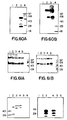
- Plasmid pRW838 was transfected into ALVAC infected primary CEF cells by using the calcium phosphate precipitation method previously described (Panicali et al., 1982; Piccini et al., 1987). Positive plaques were selected on the basis of hybridization to a specific rabies G probe and subjected to 6 sequential rounds of plaque purification until a pure population was achieved. One representative plaque was then amplified and the resulting ALVAC recombinant was designated ALVAC-RG (vCP65) ( see also Figs. 9A and 9B). The correct insertion of the rabies G gene into the ALVAC genome without subsequent mutation was confirmed by sequence analysis.
- Immunofluorescence During the final stages of assembly of mature rabies virus particles, the glycoprotein component is transported from the golgi apparatus to the plasma membrane where it accumulates with the carboxy terminus extending into the cytoplasm and the bulk of the protein on the external surface of the cell membrane.
- immunofluorescence was performed on primary CEF cells infected with ALVAC or ALVAC-RG. Immunofluorescence was performed as previously described (Taylor et al., 1990) using a rabies G monoclonal antibody. Strong surface fluorescence was detected on CEF cells infected with ALVAC-RG but not with the parental ALVAC.
- Immunoprecipitation Preformed monolayers of primary CEF, Vero (a line of African Green monkey kidney cells ATCC # CCL81) and MRC-5 cells (a fibroblast-like cell line derived from normal human fetal lung tissue ATCC # CCL171) were inoculated at 10 pfu per cell with parental virus ALVAC and recombinant virus ALVAC-RG in the presence of radiolabelled 35 S-mathionine and treated as previously described (Taylor et al., 1990). Immunoprecipitation reactions were performed using a rabies G specific monoclonal antibody.
- sample t7 of each cell substrate was inoculated undiluted onto three dishes of the same cell substrate (to provide samples to, t7 and t7A) and onto one dish of primary CEF cells. Samples to, t7 and t7A were treated as for passage one. The additional inoculation on CEF cells was included to provide an amplification step for more sensitive detection of virus which might be present in the non-avian cells.
- Virus yield in each sample was then determined by plaque titration on CEF monolayers under agarose. Summarized results of the experiment are shown in Tables 1 and 2.
- mice Inoculation of Mice .
- Groups of mice were inoculated with 50 to 100 ⁇ l of a range of dilutions of different batches of vCP65. Mice were inoculated in the footpad. On day 14, mice were challenged by intracranial inoculation of from 15 to 43 mouse LD 50 of the virulent CVS strain of rabies virus. Survival of mice was monitored and a protective dose 50% (PD 50 ) calculated at 28 days post-inoculation.
- PD 50 protective dose 50%
- Chicken embryo fibroblast cells produced from 11 day old white leghorn embryos were included as a positive control. All inoculations were performed on preformed monolayers of 2 X 10 6 cells as discussed below.
- Each sample of 2 X 10 6 cells was resuspended in 0.5 ml phosphate buffered saline (PBS) containing 40 mM EDTA and incubated for 5 minutes at 37°C.
- An equal volume of 1.5% agarose prewarmed at 42°C and containing 120 mM EDTA was added to the cell suspension and gently mixed. The suspension was transferred to an agarose plug mold and allowed to harden for at least 15 min.
- the agarose plugs were then removed and incubated for 12-16 hours at 50°C in a volume of lysis buffer (1% sarkosyl, 100 ⁇ g/ml proteinase K, 10 mM Tris HCl pH 7.5, 200 mM EDTA) that completely covers the plug.
- the lysis buffer was then replaced with 5.0 ml sterile 0.5 X TBE (44.5 mM Tris-borate, 44.5 mM boric acid, 0.5 mM EDTA) and equilibrated at 4°C for 6 hours with 3 changes of TBE buffer.
- the viral DNA within the plug was fractionated from cellular RNA and DNA using a pulse field electrophoresis system.
- Electrophoresis was performed for 20 hours at 180 V with a ramp of 50-90 sec at 15°C in 0.5 X TBE.
- the DNA was run with lambda DNA molecular weight standards. After electrophoresis the viral DNA band was visualized by staining with ethidium bromide.
- the DNA was then transferred to a nitrocellulose membrane and probed with a radiolabelled probe prepared from purified ALVAC genomic DNA.
- Dishes were inoculated exactly as described above, with the exception that input multiplicity was 0.1 pfu/cell. At 72 hours post infection, cells were lysed by three successive cycles of freezing and thawing. Virus yield was assessed by plaque titration on CEF monolayers.
- Dishes were inoculated with recombinant or parental virus at a multiplicity of 10 pfu/cell, allowing an additional dish as an uninfected virus control. After a one hour absorption period, the medium was removed and replaced with methionine free medium. After a 30 minute period, this medium was replaced with methionine-free medium containing 25 uCi/ml of 35 S-Methionine. Infected cells were labelled overnight (approximately 16 hours), then lysed by the addition of buffer A lysis buffer. Immunoprecipitation was performed as previously described (Taylor et al., 1990) using a rabies G specific monoclonal antibody. Results: Estimation of Viral Yield .
- DNA from the cell lysates was fractionated by electrophoresis, transferred to nitrocellulose and probed for the presence of viral specific DNA.
- ALVAC-RG infected CEF cells at 72 hours post-infection exhibited a strong band in the region of approximately 350 kbp representing ALVAC-specific viral DNA accumulation. No such band is detectable when the culture is incubated in the presence of the DNA synthesis inhibitor, cytosine arabinoside.
- Equivalent samples produced in Vero cells showed a very faint band at approximately 350 kbp in the ALVAC-RG infected Vero cells at time zero. This level represented residual virus. The intensity of the band was amplified at 72 hours post-infection indicating that some level of viral specific DNA replication had occurred in Vero cells which had not resulted in an increase in viral progeny.
- results of this experiment indicated that in the human cell lines analyzed, although the ALVAC-RG recombinant was able to initiate an infection and express a foreign gene product under the transcriptional control of the H6 early/late vaccinia virus promoter, the replication did not proceed through DNA replication, nor was there any detectable viral progeny produced. In the Vero cells, although some level of ALVAC-RG specific DNA accumulation was observed, no viral progeny was detected by these methods. These results would indicate that in the human cell lines analyzed the block to viral replication occurs prior to the onset of DNA replication, while in Vero cells, the block occurs following the onset of viral DNA. replication.
- mice were challenged 14 days later by the intracranial route with 300 ⁇ l of the CVS strain of rabies virus containing from 15 to 43 mouse LD 50 as determined by lethality titration in a control group of mice. Potency, expressed as the PD 50 (Protective dose 50%), was calculated at 14 days post-challenge. The results of the experiment are shown in Table 6. The results indicated that ALVAC-RG was consistently able to protect mice against rabies virus challenge with a PD 50 value ranging from 3.33 to 4.56 with a mean value of 3.73 (STD 0.48).
- mice were inoculated intracranially with 50 ⁇ l of virus containing 6.0 log 10 TCID 50 of ALVAC-RG or with an equivalent volume of an uninfected cell suspension. Mice were sacrificed on days 1, 3 and 6 post-inoculation and their brains removed, fixed and sectioned. Histopathological examination showed no evidence for neurovirulence of ALVAC-RG in mice.
- the immunological response of HIV-2 seropositive macaques to inoculation with ALVAC-RG was assessed. Animals were inoculated as described above and the presence of anti-rabies serum neutralizing antibody assessed in an RFFI test. The results, shown in Table 9, indicated that HIV-2 positive animals inoculated by the subcutaneous route developed anti-rabies antibody by 11 days after one inoculation. An anamnestic response was detected after a booster inoculation given approximately three months after the first inoculation. No response was detected in animals receiving the recombinant by the oral route. In addition, a series of six animals were inoculated with decreasing doses of ALVAC-RG given by either the intra-muscular or subcutaneous routes. Five of the six animals inoculated responded by 14 days post-vaccination with no significant difference in antibody titer.
- N.D. N.D. t7A 3.8 N.D. N.D. Pass 4 Sample to 5.2 N.D. N.D. t7 7.1 N.D. N.D. t7A 3.9 N.D. N.D. a: This sample was harvested at zero time and represents the residual input virus. The titer is expressed as log 10 pfu per ml. b: This sample was harvested at 7 days post-infection. c: This sample was inoculated in the presence of 40 ⁇ g/ml of Cytosine arabinoside and harvested at 7 days post infection. d: Not detectable Table 2.
- N.D a This sample was harvested at zero time and represents the residual input virus. The titer is expressed as log 10 pfu per ml.
- b This sample was harvested at 7 days post-infection.
- c This sample was inoculated in the presence of 40 ⁇ g/ml of Cytosine arabinoside and harvested at 7 days post-infection.
- d Not detectable.
- Table 3 Amplification of residual virus by passage in CEF cells
- ALVAC-RG Pass 2 a 7.2 5.5 5.5 3 7.2 5.0 5.1 4 7.2 N.D. N.D. 5 7.2 N.D. N.D. a: Pass 2 represents the amplification in CEF cells of the 7 day sample from Pass 1.
- b Titer expressed as log 10 pfu per ml
- c Not Detectable Table 4 .
- a Antibody at day 29 post inoculation expressed as the geometric mean titer in International Units.
- b Expressed as a ratio of survivors over animals challenged Table 8. Anti-rabies serological response of Squirrel monkeys inoculated with canarypox recombinants Monkey # Previous Exposure Rabies serum-neutralizing antibody a -196 b 0 3 7 11 21 28 22 ALVAC c NT g ⁇ 1.2 ⁇ 1.2 ⁇ 1.2 2.1 2.3 2.2 51 ALVAC c NT ⁇ 1.2 ⁇ 1.2 1.7 2.2 2.2 2.2 39 vCP37 d NT ⁇ 1.2 ⁇ 1.2 1.7 2.1 2.2 N.T.
- b Animals 176L and 185L received 8.0 log 10 pfu by the oral route in 5 ml Tang. Animal 187L received 7.0 log 10 pfu by oral route not in Tang.
- c Day of re-vaccination for animals 176L, 185L, 177L and 186L by S.C. route, and primary vaccination for animals 178L, 182L, 179L, 183L, 180L, 184L and 187L.
- d Titers expressed as reciprocal of last dilution showing inhibition of fluorescence in an RFFI test. Table 10. Inoculation of chimpanzees with ALVAC-RG Weeks post-Inoculation Animal 431 I.M. Animal 457 S.C.
- ALVAC-RG (vCP65) was generated as described in Example 9 and FIGS. 9A and 9B.
- ALVAC-RG (vCP65) was grown in primary CEF derived from specified pathogen free eggs. Cells were infected at a multiplicity of 0.1 and incubated at 37°C for three days.
- the vaccine virus suspension was obtained by ultrasonic disruption in serum free medium of the infected cells; cell debris were then removed by centrifugation and filtration.
- the resulting clarified suspension was supplemented with lyophilization stabilizer (mixture of amino-acids), dispensed in single dose vials and freeze dried.
- Three batches of decreasing titer were prepared by ten-fold serial dilutions of the virus suspension in a mixture of serum free medium and lyophilization stabilizer, prior to lyophilization.
- ALVAC-RG vCP65
- canaries chickens, ducks, geese, laboratory rodents (suckling and adult mice), hamsters, guinea-pigs, rabbits, cats and dogs, squirrel monkeys, rhesus macaques and chimpanzees
- hamsters guinea-pigs
- rabbits rabbits
- cats and dogs squirrel monkeys
- rhesus macaques and chimpanzees were inoculated with doses ranging from 10 5 to 10 8 pfu.
- routes were used, most commonly subcutaneous, intramuscular and intradermal but also oral (monkeys and mice) and intracerebral (mice).
- ALVAC-RG (vCP65) caused a "take" lesion at the site of scarification with no indication of disease or death. Intradermal inoculation of rabbits resulted in a typical poxvirus inoculation reaction which did not spread and healed in seven to ten days. There was no adverse side effects due to canarypox in any of the animal tests. Immunogenicity was documented by the development of anti-rabies antibodies following inoculation of ALVAC-RG (vCP65) in rodents, dogs, cats, and primates, as measured by Rapid Fluorescent Focus Inhibition Test (RFFIT). Protection was also demonstrated by rabies virus challenge experiments in mice, dogs, and cats immunized with ALVAC-RG (vCP65).
- RFFIT Rapid Fluorescent Focus Inhibition Test
- Volunteers Twenty-five healthy adults aged 20-45 with no previous history of rabies immunization were enrolled. Their health status was assessed by complete medical histories, physical examinations, hematological and blood chemistry analyses. Exclusion criteria included pregnancy, allergies, immune depression of any kind, chronic debilitating disease, cancer, injection of immune globins in the past three months, and seropositivity to human immunodeficiency virus (HIV) or to hepatitis B virus surface antigen.
- HIV human immunodeficiency virus
- the trial was designated as a dose escalation study.
- Three batches of experimental ALVAC-RG (vCP65) vaccine were used sequentially in three groups of volunteers (Groups A, B and C) with two week intervals between each step.
- the concentration of the three batches was 10 3.5 , 10 4.5 , 10 5.5 Tissue Culture Infectious Dose (TCID 50 ) per dose, respectively.
- the nature of the injected vaccine was not known by the participants at the time of the first injection but was known by the investigator.
- the volunteers of Group B allocated to the medium dose of experimental vaccine were injected 1 h previously with the lower dose and those allocated to the higher dose (Group C) received successively the lower and the medium dose at hourly intervals.
- ALVAC-RG (vCP65) (Group C) and HDC vaccine were offered a third dose of vaccine; they were then randomized to receive either the same vaccine as previously or the alternate vaccine.
- four groups were formed corresponding to the following immunization scheme: 1. HDC, HDC - HDC; 2. HDC, HDC - ALVAC-RG (vCP65); 3. ALVAC-RG (vCP65), ALVAC-RG (vCP65) - HDC; 4. ALVAC-RG (vCP65), ALVAC-RG (vCP65), ALVAC-RG (vCP65).
- Antibody assays were performed seven days prior to the first injection and at days 7, 28, 35, 56, 173, 187 and 208 of the study.
- the levels of neutralizing antibodies to rabies were determined using the Rapid Fluorescent Focus Inhibition test (RFFIT) (Smith et al., 1973).
- Canarypox antibodies were measured by direct ELISA.
- the antigen a suspension of purified canarypox virus disrupted with 0.1% Triton X100, was coated in microplates. Fixed dilutions of the sera were reacted for two hours at room temperature and reacting antibodies were revealed with a peroxidase labelled anti-human IgG goat serum. The results are expressed as the optical density read at 490nm.
- Tenderness was the most common symptoms and was always mild. Other local symptoms included redness and induration which were also mild and transient. All symptoms usually subsided within 24 hours and never lasted more than 72 hours.
- protective titers were achieved in 0/3 of Group A, 2/3 of Group B and 9/9 of Group C recipients of ALVAC-RG (vCP65) vaccine and persisted in all 10 HDC recipients.
- the rabies antibody titers had substantially decreased in all subjects but remained above the minimum protective titer of 0.5 IU/ml in 5/10 HCD recipients and in 5/9 ALVAC-RG (vCP65) recipients; the geometric mean titers were 0.51 and 0.45 IU/ml in groups HCD and C, respectively.
- Booster Injection The vaccine was similarly well tolerated six months later, at the time of the booster injection: fever was noted in 2/9 HDC booster recipients and in 1/10 ALVAC-RG (vCP65) booster recipients. Local reactions were present in 5/9 recipients of HDC booster and in 6/10 recipients of the ALVAC-RG (vCP65) booster.
- Figs. 11A-11D show graphs of rabies neutralizing antibody titers (Rapid Fluorescent Focus Inhibition Test or RFFIT, IU/ml): Booster effect of HDC and vCP65 (10 5.5 TCID 50 ) in volunteers previously immunized with either the same or the alternate vaccine. Vaccines were given at days 0, 28 and 180. Antibody titers were measured at days 0, 7, 28, 35, 56, 173, and 187 and 208.
- RFFIT Rapid Fluorescent Focus Inhibition Test
- the booster dose given resulted in a further increase in rabies antibody titers in every subject whatever the immunization scheme.
- the ALVAC-RG (vCP65) booster globally elicited lower immune responses than the HDC booster and the ALVAC-RG (vCP65), ALVAC-RG (vCP65) - ALVAC-RG (vCP65) group had significantly lower titers than the three other groups.
- the ALVAC-RG (vCP65) booster injection resulted in an increase in canarypox antibody titers in 3/5 subjects who had previously received the HDC vaccine and in all five subjects previously immunized with ALVAC-RG (vCP65).
- Rabies neutralizing antibodies were assayed with the Rapid Fluorescent Focus Inhibition Test (RFFIT) which is known to correlate well with the sero neutralization test in mice. Of 9 recipients of 10 5.5 TCID 50 , five had low level responses after the first dose. Protective titers of rabies antibodies were obtained after the second injection in all recipients of the highest dose tested and even in 2 of the 3 recipients of the medium dose. In this study, both vaccines were given subcutaneously as usually recommended for live vaccines, but not for the inactivated HDC vaccine.
- RFFIT Rapid Fluorescent Focus Inhibition Test
- this Example clearly demonstrates that a non-replicating poxvirus can serve as an immunizing vector in humans, with all of the advantages that replicating agents confer on the immune response, but without the safety problem created by a fully permissive virus.
- suitable dosages and modes or routes for administration or immunization of recombinants containing either rabies or other coding, or expression products thereof are within the ambit of the skilled artisan as well modes for in vitro expression.
- TABLE 11 Reactions in the 5 days following vaccination vCP65 dosage (TCID50) 10 3.5 10 4.5 10 5.5 H D C control Injection 1st 2nd 1st 2nd 1st 2nd 1st 2nd No.
- mice Male outbred Swiss Webster mice were purchased from Taconic Farms (Germantown, NY) and maintained on mouse chow and water ad libitum until use at 3 weeks of age ("normal" mice). Newborn outbred Swiss Webster mice were of both sexes and were obtained following timed pregnancies performed by Taconic Farms. All newborn mice used were delivered within a two day period.
- ALVAC was derived by plaque purification of a canarypox virus population and was prepared in primary chick embryo fibroblast cells (CEF). Following purification by centrifugation over sucrose density gradients, ALVAC was enumerated for plaque forming units in CEF cells.
- the WR(L) variant of vaccinia virus was derived by selection of large plaque phenotypes of WR (Panicali et al., 1981).
- the Wyeth New York State Board of Health vaccine strain of vaccinia virus was obtained from Pharmaceuticals Calf Lymph Type vaccine Dryvax, control number 302001B.
- Copenhagen strain vaccinia virus VC-2 was obtained from Institut Merieux, France.
- Vaccinia virus strain NYVAC was derived from Copenhagen VC-2. All vaccinia virus strains except the Wyeth strain were cultivated in Vero African green monkey kidney cells, purified by sucrose gradient density centrifugation and enumerated for plaque forming units on Vero cells. The Wyeth strain was grown in CEF cells and enumerated for plaque forming units in CEF cells.
- mice were inoculated intracranially (ic) with 0.05 ml of one of several dilutions of virus prepared by 10-fold serially diluting the stock preparations in sterile phosphate-buffered saline. In some instances, undiluted stock virus preparation was used for inoculation.
- mice Groups of 10 newborn mice, 1 to 2 days old, were inoculated ic similarly to the normal mice except that an injection volume of 0.03 ml was used.
- mice All mice were observed daily for mortality for a period of 14 days (newborn mice) or 21 days (normal mice) after inoculation. Mice found dead the morning following inoculation were excluded due to potential death by trauma.
- the lethal dose required to produce mortality for 50% of the experimental population was determined by the proportional method of Reed and Muench (Reed and Muench, 1938).
- Preformed monolayers of avian or non-avian cells were inoculated with 10 pfu per cell of parental NYVAC (vP866) or NYVAC-RG (vP879) virus.
- the inoculation was performed in EMEM free of methionine and supplemented with 2% dialyzed fetal bovine serum. After a one hour incubation, the inoculum was removed and the medium replaced with EMEM (methionine free) containing 20 ⁇ Ci/ml of 35 S-methionine.
- Protein A Sepharose CL-48 obtained from Pharmacia LKB Biotechnology Inc., Piscataway, New Jersey, was used as a support matrix. Immunoprecipitates were fractionated on 10% polyacrylamide gels according to the method of Dreyfuss et. al. (1984). Gels were fixed, treated for fluorography with 1M Nasalicylate for one hour, and exposed to Kodak XAR-2 film to visualize the immunoprecipitated protein species.
- New Zealand White rabbits were obtained from Hare-Marland (Hewitt, New Jersey).
- Three week old male Swiss Webster outbred mice, timed pregnant female Swiss Webster outbred mice, and four week old Swiss Webster nude (nu + nu + ) mice were obtained from Taconic Farms, Inc. (Germantown, New York). All animals were maintained according to NIH guidelines. All animal protocols were approved by the institutional IACUC. When deemed necessary, mice which were obviously terminally ill were euthanized.
- Virus Recovery from Inoculation Sites A single rabbit was inoculated intradermally at multiple sites with 0.1 ml of PBS containing 10 6 , 10 7 , or 10 8 pfu of each test virus or with PBS alone. After 11 days, the rabbit was euthanized and skin biopsy specimens taken from each of the inoculation sites were aseptically prepared by mechanical disruption and indirect sonication for virus recovery. Infectious virus was assayed by plaque titration on CEF monolayers.
- mice Virulence in Mice .
- Groups of ten mice, or five in the nude mice experiment were inoculated ip with one of several dilutions of virus in 0.5 ml of sterile PBS.
- LD 50 The lethal dose required to produce 50% mortality (LD 50 ) was determined by the proportional method of Reed and Muench (Reed and Muench 1938).
- mice Potency Testing of NYVAC-RG in Mice .
- Four to six week old mice were inoculated in the footpad with 50 to 100 ⁇ l of a range of dilutions (2.0 - 8.0 log 10 tissue culture infective dose 50% (TCID 50 )) of either VV-RG (Kieny et al., 1984), ALVAC-RG (Taylor et al., 1991b), or the NYVAC-RG.
- TCID 50 tissue culture infective dose 50%
- Each group consisted of eight mice. At 14 days post-vaccination, the mice were challenged by intracranial inoculation with 15 LD 50 of the rabies virus CVS strain (0.03 ml). On day 28, surviving mice were counted and protective does 50% (PD 50 ) calculated.
- FIG. 10 schematically depicts the ORFs deleted to generate NYVAC. At the top of FIG. 10 is depicted the Hin dIII restriction map of the vaccinia virus genome (VC-2 plaque isolate, COPENHAGEN strain).
- NYVAC The replication potential of NYVAC in a number of human derived cells are shown in Table 16. Both VC-2 and NYVAC are capable of productive replication in CEF cells, although NYVAC with slightly reduced yields. VC-2 is also capable of productive replication in the six human derived cell lines tested with comparable yields except in the EBV transformed lymphoblastoid cell line JT-1 (human lymphoblastoid cell line transformed with Epstein-Barr virus, see Rickinson et al., 1984). In contrast, NYVAC is highly attenuated in its ability to productively replicate in any of the human derived cell lines tested.
- MRC-5 and DETROIT 532 Consistent with the low levels of virus yields obtained in the human-derived cell lines, MRC-5 and DETROIT 532, detectable but reduced levels of NYVAC-specific DNA accumulation were noted.
- NYVAC-specific viral DNA accumulation was not observed in any of the other human-derived cells.
- Sites inoculated with NYVAC or control PBS displayed no induration or ulceration.
- skin samples from the sites of inoculation were excised, mechanically disrupted, and virus was titrated on CEF cells. The results are shown in Table 18. In no case was more virus recovered at this timepoint than was administered.
- Recovery of vaccinia strain, WR was approximately 10 6 pfu of virus at each site irrespective of amount of virus administered.
- Recovery of vaccinia strains WYETH and VC-2 was 10 3 to 10 4 pfu regardless of amount administered. No infectious virus was recovered from sites inoculated with NYVAC.
- mice inoculated with WR (10 3 to 10 4 pfu), WYETH (5 x 10 7 or 5 x 10 8 pfu) or VC-2 (10 4 to 10 9 pfu) displayed disseminated lesions typical of poxviruses first on the toes, then on the tail, followed by severe orchitis in some animals.
- WR 10 3 to 10 4 pfu
- WYETH 5 x 10 7 or 5 x 10 8 pfu
- VC-2 (10 4 to 10 9 pfu) displayed disseminated lesions typical of poxviruses first on the toes, then on the tail, followed by severe orchitis in some animals.
- WR or WYETH the appearance of disseminated lesions generally led to eventual death, whereas most mice infected with VC-2 eventually recovered.
- Calculated LD 50 values are given in Table 19.
- mice inoculated with VC-2 began to display lesions on their toes (red papules) and 1 to 2 days later on the tail. These lesions occurred between 11 and 13 days post-inoculation (pi) in mice given the highest doses (10 9 , 10 8 , 10 7 and 10 6 pfu), on day 16 pi in mice given 10 5 pfu and on day 21 pi in mice given 10 4 pfu. No lesions were observed in mice inoculated with 10 3 and 10 2 pfu during the 100 day observation period. Orchitis was noticed on day 23 pi in mice given 10 9 and 10 8 pfu, and approximately 7 days later in the other groups (10 7 to 10 4 pfu).
- Orchitis was especially intense in the 10 9 and 10 8 pfu groups and, although receding, was observed until the end of the 100 day observation period. Some pox-like lesions were noticed on the skin of a few mice, occurring around 30-35 days pi. Most pox lesions healed normally between 60-90 days pi. Only one mouse died in the group inoculated with 10 9 pfu (Day 34 pi) and one mouse died in the group inoculated with 10 8 pfu (Day 94 pi). No other deaths were observed in the VC-2 inoculated mice.
- mice inoculated with 10 4 pfu of the WR strain of vaccinia started to display pox lesions on Day 17 pi. These lesions appeared identical to the lesions displayed by the VC-2 injected mice (swollen toes, tail). Mice inoculated with 10 3 pfu of the WR strain did not develop lesions until 34 days pi. Orchitis was noticed only in the mice inoculated with the highest dose of WR (10 4 pfu). During the latter stages of the observation period, lesions appeared around the mouth and the mice stopped eating. All mice inoculated with 10 4 pfu of WR died or were euthanized when deemed necessary between 21 days and 31 days pi.
- mice injected with 10 3 pfu of WR died or were euthanized when deemed necessary between 35 days and 57 days pi. No deaths were observed in mice inoculated with lower doses of WR (1 to 100 pfu).
- CY-treated mice provided a more sensitive model for assaying poxvirus virulence than did nude mice.
- LD 50 values for the WR, WYETH, and VC-2 vaccinia virus strains were significantly lower in this model system than in the nude mouse model.
- CY-treated mice injected with NYVAC or ALVAC did not develop lesions. However, unlike nude mice, some deaths were observed in CY-treated mice challenged with NYVAC or ALVAC, regardless of the dose. These random incidences are suspect as to the cause of death.
- mice injected with all doses of WYETH displayed pox lesions on their tail and/or on their toes between 7 and 15 days pi. In addition, the tails and toes were swollen. Evolution of lesions on the tail was typical of pox lesions with formation of a papule, ulceration and finally formation of a scab.
- Mice inoculated with all doses of VC-2 (1.65 x 10 5 to 1.65 x 10 9 ) also developed pox lesions on their tails and/or their toes analogous to those of WYETH injected mice. These lesions were observed between 7-12 days post inoculation. No lesions were observed on mice injected with lower doses of WR virus, although deaths occurred in these groups.
- Table 20 demonstrates that the PD 50 values obtained with the highly attenuated NYVAC vector are identical to those obtained using a COPENHAGEN-based recombinant containing the rabies glycoprotein gene in the tk locus (Kieny et al., 1984) and similar to PD 50 values obtained with ALVAC-RG, a canarypox based vector restricted to replication to avian species.
- NYVAC deleted of known virulence genes and having restricted in vitro growth characteristics, was analyzed in animal model systems to assess its attenuation characteristics. These studies were performed in comparison with the neurovirulent vaccinia virus laboratory strain, WR, two vaccinia virus vaccine strains, WYETH (New York City Board of Health) and COPENHAGEN (VC-2), as well as with a canarypox virus strain, ALVAC ( See also Example 11).
- viruses provided a spectrum of relative pathogenic potentials in the mouse challenge model and the rabbit skin model, with WR being the most virulent strain, WYETH and COPENHAGEN (VC-2) providing previously utilized attenuated vaccine strains with documented characteristics, and ALVAC providing an example of a poxvirus whose replication is restricted to avian species.
- Results from these in vivo analyses clearly demonstrate the highly attenuated properties of NYVAC relative to the vaccinia virus strains, WR, WYETH and COPENHAGEN (VC-2) (Tables 14-20).
- the LD 50 values for NYVAC were comparable to those observed with the avian host restricted avipoxvirus, ALVAC.
- Example 10 demonstrate the highly attenuated nature of NYVAC relative to WR, and the previously utilized vaccinia virus vaccine strains, WYETH and COPENHAGEN.
- the pathogenic profile of NYVAC, in the animal model systems tested was similar to that of ALVAC, a poxvirus known to productively replicate only in avian species.
- NYVAC-based vaccine candidates have been shown to be efficacious.
- NYVAC recombinants expressing foreign gene products from a number of pathogens have elicited immunological responses towards the foreign gene products in several animal species, including primates.
- a NYVAC-based recombinant expressing the rabies glycoprotein was able to protect mice against a lethal rabies challenge.
- the potency of the NYVAC-based rabies glycoprotein recombinant was comparable to the PD 50 value for a COPENHAGEN-based recombinant containing the rabies glycoprotein in the tk locus (Table 20).
- NYVAC-based recombinants have also been shown to elicit measles virus neutralizing antibodies in rabbits and protection against pseudorabies virus and Japanese encephalitis virus challenge in swine.
- the highly attenuated NYVAC strain confers safety advantages with human, animal, medical and veterinary applications (Tartaglia et al., 1992).
- the use of NYVAC as a general laboratory expression vector system may greatly reduce the biological hazards associated with using vaccinia virus.
- Example 10 shows NYVAC to be highly attenuated: a) no detectable induration or ulceration at site of inoculation (rabbit skin); b) rapid clearance of infectious virus from intradermal site of inoculation (rabbit skin); c) absence of testicular inflammation (nude mice); d) greatly reduced virulence (intracranial challenge, both three-week old and newborn mice); e) greatly reduced pathogenicity and failure to disseminate in immunodeficient subjects (nude and cyclophosphamide treated mice); and f) dramatically reduced ability to replicate on a variety of human tissue culture cells.
- b Titer expressed as LOG 50 pfu per ml.
- c Sample was incubated in the presence of 40 ⁇ g/ml of cytosine arabinoside.
- d Not determined. *: ATCC #CCL25 Human amnionic cells. Table 18. Persistence of poxviruses at the site of intradermal inoculation Virus Inoculum Dose Total Virus Recovered WR 8.0 a 6.14 7.0 6.26 6.0 6.21 WYETH 8.0 3.66 7.0 4.10 6.0 3.59 VC-2 8.0 4.47 7.0 4.74 6.0 3.97 NYVAC 8.0 0 7.0 0 6.0 0 a: expressed as log 10 pfu. Table 19.
- mice Recombinant PD 50 a VV-RG 3.74 ALVAC-RG 3.86 NYVAC-RG 3.70 a Four to six week old mice were inoculated in the footpad with 50-100 ⁇ l of a range of dilutions (2.0 - 8.0 log 10 tissue culture infection dose 50% (TCID 50 ) of either the VV-RG (Kieny et al., 1984), ALVAC-RG (vCP65) or NYVAC-RG (vP879).
- mice of each group were challenged by intracranial inoculation of 30 ⁇ l of a live CVS strain rabies virus corresponding to 15 lethal dose 50% (LD 50 ) per mouse.
- LD 50 lethal dose 50%
- CMVM5 SEQ ID NO:74
- CMVM3 SEQ ID NO:75
- the plasmid containing the modified gB was designated 24CMVgB (5+3).
- the DNA sequence of the CMVgB gene is shown in FIG. 12 (SEQ ID NO:37).
- Plasmid pMP2VCL (containing a polylinker region with vaccinia sequences upstream of the K1L host range gene) was digested within the polylinker with Hin dIII and Xho I and ligated to annealed oligonucleotides SPHPRHA A through D generating SP131 containing a Hin dIII site, H6 promoter -124 through -1 (Perkus et al., 1989) and a polylinker region.
- SPHPRHA A (SEQ ID NO:76) (5'-AG CTTCTTTATTCTATACTTAAAAAGTGAAAATAAATACAAAGGTTCTTGAGGGT-3')
- SPHPRHA C (SEQ ID NO:78) (3'-TT ATTAGTATTTAATAAAGTAATAGCGCTATAGGCAATTCAAACATAGCATGAGCT-5')
- the 2900 bp Eco RV- Bam HI fragment of 24CMVgB (5+3) was cloned into the 3100 bp Bco RV- Bgl II fragment of SP131. This cloning step put the gB gene under the control of the H6 promoter. The resulting plasmid was designated SP131CMVgB.
- Plasmid pSD22-H contains a 2.9 kb Bgl II fragment derived from the Hin dIII F region of the WR strain of vaccinia virus ligated into the Bam HI site of pUC8.
- the unique Bam HI site in pSD22-H is a nonessential site used as an insertion locus for foreign genes (Panicali and Paoletti, 1982).
- Plasmid pMP22BHP is a derivative of pSD22-H in which the unique Bam HI site was modified by the addition of an expanded polylinker region for the insertion of foreign DNA.
- Plasmid pMP22BHP was digested with Hin dIII and ligated to a 2.9 kb Hin dIII fragment from SP131CMVgB (containing the H6 promoted gB gene) generating plasmid SAg22CMVgB.
- the plasmid was digested with Bam HI followed by partial digestion with Hin dIII and purified. Ligation to a 50 bp Bam HI/ Hin dIII polylinker derived from IBI24 resulted in plasmid 22CMVgB.
- Plasmid pSD542 (a NYVAC TK locus donor plasmid) was derived from plasmid pSD513 (Tartaglia et al., 1992). The polylinker region in pSD513 was modified by cutting with Pst I/ Bam HI and ligating to annealed synthetic oligonucleotides MPSYN288 (SEQ ID NO:80) (5'-GGTCGACGGATCCT-3') and MPSYN289 (SEQ ID NO:81) (5'-GATCAGGATCCGTCGACCTGCA-3') resulting in plasmid pSD542.
- MPSYN288 SEQ ID NO:80
- MPSYN289 SEQ ID NO:81
- 22CMVgB was digested with Bam HI and Nsi I to generate a fragment containing the H6 promoter and part of the gB gene, and with Nsi I and Pst I to generate a fragment containing the remainder of the gB gene. These two fragments were ligated to pSD542 that had been digested with Bam HI and Pst I within its' polylinker creating the NYVAC donor plasmid 542CMVgB.
- the DNA sequence of the CMVgB gene and flanking sequences contained in 542CMVgB is shown in FIGS. 13A and B (SEQ ID NO:38).
- PCR primers were used to amplify the 5' and 3' sequences relative to C3.
- Primers for the 5' sequence were RG277 (SEQ ID NO:82) (5'-CAGTTGGTACCACTGGTATTTTATTTCAG-3') and RG278 (SEQ ID NO:83) (5'-TATCTGAATTCCTGCAGCCCGGGTTTTTATAGCTAATTAGTCAAATGTGAGTTAATATTA G -3').
- Primers for the 3" sequences were RG279 (SEQ ID NO:84) (5'-TCGCTGAATTCGATATCAAGCTTATCGATTTTTATGACTAGTTAATCAAATAAAAAGCAT ACAAGC-3') and RG280 (SEQ ID NO:85) (5'-TTATCGAGCTCTGTAACATCAGTATCTAAC-3').
- the primers were designed to include a multiple cloning site flanked by vaccinia transcriptional and translational termination signals.
- a 908 bp fragment of canarypox DNA, immediately upstream of the C3 locus was obtained by digestion of plasmid pWW5 with Nsi I and Ssp I.
- a 604 bp fragment of canarypox DNA was derived by PCR (Engelke et al., 1988) using plasmid PWW5 as template and oligonucleotides CP16 (SEQ ID NO:86) (5'-TCCGGTACCGCGGCCGCAGATATTTGTTAGCTTCTGC-3') and CP17 (SEQ ID NO:87) (5'-TCGCTCGAGTAGGATACCTACCTACTACCTACG-3').
- the 604 bp fragment was digested with Asp 718 and Xho I (sites present at the 5' ends of oligonucleotides CP16 and CP17, respectively) and cloned into Asp 718- Xho I digested and alkaline phosphatase treated IBI25 (International Biotechnologies, Inc., New Haven, CT) generating plasmid SPC3LA.
- SPC3LA was digested within IBI25 with Eco RV and within canarypox DNA with Nsi I and ligated to the 908 bp Nsi I- Ssp I fragment generating SPCPLAX which contains 1444bp of canarypox DNA upstream of the C3 locus.
- a 2178 bp Bgl II- Sty I fragment of canarypox DNA was isolated from plasmids pXX4 (which contains a 6.5 kb Nsi I fragment of canarypox DNA cloned into the Pst I site of pBS-SK).
- a 279 bp fragment of canarypox DNA was isolated by PCR (Engelke et al., 1988) using plasmid pXX4 as template and oligonucleotides CP19 (SEQ ID NO:88) (5'-TCGCTCGAGCTTTCTTGACAATAACATAG-3') and CP20 (SEQ ID NO:89) (5'-TAGGAGCTCTTTATACT ACTGGGTTACAAC-3').
- the 279 bp fragment was digested with Xho I and Sac I (sites present at the 5' ends of oligonucleotides CP19 and CP20, respectively) and cloned into Sac I- Xho I digested and alkaline phosphatase treated IBI25 generating plasmid SPC3RA.
- pC3I was digested within the polylinker region with Eco RI and Cla I, treated with alkaline phosphatase and ligated to kinased and annealed oligonucleotides CP12 (SEQ ID NO:90) (5'-AATTCCTCGAGGGATCC-3') and CP13 (SEQ ID NO:91) (5'-CGGGATCCCTCGAGG-3') (containing an Eco RI sticky end, X ho I site, Bam HI site and a sticky end compatible with Cla I) generating plasmid SPCP3S.
- SPCP3S was digested within the canarypox sequences downstream of the C3 locus with Sty I and Sac I (pBS-SK) and ligated to a 261 bp Bal II- Sac I fragment from SPC3RA and the 2178 bp Bal II- Sty I fragment from pXX4 generating plasmid CPRAL containing 2572 bp of canarypox DNA downstream of the C3 locus.
- SPCP3S was digested within the canarypox sequences upstream of the C3 locus with Asp 718 (in pBS-SK) and Acc I and ligated to a 1436 bp Asp 718- Acc I fragment from SPCPLAX generating plasmid CPLAL containing 1457 bp of canarypox DNA upstream of the C3 locus.
- CPLAL was digested within the canarypox sequences downstream of the C3 locus with Sty I and Sac I (in pBS-SK) and ligated to a 2438 bp Sty I- Sac I fragment from CPRAL generating plasmid CP3L containing 1457 bp of canarypox DNA upstream of the C3 locus, stop codons in six reading frames, early transcription termination signal, a polylinker region, early transcription termination signal, stop codons in six reading frames, and 2572 bp of canarypox DNA downstream of the C3 locus.
- the early/late H6 vaccinia virus promoter (Taylor et al., 1988a,b; Perkus et al., 1989) was derived by PCR (Engelke et al., 1988) using pRW838 (a plasmid containing the rabies glycoprotein gene (Kieny et al., 1984) linked to the H6 promoter) as template and oligonucleotides CP21 (SEQ ID NO:92) (5'-TCGGGATCCGGGTTAATTAATTAGTTATTAGACAAGGTG-3') and CP22 (SEQ ID NO:93) (5'-, TAGGAATTCCTCGAGTACGATACAAACTTAAGCGGATATCG-3').
- the PCR product was digested with Bam HI and Eco RI (sites present at the 5' ends of oligonucleotides CP21 and CP22, respectively) and ligated to CP3L that was digested with Bam HI and Eco RI in the polylinker generating plasmid VQH6CP3L.
- ALVAC donor plasmid VQH6CP3L was digested within the polylinker with Xho I and within the H6 promoter with Nru I and ligated to a Nru I/ Hin dIII fragment from 22CMVgB containing part of the H6 promoter and gB gene and a polylinker derived from pIBI24 by Xho I and Hin dIII digestion generating the ALVAC donor plasmid CP3LCMVgB.
- the DNA sequence of the CMVgB gene plus additional flanking DNA sequences in plasmid CP3LCMVgB is shown in FIGS. 15A-C (SEQ ID NO:40).
- Plasmid pSD553 is a vaccinia deletion/insertion plasmid of the COPAK series. It contains the vaccinia K1L host range gene (Gillard et al., 1986; Perkus et al., 1990) within flanking Copenhagen vaccinia arms, replacing the ATI region (ORFs A25L, A26L; Goebel et al., 1990a,b). pSD553 was constructed as follows.
- Left and right vaccinia flanking arms were constructed by polymerase chain reaction (PCR) using pSD414, a pUC8-based clone of vaccinia Sal I B (Goebel et al., 1990a,b) as template.
- the left arm was synthesized using synthetic deoxyoligonucleotides MPSYN267 (SEQ ID NO:94) (5'-GGGCTGAAGCTTGCTGGCCGCTCATTAGACAAGCGAATGAGGGAC-3') and MPSYN268 (SEQ ID NO:95) (5'-AGATCTCCCGGGCTCGAGTAATTAATTAATTTTTATTACACCAGAAAAGACGGCTTGAGA T C-3') as primers.
- the right arm was synthesized using synthetic deoxyoligonucleotides MPSYN269 (SEQ ID NO:96) (5'-TAATTACTCGAGCCCGGGAGATCTAATTTAATTTAATTTATATAACTCATTTTTTGAATA T ACT-3') and MPSYN270 (SEQ ID NO:97) (5'-TATCTCGAATTCCCGCGGCTTTAAATGGACGGAACTCTTTTCCCCC-3') as primers.
- the two PCR-derived DNA fragments containing the left and right arms were combined in a further PCR reaction.
- the resulting product was cut with Eco RI/ Hin dIII and a 0.9 kb fragment isolated.
- pSD541 was cut with Bgl II/ Xho I and ligated with annealed complementary synthetic oligonucleotides MPSYN333 (SEQ ID NO:98) (5'-GATCTTTTGTTAACAAAAACTAATCAGCTATCGCGAATCGATTCCCGGGGGATCCGGTAC CC-3') and MPSYN334 (SEQ ID NO:99) (5'-TCGAGGGTACCGGATCCCCCGGGAATCGATTCGCGATAGCTGATTAGTTTTTGTTAACAA A A-3') generating plasmid pSD552.
- MPSYN333 SEQ ID NO:98
- MPSYN334 5'-TCGAGGGTACCGGATCCCCCGGGAATCGATTCGCGATAGCTGATTAGTTTTTGTTAACAA A A-3'
- the K1L host range gene was isolated as a 1 kb Bgl II (partial)/ Hpa I fragment from plasmid pSD452 (Perkus et al., 1990).
- pSD552 was cut with Bgl II/ Hpa I and ligated with the K1L containing fragment, generating pSD553.
- a Hin dIII fragment from SP131CMVgB (containing the HCMVgB gene under the control of the H6 promoter) was filled in with the klenow fragment of DNA polymerase I and ligated into plasmid pSD553 which had been Sma I digested and alkaline phosphatase treated.
- the resulting NYVAC donor plasmid (in which the H6 promoted gB is in the same orientation as K1L) was designated 553H6CMVgB.
- the DNA sequence of the CMVgB gene plus additional flanking DNA sequences in plasmid 553H6CMVgB is shown in FIGS. 16A and B (SEQ ID NO:41).
- FIG. 17 The sequence of CMVgB deleted of its transmembrane region is presented in FIG. 17 (SEQ ID NO:42).
- the nucleotides encoding the transmembrane region were deleted in the following manner.
- Oligonucleotides SPgB3 (SEQ ID NO:100) (5'-GATCCATGGACTCGACAGCGGCGTCTCTGCATGCAGCCGCTGCAGA-3') and SPgB4 (SEQ ID NO:101) (5'-AGCTTCTGCAGCGGCTGCATGCAGAGACGCCGCTGTCGAGTCCATG-3') were kinased, annealed and cloned into Bam HI/ Hin dIII digested and alkaline phosphatase treated IBI24 generating plasmid SPCMVgB2.
- Oligonucleotides SPgB1 (SEQ ID NO:102) (5'-TACGAATTCTGCAGTTCACCTATGACACGTTGC-3') and SPgB2 (SEQ ID NO:103) (5'-ATAGGATCCATGGTCGTCCAGACCCTTGAGGTAGGGC-3') were used in PCR with plasmid SP131CMVgB as template to generate a 0.7 kb fragment. This fragment was digested with Eco RI/ Bam HI and cloned into Eco RI/ Bam HI digested and alkaline phosphase treated IBI24 generating plasmid SPCMVgB1.
- a 0.7 kb Eco RI/ Nco I fragment from SPCMVgB1 was ligated to Eco RI/ Nco I digested and phosphatase treated SPCMVgB2 generating plasmid SPCMVgB3.
- the unique Nco I site in SPCMVgB3 was deleted by mutagenesis (Mandecki, 1986) using oligonucleotide SPgB5 (SEQ ID NO:104) (5'-GCCCTACCTCAAGGGTCTGGACGACACTCGACAGCGGCGTCTCTGCAT-3') generating plasmid SPCMVgB4.
- a 0.7 kb PstI fragment from SPCMVgB4 was ligated to a 6.6 kb PstI fragment from 553H6CMVgB generating NYVAC donor plasmid 553H6CMVgBTM-.
- This plasmid contains the gB gene under the control of the H6 promoter with its transmembrane region deleted (amino acids 715-772; Spaete et al., 1988).
- the DNA sequence of the transmembrane deleted CMVgB gene plus additional flanking DNA sequences in plasmid 553H6CMVgBTM - is shown in FIGS. 18A and B (SEQ ID NO:43).
- Oligonucleotides SPgB8 (SEQ ID NO:105) (5'-AATTGGTGACCG-3') and SPgB9 (SEQ ID NO:106) (5'-GATCCGGTCACC-3') were kinased, annealed and cloned into Eco RI/ Bam HI digested and alkaline phosphatase treated IBI24 generating plasmid Bst IBI.
- a 1.4 kb Bst EII/SpHI fragment from 553H6CMVgBTM - was cloned into Bst EII/SpHI digested and alkaline phosphatase treated Bst IBI generating plasmid SPCMVgB5.
- Oligonucleotides SPgB10 (SEQ ID NO:107) (5'-TGAAAGACCGAATTCTGCGT-3') plus SPgB11 (SEQ ID NO:108) (5'-TGCGATTCATCGGTTTGTTGTAGAT-3') and SPgB12 (SEQ ID NO:109) (5'-GACCCTTGAGGTAGGGCGGC-3') plus SPgB13 (SEQ ID NO:110) (5'-ACTCATAATAGAACCATAAGATCTACAGATGGCAACAAT-3') were used in PCR with plasmid 553H6CMVgBTM- to generate 0.7 and 0.8 kb fragments.
- a 1.4 kb Bst EII/SpHI fragment from SPCNVgB7 was used to replace the corresponding fragment in 553H6CMVgB generating NYVAC donor plasmid 553H6gBC - TM - .
- This plasmid contains the gB gene under the control of the H6 promoter with its transmembrane region deleted (amino acids 715-772) and an alteration at the cleavage site (RTKR*ST modified to RTIRST where the asterisk indicated where cleavage normally occurs (Spaete et al., 1988) the S codon was modified to create a BglII restriction site).
- FIGS. 20A and B The DNA sequence of the cleavage site altered and transmembrane deleted CMVgB gene plus additional flanking DNA sequences in plasmid 553H6gBC - TM - is shown in FIGS. 20A and B (SEQ ID NO:45).
- Plasmid 542CMVgB was transfected into NYVAC (vP866) infected Vero cells (ATCC CCL#81) to generate the recombinant vP1001 (NYVAC-gB).
- Plasmid CP3LCMVgB was transfected into ALVAC infected primary chicken embryo fibroblast (CEF) cells to generate the recombinant vCP139 (ALVAC-gB). Plasmids 553H6CMVgB, 553H6CMVgBTM - and 553H6gBC - TM - were transfected into NYVAC infected Vero cells to generate the recombinants vP1126, vP1128 and vP1145, respectively. Plasmid 22CMVgB was transfected into Vero cells infected with the WR L variant vaccinia virus (Panicali et al., 1981) to generate the recombinant vP992.
- Immunoprecipitation assays were performed as described previously (Taylor et al., 1990) using gB specific guinea pig polyclonal serum (Gönczöl et al., 1990).
- the apparent molecular weights of the gB specific bands corresponded to previously published results (Britt and Auger, 1986; Britt and Vugler, 1989; Reis et al., 1993).
- the intracellular fraction from vP992, vP1001, vCP139, vP1126, vP1128 and vPl145 contained a major band of apparent molecular weight 130-140 kDa, identifiable as the glycosylated uncleaved gB precursor.
- ALVAC-gB (vCP139) was evaluated for safety and immunogenicity in human volunteers. After two inoculations with 10 6.3 TCID 50 of this recombinant, no serious reactions were noted. Table 21 HCMV Neutralizing Antibodies in CBA mice Immunization. Days After Immunization 14-21 21-28 28-60 NYVAC-gB 16 16 32 24 32 24 ALVAC-gB 32 64 128 64 64 128 128 96 Immunization was i.p. with 2-4 X 10 8 PFU of recombinant viruses.
- Guinea pigs were immunized twice with ALVAC-gB (days 0 and 28) and sera were tested for the presence of HCMV neutralizing antibody.
- immunization of guinea pigs with ALVAC-gB elicited antibodies able to neutralize the infectivity of HCMV.
- the HCMVgH gene was isolated from genomic DNA (Towne strain) by PCR using oligonucleotides SPgH1 (SEQ ID NO:111) (5'-TATCTGCAGATGCGGCCAGGCCTCCCCTCCTAC-3') and SPgH2 (SEQ ID NO:112) (5'-CCGAAGCTTTCAGCATGTCTTGAGCATGC-3').
- the resulting 2.3 kb fragment was digested with PstI (site at the 5' end of SPgH1) and Hind III (site at the 5' end of SPgH2) and cloned into Pst I/ Hin dIII digested and alkaline phosphatase treated IBI24 generating plasmid SPgH1.
- the sequence of CMVgH is presented in FIG. 21 (SEQ ID NO:46).
- SPgH1 The 3' end of the gH gene in SPgH1 was modified to contain a vaccinia virus early transcription termination signal (Yuen and Moss, 1987) and a unique Xho I restriction site in the following manner.
- SPgH1 was digested within the 3' end of the gH gene with SpHI and within IBI24 with Hin dIII and the fragment containing gH was purified and ligated to kinased and annealed oligonucleotides SPgH16 (SEQ ID NO:113) (5'-CTCAAGACATGCTGATTTTTATCTCGAGA-3') and SPgH17 (SEQ ID NO:114) (5'-AGCTTCTCGAGATAAAAATCAGCATGTCTTGAGCATG-3') generating plasmid SPgH2.
- Oligonucleotides SPgH18 (SEQ ID NO:119) (5'-TTAGAATTCCCCGGGCTCCCCTCCTACCTCATCGT-3') and SPgH19 (SEQ ID NO:120) (5'-TTACTGCAGTAAGTGTTAAGTCTCTGTTGGTATC-3') were used in PCR with plasmid SPgH1 as template to derive a 0.4 kb fragment.
- This fragment was digested with Sma I and PstI and cloned into Sma I/PstI digested and alkaline phosphatase treated SPgH3 generating plasmid SPgH5 which contains a unique Xho I site, the 42K promoter and 5' 15% of the HCMVgH gene.
- a 0.4 kb EcoRI/BglII fragment from SPgH5 was ligated to a 4.7 kb EcoRI/BglII fragment from SPgH3 generating plasmid SPgH6 which contains the 42K promoted gH gene flanked by Xho I sites.
- Plasmid pSD550 (an 14L locus donor plasmid) was derived from plasmid pSD548 (Tartaglia et al., 1992).
- the polylinker region in pSD548 was modified by cutting with BglII and Sma I and ligating to annealed synthetic oligonucleotides 539A (SEQ ID NO:121) (5'-AGAAAAATCAGTTAGCTAAGATCTCCCGGGCTCGAGGGTACCGGATCCTGATTAGTTAAT T TTTGT-3') and 539B (SEQ ID NO:122) (5'-GATCACAAAAATTAACTAATCAGGATCCGGTACCCTCGAGCCCGGGAGATCTTAGCTAAC T GATTTTTCT-3') resulting in plasmid pSD550.
- the 2.3 kb Xho I fragment from SPgH6 was cloned into Xho I digested and alkaline phosphatase treated pSD550 generating the NYVAC donor plasmid I4L42KgH in which the orientation of gH is in the same direction as the replaced I4L gene.
- the DNA sequence of CMVgH plus additional flanking DNA sequences in plasmid I4L42KgH are shown in FIGS. 22A and B (SEQ ID NO:47).
- a genomic library of canarypox DNA was constructed in the cosmid vector puK102 (Knauf and Nester, 1982) probed with pRW764.5 (a PuC9 based plasmid containing an 880 bp canarypox PvuII fragment which includes the C5 ORF Nucleotides 1372 to 2251 in FIG. 8 (SEQ ID NO:27)) and a clone containing a 29 kb insert identified (pHCOS1).
- pHCOS1 a clone containing a 29 kb insert identified
- the C5 insertion vector was constructed in two steps.
- the 1535 bp upstream sequence was generated by PCR amplification using oligonucleotides C5A (SEQ ID NO:123) (5'-ATCATCGAATTCTGAATGTTAAATGTTATACTTTG-3') and C5B (SEQ ID NO:124) (5'-GGGGGTACCTTTGAGAGTACCACTTCAG-3') and purified genomic canarypox DNA as template.
- This fragment was digested with Eco RI (within oligoC5A) and cloned into Eco RI/ Sma I digested pUC8 generating C5LAB.
- the 404 bp arm was generated by PCR amplification using oligonucleotides C5C (SEQ ID NO:125) (5'-GGGTCTAGAGCGGCCGCTTATAAAGATCTAAAATGCATAATTTC-3') and C5DA (SEQ ID NO:126) (5'-ATCATCCTGCAGGTATTCTAAACTAGGAATAGATG-3'). This fragment was digested with Pst I (within oligoC5DA) and cloned into Sma I/ Pst I digested C5LAB generating pC5L.
- pC5L was digested within the polylinker with Asp 718 and Not I, treated with alkaline phosphatase and ligated to kinased and annealed oligonucleotides CP26 (SEQ ID NO:127) (5'-GTACGTGACTAATTAGCTATAAAAAGGATCCGGTACCCTCGAGTCTAGAATCGATCCCGG GTTTTTATGA CTAGTTAATCAC-3') and CP27 (SEQ ID NO:128) (5'-GGCCGTGATTAACTAGTCATAAAAACCCGGGATCGATTCTAGACTCGAGGGTACCGGATC C TTTTTATAGCTAATTAGTCAC-3') (containing a disabled Asp 718 site, translation stop codons in six reading frames, vaccinia early transcription termination signal (Yuen and Moss, 1987), Bam HI Kpn I Xho I Xba I Cla I and Sma I restriction sites, vaccinia early transcription termination signal, translation stop codons in six
- the polylinker region in C5LSP was further modified by digesting with Bam HI and ligating to annealed oligonucleotides CP32 (SEQ ID NO:129) (5'-GATCTTAATTAATTAGTCATCAGGCAGGGCGAGAACGAGACTATCTGCTCGTTAATTAAT T AGGTCGACG-3') and CP33 (SEQ ID NO:130) (5'-GATCCGTCGACCTAATTAATTAACGAGCAGATAGTCTCGTTCTCGCCCTGCCTGATGACT A ATTAATTAA-3') generating plasmid VQC5LSP.
- VQC5LSP was digested with Eco RI.
- Plasmid pHK (which contains the WR vaccinia Hin dIII K fragment cloned in pBR322) was digested with Hin dIII/ Bg lII and a 1.2 kb fragment isolated and cloned into Bam HI/ Hin dIII digested PBS-SK + yielding plasmid pBS-HKARM.
- pBS-HKARM was digested with Asp718 in the polylinker region, blunt ended with the klenow fragment of E. Coli DNA polymerase, and digested with Hin dIII at the pBS/vaccinia junction.
- the resulting 4.1 kb vector fragment was ligated to a 2.0 kb Nru I/ Hin dIII fragment from pHM-1 (pHM-1 contains the WR vaccinia virus Hin dIII M fragment cloned in pBR322) resulting in plasmid pMPWRMK.
- pMPWRMK was cut with Hpa I and ligated with annealed synthetic oligonucleotides MPSYN527 (SEQ ID NO:132) (5'-ATAAAAATTAGCTACTCAGGTACCCTGCAGTCGCGAGGATCCGAATTCCCCGGGCTCGAG T GATTAATTAGTTTTTAT-3') and MPSYN528 (SEQ ID NO:133) (5'-ATAAAAACTAATTAATCACTCGAGCCCGGGGAATTCGGATCCTCGCGACTGCAGGGTACC T GAGTAGCTAATTTTTAT-3').
- the resulting plasmid is pSDI57KlLINS.
- pSD157KlLINS was digested within its polylinker region with Xho I, treated with alkaline phosphatase and ligated to the 2.3 kb Xho I fragment from SPgH6 yielding plasmid MP804-42KgH (which contains the HCMVgH gene and vaccinia K1L gene both in the same orientation.)
- the DNA sequence of CMVgH plus additional flanking DNA sequences in plasmid MP804-42KgH are shown in FIG. 24 (SEQ ID NO:49).
- Plasmid I4L42kgH was transfected into NYVAC infected CEF cells to generate the recombinant vP1173 (containing HCMVgH).
- the same plasmid was transfected into vP1001 infected Vero cells to generate the recombinant vP1183 (containing HCMVgB and gH).
- Plasmid NVQC5L42KgH was transfected into ALVAC infected CEF cells to generate the recombinant vCP236 (containing HCMVgH). The same plasmid was transfected into vCP139 infected CEF cells to generate the recombinant vCP233 (containing HCMVgB and gH).
- Vaccinia virus vP1170 (which contains Ecog pt under the transcriptional control of the entomopoxvirus 42K promoter in place of the deleted K1L gene) was used to infect Vero cells transfected with plasmid MP804-42KgH to generate the recombinant vP1205B.
- Immunoprecipitation performed with a monoclonal antibody specific for HCMVgH demonstrated the expression of an 86 kDa gH protein (Pachl et al., 1989) by recombinants vP1173, vP1183, vP1205B, vCP233 and vCP236.
- Immunoprecipitation with the gB specific guinea pig polyclonal serum demonstrated correct expression of gB by recombinants VP1183 and vCP233.
- the HCMV 72-kDa immediate early 1 protein (IE1) is a target for CD8 + cytotoxic T cells in humans (Borysiewicz et al., 1988) and is recognized by CD4 + T cells (Alp et al., 1991).
- IE1 immediate early 1 protein
- the peptide specificities of proliferative and MHC-class I-restricted cytotoxic determinants on IE1 were determined and found to be spatially distinct segments of the exon 4 coding region (Alp et al., 1991).
- the IE1 protein has been shown to up-regulate expression from its own promoter (Cherrington and Mocarski, 1989) as well as expression from the HIV LTR (Biegalke and Geballe, 1991; Ghazal et al., 1991) and expression of the promoters for the cellular genes c-myc, c-fos and hsp70 (Hagemeier et al., 1992; Santomenna and Colberg-Poley, 1990; Colberg-Poley et al., 1992).
- Example 45 demonstrates that an ALVAC recombinant expressing an altered form of the IE1 protein (deleted of amino acids 2-32) which unlike the full length gene product is found in both the nucleus and cytomplasm of infected cells, can re-stimulate cytotoxic effector cells from HCMV seropositive individuals.
- HCMV IE1 Cloning of the HCMV IE1 gene into the vaccinia donor Plasmid pSD22-H .
- the entire HCMV IE1 gene (AD169 strain) was derived as a 1.5 kb fragment by PCR using plasmid pJD083 as template (Akrigg et al., 1985) along with oligonucleotides IE3 (SEQ ID NO:134) (5'-ACGGATCCATAAAAATTACTGGTCAGCCTTGCTTC-3') and IE5 (SEQ ID NO:135) (5'-ATCCGTTAAGTTTGTATCGTAATGGAGTCCTCTGCCAAGAGA-3').
- IE3 SEQ ID NO:134
- IE5 SEQ ID NO:1305
- the DNA sequence of CMV IE1 is presented in FIG. 25 (SEQ ID NO:50).
- Plasmid pSD486H6340 (which contains an irrelevant gene linked precisely to H6 promoter) was digested (within the H6 promoter) with Nru I and (at the 3' end of the irrelevant gene) with Bam HI and ligated to the Bam HI digested 1.5 kb PCR fragment ( Bam HI site located at the 5' end of oligonucleotide IE3) generating plasmid pSD486H6HCMVIEI.
- the H6 promoted IE1 gene was obtained from pSD486H6HCMVIE1 as a 1.6 kb fragment by digestion with Bam HI followed by partial BglII digestion and ligated to Bam HI digested pSD22-H yielding plasmid pSD22-HCMVIE1.
- the DNA sequence of CMV IE1 plus additional flanking DNA sequences in plasmid pSD22-HCMVIE1 are shown in FIG. 26 (SEQ ID NO:51).
- This fragment was digested with Xba I and Hin dIII and cloned into Xba I/ Hin dIII digested and alkaline phosphatase treated IBI24 generating plasmid SPIE2 containing the 3' end of the IE1 gene, a vaccinia early transcription termination signal and an Xho I site.
- SPIE1 was digested at the 3' end of the inserted fragment of the IE1 gene with Hin dII and within the IBI24 polylinker with Hin dIII, alkaline phosphatase treated and ligated to a 903 bp Hin dII- Bgl II fragment from pSD486H6HCMVIE1 and a 464 bp Bgl II- Hin dIII fragment from SPIE2 generating plasmid SPIE3 containing the entire IE1 gene linked to part of the H6 promoter.
- Plasmid pSD553 was cut with Nru I and ligated with a Sma I/ Nru I fragment containing the synthetic H6 promoter (Perkus et al., 1989) upstream from the Nru I site located at -26 relative to the translation initiation codon.
- the resulting plasmid, pSD554 contains the entire H6 promoter region through nucleotide -1 relative to the initiation codon, followed by a polylinker region.
- pSD554 was digested with Nru I and Xho I and ligated to a 1.5 kb Nru I/ Xho I fragment from SPIE3 generating plasmid COPAKH6IE.
- the DNA sequence of CMV IE1 plus flanking DNA sequences in plasmid COPAKH6IE are shown in FIGS. 27A and B (SEQ ID NO:52).
- Plasmid pSD22-HCMVIE1 was transfected into Vero cells infected with the WR L variant to generate the recombinant vP893. Plasmid COPAKH6IE was transfected into NYVAC infected Vero cells to generate the recombinant vP1161.
- Immunoprecipitation studies performed with a monoclonal antibody specific for HCMVIE1 demonstrated the expression of a 72 kDa IE1 protein (Blanton and Tevethia, 1981; Cameron and Preston, 1981) by recombinants vP893 and vP1161.
- Immunofluorescence studies (performed as described in Taylor et al., 1990) revealed nuclear localization of the IE1 gene product.
- the DNA sequence of CMVIE1 lacking amino acids 292-319 is shown in FIG. 28 (SEQ ID NO:53). This deletion was made in the following manner. Plasmid SPIE3 was digested with SpeI and a 4239 bp fragment isolated (which lacks nucleotides 868-958 encoding amino acids 292-319). This fragment was self ligated generating plasmid SPIE4. A 1.4 kb Nru I/ Xho I fragment from SPIE4 was ligated to Nru I/ Xho I digested pSD554 generating plasmid COPAKH6IEN - . The DNA sequence of CMVIE1 lacking amino acids 292-319 plus flanking DNA sequences in plasmid COPAKH6IEN - are shown in FIGS. 29A and B (SEQ ID NO:54).
- Plasmid COPAKH6IEN- was transfected into NYVAC infected Vero cells to generate the recombinant vP1160.
- Immunoprecipitation assays demonstrated the expression of a 69 kDa protein in cells infected with vP1160 consistent with the deletion of amino acids 292-319. Immunofluorescence studies revealed nuclear localization of this gene product.
- Plasmid SPIE3 was digested with Eco RI and Nco I and a 3.6 kb fragment purified and ligated to a 0.47 kb Eco RI- Nco I fragment from SPIES generating plasmid SPIE6 which contains the Exon 4 segment of IE1 linked to part of the H6 promoter.
- the early/late H6 vaccinia virus promoter (Guo et al., 1989; Perkus et al., 1989) was derived by PCR using PRW823 as template (a plasmid containing the H6 promoter linked to an irrelevant gene) and oligonucleotides CP30 (SEQ ID NO:144) (5'-TCGGGATCCGGGTTAATTAATTAGTCATCAGGCAGGGCG-3') and CP31 (SEQ ID NO:145) (5'-TAGCTCGAGGGTACCTACGATACAAACTTAACGGATATCG-3').
- the PCR product was digested with Bam HI and Xho I (sites present at the 5' end of CP30 and CP31, respectively) and ligated to Bam HI/ Xho I digested C5LSP generating plasmid VQH6C5LSP.
- This plasmid was used as template in PCR with oligonucleotides CP31 and RUB1 (SEQ ID NO:146) (5'-TCGGGATCCTTCTTTATTCTATACTTA-3').
- the PCR product was digested with Bam HI and Xho I (site present at the 5' ends of RUB1 and CP31, respectively) and ligated to Bam HI/ Xho I digested pSD550 generating plasmid SPI4LH6.
- a 1.3 kb Nru I/ Xho I fragment isolated from SPIE6 was cloned into Nru I/ Xho I digested and alkaline phosphatase treated SPI4LH6 generating plasmid I4LH6IE-Ex4 (in which the H6 promoted IE1 Exon 4 gene is in the same orientation as the replaced I4L gene).
- the DNA sequence of the Exon 4 segment of HCMVIE1 plus flanking DNA sequences in plasmid I4LH6IE-Ex4 are shown in FIG. 31 (SEQ ID NO:56).
- Plasmid VQH6C5LSP was digested with EcoRI, treated with alkaline phosphatase, ligated with kinased and annealed oligonucleotide CP29 and digested with NotI. The linearized plasmid was purified and self ligated generating plasmid NVQH6C5LSP.
- NruI/ Xho I fragment from SPIE6 was cloned into NruI/XhoI digested and alkaline phosphatase treated NVQH6C5LSP generating plasmid NVQH6IE-Ex4 (in which the H6 promoted IE1 Exon 4 gene is in the same orientation as the replaced C5 gene).
- the DNA sequence of the Exon 4 segment of HCMVIE1 plus flanking DNA sequences in plasmid NVQH6IE-Ex4 are shown in FIG. 32A and B (SEQ ID NO:57).
- Plasmid I4LH6IE-Ex4 was transfected into NYVAC infected CEF cells to generate the recombinant vP1186. Plasmid NVQH6IE-Ex4 was transfected into ALVAC infected CEF cells to generate the recombinant vCP244.
- Immunofluorescence experiments revealed cytoplasmic localization of the IE-Exon 4 protein expressed by recombinants vP1186 and vCP244.
- Immunoprecipitation experiments with a monoclonal antibody specific for IE-Exon 4 demonstrated the expression of a 60 kDa protein in cells infected with vCP244 consistent with the predicted size of the exon 4 segment.
- Immunoprecipitation with a polyclonal rabbit serum raised against a bacterial Exon 4 fusion protein revealed the expression of a 60 kDa protein in cells infected with vP1186 and VCP244.
- Oligonucleotides SPIE9 (SEQ ID NO:147) (5'-AATTCTCGCGATATCCGTTAAGTTTGTATCGTAATGACGACGTTCCTGCAGACTATGTTG A GGAAGGAGGTT-3') and SPIE10 (SEQ ID NO:148) (5'-AACCTCCTTCCTCAACATAGTCTGCAGGAACGTCGTCATTACGATACAAACTTAACGGAT ATCGC GAG-3') were kinased, annealed and ligated to a 4.2 kb Hin dII/ Eco RI digested and alkaline phosphatase treated fragment from SPIE3 generating plasmid SPIE8.
- Nru I/ Xho I fragment from SPIE8 (containing part of the H6 promoter and IE1 lacking amino acids 2-32) was ligated to Nru I/ Xho I digested and alkaline phosphatase treated SPI4LH6 generating plasmid I4LH6IEd32.
- the DNA sequence of HCMVIE1 lacking amino acids 2-32 plus flanking DNA sequences in plasmid I4LH6IEd32 are shown in FIG. 34 (SEQ ID NO:59).
- HCMVIE1 Cloning of the HCMVIE1 gene (lacking amino acids 2-321 in ALVAC donor plasmid NVOH6C5LSP .
- the 1.4 kb Nru I/ Xho I fragment from SPIE8 was cloned into Nru I/ Xho I digested and alkaline phosphatase treated KVQH6C5LSP generating plasmid NVQH6IEd32.
- the DNA sequence of HCMVIE1 lacking amino acids 2-32 plus flanking DNA sequences in plasmid NVQH6IEd32 are shown in FIGS. 35A and B (SEQ ID NO:60).
- Plasmid I4LH6IEd32 was transfected into NYVAC infected CEF cells to generate the recombinant vP1201. Plasmid NVQH6IEd32 was transfected into ALVAC infected CEF cells to generate the recombinant vCP256.
- EXAMPLE 30 EXPRESSION OF IE1 LACKING AMINO ACIDS 2-32 BY POXVIRUS RECOMBINANTS
- Immunofluorescence experiments revealed both nuclear and cytoplasmic localization of the IE1 protein lacking amino acids 2-32 by recombinants vP1201 and vCP256.
- Immunoprecipitation with a polyclonal rabbit serum raised against a bacterial exon 4 fusion protein revealed the expression of a 68 kDa protein in cells infected with vP1201 consistent with the predicted size.
- pSD456 is a subclone of Copenhagen vaccinia DNA containing the HA gene (A56R; Goebel et al., 1990a,b) and surrounding regions. pSD456 was used as a template in PCR for synthesis of left and right vaccinia arms flanking the A56R ORF.
- the left arm was synthesized using oligonucleotides MPSYN279 (SEQ ID NO:149) (5'-CCCCCCGAATTCGTCGACGATTGTTCATGATGGCAAGAT-3') and MPSYN280 (SEQ ID NO:150) (5'-CCCGGGGGATCCCTCGAGGGTACCAAGCTTAATTAATTAAATATTAGTATAAAAAGTGAT TTATTTTT-3').
- the right arm was synthesized using oligonucleotides MPSYN281 (SEQ ID NO:151) (5'-AAGCTTGGTACCCTCGAGGGATCCCCCGGGTAGCTAGCTAATTTTTCTTTTACGTATTAT A TATGTAATAAACGTTC-3') and MSYN312 (SEQ ID NO:152) (5'-TTTTTTCTGCAGGTAAGTATTTTTAAAACTTCTAACACC-3').
- the purified PCR fragments for the left and right arms were combined in a further PCR reaction.
- the resulting product was digested with EcoRI/ Hin dIII.
- the resulting 0.9 kb fragment was cloned into Eco RI/ Hin dIII digested pUC8 resulting in plasmid pSD544.
- pSD544 was digested within its polylinker with Xho I, filled in with klenow and treated with alkaline phosphatase. Plasmid SP126 (equivalent to SP131) was digested with Hin dIII, treated with klenow and the H6 promoter isolated by digestion with Sma I. Ligation of the H6 promoter fragment to pSD544 generated SPHA-H6.
- the HCMV pp65 gene was PCR amplified using HCMV genomic DNA as template (Towne strain) and oligonucleotides pp651 (SEQ ID NO:153) (5'-GATTATCGCGATATCCGTTAAGTTTGTATCGTAATGGCATCCGTACTGGGTCCCATTTCG GG-3') and pp651R (SEQ ID NO:154) (5'-GCATAGGTACCGGATCCATAAAAATCAACCTCGGTGCTTTTTGGGCG-3').
- the DNA sequence of CMVpp65 is shown in FIG. 36 (SEQ ID NO:61).
- the 1.6 kb product was digested with Nru I and Bam HI (site present at the 5' end of oligonucleotides pp651 and pp651R, respectively) and cloned into Nru I/ Bam HI digested SPHA-H6 generating plasmid CMV65.1.
- This plasmid contained the pp65 gene linked to the H6 promoter, however, the first 30 bp of the pp65 gene were missing.
- RNApp65I SEQ ID NO:155
- pp65R4 SEQ.ID NO:156
- Bam HI Bam HI sites present at the 5' ends of both oligonucleotides
- Plasmid pp65.7 was used in PCR with oligonucleotides pp651B (SEQ ID NO:157) (5'-GATTATCGCGATATCCGTTAAGTTTGTATCGTAATGGAGTCGCGCGGTCGCCGTTGTCCC G -3') and pp65BstXI (SEQ ID NO:158) (5'-ACCTGCATCTTGGTTGC-3') to generate a 0.5 kb fragment.
- This fragment was digested with Nru I and Bst XI (sites at the 5' ends of oligonucleotides pp651B and pp65BstXI, respectively) and ligated to a 4.8 kb Nru I/ Bst XI fragment of CMV65.1 generating plasmid pCMV65.2.
- This plasmid contains the entire pp65 gene linked precisely to the H6 promoter oriented in the same direction as the replaced HA gene.
- the DNA sequence of CMVpp65 plus flanking DNA sequences in plasmid pCMV65.2 are shown in FIG. 37 (SEQ ID NO:62).
- FIGS. 38A and B is the sequence of a 3.7 kb segment of canarypox DNA. Analysis of the sequence revealed a reading frame designate C6L initiated at position 377 and terminated at position 2254.
- a C6 insertion vector containing 370 bp upstream of C6, polylinker containing Sma I, Pst I, Xho I and Eco RI sites, and 1156 bp of downstream sequence was derived in the following manner.
- the 0.4 bp upstream sequence was generated by PCR amplification of a cosmid clone derived from purified genomic canarypox DNA using oligonucleotides C6A1SG (SEQ ID NO:159) (5'-ATCATCGAGCTCGCGGCCGCCTATCAAAAGTCTTAATGAGTT-3') and C6B1SG (SEQ ID NO:160) (5'-GAATTCCTCGAGCTGCAGCCCGGGTTTTTATAGCTAATTAGTCATTTTTTCGTAAGTAAG T ATTTTATTTAA-3').
- the 1.2 kb downstream arm was generated by PCR amplification of the same template using oligonucleotides C6C1SG (SEQ ID NO:161) (5'-CCCGGGCTGCAGCTCGAGGAATTCTTTTTATTGATTAACTAGTCAAATGAGTATATATAA T TGAAAAAGTAA-3') and C6D1SG (SEQ ID NO:162) (5'-GATGATGGTACCTTCATAAATACAAGTTTGATTAAACTTAAGTTG-3'). These fragments were fused by a third PCR employing gel purified 0.4 and 1.2 kb fragments as template for primers C6A1SG (SEQ ID NO:159) and C6D1SG (SEQ ID NO:162). The resulting 1.6 kb fragment was isolated from an agarose gel, digested with Sac I and KpnI and ligated to similarly digested pBS generating C6 insertion plasmid pC6L.
- Plasmid pMPC616E6VQ was derived by cloning a Hpa I- Xho I fragment containing the H6 promoter precisely linked to an irrelevant gene into Sma - Xho I digested pC6L.
- pMPC616E6VQ was digested with Nru I and Bam HI and the 4 kb vector fragment (Nru I- Bam HI) and 0.6 kb C6 flanking arm fragment ( Bam HI- Bam HI) isolated.
- CMV65C6.1 was digested with Bam HI. treated with alkaline phosphatase and ligated to the 0.6 kb C6 flanking arm generating plasmid CMV65C6.2 in which C6 flanking arms are present on both sides of the H6-pp65 insert.
- the DNA sequence of CMVpp65 plus flanking DNA sequences in plasmid CMV65C6.2 are shown in FIGS. 39A and B (SEQ ID NO:64).
- Plasmid pCMV65.2 was digested with Kpn I, treated with Mung Bean Nuclease and digested with Bam HI generating a 1.7 kb fragment containing H6-pp65.
- PSD157K1LINS was digested with Bam HI and Sma I and ligated to the 1.7 kb fragment generating plasmid CMV65.WR.
- the DNA sequence of CMVpp65 plus flanking DNA sequences in plasmid CMV65.WR are shown in FIG. 40 (SEQ ID NO:65).
- Plasmid pCMV65.2 was transfected into NYVAC infected Vero cells to generate the recombinant vP1184 (containing HCMVpp65), into vP1001 infected Vero cells to generate the recombinant vP1196 (containing HCMVgB and pp65) and into vP1183 infected Vero cells to generate the recombinant vP1210 (containing HCMVgB, gH and pp65).
- Plasmid CMV65C6.2 was transfected into ALVAC infected CEF cells to generate the recombinant vCP260 (containing HCMVpp65).
- Plasmid CMV65.WR was transfected into vP1170 infected Vero cells to generate the recombinant vP1214 (WR-pp65).
- Immunoprecipitation experiments with a monoclonal antibody specific for HCMV pp65 demonstrated the expression of a 65 kDa protein (Pande et al., 1991) by recombinants vP1184, vP1214, vCP260, vP1196 and vP1210.
- immunoprecipitation with gB specific guinea pig polyclonal sera demonstrated correct expression of gB by recombinants vP1196 and vP1210
- immunoprecipitation with a gH specific monoclonal antibody demonstrated correct expression of gH by recombinant vP1210.
- Oligonucleotides pp150.9 (SEQ ID NO:165) (5'-TTCGGATCCGGCTTTCAGTCTCGTCTCC-3') and pp150END2 (SEQ ID NO:166) (5'-TTCGGATCCATGCAATTGCCCGCGGACAAC-3') were used in PCR with Towne DNA to generated a 1.8 kb fragment which includes the 3' end of the gene. This fragment was digested with Bam HI and cloned into Bam HI digested and alkaline phosphatase treated PUC8 yielding pp150.3.
- Oligonucleotides SP150-3 (SEQ ID NO:167) (5'-TTCGAATTCGCTAGCTTTATTGGGAAGA A TATGATAATATTTTGGGATTTCAAAATTGAA A ATATATAATTACAATATAAAATGAGTTTGCAGTTTATC-3') and SP150-4 (SEQ ID NO:168) (5'-TTCTCTAGATGAGCTCGTTGAACAGCAC-3') were used in PCR with plasmid pp150.5 as template to generate a 259 bp fragment. This fragment was digested with Eco RI and Xba I and cloned into Eco RI/ Xba I digested and alkaline phosphatase treated IBI24 generating plasmid 150.5MP.
- This plasmid contains a Nhe I site, 65 bp entomopoxvirus 42K promoter and bases 1-170 from the 5' end of the pp150 gene.
- the underlined base in the sequence of oligonucleotide SP150-3 (position -53 of the promoter) is missing in this clone.
- Oligonucleotides SP150-1 (SEQ ID NO:169) (5'-CCGAAGCTTGCTAGCAATAAAAACTATTCCTCCGTGTTCTTAAT-3') and SP150-2 (SEQ ID NO:170) (5'-GCCTCTAGATACGTAAAGCTAAGTTATC-3') were used in PCR with plasmid pp150.3 as template to generate a 907 bp fragment. This fragment was digested with Xba I and Hin dIII and cloned into Xba I/ Hin dIII digested and alkaline phosphatase treated IBI24 yielding plasmid 150.3MP.
- This plasmid contains nucleotides 2273-3141 from pp150 followed by a vaccinia early transcription termination signal (T 5 ATT) (Yuen and Moss, 1987) and a Nhe I site.
- pp150 nucleotide 2748 (FIG. 41; SEQ ID NO:66) in this clone is an A not a C as in pp150.3, this change is silent.
- Plasmid pp150.3 was digested with Sna BI and Hin dIII and a 3451 bp fragment isolated. Plasmid 150.3MP was digested with Sna BI and Hin dIII and 873 bp fragment isolated. Ligation of these two fragments yielded plasmid 150.3MC which contains pp150 nucleotides 1473-3141 followed by T 5 ATT and a Nhe I site.
- Plasmid 150.5MP was digested with Sac I and Hin dIII and a 3056 bp fragment isolated.
- Plasmid pp150.5 was digested with Sac I and Hin dIII and a 1816 bp fragment isolated. Ligation of these two fragments yielded plasmid 150.5MC which contains a Nhe I site, 65bp 42K promoter and pp150 nucleotides 1-1981.
- Plasmid 150.5MC was digested with Hpa I and Hin dIII and a 4634 bp fragment isolated.
- Plasmid 150.3MC was digested with Hpa I and Hin dIII and a 1412 bp fragment isolated. Ligation of these two fragments yielded plasmid 150.1 which contains a Nhe I site, 65bp 42K promoter, nucleotides 1-3141 pp150, T 5 ATT and a NheI site.
- Plasmid pSD541 is a vaccinia insertion plasmid which is deleted for vaccinia sequences encompassing the A25L and A26L ORFs (Goebel et al., 1990a,b).
- the deletion junction consists of a polylinker region containing Xho I, Sma I and Bgl II restriction sites, flanked on both sides by stop codons and early vaccinia transcriptional terminators (Yuen and Moss, 1987).
- pSD541 was constructed by polymerase chain reaction (PCR) using cloned vaccinia Sal I E plasmid pSD414 as template.
- Synthetic oligonucleotides MPSYN267 (SEQ ID NO:94) (5'-GGGCTCAAGCTTGCGGCCGCTCATTAGACAAGCGAATGAGGGAC-3') and MPSYN268 (SEQ ID NO:95) (5'-AGATCTCCCGGGCTCGAGTAATTAATTAATTTTTATTACACCAGAAAAGACGGCTTGAGA T C-3') were used as primers to generate the left vaccinia arm and synthetic oligonucleotides MPSYN269 (SEQ ID NO:96) (5'-TAATTACTCGAGCCCGGGAGATCTAATTTAATTTAATTTATATAACTCATTTTTTGAATA T ACT-3') and MPSYN270 (SEQ ID NO:97) (5'-TATCTCGAATTCCCGCGGCTTTAAATGGACGGAACTCTTTTCCCC-3') were used to generate the right vaccinia arm.
- PCR products consisting of the left and right vaccinia arms were combined, and subjected to PCR amplification.
- the PCR product was digested with Eco RI and Hin dIII and electrophoresed on a agarose gel.
- the 0.8 kb fragment was isolated and ligated into pUC8 cut with Eco RI/ Hind III, resulting in plasmid pSD541.
- Plasmid pSD541 was digested in its polylinker region with Sma I and alkaline phosphatase treated. Plasmid 150.1 was digested with Nhe I, treated with klenow and a 3224bp fragment (containing 42K-pp150) isolated. Ligation of these two fragments yielded plasmid 150.7.
- the DNA sequence of CMVpp150 plus flanking DNA sequences in plasmid 150.7 are shown in FIGS. 42A and B (SEQ ID NO:68).
- Plasmid PMM117 is a derivative of pC6L with a modified polylinker region. PMM117 was digested in its polylinker with EcoRI filled in with klenow and treated with alkaline phosphatase. Plasmid 150.1 was digested with Nhe I, treated with klenow and a 3224bp fragment (containing 42K-pp150) isolated. Ligation of these two fragments generated plasmid 150.6.
- the DNA sequence of CMVpp150 plus flanking DNA sequences in plasmid 150.6 are shown in FIGS. 43A and B (SEQ ID NO:68).
- Plasmid pSD1571LINS was digested in its polylinker region with SmaI and alkaline phosphatase treated. Plasmid 150.1 was digested with Nhe I, treated with klenow and a 3224 bp fragment (containing 42K-pp150) isolated. Ligation of these two fragments generated plasmid 150.4.
- the DNA sequence of CMVpp150 plus flanking DNA sequences in plasmid 150.4 are shown in FIGS. 44A and B (SEQ ID NO:69).
- Plasmid 150.4 was transfected into vP1170 infected CEF cells to generate the recombinant vP1238 (WR-pp150).
- Plasmid 150.7 was transfected into NYVAC infected CEF cells to generate the recombinant vP1247 (NYVAC-pp150).
- Plasmid 150.6 was transfected into ALVAC infected CEF cells to generate the recombinant vCP284 (ALVAC-pp150).
- Plasmid I4LH6IE-Ex4 was linearized with Bam HI, filled in with klenow and treated with alkaline phosphatase yielding a 4.9 kb fragment.
- Plasmid gH6-3 was digested with Xho I, filled in with klenow and a 2.3 kb fragment (containing 42K-gH) isolated. These two fragments were ligated to generate plasmid I4L42KgHH6IE -Ex4.
- the DNA sequence of CMVgH and IE-Exon4 plus additional flanking sequences in plasmid I4L42KgHH6IE-Ex4 are shown in FIGS. 45A and B (SEQ ID NO:70).
- EXAMPLE 38 CONSTRUCTION OF NYVAC RECOMBINANTS CONTAINING HCMVgB. + gH. + pp65. + IE-EXON 4, HCMVgB. + gh. + pp65. + pp150 OR HCMVgB. + gh. + pp65. + IE-EXON 4 AND pp150
- Plasmid I4L42KgHH6IE-Ex 4 was transfected into vP1196 infected Vero cells to generate the recombinant vP1216 (containing HCMVgB, gH, pp65, IE-Exon 4). Plasmid 150.7 was transfected into vP1216 infected CEF cells to generate the recombinant vP1251 (containing HCMVgB, gH, IE-Exon 4, pp65, pp150). Plasmid 150.7 was transfected into vP1210 infected Vero cells to generate the recombinant vP1262 (containing HCMV-gB, gH, pp65, pp150).
- Immunoprecipitation with monoclonal antibodies specific for gB, gH, pp65 and IE-Exon 4 demonstrated the correct expression of all four genes by recombinant vP1216. Immunoprecipitation with monoclonal antibodies specific for gB, gH, pp65 and IE-Exon 4 demonstrated the correct expression of these four genes by recombinant vP1251. Immunoprecipitation with monoclonal antibodies specific for gB, gH and pp65 demonstrated the correct expression of these three genes by recombinant vP1262. Western blot with a monoclonal antibody specific for pp150 demonstrated the correct expression of this gene by recombinants vP1251 and vP1262.
- Plasmid CMV65C6.2 was linearized with Eco RI, filled in with klenow and treated with alkaline phosphatase generating a 6.3 kb fragment. Plasmid 150.1 was digested with Nhe I, filled in with klenow and a 3.2 kb fragment (42K-pp150) isolated. Ligation of these two fragments yielded plasmid 150.8.
- the DNA sequence of CMVpp65 and pp150 plus additional flanking sequences in plasmid 150.8 are shown in FIGS. 46A to C (SEQ ID NO:71).
- Plasmid 150.8 was transfected into vPC233 infected CEF cells to generate an ALVAC-gB, gH, pp65, pp150 recombinant (vCP280).
- Oligonucleotides UL115A (SEQ ID NO:171) (5'-GCCTCTAGAATGTGCCGCCGCCCGGATTGC-3') and UL115B (SEQ ID NO:172) (5'-CGCAAGCTTAGCGAGCATCCACTGCTTGAGGGC-3') were used in PCR with Towne DNA as template to generate a 853bp fragment. This fragment was digested with Xba I and Hin dIII and cloned into Xba I/ Hin dIII digested and alkaline phosphatase treated IBI24 generating plasmid UL115.1. The sequence of CMVgL is presented in FIG. 47 (SEQ ID NO:72).
- Oligonucleotides UL115M (SEQ ID NO:173) (5'-TCCAAGCTTAGATCTATAAAAATTAGCGAGCATCCACTGCTTGAGGGCCATAGC-3') and UL115N (SEQ ID NO:174) (5'-GCCTCTAGATGCTGACGCTGTTGAGCTCGGAC-3') were used in PCR with plasmid UL115.1 as template to generate a 498bp fragment. This fragment was digested with Hin dIII and Xba I and cloned into Hin dIII/ Xba I digested and alkaline phosphatase treated IBI24 generating plasmid UL115.2.
- Oligonucleotides UL115G2 (SEQ ID NO:175) (5'-CGCGAATTCTCGCGATATCCGTTAAGTTTGTATCGTAATGTGCCGCCGCCCGGATTGC-3') and UL115H2 (SEQ ID NO:176) (5'-GCCTCTAGATTCCAGCGCGGCGCTGTGTCCGAGC-3') were used in PCR with plasmid UL115.1 as template to generate a 450bp fragment. This fragment was digested with Eco RI and Xba I and cloned into Eco RI/ Xba I digested and alkaline phosphatase treated IBI24 generating plasmid UL115.3.
- Plasmid UL115.3 was digested with Hin dIII and Sac I and a 3226bp fragment isolated. Plasmid UL115.2 was digested with Hin dIII and Sac I and a 469bp fragment isolated. Ligation of these two fragments yielded plasmid UL115.4.
- Plasmid UL115.4 was digested with Nru I and Bgl II and a 865bp fragment isolated.
- Plasmid I4LH6 was digested with Nru I and Bgl II and a 3683bp fragment isolated. Ligation of these two fragments yielded plasmid I4LH6gL.
- Plasmid 542CMVgB was linearized with Bam HI and treated with alkaline phosphatase. Plasmid I4LH6CgL was digested with Bam HI and Bgl II and a 968bp fragment (containing the H6 promoted gL gene) isolated. Ligation of these two fragments generated plasmid 542CMVgBgL.
- the DNA sequence of CMVgL and CMVgB plus additional flanking DNA sequences in plasmid 542CMVgBgL are shown in FIGS. 48A and B (SEQ ID NO:73).
- EXAMPLE 44 DEVELOPING A NYVAC RECOMBINANT CONTAINING gB, gH, gL, pp65, pp150, IE1-EXON 4 OR gB, gH, gL, pp65, pp150
- Plasmid 542CMVgBgL was transfected into vP1251 infected CEF cells to generate a NYVAC gB, gH, gL, pp65, pp150, IE1-Exon 4 recombinants (NYVAC-CMV6: vP1302 and vP1302B).
- Plasmid 542CMVgBgL is transfected into vP1262 infected cells to generate NYVAC recombinant vP1312 (NYVAC-CMV5).
- Lymphocytes comprising the antigen-specific.segment of the immune system may functionally react to antigen by producing antibodies (B-lymphocytes) or by becoming cytotoxic T lymphocytes (CD8+ T-lymphocytes).
- B-lymphocytes antibodies
- CD8+ T-lymphocytes cytotoxic T lymphocytes
- ALVAC recombinants expressing HCMV proteins that are known to be recognized by human cytotoxic T lymphocytes (CTLs) are capable of re-stimulating human cellular immune responses with characteristics of classical CTLs.
- EBV-transformed B-cell lines were screened for CTL responses to HCMV gB, IE1, and pp65. Although only one of these volunteer blood donors had an established clinical history of HCMV infection, seven were found to be HCMV seropositive by virtue of their sera containing antibodies which neutralized HCMV.
- Stimulation of HCMV 1E1 CTLs by ALVAC-1E1 (vCP256): Whole blood was collected into heparinized Vacutainer tubes from each volunteer donor by venipuncture. The mononuclear cell fraction was separated from the remainder of the blood components by centrifugation over Leucoprep gradients, washed several times by centrifugation in Stim Medium (MEM containing 5% fetal bovine serum [FBS], 2 mM L-glutamine, 10 -4 M 2-mercaptoethanol, 100 IU/ml penicillin, and 100 ⁇ g/ml streptomycin), counted for viable cells with trypan blue, and resuspended at 5 x 10 6 cells/ml in Stim Medium (responder cells).
- MEM containing fetal bovine serum [FBS], 2 mM L-glutamine, 10 -4 M 2-mercaptoethanol, 100 IU/ml penicillin, and 100 ⁇ g/ml streptomycin
- a portion of the mononuclear cells were resuspended at 10 7 cells/ml in MEM containing 2% FBS and infected with recombinant ALVAC expressing HCMV 1E1 (vCP256) at a multiplicity of infection of 25 for approximately 1 hour at 37C. Following incubation, sufficient Stim Medium was added to dilute the infected cells to 5 x 10 5 cells/ml (stimulator cells). Equal volumes of responder cells and stimulator cells were added to upright 25 cm 2 tissue culture flasks or to the wells of 24-well tissue culture plates and incubated in 5% CO 2 /95% air at 37C for 6 days.
- Target cells were prepared by infecting LBCLs with recombinant WR vaccinia virus expressing HCMV IE1 (vP893) similarly to the infection of stimulator cells except the target cells were incubated overnight at 4 x 10 5 cells/ml in RPMI 1640 medium containing 20% FBS. Following incubation, the mononuclear cells and the target cells were washed by centrifugation in Assay Medium (RPMI 1640 medium containing 10% FBS, 2 mM L-glutamine, 5 x 10 -5 M 2-mercaptoethanol, 100 IU/ml penicillin, and 100 ⁇ g/ml streptomycin).
- Assay Medium RPMI 1640 medium containing 10% FBS, 2 mM L-glutamine, 5 x 10 -5 M 2-mercaptoethanol, 100 IU/ml penicillin, and 100 ⁇ g/ml streptomycin).
- Target cells were incubated in Na 2 51 Cro 4 for 1 hour, washed by centrifugation in Assay Medium, resuspended to 10 5 cells/ml in Assay Medium, and held on ice until use. Following centrifugation, the mononuclear cells were diluted to 2x10 6 cells/ml in Assay Medium. One tenth ml of mononuclear cells and 0.1 ml of 51 Cr labelled, infected target cells were added to the wells of 96-well round bottom tissue culture plates. These volumes and cell densities resulted in an effector to target ratio (E:T) of 20:1.
- E:T effector to target ratio
- tissue culture plates were centrifuged at 250g for 2 minutes and incubated in 5% CO 2 /95% air at 37C for 4 to 5 hours. Following incubation, 0.1 ml of supernatant fluid from each well was collected using Skatron filter wicks and counted for released radioactivity. Percent cytoxicity was calculated as: EXPERIMENTAL CR 51 RELEASE - SPONTANEOUS CR 51 RELEASE MAXIMUM CR 51 RELEASE - SPONTANEOUS CR 51 RELEASE ⁇ 100.
- the mononuclear cells from HCMV seronegative volunteer donors when re-stimulated similarly to the mononuclear cells of the HCMV seropositive donors, failed to lyse autologous target cells expressing HCMV IE1 or HCMV pp65 (FIG. 49 and FIG. 50, respectively).
- the cytotoxic effector cells only lysed autologous, but not nonautologous, target cells expressing the appropriate HCMV protein.
- Donor 7C and the donor for the nonautologous target cell line share HLA-B7 of the human major histocompatibility complex (MHC).
- MHC human major histocompatibility complex
- HCMV IE1 CTLs Stimulation of HCMV IE1 CTLs by ALVAC-IE1 (vCP256): Human CTLs were stimulated in vitro and assayed for HCMV IE1 CTLs using similar methodology as in FIG. 49 except that following 6 days incubation for restimulation, the responder mononuclear cells were incubated with immunomagnetic beads coupled to monoclonal anti-human CD3, CD4, or CD8. Following incubation, the beads were removed by a magnet and therefore the CD3+, CD4+ or CD8+ cells. The cells adhering to the magnetic beads were uncoupled, washed and used in the cytotoxicity assay.
- the ALVAC-IE1 (vCP256) re-stimulated mononuclear cells from Donor 2A failed to lyse IE1-expressing targets following depletion of lymphocytes expressing CD3 and CD8, but not CD4 (FIG. 51). Furthermore, re-stimulated mononuclear cells that had been enriched for CD8, but not CD4, retained cytotoxic activity.
- cytotoxic effector cells derived from HCMV seropositive volunteer donors by re-stimulation in vitro with ALVAC recombinants expressing HCMV IE1 (vCP256) or HCMV pp65 (vCP260) were antigen specific, MHC-restricted, and expressed CD3 and CD8. These characteristics are consistent with those of classical cytotoxic T lymphocytes (CTLs).
- CTLs cytotoxic T lymphocytes
- ALVAC recombinants expressing HCMV proteins can serve as vaccines for the purpose of eliciting human cytotoxic T lymphocytes capable of mediating the destruction of HCMV-infected human cells. Furthermore, these data also show that these recombinant viruses can serve as reagents for the ex vivo stimulation and expansion of cytotoxic T lymphocyte clones for the purpose of immunotherapeutic applications (Riddell et al., 1992).
- HCMV-gB can serve to elicit protective immunity in humans since 1) HCMV neutralizing antibody titer is reduced significantly when gB specific antibody is absorbed from human sera (Gönczöl et al., 1991; Marshall et al., 1992) and 2) there is evidence for the activation of helper T cells by the gB protein in seropositive individuals (Liu et al., 1991). Gönczöl et al., (1990) reported the immunoaffinity purified gB was immunogenic in human volunteers. In this study a single injection of the purified gB was able to induce high titers of HCMV neutralizing antibodies and lymphocyte proliferation in naturally seropositive individuals. In seronegative individuals three injections of the gB preparation induced transient HCMV neutralizing antibodies, a fourth injection induced a rapid reappearance and increase in titer of HCMV neutralizing antibodies.
- purified gB as a subunit vaccine. Additionally purified gB can also be used in prime/boost protocols in combination with NYVAC or ALVAC-gB recombinants. Recent studies have indicated that a prime/boost protocol, whereby immunization with a poxvirus recombinant expressing a foreign gene product is followed by a boost with a purified form of that gene product, elicits an enhanced immune response relative to the response elicited with either product alone.
- humans immunized with a vaccinia recombinant expressing the HIV-1 envelope glycoprotein and boosted with purified HIV-1 envelope glycoprotein from a baculovirus recombinant exhibit higher HIV-1 neutralizing antibody titers than individuals immunized with just the vaccinia recombinant or purified envelope glycoprotein alone (Graham et al., 1993; Cooney et al., 1993).
- Boosting with purified rgp160 from a vaccinia virus recombinant resulted in detectable HIV-1 neutralizing antibodies.
- This Example involves purification of CMV glycoprotein B produced by a vaccinia recombinant, and the testing of its immunogenicity in laboratory animals in combination with ALVAC-CMV gB (vCP139).
- COPAX recombinants vP1126, vP1128, and vP1145 each expressing a different form of gB, elicit CMV neutralizing antibodies in mice (Table 23) and therefore express gB in an immunogenic form.
- gB expression by the three COPAK recombinants was compared by an immunoprecipitation assay, utilizing 5 different gB-specific monoclonal antibodies. Based on the assay results, a scheme was developed to purify gB from the medium of vP1145-infected VERO cells.
- Immunoaffinity column bed material was prepared by crosslinking CMV gB-specific monoclonal antibody (mAb) CH380 to Protein A-agarose. This material was used to purify gB in a one-step procedure. Batches of gB were produced and evaluated for purity, as described in section III.
- mAb monoclonal antibody
- vP1128 COPAK-CMV gB (TM - ). Lacks transmembrane region
- vP1145 COPAK-CMV gB (TM - , Cl - lacks transmembrane region and has an altered cleavage site.
- vP993 COPAK control Reagents Guinea pig anti-CMV gB: Obtained from Eva Gönczöl (Wistar Institute) Monoclonal CH380: Obtained from PMs&v (Pereria and Hoffman, 1986) Monoclonal 13-127 Advanced Biotechnologies, Inc.
- Monoclonal 13-128 Advanced Biotechnologies, Inc. neutralizing, conformationally dependent Monoclonal HCMV-34 Cogent Diagnostics, neutralizing Monoclonal HCMV-37 Cogent Diagnostics, neutralizing Rabbit anti-p25 (Vaccinia E3L) (obtained from Bert Jacobs, U. Arizona)
- immunoaffinity chromatography bed material Preparation of immunoaffinity chromatography bed material .
- One ml of immunoaffinity column bed material consisting of approximately 2.4 mg of mAb CH380 coupled to Protein A-agarose with the crosslinking agent dimethylpimelimidate was provided by Stephen Cockle, Connaught Laboratories, Limited (Willowdale, Ontario, Canada).
- mAb CH380 Pereria and Hoffman, 1986 was used previously to purify CMV gB from a CMV viral envelope preparation (Gönczöl et.al., 1990).
- the material from S. Cockle was used in preliminary experiments to further determine its utility in gB purification. To scale up gB production, additional bed material was prepared by the same method used by S. Cockle, as described below.
- MAPS binding buffer Bio-Rad cat# 153-6161; 31.4% w/v in milli-Q water, adjusted to pH 9, and filtered through a 22 mm membrane.
- MAPS binding buffer Bio-Rad cat# 153-6161; 31.4% w/v in milli-Q water, adjusted to pH 9, and filtered through a 22 mm membrane.
- the antibody preparation in MAPS buffer was centrifuged at 16,000 x g for 30 min, and the protein concentration of the supernate was calculated from the absorbence at 280 nm, using 1.44 as the absorbence coefficient for IgG.
- DMP was added to the beads to produce a final concentration of 20 mM, and the beads were mixed in a closed tube, end-over-end, for 30 min at room temperature.
- Another sample of beads (100 ⁇ l) was removed for evaluation of cross-linking.
- the beads were washed 2 times with 6 ml (2 volumes) of 200mM ethanolamine, pH8 and then incubated in 30 ml (10 volumes) of 200mM ethanolamine, pH8 by mixing end-over-end for 2 hours at room temperature. Finally the beads were washed 4 times with 6 ml (2 volumes) of PBS and stored in 6 ml of PBS with 0.01% NaN 3 .
- the gel bead samples taken before and after DMP incubation were pelleted, supernates discarded, and the beads mixed with 2X SDS-PAGE sample buffer containing reducing agent. These samples were boiled and electrophoretically separated on a 10% polyacrylamide gel. After staining with Coomassie Blue, IgG heavy and light chains could be detected in the "before" samples, but not in the "after” samples, indicating good efficiency of crosslinking.
- the resulting bed material was estimated to contain approximately 5 mg of monoclonal antibody per ml of protein A-agarose beads.
- the supernatant was recovered and further clarified by centrifugation at 20,000 rpm in a Beckman SW28 rotor for 60 min.
- the clarified medium was then concentrated (10 to 40-fold) by ultrafiltration with buffer exchange to PBS, pH 7.4, using one or more of the following ultrafiltration devices having 30,000 MWCO: Centricell-60 (Polysciences #19182-6), Centriprep-30 (Amicon #4306), or polysulfone immersible filter units (Polysciences #2250). This material was applied to the column as described below.
- Bound material was eluted with 10 bv of 0.1 M glycine, pH 2.5, collecting 500 ⁇ l (Batch 1,3) or 1 ml (batch 2,4,5) fractions into tubes containing 50 ⁇ l (Batch 1,3) or 100 ⁇ l (batch 2,4,5) of 1.0 M Tris, pH 8.5.
- One column (#28) was eluted with 0.1N glycine + 0.1M Tris, pH7.
- CMV gB fractions were identified by SDS-PAGE on a 10% gel, under reducing conditions, followed by silver stain (Bio-Rad kit #161-0443).
- Immunoprecipitation assay comparing expression of CMV gB by three vaccinia COPAK recombinants .
- COPAK recombinants expressing 3 different forms of gB were compared by immunoprecipitation assay using guinea pig anti-gB and a panel of monoclonal antibodies.
- Recombinants vP1126, vP1128, and vP1145 elicit CMV neutralizing antibodies in mice and therefore express gB in an immunogenic form (Table 23). All of the CMV gB antibodies tested produced similar IP results.
- FIGS. 52A to D A representative assay, with guinea pig serum using both medium and cell fractions from HeLa and Vero cell infections, is shown in FIGS. 52A to D.
- CMV gB specific material was precipitated from both the cell and medium fractions of vP1128 and vP1145 infected cells, but in only the cell fraction with vP1126 infected cells.
- the apparent molecular weights of the gB specific bands correspond to previously published results (Britt and Auger, 1986; Britt and Vugler, 1989; Reis et.al., 1993).
- the cell fractions of all three CMV gB recombinants contained a major band of apparent molecular weight 130-140 kDa, consistent with the apparent molecular weight of the glycosylated uncleaved gB precursor. Less intense protein species with apparent MW of 110 kDa and 55 kDa were observed in the cell fractions and are consistent with the proteolytically processed mature protein species. The N-terminal product was previously reported to be 90-110 kDa and the C-terminal product 55-58 kDa (Britt and Auger, 1986). In HeLa cells a protein species with an apparent higher molecular mass (approximately 150 kDa) was also present (e.g., FIG. 52D, lane 4).
- This species probably also represents an uncleaved precursor form that is more highly glcosylated.
- the medium fractions three gB bands were precipitated from vP1128 and vP1145 infected cells, representing the uncleaved precursor, and N-terminal and C-terminal processed polypeptides.
- densitometric analysis there was more gB-specific material precipitated from the medium fractions of Vero cells compared to HeLa, with recombinant vP1145 producing more gB-specific material than vP1128.
- the three different sizes of gB precipitated from the medium of HeLa infected cells appear to have higher molecular weights than the three species produced in Vero cells (compare FIG. 52A vs. 52B). These differences may be due to different levels of glycosylation in HeLa cells compared to Vero, but this hypothesis was not examined further.
- a western blot assay was performed comparing the gB proteins in the medium of vP1145 infected HeLa, MRC-5, and Vero cells using monoclonal CH380 (FIG. 54). The result shows that the two gB bands detectable in this assay, gB precursor (approx.
- N-terminal processing fragment 140 kDa and C terminal processing fragment (55-58 kDa), had apparently higher molecular weights in HeLa and MRC-5 than in VERO cells.
- the N-terminal processing fragment is not detectable by western blot using either monoclonal CH380 or the guinea pig anti-CMV gB serum.
- MAb CH380 was chosen for use in immunoaffinity purification of gB, since a large quantity was readily available and no apparent differences were seen in the gB-specific proteins detected by the five different monoclonals in the IP assay (FIG. 55). Based on the IP analysis and the considerations that purification of secreted gB from the medium of infected cells eliminates the need to solubilize gB from cell membranes and purify it from cellular proteins, purification of CMV gB was initiated using the medium fraction of vP1145-infected Vero cells. Infection was done in serum-free medium, further reducing contaminating proteins in the crude material.
- Table 24 lists the Crude gB material applied to each column in terms of the number of vP1145-infected Vero roller bottles (1x10 8 cells per RB) from which the crude material was derived, and amount of total protein and gB-specific protein in the crude. Based on analysis of 8 samples, the total protein content of the crude preparations ranged from 1.2 to 3.7 mg /RB with a mean value of 2.4 mg/RB (24 ⁇ g per 10 6 cells). Utilizing a slot blot assay with purified gB as standard, the amount of gB present in the crude material was measured for 7 of the preparations: values ranged from 50 to 350 ⁇ g/RB with a mean of 153 ⁇ g/RB (1.5 ⁇ g/10 6 cells).
- CMV gB yields ranged from 8 to 29 ⁇ g/RB with a mean of 20 ⁇ g/RB (0.2 ⁇ g/10 6 cells) (Table 24). Approximately fifty roller bottles (1x10 9 cells) were required to produce 1 mg of CMV gB.
- the capacity of the immunoabsorbent gel for gB was not fully evaluated.
- the quantity of gB in the crude material applied to the columns was overestimated, and saturation was not demonstrated.
- the highest binding result (from column 19C) was used as an estimate of column capacity (300 ⁇ g/ml bed material).
- the amount of gB eluted from the mini-columns represented 8 to 25% of the gB protein applied to the columns (Table 24).
- the gB applied to the columns but not eluted as purified material has not been quantitatively accounted for. Since only 8-25% of the gB applied to the column was recovered as purified gB, the remainder of the gB must be present in flow-through fractions, wash fractions, eluted fractions not pooled with the product, or bound to the column. CMV gB could be detected by western blot in the flow-through fractions (e.g., FIG. 56, lane 6). However, when the amount of gB in the flow-through fractions was estimated by slot blot analysis, it did not account for more than 20% of the applied gB. The wash fractions have not been evaluated.
- FIG. 57 shows sequential fractions eluted from column 8. Fractions 8.17-8.21 were pooled for the gB product, but trace amounts remained in fractions 8.16 and 8.22. Evidence exists also for the retention of gB in the immunoabsorbent gel. Gel material, taken from columns 11 and 19C after elution and washes, contains gB specific material detectable by western blot (FIG. 56, lanes 2 and 3). The amount of gB remaining on the column has not been quantitatively evaluated.
- evaluation of purified gB consisted of 1) determination of total protein concentration, 2) SDS-PAGE analysis to identify gB specific and non-specific bands, and 3) confirmation of these bands with immunological reagents. Additionally, the purified gB was analyzed for degree of purity by densitometer scan, and for native conformation by ability to bind to a panel of CMV monoclonal antibodies.
- gB protein bands Two more possible gB protein bands, one of greater than 200 kDa and the other 30-35 kDa are present in every batch (FIGS. 58, 59, and 59A; Table 26).
- Evidence that the large (-200 kDa) protein is gB is derived from western blot analysis with monoclonal CH380 which detects two proteins with molecular weights greater than 200 kDa (FIG. 60B, lanes 2 & 3). It is possible that the protein of approximately 30-35 kDa is also gB-specific (FIG. 58).
- the preparation was probed for the presence of mouse IgG, Vero cell proteins, and vaccinia proteins. Proteins derived from Vero cells or mouse IgG could not be detected by western blot analysis (FIGS. 60A and 62A). However, contaminating vaccinia-specific proteins with molecular weights of approximately 35 and 20 kDa were detected in trace amounts (FIG. 62B, lane 5).
- the analysis of eluted gB in batches 1-5 demonstrates that the product contains at least two known gB-specific proteins, the precursor gB and C-terminal fragment, which together account for approximately 50% of the protein content (FIG. 58 and Table 26). Three other protein species, which account for 20-25% of total protein content (Table 26), could also be gB-specific although direct evidence has not been provided.
- Purified gB was used in a prime/boost protocol in combination with ALVAC-gB (vCP139) in mice.
- Table 28 demonstrates that mice receiving ALVAC gB (vCP139) on day 0 and boosted on Day 29 with purified gB adjuvanted with QS21 or Alum developed higher levels of HCMV neutralizing antibody than mice receiving a second dose of ALVAC-gB (vCP1319).
- each band is calculated from a 2 dimensional scan line through the band: the average pixel OD across the sample width is integrated under the curve to the baseline to obtain density (ODxcm).
- Relative quantity is the percentage of the total density of all bands in the lane. (refer to FIG. 59, 59A).
- results presented here demonstrate the ability of the NYVAC and ALVAC-HCMV recombinants and products therefrom to be employed in the compositions and utilities aforementioned, for instance, immunological, antigenic or vaccine compositions, or for use in preparing antigens or antibodies for assays, kits or tests, and, for example, as suitable for uses in vaccine or immunization strategies capable of preventing infection by HCMV; and, that the DNA of the recombinants is useful for probes or for preparing PCR primers.
- Immunoprecipitation with monoclonal antibodies specific for gB, gH, pp65, pp150 and IE1-exon4 demonstrated the correct expression of these five genes by NYVAC-CMV6.
- FACScan analysis (Becton-Dickinson) demonstrated surface expression of gH in vP1302B infected cells but not in cells infected with its parent (vP1251) indicating that a functional gL gene product is expressed in vP1302B.
- Plasmid CMV65C6.2 was linearized with Eco RI, filled in with klenow and treated with alkaline phosphatase generating a 6.3kb fragment.
- Plasmid 150.1 was digested with Nhe I, filled in with klenow and a 3.2kb fragment (42K-pp150) isolated. Ligation of these two fragments yielded plasmid 150.8R1 in which transcription of pp65 and pp150 are in the same direction and pp150 is reversed from plasmid 150.8 in example 40.
- the DNA sequence of CMVpp65 and CMVpp150 plus additional flanking sequences in plasmid 150.8R1 are shown in FIGS. 63 A-C (SEQ ID NO:177).
- Plasmid 150.8R1 was transfected into vCP233 infected CEF cells to generate ALVAC-CMV4 (vP1360).
- FIGS. 64A and B (SEQ ID NO:178) is the sequence of a 5.8 kd segment of canarypox DNA contained in plasmid pCPtk.
- the canarypox thymidine kinase gene (tk) is encoded within this segment initiating at nucleotide 4412 and terminating at nucleotide 4951.
- the polylinker region in pC7 was modified in the following manner.
- pC7 was digested with Eco RI and Stu I, purified and ligated to annealed oligonucleotides SDSYN154 (SEQ ID NO:183) (5'-AATTCGTCGACGGAT CCCTCGAGGGTACCGCATGC-3') and SDSYN155 (SEQ ID NO:184) (5'-GCATGCGGTACCCTCGAGGGATCCGTCGACG-3') generating plasmid pC7 + .
- Plasmid pC7 + was digested with Bam HI and treated with alkaline phosphatase.
- Plasmid I4LH6CgL was digested with Bam HI and Bal II and a 968bp fragment (containing the H6 promoted gL gene) isolated. Ligation of these two fragments generated plasmid C7gL in which transcription of gL is in the same direction as the deleted tk gene.
- the DNA sequence of HCMV gL plus additional flanking sequences in plasmid C7gL is shown in FIGS. 65A and B.
- Plasmid C7gL was digested with Bam HI and Psp AI and treated with alkaline phosphatase. Plasmid I4LH6IEEX4 was digested with Bam HI and Psp AI and a 1363bp fragment (containing the H6 promoted IE1-exon4 gene) isolated. Ligation of these two fragments yielded plasmid C7gLIES2.
- the DNA sequence of HCMV gL and IE1-exon4 plus additional flanking sequences in plasmid C7gLIES2 is shown in FIGS. 66A and B.
- Plasmid C7gLIES2 is transfected into vP1360 infected cells to generate ALVAC-CMV6 (gB, gH, gL, pp65, pp150, IE1-exon4).
- Plasmid C7gL is transfected into vP1360 infected cells to generate ALVAC-CMV5 (gB, gH, gL, pp65, pp150).
- Plasmid SPgH1 was used in PCR with oligonucleotides SPgHS1 (SEQ ID NO:185) (5'-CCGAAGCTTCTCGAGAT AAAAATCAACGACTGTCGGTAGCGTCCACGACGAC-3') and SPgH8 (SEQ ID NO:186) (5'-TCCACTCCATGCTAGT-3') to generate a 756bp fragment. This fragment was digested with Ns iI and Hind III and a 275bp fragment isolated.
- Plasmid SPgH6 was digested with Nsi I and Hind III and a 4779bp fragment isolated. Ligation of these two fragments yielded plasmid SPgH7 which contains the 42K promoted gH gene lacking its transmembrane region and cytoplasmic tail.
- NYVAC insertion plasmid pSD553VC was digested with Bam HI and treated with alkaline phosphatase. Plasmid I4LH6CgL was digested with Bam HI and Bal II and a 970bp fragment (containing the H6 promoter and gL gene) isolated. Ligation of these two fragments generated plasmid COPAKgL-24.
- Plasmid gH7 was digested with Xho I and Seal and a 2239bp fragment isolated (containing the 42K promoter and truncated gH gene).
- Plasmid COPAKgL-24 was digested with Xho I, treated with alkaline phosphatase and ligated to the 2239bp fragment generating plasmid COPAKHL-15.
- the DNA sequence of gL and the truncated gH plus additional flanking DNA sequences in plasmid COPAKHL-15 is shown in FIGS. 68A and B (SEQ ID NO:187).
- Plasmid COPAKHL-15 was transfected into NYVAC infected CEF cells to generate the recombinant vP1399.
- Immunoprecipitation with a monoclonal antibody specific for gH revealed the expression of a secreted gH protein of approximately 97kDa by recombinant vP1399.
Landscapes
- Health & Medical Sciences (AREA)
- Chemical & Material Sciences (AREA)
- Life Sciences & Earth Sciences (AREA)
- Organic Chemistry (AREA)
- General Health & Medical Sciences (AREA)
- Medicinal Chemistry (AREA)
- Virology (AREA)
- Immunology (AREA)
- Molecular Biology (AREA)
- Biophysics (AREA)
- Biochemistry (AREA)
- Genetics & Genomics (AREA)
- Proteomics, Peptides & Aminoacids (AREA)
- Nuclear Medicine, Radiotherapy & Molecular Imaging (AREA)
- Veterinary Medicine (AREA)
- Animal Behavior & Ethology (AREA)
- Pharmacology & Pharmacy (AREA)
- General Chemical & Material Sciences (AREA)
- Chemical Kinetics & Catalysis (AREA)
- Public Health (AREA)
- Communicable Diseases (AREA)
- Gastroenterology & Hepatology (AREA)
- Oncology (AREA)
- Tropical Medicine & Parasitology (AREA)
- Engineering & Computer Science (AREA)
- Bioinformatics & Cheminformatics (AREA)
- Micro-Organisms Or Cultivation Processes Thereof (AREA)
- Medicines Containing Antibodies Or Antigens For Use As Internal Diagnostic Agents (AREA)
- Preparation Of Compounds By Using Micro-Organisms (AREA)
- Medicines That Contain Protein Lipid Enzymes And Other Medicines (AREA)
- Medicines Containing Material From Animals Or Micro-Organisms (AREA)
- Peptides Or Proteins (AREA)
- Measuring Or Testing Involving Enzymes Or Micro-Organisms (AREA)
Applications Claiming Priority (3)
| Application Number | Priority Date | Filing Date | Title |
|---|---|---|---|
| US47101495A | 1995-06-06 | 1995-06-06 | |
| US08/658,665 US5997878A (en) | 1991-03-07 | 1996-06-05 | Recombinant poxvirus-cytomegalovirus, compositions and uses |
| EP96919200A EP0837928B1 (fr) | 1995-06-06 | 1996-06-06 | Cytomegalovirus-poxvirus recombinant, compositions et utilisations |
Related Parent Applications (1)
| Application Number | Title | Priority Date | Filing Date |
|---|---|---|---|
| EP96919200A Division EP0837928B1 (fr) | 1995-06-06 | 1996-06-06 | Cytomegalovirus-poxvirus recombinant, compositions et utilisations |
Publications (1)
| Publication Number | Publication Date |
|---|---|
| EP1925662A1 true EP1925662A1 (fr) | 2008-05-28 |
Family
ID=27043299
Family Applications (2)
| Application Number | Title | Priority Date | Filing Date |
|---|---|---|---|
| EP96919200A Expired - Lifetime EP0837928B1 (fr) | 1995-06-06 | 1996-06-06 | Cytomegalovirus-poxvirus recombinant, compositions et utilisations |
| EP07023799A Withdrawn EP1925662A1 (fr) | 1995-06-06 | 1996-06-06 | Composition de Poxvirus Cytomegalovirus recombinant et utilisations |
Family Applications Before (1)
| Application Number | Title | Priority Date | Filing Date |
|---|---|---|---|
| EP96919200A Expired - Lifetime EP0837928B1 (fr) | 1995-06-06 | 1996-06-06 | Cytomegalovirus-poxvirus recombinant, compositions et utilisations |
Country Status (7)
| Country | Link |
|---|---|
| US (1) | US5997878A (fr) |
| EP (2) | EP0837928B1 (fr) |
| JP (1) | JP3940959B2 (fr) |
| AT (1) | ATE386106T1 (fr) |
| AU (1) | AU722867B2 (fr) |
| CA (1) | CA2223282A1 (fr) |
| WO (1) | WO1996039491A1 (fr) |
Families Citing this family (72)
| Publication number | Priority date | Publication date | Assignee | Title |
|---|---|---|---|---|
| US7767449B1 (en) | 1981-12-24 | 2010-08-03 | Health Research Incorporated | Methods using modified vaccinia virus |
| US7374768B1 (en) | 1990-09-25 | 2008-05-20 | Xenova Research Limited | Viral vaccines |
| DE69229390T2 (de) * | 1991-08-26 | 1999-11-11 | Immuno Ag, Wien | Direkt molekuläre Klonierung eines modifizierten Genoms eines Chordopocken-Virus |
| ATE298370T1 (de) * | 1994-04-29 | 2005-07-15 | Baxter Healthcare Sa | Rekombinante poxviren mit fremdenpolynukleotiden in wichtigen regionen |
| US5858373A (en) * | 1995-12-01 | 1999-01-12 | Virogenetics Corporation | Recombinant poxvirus-feline infectious peritionitis virus, compositions thereof and methods for making and using them |
| FR2757169B1 (fr) * | 1996-12-13 | 1999-03-05 | Inst Nat Sante Rech Med | Proteine de fusion comportant tout ou partie de la proteine pp65 de cmv humain, utile notamment dans la preparation d'un vaccin |
| US6517843B1 (en) | 1999-08-31 | 2003-02-11 | Merial | Reduction of porcine circovirus-2 viral load with inactivated PCV-2 |
| JP5639736B2 (ja) * | 1999-05-28 | 2014-12-10 | ヘルムホルツ・ツェントルム・ミュンヒェン・ドイチェス・フォルシュンクスツェントルム・フューア・ゲズントハイト・ウント・ウムベルト(ゲーエムベーハー)Helmholtz Zentrum MuenchenDeutsches Forschungszentrum fuer Gesundheit und Umwelt (GmbH) | ポックスウイルスゲノムへの異種遺伝子の組込みのためのベクター |
| EP1903056A3 (fr) | 2002-12-10 | 2008-05-07 | Idm Pharma, Inc. | Peptides se liant au HLA-A1, -A2, -A3, -A24, -B7 and -B44 et comprenant des épitopes d'antigène associé à une tumeur, et compositions les comprenant |
| EP1606419A1 (fr) | 2003-03-18 | 2005-12-21 | Quantum Genetics Ireland Limited | Systemes et procedes pour accroitre la production de proteines et de lait chez des bovins laitiers |
| PT1620456E (pt) | 2003-04-18 | 2014-04-15 | Biotech Synergy Inc | Péptidos antigénicos hla-a2 associados a tumor e suas composições |
| US7468273B2 (en) | 2003-05-01 | 2008-12-23 | Meial Limited | Canine GHRH gene, polypeptides and methods of use |
| WO2005049794A2 (fr) | 2003-11-13 | 2005-06-02 | University Of Georgia Research Foundation, Inc. | Procedes de caracterisation du virus de la bursite infectieuse |
| WO2005112544A2 (fr) | 2004-02-19 | 2005-12-01 | The Governors Of The University Of Alberta | Polymorphismes du promoteur de la leptine et utilisations correspondantes |
| US20080311564A1 (en) * | 2004-08-06 | 2008-12-18 | Fort Thomas L | Sequences and Methods for Detection of Cytomegalovirus |
| RS51324B (sr) | 2005-04-25 | 2010-12-31 | Merial Ltd. | Vakcine protiv nipah virusa |
| US20080241184A1 (en) | 2005-08-25 | 2008-10-02 | Jules Maarten Minke | Canine influenza vaccines |
| EP3147296A1 (fr) | 2005-11-14 | 2017-03-29 | Merial, Inc. | Thérapie génique pour traiter l'insuffisance rénale |
| US7771995B2 (en) | 2005-11-14 | 2010-08-10 | Merial Limited | Plasmid encoding human BMP-7 |
| US7862821B2 (en) | 2006-06-01 | 2011-01-04 | Merial Limited | Recombinant vaccine against bluetongue virus |
| US20080138354A1 (en) * | 2006-07-21 | 2008-06-12 | City Of Hope | Cytomegalovirus vaccine |
| US8202967B2 (en) | 2006-10-27 | 2012-06-19 | Boehringer Ingelheim Vetmedica, Inc. | H5 proteins, nucleic acid molecules and vectors encoding for those, and their medicinal use |
| GB0700133D0 (en) | 2007-01-04 | 2007-02-14 | Humabs Llc | Human cytomegalovirus neutralising antibodies and use thereof |
| US7947274B2 (en) | 2007-01-04 | 2011-05-24 | Humabs, LLC. | Human cytomegalovirus neutralising antibodies and use thereof |
| DK2352759T3 (en) | 2008-07-16 | 2017-12-18 | Inst Res Biomedicine | ANTIBODIES THAT NEUTRALIZE HUMAN CYTOMEGALOVIRUS AND USE THEREOF |
| EP2414386B1 (fr) | 2009-04-03 | 2016-01-27 | Merial Limited | Vaccins aviaires transportés par le virus de la maladie de newcastle |
| AR078253A1 (es) | 2009-09-02 | 2011-10-26 | Boehringer Ingelheim Vetmed | Metodos para reducir la actividad antivirica en composiciones pcv-2 y composiciones pcv-2 con mejor inmunogenicidad |
| EP2611460B1 (fr) | 2010-08-31 | 2016-10-05 | Merial, Inc. | Vaccins contre le virus herpétique à base de vecteurs du virus de la maladie de newcastle |
| AR083533A1 (es) | 2010-10-22 | 2013-03-06 | Boehringer Ingelheim Vetmed | Proteinas de hemaglutinina 5 (h5) para el tratamiento y prevencion de las infecciones de gripe |
| WO2012090073A2 (fr) | 2010-12-30 | 2012-07-05 | The Netherlands Cancer Institute | Procédés et compositions pour prédire la sensibilité à la chimiothérapie |
| ES2645156T3 (es) | 2011-03-21 | 2017-12-04 | Altimmune Inc. | Agente terapéutico inmunológico de acción rápida y prolongada |
| EP2694678A2 (fr) | 2011-04-04 | 2014-02-12 | Netherland Cancer Institute | Procédés et compositions pour prédire une résistance à un traitement anti-cancéreux |
| US20140296248A1 (en) | 2011-04-04 | 2014-10-02 | Stichting het Nederlands Kanker Instiuut-Antoni van Leeuwenhoek ziekenhuis | Methods and compositions for predicting resistance to anticancer treatment |
| US9216213B2 (en) | 2011-04-20 | 2015-12-22 | Merial, Inc. | Adjuvanted rabies vaccine with improved viscosity profile |
| WO2012149038A1 (fr) | 2011-04-25 | 2012-11-01 | Advanced Bioscience Laboratories, Inc. | Protéines tronquées d'enveloppe (env) du vih, procédés et compositions associés à celles-ci |
| CA2837375C (fr) | 2011-06-01 | 2019-07-16 | Merial Limited | Administration sans aiguille de vaccins contre le vsrrp |
| MX350096B (es) | 2011-08-12 | 2017-08-25 | Merial Inc | Preservación asistida con vacío de productos biológicos, en particular de vacunas. |
| AR088028A1 (es) | 2011-08-15 | 2014-05-07 | Boehringer Ingelheim Vetmed | Proteinas h5, de h5n1 para un uso medicinal |
| WO2013033092A2 (fr) | 2011-09-03 | 2013-03-07 | Boehringer Ingelheim Vetmedica Gmbh | Antigènes de poil de streptococcus suis |
| WO2013093629A2 (fr) | 2011-12-20 | 2013-06-27 | Netherlands Cancer Institute | Vaccins modulaires, procédés et compositions qui y sont liés |
| WO2013138776A1 (fr) | 2012-03-16 | 2013-09-19 | Merial Limited | Nouveaux procédés pour fournir une immunité de protection à long terme contre la rage chez des animaux, basés sur l'administration d'un flavivirus défectueux en termes de réplication, exprimant la rage g |
| EP2958586B1 (fr) | 2013-02-21 | 2018-09-05 | Boehringer Ingelheim Vetmedica GmbH | Protéines h5 du virus de la grippe h5n1 pour utilisation en tant que médicament |
| HK1216006A1 (zh) | 2013-03-12 | 2016-10-07 | Boehringer Ingelheim Animal Health USA Inc. | 反向遺傳學施馬倫貝格病毒疫苗組合物,和其使用方法 |
| JP6395855B2 (ja) | 2014-04-03 | 2018-09-26 | ベーリンガー インゲルハイム フェトメディカ インコーポレイテッド | ブタ流行性下痢ウイルスワクチン |
| WO2016073410A1 (fr) | 2014-11-03 | 2016-05-12 | Merial, Inc. | Procédés d'utilisation de formulations de vaccin par micro-aiguilles pour éliciter une immunité de protection contre le virus de la rage chez les animaux |
| ES3050936T3 (en) | 2015-06-23 | 2025-12-23 | Boehringer Ingelheim Vetmedica Gmbh | Prrsv minor protein-containing recombinant viral vectors and methods of making and use thereof |
| TWI799365B (zh) | 2015-08-31 | 2023-04-21 | 德商百靈佳殷格翰維美迪加股份有限公司 | 用於先天性痙攣症之瘟疫病毒疫苗 |
| US20170072042A1 (en) | 2015-09-16 | 2017-03-16 | Boehringer Ingelheim Vetmedica, Inc. | Salmonella choleraesuis-salmonella typhimurium vaccines |
| US10611800B2 (en) | 2016-03-11 | 2020-04-07 | Pfizer Inc. | Human cytomegalovirus gB polypeptide |
| NZ751966A (en) | 2016-09-20 | 2023-03-31 | Boehringer Ingelheim Vetmedica Gmbh | New promoters |
| CA3036310A1 (fr) | 2016-09-20 | 2018-03-29 | Boehringer Ingelheim Vetmedica Gmbh | Nouveau vaccin contre la grippe porcine |
| WO2018057441A1 (fr) | 2016-09-20 | 2018-03-29 | Boehringer Ingelheim Vetmedica Gmbh | Vecteurs d'adénovirus canin |
| MX2019003161A (es) | 2016-09-20 | 2019-05-27 | Boehringer Ingelheim Vetmedica Gmbh | Nuevo sitio de insercion orf70 de ehv. |
| PT3534939T (pt) | 2016-11-03 | 2023-04-20 | Boehringer Ingelheim Vetmedica Gmbh | Vacina contra parvovírus porcino e vírus da síndrome reprodutiva e respiratória porcina e métodos da sua produção |
| US10485866B2 (en) | 2016-11-03 | 2019-11-26 | Boehringer Ingelheim Vetmedica Gmbh | Vaccine against porcine parvovirus |
| EA201991762A1 (ru) | 2017-01-30 | 2020-01-23 | Бёрингер Ингельхайм Энимал Хелс Ю-Эс-Эй Инк. | Вакцины против коронавируса свиней |
| US11179457B2 (en) | 2017-07-12 | 2021-11-23 | Boehringer Ingelheim Animal Health USA Inc. | Senecavirus a immunogenic compositions and methods thereof |
| MX2020003071A (es) | 2017-09-23 | 2020-07-28 | Boehringer Ingelheim Vetmedica Gmbh | Sistema de expresion de paramyxoviridae. |
| WO2019092027A1 (fr) | 2017-11-09 | 2019-05-16 | Boehringer Ingelheim Vetmedica Gmbh | Compositions immunogènes à base de sapelovirus et leurs utilisations |
| WO2019162294A1 (fr) | 2018-02-23 | 2019-08-29 | Boehringer Ingelheim Vetmedica Gmbh | Systèmes de vecteurs viraux recombinants exprimant des gènes de paramyxovirus félin exogènes et vaccins fabriqués à partir de ceux-ci |
| EA202092165A1 (ru) | 2018-03-19 | 2021-02-25 | Бёрингер Ингельхайм Ветмедика Гмбх | Новый ehv сайт инсерции ul43 |
| US11384365B2 (en) | 2018-03-19 | 2022-07-12 | Boehringer Ingelheim Vetmedica Gmbh | EHV with inactivated UL18 and/or UL8 |
| CN119488586A (zh) | 2018-03-26 | 2025-02-21 | 勃林格殷格翰动物保健美国有限公司 | 制备免疫原性组合物的方法 |
| US10905758B2 (en) | 2018-09-20 | 2021-02-02 | Boehringer Ingelheim Vetmedica Gmbh | Intranasal vector vaccine against porcine epidemic diarrhea |
| KR102879825B1 (ko) | 2018-09-20 | 2025-11-03 | 베링거잉겔하임베트메디카게엠베하 | 변형된 pedv 스파이크 단백질 |
| US11629172B2 (en) | 2018-12-21 | 2023-04-18 | Pfizer Inc. | Human cytomegalovirus gB polypeptide |
| CN115768784A (zh) | 2020-02-06 | 2023-03-07 | 勃林格殷格翰动物保健有限公司 | 用于检测抗弹状病毒抗体的多肽 |
| TWI810589B (zh) | 2020-06-21 | 2023-08-01 | 美商輝瑞股份有限公司 | 人巨細胞病毒糖蛋白B(gB)多肽 |
| CA3195910A1 (fr) | 2020-10-05 | 2022-04-14 | Boehringer Ingelheim Vetmedica Gmbh | Proteine de fusion utile pour la vaccination contre les rotavirus |
| US20220160864A1 (en) | 2020-10-05 | 2022-05-26 | Boehringer Ingelheim Animal Health USA Inc. | Fusion protein comprising circoviridae capsid protein, and chimeric virus-like particles composed thereof |
| WO2023194913A1 (fr) | 2022-04-05 | 2023-10-12 | Boehringer Ingelheim Vetmedica Gmbh | Composition immunogène utile pour la vaccination contre les rotavirus |
| WO2025184411A1 (fr) | 2024-02-27 | 2025-09-04 | Calidi Biotherapeutics (Nevada), Inc. | Virus eev résistants au sérum et leurs utilisations |
Citations (11)
| Publication number | Priority date | Publication date | Assignee | Title |
|---|---|---|---|---|
| US4196265A (en) | 1977-06-15 | 1980-04-01 | The Wistar Institute | Method of producing antibodies |
| US4376110A (en) | 1980-08-04 | 1983-03-08 | Hybritech, Incorporated | Immunometric assays using monoclonal antibodies |
| US4603112A (en) | 1981-12-24 | 1986-07-29 | Health Research, Incorporated | Modified vaccinia virus |
| US4722848A (en) | 1982-12-08 | 1988-02-02 | Health Research, Incorporated | Method for immunizing animals with synthetically modified vaccinia virus |
| US4769330A (en) | 1981-12-24 | 1988-09-06 | Health Research, Incorporated | Modified vaccinia virus and methods for making and using the same |
| US4772848A (en) | 1986-12-16 | 1988-09-20 | Leybold-Heraeus Gmbh | Gas measuring chamber for paramagnetic measuring instruments |
| US5100587A (en) | 1989-11-13 | 1992-03-31 | The United States Of America As Represented By The Department Of Energy | Solid-state radioluminescent zeolite-containing composition and light sources |
| WO1992015672A1 (fr) * | 1991-03-07 | 1992-09-17 | Virogenetics Corporation | Souche de vaccin mise au point par genie genetique |
| US5179993A (en) | 1991-03-26 | 1993-01-19 | Hughes Aircraft Company | Method of fabricating anisometric metal needles and birefringent suspension thereof in dielectric fluid |
| US5338683A (en) | 1981-12-24 | 1994-08-16 | Health Research Incorporated | Vaccinia virus containing DNA sequences encoding herpesvirus glycoproteins |
| US5364773A (en) | 1991-03-07 | 1994-11-15 | Virogenetics Corporation | Genetically engineered vaccine strain |
Family Cites Families (3)
| Publication number | Priority date | Publication date | Assignee | Title |
|---|---|---|---|---|
| CA1335429C (fr) * | 1986-03-07 | 1995-05-02 | Geoffrey L. Smith | Procedes de fabrication de glycoproteines hcmv, les anticorps et les vaccins diriges contre ces glycoproteines hcmv, et leurs vecteurs recombinants |
| DE68929478T2 (de) * | 1988-01-29 | 2004-04-29 | Aventis Pasteur | Rekombinante CMV-Neutralisierungsproteine |
| EP0708658A4 (fr) * | 1993-04-16 | 1997-05-21 | Wistar Inst | Vaccin contenant un cytomegalovirus de recombinaison |
-
1996
- 1996-06-05 US US08/658,665 patent/US5997878A/en not_active Expired - Fee Related
- 1996-06-06 AU AU61601/96A patent/AU722867B2/en not_active Expired
- 1996-06-06 WO PCT/US1996/009454 patent/WO1996039491A1/fr not_active Ceased
- 1996-06-06 AT AT96919200T patent/ATE386106T1/de not_active IP Right Cessation
- 1996-06-06 EP EP96919200A patent/EP0837928B1/fr not_active Expired - Lifetime
- 1996-06-06 EP EP07023799A patent/EP1925662A1/fr not_active Withdrawn
- 1996-06-06 JP JP50176997A patent/JP3940959B2/ja not_active Expired - Fee Related
- 1996-06-06 CA CA002223282A patent/CA2223282A1/fr not_active Abandoned
Patent Citations (12)
| Publication number | Priority date | Publication date | Assignee | Title |
|---|---|---|---|---|
| US4196265A (en) | 1977-06-15 | 1980-04-01 | The Wistar Institute | Method of producing antibodies |
| US4376110A (en) | 1980-08-04 | 1983-03-08 | Hybritech, Incorporated | Immunometric assays using monoclonal antibodies |
| US4603112A (en) | 1981-12-24 | 1986-07-29 | Health Research, Incorporated | Modified vaccinia virus |
| US4769330A (en) | 1981-12-24 | 1988-09-06 | Health Research, Incorporated | Modified vaccinia virus and methods for making and using the same |
| US5338683A (en) | 1981-12-24 | 1994-08-16 | Health Research Incorporated | Vaccinia virus containing DNA sequences encoding herpesvirus glycoproteins |
| US5482713A (en) | 1981-12-24 | 1996-01-09 | Health Research Incorporated | Equine herpesvirus recombinant poxvirus vaccine |
| US4722848A (en) | 1982-12-08 | 1988-02-02 | Health Research, Incorporated | Method for immunizing animals with synthetically modified vaccinia virus |
| US4772848A (en) | 1986-12-16 | 1988-09-20 | Leybold-Heraeus Gmbh | Gas measuring chamber for paramagnetic measuring instruments |
| US5100587A (en) | 1989-11-13 | 1992-03-31 | The United States Of America As Represented By The Department Of Energy | Solid-state radioluminescent zeolite-containing composition and light sources |
| WO1992015672A1 (fr) * | 1991-03-07 | 1992-09-17 | Virogenetics Corporation | Souche de vaccin mise au point par genie genetique |
| US5364773A (en) | 1991-03-07 | 1994-11-15 | Virogenetics Corporation | Genetically engineered vaccine strain |
| US5179993A (en) | 1991-03-26 | 1993-01-19 | Hughes Aircraft Company | Method of fabricating anisometric metal needles and birefringent suspension thereof in dielectric fluid |
Non-Patent Citations (174)
| Title |
|---|
| AKRIGG, A.; WILKINSON, G.W.G.; ORAM, J.D., VIRUS RES., vol. 2, 1985, pages 107 - 121 |
| ALBRECHT, T.; RAPP, F., VIROLOGY, vol. 55, 1973, pages 53 - 61 |
| ALP, N.J. ET AL., J. VIROL., vol. 65, 1991, pages 4812 - 4820 |
| ALTENBURGER, W.; C-P. SUTER; J. ALTENBURGER, ARCHIVES VIROL., vol. 105, 1989, pages 15 - 27 |
| AULITZKY, W.; SCHULZ, T.; TILG, H. ET AL., J. INFECT. DIS., vol. 163, 1991, pages 1344 - 1347 |
| BABOONIAN, C. ET AL., J. MED. VIROL., vol. 29, 1989, pages 139 - 145 |
| BEHBEHANI, A.M., MICROBIOLOGICAL REVIEWS, vol. 47, 1983, pages 455 - 509 |
| BENINGA, J.; KROPFF, B.; MACH, M., J. GEN. VIROL., vol. 76, 1995, pages 153 - 160 |
| BERGOIN, M.; DALES, S.: "Comparative Virology", 1971, ACADEMIC PRESS, pages: 169 - 205 |
| BERTHOLET, C.; DRILLIEN, R.; WITTEK, R., PROC. NATL. ACAD. SCI., vol. 82, 1985, pages 2096 - 2100 |
| BIEGALKE, B.J.; GEBALLE, A.P., VIROLOGY, vol. 183, 1991, pages 381 - 385 |
| BLANTON, R.; TEVETHIA, M., VIROLOGY, vol. 112, 1981, pages 262 - 273 |
| BORYSIEWICZ, L.K. ET AL., J. EXP. MED., vol. 168, 1988, pages 919 - 931 |
| BRITT, W.J., VIROLOGY, vol. 135, 1984, pages 369 - 378 |
| BRITT, W.J.; AUGER, D., J. VIROL., vol. 58, 1986, pages 185 - 191 |
| BRITT, W.J.; VUGLER, L.G., J. VIROL., vol. 63, 1989, pages 403 - 410 |
| BROCKMEIER, S. ET AL., VETERINARY MICROBIOLOGY, vol. 38, 1993, pages 41 - 58 |
| BULLER, R.M.L. ET AL., J.VIROL., vol. 62, 1988, pages 866 - 874 |
| BULLER, R.M.L. ET AL., NATURE, vol. 317, 1985, pages 813 - 815 |
| CADOZ, M. ET AL., THE LANCET, vol. 339, 1992, pages 1429 - 1432 |
| CAMERON, J.; PRESTON, C., J. GEN. VIROL., vol. 54, 1981, pages 421 - 424 |
| CHARPENTIER, B.; MICHELSON, S.; MARTIN, B., J. OF IMMUNOLOGY, vol. 137, 1986, pages 330 - 336 |
| CHERRINGTON, J.M.; MOCARSKI, E.S., J. VIROL., vol. 63, 1989, pages 1435 - 1440 |
| CHILD, S.J. ET AL., VIROLOGY, vol. 174, 1990, pages 625 - 629 |
| CLEWELL, D.B., J. BACTERIOL, vol. 110, 1972, pages 667 - 676 |
| CLEWELL, D.B.; D.R. HELINSKI, PROC. NATL. ACAD. SCI., vol. 62, 1969, pages 1159 - 1166 |
| COLBERG-POLEY, A.M. ET AL., J. VIROL., vol. 66, 1992, pages 95 - 105 |
| COLINAS, R.J.; R.C. CONDIT; E. PAOLETTI, VIRUS RESEARCH, vol. 18, 1990, pages 49 - 70 |
| COONEY, E. ET AL., PROC. NATL. ACAD. SCI., vol. 90, 1993, pages 1882 - 1886 |
| COONEY, E.L.; CORRIER, A.C.; GREENBERG, P.D. ET AL., LANCET, vol. 337, 1991, pages 567 - 572 |
| CRANAGE, M.P. ET AL., EMBO J., vol. 5, 1986, pages 3057 - 3063 |
| CRANAGE, M.P. ET AL., J. VIROL., vol. 62, 1988, pages 1416 - 1422 |
| DEMARCHI, J.M.; SCHMIDT, C.A.; KAPLAN, A.S., J. VIROL., vol. 35, 1980, pages 277 - 286 |
| DREYFUS, G.; ADAM, S.A.; CHOI, Y.D., MOL. CELL. BIOL., vol. 4, 1984, pages 415 - 423 |
| DRILLIEM, R.; KOEHREN, F.; KIRN, A., VIROLOGY, vol. 111, 1981, pages 448 - 499 |
| EDBAUER, C. ET AL., VIROLOGY, vol. 179, 1990, pages 901 - 904 |
| EHRLICH, P.H. ET AL., HYBRIDOMA, vol. 7, 1988, pages 385 - 395 |
| ENGELKE, D.R.; HOENER, P.A.; COLLINS, F.S., PROC. NATL. ACAD. SCI., vol. 85, 1988, pages 544 - 548 |
| ETINGER, H.M.; ALTENBURGER, W., VACCINE, vol. 9, 1991, pages 470 - 472 |
| FENNER, F., VIROLOGY, vol. 5, 1958, pages 502 - 529 |
| FLEXNER, C.; HUGEN, A.; MOSS, B., NATURE, vol. 330, 1987, pages 259 - 262 |
| FORMAN, S.J. ET AL., J. IMMUNOL., vol. 134, 1985, pages 3391 - 3395 |
| FRIES ET AL., 32ND INTERSCIENCE CONFERENCE ON ANTIMICROBIAL AGENTS AND CHEMOTHERAPY, October 1992 (1992-10-01) |
| FUNAHASHI, S.; T. SATO; H. SHIDA, J. GEN. VIROL., vol. 69, 1988, pages 35 - 47 |
| GALLANT, J.E.; MOORE, R.D.; RICHMAN, D.D. ET AL., J. INFECT. DIS., vol. 166, 1992, pages 1223 - 1227 |
| GALLOWAY, D.A. ET AL.: "Cancer Cells", vol. 4, 1986, COLD SPRING HARBOR LABORATORY, article "DNA Tumor Viruses: Control of Gene Expression and Replication", pages: 355 - 361 |
| GHAZAL, P. ET AL., J. VIROL., vol. 65, 1991, pages 6735 - 6742 |
| GHENDON, Y.Z.; CHERNOS, V.I., ACTA VIROL., vol. 8, 1964, pages 359 - 368 |
| GILBERT, M.J. ET AL., J. VIROL., vol. 67, 1993, pages 3461 - 3469 |
| GILLARD, S. ET AL., PROC. NATL. ACAD. SCI., vol. 83, 1986, pages 5573 - 5577 |
| GLENN, J., REV. INFECT. DIS., vol. 3, 1981, pages 1151 - 1178 |
| GOEBEL, S.J. ET AL., VIROLOGY, vol. 179, 1990, pages 247 - 266 |
| GOEBEL, S.J. ET AL., VIROLOGY, vol. 179, 1990, pages 517 - 563 |
| GOLDSTEIN, D.J.; S.K. WELLER, VIROLOGY, vol. 166, 1988, pages 41 - 51 |
| GONCZOL E. ET AL., J. VIROL. METH., vol. 14, 1986, pages 37 - 41 |
| GÖNCZÖL, E. ET AL., VACCINE, vol. 8, 1990, pages 130 - 136 |
| GRAHAM, B. ET AL.: "the HIAID AIDS Vaccine Clinical Trials Network", J. INFECT. DIS., vol. 167, 1993, pages 533 - 537 |
| GRETCH, D.R. ET AL., J. VIROL., vol. 62, 1988, pages 1956 - 1962 |
| GRETCH, D.R. ET AL., J. VIROL., vol. 62, 1988, pages 875 - 881 |
| GROSS, J.G.; BOZZETTE, S.A.; MATHEWS, W.C. ET AL., OPHTHALMOLOGY, vol. 97, 1990, pages 681 - 686 |
| GSNCZOL, E. ET AL., VACCINE, vol. 9, 1991, pages 631 - 637 |
| GUO ET AL., J. VIROL., vol. 64, 1990, pages 2399 - 2406 |
| GUO, P. ET AL., J. VIROL., vol. 63, 1989, pages 4189 - 4198 |
| HAGEMEIER, C. ET AL., J. GEN. VIROL., vol. 73, 1992, pages 2385 - 2393 |
| HARLOW, E.; LANE D.: "Antibodies: A Laboratory Manual", 1988, COLD SPRING HARBOR UNIVERSITY |
| HRUBY, D.E. ET AL., PROC. NATL. ACAD. CI., vol. 80, 1983, pages 3411 - 3415 |
| HRUBY, D.E.; L.A. BALL, J. VIROL., vol. 43, 1982, pages 403 - 409 |
| HU, S.-L. ET AL., AIDS RES. HUM. RETROVIRUSES, vol. 3, 1991, pages 615 - 620 |
| HU, S.-L. ET AL., SCIENCE, vol. 255, 1992, pages 456 - 459 |
| ICHIHASHI, Y.; DALES, S., VIROLOGY, vol. 46, 1971, pages 533 - 543 |
| ITAMURA, S. ET AL., J. GEN. VIROL., vol. 71, 1990, pages 2859 - 2865 |
| JACOBSON, J.G. ET AL., VIROLOGY, vol. 173, 1989, pages 276 - 283 |
| JAHN, G. ET AL., J. GEN. VIROL., vol. 68, 1987, pages 1327 - 1337 |
| JAMIESON, A.T.; G.A. GENTRY; J.H. SUBAK-SHARPE, J. GEN. VIROL., vol. 24, 1974, pages 465 - 480 |
| JONJIC, S. ET AL., J. VIROL., vol. 62, 1988, pages 1653 - 1658 |
| KARI, B. ET AL., J. GEN. VIROL., vol. 71, 1990, pages 2673 - 2680 |
| KARI, B. ET AL., J. VIROL., vol. 60, 1986, pages 345 - 352 |
| KARI, B; GEHRZ, R., ARCH. VIROL., vol. 114, 1990, pages 213 - 228 |
| KATO, S. ET AL., BIKEN'S, vol. 2, 1959, pages 353 - 363 |
| KAYE, J.F.; GOMPELS, U.A.; MINSON, A.C., J. GEN. VIROL., vol. 72, 1992, pages 2693 - 2698 |
| KIENY, M.P. ET AL., NATURE, vol. 312, 1984, pages 163 - 166 |
| KLEITMAN, W.; SCHOTTLE, A.; KLEITMANN, B. ET AL.: "International Green Cross-Geneva", 1981, article "Cell Culture Rabies Vaccines and Their Protective Effect in Man.", pages: 330 - 337 |
| KLIETMANN ET AL., GENEVA, 1981 |
| KNAUF, V.C.; NESTER, E.W., PLASMID, vol. 8, 1982, pages 45 - 54 |
| KONISHI, E. ET AL., VIROLOGY, vol. 190, 1992, pages 454 - 458 |
| KOTWAL, G.J. ET AL., SCIENCE, vol. 250, 1990, pages 827 - 830 |
| KOTWAL, G.J.; A.W. HUGIN; B. MOSS, VIROLOGY, vol. 171, 1989, pages 579 - 587 |
| KOTWAL, G.J.; B. MOSS, J. VIROL., vol. 63, 1989, pages 600 - 606 |
| KOTWAL, G.J.; MOSS, B., NATURE, vol. 335, 1988, pages 176 - 178 |
| KOTWAL, G.J.; MOSS, B., VIROLOGY, vol. 167, 1988, pages 524 - 537 |
| KUNKEL, T.A., PROC. NATL. ACAD. SCI., vol. 82, 1985, pages 488 - 492 |
| KUWERT, E.K.; BARSENBACH, C.; WERNER, J. ET AL.: "International Green Cross-Geneva", 1981, article "Cell Culture Rabies Vaccines and Their Protective Effect in Man.", pages: 160 - 167 |
| LAFEMINA, R. ET AL., VIROLOGY, vol. 172, 1989, pages 584 - 600 |
| LAI, C.-K.; B.G-T., POGO VIRUS RES., vol. 12, 1989, pages 239 - 250 |
| LIU, Y.-N.C. ET AL., J. VIROLOGY, vol. 65, 1991, pages 1644 - 1648 |
| MANDECKI, W., PROC. NATL. ACAD. SCI., vol. 83, 1986, pages 7177 - 7182 |
| MANIATIS, T.; FRITSCH, E.F.; SAMBROOK, J.: "Molecular cloning: a laboratory manual", 1982, COLD SPRING HARBOR LABORATORY |
| MARSHALL, G.S. ET AL., J. INFECT. DIS., vol. 165, 1992, pages 381 - 384 |
| MASUHO, Y. ET AL., J. GEN. VIROL., vol. 68, 1987, pages 1357 - 1461 |
| MATTHEWS, R.E.F., INTERVIROLOGY, vol. 17, 1982, pages 42 - 44 |
| MCDONOUGH, S.; SPECTOR, D., VIROLOGY, vol. 125, 1983, pages 31 - 46 |
| MILSTEIN, C., SCIENTIFIC AMERICAN, vol. 243, no. 66, 1980, pages 70 |
| MORGAN, A.J. ET AL., J. MED. VIROL., vol. 25, 1988, pages 189 - 195 |
| MOSS, B.; E. WINTERS; J.A. COOPER, J. VIROL., vol. 40, 1981, pages 387 - 395 |
| PACHL, C. ET AL., VIROLOGY, vol. 169, 1989, pages 418 - 426 |
| PAEZ, E.; DALLO, S.; ESTEBAN, M., PROC. NATL. ACAD. SCI., vol. 82, 1985, pages 3365 - 3369 |
| PALUMBO, G.J. ET AL., VIROLOGY, vol. 172, 1989, pages 262 - 273 |
| PANDE, H. ET AL., VIROLOGY, vol. 182, 1991, pages 220 - 228 |
| PANICALI, D. ET AL., J. VIROL., vol. 37, 1981, pages 1000 - 1010 |
| PANICALI, D.; E. PAOLETTI, PROC. NATL. ACAD. SCI., vol. 79, 1982, pages 4927 - 4931 |
| PATEL, D.D. ET AL., PROC. NATL. ACAD. SCI., vol. 85, 1988, pages 9431 - 9435 |
| PATEL, D.D.; PICKUP, D.J., EMBO, vol. 6, 1987, pages 3787 - 3794 |
| PEREIRA, L. ET AL., VIROLOGY, vol. 139, 1984, pages 73 - 86 |
| PEREIRA, L.; HOFFMAN, M.: "Human Herpesvirus Infections: Pathogenesis, Diagnosis and Treatments", 1986, RAVEN PRESS, article "Second International Conference on Immunobiology and Prophyaxis of Human Herpesvirus Infections Oct. 13-16, 1985", pages: 69 - 92 |
| PERKUS, M.E. ET AL., J. TISS. CULT. METH., vol. 15, 1993, pages 72 - 81 |
| PERKUS, M.E. ET AL., SCIENCE, vol. 229, 1985, pages 981 - 984 |
| PERKUS, M.E. ET AL., VIROLOGY, vol. 152, 1986, pages 285 - 297 |
| PERKUS, M.E. ET AL., VIROLOGY, vol. 179, 1990, pages 276 - 286 |
| PERKUS, M.E. ET AL., VIROLOGY, vol. 180, 1991, pages 406 - 410 |
| PERKUS, M.E. ET AL.: "Annals of the New York Academy of Sciences", 1994, IN PRESS, article "Combined Vaccines and Simultaneous Administration: Current Issues and Perspective" |
| PERKUS, M.E.; LIMBACH, K.; PAOLETTI, E., J. VIROL., vol. 63, 1989, pages 3829 - 3836 |
| PIALOUX, G.; EXCLER, J.-L.; RIVIERE, Y. ET AL., AIDS RESEARCH AND HUMAN RETROVIRUSES, vol. 11, 1995, pages 373 - 381 |
| PICCINI, A.; M.E. PERKUS; E. PAOLETTI, METHODS IN ENZYMOLOGY, vol. 153, 1987, pages 545 - 563 |
| PICKUP, D.J. ET AL., PROC. NATL. ACAD. SCI., vol. 81, 1984, pages 6817 - 6821 |
| PICKUP, D.J. ET AL., PROC. NATL. ACAD. SCI., vol. 83, 1986, pages 7698 - 7702 |
| PLACHTER, B. ET AL., J. CLIN. MICROBIOL., vol. 28, 1990, pages 1229 - 1235 |
| PLOTKIN, S.A. ET AL., J. INFECT. DIS., vol. 159, 1989, pages 860 - 865 |
| PLOTKIN, S.A. ET AL., LANCET, vol. 1, 1984, pages 528 - 530 |
| PLOTKIN, S.A.; FARQUHAR, J.; HORNBERGER, E., J. INFECT. DIS., vol. 134, 1976, pages 470 - 475 |
| RASMUSSEN, L. ET AL., J. INFECT. DIS., vol. 164, 1991, pages 835 - 842 |
| RASMUSSEN, L. ET AL., PROC. NATL. ACAD. SCI., vol. 81, 1984, pages 876 - 880 |
| RASMUSSEN, L. ET AL., VIROLOGY, vol. 163, 1988, pages 308 - 318 |
| REED, J.; MUENCH, H., AM. J. HYG., vol. 27, 1938, pages 493 - 497 |
| REIS, B. ET AL., J. GEN. VIROL., vol. 74, 1993, pages 1371 - 1379 |
| RIDDELL, S.R. ET AL., J. IMMUNOL., vol. 146, 1991, pages 2795 - 2804 |
| RIDDELL, S.R. ET AL., SCIENCE, vol. 257, 1992, pages 238 - 241 |
| ROARTY, J.D.; FISHER, E.J.; NUSSBAUM, J.J., OPHTHALMOLOGY, vol. 100, 1993, pages 1685 - 1688 |
| SANGER, F.; NICKEL, S.; COULSON, A.R., PROC. NATL. ACAD. SCI., vol. 74, 1977, pages 5463 - 5467 |
| SANTOMENNA, L.D.; COLBERG-POLEY, A.M., J. VIROL., vol. 64, 1990, pages 2033 - 2040 |
| SCHMIDTT, J.F.C.; H.G. STUNNENBERG, J. VIROL., vol. 62, 1988, pages 1889 - 1897 |
| SELIGMANN, E.B.: "World Health Organization, Geneva", 1973, article "Laboratory Techniques in Rabies", pages: 279 - 285 |
| SHAPIRA, S.K. ET AL., GENE, vol. 25, 1983, pages 71 - 82 |
| SHIDA, H. ET AL., EMBO, vol. 6, 1987, pages 3379 - 3384 |
| SHIDA, H. ET AL., J. VIROL., vol. 62, 1988, pages 4474 - 4480 |
| SHIDA, H., VIROLOGY, vol. 150, 1986, pages 451 - 462 |
| SLABAUGH, M. ET AL., J. VIROL., vol. 62, 1988, pages 519 - 527 |
| SMITH ET AL., RAPID FLUORESCENT FOCUS INHIBITION ASSAY, 1973 |
| SMITH, J.S.; P.A. YAGER; G.M. BAER: "WHO Geneva", 1973, article "Laboratory Techniques in Rabies", pages: 354 - 357 |
| SPAETE, R.R. ET AL., VIROLOGY, vol. 167, 1988, pages 207 - 225 |
| SPAETE, R.R. ET AL., VIROLOGY, vol. 193, 1993, pages 853 - 861 |
| SPAETE, R.R. ET AL.: "Progress in cytomegalovirus research", 1991, pages: 133 - 136 |
| SPAETE, R.R.; GEHRZ, R.C.; LANDINI, M.P., J. GEN. VIROL., vol. 75, 1994, pages 3287 - 3308 |
| SPEIR, E.; MODALI, R.; HUANG, E.-S ET AL., SCIENCE, vol. 265, 1994, pages 391 - 394 |
| STAGNO, S. ET AL., SEMIN. PERINATOL, vol. 7, 1983, pages 31 - 42 |
| STANBERRY, L.R.; KIT, S.; MYERS, M.G., J. VIROL., vol. 55, 1985, pages 322 - 328 |
| TABOR, S.; C. C. RICHARDSON, PROC. NATL. ACAD. SCI., vol. 84, 1987, pages 4767 - 4771 |
| TARTAGLIA, J. ET AL., J. VIROL., vol. 67, 1993, pages 2370 - 2375 |
| TARTAGLIA, J. ET AL., VIROLOGY, vol. 188, 1992, pages 217 - 232 |
| TARTAGLIA, J. ET AL.: "In AIDS Research Reviews", vol. 3, 1993, MARCEL DEKKER, pages: 361 - 378 |
| TARTAGLIA, J.; PAOLETTI, E.: "Immunochemistry of Viruses", 1990, ELSEVIER SCIENCE PUBLISHERS, pages: 125 - 151 |
| TARTAGLIA, J.; PINCUS, S.; PAOLETTI, E., CRITICAL REVIEWS IN IMMUNOLOGY, vol. 10, 1990, pages 13 - 30 |
| TARTAGLIA, J.; R. GETTIG; E. PAOLETTI, VIROLOGY |
| TAYLOR, G. ET AL., J. GEN. VIROL., vol. 72, 1991, pages 125 - 130 |
| TAYLOR, J. ET AL., J. VIROL., vol. 64, 1990, pages 1441 - 1450 |
| TAYLOR, J. ET AL., VACCINE, vol. 6, 1988, pages 497 - 503 |
| TAYLOR, J. ET AL., VACCINE, vol. 6, 1988, pages 504 - 508 |
| TAYLOR, J. ET AL., VACCINE, vol. 9, 1991, pages 190 - 193 |
| TAYLOR, J. ET AL., VIROLOGY, vol. 187, 1992, pages 321 - 328 |
| TAYLOR, J. ET AL.: "Univ of Wisconsin- Madison", 1993, MADISON, WISCONSIN, article "Proceedings of the Third International Symposium on Avian Influenza", pages: 311 - 335 |
| WATHEN, M.W.; THOMSEN, D.R.; STINSKI, M.F., J. VIROL., vol. 38, 1981, pages 446 - 459 |
| WEIR, J.P.; B. MOSS, J. VIROL., vol. 46, 1983, pages 530 - 537 |
| WELLER, T.H., N. ENGL. J. OF MED., vol. 285, 1971, pages 203 - 214 |
| YUEN, L.; MOSS, B., PROC. NATL. ACAD. SCI., vol. 84, 1987, pages 6417 - 6421 |
| ZHOU, J. ET AL., J. GEN. VIROL., vol. 71, 1990, pages 2185 - 2190 |
Also Published As
| Publication number | Publication date |
|---|---|
| JP2001520507A (ja) | 2001-10-30 |
| US5997878A (en) | 1999-12-07 |
| JP3940959B2 (ja) | 2007-07-04 |
| EP0837928B1 (fr) | 2008-02-13 |
| ATE386106T1 (de) | 2008-03-15 |
| EP0837928A1 (fr) | 1998-04-29 |
| CA2223282A1 (fr) | 1996-12-12 |
| AU722867B2 (en) | 2000-08-10 |
| EP0837928A4 (fr) | 2003-01-22 |
| WO1996039491A1 (fr) | 1996-12-12 |
| AU6160196A (en) | 1996-12-24 |
Similar Documents
| Publication | Publication Date | Title |
|---|---|---|
| EP0837928B1 (fr) | Cytomegalovirus-poxvirus recombinant, compositions et utilisations | |
| US7144578B2 (en) | Poxvirus-rabies recombinants and compositions and methods employing the recombinants | |
| US5756102A (en) | Poxvirus-canine distemper virus (CDV) recombinants and compositions and methods employing the recombinants | |
| US5688920A (en) | Nucleotide and amino acid sequences for canine herpesvirus GB, GC and GD and uses therefor | |
| EP0752887A1 (fr) | Virus d'immunodeficience de recombinaison de la variole | |
| US20030064077A1 (en) | Recombinant poxvirus cytomegalovirus, compositions, and uses | |
| AU721567B2 (en) | Recombinant poxvirus-calicivirus {rabbit hemorrhagic disease virus (RHDV)} compositions and uses | |
| EP0752888B1 (fr) | SEQUENCES DE NUCLEOTIDES ET D'ACIDES AMINES DES VIRUS gB, gC ET gD DE L'HERPES DU CHIEN ET LEURS UTILISATIONS | |
| WO1996039177A9 (fr) | Compositions a base de poxvirus-calicivirus de recombinaison [virus de la maladie hemorragique du lapin (rhdv)] et leurs utilisations | |
| CA2210294C (fr) | Compositions immunogenes contenant des virus pox attenues exprimant des antigenes de htlv | |
| AU770916B2 (en) | Recombinant poxvirus-cytomegalovirus, compositions and uses | |
| DE69637431T2 (de) | Rekombinante poxvirus-cytomegalovirus zusammensetzungen und ihre verwendung | |
| CA2187207C (fr) | Produits de recombinaison du poxvirus et du virus de la maladie de carre du chien (cdv) et compositions et procedes utilisant lesdits produits | |
| AU720448B2 (en) | Poxvirus-canine distemper virus (CDV) recombinants and compositions and methods employing the recombinants | |
| AU717809B2 (en) | Immunogenic compositions containing recombinant attenuated poxviruses expressing HTLV antigens | |
| AU6247000A (en) | Recombinant poxvirus-calicivirus (rabbit hemorrhagic disease virus (RHDV)) compositions and uses | |
| AU2275595A (en) | Immunodeficiency recombinant poxvirus |
Legal Events
| Date | Code | Title | Description |
|---|---|---|---|
| PUAI | Public reference made under article 153(3) epc to a published international application that has entered the european phase |
Free format text: ORIGINAL CODE: 0009012 |
|
| 17P | Request for examination filed |
Effective date: 20071207 |
|
| AC | Divisional application: reference to earlier application |
Ref document number: 0837928 Country of ref document: EP Kind code of ref document: P |
|
| AK | Designated contracting states |
Kind code of ref document: A1 Designated state(s): AT BE CH DE DK ES FI FR GB GR IE IT LI LU MC NL PT SE |
|
| AX | Request for extension of the european patent |
Extension state: AL LT LV RO SI |
|
| RIN1 | Information on inventor provided before grant (corrected) |
Inventor name: KAUFFMAN, ELIZABETH B. Inventor name: COX, WILLIAM I. Inventor name: PINCUS, STEVEN E. Inventor name: PAOLETTI, ENZO |
|
| AKX | Designation fees paid |
Designated state(s): AT BE CH DE DK ES FI FR GB GR IE IT LI LU MC NL PT SE |
|
| STAA | Information on the status of an ep patent application or granted ep patent |
Free format text: STATUS: THE APPLICATION IS DEEMED TO BE WITHDRAWN |
|
| 18D | Application deemed to be withdrawn |
Effective date: 20081202 |