EP1931996B1 - Freies ngal als biomarker für krebs - Google Patents
Freies ngal als biomarker für krebs Download PDFInfo
- Publication number
- EP1931996B1 EP1931996B1 EP07751053.5A EP07751053A EP1931996B1 EP 1931996 B1 EP1931996 B1 EP 1931996B1 EP 07751053 A EP07751053 A EP 07751053A EP 1931996 B1 EP1931996 B1 EP 1931996B1
- Authority
- EP
- European Patent Office
- Prior art keywords
- cancer
- ngal
- level
- free ngal
- free
- Prior art date
- Legal status (The legal status is an assumption and is not a legal conclusion. Google has not performed a legal analysis and makes no representation as to the accuracy of the status listed.)
- Not-in-force
Links
Images






Classifications
-
- G—PHYSICS
- G01—MEASURING; TESTING
- G01N—INVESTIGATING OR ANALYSING MATERIALS BY DETERMINING THEIR CHEMICAL OR PHYSICAL PROPERTIES
- G01N33/00—Investigating or analysing materials by specific methods not covered by groups G01N1/00 - G01N31/00
- G01N33/48—Biological material, e.g. blood, urine; Haemocytometers
- G01N33/50—Chemical analysis of biological material, e.g. blood, urine; Testing involving biospecific ligand binding methods; Immunological testing
- G01N33/53—Immunoassay; Biospecific binding assay; Materials therefor
- G01N33/575—Immunoassay; Biospecific binding assay; Materials therefor for cancer
- G01N33/5758—Immunoassay; Biospecific binding assay; Materials therefor for cancer involving compounds serving as markers for tumours, cancers or neoplasias, e.g. cellular determinants, receptors, heat shock/stress proteins, A-protein, oligosaccharides or metabolites
- G01N33/57595—Immunoassay; Biospecific binding assay; Materials therefor for cancer involving compounds serving as markers for tumours, cancers or neoplasias, e.g. cellular determinants, receptors, heat shock/stress proteins, A-protein, oligosaccharides or metabolites involving intracellular compounds
-
- G—PHYSICS
- G01—MEASURING; TESTING
- G01N—INVESTIGATING OR ANALYSING MATERIALS BY DETERMINING THEIR CHEMICAL OR PHYSICAL PROPERTIES
- G01N33/00—Investigating or analysing materials by specific methods not covered by groups G01N1/00 - G01N31/00
- G01N33/48—Biological material, e.g. blood, urine; Haemocytometers
- G01N33/50—Chemical analysis of biological material, e.g. blood, urine; Testing involving biospecific ligand binding methods; Immunological testing
- G01N33/53—Immunoassay; Biospecific binding assay; Materials therefor
- G01N33/575—Immunoassay; Biospecific binding assay; Materials therefor for cancer
- G01N33/57515—Immunoassay; Biospecific binding assay; Materials therefor for cancer of the breast
-
- G—PHYSICS
- G01—MEASURING; TESTING
- G01N—INVESTIGATING OR ANALYSING MATERIALS BY DETERMINING THEIR CHEMICAL OR PHYSICAL PROPERTIES
- G01N33/00—Investigating or analysing materials by specific methods not covered by groups G01N1/00 - G01N31/00
- G01N33/48—Biological material, e.g. blood, urine; Haemocytometers
- G01N33/50—Chemical analysis of biological material, e.g. blood, urine; Testing involving biospecific ligand binding methods; Immunological testing
- G01N33/53—Immunoassay; Biospecific binding assay; Materials therefor
- G01N33/575—Immunoassay; Biospecific binding assay; Materials therefor for cancer
- G01N33/57545—Immunoassay; Biospecific binding assay; Materials therefor for cancer of the ovaries
-
- G—PHYSICS
- G01—MEASURING; TESTING
- G01N—INVESTIGATING OR ANALYSING MATERIALS BY DETERMINING THEIR CHEMICAL OR PHYSICAL PROPERTIES
- G01N33/00—Investigating or analysing materials by specific methods not covered by groups G01N1/00 - G01N31/00
- G01N33/48—Biological material, e.g. blood, urine; Haemocytometers
- G01N33/50—Chemical analysis of biological material, e.g. blood, urine; Testing involving biospecific ligand binding methods; Immunological testing
- G01N33/53—Immunoassay; Biospecific binding assay; Materials therefor
- G01N33/575—Immunoassay; Biospecific binding assay; Materials therefor for cancer
- G01N33/5758—Immunoassay; Biospecific binding assay; Materials therefor for cancer involving compounds serving as markers for tumours, cancers or neoplasias, e.g. cellular determinants, receptors, heat shock/stress proteins, A-protein, oligosaccharides or metabolites
- G01N33/57585—Immunoassay; Biospecific binding assay; Materials therefor for cancer involving compounds serving as markers for tumours, cancers or neoplasias, e.g. cellular determinants, receptors, heat shock/stress proteins, A-protein, oligosaccharides or metabolites involving compounds identifiable in body fluids
-
- G—PHYSICS
- G01—MEASURING; TESTING
- G01N—INVESTIGATING OR ANALYSING MATERIALS BY DETERMINING THEIR CHEMICAL OR PHYSICAL PROPERTIES
- G01N2333/00—Assays involving biological materials from specific organisms or of a specific nature
- G01N2333/435—Assays involving biological materials from specific organisms or of a specific nature from animals; from humans
- G01N2333/46—Assays involving biological materials from specific organisms or of a specific nature from animals; from humans from vertebrates
- G01N2333/47—Assays involving proteins of known structure or function as defined in the subgroups
Definitions
- the invention relates to non-invasive methods for the diagnosis and prognosis of cancer, e.g ., cancers of epithelial origin including breast and ovarian cancer, by determining free NGAL levels in urine samples obtained from a patient.
- cancer e.g ., cancers of epithelial origin including breast and ovarian cancer
- Urinalysis presents probably the most patient-friendly option (e.g ., it does not require phlebotomy) and is widely used by health care providers, but to date there appear to be few options available for diagnosing cancer in this manner.
- MUC1 Polymorphic epithelial mucin
- NGAL neutrophil gelatinase associated lipocalin
- WO02/3150 describes methods for diagnosing and prognosing cancers by the identification of high molecular weight enzyme complexes comprising MMPs. The detection of MMP9/NGAL complexes in urine of cancer patients is described.
- WO02/07192 describes various markers associated with ovarian cancer, identified by transcriptional profiling of mRNA from ovarian cancer tumours.
- the markers include NGAL.
- the publication suggests that detecting levels of expression of the markers in patients may be useful in determining whether a patient has ovarian cancer or has an increased risk of developing cancer.
- the present invention provides a method for determining if an individual is at risk of developing, or has developed, cancer of epithelial origin, comprising determining a level of free NGAL in a test urine sample obtained from an individual and comparing the level of free NGAL in the test urine sample with a control level of free NGAL, in accordance with the method of claim 1.
- the invention also provides a method for staging a cancer of epithelial origin comprising determining a level of free NGAL in a test urine sample, in accordance with independent claim 12.
- Also provided is a method for monitoring cancer or risk of developing cancer of epithelial origin comprising determining levels of free NGAL in first and second urine samples in accordance with independent claim 15.
- Identifying biomarkers is particularly relevant to improving diagnosis, prognosis, and treatment of cancers, e.g ., cancers of epithelial origin including breast and ovarian cancer, and as such there is a need in the art for biomarkers that can be quickly, easily and safely detected.
- the invention described herein utilizes a biomarker, free urinary neutrophil gelatinase associated lipocalin (NGAL), to diagnose, to stage, or to monitor the progression or treatment of a subject with cancer of epithelial origin, in particular, an invasive, potentially metastatic stage of the disease.
- NGAL free urinary neutrophil gelatinase associated lipocalin
- the prior work in this field has not shown or suggested this as a reliable, non-invasive way to detect cancers, e.g ., of epithelial origin including breast cancer or ovarian cancer.
- NGAL uncomplexed or "free" NGAL has been found to be present at increased levels in the urine of individuals with atypical ductal hyperplasia (ADH), a major risk factor for future breast cancer development; in individuals that have ovarian cancer; and in individuals that have breast cancer, both invasive and non-invasive.
- ADH atypical ductal hyperplasia
- the invention encompasses measuring uncomplexed urinary NGAL as a primary screen to determine if an individual is either at risk of developing, or has developed, a cancer of epithelial origin such as ovarian, prostate, or breast cancer; and other cancers such as basal cell carcinoma, renal cell carcinoma, adenocarcinoma, gastrointestinal cancer, lip, mouth, esophageal, small bowel, stomach, colon, liver, bladder, pancreatic, cervical, lung, skin, and kidney cancer.
- Methods for monitoring levels of urinary NGAL as a marker for therapeutic efficacy are also disclosed.
- the invention includes methods for determining if an individual is at risk of developing, or has developed, cancer of epithelial origin.
- a level of free NGAL in a test urine sample obtained from an individual is determined, and this level is compared with a control level of free NGAL.
- a level of free NGAL in the test sample higher than a control level of free NGAL indicates that the individual is either at increased risk for developing cancer or has cancer of epithelial origin.
- the individual is a post-cancer treatment patient, and when a level of free NGAL in the test sample is higher than a control level of free NGAL, the post-cancer treatment patient is referred for treatment of recurrence of the cancer.
- the cancer is a cancer of epithelial origin, e.g ., breast cancer, basal cell carcinoma, adenocarcinoma, gastrointestinal cancer, lip cancer, mouth cancer, esophageal cancer, small bowel cancer, stomach cancer, colon cancer, liver cancer, bladder cancer, pancreas cancer, ovary cancer, cervical cancer, lung cancer, skin cancer, kidney cancer, prostate cancer, or renal cell carcinoma.
- the level of free NGAL detected is monomeric NGAL.
- the level of free NGAL detected includes monomeric NGAL.
- an antibody-based binding moiety e.g .
- the antibody-based binding moiety may be labelled with a detectable label such as a radioactive label, a hapten label, fluorescent label, or an enzymatic label.
- the invention includes methods for staging a cancer of epithelial origin.
- a level of free NGAL in a test urine sample obtained from an individual is determined, and this level is compared with a control level of free NGAL.
- a level of free NGAL, e.g ., monomeric NGAL, in the test sample higher than a control level of free NGAL indicates that the individual is either at increased risk for developing or has malignant or metastatic cancer.
- the control level of free NGAL may be associated with a healthy individual.
- a subject is tested for levels of free NGAL in a urine sample obtained from the subject, a clinician reviews the results, and if the urine has a higher level of free NGAL than the control level of free NGAL, the clinician directs the subject to be further tested for cancer.
- the test may be performed in the same country where the subject resides, or in another country, and the results are made available, for example via a Web site, or are transmitted to the clinician.
- a level of free NGAL in a first test urine sample obtained from an individual to obtain an initial level of free NGAL is determined, and the level of free NGAL in the test urine sample is compared with a level of free NGAL in a second urine sample obtained from the individual to obtain a second level of free NGAL.
- the individual is a post-cancer treatment patient, and when the second level is greater than the first level, the post-cancer treatment patient is referred for treatment of recurrence of the cancer
- kits for detecting free NGAL in a urine sample include a container for holding a urine sample, and at least one antibody that preferentially binds to free NGAL; and directions for use.
- the kit may alternatively include at least two antibodies that preferentially bind to NGAL, one of the antibodies being immobilized on a solid phase and the other of the antibodies being detectably labeled.
- NGAL is found in urine as both free NGAL, i.e ., not bound to matrix metalloproteinase 9 (MMP-9) and as complexed NGAL, i.e. NGAL/MMP-9 complex. In the methods described herein, free NGAL is measured.
- MMP-9 matrix metalloproteinase 9
- a method determining if an individual is at risk of developing, or has developed a cancer of epithelial origin including breast and ovarian cancer comprises measuring levels of free and complexed NGAL present in urine of a test sample and comparing the levels obtained in the test sample with a control sample, wherein higher levels of NGAL in the urine of the test sample indicates that the individual is either at increased risk for developing cancer or already has a cancer. A positive result (higher levels) therefore indicates that the individual should be further tested for cancer and monitored accordingly.
- the level of free NGAL is 1.5 fold, more preferably 2 fold or greater, more than that of the control. This level of increase can also be detected against a control level.
- the invention is advantageously used for risk assessment or early detection of cancer of epithelial origin including breast and ovarian cancer.
- a subject can be screened by a physician during their annual physicals.
- a positive test result wherein the levels of free NGAL are higher than that of the control, would warrant further diagnostic evaluation to determine whether or not the individual has a cancer precursor lesion or cancer.
- the invention may also be used to assess the level of urinary free NGAL present in multiple test samples obtained from the same patient, where a progressive increase in the amount of free NGAL over time indicates an increased aggressiveness (e.g ., metastatic potential) of the cancer of epithelial origin.
- the NGAL levels serve as a predictor of disease status and stage.
- NGAL levels are assessed at one or more intervals to monitor the therapeutic efficacy of a treatment regime designed to treat cancer, e.g ., cancer of epithelial origin including breast and ovarian cancer, patient.
- cancer e.g ., cancer of epithelial origin including breast and ovarian cancer, patient.
- NGAL levels present in a test urine sample are measured by contacting the test sample, or preparation thereof, with an antibody-based binding moiety that preferentially binds specifically to (free) NGAL protein, or to a portion thereof.
- an antibody-based binding moiety that preferentially binds specifically to (free) NGAL protein, or to a portion thereof.
- preferentially is meant that the antibody binds to free NGAL, rather than NGAL that may be complexed (e.g ., to MMP-9).
- the free NGAL is monomeric NGAL.
- the antibody-based binding moiety forms a complex with NGAL that can be detected, thereby allowing the levels of NGAL to be measured.
- an antibody-based binding moiety that preferentially interacts with free NGAL can be used, e.g ., in an ELISA.
- Such antibody-binding moieties may be raised against an epitope inside the MMP-9/NGAL binding region.
- Antibody-based immunoassays are the preferred means for measuring levels of NGAL protein. However, any means known to those skilled in art can be used to assess NGAL levels. For example, in some embodiments NGAL expression levels are assayed by mass spectrometry, including SELDI mass spectrometry.
- Figures 1A and 1B show NGAL transcript expression levels in ovarian cancer cell lines. Expression levels were determined using real-time PCR.
- Figure 1A shows a transcript separated on agarose as compared to GAPDH control transcript.
- Figure 1B is a graph depicting the ratio of NGAL/GAPDH on the Y-axis.
- HOSE is a normal ovarian cell line;
- OVCAR-3, OVCAR-5 and SKOV-3 are ovarian cancer cell lines.
- Figure 2 is a graph illustrating the amount of NGAL protein secretion in conditioned medium from ovarian cancer cell lines, OVCAR-3, OVCAR-5, and SKOV-3, as determined by Borregaard ELISA.
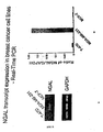
- Figures 3A and 3B show NGAL transcript expression levels in breast cancer cell lines. Expression levels were determined using real-time PCR.
- Figure 3A shows a transcript separated on agarose as compared to GAPDH control transcript.
- Figure 3B is a graph depicting the ratio of NGAL/GAPDH on the Y-axis. T-47D and MCF-7 are "organ confined" breast cancer cell lines, while MDA-MB-231 is a "highly metastatic cell line”.
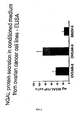
- Figure 4 is a graph illustrating the amount of NGAL protein secretion in conditioned medium from breast cancer cell lines, T-47D, MCF-7 and MDA-MB-231, as determined by Borregaard ELISA.
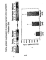
- Figures 5A and 5B show the levels of NGAL protein in urine samples from patients with either no disease, benign ovarian disease, and malignant ovarian cancer.
- Figure 5A shows a Western blot analysis of urine samples using anti-NGAL antibody.
- Figure 5B shows quantitative analysis of Western blots as described in Figure 5A . Blots were quantitated using densitometry and levels are represented in arbitrary densitometric units.
- Figure 6 is the amino acid sequence of human NGAL (SEQ ID NO:1).
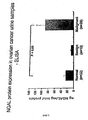
- Figure 7 is a graph of NGAL protein expression in ovarian cancer urine samples compared to healthy control samples as determined by Borregaard ELISA.
- Figure 8 is a graph of NGAL protein expression in breast cancer urine samples compared to healthy control samples as determined by Borregaard ELISA.
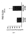
- Figure 9 is a graph of NGAL protein expression in individuals with atypical ductal hyperplasia (ADH) or lobular carcinoma in situ (LCIS), a major risk factor for future breast cancer development, as compared to levels seen in healthy individuals.
- ADH atypical ductal hyperplasia
- LCIS lobular carcinoma in situ
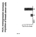
- Figure 10 is a graph illustrating the amount of migrated cells per field (y-axis) in a migration assay of breast cancer cells: MCF-7 cells, which express little NGAL, and two cell line clones that overexpress NGAL, N1 and N2.
- Figure 11 is a graph illustrating the amount of cell invasion in a tumor invasion assay of breast cancer cells: MCF-7 cells, which express little NGAL, and two cell line clones that overexpress NGAL, N 1 and N2.
- Figure 12 is a graph of NGAL protein expression in individuals with atypical ductal hyperplasia (ADH), a major risk factor for future breast cancer development, with ductal carcinoma in situ (DCIS), the most common type of noninvasive breast cancer, and with invasive breast cancer (IBC) as compared to levels seen in healthy individuals.
- ADH atypical ductal hyperplasia
- DCIS ductal carcinoma in situ
- IBC invasive breast cancer
- Figure 13 is a graph showing the amount of free NGAL monomer present in ovarian cancer urine samples, from patients having normal, benign and malignant tumors.
- the malignant tumor samples evidence a higher free NGAL level than the benign sample population.
- Free NGAL is used interchangeably with the term “uncomplexed NGAL” and refers to NGAL that is not complexed with a matrix metalloproteinase, e.g ., MMP-9.
- Cancers of epithelial origin include cancers that arise from epithelial cells which include, but are not limited to, breast cancer, basal cell carcinoma, adenocarcinoma, gastrointestinal cancer, lip cancer, mouth cancer, esophageal cancer, small bowel cancer and stomach cancer, colon cancer, liver cancer, bladder cancer, pancreatic cancer, ovarian cancer, cervical cancer, lung cancer, breast cancer and skin cancer, such as squamous cell and basal cell cancers, prostate cancer, renal cell carcinoma, and other known cancers that effect epithelial cells throughout the body.
- a cancer of epithelial origin may be a hormone-dependent cancer, e.g ., kidney, breast, endometrial, ovarian or prostate cancer.
- Control sample includes urine sample(s) obtained from a "normal” or “healthy” individual(s) believed not to have cancer. Controls may be selected using methods that are well known in the art. Once a level has become well established for a control population, array results from test urine samples can be directly compared with the known levels. Preferably the control sample is an age and sex.matched control urine sample.
- a "control level” is a level of analyte against which a tested level of the analyte is compared.
- the control level can be an empirically-determined mean or median analyte level from a group of individuals believed not to have the disease of interest (e.g ., cancer) or having a particular stage of the disease of interest, e.g ., benign or early stage cancer vs. malignant or late stage cancer; can be a known analyte level in an actual sample ( e.g ., a positive control); or can be a previously-determined analyte level for a particular individual ( e.g ., when monitoring an individual at two or more time points).
- the analyte is, for example, free NGAL.
- Test sample includes a urine sample obtained from a subject being tested.
- Invasive or “invasive” includes the proclivity of a tumor for expanding beyond its boundaries into adjacent tissue ( Darnell, J. (1990), Molecular Cell Biology, Third Ed., W.H. Freeman, NY ). Invasive cancer can be contrasted with organ-confined cancer wherein the tumor is confined to a particular organ.
- the invasive property of a tumor is often accompanied by the elaboration of proteolytic enzymes, such as collagenases, that degrade matrix material and basement membrane material to enable the tumor to expand beyond the confines of the capsule, and beyond confines of the particular tissue in which that tumor is located.
- Methodastasis includes the condition of spread of cancer from the organ of origin to additional distal sites in the patient.
- the process of tumor metastasis is a multistage event involving local invasion and destruction of intercellular matrix, intravasation into blood vessels, lymphatics or other channels of transport, survival in the circulation, extravasation out of the vessels in the secondary site and growth in the new location (see, e.g., Fidler, et al., Adv. Cancer Res. 28, 149-250 (1978 ), Liotta, et al., Cancer Treatment Res. 40, 223-238 (1988 ), Nicolson, Biochim. Biophy. Acta 948, 175-224 (1988 ) and Zetter, N. Eng. J. Med.
- a “primary tumor” includes a tumor appearing at a first site within the subject and can be distinguished from a “metastatic tumor” which appears in the body of the subject at a remote site from the primary tumor.
- NGAL includes the NGAL protein of Genebank accession, Genpept, CAA58127 (Homo sapiens) (SEQ ID NO:1) ( Fig. 6 ). NGAL is also referred to as neutrophil gelatinase-associated lipocalin and lipocalin-2. “NGAL” also encompasses species variants, homologues, allelic forms, mutant forms, and equivalents thereof.
- 'higher level' includes a level that is statistically significant or significantly above levels found in the control sample, e.g ., 1.3 fold higher or 1.5 fold higher.
- the 'higher level' is at least 2 fold higher or greater.
- Statistically significant or “significantly” includes statistical significance, and generally means a two standard deviation (2SD) above normal, or higher, concentration of the marker.
- LCIS is lobular carcinoma in situ. LCIS is also called lobular neoplasia and is sometimes classified as a type of noninvasive breast cancer. It does not penetrate through the wall of the lobules. Although it does not itself usually become an invasive cancer, women with this condition have a higher risk of developing an invasive breast cancer in the same or opposite breast.
- DCIS is ductal carcinoma in situ. Ductal carcinoma in situ is the most common type of noninvasive breast cancer. In DCIS, the malignant cells have not metastasized through the walls of the ducts into the fatty tissue of the breast. Comedocarcinoma is a type of DCIS that is more likely than other types of DCIS to come back in the same area after lumpectomy, and is more closely linked to eventual development of invasive ductal carcinoma than other forms of DCIS.
- “Increased risk” of cancer includes a risk of cancer increased from that of the general population. A person said to have increased risk of cancer is in need of monitoring for the development of cancer and for the continued presence of cancer risk markers.
- cell lines derived from highly metastatic breast cancer express and secrete higher amounts of NGAL than cell lines derived from benign, organ defined breast cancers, such as T-47D and MCF-7.
- overexpression of NGAL in a NGAL(-) breast cancer cell line (MCF-7), induces a mesenchymal-like phenotype, increases migration and invasion, and regulates Epithelial Mesenchymal Transition (EMT) marker transcription and expression, indicating a role for NGAL in metastatic disease.
- EMT Epithelial Mesenchymal Transition
- free urinary NGAL can be used as a biomarker to detect the presence and/or progression of cancer, e.g ., cancer of epithelial origin including breast and ovarian cancer, and that non-invasive assays and tests can be used as an essential part of a cancer diagnosis and treatment.
- cancer e.g ., cancer of epithelial origin including breast and ovarian cancer
- non-invasive assays and tests can be used as an essential part of a cancer diagnosis and treatment.
- the non-invasive nature of the tests ameliorates somewhat the apprehension associated with the process.
- ELISA ELISA
- Other cancers include peritoneal cancer, endometrial cancer, cervical cancer, colorectal cancer, uterine cancer, thyroid cancer, lung cancer, kidney cancer, and pancreatic cancer.
- a determination of the likelihood for cancer recurrence, spread, or patient survival can assist in determining whether a more conservative or more radical approach to therapy should be taken, or whether treatment modalities should be combined.
- the methods disclosed herein may be used to determine whether or not an individual has a cancer of epithelial origin including breast and ovarian cancer, or is at increased risk of developing cancer.
- the methods involve measuring levels of free NGAL in a test sample obtained from an individual, and comparing the observed levels to control levels of free NGAL, e.g ., those found in a control sample, or as control level is defined above.
- levels of NGAL higher than control levels indicate that the individual is either at an increased risk of developing cancer, or has cancer.
- the levels of NGAL can be represented by arbitrary units, for example as units obtained from a densitometer, luminometer, or an ELISA plate reader.
- test sample and control sample are of the same type, i.e ., obtained from urine.
- control sample can also be a standard sample that contains the same concentration of NGAL that is normally found in a urine sample obtained from a healthy individual.
- the urine sample is preferably treated to prevent degradation of the NGAL protein.
- Methods for inhibiting or preventing degradation include, but are not limited to, treatment of the sample with protease inhibitors, freezing the sample, or placing the sample on ice.
- the samples are constantly kept under conditions as to prevent degradation of the NGAL protein.
- a secondary diagnostic step can be performed. For example, if a level of NGAL is found to indicate either the risk or the presence of cancer, then an additional method of detecting the risk of cancer, or the presence of cancer can be performed to confirm.
- the individual can be tested for atypical ductal carcinoma (ADH) or lobular carcinoma in situ (LCIS) by means known to those in the art, such as atypia of the breast can be diagnosed by biopsy, random periareolar fine needle aspiration or ductal lavage. Genetic markers of risk can also be assessed, such as BRCA1 or BRCA2 or any other known genetic marker for cancer.
- any of a variety of additional diagnostic steps can be used to confirm the presence of cancer, such as ultrasound, mammography, PET scanning, MRI, thermal imaging, or any other imaging techniques, biopsy, ductal lavage, ductogram, clinical examination, or any other method known to those skilled in the art.
- a patient with increased risk of cancer is preferably placed on a cancer detection regime, such as regular pap smears and mammographies with assessment of other risk markers.
- a patient may also be placed on preventative regimes, such as administration of angiogenesis inhibitors or selective hormone receptor modulators, preventive regimes are well known to those in the art.
- preventative regimes such as administration of angiogenesis inhibitors or selective hormone receptor modulators, preventive regimes are well known to those in the art.
- a patient with cancer is directed to cancer treatments well known to those in the art.
- disease progression can be assessed by following NGAL levels, such as free-NGAL levels, in an individual patient.
- NGAL levels such as free-NGAL levels
- changes in an individual's condition can be monitored by comparing changes in NGAL expression levels in the individual over time, e.g ., via standard methods for carrying out longitudinal studies in patients for the relevant condition.
- Progressive increases in NGAL levels are indicative of increased potential for having cancer, tumor invasion and metastasis, and/or a later stage of cancer, depending upon the reason for monitoring the individual over time.
- therapeutic efficacy of a treatment regimen can be monitored by monitoring the level of NGAL over time in an individual.
- decreases in NGAL levels over time can be indicative of efficacy of the treatment regimen. Longitudinal monitoring can occur at two or more time points.
- the methods outlined in the previous paragraph may also be advantageously used to longitudinally monitor patients who have been treated for cancer, e.g ., for recurrence of the cancer.
- urinary NGAL e.g ., free NGAL
- urinary NGAL has been found in the present disclosure to be a useful cancer biomarker
- an increased level of urinary NGAL in a patient post-treatment can be acted on accordingly, e.g ., by further diagnosis, or further treatment, if appropriate.
- a control level of urinary NGAL would be established, e.g ., using either one or several values of measured urinary NGAL, whichever is analytically appropriate.
- Changes in the post-treatment patients' condition can then be monitored by comparing changes in NGAL expression levels in the individual as needed.
- a progressive or gradual increase, or a rapid elevation, of urinary NGAL levels can be indicative of a recurrence. Determining cancer recurrence can assist in determining whether a more conservative or more radical approach to therapy should be taken, or whether treatment modalities should be combined.
- it can be advantageous to precede or follow surgical treatment with chemotherapy, radiation, immunotherapy, biological modifier therapy, gene therapy, vaccines, and the like, or adjust the span of time during which the patient is treated.
- urinary free NGAL levels in an individual being screened for recurrence of cancer can be compared against an empirically-determined control level. A test level from the individual greater than the control level indicates recurrence, which can be followed up with further therapy or monitoring, as described above.
- Free urinary NGAL levels may be measured preferably through the use of antibodies, or antibody equivalents, to detect free NGAL levels. Other methods, such as disclosed in WO 2004/088276 , are also suitable; and NGAL levels may also be monitored by mass spectrometric analysis.
- levels of NGAL protein are measured by contacting the urine sample with an antibody-based binding moiety that preferentially binds to free NGAL, or to a fragment of free NGAL. Formation of the antibody-NGAL complex is then detected as a measure of free NGAL levels.
- Antibody-based binding moiety or “antibody” includes immunoglobulin molecules and immunologically active determinants of immunoglobulin molecules, e.g ., molecules that contain an antigen binding site which preferentially binds (immunoreacts with) to NGAL, or to the additional biomarkers of the invention.
- Antibody-based binding moiety includes whole antibodies, e.g ., of any isotype (IgG, IgA, IgM, IgE, etc), and fragments thereof which are also specifically reactive with NGAL protein, more preferably those which preferentially bind to uncomplexed (free) NGAL. Antibodies can be fragmented using conventional techniques.
- the term includes segments of proteolytically-cleaved or recombinantly-prepared portions of an antibody molecule that are capable of selectively reacting with a certain protein.
- proteolytic and/or recombinant fragments include Fab, F(ab')2, Fab' , Fv, dAbs and single chain antibodies (scFv) containing a VL and VH domain joined by a peptide linker.
- the scFvs may be covalently or non-covalently linked to form antibodies having two or more binding sites.
- antibody-based binding moiety includes polyclonal, monoclonal, or other purified preparations of antibodies and recombinant antibodies.
- antibody-based binding moiety is further intended to include humanized antibodies, bispecific antibodies, and chimeric molecules having at least one antigen binding determinant derived from an antibody molecule.
- the antibody-based binding moiety is detectably labeled.
- Labeled antibody includes antibodies that are labeled by a detectable means and include antibodies that are enzymatically, radioactively, fluorescently, and chemiluminescently labeled. Antibodies can also be labeled with a detectable tag, such as c-Myc, HA, VSV-G, HSV, FLAG, V5, or HIS.
- a detectable tag such as c-Myc, HA, VSV-G, HSV, FLAG, V5, or HIS.
- the level of free NGAL protein present in the urine samples correlate to the intensity of the signal emitted from the detectably labeled antibody.
- the antibody-based binding moiety is detectably labeled by linking the antibody to an enzyme.
- the enzyme when exposed to its substrate, will react with the substrate in such a manner as to produce a chemical moiety which can be detected, for example, by spectrophotometric, fluorometric or by visual means.
- Enzymes which can be used to detectably label the antibodies of the present invention include malate dehydrogenase, staphylococcal nuclease, delta-V-steroid isomerase, yeast alcohol dehydrogenase, alpha-glycerophosphate dehydrogenase, triose phosphate isomerase, horseradish peroxidase, alkaline phosphatase, asparaginase, glucose oxidase, beta-galactosidase, ribonuclease, urease, catalase, glucose-VI-phosphate dehydrogenase, glucoamylase and acetylcholinesterase.
- Chemiluminescence is another method that can be used to detect an antibody-based binding moiety.
- Detection may also be accomplished using any of a variety of other immunoassays.
- by radioactively labeling an antibody it is possible to detect the antibody through the use of radioimmunoassays.
- the radioactive isotope can be detected by such means as the use of a gamma counter or a scintillation counter or by autoradiography.
- Isotopes which are particularly useful for the purpose of the present invention are 3 H, 131 I, 35 S, 14 C, and preferably 125 I.
- fluorescent labeling compounds include CYE dyes, fluorescein isothiocyanate, rhodamine, phycoerytherin, phycocyanin, allophycocyanin, o-phthaldehyde and fluorescamine.
- An antibody can also be detectably labeled using fluorescence emitting metals such as 152 Eu, or others of the lanthanide series. These metals can be attached to the antibody using such metal chelating groups as diethylenetriaminepentaacetic acid (DTPA) or ethylenediaminetetraacetic acid (EDTA).
- DTPA diethylenetriaminepentaacetic acid
- EDTA ethylenediaminetetraacetic acid
- An antibody also can be detectably labeled by coupling it to a chemiluminescent compound. The presence of the chemiluminescent-antibody is then determined by detecting the presence of luminescence that arises during the course of a chemical reaction.
- chemiluminescent labeling compounds are luminol, luciferin, isoluminol, theromatic acridinium ester, imidazole, acridinium salt and oxalate ester.
- NGAL levels can be detected by immunoassays, such as enzyme linked immunoabsorbant assay (ELISA), radioimmunoassay (RIA), Immunoradiometric assay (IRMA), Western blotting, or immunohistochemistry, each of which are described in more detail below.
- immunoassays such as enzyme linked immunoabsorbant assay (ELISA), radioimmunoassay (RIA), Immunoradiometric assay (IRMA), Western blotting, or immunohistochemistry, each of which are described in more detail below.
- ELISA enzyme linked immunoabsorbant assay
- RIA radioimmunoassay
- IRMA Immunoradiometric assay
- Western blotting or immunohistochemistry
- Radioimmunoassay is a technique for detecting and measuring the concentration of an antigen using a labeled (e.g ., radioactively labeled) form of the antigen.
- radioactive labels for antigens include 3 H, 14 C, and 125 I.
- the concentration of free NGAL antigen in a biological sample is measured by having the antigen in the biological sample compete with the labeled (e.g ., radioactively) antigen for binding to an antibody to the antigen.
- the labeled antigen is present in a concentration sufficient to saturate the binding sites of the antibody. The higher the concentration of antigen in the sample, the lower the concentration of labeled antigen that will bind to the antibody.
- the antigen-antibody complex In a radioimmunoassay, to determine the concentration of labeled antigen bound to antibody, the antigen-antibody complex must be separated from the free antigen.
- One method for separating the antigen-antibody complex from the free antigen is by precipitating the antigen-antibody complex with an anti-isotype antiserum.
- Another method for separating the antigen-antibody complex from the free antigen is by precipitating the antigen-antibody complex with formalin-killed S. aureus.
- Yet another method for separating the antigen-antibody complex from the free antigen is by performing a "solid-phase radioimmunoassay" where the antibody is linked ( e.g ., covalently) to Sepharose beads, polystyrene wells, polyvinylchloride wells, or microtiter wells.
- a solid-phase radioimmunoassay where the antibody is linked ( e.g ., covalently) to Sepharose beads, polystyrene wells, polyvinylchloride wells, or microtiter wells.
- a "Immunoradiometric assay” is an immunoassay in which the antibody reagent is radioactively labeled.
- An IRMA requires the production of a multivalent antigen conjugate, by techniques such as conjugation to a protein e.g ., rabbit serum albumin (RSA).
- the multivalent antigen conjugate must have at least 2 antigen residues per molecule and the antigen residues must be of sufficient distance apart to allow binding by at least two antibodies to the antigen.
- the multivalent antigen conjugate can be attached to a solid surface such as a plastic sphere.
- sample antigen and antibody to antigen which is radioactively labeled are added to a test tube containing the multivalent antigen conjugate coated sphere.
- the antigen in the sample competes with the multivalent antigen conjugate for antigen antibody binding sites.
- the unbound reactants are removed by washing and the amount of radioactivity on the solid phase is determined.
- the amount of bound radioactive antibody is inversely proportional to the concentration of antigen in the sample.
- ELISA Enzyme-Linked Immunosorbent Assay
- an antibody e.g ., anti-NGAL or anti-free NGAL
- a solid phase i.e ., a microtiter plate
- antigen e.g ., NGAL
- a labeled antibody e.g ., enzyme linked
- enzymes that can be linked to the antibody are alkaline phosphatase, horseradish peroxidase, luciferase, urease, and ⁇ -galactosidase.
- the enzyme linked antibody reacts with a substrate to generate a colored reaction product that can be measured.
- antibody e.g ., anti-NGAL or anti-free NGAL
- a sample containing antigen i.e ., NGAL
- the antigen-antibody mixture is then contacted with a solid phase (e.g ., a microtiter plate) that is coated with antigen (i.e. , NGAL).
- a solid phase e.g ., a microtiter plate
- antigen i.e. , NGAL
- a labeled (e.g ., enzyme linked) secondary antibody is then added to the solid phase to determine the amount of primary antibody bound to the solid phase.
- a section of tissue is tested for specific proteins by exposing the tissue to antibodies that are specific for the protein that is being assayed.
- the antibodies are then visualized by any of a number of methods to determine the presence and amount of the protein present. Examples of methods used to visualize antibodies are, for example, through enzymes linked to the antibodies (e.g ., luciferase, alkaline phosphatase, horseradish peroxidase, or ⁇ -galactosidase), or chemical methods (e.g ., DAB/Substrate chromagen).
- NGAL neurotrophic factor
- Detectably labeled antibodies that preferentially bind to NGAL can then be used to assess NGAL levels, where the intensity of the signal from the detectable label corresponds to the amount of NGAL present. Levels can be quantitated, for example by densitometry.
- NGAL e.g ., free NGAL
- Mass Spectrometry such as MALDI/TOF (time-of-flight), SELDI/TOF, liquid chromatography-mass spectrometry (LC-MS), gas chromatography-mass spectrometry (GC-MS), high performance liquid chromatography-mass spectrometry (HPLC-MS), capillary electrophoresis-mass spectrometry, nuclear magnetic resonance spectrometry, or tandem mass spectrometry (e.g ., MS/MS, MS/MS/MS, ESI-MS/MS, etc.). see, e.g., U.S. Publication Nos. 20030199001 , 20030134304 , and 20030077616 .
- Mass spectrometry methods are well known in the art and have been used to quantify and/or identify proteins (see, e.g., Li et al. (2000) Tibtech 18:151-160 ; Rowley et al. (2000) Methods 20: 383-397 ; and Kuster and Mann (1998) Curr. Opin. Structural Biol. 8: 393-400 ). Further, mass spectrometric techniques have been developed that permit at least partial de novo sequencing of isolated proteins. Chait et al., Science 262:89-92 (1993 ); Keough et al., Proc. Natl. Acad. Sci. USA. 96:7131-6 (1999 ); reviewed in Bergman, EXS 88:133-44 (2000 ).
- a gas phase ion spectrophotometer is used.
- laser-desorption/ionization mass spectrometry is used to analyze the sample.
- Modem laser desorption/ionization mass spectrometry (“LDI-MS”) can be practiced in two main variations: matrix assisted laser desorption/ionization (“MALDI”) mass spectrometry and surface-enhanced laser desorption/ionization (“SELDI”).
- MALDI matrix assisted laser desorption/ionization
- SELDI surface-enhanced laser desorption/ionization
- MALDI Metal-organic laser desorption ionization
- the substrate surface is modified so that it is an active participant in the desorption process.
- the surface is derivatized with adsorbent and/or capture reagents that selectively bind the protein of interest.
- the surface is derivatized with energy absorbing molecules that are not desorbed when struck with the laser.
- the surface is derivatized with molecules that bind the protein of interest and that contain a photolytic bond that is broken upon application of the laser.
- the derivatizing agent generally is localized to a specific location on the substrate surface where the sample is applied. See, e.g., U.S. Pat. No. 5,719,060 and WO 98/59361 .
- the two methods can be combined by, for example, using a SELDI affinity surface to capture an analyte and adding matrix-containing liquid to the captured analvte to provide the energy absorbing material.
- Detection of the presence of a marker or other substances will typically involve detection of signal intensity. This, in turn, can reflect the quantity and character of a polypeptide bound to the substrate. For example, in certain embodiments, the signal strength of peak values from spectra of a first sample and a second sample can be compared ( e.g ., visually, by computer analysis etc.), to determine the relative amounts of particular biomolecules.
- Software programs such as the Biomarker Wizard program (Ciphergen Biosystems, Inc., Fremont, Calif.) can be used to aid in analyzing mass spectra. The mass spectrometers and their techniques are well known to those of skill in the art.
- any of the components of a mass spectrometer e.g ., desorption source, mass analyzer, detect, etc.
- varied sample preparations can be combined with other suitable components or preparations described herein, or to those known in the art.
- a control sample may contain heavy atoms (e.g . 13 C) thereby permitting the test sample to be mixed with the known control sample in the same mass spectrometry run.
- a laser desorption time-of-flight (TOF) mass spectrometer is used.
- TOF time-of-flight
- a substrate with a bound marker is introduced into an inlet system.
- the marker is desorbed and ionized into the gas phase by laser from the ionization source.
- the ions generated are collected by an ion optic assembly, and then in a time-of-flight mass analyzer, ions are accelerated through a short high voltage field and let drift into a high vacuum chamber. At the far end of the high vacuum chamber, the accelerated ions strike a sensitive detector surface at a different time. Since the time-of-flight is a function of the mass of the ions, the elapsed time between ion formation and ion detector impact can be used to identify the presence or absence of molecules of specific mass to charge ratio.
- the relative amounts of one or more biomolecules present in a first or second sample is determined, in part, by executing an algorithm with a programmable digital computer.
- the algorithm identifies at least one peak value in the first mass spectrum and the second mass spectrum.
- the algorithm compares the signal strength of the peak value of the first mass spectrum to the signal strength of the peak value of the second mass spectrum.
- the relative signal strengths are an indication of the amount of the biomolecule (e.g ., NGAL or free NGAL) that is present in the first and second samples.
- a standard containing a known amount of a biomolecule can be analyzed as the second sample to better quantify the amount of the biomolecule present in the first sample.
- the identity of the biomolecules in the first and second sample can also be determined.
- biomarker levels are measured by MALDI-TOF mass spectrometry.
- the free NGAL-binding antibodies for use in the invention can be obtained from a commercial source (for example, the anti-free NGAL antibody available in NGAL ELISA kit #DLCN20 from R&D Systems, Minneapolis, MN USA.)
- antibodies can be raised against free NGAL, or a portion of the biomarker polypeptide, e.g ., an amino acid portion that is normally masked in the MMP-9/NGAL complex, in order to generate an antibody that only interacts with free NGAL and not MMP-9/NGAL.
- Such antibody-binding moieties may be raised against an epitope inside the MMP-9/NGAL binding region by those of ordinary skill in the art, and need not be disclosed in further detail here.
- Antibodies for use in the present invention can be produced using standard methods to produce antibodies, for example, by monoclonal antibody production ( Campbell, A.M., Monoclonal Antibodies Technology: Laboratory Techniques in Biochemistry and Molecular Biology, Elsevier Science Publishers, Amsterdam, the Netherlands (1984 ); St. Groth et al., J. Immunology, (1990) 35: 1-21 ; and Kozbor et al., Immunology Today (1983) 4:72 ). Antibodies can also be readily obtained by using antigenic portions of the protein to screen an antibody library, such as a phage display library by methods well known in the art. For example, U.S. Patent No. 5,702,892 and WO 01/18058 disclose bacteriophage display libraries and selection methods for producing antibody binding domain fragments.
- kits for the detection and prognostic evaluation of ovarian cancer and for the detection and prognostic evaluation of breast cancer are provided according to this disclosure.
- the kits may be in any configuration well known to those of ordinary skill in the art and are useful for performing one or more of the methods described herein for the detection of NGAL.
- the kits are convenient in that they supply many, if not all, of the essential reagents for conducting an assay for the detection of NGAL in a urine sample.
- the assay is preferably performed simultaneously with a standard or multiple standards that are included in the kit, such as a predetermined amount of NGAL protein, so that the results of the test can be quantitated or validated.
- kits include the means for detecting NGAL levels i.e., NGAL binding antibodies or antibody fragments which selectively bind to NGAL protein.
- the diagnostic assay kit is preferentially formulated in a standard two-antibody binding format, in which one NGAL specific antibody captures NGAL in a patient sample and another NGAL specific antibody is used to detect captured NGAL.
- the capture antibody is immobilized on a solid phase, e.g ., an assay plate, an assay well, a nitrocellulose membrane, a bead, a dipstick, or a component of an elution column.
- the second antibody i.e ., the detection antibody, is typically tagged with a detectable label such as a calorimetric agent or radioisotope.
- the kit comprises a means for detecting levels of free NGAL in a sample of urine.
- the kit comprises a "dipstick" with anti-NGAL antibodies or fragments, immobilized thereon, which preferentially bind NGAL protein.
- Preferentially bound NGAL protein can then be detected using, for example, a second antibody that is detectably labeled with a colorimetric agent or radioisotope, whereupon the amount of free NGAL may be quantitatively determined as those of ordinary skill in the art will understand.
- the assay kits may employ (but are not limited to) the following techniques: competitive and non-competitive assays, radioimmunoassay (RIA) , bioluminescence and chemiluminescence assays, fluorometric assays, sandwich assays, immunoradiometric assays, dot blots, enzyme linked assays including ELISA, microtiter plates, and immunocytochemistry.
- RIA radioimmunoassay
- bioluminescence and chemiluminescence assays fluorometric assays
- sandwich assays sandwich assays
- immunoradiometric assays sandwich assays
- dot blots enzyme linked assays including ELISA, microtiter plates, and immunocytochemistry.
- enzyme linked assays including ELISA, microtiter plates, and immunocytochemistry.
- NGAL as a urinary biomarker for ovarian and breast cancer
- NGAL can serve as a biomarker for cancer, e.g ., of epithelial origin such as breast and/or ovarian cancer. Moreover, it demonstrates that free NGAL is useful for diagnosing, prognosing and staging of cancers.
- Figures 1a and 1b show the NGAL transcript expression levels in various ovarian cancer cell lines.
- Figures 3A and 3B show the NGAL transcript expression levels in various breast cancer cell lines. Expression levels were determined using real-time PCR. These results confirm that NGAL is overexpressed in cancer cell lines as compared to non-cancerous (HOSE) cell lines.
- HOSE non-cancerous
- Figure 2 shows a graph illustrating the amount of NGAL protein secretion in conditioned medium from ovarian cancer cell lines as determined by Borregaard ELISA as described in Kjeldsen L, Koch C, Arnljots K, Borregaard N., Characterization of two ELISAs for NGAL, a newly described lipocalin in human neutrophils. J Immunol Methods. 1996 Nov 13;198(2):155-64 , using anti-NGAL antibody provided by Dr. Borregaard.
- OVCAR-5 shows the highest level of secretion while SKOV-3 had significantly less secretion than OVCAR-5.
- Figure 4 shows a graph illustrating the amount of NGAL protein secretion in conditioned medium from breast cancer cell lines using Borregaard ELISA.
- MDA-MB-231 a cell line derived from a highly metastatic cancer, shows the highest level of NGAL secretion, while there is significantly less secretion in cell lines derived from organ confined cancers (MCF-7 and T-47D).
- free urinary NGAL protein serves a biomarker for breast and/or ovarian cancer.
- FIG. 5A shows a Western blot analysis of urine samples using anti-human NGAL from R&D Systems (Minneapolis, MN USA).
- Figure 5B shows the quantitative analysis of the results from at least 5 samples of urine from patients with either no disease, benign ovarian disease, or malignant ovarian cancer. Patients with malignant ovarian cancer showed higher amounts of NGAL in urine as compared to samples from individuals without disease. This was confirmed by a Borregaard ELISA assay ( Figure 7 ). Similar analysis of urine samples from breast cancer patients also showed higher levels of NGAL in urine from patients that have malignant cancer as compared to patients without disease ( Figure 8 ).
- Free NGAL is present in urine in at least monomeric, dimeric and trimeric forms. Detection of free monomeric urinary NGAL is possible in accordance with the present disclosure. In this instance, the studies were carried out with NGAL ELISA kit #DLCN20 from R&D Systems, Minneapolis, MN USA. The presence of monomeric NGAL appears to be particularly indicative of benign and malignant ovarian cancers, as shown in Figure 13.
- Figure 13 is a graph showing the amount of free NGAL monomer present in ovarian cancer urine samples, from patients having benign and malignant tumors, in comparison with normal samples. The malignant tumor samples evidence a much higher free NGAL level than the benign sample population.
- measuring the level of free NGAL in urine provides a quick, easy, and safe screen that can be used to determine whether or not an individual either has a cancer, e.g ., of epithelial origin such as breast and/or ovarian cancer, or is at an increased risk for developing a cancer.
- a higher level of free NGAL in urine as compared to an age and sex matched control sample indicates that the individual either has a cancer or is at increased risk for developing a breast and/or ovarian cancer.
- a positive test indicates that the individual should be tested further for cancer and monitored accordingly.
- EMT Epithelial Mesenchymal Transition
- MCF-7 is a breast cancer cell line that expresses little NGAL.
- N1 and N2 cell clones MCF-7 cells that overexpress NGAL
- MCF-7 cells that overexpress NGAL exhibit a mesenchymal-like phenotype, these cells lose cell-contacts and scatter more evenly across the cell surface (data not shown).
- NGAL overexpressing cells were evaluated using BD Falcon HTS FluoroBlok 24-Multiwell Insert System (BD Bioscience, Bedford, MA) according to manufacture's manual. MCF-7 control and NGAL-overexpressing cells (N1/N2) were grown to 70% confluence and were then trypsinized and resuspended in serum-free medium. 5 ⁇ 10 4 cells were seeded into the top chambers. Full media (750 ⁇ l) were used as chemoattractant and were added into the lower chambers. After incubation in 37°C, 5% CO 2 incubator for 24 hours, medium was removed from upper chambers.
- the insert plates were then transferred to a new 24-well plate containing 0.5ml/well of 10 ⁇ M CellTracker CMFDA (Molecular Probes) and were incubated for 1 hour at 37°C, 5% CO 2 .
- the BD FluoroBlok membrane only allows the labeled cells that migrated through the membrane to be visualized under fluorescence microscope. The results of this assay clearly show that overexpression of NGAL increases motility. Cells overexpressing NGAL migrated more than the parental MCF-7 cells ( Figure 10 ).
- NGAL neurotrophic factor
- BD Bioscience, Bedford, MA FluoroBlok membranes
- MCF-7 control and NGAL-overexpressing cells were grown to 70% confluence and were then trypsinized and resuspended in serum-free medium. 5 ⁇ 10 4 cells were seeded into the top chambers. Full media (750 ⁇ l) were used as chemoattractant and were added into the lower chambers.
- NGAL Epithelial Mesenchymal Transition
- NGAL is involved in the development of cell phenotypes consistent with what is found in aggressive cancers.
- Neutrophil gelatinase-associated lipocalin is a marker for dysregulated keratinocyte differentiation in human skin.
Landscapes
- Health & Medical Sciences (AREA)
- Life Sciences & Earth Sciences (AREA)
- Immunology (AREA)
- Engineering & Computer Science (AREA)
- Urology & Nephrology (AREA)
- Biomedical Technology (AREA)
- Hematology (AREA)
- Chemical & Material Sciences (AREA)
- Molecular Biology (AREA)
- Cell Biology (AREA)
- Biotechnology (AREA)
- Microbiology (AREA)
- Food Science & Technology (AREA)
- Medicinal Chemistry (AREA)
- Physics & Mathematics (AREA)
- Analytical Chemistry (AREA)
- Biochemistry (AREA)
- General Health & Medical Sciences (AREA)
- General Physics & Mathematics (AREA)
- Pathology (AREA)
- Investigating Or Analysing Biological Materials (AREA)
- Measuring Or Testing Involving Enzymes Or Micro-Organisms (AREA)
- Other Investigation Or Analysis Of Materials By Electrical Means (AREA)
Claims (18)
- Verfahren zur Bestimmung, ob ein Individuum ein Risiko für die Entwicklung von Krebs aufweist oder Krebs entwickelt hat, umfassend die folgenden Schritte:a) Bestimmung eines Spiegels von freiem NGAL in einer von einem Individuum gewonnenen Testurinprobe;b) Vergleich des Spiegels von freiem NGAL in der Testurinprobe mit einem Kontrollspiegel von freiem NGAL;wobei ein Spiegel von freiem NGAL in der Testprobe höher als ein Kontrollspiegel von freiem NGAL anzeigt, dass das Individuum entweder ein erhöhtes Risiko für die Entwicklung von Krebs aufweist, oder an Krebs leidet;
wobei der Krebs ein Krebs epithelialen Ursprungs ist. - Verfahren nach Anspruch 1, wobei der Krebs epithelialen Ursprungs ausgewählt ist aus der Gruppe, bestehend aus Brustkrebs, einem Basalzellkarzinom, einem Adenokarzinom, Gastrointestinalkrebs, Lippenkrebs, Mundkrebs, Ösophaguskrebs, Dünndarmkrebs, Magenkrebs, Kolonkrebs, Leberkrebs, Blasenkrebs, Pankreaskrebs, Ovarialkrebs, Cervixkrebs, Lungenkrebs, Hautkrebs, Nierenkrebs, Prostatakrebs und einem Nierenzellkarzinom.
- Verfahren nach Anspruch 1, wobei der Krebs Brust- und/oder Ovarialkrebs ist.
- Verfahren nach Anspruch 1, wobei der Spiegel von freiem NGAL mittels eines Verfahrens gemessen wird, umfassend die folgenden Schritte:a) Inkontaktbringen der Testprobe oder einer Aufbereitung davon, mit einem Antikörper-basierten Bindungsteil, der zur Bildung eines Antikörper-NGAL-Komplexes bevorzugt an freies NGAL bindet; undb) Nachweis des Vorliegens des Antikörper-NGAL-Komplexes, wobei so der Spiegel von vorliegendem freiem NGAL gemessen wird.
- Verfahren nach Anspruch 1, wobei der Antikörper-basierte Bindungsteil mit einer nachweisbaren Markierung markiert ist.
- Verfahren nach Anspruch 1, wobei die Markierung aus der Gruppe ausgewählt ist, bestehend aus einer radioaktiven Markierung, einer Haptenmarkierung, einer Fluoreszenzmarkierung und einer Enzymmarkierung.
- Verfahren nach Anspruch 1, wobei der Antikörper-basierte Bindungsteil ein Antikörper ist.
- Verfahren nach Anspruch 1, wobei der Antikörper ein monoklonaler Antikörper ist.
- Verfahren nach Anspruch 1, ferner umfassend den Schritt des Überprüfens der Ergebnisse, und wenn der Urin einen höheren Spiegel von freiem NGAL aufweist als der Kontrollspiegel von freiem NGAL, Überweisung des Individuums zum weiteren Testen auf Krebs.
- Verfahren nach Anspruch 1, wobei der Spiegel von freiem NGAL mittels massenspektrometrischer Verfahren gemessen wird.
- Verfahren nach Anspruch 1, wobei das Individuum ein Patient nach einer Krebsbehandlung ist, und wenn ein Spiegel von freiem NGAL in der Testprobe höher ist als ein Kontrollspiegel von freiem NGAL, der Patient nach einer Krebsbehandlung zur Behandlung eines Rezidivs des Krebses überwiesen wird.
- Verfahren zum Staging eines Krebses epithelialen Ursprungs, umfassend die folgenden Schritte:a) Bestimmung eines Spiegels von freiem NGAL in einer von einem Individuum gewonnenen Testurinprobe;b) Vergleich des Spiegels von freiem NGAL in der Testurinprobe mit einem Kontrollspiegel von freiem NGAL;wobei ein Spiegel von freiem NGAL in der Testprobe höher als ein Kontrollspiegel von freiem NGAL anzeigt, dass das Individuum entweder ein erhöhtes Risiko für die Entwicklung eines malignen oder metastatischen Krebses aufweist oder an einem malignen oder metastatischen Krebs leidet.
- Verfahren nach Anspruch 1 oder 12, wobei das freie NGAL monomeres NGAL umfasst.
- Verfahren nach Anspruch 12, wobei der Kontrollspiegel ein Spiegel von freiem NGAL ist, der mit einem gesunden Individuum assoziiert wird.
- Verfahren zur Überwachung von Krebs oder eines Risikos für die Entwicklung von Krebs bei einem Individuum, umfassend die folgenden Schritte:a) Bestimmung eines Spiegels von freiem NGAL in einer ersten Testurinprobe, die von einem Individuum zum Erhalt eines ersten Spiegels von freiem NGAL gewonnen wird;b) Bestimmung eines Spiegels von freiem NGAL in einer zweiten Urinprobe, die von dem Individuum zum Erhalt eines zweiten Spiegels von freiem NGAL gewonnen wird; undc) Vergleich der ersten und zweiten Spiegel von freiem NGAL;wobei der Krebs ein Krebs epithelialen Ursprungs ist.
- Verfahren nach Anspruch 16, wobei, wenn der zweite Spiegel höher als der erste Spiegel ist, das Individuum ein größeres Risiko, an Krebs zu leiden oder für die Entwicklung eines Krebses aufweist oder ein größeres Risiko, an metastasierendem Krebs zu leiden oder für die Entwicklung eines metastasierenden Krebses aufweist.
- Verfahren nach Anspruch 15, wobei das Individuum ein Patient nach der Krebsbehandlung ist, und wenn der zweite Spiegel höher als der erste Spiegel ist, der Patient nach der Krebsbehandlung zur Behandlung eines Rezidivs des Krebses überwiesen wird.
- Verfahren zur Überwachung auf ein Krebsrezidiv bei einem Patienten nach der Krebsbehandlung, umfassend die folgenden Schritte:a) Bestimmung eines Spiegels von freiem NGAL in einer ersten Testurinprobe, die von einem Patienten nach der Krebsbehandlung zum Erhalt eines ersten Spiegels von freiem NGAL gewonnen wird;b) Bestimmung eines Spiegels von freiem NGAL in einer zweiten Urinprobe, die von dem Patienten nach der Krebsbehandlung zum Erhalt eines zweiten Spiegels von freiem NGAL gewonnen wird; undc) Vergleich des ersten und des zweiten Spiegels von freiem NGAL, wobei, wenn der zweite Spiegel höher als der erste Spiegel ist, der Patient nach der Krebsbehandlung zur Behandlung eines Rezidivs des Krebses überwiesen wird;wobei der Krebs ein Krebs epithelialen Ursprungs ist.
Priority Applications (1)
| Application Number | Priority Date | Filing Date | Title |
|---|---|---|---|
| EP11156071A EP2375254A1 (de) | 2006-02-17 | 2007-02-16 | Freies NGAL als Biomarker für Krebs |
Applications Claiming Priority (2)
| Application Number | Priority Date | Filing Date | Title |
|---|---|---|---|
| US77482306P | 2006-02-17 | 2006-02-17 | |
| PCT/US2007/004265 WO2007098102A2 (en) | 2006-02-17 | 2007-02-16 | Free ngal as a biomarker for cancer |
Publications (2)
| Publication Number | Publication Date |
|---|---|
| EP1931996A2 EP1931996A2 (de) | 2008-06-18 |
| EP1931996B1 true EP1931996B1 (de) | 2013-11-20 |
Family
ID=38180692
Family Applications (2)
| Application Number | Title | Priority Date | Filing Date |
|---|---|---|---|
| EP07751053.5A Not-in-force EP1931996B1 (de) | 2006-02-17 | 2007-02-16 | Freies ngal als biomarker für krebs |
| EP11156071A Withdrawn EP2375254A1 (de) | 2006-02-17 | 2007-02-16 | Freies NGAL als Biomarker für Krebs |
Family Applications After (1)
| Application Number | Title | Priority Date | Filing Date |
|---|---|---|---|
| EP11156071A Withdrawn EP2375254A1 (de) | 2006-02-17 | 2007-02-16 | Freies NGAL als Biomarker für Krebs |
Country Status (7)
| Country | Link |
|---|---|
| US (6) | US20070196876A1 (de) |
| EP (2) | EP1931996B1 (de) |
| JP (1) | JP2009527735A (de) |
| CN (1) | CN101421622A (de) |
| AU (1) | AU2007217861A1 (de) |
| CA (1) | CA2642833A1 (de) |
| WO (1) | WO2007098102A2 (de) |
Families Citing this family (39)
| Publication number | Priority date | Publication date | Assignee | Title |
|---|---|---|---|---|
| WO2002031507A2 (en) * | 2000-10-13 | 2002-04-18 | Children's Medical Center Corporation | Non-invasive enzyme screen for tissue remodelling-associated conditions |
| ES2818028T3 (es) * | 2004-12-20 | 2021-04-09 | Antibodyshop As | Determinación de lipocalina asociada a gelatinasa de neutrófilos (NGAL) como marcador diagnóstico para trastornos renales |
| CN101421622A (zh) * | 2006-02-17 | 2009-04-29 | 儿童医学中心公司 | 作为癌症生物标志物的游离ngal |
| PL2035835T3 (pl) * | 2006-05-30 | 2012-05-31 | Antibodyshop As | Sposoby szybkiej oceny ciężkości urazu |
| US20080033755A1 (en) * | 2006-06-07 | 2008-02-07 | Krusz John C | Medical treatment modality for headache, pain, and other medical disorders |
| US7645616B2 (en) * | 2006-10-20 | 2010-01-12 | The University Of Hong Kong | Use of lipocalin-2 as a diagnostic marker and therapeutic target |
| WO2008113363A1 (en) | 2007-03-21 | 2008-09-25 | Bioporto Diagnostics A/S | Diagnostic test for renal injury |
| WO2008132167A2 (en) * | 2007-04-26 | 2008-11-06 | Dublin City University | Diagnostic, prognostic and/or predictive indicators of breast cancer |
| WO2009052400A1 (en) * | 2007-10-19 | 2009-04-23 | Abbott Laboratories | Antibodies that bind to mammalian ngal and uses thereof |
| US8846036B2 (en) | 2007-10-19 | 2014-09-30 | Abbott Laboratories | Antibodies that bind to mammalian NGAL and uses thereof |
| ES2475990T5 (es) * | 2007-11-15 | 2017-07-06 | Bioporto Diagnostics A/S | Uso diagnóstico de formas moleculares individuales de un biomarcador |
| EP2257813A4 (de) * | 2008-03-12 | 2011-11-02 | Univ Columbia | Ngal mit hohem molekulargewicht als biomarker für chronische nierenerkrankung |
| JP2009244154A (ja) * | 2008-03-31 | 2009-10-22 | Nationa Hospital Organization | 老化、および血管障害を伴う疾患の検定のための組成物、キットおよび方法 |
| US20100105150A1 (en) * | 2008-10-24 | 2010-04-29 | Abbott Laboratories | Isolated human autoantibodies to neutrophil gelatinase-associated lipocalin (ngal) and methods and kits for the detection of human autoantibodies to ngal |
| JP2012508177A (ja) * | 2008-11-05 | 2012-04-05 | アボット・ラボラトリーズ | 尿および組換えチャイニーズハムスター卵巣(cho)細胞から濃縮された好中球ゼラチナーゼ関連リポカリン(ngal)タンパク質アイソフォーム、ならびに関連する組成物、抗体、ならびに濃縮、分析および使用の方法 |
| KR101939964B1 (ko) * | 2008-11-21 | 2019-01-18 | 퓨처 메디칼 다이아그노스틱스 코 엘티디 | 급성 신장 손상의 발견 또는 감시 방법, 장치 및 키트 |
| US20100233740A1 (en) * | 2009-02-12 | 2010-09-16 | Jonathan Barasch | Use of urinary ngal to distinguish kidney disease and predict mortality in subjects with cirrhosis |
| WO2010148145A1 (en) * | 2009-06-16 | 2010-12-23 | Fred Hutchinson Cancer Research Center | Methods and kits for detecting ovarian cancer from blood |
| EP2273272A1 (de) | 2009-07-06 | 2011-01-12 | Stichting Katholieke Universiteit | Verfahren zum Vorhersagen der Reaktion auf eine Chemotherapie bei Eierstockkrebs |
| WO2011053832A1 (en) * | 2009-10-29 | 2011-05-05 | The Trustees Of Columbia University In The City Of New York | Use of urinary ngal to diagnose sepsis in very low birth weight infants |
| US20110229921A1 (en) * | 2010-03-18 | 2011-09-22 | Abbott Laboratories | METHODS OF ASSAYING URINARY NEUTROPHIL GELATINASE-ASSOCIATED LIPOCALIN (uNGAL) IN THE PROGNOSIS OF CADAVERIC KIDNEY TRANSPLANT FUNCTION IN A PATIENT, INCLUDING A PATIENT DIAGNOSED WITH DELAYED GRAFT FUNCTION (DGF), A METHOD OF ASSAYING uNGAL IN THE ASSESSMENT OF RISK OF DGF IN A PATIENT DIAGNOSED WITH EARLY GRAFT FUNCTION (EGF), AND RELATED KITS |
| CN102072960A (zh) * | 2010-03-29 | 2011-05-25 | 武汉生之源生物科技有限公司 | 一种样本中中性粒细胞明胶酶相关脂质运载蛋白的检测方法 |
| WO2011140554A1 (en) * | 2010-05-07 | 2011-11-10 | The Trustees Of Columbia University In The City Of New York | Ngal and urinary tract infection |
| WO2011149962A1 (en) | 2010-05-24 | 2011-12-01 | The Trustees Of Columbia University In The City Of New York | Mutant ngal proteins and uses thereof |
| CN102253217B (zh) * | 2011-04-07 | 2013-08-07 | 武汉生之源生物科技有限公司 | 一种胶乳颗粒增强型中性粒细胞明胶酶相关脂质运载蛋白检测试剂盒 |
| EP2925337B1 (de) | 2012-11-21 | 2019-07-03 | The Trustees of Columbia University in the City of New York | Mutante ngal-proteine und verwendungen davon |
| RU2758608C2 (ru) * | 2014-11-19 | 2021-11-01 | Конинклейке Филипс Н.В. | Способ диагностики с использованием hnl |
| JP5866053B1 (ja) * | 2014-12-15 | 2016-02-17 | 株式会社ファンケル | 皮膚粘弾性のマーカー及びその利用 |
| GB201501930D0 (en) | 2015-02-05 | 2015-03-25 | Univ London Queen Mary | Biomarkers for pancreatic cancer |
| CN106018813A (zh) * | 2015-11-15 | 2016-10-12 | 陈博 | 一种唇癌特异性检测的试剂盒 |
| US12019068B2 (en) | 2017-03-31 | 2024-06-25 | Hirotsu Bio Science Inc. | Method for predicting therapeutic effect and/or recurrence monitoring in cancer patients |
| ES2876038T3 (es) * | 2018-02-18 | 2021-11-11 | Marquez Marquez Lidia | Método, aparato y kit para la detección temprana de cáncer de mama |
| CN109096373B (zh) * | 2018-09-27 | 2022-06-21 | 江苏亨瑞生物医药科技有限公司 | 合成寡肽及其用途 |
| CN109206485B (zh) * | 2018-09-27 | 2022-04-08 | 山东聚胜生物科技有限公司 | 一种防治肺癌转移的寡肽 |
| CN109096374B (zh) * | 2018-09-27 | 2022-11-18 | 深圳乐土沃森精准医疗有限公司 | 一种抑制肺癌转移的合成寡肽 |
| CN109180789B (zh) * | 2018-09-27 | 2022-08-19 | 上海揽微赛尔生物科技有限公司 | 一种寡肽及制药应用 |
| CN109608542B (zh) * | 2018-12-29 | 2022-04-05 | 江苏众红生物工程创药研究院有限公司 | 抗人ngal抗体及其在检测试纸卡中的应用 |
| CN109553682B (zh) * | 2018-12-29 | 2022-04-05 | 江苏众红生物工程创药研究院有限公司 | 抗人中性粒细胞明胶酶相关脂质运载蛋白抗体及其在检测试纸卡中的应用 |
| CN110333356B (zh) * | 2019-05-30 | 2023-09-19 | 桂林优利特医疗电子有限公司 | 一种尿液特种蛋白复合质控物及制备方法 |
Family Cites Families (22)
| Publication number | Priority date | Publication date | Assignee | Title |
|---|---|---|---|---|
| US5118937A (en) | 1989-08-22 | 1992-06-02 | Finnigan Mat Gmbh | Process and device for the laser desorption of an analyte molecular ions, especially of biomolecules |
| US5045694A (en) | 1989-09-27 | 1991-09-03 | The Rockefeller University | Instrument and method for the laser desorption of ions in mass spectrometry |
| EP0700521B1 (de) | 1993-05-28 | 2003-06-04 | Baylor College Of Medicine | Verfahren und massenspektrometer zur desorption und ionisierung von analyten |
| KR100251594B1 (ko) * | 1995-04-12 | 2000-06-01 | 알카 쿠보넨 | 치근막 질환의 진단과 이의 진행위험을 예견하는 방법과 이에 사용되는 테스트 키트 |
| US5702892A (en) | 1995-05-09 | 1997-12-30 | The United States Of America As Represented By The Department Of Health And Human Services | Phage-display of immunoglobulin heavy chain libraries |
| JP2000510949A (ja) * | 1996-04-26 | 2000-08-22 | チルドレンズ メディカル センター コーポレーション | 組織のリモデリングに関連した状態のための非観血的酵素スクリーン |
| NZ516848A (en) | 1997-06-20 | 2004-03-26 | Ciphergen Biosystems Inc | Retentate chromatography apparatus with applications in biology and medicine |
| US6406921B1 (en) | 1998-07-14 | 2002-06-18 | Zyomyx, Incorporated | Protein arrays for high-throughput screening |
| US6610484B1 (en) * | 1999-01-26 | 2003-08-26 | Cytyc Health Corporation | Identifying material from a breast duct |
| WO2001018058A2 (en) | 1999-09-07 | 2001-03-15 | Viventia Biotech Inc. | Enhanced phage display library of human vh fragments and methods for producing same |
| US7429466B2 (en) | 2000-01-24 | 2008-09-30 | Hypromatrix, Inc | Methods and arrays for detecting biological molecules |
| WO2002031507A2 (en) * | 2000-10-13 | 2002-04-18 | Children's Medical Center Corporation | Non-invasive enzyme screen for tissue remodelling-associated conditions |
| AU2002258518A1 (en) * | 2001-03-14 | 2002-09-24 | Millennium Pharmaceuticals, Inc. | Nucleic acid molecules and proteins for the identification, assessment, prevention, and therapy of ovarian cancer |
| CN1463291A (zh) | 2001-04-19 | 2003-12-24 | 赛弗根生物系统股份有限公司 | 用质谱法和亲和标记物鉴定生物分子的特性 |
| US7112408B2 (en) | 2001-06-08 | 2006-09-26 | The Brigham And Women's Hospital, Inc. | Detection of ovarian cancer based upon alpha-haptoglobin levels |
| WO2002102235A2 (en) | 2001-06-18 | 2002-12-27 | Eos Biotechnology Inc. | Methods of diagnosis of ovarian cancer, compositions and methods of screening for modulators of ovarian cancer |
| US7189507B2 (en) | 2001-06-18 | 2007-03-13 | Pdl Biopharma, Inc. | Methods of diagnosis of ovarian cancer, compositions and methods of screening for modulators of ovarian cancer |
| US20030013208A1 (en) | 2001-07-13 | 2003-01-16 | Milagen, Inc. | Information enhanced antibody arrays |
| WO2003017177A2 (en) | 2001-08-13 | 2003-02-27 | Beyong Genomics, Inc. | Method and system for profiling biological systems |
| WO2003091735A1 (en) | 2002-04-23 | 2003-11-06 | Millipore Corporation | Sample preparation of biological fluids for proteomic applications |
| DE602004022150D1 (de) | 2003-03-27 | 2009-09-03 | Childrens Hosp Medical Center | Verfahren und kit zum nachweis des frühstadiums einer nierentubuluszellenverletzung |
| CN101421622A (zh) * | 2006-02-17 | 2009-04-29 | 儿童医学中心公司 | 作为癌症生物标志物的游离ngal |
-
2007
- 2007-02-16 CN CNA2007800135952A patent/CN101421622A/zh active Pending
- 2007-02-16 EP EP07751053.5A patent/EP1931996B1/de not_active Not-in-force
- 2007-02-16 AU AU2007217861A patent/AU2007217861A1/en not_active Abandoned
- 2007-02-16 EP EP11156071A patent/EP2375254A1/de not_active Withdrawn
- 2007-02-16 JP JP2008555404A patent/JP2009527735A/ja not_active Withdrawn
- 2007-02-16 CA CA002642833A patent/CA2642833A1/en not_active Abandoned
- 2007-02-16 WO PCT/US2007/004265 patent/WO2007098102A2/en not_active Ceased
- 2007-02-16 US US11/707,749 patent/US20070196876A1/en not_active Abandoned
-
2010
- 2010-04-08 US US12/756,501 patent/US20110081650A1/en not_active Abandoned
-
2013
- 2013-09-18 US US14/030,868 patent/US20140242616A1/en not_active Abandoned
-
2016
- 2016-04-21 US US15/135,460 patent/US20170082631A1/en not_active Abandoned
-
2017
- 2017-09-22 US US15/712,692 patent/US20180238896A1/en not_active Abandoned
-
2019
- 2019-01-03 US US16/238,940 patent/US20190361028A1/en not_active Abandoned
Also Published As
| Publication number | Publication date |
|---|---|
| EP1931996A2 (de) | 2008-06-18 |
| JP2009527735A (ja) | 2009-07-30 |
| US20190361028A1 (en) | 2019-11-28 |
| CA2642833A1 (en) | 2007-08-30 |
| US20180238896A1 (en) | 2018-08-23 |
| WO2007098102A2 (en) | 2007-08-30 |
| US20170082631A1 (en) | 2017-03-23 |
| CN101421622A (zh) | 2009-04-29 |
| US20070196876A1 (en) | 2007-08-23 |
| EP2375254A1 (de) | 2011-10-12 |
| US20110081650A1 (en) | 2011-04-07 |
| US20140242616A1 (en) | 2014-08-28 |
| WO2007098102A3 (en) | 2007-10-25 |
| AU2007217861A1 (en) | 2007-08-30 |
Similar Documents
| Publication | Publication Date | Title |
|---|---|---|
| EP1931996B1 (de) | Freies ngal als biomarker für krebs | |
| US7858324B2 (en) | Cyr61 as a biomarker for diagnosis and prognosis of cancers of epithelial origin | |
| US8685659B2 (en) | Method for diagnosis and prognosis of epithelial cancers | |
| US20100209944A1 (en) | Adamts-7 as a biomarker for cancers of epithelial origin | |
| JP2007519903A (ja) | 上皮起源の癌を診断および予後診断するための方法 | |
| WO2009017475A1 (en) | Methods for diagnosis and prognosis of epithelial cancers |
Legal Events
| Date | Code | Title | Description |
|---|---|---|---|
| PUAI | Public reference made under article 153(3) epc to a published international application that has entered the european phase |
Free format text: ORIGINAL CODE: 0009012 |
|
| 17P | Request for examination filed |
Effective date: 20080331 |
|
| AK | Designated contracting states |
Kind code of ref document: A2 Designated state(s): AT BE BG CH CY CZ DE DK EE ES FI FR GB GR HU IE IS IT LI LT LU LV MC NL PL PT RO SE SI SK TR |
|
| 17Q | First examination report despatched |
Effective date: 20090217 |
|
| DAX | Request for extension of the european patent (deleted) | ||
| GRAP | Despatch of communication of intention to grant a patent |
Free format text: ORIGINAL CODE: EPIDOSNIGR1 |
|
| INTG | Intention to grant announced |
Effective date: 20130327 |
|
| GRAJ | Information related to disapproval of communication of intention to grant by the applicant or resumption of examination proceedings by the epo deleted |
Free format text: ORIGINAL CODE: EPIDOSDIGR1 |
|
| RAP1 | Party data changed (applicant data changed or rights of an application transferred) |
Owner name: CHILDREN'S MEDICAL CENTER CORPORATION |
|
| GRAP | Despatch of communication of intention to grant a patent |
Free format text: ORIGINAL CODE: EPIDOSNIGR1 |
|
| INTG | Intention to grant announced |
Effective date: 20130606 |
|
| GRAS | Grant fee paid |
Free format text: ORIGINAL CODE: EPIDOSNIGR3 |
|
| GRAA | (expected) grant |
Free format text: ORIGINAL CODE: 0009210 |
|
| AK | Designated contracting states |
Kind code of ref document: B1 Designated state(s): AT BE BG CH CY CZ DE DK EE ES FI FR GB GR HU IE IS IT LI LT LU LV MC NL PL PT RO SE SI SK TR |
|
| REG | Reference to a national code |
Ref country code: GB Ref legal event code: FG4D |
|
| REG | Reference to a national code |
Ref country code: CH Ref legal event code: EP |
|
| REG | Reference to a national code |
Ref country code: AT Ref legal event code: REF Ref document number: 641936 Country of ref document: AT Kind code of ref document: T Effective date: 20131215 |
|
| REG | Reference to a national code |
Ref country code: IE Ref legal event code: FG4D |
|
| REG | Reference to a national code |
Ref country code: DE Ref legal event code: R096 Ref document number: 602007033897 Country of ref document: DE Effective date: 20140116 |
|
| REG | Reference to a national code |
Ref country code: NL Ref legal event code: VDEP Effective date: 20131120 |
|
| REG | Reference to a national code |
Ref country code: AT Ref legal event code: MK05 Ref document number: 641936 Country of ref document: AT Kind code of ref document: T Effective date: 20131120 |
|
| REG | Reference to a national code |
Ref country code: LT Ref legal event code: MG4D |
|
| PG25 | Lapsed in a contracting state [announced via postgrant information from national office to epo] |
Ref country code: FI Free format text: LAPSE BECAUSE OF FAILURE TO SUBMIT A TRANSLATION OF THE DESCRIPTION OR TO PAY THE FEE WITHIN THE PRESCRIBED TIME-LIMIT Effective date: 20131120 Ref country code: IS Free format text: LAPSE BECAUSE OF FAILURE TO SUBMIT A TRANSLATION OF THE DESCRIPTION OR TO PAY THE FEE WITHIN THE PRESCRIBED TIME-LIMIT Effective date: 20140320 Ref country code: NL Free format text: LAPSE BECAUSE OF FAILURE TO SUBMIT A TRANSLATION OF THE DESCRIPTION OR TO PAY THE FEE WITHIN THE PRESCRIBED TIME-LIMIT Effective date: 20131120 Ref country code: SE Free format text: LAPSE BECAUSE OF FAILURE TO SUBMIT A TRANSLATION OF THE DESCRIPTION OR TO PAY THE FEE WITHIN THE PRESCRIBED TIME-LIMIT Effective date: 20131120 Ref country code: LT Free format text: LAPSE BECAUSE OF FAILURE TO SUBMIT A TRANSLATION OF THE DESCRIPTION OR TO PAY THE FEE WITHIN THE PRESCRIBED TIME-LIMIT Effective date: 20131120 |
|
| PG25 | Lapsed in a contracting state [announced via postgrant information from national office to epo] |
Ref country code: AT Free format text: LAPSE BECAUSE OF FAILURE TO SUBMIT A TRANSLATION OF THE DESCRIPTION OR TO PAY THE FEE WITHIN THE PRESCRIBED TIME-LIMIT Effective date: 20131120 Ref country code: BE Free format text: LAPSE BECAUSE OF FAILURE TO SUBMIT A TRANSLATION OF THE DESCRIPTION OR TO PAY THE FEE WITHIN THE PRESCRIBED TIME-LIMIT Effective date: 20131120 Ref country code: ES Free format text: LAPSE BECAUSE OF FAILURE TO SUBMIT A TRANSLATION OF THE DESCRIPTION OR TO PAY THE FEE WITHIN THE PRESCRIBED TIME-LIMIT Effective date: 20131120 Ref country code: LV Free format text: LAPSE BECAUSE OF FAILURE TO SUBMIT A TRANSLATION OF THE DESCRIPTION OR TO PAY THE FEE WITHIN THE PRESCRIBED TIME-LIMIT Effective date: 20131120 |
|
| PG25 | Lapsed in a contracting state [announced via postgrant information from national office to epo] |
Ref country code: PT Free format text: LAPSE BECAUSE OF FAILURE TO SUBMIT A TRANSLATION OF THE DESCRIPTION OR TO PAY THE FEE WITHIN THE PRESCRIBED TIME-LIMIT Effective date: 20140320 |
|
| PG25 | Lapsed in a contracting state [announced via postgrant information from national office to epo] |
Ref country code: EE Free format text: LAPSE BECAUSE OF FAILURE TO SUBMIT A TRANSLATION OF THE DESCRIPTION OR TO PAY THE FEE WITHIN THE PRESCRIBED TIME-LIMIT Effective date: 20131120 |
|
| REG | Reference to a national code |
Ref country code: DE Ref legal event code: R097 Ref document number: 602007033897 Country of ref document: DE |
|
| PG25 | Lapsed in a contracting state [announced via postgrant information from national office to epo] |
Ref country code: RO Free format text: LAPSE BECAUSE OF FAILURE TO SUBMIT A TRANSLATION OF THE DESCRIPTION OR TO PAY THE FEE WITHIN THE PRESCRIBED TIME-LIMIT Effective date: 20131120 Ref country code: PL Free format text: LAPSE BECAUSE OF FAILURE TO SUBMIT A TRANSLATION OF THE DESCRIPTION OR TO PAY THE FEE WITHIN THE PRESCRIBED TIME-LIMIT Effective date: 20131120 Ref country code: CZ Free format text: LAPSE BECAUSE OF FAILURE TO SUBMIT A TRANSLATION OF THE DESCRIPTION OR TO PAY THE FEE WITHIN THE PRESCRIBED TIME-LIMIT Effective date: 20131120 Ref country code: SK Free format text: LAPSE BECAUSE OF FAILURE TO SUBMIT A TRANSLATION OF THE DESCRIPTION OR TO PAY THE FEE WITHIN THE PRESCRIBED TIME-LIMIT Effective date: 20131120 |
|
| PLBE | No opposition filed within time limit |
Free format text: ORIGINAL CODE: 0009261 |
|
| STAA | Information on the status of an ep patent application or granted ep patent |
Free format text: STATUS: NO OPPOSITION FILED WITHIN TIME LIMIT |
|
| PG25 | Lapsed in a contracting state [announced via postgrant information from national office to epo] |
Ref country code: DK Free format text: LAPSE BECAUSE OF FAILURE TO SUBMIT A TRANSLATION OF THE DESCRIPTION OR TO PAY THE FEE WITHIN THE PRESCRIBED TIME-LIMIT Effective date: 20131120 Ref country code: LU Free format text: LAPSE BECAUSE OF FAILURE TO SUBMIT A TRANSLATION OF THE DESCRIPTION OR TO PAY THE FEE WITHIN THE PRESCRIBED TIME-LIMIT Effective date: 20140216 Ref country code: MC Free format text: LAPSE BECAUSE OF FAILURE TO SUBMIT A TRANSLATION OF THE DESCRIPTION OR TO PAY THE FEE WITHIN THE PRESCRIBED TIME-LIMIT Effective date: 20131120 |
|
| 26N | No opposition filed |
Effective date: 20140821 |
|
| REG | Reference to a national code |
Ref country code: IE Ref legal event code: MM4A |
|
| REG | Reference to a national code |
Ref country code: DE Ref legal event code: R097 Ref document number: 602007033897 Country of ref document: DE Effective date: 20140821 |
|
| PG25 | Lapsed in a contracting state [announced via postgrant information from national office to epo] |
Ref country code: IE Free format text: LAPSE BECAUSE OF NON-PAYMENT OF DUE FEES Effective date: 20140216 |
|
| PG25 | Lapsed in a contracting state [announced via postgrant information from national office to epo] |
Ref country code: SI Free format text: LAPSE BECAUSE OF FAILURE TO SUBMIT A TRANSLATION OF THE DESCRIPTION OR TO PAY THE FEE WITHIN THE PRESCRIBED TIME-LIMIT Effective date: 20131120 |
|
| PG25 | Lapsed in a contracting state [announced via postgrant information from national office to epo] |
Ref country code: IT Free format text: LAPSE BECAUSE OF FAILURE TO SUBMIT A TRANSLATION OF THE DESCRIPTION OR TO PAY THE FEE WITHIN THE PRESCRIBED TIME-LIMIT Effective date: 20131120 |
|
| REG | Reference to a national code |
Ref country code: FR Ref legal event code: PLFP Year of fee payment: 10 |
|
| PG25 | Lapsed in a contracting state [announced via postgrant information from national office to epo] |
Ref country code: BG Free format text: LAPSE BECAUSE OF FAILURE TO SUBMIT A TRANSLATION OF THE DESCRIPTION OR TO PAY THE FEE WITHIN THE PRESCRIBED TIME-LIMIT Effective date: 20131120 |
|
| PG25 | Lapsed in a contracting state [announced via postgrant information from national office to epo] |
Ref country code: CY Free format text: LAPSE BECAUSE OF FAILURE TO SUBMIT A TRANSLATION OF THE DESCRIPTION OR TO PAY THE FEE WITHIN THE PRESCRIBED TIME-LIMIT Effective date: 20131120 Ref country code: GR Free format text: LAPSE BECAUSE OF FAILURE TO SUBMIT A TRANSLATION OF THE DESCRIPTION OR TO PAY THE FEE WITHIN THE PRESCRIBED TIME-LIMIT Effective date: 20140221 |
|
| PG25 | Lapsed in a contracting state [announced via postgrant information from national office to epo] |
Ref country code: TR Free format text: LAPSE BECAUSE OF FAILURE TO SUBMIT A TRANSLATION OF THE DESCRIPTION OR TO PAY THE FEE WITHIN THE PRESCRIBED TIME-LIMIT Effective date: 20131120 Ref country code: HU Free format text: LAPSE BECAUSE OF FAILURE TO SUBMIT A TRANSLATION OF THE DESCRIPTION OR TO PAY THE FEE WITHIN THE PRESCRIBED TIME-LIMIT; INVALID AB INITIO Effective date: 20070216 |
|
| REG | Reference to a national code |
Ref country code: FR Ref legal event code: PLFP Year of fee payment: 11 |
|
| REG | Reference to a national code |
Ref country code: FR Ref legal event code: PLFP Year of fee payment: 12 |
|
| PGFP | Annual fee paid to national office [announced via postgrant information from national office to epo] |
Ref country code: GB Payment date: 20220225 Year of fee payment: 16 Ref country code: DE Payment date: 20220225 Year of fee payment: 16 Ref country code: CH Payment date: 20220302 Year of fee payment: 16 |
|
| PGFP | Annual fee paid to national office [announced via postgrant information from national office to epo] |
Ref country code: FR Payment date: 20220223 Year of fee payment: 16 |
|
| P01 | Opt-out of the competence of the unified patent court (upc) registered |
Effective date: 20230524 |
|
| REG | Reference to a national code |
Ref country code: DE Ref legal event code: R119 Ref document number: 602007033897 Country of ref document: DE |
|
| REG | Reference to a national code |
Ref country code: CH Ref legal event code: PL |
|
| GBPC | Gb: european patent ceased through non-payment of renewal fee |
Effective date: 20230216 |
|
| PG25 | Lapsed in a contracting state [announced via postgrant information from national office to epo] |
Ref country code: LI Free format text: LAPSE BECAUSE OF NON-PAYMENT OF DUE FEES Effective date: 20230228 Ref country code: CH Free format text: LAPSE BECAUSE OF NON-PAYMENT OF DUE FEES Effective date: 20230228 |
|
| PG25 | Lapsed in a contracting state [announced via postgrant information from national office to epo] |
Ref country code: GB Free format text: LAPSE BECAUSE OF NON-PAYMENT OF DUE FEES Effective date: 20230216 |
|
| PG25 | Lapsed in a contracting state [announced via postgrant information from national office to epo] |
Ref country code: GB Free format text: LAPSE BECAUSE OF NON-PAYMENT OF DUE FEES Effective date: 20230216 Ref country code: FR Free format text: LAPSE BECAUSE OF NON-PAYMENT OF DUE FEES Effective date: 20230228 Ref country code: DE Free format text: LAPSE BECAUSE OF NON-PAYMENT OF DUE FEES Effective date: 20230901 |