EP1932539A2 - Bifunktionelle Signal-1/Signal-2-Peptidhemmer - Google Patents
Bifunktionelle Signal-1/Signal-2-Peptidhemmer Download PDFInfo
- Publication number
- EP1932539A2 EP1932539A2 EP07014573A EP07014573A EP1932539A2 EP 1932539 A2 EP1932539 A2 EP 1932539A2 EP 07014573 A EP07014573 A EP 07014573A EP 07014573 A EP07014573 A EP 07014573A EP 1932539 A2 EP1932539 A2 EP 1932539A2
- Authority
- EP
- European Patent Office
- Prior art keywords
- peptide
- sequence
- signal
- bpi
- amino acid
- Prior art date
- Legal status (The legal status is an assumption and is not a legal conclusion. Google has not performed a legal analysis and makes no representation as to the accuracy of the status listed.)
- Withdrawn
Links
Images










Classifications
-
- A—HUMAN NECESSITIES
- A61—MEDICAL OR VETERINARY SCIENCE; HYGIENE
- A61K—PREPARATIONS FOR MEDICAL, DENTAL OR TOILETRY PURPOSES
- A61K39/00—Medicinal preparations containing antigens or antibodies
- A61K39/0005—Vertebrate antigens
- A61K39/001—Preparations to induce tolerance to non-self, e.g. prior to transplantation
-
- A—HUMAN NECESSITIES
- A61—MEDICAL OR VETERINARY SCIENCE; HYGIENE
- A61P—SPECIFIC THERAPEUTIC ACTIVITY OF CHEMICAL COMPOUNDS OR MEDICINAL PREPARATIONS
- A61P37/00—Drugs for immunological or allergic disorders
- A61P37/02—Immunomodulators
-
- C—CHEMISTRY; METALLURGY
- C07—ORGANIC CHEMISTRY
- C07K—PEPTIDES
- C07K14/00—Peptides having more than 20 amino acids; Gastrins; Somatostatins; Melanotropins; Derivatives thereof
- C07K14/435—Peptides having more than 20 amino acids; Gastrins; Somatostatins; Melanotropins; Derivatives thereof from animals; from humans
- C07K14/705—Receptors; Cell surface antigens; Cell surface determinants
- C07K14/70546—Integrin superfamily
- C07K14/70553—Integrin beta2-subunit-containing molecules, e.g. CD11, CD18
-
- C—CHEMISTRY; METALLURGY
- C07—ORGANIC CHEMISTRY
- C07K—PEPTIDES
- C07K16/00—Immunoglobulins [IG], e.g. monoclonal or polyclonal antibodies
- C07K16/18—Immunoglobulins [IG], e.g. monoclonal or polyclonal antibodies against material from animals or humans
- C07K16/28—Immunoglobulins [IG], e.g. monoclonal or polyclonal antibodies against material from animals or humans against receptors, cell surface antigens or cell surface determinants
- C07K16/2803—Immunoglobulins [IG], e.g. monoclonal or polyclonal antibodies against material from animals or humans against receptors, cell surface antigens or cell surface determinants against the immunoglobulin superfamily
- C07K16/2821—Immunoglobulins [IG], e.g. monoclonal or polyclonal antibodies against material from animals or humans against receptors, cell surface antigens or cell surface determinants against the immunoglobulin superfamily against ICAM molecules, e.g. CD50, CD54, CD102
-
- C—CHEMISTRY; METALLURGY
- C07—ORGANIC CHEMISTRY
- C07K—PEPTIDES
- C07K16/00—Immunoglobulins [IG], e.g. monoclonal or polyclonal antibodies
- C07K16/18—Immunoglobulins [IG], e.g. monoclonal or polyclonal antibodies against material from animals or humans
- C07K16/28—Immunoglobulins [IG], e.g. monoclonal or polyclonal antibodies against material from animals or humans against receptors, cell surface antigens or cell surface determinants
- C07K16/2803—Immunoglobulins [IG], e.g. monoclonal or polyclonal antibodies against material from animals or humans against receptors, cell surface antigens or cell surface determinants against the immunoglobulin superfamily
- C07K16/2833—Immunoglobulins [IG], e.g. monoclonal or polyclonal antibodies against material from animals or humans against receptors, cell surface antigens or cell surface determinants against the immunoglobulin superfamily against MHC-molecules, e.g. HLA-molecules
-
- C—CHEMISTRY; METALLURGY
- C12—BIOCHEMISTRY; BEER; SPIRITS; WINE; VINEGAR; MICROBIOLOGY; ENZYMOLOGY; MUTATION OR GENETIC ENGINEERING
- C12N—MICROORGANISMS OR ENZYMES; COMPOSITIONS THEREOF; PROPAGATING, PRESERVING, OR MAINTAINING MICROORGANISMS; MUTATION OR GENETIC ENGINEERING; CULTURE MEDIA
- C12N9/00—Enzymes; Proenzymes; Compositions thereof; Processes for preparing, activating, inhibiting, separating or purifying enzymes
- C12N9/88—Lyases (4.)
-
- A—HUMAN NECESSITIES
- A61—MEDICAL OR VETERINARY SCIENCE; HYGIENE
- A61K—PREPARATIONS FOR MEDICAL, DENTAL OR TOILETRY PURPOSES
- A61K39/00—Medicinal preparations containing antigens or antibodies
- A61K2039/57—Medicinal preparations containing antigens or antibodies characterised by the type of response, e.g. Th1, Th2
-
- C—CHEMISTRY; METALLURGY
- C07—ORGANIC CHEMISTRY
- C07K—PEPTIDES
- C07K2319/00—Fusion polypeptide
Definitions
- the present invention concerns immune responses initiated by the recognition of a peptide:MHC complex on the surface of antigen presenting cells by T-cells.
- the present invention also concerns immune responses initiated by the binding of a Signal-2 moiety to its complement protein on the surface of an antigen presenting cell. More particularly, the present invention concerns the immune responses initiated by the recognition of the peptide:MHC by the T-cell and by the binding of a Signal-2 moiety to its complement protein. Still more particularly, the present invention concerns the modification of the typical immune response generated by a particular individual in response to this binding.
- the present invention concerns the conjugation of peptides derived from the peptide portion of the peptide:MHC complex to the preferred Signal-2 moiety in order to modify or shift a given immune response from type-1 to type-2 or from type-2 to type-1.
- This may include specific phenotypes of regulatory T-cells including suppressor T-cells.
- T-cells Autoimmune diseases are characterized by the activation of T-cells against self-antigens. These T-cells then destroy cells presenting these antigens.
- IDDM insulin-dependent diabetes mellitus
- IDL insulin-dependent diabetes mellitus
- T-cells characterized by the activation of T-cells against the insulin-producing cells of the pancreas and their subsequent destruction by these T-cells.
- MHC major histocompatability complex
- MHC molecules bind fragments (peptides) of proteins from infectious agents, allergens, and selfproteins, and this MHC:peptide complex is the structure that T-cells recognize with their receptor (called the T-cell receptor, or TCR).
- TCR T-cell receptor
- the MHC:peptide complex is displayed on the surfaces of other cells of the immune system (i.e., B cells, dendritic cells and macrophages) which are called antigen presenting cells (APC).
- B cells i.e., B cells, dendritic cells and macrophages
- APC antigen presenting cells
- the major regulatory cell of the immune system the undifferentiated T-cell, must be presented with small breakdown products (peptides) of the foreign invader. This presentation occurs on the surface of the APC.
- Signal-2 a costimulatory signal
- Signal-1 the signal generated by the TCR recognition of the MHC:peptide complex
- a defining stage of the immune response is the differentiation of CD4 + T-cells into either type-1 helper T-cells (T H 1 cells) or type-2 helper T-cells (T H 2 cells) as a result of the two signals.
- T H 1 cells type-1 helper T-cells
- T H 2 cells type-2 helper T-cells
- T H 1 cells type-1 helper T-cells
- T H 2 cells type-2 helper T-cells
- Interleukin-12 a cytokine produced by immune cells known as macrophages and dendritic cells.
- Interleukin-12 induces or stimulates the naive T-cell (CD4 + T-cells) to produce interferon- ⁇ (IFN- ⁇ ) and interleukin-2 (IL-2).
- IFN- ⁇ interferon- ⁇
- IL-2 and IFN- ⁇ are involved in classic cell-mediated functions such as clonal expansion of cytotoxic T-lymphocytes (CTLs), macrophage activation, and class switching to IgG isotypes that mediate complement lysis of sensitized cells.
- T H 1 immune response is enhanced by the presence of IFN- ⁇ which up-regulates expression of the interleukin-12 (IL-12) receptor while inhibiting the development of T H 2 cells.
- T H 2 immunity results from the production of interleukin-4(IL-4) by the naive T-cell.
- IL-4 induces T H 2 development and the subsequent production of interleukins-4 (IL-4), -5 (IL-5), -10 (IL-10), and -13 (IL-13).
- IL-4 also operates to down-regulate expression of the IL-12 receptor on developing cells, thereby inhibiting T H 1 development and helping undifferentiated T-cells to commit to T H 2 cell development.
- IL-4 and IL-5 are known to activate B cells and switch to neutralizing antibody (IgG1 in the mouse) and IgE, the initiator of immediate hypersensitivity.
- S-1 occurs when the T-cell antigen receptor (TCR) recognizes the peptide:MHC-II complex on the surface ofan antigen presenting cell (APC).
- TCR T-cell antigen receptor
- APC an antigen presenting cell
- This first signal passes through the T-cell receptor and initiates a cascade of tyrosine phosphorylation/dephosphorylation events mediated by kinases and phosphatases and leads to the activation of Ca ++ flux, nuclear factor of activated T cells (NF-AT) and NF ⁇ B transcription factors. These factors enter the nucleus of the T-cell and bind to promoters of genes responsible for effector functions.
- NF-AT nuclear factor of activated T cells
- Signal-2 arises from the binding of Signal-2 receptors to their ligands on the surface of an APC.
- Signal-2 receptors include CD28 and its ligand B7 as well as LFA-1 and its ligand ICAM-1.
- a series of signaling events occur. These events include serine/threonine phosphorylation/dephosphory-lation and activation of guanine nucleotide exchange factors that activate adapter proteins with GTPase activity. These signaling events activate a separate set of transcription factors.
- the signal delivered through the CD28:B7 complex is distinct from that delivered from the ICAM-1:LFA-1 complex, particularly with respect to the differentiation of CD4 + T-cells into T H 1 versus T H 2 effector populations.
- the CD4 + T-cell differentiation favors T H 1 cells which are abundant producers of IL-2 and IFN ⁇ , the preeminent initiators of inflammatory immune responses including delayed-type hypersensitivity (DTH), immunity to intracellular pathogens, and several autoimmune diseases.
- DTH delayed-type hypersensitivity
- the CD4 + T-cells differentiate into T H 2 cells.
- T H 2 cells In contrast to T H 1 cells, T H 2 cells do not produce abundant IL-2 or IFN ⁇ cytokines, but instead release the mediators of immediate-type hypersensitivity such as allergy and asthma, i.e., IL-4, IL-5, IL-10, and IL-13.
- the ability to manipulate the relative contribution of the complex providing the second signal has a profound effect on the type of immune response that is elicited against a given self-tissue antigen.
- T H 1 cells and type-1 immunity a program that ultimately activates IL-4 production
- T H 2 cells and type-2 immunity a program that ultimately activates IL-4 production
- the TCR recognizes the peptide:MHC-II complex and sends Signal-1 to the T-cell. Additionally, LFA-1 binds to ICAM-1, and these molecules, along with the peptide:MHC-II complex, translocate to form the end-stage immune synapse. This leads to the effective expression of the CD40 ligand (CD154) by the uncommited T H cell. CD40 interaction (expressed on the antigen presenting cell) with its ligand generates NF ⁇ B up-regulation of the inflammatory cytokine, IL-12.
- CD40 ligand CD154
- EL-12 then binds to its receptor on the undifferentiated T H cell and initiates the T H 1 program, including the up-regulation of the transcription regulators, Stat4 and Tbet.
- the TCR can recognize the same peptide:MHC-II complex, thereby sending Signal-1.
- a weaker strength of Signal-1 and/or altered or blocked binding between Signal-2 moieties leads to an altered form of the end-stage immune synapse.
- this lower strength of Signal-1 or distinct participation of the LFA-1 second signal leads to this different result, i.e., dominant T H 2 differentiation.
- the altered immune synapse can dictate that the CD40 ligand is not expressed and IL-12 is therefore not released by the APC.
- This pathway is schematically represented in Fig. 2.
- IL-4 appears to accumulate, thereby leading to the up-regulation of Stat6 and GATA-3 within the T-cell and hence commitment to a T H 2 pattern of differentiation.
- T H 1-dominant immunity e.g., as seen in autoimmune diseases and transplant rejection
- T H 2 responses against these same tissue antigens In other cases, it would be extremely valuable to replace weak T H 2 immunity with T H 1 dominance leading to strong T-cell proliferation and the effective generation of cytotoxic T-cells (CTL).
- CTL cytotoxic T-cells
- These cases may include chronic viral illnesses, like hepatitis-C and AIDS; and could include certain cancers like melanoma. Accordingly, what is needed in the art is modifiers of these immune responses so that type-2 immunity can be replaced with type-1 immunity or type-1 immunity can be replaced with type-2 immunity, as desired in order to combat different human disease states or health conditions.
- Interleukin-12 induces or stimulates the naive T-cell to produce interferon- ⁇ (IFN- ⁇ ) and interleukin-2 (IL-2).
- IFN- ⁇ interferon- ⁇
- IL-2 and IFN- ⁇ are involved in classic cell-mediated functions such as clonal expansion of cytotoxic T-lymphocytes (CTLs), macrophage activation, and class switching to IgG isotypes that mediate complement lysis of sensitized cells.
- CTLs cytotoxic T-lymphocytes
- macrophage activation clonal expansion of cytotoxic T-lymphocytes
- IgG interleukin-12
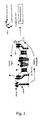
- Commitment to a T H 1 immune response is enhanced by the presence of IFN- ⁇ which up-regulates expression of the interleukin-12 (IL-12) receptor while inhibiting the development of T H 2 cells. This pathway is shown schematically in Fig. 3.
- T H 2 immunity results from the production ofinterleukin-4 (IL-4) by the naive T-cell.
- IL-4 induces T H 2 development and the subsequent production of interleukins 4 (IL-4), 5 (IL-5) and 13 (IL-13), through activation of the transcription regulator Stat6.
- IL-4 also operates to down-regulate expression of the IL-12 receptor on developing cells, thereby inhibiting T H 1 development and helping undifferentiated T-cells to commit to T H 2 cell development.
- IL-4 and IL-5 are known to activate B cells and switch to neutralizing antibody (IgG 1 in the mouse) and IgE, the initiator of immediate hypersensitivity. Again, a schematic representation of this process is depicted in Fig. 2.
- Signal-2 receptors include CD28 and its ligand B7 as well as LFA-1 and its ligand ICAM-1.
- a Signal-2 receptor and its ligand form a complex at the interface between the T-cell and APC membranes, a series of signaling events occurs including serine/threonine phosphorylation/dephosphorylation along with actuation of guanine nucleotide exchange factors that activate adapter proteins with GTPase activity. These signaling events activate a separate set of transcription factors.
- the signal delivered through the CD28:B7 complex is distinct from that delivered from the ICAM-1:LFA-1 complex, particularly with respect to the differentiation of CD4 + T-cells into T H 1 versus T H 2 effector populations.
- FIG. 4 A schematic representation of this signaling is provided herein as Fig. 4.
- the CD4 + T-cells differentiate into T H 1 cells.
- the CD4 + T-cells of the T H 1 differentiation state are abundant producers of IL-2 and IFN ⁇ , two cytokines that are the preeminent initiators of inflammatory immune responses, such as delayed-type hypersensitivity (DTH), immunity to intracellular pathogens, and several autoimmune diseases.
- DTH delayed-type hypersensitivity
- the CD4 + T-cells differentiate into T H 2 cells.
- T H 2 cells In contrast to T H 1 cells, T H 2 cells do not produce IL-2 and IFN ⁇ cytokines, but instead release the mediators of immediate-type hypersensitivity such as allergy and asthma, i.e., IL-4, IL-5, IL-10, and IL-13.
- IL-4, IL-5, IL-10, and IL-13 the mediators of immediate-type hypersensitivity
- IL-13 the mediators of immediate-type hypersensitivity
- the TCR:peptide:MHC-II complex is in the center of the dark circle which represents the protein:protein pair constituting the Signal-2 receptor and the Signal-2 ligand.
- adhesion molecules such as LFA-1 and the peptide-recognition receptor (TCR) to form a doughnut-like structure with the TCR on the inside and LFA-1 on the outside.
- TCR and LFA-1 molecules actually translocate past one another within the T-cell lipid bilayer. If these molecules do not translocate within the immune synapse then the T-cell signal is not fully received and a different program of gene activity may occur within the T-cell.
- known TCR epitopes are used as the first peptide portion of the BPI.
- minimal peptide sequences that are potent immunogens are utilized.
- These minimal peptide sequences e.g. antigenic peptides
- effectively engage the TCR involved in immune responses of interest i.e. autoimmune diseases, infectious diseases, allergies, cancers, etc.
- TCR epitopes of interest e.g. autoimmune diseases, infectious diseases, allergies, cancers, etc.
- T-cells are first cloned from patients. These cloned T-cells are placed separately in cultures with overlapping peptides (again, representing individual portions of the antigen involved, e.g., HIV-1, p24 (SEQ ID No. 8)). Again, the peptide to which most T-cell clones respond is the dominant TCR epitope.
- peptides derived from Signal-2 receptors are used to alter interactions between the nominal receptors on T-cells and their complementary ligands on the APC surface.
- Table 3 includes a representative list of some known Signal-2 receptor moieties. Of course, those of ordinary skill in the art will be able to identify other Signal-2 moieties not listed therein, as this list is representative and not all-inclusive.
- Another aspect of the present invention is the linking of the TCR epitope (i.e. the Signal-1 moiety) to a Signal-2 receptor peptide mimic (i.e., the Signal-2 moiety) in order to modify the resultant immune response.
- This linkage can be between the Signal-1 moiety and the Signal-2 moiety directly, or through flanking residues.
- this linking can be done via a linker which is positioned between the Signal-1 moiety and the Signal-2 moiety.
- the linker could be any amino acid including naturally occurring or chemically synthesized amino acids.
- non-substrate amino acids will be used due to their resistance to protease attack.
- the linker will comprise a non-substrate amino acid alternating with a small or hydrophilic amino acid. Even more preferably, the linker is synthesizable as one continuous sequence along with the Signal-1 and Signal-2 moieties, which flank the linker at each respective end. Still more preferably, the linker has the general formula (A,B) x , wherein A and B are amino acid residues, and the A amino acid residue is individually and respectively selected from the group consisting of aminocaproic acid, aminohexanoic acid, aminododecanoic acid, and ⁇ -alanine, and the B amino acid residue is a small or hydrophilic amino acid. In this formula, X can range from 1 to 100.
- a particularly representative B residue is glycine.
- a linker could potentially have aminocaproic acid (Ac), aminohexanoic acid (Ahx), aminododecanoic acid (Ado), and ⁇ -alanine ( ⁇ A) alternating with glycine residues (G) (e.g., Ac-G-Ahx-G-Ado-G- ⁇ A).
- the choice of the residues used to construct the linker can be based upon the desired length of the linker as well as steric hindrance considerations.
- One preferred linker comprises alternating Ac and G residues. This linker can be lengthened or shortened by the inclusion of the other amino acid residue choices (Ahx, Ado, ⁇ A).
- Some representative linkers are included in Table 2 as SEQ ID Nos. 26-29.
- TCR tumor antigens and the myriad of allergenic substances in the environment.
- TCR epitope of a given BPI we direct the immunomodulating capacity of the BPI to a select group of TCR.
- the selection of a TCR epitope to incorporate into the BPI targets T-cells that are involved in a particular human disease in a highly specific fashion.
- incorporating the GAD65 epitope into a BPI targets autoaggressive T-cells involved in the induction of type-1 diabetes.
- BPI offer the possibility to specifically modulate T-cell immunity to one antigen while leaving intact the T-cell repertoire necessary for protective immunity to infectious agents and developing cancers.
- the Signal-1 moieties of the present invention are preferably derived from TCR epitopes and a list of representative known epitopes is provided in Table 1 wherein these known epitopes are presented as SEQ ID Nos. 1-25.
- Table 1 wherein these known epitopes are presented as SEQ ID Nos. 1-25.
- the TCR epitope selected will be correlated with a known health condition or disease state.
- the peptides include a sequence having at least about 10% sequence homology with a sequence selected from the group consisting of SEQ ID Nos. 1-25.
- the peptide will have at least 30% sequence homology with a sequence selected from the group consisting of SEQ ID Nos. 1-25. More preferably, the peptide will have at least 50% sequence homology with a sequence selected from the group consisting of SEQ ID Nos. 1-25. Even more preferably, the peptide will have at least 70% sequence homology with a sequence selected from the group consisting of SEQ ID Nos.1-25. Most preferably, the peptide will have at least about 95% sequence homology with a sequence selected from the group consisting of SEQ ID Nos. 1-25.
- the peptidomimetic will be a mimetic of a peptide selected from the group consisting of SEQ ID Nos. 1-25.
- the Signal-1 moiety will be a derivative of a TCR epitope or a peptide selected from the group consisting of SEQ ID Nos. 1-25.
- this first portion of the BPI (or the portion responsible for initiating the first signal) be capable of binding with a major histocompatability complex (MHC) on an antigen presenting cell (APC).
- MHC major histocompatability complex
- APC antigen presenting cell
- this resulting peptide:MHC complex be capable of engaging important TCR and initiating some form of the signal to the T-cell.
- the peptides used on the side of the linker opposite the Signal-1 moiety are preferably derived from Signal-2 receptors.
- This second portion of the BPI is connected to the first portion either directly or via the linker.
- the second portion includes a sequence having at least about 10% sequence homology with a sequence selected from a group consisting of SEQ ID Nos. 30-41. More preferably, the second portion peptide has at least about 30% sequence homology with a sequence selected from the group consisting of SEQ ID Nos. 30-41. Still more preferably, the second portion peptide has at least about 50% sequence homology with a sequence selected from the group consisting of SEQ ID Nos. 30-41.
- the second portion peptide has at least about 70% sequence homology with a sequence selected from the group consisting of SEQ ID Nos. 30-41. Most preferably, the second portion peptide includes a sequence having at least about 95% sequence homology with a sequence selected from the group of SEQ ID Nos. 30-41.
- peptidomimetics can be used in place of all or some of the amino acid residues of the second portion. In preferred forms the peptidomimetic of the second portion will be a mimic of a peptide selected from the group consisting of SEQ ID Nos. 30-41.
- the second portion of the BPI will comprise a derivative of a peptide selected from the group consisting of SEQ ID Nos. 30-41.
- the second portion be capable of binding with a complementary ligand (e.g. the Signal-2 ligand) on an antigen presenting cell.
- a complementary ligand e.g. the Signal-2 ligand
- the second portion be capable of binding with a complementary ligand (e.g. the Signal-2 ligand) on an antigen presenting cell.
- a complementary ligand e.g. the Signal-2 ligand
- the Signal-2 ligand e.g. the Signal-2 ligand
- BPI may operate via the activation ofvery specific T-cell phenotypes, e.g., peptide-specific suppressor T-cells.
- T-cell phenotypes e.g., peptide-specific suppressor T-cells.
- the response generated when a BPI similar to the GAD 65-CD11a BPI is introduced into the immune synapse is quite different and operates to shift the response from type-1 to type-2.
- This situation is depicted schematically in Fig. 2.
- a BPI comprising a Signal-1 moiety, a flexible, non-substrate linker, and a Signal-2 moiety is formed and introduced into the immune synapse.
- the TCR recognizes the peptide:MHC complex on the APC and initiates the first signal.
- the second portion of the BPI (the Signal-2 moiety) blocks the typical Signal-2 interaction occurring between LFA-1/ICAM-1, (or for other BPI:CTLA-4/B7, or CD40L/CD40, or FasL:Fas) and the translocation of the TCR into the central cluster.
- LFA-1/ICAM-1 or CTLA-4/B7 interaction is targeted by the specific BPI construction, perhaps by tethering the MHC-II:peptide complex to the second signal ligand, the signal will be altered in a different direction of differentiation.
- the Signal-2 moiety peptide portion of the BPI is derived from CTLA-4
- the normal binding of CTLA-4 and CD-28 to B7 ligands is affected and thus more CD40 ligand is expressed (i.e., a greater role for high affinity LFA-1:ICAM-1 is dictated by blocking the B7 receptors); hence, the release of IL-12 increases.
- Interleukin-12 induces or stimulates the naive T-cell to produce more IFN- ⁇ and IL-2, thus providing a positive feedback toward type-1 immunity.
- cytokines IL-2 and IFN- ⁇
- CTLs cytotoxic T-lymphocytes
- macrophage activation and class switching to IgG isotypes that mediate complement lysis of sensitized cells.
- IgG cytotoxic T-lymphocytes
- Fas Fas:FasL interaction governs apoptosis, it will be possible to increase the frequency of specific TCR-bearing cells by blocking the apoptotic event. This will be important for BPI design against HIV, HPV, HCV, and cancers.
- an important aspect of the present invention is that tethering a specific TCR epitope to a Signal-2 receptor peptide mimic leads to alteration of T-cell differentiation involving T-cells bearing only these receptors and/or T-cell populations indirectly linked to these peptide specific subsets.
- the ability to block or alter T-cell responses to a given immunodominant peptide antigen would offer extremely precise treatments for immunopathological conditions.
- a major drawback to current immunotherapies is that broad specificities of T-cells are affected leaving the host more susceptible to infections and cancers.
- the BPI of the present invention should block and/or alter only the desired T-cell population and subsequent responses that depend on these initial T-cells. Also, BPI will target a specific TCR-bearing population for activation toward a desired effector function.
- the relative strength of signal generated by the T-cell-APC interaction has an affect on whether the ultimate immune response is a type-1 or a type-2 response.
- the teachings of Murray in How the MHC Selects TH1/TH2 Immunity, 19 Immunology Today 157-163 (1998 ) are hereby incorporated by reference.
- an immune response is modified by contacting an APC with a peptide capable of binding to an MHC and to a Signal-2 ligand on the APC and causing an altered signal to be transmitted to the T-cell.
- the immune response is deviated from the immune response generally associated with the immunogenic peptide and its corresponding antigen (i.e., infectious agent, self protein, or allergen).
- a peptide having the general formula AXB is provided.
- the A, X, and B represent a chain of amino acid residues wherein the A chain has at least about five residues and at least about 10% sequence homology with a TCR epitope, the B chain has at least four residues and at least about 10% sequence homology with a peptide derived from a Signal-2 moiety, and the X chain is a linker.
- the linker could be any amino acid including naturally occurring or chemically synthesized amino acids.
- the X chain has at least one residue.
- link A to B directly without X it is possible to link A to B directly without X as well, although a linker of some size is preferred in order to span the distance between the MHC-II and second signal ligands on the APC surface.
- non-substrate amino acids will be used due to their resistance to protease attack.
- the linker will comprise a non-substrate amino acid alternating with a small or hydrophilic amino acid.
- the linker is synthesizable as one continuous sequence along with the Signal-1 and Signal-2 moieties, which flank the linker at each respective end.
- the linker has the general formula (A,B) x , wherein A and B are amino acid residues, and the A amino acid residue is individually and respectively selected from the group consisting of aminocaproic acid, aminohexanoic acid, aminododecanoic acid, and ⁇ -alanine, and the B amino acid residue is a small or hydrophilic amino acid.
- X can range from 1 to 100.
- a particularly representative B residue is glycine.
- the X chain is positioned between the A chain and the B chain and the entire peptide can be synthesized as one continuous sequence.
- Some preferred sequences will have an A chain having at least about 10% sequence homology with any one of SEQ ID Nos. 1-25, an X chain having at least about 2% sequence homology with any one of SEQ ID Nos. 26-29, and a B chain having at least about 10% sequence homology with any one of SEQ ID Nos. 30-41.
- the peptide is capable of shifting a type-1 response to a type-2 response, or vice versa.
- peptidomimetics may be synthesized to mimic any part of the BPI, including the linker.
- the A chain binds to the MHC on an APC to form a peptide:MHC complex.
- This complex is capable of engaging the TCR on critical T-cell populations.
- the B chain is capable of binding to a Signal-2 ligand on the APC at the same time as the formation of the peptide:MHC complex. This combined binding to the APC should be capable of altering the signal delivered to the T-cell.
- the first peptide sequence should be associated with a known health condition and be capable of binding with an MHC on an APC.
- the second peptide sequence be capable of binding with a Signal-2 ligand moiety on the APC.
- the method can further comprise the step of contacting the nominal peptide immunogen with the TCR, thereby binding the first peptide sequence to the MHC and the second peptide sequence to the Signal-2 ligand, thereby generating APC bearing potent first and altered/blocked second signal ligands which activate a desired immune response.
- T-cell clones to possible peptide epitopes were generated and tested for binding to the immunodominant TCR of a response and specifically stimulate T-cell functions in vitro by the ELISPOT assay. Because modifications in the peptide residues that actually contact the TCR are part of the BPI development, it is also preferable that known crystallographic structures of the epitope bound to MHC molecules are available. This allows for precise three-dimensional predictions of how a particular amino acid substitution or mimetic will affect the actual structure encountered by the developing T-cells. However, in the absence of known crystal structures, it is possible to predict the shape of a hypothetical peptide:MHC structure based upon the available coordinates of other peptide:MHC structures.
- TCR contact positions It is important to identify (or at least predict) these TCR contact positions. It is well-known that certain alterations to TCR-contact positions can change the functional differentiation of T-cells into the T H 1 or T H 2 types that can determine the course of immunity (see Murray, et al., Major Histocompatibility Complex (MHC) Class II Molecules Direct TCR-Specificity for Opposite Ends of the Same Immunogenic Peptide in T H 1 or T H 2 responses (unpublished manuscript, 2000); and Murray; 19 Immunology Today 157-163 (1998 ), the teachings of which are hereby incorporated by reference.
- MHC Major Histocompatibility Complex
- spleen cell density-gradient fractions from mice
- PBL from PBL
- APC lines from humans
- Avidin-FITC was incubated with the cells on ice for 30 minutes, followed by biotinylated anti-Avidin for 1 hour, then again with Avidin-FITC.
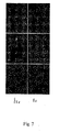
- Unlabeled peptide was used in a competition assay to derive the relative affinity of the BPI for the APC and the ability of the BPI to crosslink the MHC and ICAM molecules on the APC was examined by cocapping ICAM with an anti-MHC mAb.
- cells were incubated with the BPI.
- an anti-MHC mAb was added. After 30 minutes at 37°C, the cells were transferred to ice and stained with PE-labeled anti-ICAM (Fig. 7).
- T-cell clones were generated for determination of TCR epitopes for later use in BPI. These experiments utilized CD4+ or CD8+ T-cell clones from humans or mice immunized against predicted TCR epitopes using previously described methods ( Murray et al., 24 Eur. J. Immunol. 2337-2344 (1994 ); Murray; 19 Immunology Today 157-163 ; and Schountz et al., 157 The Journal of Immunology 3893-3901 (1996 )). These clones were maintained by biweekly restimulation with irradiated histocompatible lymphocytes, the peptide, and recombinant IL2.
- ELISPOT assay To determine if a given TCR-epitope is effective for the activation to cytokine synthesis, an ELISPOT assay was used. Of course, other cytokine assays could also be used.
- BPI that have been substituted at predicted TCR-contact positions will be used to determine which of these BPI variants are most effective in the inhibition of proliferation and cytokine release from individual clones as analyzed above. Predicted positions will be scanned with different amino acids or mimetics to alter the interaction with the TCR in the structures generated by molecular modeling.
- MDEM Molecular Dynamics/Energy Minimizations
- the second stage in the BPI process is selection of peptide mimics of established second signal receptor molecules involved in the functional differentiation of T-cells.
- the crystallographic structures and available models ofthe second signal receptors bound to their physiological ligands will be used to predict the regions of the receptors that make contact with the ligand.
- This approach was used to design the EGAD-BPI which can be depicted as (EIAPVFVLLE)-(AcGAcGAc)-(ITDGEATDSG) GAD65(208-217) linker LFA-1(237-247)
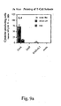
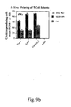
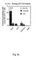
- BPI are tested for binding to isolated MHC and second signal ligands, and NMR, molecular modeling and crystallography are used to determine their exact 3D structures. Finally, it was determined whether a given BPI was biologically active in vivo. Mice were treated with the BPI and immune cells isolated and tested for cytokine production by ELISPOT (see Figs. 9 & 10). Of course other cytokine assays will be well known to those of ordinary skill in the art and can be used in place of ELISPOT.
- T-cells 15 million disease-linked lymphocytes (i.e., patient T-cells, or T-cell populations linked to the disease process) were injected with or without T-cells that received the BPI compound (in vivo or in vitro) and were expanded for 24 hours in recombinant IL-2.
- Some experiments will deplete specific subsets of the T-cells using mAb to CD154, CD25, CD62L, CD152, etc. and magnetic particles prior to adoptive transfer.
- T-cells (from mice or humans) treated with the individual moieties of the BPI will be used as negative controls along with CD4+ cells from mice treated with saline alone (Fig. 9).
- mice For blocking spontaneous diabetes, five groups of ten female NOD mice (12 weeks of age) were used and monitored for nondiabetic blood glucose levels with a standard glucometer (AccuChek-complete, Roche Diagnostic). Each mouse was labeled and individually monitored for blood glucose levels weekly for the course of the experiment.
- the five groups received either (a) intravenous (i.v.) injection of the BPI (100 ⁇ g in 100 ⁇ l endotoxin-free saline/injection) at 8 weeks of age, (b) same dose GAD65 (208-217) epitope alone, (c) same dose CD11a (237-247) peptide alone, or (d) saline alone.
- similar treatment groups involving the different Signal-1 and Signal-2 peptides and BPI will be used. Mice will be tested by challenging with the appropriate infectious agent or antigen depending upon the particular BPI in question.
- biotinylated mAb to various cell surface antigens will be incubated individually with the Cryostat sections (2 hours), followed by avidin-alkaline phosphatase (Vector laboratories).
- cell subsets will be phenotyped by standard flow cytometry methods as described in Murray et al., 24 Eur. J. Immunol. 2337-2344 (1994 ); Murray; 19 Immunology Today 157-163 ; and Schountz et al., 157 The Journal of Immunology 3893-3901 (1996 ).
- the students t-test or ANOVA will be used to estimate the statistical significance of differences observed between groups and individual mice.
- Sequence Identity refers to a relationship between two or more polypeptide sequences or two or more polynucleotide sequences, namely a reference sequence and a given sequence to be compared with the reference sequence. Sequence identity is determined by comparing the given sequence to the reference sequence after the sequences have been optimally aligned to produce the highest degree of sequence similarity, as determined by the match between strings of such sequences. Upon such alignment, sequence identity is ascertained on a position-by-position basis, e.g., the sequences are "identical” at a particular position if at that position, the nucleotides or amino acid residues are identical.
- Sequence identity can be readily calculated by known methods, including but not limited to, those described in Computational Molecular Biology, Lesk, A. N., ed., Oxford University Press, New York (1988 ), Biocomputing: Informatics and Genome Projects, Smith, D.W., ed., Academic Press, New York (1993 ); Computer Analysis of Sequence Data, Part I, Griffin, A.M., and Griffin, H.
- a polynucleotide having a nucleotide sequence having at least 95% identity relative to the reference nucleotide sequence up to 5% of the nucleotides in the reference sequence may be deleted or substituted with another nucleotide, or a number of nucleotides up to 5% of the total nucleotides in the reference sequence may be inserted into the reference sequence.
- These mutations of the reference sequence may occur at the 5' or 3' terminal positions of the reference nucleotide sequence or anywhere between those terminal positions, interspersed either individually among nucleotides in the reference sequence or in one or more contiguous groups within the reference sequence.
- a polypeptide having a given amino acid sequence having at least, for example, 95% sequence identity to a reference amino acid sequence it is intended that the given amino acid sequence of the polypeptide is identical to the reference sequence except that the given polypeptide sequence may include up to 5 amino acid alterations per each 100 amino acids of the reference amino acid sequence.
- up to 5% of the amino acid residues in the reference sequence may be deleted or substituted with another amino acid, or a number of amino acids up to 5% of the total number of amino acid residues in the reference sequence may be inserted into the reference sequence.
- alterations ofthe reference sequence may occur at the amino or the carboxy terminal positions of the reference amino acid sequence or anywhere between those terminal positions, interspersed either individually among residues in the reference sequence or in the one or more contiguous groups within the reference sequence.
- residue positions which are not identical differ by conservative amino acid substitutions. However, conservative substitutions are not included as a match when determining sequence identity.
- sequence homology also refers to a method of determining the relatedness of two sequences. To determine sequence homology, two or more sequences are optimally aligned as described above, and gaps are introduced if necessary. However, in contrast to “sequence identity”, conservative amino acid substitutions are counted as a match when determining sequence homology.
- 95% of the amino acid residues or nucleotides in the reference sequence must match or comprise a conservative substitution with another amino acid or nucleotide, or a number of amino acids or nucleotides up to 5% of the total amino acid residues or nucleotides, not including conservative substitutions, in the reference sequence may be inserted into the reference sequence.
- Isolated means altered “by the hand of man” from its natural state., i.e., if it occurs in nature, it has been changed or removed from its original environment, or both.
- a polynucleotide or polypeptide naturally present in a living organism is not “isolated,” but the same polynucleotide or polypeptide separated from the coexisting materials of its natural state is “isolated", as the term is employed herein.
- Sequences including or having a sequence which has at least about 10% sequence identity with any one of SEQ ID Nos. 1-46 and which exhibit similar binding properties to APC or linking properties between two peptide sequences are within the scope of the present invention.
- sequences will have at least about 30% sequence identity with any one of SEQ ID Nos. 1-46, still more preferably at least about 50% sequence identity, even more preferably, at least about 70% sequence identity, and most preferably at least about 95% sequence identity.
- sequences including or having a sequence which has at least about 10% sequence homology with any one of SEQ ID Nos. 1-46 and which exhibit similar binding properties to APC or linking properties between the two adjacent peptide sequences are embraced in the present invention.
- a “linker” is defined as any amino acid including naturally occurring or chemically synthesized amino acids.
- a “linker” is a flexible, non-substrate sequence of amino acid residues resistant to proteolytic degradation which can be used to conjugate and/or couple a Signal-1 moiety to a Signal-2 moiety.
- a “Signal-1 moiety” is defined as a peptide epitope, i.e., the peptide portion of an antigen and/or mimetics of these antigenic peptides to which important TCRs bind.
- a “Signal-2 moiety” or a “Signal-2 receptor moiety” is defined as a peptide portion of a second signal receptor known to bind to and/or affect binding of the receptor to its complimentary ligand on the APC. This can include peptide mimics and mimetics of the receptor/ligand structure of interest.
- a “Signal-2 ligand” is the complementary protein of the Signal-2 receptor moiety on the APC to which the receptor portion and/or the Signal-2 receptor moiety has significant affinity and binds.
- BPI were generated using automated peptide synthesis by a robotic multiple peptide synthesizer employing Fmoc amino acid chemistry by standard methods.
- Wang resin p- benzyloxybenzyl alcohol polystyrene
- Peptides were characterized byreversed-phase HPLC and electrospray mass-spectrometry. This synthesis, referred to as Merrifield peptide synthesis, utilizes traditional organic chemical reactions carried out on a solid material so that the peptide chain is lengthened while attached to the support structure. The peptides will be cleaved from the resin using TFA, and purified by reverse-phase HPLC and analyzed by mass spectroscopy.
- nucleotide sequences encoding the peptides of the invention may be synthesized, and/or cloned, and expressed according to techniques well known to those of ordinary skill in the art. See, for example, Sambrook, et al., 1989, Molecular Cloning, A is Laboratory Manual, Vols. 1-3, Cold Spring Harbor Press, New York .
- the peptides of the invention may be synthesized such that one or more of the bonds which link the amino acid residues of the peptides are non-peptide bonds.
- These alternative non-peptide bonds may be formed by utilizing reactions well known to those in the art, and may include, but are not limited to amino, ester, hydrazide, semicarbazide, and azo bonds, to name but a few.
- peptides comprising the sequences described above may be synthesized with additional chemical groups present at their amino and/or carboxy termini, such that, for example, the stability, bioavailability, and/or inhibitory activity of the peptides is enhanced.
- hydrophobic groups such as carbobenzoxyl, dansyl, or t-butyloxycarbonyl groups, may be added to the peptides' amino termini.
- an acetyl group or a 9-fluorenylmethoxy-carbonyl group may be placed at the peptides' amino termini.
- the hydrophobic group, t-butyloxycarbonyl, or an amido group may be added to the peptides' carboxy termini.
- peptide mimetic compounds may be synthesized in place of the peptide moieties and linked by the same chemistry.
- the design of peptidomimetics is an established technique and known correlates of key amino acids of the peptide can be synthesized by previously published methods.
- peptidomimetics may be developed which have the same modulation properties as the preferred peptides detailed herein. As these peptidomimetics require no more than routine skill in the art to produce, such peptidomimetics are embraced within the present application.
- the side chains of these peptidomimetics will be very similar in structure to the side chains of the preferred peptides herein, however, their peptide backbone may be very different or even entirely dissimilar.
- the peptides of the present invention could be cyclized by any well known method.
- One such method adds Penicillamine (Pen) and cysteine (Cys) residues to the N- and C-termini to form cyclic peptides via a disulfide bond between the Pen and Cys residues.
- Pen Penicillamine
- Cys cysteine residues
- the formation of this cyclic peptide restricts the peptide conformation to produce a conformational stability, thereby providing better selectivity for cell surface receptors than its linear counterpart.
- linker The portion of the BPI which spans between the Signal-1 moiety and the Signal-2 moiety is referred to as a linker.
- the linker is not essential in forming a BPI.
- the linker can be any naturally occurring or chemically synthesized amino acid.
- the linker is a non-substrate amino acid residue chain which helps to prevent protease attack.
- a particularly preferred linker is a repeating chain of the non-natural amino acid, aminocaproic acid (Ac), and the amino acid glycine (G) (e.g. Ac-G-Ac-G-Ac).
- beta-alanine residues ( ⁇ Ala) could be substituted for one or more of the Ac residues.
- amino-dodecanoic acid residues (Adod) could be substituted for one or more of the Ac residues.
- peptide mimetics of these linker amino acids may also be synthesized and inserted into the BPI structure.
- This example describes the methods used to generate the BPI.
- Synthesis of peptides was via Fmoc on chlorotrity resins. Protected amino acids were double coupled at 8-fold excess for one hour. Resins were dimethylforamide (DMF) and methanol (MeOH) washed and cleaved in Reagent R: trifluorolacetic acid (TFA), ethylene diamine tetraacetic acid (EDTA), Thioanisole, Anisole. The TFA mixture containing the peptide in solution is precipitated in ether and washed extensively. Preparative HPLC ofpeptides was accomplished by a gradient of 0-80% acetonitrile in 0.1 % TFA. Lyophilization of the various fractions and verification by mass spectroscopy yielded the synthetic peptides as a TFA salt. Modeling, crystallography and binding studies are as described above.
- the peptides produced in this example are provided in Table 1 and are also listed as SEQ ID Nos. 1- 46. These peptides include the Signal-1 moiety, the Signal-2 moiety and the non-substrate linker between the two moieties.
- any Signal-1 moiety could be linked with any Signal-2 moiety via any linker using the peptide synthesis described above.
- the BPI are generated as one continuous peptide chain comprising a Signal-1 peptide sequence followed by a linker sequence followed by a Signal-2 peptide sequence. Additionally, some representative BPI were generated for later use in the experiments. These BPI are included herein in Table 4.
- the entire BPI can be synthesized using the above-described methods.
- Signal-1 moieties which could be used in connection with the present invention. Each of these moieties may be associated with a distinct immunological response or disease state.
- an appropriate linker could be selected and the other portion of the BPI, i.e., the second signal moiety, can be chosen.
- BPI can be designed using the peptide sequences themselves, peptidomimetics, or combinations of the two. Construction of appropriate peptidomimetics is detailed by Falcioni, et al, 17 Nature Biotechnology, 562-567 (1999 ), the content and teachings of which are hereby incorporated by reference herein.
- Fig. 8 illustrates the structure of the GAD65 (208-217), TCR epitope linked to the CD11a (237-247) second signal moiety produced by the present methods. It is shown bound to the groove of I-A g7 and the D1 domain of ICAM-1.
- ICAM-1 ICAM-1 domain structure
- FIG. 8 For modeling the I-A g7 :GAD65 peptide structure, docking studies were performed on a Silicon Graphic Ocatane work station using InSight II software (MSI/Biosym).
- the LFA-1 peptide:ICAM-1 domain structure is based on the docking model of Edwards, C.P. et al. J Biol. Chem. 273:28937 (1998 ), the teachings and disclosure of which is incorporated by reference herein.
- the alpha carbon ribbon of I-A g7 is shown in pink; D1 of ICAM-1 is in light-blue; the BPI is shown by atom, carbon in green, oxygen in red, and nitrogen in blue.
- This structure can be denominated as GAD65 (208-217) - [Ac-G-Ac-G-Ac] - CD11a(237-247).
- the length of the linker may be modified as needed or as indicated by any experimental data obtained in order to span between the Signal-1 and Signal-2 moieties at an optimum length.
- the linker used has the sequence -[Ac-G-Ac-G-Ac]-.
- Ac aminocaproic acid
- beta-2 alanine can be used as a substitute for aminocaproic acid.
- This example uses biotinylated BPI to test for competitive inhibition of BPI binding by unlabeled peptides or monoclonal antibodies to MHC-II and ICAM-1 on live APC, and to verify antigenic peptide binding to live APC. Additionally, it was shown that monoclonal antibodies to MHC-II or ICAM-1 effectively block binding of the diabetes BPI (GAD65 (208-217) - [Ac-G-Ac-G-Ac] - CD11a (237-247)) (hereinafter referred to as EGAD-BPI) to NOD spleenocytes.
- diabetes BPI GID65 (208-217) - [Ac-G-Ac-G-Ac] - CD11a (237-247)
- the synthesized EGAD BPI was biotinylated with NHS-Biotin as described in Murray et al., 24 Eur. J. Immunol. 2337-2344 (1994 ). Spleen cell density-gradient fractions from normal (unimmunized) NOD, BALB/c and other MHC congenic strains were incubated in round bottom 96-well plates with increasing concentrations of individual biotinylated peptides at 37°C, 5% CO 2 for 16 hours.
- Negative selection methods with monoclonal antibodies conjugated to magnetic particles were used to enrich the spleen cell fractions for B cells, macrophages, or dendritic cells as well as to examine differences in BPI binding to these different populations. These methods are detailed in Schountz et al., 157 The Journal of Immunology 3893-3901 (1996 ), the teachings and content of which were incorporated by reference above.
- initial EGAD-BPI were screened for selective binding to NOD (I-A g7 ) APC and assayed for simultaneous binding using monoclonal antibodies against either MHC-II or ICAM-1 by flow cytometry methods using live APC ( Murray et al., 24 Eur. J. Immunol. 2337-2344 (1994 ); Murray; 19 Immunology Today 157-163 ; and Schountz et al., 157 The Journal of Immunology 3893-3901 (1996 )).
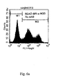
- biotinylated-BPI, -CD11a(237-247) or -GAD65(208-217) peptide were incubated overnight with spleenocytes from each inbred strain. Bound peptide was detected with amplification of avidin-FITC fluorescence by the use of a biotinylated anti-avidin reagent, followed by a second round of avidin-FITC binding. Biotinylated peptide was incubated with 10 6 viable cells at a peptide concentrations of 50 ⁇ M. Bound peptide was detected with avidin-FITC/biotinylated anti-avidin-FITC.
- M2 median channel fluorescence
- NOD spleen cells bind the diabetes BPI (EGAD-BPI) at a higher density than spleenocytes identically purified from BALB/c, A.SW, or A.BY.
- B cells are the major antigenic peptide binding cells in these spleen cell preparations isolated by lymphocyte separation media (LSM) density gradient centrifugation ( Murray et al., 24 Eur. J. Immunol. 2337-2344 (1994 ); Murray; 19 Immunology Today 157-163 ; and Schountz et al., 157 The Journal of Immunology 3893-3901 (1996 )).
- LSM lymphocyte separation media
- Fig. 5b illustrates direct binding of biotinylated LFA-1 (CD11a 237-247) peptide to the same spleenocyte preparations as those shown in Fig. 5a. Note that this Signal-2 moiety bound similarly to all strain spleenocytes.
- Fig. 5b illustrates direct binding of biotinylated LFA-1 (CD11a 237-247) peptide to the same spleenocyte preparations as those shown in Fig. 5a. Note that this Signal-2 moiety bound similarly to all strain spleenocytes.
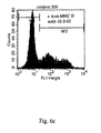
- FIG. 5c illustrates direct binding of biotinylated GAD65 (208-217) peptide to the same spleenocyte preparations as those depicted in Figs. 5a and 5b.
- BPI could be engineered to fit particular MHC peptide binding motifs as discussed by Corper et al. in A Structural Framework for Deciphering the Link Between I-Ag7 and Autoimmune Diabetes, 288 Science 505-511 (2000 ), and by Dessen et al.
- This example utilizes co-capping experiments to demonstrate simultaneous binding of the BPI to MHC-II and ICAM-1 molecules.
- Fig. 7 illustrate the results from the T-depleted spleenocytes from mice treated 16 hours previously with EGAD-BPI (i.v.), wherein ICAM-1 was co-capped with MHC-II in the presence of bio-10-3.62/streptavidin.
- the bottom panels of Fig. 7 illustrate the results from the mice treated with saline only wherein co-capping is not exhibited. This is evidenced by having ICAM-1 remain dispersed on the B-cell membranes.
- BPI have the capacity to bind simultaneously to MHC-II and ICAM-1 structures on the surface of live APC and therefore may provide signal alterations involving pathways necessary for T H 1/T H 2 differentiation.
- T-cells from mice injected with EGAD-BPI were examined for cytokine analysis.
- This example used an ELISPOT to determine T H 1/T H 2 frequency as altered by BPI injection.
- mice Groups of 3-5 NOD mice were immunized subcutaneously (s.c.) with the GAD 65 peptide in CFA (40 nanomoles/mouse) at the tail base.
- Different groups received either the EGAD-BPI, its single TCR epitope (Signal-1 moiety), or its CD11a peptide (Signal-2 moiety) i.v. (all 40 nanomoles/mouse).
- the EGAD-BPI its single TCR epitope
- CD11a peptide Signal-2 moiety
- lymph nodes draining the site of the s.c. injection were made into single cell suspensions for culture. Identical primary cultures were incubated for 96 hours; then, viable T-cells were recovered by density gradient centrifugation.
- mice were combined in nitrocellulose-bottomed 96-well plates (Millititer-HA, Millipore, Bedford, MA), previously coated (50 ⁇ l/well) with mAb to either mouse IFN ⁇ (clone R4-6A2), or mouse IL-4 (clone BVD4-1D11) at a concentration of 10 ⁇ g/ml in PBS.
- Groups of triplicate cultures were incubated with either Concanavalin-A (2 ⁇ g/ml), or the Signal-1 peptide moiety plus 20 U/ml recombinant IL2 (R & D Systems). After 72-96 hours of culture at 37°C and 5% CO 2 , plates were washed three times with PBS-0.05% Tween-20.
- biotinylated anti-IFN ⁇ (clone XMG1.2) or biotinylated anti-IL-4 (clone BVD6-24G2) at a concentration of 1 ⁇ g/ml and incubated for 1 hour at room temperature.
- Positive control wells receive known T H 1 or T H 2 clones in place of normal T-cells.
- MAb pairs to IL12, IL10, and IL2 are also available and will be used to test for these cytokines in the same assay. All mAb and recombinant controls are purchased from PharMingen (San Diego, CA).
- CD4+ T-cell clones from NOD mice immunized with the GAD65(208-217) peptide by our previously described methods can be used in the same assay.
- the clones were generated using the methods described in Murray et al., 24 Eur. J. Immunol. 2337-2344 (1994 ), the teachings of which are hereby incorporated by reference. These clones will be maintained by biweekly restimulation with irradiated NOD lymphocytes, the GAD peptide, and recombinant IL-2.
- BPI that have been substituted at predicted TCR-contact positions will be used to determine which of these BPI variants are most effective in the inhibition of proliferation and cytokine release from individual clones as analyzed above.
- Predicted positions that will be scanned with all amino acids except cysteine are amino acids 208, 213, and 216. These residues point toward the TCR in the recently solved crystal structure of the GAD65 peptide bound to I-A 87 ( Corper et al., 288 Science 505-511 (2000 ) and see Fig. 8).
- IL-4 production increases by approximately 10-fold when T-cells are from the BPI treated animals stimulated in vitro with mitogen.
- IFN- ⁇ production also increased, although to a lesser extent (see Figs. 9b and 9d).
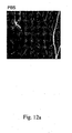
- This example tested the capacity of the BPI to inhibit lymphocytic infiltration of pancreatic islets in NOD mice. Lymphocytic infiltration is a hallmark of insulitis and the development of type-1 diabetes.
- Fig. 11 represents the cumulative data of this analysis wherein the severity of islet infiltration is scored and plotted as the percentage of islets examined. Over 100 islets from each group in greater than 5 tissue sections were analyzed by three independent observers. As shown in Fig. 11, there was a clear inhibitory effect of the EGAD-BPI treatment on mononuclear cell infiltration (insulitis). Over 95% of the islets from the BPI treated animals were intact and did not show infiltration (i.e., grade-0 islets). All of the other groups showed some signs ofinsulitis even at this early stage ofthe disease.
- This example tested BPI blocking of diabetes development in the well described intact immune system of immunologically reconstituted NOD.Scid mice to study diabetes progression.
- inducer cells (15 x 10 6 per mouse) were injected (i.v.) into 6 week NOD.Scid females (Jackson Labs) together with 3 x 10 6 CD4+ T-cells from mice either treated with the EGAD-BPI or treated identically except with PBS in place of the EGAD-BPI.
- In vitro clonal expansion was with either recombinant IL-2 (R&D systems), or ConA, as described for the ELISPOT experiments. All manipulations of animals and cells were performed in laminar flow hoods, and animals were maintained continuously behind laminar flow barriers on autoclaved food and water in microisolator cages.
- BPI for other autoimmune diseases
- BPI containing immunodominant TCR epitopes for collagen-induced arthritis (CIA) and myelin basic protein-induced experimental allergic encephalomyelitis (EAE) will be discussed.
- CD40L peptide mimic is predicted to favor T H 2 immunity, as blocking the CD40 signal would be expected to decrease IL12 production ( Ruedl, et al., The Antigen Dose Determines T Helper Subset Development by Regulation of CD40 Ligand, 30 Eur. J. Immunol. 2056-2064 (2000 )).
- peripheral blood mononuclear cells PBMCs
- CD8+ cells removed by the negative selection protocol with magnetic particles.
- PBMCs peripheral blood mononuclear cells
- These cells are grown in tissue culture medium at 4 x 10 6 cells in 1 ml for 6 days in the presence of 20 ⁇ M of the p24 peptide, and blast cells isolated by density gradient centrifugation. Secondary cultures of these cells contained recombinant human IL-2 (20 U/ml) and are continued for a maximum of 10-14 days.
- Clones were prepared by limiting dilution cloning in fresh plates containing irradiated APC, peptide and IL-2 (50 U/ml) ( Murray et al., 24 Eur. J. Immunol. 2337-2344 (1994 ); Murray; 19 Immunology Today 157-163 ; and Schountz et al., 157 The Journal of Immunology 3893-3901 (1996 )).
- BPI will be tested for their ability to inhibit the proliferation and cytokine release of these established lines of human T-cells by our detailed methods described in Schountz et al., Unique T Cell Antagonist Properties of the Exact Self-Correlate of a Peptide Antigen Revealed by Self-Substitution of Non-Self-Positions in the Peptide Sequence, 168 Cellular Immunology 193-200 (1996 ).
- peptides changed by amino acid substitutions at key TCR-contact residues alter the proliferation and cytokine release of cloned T-cells.
- the Scid-human mouse model in which human tissues are adoptively transferred to the Scid mouse could be used to examine protective immunity to HIV ( Jenkins, et al., Blood (1998) 8:2672 , hepatitis-C virus (HCV) ( Bronowicki, J., et al. Hepatology (1998)28:211 ), human pappiloma virus (HPV) ( Tewari, et al. Gynecol. Oncol. (2000)77:137 , and respiratory syncitial virus (RSV) ( Nadal, et al. Clin. Exp. Immunol.85:358 ) infections.
- HCV hepatitis-C virus
- HPV human pappiloma virus
- RSV respiratory syncitial virus
- CD95 also called Fas
- FasL CD95-ligand
- BPI with the peptide mimic of the FasL which is from the Y218 loop of FasL, predicted to interact with Fas at the T-cell:APC interface, will be used to favor T H 1 activation and CTL responses against chronic T H 2-dominated diseases.
- mice offer the advantage of having the human MHC molecules and thus, BPI binding will be very similar in this model as in the human allergic condition. If a given BPI blocks T H 2 activation in these mice against the native allergen, then adoptive transfer studies with human Scid mice (similar to those used in Example 8) can be used as a precursor to human clinical trials.
- Blocking the CTLA4/CD28 pathway in preference for LFA-1 signaling will favor T H 1 differentiation against these allergens in the presence of the BPI.
- BPI with the FasL moiety should favor type-1 immunity. It has been a long-held practice to "desensitize" atopic patients with allergens given by injection and desensitization is thought to operate via a shift toward T H 1 responses to the specific allergens ( Holt, et al., Nature (1999) 402:6760 suppl:B 12-17 ).
Landscapes
- Health & Medical Sciences (AREA)
- Chemical & Material Sciences (AREA)
- Life Sciences & Earth Sciences (AREA)
- Immunology (AREA)
- Organic Chemistry (AREA)
- General Health & Medical Sciences (AREA)
- Medicinal Chemistry (AREA)
- Genetics & Genomics (AREA)
- Molecular Biology (AREA)
- Biochemistry (AREA)
- Proteomics, Peptides & Aminoacids (AREA)
- Biophysics (AREA)
- Zoology (AREA)
- Engineering & Computer Science (AREA)
- Bioinformatics & Cheminformatics (AREA)
- Wood Science & Technology (AREA)
- Microbiology (AREA)
- Pharmacology & Pharmacy (AREA)
- Veterinary Medicine (AREA)
- Public Health (AREA)
- Animal Behavior & Ethology (AREA)
- General Engineering & Computer Science (AREA)
- Toxicology (AREA)
- Epidemiology (AREA)
- Mycology (AREA)
- Biomedical Technology (AREA)
- Biotechnology (AREA)
- Cell Biology (AREA)
- Transplantation (AREA)
- Gastroenterology & Hepatology (AREA)
- Nuclear Medicine, Radiotherapy & Molecular Imaging (AREA)
- Chemical Kinetics & Catalysis (AREA)
- General Chemical & Material Sciences (AREA)
- Peptides Or Proteins (AREA)
- Medicines That Contain Protein Lipid Enzymes And Other Medicines (AREA)
Applications Claiming Priority (2)
| Application Number | Priority Date | Filing Date | Title |
|---|---|---|---|
| US09/739,466 US7786257B2 (en) | 2000-12-18 | 2000-12-18 | Signal-1/signal-2 bifunctional peptide inhibitors |
| EP01991168A EP1404362B1 (de) | 2000-12-18 | 2001-12-17 | Signal-1/signal-2-bifunktionelle peptidinhibitoren |
Related Parent Applications (1)
| Application Number | Title | Priority Date | Filing Date |
|---|---|---|---|
| EP01991168A Division EP1404362B1 (de) | 2000-12-18 | 2001-12-17 | Signal-1/signal-2-bifunktionelle peptidinhibitoren |
Publications (2)
| Publication Number | Publication Date |
|---|---|
| EP1932539A2 true EP1932539A2 (de) | 2008-06-18 |
| EP1932539A3 EP1932539A3 (de) | 2008-10-29 |
Family
ID=24972431
Family Applications (2)
| Application Number | Title | Priority Date | Filing Date |
|---|---|---|---|
| EP01991168A Expired - Lifetime EP1404362B1 (de) | 2000-12-18 | 2001-12-17 | Signal-1/signal-2-bifunktionelle peptidinhibitoren |
| EP07014573A Withdrawn EP1932539A3 (de) | 2000-12-18 | 2001-12-17 | Bifunktionelle Signal-1/Signal-2-Peptidhemmer |
Family Applications Before (1)
| Application Number | Title | Priority Date | Filing Date |
|---|---|---|---|
| EP01991168A Expired - Lifetime EP1404362B1 (de) | 2000-12-18 | 2001-12-17 | Signal-1/signal-2-bifunktionelle peptidinhibitoren |
Country Status (8)
| Country | Link |
|---|---|
| US (1) | US7786257B2 (de) |
| EP (2) | EP1404362B1 (de) |
| JP (1) | JP2004536783A (de) |
| AT (1) | ATE400293T1 (de) |
| AU (1) | AU2002230911A1 (de) |
| CA (1) | CA2432767A1 (de) |
| DE (1) | DE60134791D1 (de) |
| WO (1) | WO2002050250A2 (de) |
Families Citing this family (11)
| Publication number | Priority date | Publication date | Assignee | Title |
|---|---|---|---|---|
| US8188218B2 (en) | 2006-10-27 | 2012-05-29 | University Of Kansas | Bi-functional peptides for multiple sclerosis treatment and diagnosis |
| US8735154B2 (en) | 2006-10-30 | 2014-05-27 | The University Of Kansas | Templated islet cells and small islet cell clusters for diabetes treatment |
| EP2083715B1 (de) * | 2006-11-30 | 2013-03-13 | Cook Medical Technologies LLC | Entfernbarer handgriff für ein medizinprodukt |
| GB0710529D0 (en) | 2007-06-01 | 2007-07-11 | Circassia Ltd | Vaccine |
| NZ583362A (en) | 2007-08-15 | 2012-06-29 | Circassia Ltd | Peptide with reduced dimer formation |
| KR101557173B1 (ko) * | 2007-10-25 | 2015-10-05 | 더 스크립스 리서치 인스티튜트 | 박테리아에서의 쿼럼 센싱의 항체 매개성 파괴 |
| DE102009040716B4 (de) * | 2009-09-10 | 2011-07-14 | Miltenyi Biotec GmbH, 51429 | Verwendung von CD154 zur Identifizierung und Abtrennung von nicht-regulatorischen T-Zellen aus einem Gemisch mit regulatorischen T-Zellen |
| EP2585108A4 (de) | 2010-06-24 | 2016-04-13 | Univ Kansas | Konjugate mit einem n-oxim-bindung und zugehörige verfahren |
| EP2736536A4 (de) | 2011-07-27 | 2015-03-11 | Univ Kansas | Template-inselzellen und kleine inselzellcluster zur behandlung von diabetes |
| CN105708834A (zh) * | 2011-10-18 | 2016-06-29 | 株式会社爱茉莉太平洋 | 包含丁香脂素的sirt1活化剂 |
| US11919939B2 (en) * | 2017-05-23 | 2024-03-05 | Université De Genève | Beta-2-glycoprotein 1 derived peptide and use thereof for treating antiphospholipid syndrome |
Family Cites Families (5)
| Publication number | Priority date | Publication date | Assignee | Title |
|---|---|---|---|---|
| US5844095A (en) * | 1991-06-27 | 1998-12-01 | Bristol-Myers Squibb Company | CTLA4 Ig fusion proteins |
| DE59310308D1 (de) * | 1992-06-11 | 2002-11-21 | Merck Patent Gmbh | Verfahren und Mittel zum Nachweis von Listerien |
| US5540926A (en) | 1992-09-04 | 1996-07-30 | Bristol-Myers Squibb Company | Soluble and its use in B cell stimulation |
| DE69840326D1 (de) | 1997-03-27 | 2009-01-22 | Csl Ltd | Steigerung der immunantwort unter verwendung von zielgerichteten molekülen |
| WO2001026608A2 (en) * | 1999-10-14 | 2001-04-19 | Ledbetter Jeffrey A | Dna vaccines encoding antigen linked to a domain that binds cd40 |
-
2000
- 2000-12-18 US US09/739,466 patent/US7786257B2/en not_active Expired - Fee Related
-
2001
- 2001-12-17 EP EP01991168A patent/EP1404362B1/de not_active Expired - Lifetime
- 2001-12-17 WO PCT/US2001/048632 patent/WO2002050250A2/en not_active Ceased
- 2001-12-17 AU AU2002230911A patent/AU2002230911A1/en not_active Abandoned
- 2001-12-17 DE DE60134791T patent/DE60134791D1/de not_active Expired - Fee Related
- 2001-12-17 EP EP07014573A patent/EP1932539A3/de not_active Withdrawn
- 2001-12-17 JP JP2002552127A patent/JP2004536783A/ja active Pending
- 2001-12-17 CA CA002432767A patent/CA2432767A1/en not_active Abandoned
- 2001-12-17 AT AT01991168T patent/ATE400293T1/de not_active IP Right Cessation
Also Published As
| Publication number | Publication date |
|---|---|
| AU2002230911A1 (en) | 2002-07-01 |
| US20050107585A1 (en) | 2005-05-19 |
| EP1404362A4 (de) | 2005-06-01 |
| EP1932539A3 (de) | 2008-10-29 |
| WO2002050250A3 (en) | 2004-01-08 |
| EP1404362A2 (de) | 2004-04-07 |
| ATE400293T1 (de) | 2008-07-15 |
| CA2432767A1 (en) | 2002-06-27 |
| JP2004536783A (ja) | 2004-12-09 |
| US7786257B2 (en) | 2010-08-31 |
| WO2002050250A2 (en) | 2002-06-27 |
| DE60134791D1 (de) | 2008-08-21 |
| EP1404362B1 (de) | 2008-07-09 |
Similar Documents
| Publication | Publication Date | Title |
|---|---|---|
| US11981743B2 (en) | Monoclonal antibody and a method thereof | |
| US6984625B2 (en) | Methods and compositions for modulating autoimmunity | |
| Rodgers et al. | MHC class Ib molecules bridge innate and acquired immunity | |
| US7553811B2 (en) | Methods and compositions for modulating immunity | |
| US8349320B2 (en) | Compositions and methods for modulating responses mediated or associated with BTLA activity | |
| RU2598719C2 (ru) | Средства для лечения заболевания | |
| CA2250166A1 (en) | Soluble divalent and multivalent heterodimeric analogs of proteins | |
| EP1404362B1 (de) | Signal-1/signal-2-bifunktionelle peptidinhibitoren | |
| JPH04506061A (ja) | Cd8に基づく製剤 | |
| US11701387B2 (en) | Chimeric antigen receptor specific for BDCA2 antigen | |
| JPH10501791A (ja) | クラスimhcペプチドによる細胞傷害性t細胞リンパ球(「ctl」)活性の調節 | |
| JP2021503276A (ja) | 制御性t細胞エピトープ | |
| WO2001026679A2 (en) | T-cells and molecules involved in immune regulation | |
| Miloro | Deciphering the role of the death receptor Fas/CD95 in T cell co-stimulation | |
| Wall | Structural and biophysical characterisation of T-cell receptor cross-reactivity in health and disease | |
| Pino-Lagos et al. | Searching for Immune Tolerance Manipulating New Molecules and Exploiting New Concepts on Lymphocyte Biology | |
| Jangalwe | Regulation of Alloreactive CD8 T Cell Responses by Costimulation and Inflammation | |
| KR20220160640A (ko) | 조절 t-세포 에피토프 | |
| Chen et al. | Specific tolerance induction of allo-Kb-skin grafts by FK506 in the CD8-depleted H-2k recipients required low amounts of Kb-antigen | |
| JP2023538002A5 (de) | ||
| Chlewicki | Structure, function and engineering of peptide-MHC binding receptors | |
| Clement | The role of the CD8 co-receptor in CD8+ T-cell activation | |
| Zajac | Antiviral cell-mediated immune responses in beta-2-microglobulin deficient mice | |
| Pino-Lagos et al. | Searching for Immune Tolerance Manipulating New Molecules and Exploiting New Concepts on Lymphocyte Biology. Lausanne: Frontiers Media. doi: 10.3389 | |
| Barbaro et al. | A New Strategy of Tumor Vaccination Based on Mammary Adenocarcinoma Cells Transduced with the MHC Class II Transactivator CIITA. L. Mortara1, P. Castellani2, R. Meazza3, G. Tosi1, A. De |
Legal Events
| Date | Code | Title | Description |
|---|---|---|---|
| PUAI | Public reference made under article 153(3) epc to a published international application that has entered the european phase |
Free format text: ORIGINAL CODE: 0009012 |
|
| AC | Divisional application: reference to earlier application |
Ref document number: 1404362 Country of ref document: EP Kind code of ref document: P |
|
| AK | Designated contracting states |
Kind code of ref document: A2 Designated state(s): AT BE CH CY DE DK ES FI FR GB GR IE IT LI LU MC NL PT SE TR |
|
| PUAL | Search report despatched |
Free format text: ORIGINAL CODE: 0009013 |
|
| AK | Designated contracting states |
Kind code of ref document: A3 Designated state(s): AT BE CH CY DE DK ES FI FR GB GR IE IT LI LU MC NL PT SE TR |
|
| 17P | Request for examination filed |
Effective date: 20090424 |
|
| AKX | Designation fees paid |
Designated state(s): AT BE CH CY DE DK ES FI FR GB GR IE IT LI LU MC NL PT SE TR |
|
| 17Q | First examination report despatched |
Effective date: 20090703 |
|
| R17C | First examination report despatched (corrected) |
Effective date: 20091218 |
|
| STAA | Information on the status of an ep patent application or granted ep patent |
Free format text: STATUS: THE APPLICATION IS DEEMED TO BE WITHDRAWN |
|
| 18D | Application deemed to be withdrawn |
Effective date: 20100429 |