EP1942116A1 - Anticorps monoclonaux destinés à la détermination immunologique sélective de formes de laminine hautement moléculaires intactes dans des liquides corporels - Google Patents
Anticorps monoclonaux destinés à la détermination immunologique sélective de formes de laminine hautement moléculaires intactes dans des liquides corporels Download PDFInfo
- Publication number
- EP1942116A1 EP1942116A1 EP08075098A EP08075098A EP1942116A1 EP 1942116 A1 EP1942116 A1 EP 1942116A1 EP 08075098 A EP08075098 A EP 08075098A EP 08075098 A EP08075098 A EP 08075098A EP 1942116 A1 EP1942116 A1 EP 1942116A1
- Authority
- EP
- European Patent Office
- Prior art keywords
- laminin
- antibody
- cell line
- monoclonal antibody
- dsm
- Prior art date
- Legal status (The legal status is an assumption and is not a legal conclusion. Google has not performed a legal analysis and makes no representation as to the accuracy of the status listed.)
- Granted
Links
Images




Classifications
-
- C—CHEMISTRY; METALLURGY
- C07—ORGANIC CHEMISTRY
- C07K—PEPTIDES
- C07K16/00—Immunoglobulins [IG], e.g. monoclonal or polyclonal antibodies
- C07K16/18—Immunoglobulins [IG], e.g. monoclonal or polyclonal antibodies against material from animals or humans
Definitions
- the present invention relates to monoclonal antibodies for the selective immunological determination of high molecular weight laminin forms in body fluids, a method for producing these antibodies and their use for the diagnosis of diseases.
- Laminin is a multidomain protein found in all basement membranes and complexed with various other typical components of the basement membrane, such as nidogen, heparan sulfate proteoglycan or collagen IV ( Timpl, R. (1989) Eur. J. Biochem. 180; 487 to 502 ). It is composed of three different disulfide-linked polypeptides. The structure of an asymmetric cross is formed ( FIGS. 1a and 1b ). Recently, many structural variants have been identified that result from the assembly of (currently known) 8 different subunits.
- laminin In order to obtain a laminin isoform, it is always necessary to assemble polypeptides from three different classes of molecules: an ⁇ chain (formerly A chain), a ⁇ chain (formerly B1 chain), and a ⁇ chain (formerly B2 chain) ( Burgeson, RE; et al. (1994) Matrix Biology, Vol. 14, 209-211 ).
- the laminins are assigned a variety of interesting biological functions, such as influencing cell growth, cell spreading and neurite outgrowth, and induction of differentiation processes ( Timpl, R. (1989) Eur. J. Biochem. 180; 487 to 502 ).
- This will be used in a diagnostic procedure based on such a laminin determination method, inaccuracies in the diagnostic statement, as they had to be taken in the previously known determination methods, eliminated.
- the object is further achieved by a hybridoma cell line which produces an antibody having the properties shown at No. 1 to 4, producible by fusion of cells from a myeloma cell line and lymphocytes from a vertebrate animal previously immunized with laminin P1 and subsequent selection the hybrid after that the antibody produced by the hybrid, in addition to a good binding property to the purified placental human laminin, also shows a good reaction with the high molecular weight form of human serum derived laminin.
- Such a hybridoma cell line is preferably characterized by the fact that the lymphocytes are taken from Balb / c strain mice immunized with laminin P1 and the myeloma cell line is the mouse myeloma cell line P3X63AG8.653.
- DSM ACC2181, DSM ACC2180 and DSM ACC2182 have been deposited.
- hybridoma cell lines produce monoclonal antibodies having the advantageous properties shown in No. 1 to 4.
- the hybridoma cell lines DSM ACC2181 and DSM ACC2180 each produce an antibody of the subclass IgG2a, the hybridoma cell line DSM ACC2182 an antibody of the subclass IG 1.
- This method is preferably characterized in that the hybridoma cell lines DSM ACC2181, DSM ACC2180 or DSM ACC2182 are used to carry out the method step d).
- the object is also achieved by a method for the immunological determination of native laminin using a adhesive or covalently fixed to a substrate layer antibody and a labeled second antibody which recognizes the antigen bound to the layered antibody, characterized in that the layered antibody is a monoclonal antibody according to the present invention.
- the stated object is achieved by a method for the immunological determination of native laminin using a layered antibody adhesively or covalently attached to a support material and a labeled second antibody which recognizes the antigen bound to the layered antibody, characterized in that the labeled secondary antibody is a monoclonal Antibody according to the present invention.
- the layered antibody may also be a monoclonal antibody according to the present invention, preferably it is the one produced by the hybridoma cell line DSM ACC2181 and having the binding constants shown in Table 3.
- the monoclonal antibodies of the present invention may find particular use in methods of diagnosing diseases associated with a change in the level of lamina in body fluids, wherein the bodily fluids are taken from the living body but not returned to it.
- animals preferably rodents, e.g. Mice, rats, rabbits, guinea pigs, with laminin P1, which was isolated by the method described in Example 1, are immunized in the presence of adjuvant.
- rodents e.g. Mice, rats, rabbits, guinea pigs, with laminin P1, which was isolated by the method described in Example 1, are immunized in the presence of adjuvant.
- mice are particularly preferably used, in particular those of the Balb / c strain. With repeated secondary injections, for example at intervals of 4 to 8 weeks, the immune response is enhanced. The success of the immunization is controlled by determining the concentration of specific antibodies with an ELISA known to those skilled in the art.
- a few days before fusion of the lymphocytes with a myeloma cell line the animals are treated with laminin P1 without adjuvant. Lymphocytes of the animals are obtained and fused with a myeloma cell line, which may also be derived from one of the above animal species, but preferably from the mouse, especially with the cell line P3X63AG8.653.
- lymphocytes are fused with myeloma cell lines of the same species.
- the fusion and further growth of the cell clones are carried out in a manner known to those skilled in the art, wherein the supernatant of the cell culture by means of immunological binding tests, the concentration of specific antibodies is determined.
- suitable clones are selected for use in immunological procedures using a screening sequence set out in Tables 4 and 5. Particular preference is given to working with cell lines which are produced by fusion of lymphocytes from mice of the Balb / c strain against laminin P1 with the mouse myeloma cell line P3X63AG8.653.
- the monoclonal antibodies of the invention belong to the group of immunoglobulins, preferably to the class of IgG, IgA, and IgM proteins. Antibodies of the subclass IgG2a and IgG1 can be used particularly advantageously.
- the antibody according to the invention is distinguished, in particular, by its affinity for intact laminin being approximately equal to its affinity for the immunization antigen laminin P1 produced by pepsin digestion (Tables 2 and 3). Indeed, the screening strategy outlined in Tables 4 and 5 illustrates that, in effect, the vast majority of clones recovered from the fusion produce monoclonal antibodies that recognize only the laminin P1 artificial fragment produced by the pepsin treatment.
- Human laminin P1 and various laminin preparations are purified from human placenta by the methods described in the Examples. From this organ at least two different laminin isoforms can be obtained ( Brown, CJ; Wiedemann, H .; Timpl, R. (1994) J. Cell Sci. 107; 329 to 338 ).
- the antibodies of the invention can be used in various immunological Methods such as immunoradiometric assays following labeling of the tracer antibody with chloramine T or Bolton-Hunter reagent, as well as other competitive and non-competitive binding assays such as fluorescence, enzyme, chemiluminescence or other immunoassays, including all forms of radioimmunoassay ( Harlow, E .; Lane, d. (1988) Antibodies: A Laboratory Manual, CSH; 2nd Edn .; New York; 319 to 359; 553 to 612 ).
- the monoclonal antibodies can therefore be used in immunological methods for the isolation and characterization as well as for the quantitative determination of laminin in tissues and body fluids.
- the procedure according to methods known to the person skilled in the art is to react a liquid sample containing laminin with one of the monoclonal antibodies according to the invention, either in solution or preferably on a solid support, and the amount of laminin over the antigen formed Antibody complex is determined.
- the so far in the immunological determination miterfrickten, or exclusively recognized degradation products of laminin in body fluids are not recognized in these immunoassays, but only intact laminin structures.
- the immunizing antigen laminin P1 is a 200 to 250 kDa fragment of the laminin that can be extracted and isolated after extensive pepsin digestion of placental tissue. This fragment contains the inner rod-like domains (domains III) of the short arms of the laminin chains ⁇ , ⁇ and ⁇ ( Risteli, L; Timpl, R. (1981) Biochem. J. 193, 749-755 ).
- the three polypeptide chains (of which multiple isoforms exist) are linked by disulfide bridges and are characterized by an identical three-dimensional structure.
- EGF-like repeats As in Figure 1b are represented in the individual chain sections of laminin subunits seven to fourteen linear juxtaposed structural motifs, so-called EGF-like repeats, which are all characterized by the same folding pattern.
- Each 50 to 60 amino acids of an EGF-like repeats adopt a clover-shaped structure through a specified typical sequence of disulfide linkages.
- On Sequence level is an identity of> 30% between the EGF-like repeats of the short laminin chains ( Engel, J. (1989) FEBS Lett. 251, No. 1.2; 1 to 7 ).
- the extracted protein is rebuffered with an Ultrasette (300 kDa) in 2 M urea, 0.05 M Tris / HCl, 0.02 M NaCl, 2 mM EDTA, pH 7.4 and applied to a Q-Sepharose FF 60/14 ,
- the bound protein can be eluted at 0.15 M NaCl dissolved in the loading buffer.
- the eluate is again concentrated with the Ultrasette (300kDa) and prepped over a Superose 6. grade 16/50 at a flow rate of 1 ml / min in PBS, 2 mM EDTA.
- the fractions containing laminin are combined (with RIA-gnost ® laminin P1 determines), with the Ultrasette concentrated and incubated at RT, with 5000 Units Benzonase (purity II)> 2h.
- the sample is then applied to a ConA-Sepharose 4B (2.6 x 10 cm) equilibrated with 0.15 M NaCl, 0.05 M Tris / HCl, pH 7.4.
- the laminin can be re-eluted with 0.4 M methyl ⁇ -D-mannopyranoside in the equilibration buffer.
- the eluate is used for further purification and for transfer into a PBS + 2 mM EDTA buffer with a Sephacryl S500 Superfine (2.6 x 140 cm, 1 ml / min) chromatographed.
- the affinity column (equilibrated in 0.1 M NaCl, 0.05 M Tris / HCl, 10 mM EDTA, 0.1 M Pefabloc, pH 7.4) is applied with 25 mL EDTA extract at a flow rate of 1 mL / min and then eluted with 0.1 M glycine / HCl pH 2.7.
- the pH of the eluate must be neutralized immediately with the aid of a 0.8 M Tris solution, finally it is rebuffered with the aid of a Macrosep 100 kDa in 0.1 M NH 4 HCO 3 + 2 mM EDTA.
- Balb / c strain mice are treated with 20 ⁇ g of laminin P1 according to Example 1 was immunized subcutaneously in the presence of complete Freund's adjuvant. After 4 weeks and after three months, the immune response is enhanced by another subcutaneous injection of 20 ⁇ g laminin P1 in the presence of incomplete Freund's adjuvant. Three days before fusion, the immune response is boosted by intraperitoneal injection of another 100 ⁇ g of laminin P1.
- the animals are killed and the spleen cells are isolated.
- the spleen cells are fused with the myeloma cell line P3X63AG8.653.
- x 3 P3X63AG8.653 hybrids are selected for spleen cell.
- the cell clones obtained are subcloned several times. The resulting cell colonies are tested for antibody production in various immunological binding assays.
- Peak 2 contains degradation products of serum laminin with molecular weights on the order of the immunizing antigen (about 200 kDa), or smaller fragments.
- the antibodies clones are selected which, in addition to their binding property to the purified human laminin from placenta, also show a good reaction with the "serum laminin", preferably with the high molecular weight form.
- Another selection criterion is the ability to couple in pairs with a second monoclonal antibody to the antigen.
- Table 6 the same immunization is demonstrated using the example of some monoclonal antibodies, such as the individual "screening components" (see Tables 4 and 5) in their synopsis allowing characterization and selection of the monoclonal antibodies according to the invention.
- Table 7 summarizes studies aimed at analyzing the binding properties to serum laminin compared to standard detection.
- 0.2 ml of a solution containing 40 ⁇ g of the monoclonal antibodies in 0.05 M phosphate buffer pH 7.4 are placed in a polystyrene test tube (12 ⁇ 55 mm) and filled with 100 MBq of Na 125 J solution buffered with 0.5 M phosphate buffer pH 7.4, added. After addition of 50 .mu.l of an aqueous solution of 20 .mu.g of chloramine T is mixed for 1 min. Subsequently, the iodination reaction is terminated by adding 50 .mu.l of an aqueous solution of 20 .mu.g of sodium disulphite.
- the unreacted Na 125 I is then separated from the 125 I-labeled monoclonal antibodies by chromatography on an anion exchanger or PD-10 Siebgelchromatographie.
- the purified 125 I-labeled antibodies have a specific activity of 5 to 12 mCi / mg (180 to 450 MBq / mg).
- coated tubes are pipetted at 17 to 25 ° C per 200 ul sample or standard. Then 200 ⁇ l of tracer are added and incubated for 4 hours at RT. After aspiration and two washes, the bound activity in the ⁇ -counter can be determined.
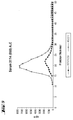
- Laminin P1 human laminin (lot II) and various sera are fractionated by molecular weight using a Sepharose S-400 column. The size distribution of the laminin antigenicity is then examined by immunoradiometric testing. In the case of a normal serum pool, the results are compared with the RIA-gnost® Lam P1. In the FIGS. 2 to 4 the chromatograms of the standards and a normal serum pool are shown. Column dimension: 1.0 x 30 cm Running buffer: PBS + 0.04% Tween-20 + 0.02% Na-azide Flow rate: 0.2 ml / min
- the column was calibrated with the molecular weight markers thyroglobulin (670 kDa), immunoglobulin (156 kDa), ovalbumin (44 kDa) and myoglobin (17 kDa).
- FIGS. 5 to 7 are single sera from patients with Alcoholic Liver Disease, PBC, and CAH.
- FIG. 8 shows a semidry blot according to the standard procedure with a discontinuous buffer system after separation of about 1 ⁇ g of laminin batch II (example 3) by SDS gel electrophoresis (Novex finished gel 4 to 12% polyacrylamide) under reducing (+ SH) or non-reducing conditions (- SH).
- the nitrocellulose membrane was cut into strips and the individual strips were incubated with the monoclonal antibodies A9 / 2/1, A27 / 2/1 and A33 / 2/20.
- Phosphatase Sigma A 5153
- the monoclonal antibodies have no reaction with the denatured structures produced by reduction. Accordingly, the monoclonal antibodies of the present invention specifically recognize native folded motifs of the laminin P1 domain.
- Table 8 shows that in the two new laminin assays, no appreciable cross-reactions to selected human connective tissue and serum proteins are detectable. Likewise, no cross-reaction to laminin / nidogen and laminin P1 from mouse EHS tumor can be detected.
- EDTA ethylenediamine-tetraacetic acid
- NEM N-ethylmaleimide
- PBS Phosphate buffered saline (Buffer solution PM 16, Serva)
- PCMB 4-Hydroxymercuri-benzoic acid sodium salt
- PMSF phenylmethylsulfonyl fluoride
- the BIAcore ® system from Pharmacia Biosensor allows biospecific interactions to be monitored on-line.
- the principle of measurement is based on an optical phenomenon (surface plasmon resonance), which is influenced by the mass bound on a gold film.
- this system is a miniaturized affinity chromatography on a gold sensor surface.
- the amount of a specifically bound ligand can be depicted in the form of a resonance signal ( Chaiken, I .; Rose, S .; Karlsson, R. (1992) Anal. Biochem.
- a double pulse of 4 ⁇ l of 100 mM HCl can be carried out.
- the laminin P1 layer is extremely stable and can be used for about 2 months (> 300 individual analyzes) without loss of quality.
- 4 to 25 ⁇ l culture supernatant from the individual cell colonies were passed directly over the affinity matrix, after 1 to 5 minutes the binding (relative strength) at the RU and the constancy of the signal can be recognized. This allows a quick and meaningful selection of interesting clones. Successive injections of various culture supernatants without intervening regeneration make this possible Screening of antibodies that can simultaneously bind to different epitopes of the antigen.
- Hybridoma supernatants as described above were conducted ® on the Lam-P1 layer of the BIAcore chip. After binding of the antibody (flow through the sample), 4 ⁇ l of subclass-specific anti-mouse antibodies were then sequentially injected. An increase in signal is in turn the expression of a mutual bond. This allows a clear classification of the subclass within 5 minutes.
- a selective and reversible binding of mouse antibodies can be applied to the sensor chip of BIAcore ®, a special antibody (R Abbit a nti m ouse Fc -Specific; RAM-Fc) can be immobilized using a standard method. Since the signal of the measuring system depends directly on the mass of the bound ligand, it is possible to carry out concentration determinations, especially if the standard material and the sample to be analyzed have the same molecular weights. Concentration determination of a specific protein is possible even in complex mixtures since, as in the present example, only the mass of the desired ligand is retained on the sensor chip by the specificity of the binding.
- a standard curve for determining the amount of antibody present in the culture supernatants was generated using a well-characterized monoclonal antibody (MAK 238) present in the laboratory.
- MAK 2328 monoclonal antibody
- the signals obtained from duplicate determinations of unknown samples (culture supernatants) can be converted into concentrations ( ⁇ g / ml monoclonal antibody) using the standard curve.
- the BIAcore system makes use of such complex and time-consuming serial measurements of a randomization program to negate those sources of error that might arise from the affinity matrix or the different downtime.
- the program determines at user-defined intervals the RU values and the current slope of the curve so that a slope / R plot is possible.
- the association constant k ass can be determined if the slopes of this function for all analyzed concentrations are related to the present actual concentrations (in nM).
- the dissociation constant k diss can be determined from the plot InR1 / Rn versus time.
- the equilibrium constant KD results from the formula k ass / k diss .
- the three tables show that the three monoclonal antibodies have very good binding properties, especially the antibodies are characterized by a very strong binding to the immobilized antigen.
- the laminin layer eg during the analysis period (30 min) no dissociation can be detected in any of the antibodies. If, however, dissolved antigen is passed over an A27 / 2/1 layer, for example, the association takes place with unchanged fast kinetics (kass1,2x10 5 / Ms). But then follows a significant dissociation phase, so that an equilibrium constant of about 1x10 9 can be determined for the binding of the laminin.
- LamP1 differs by a factor of 1000, depending on whether the LamP1 is immobilized (locally high concentration) or whether it has to be bound from the solution. This effect could be the basis for exclusive recognition of the high molecular weight peak (intact serum laminin) in human serum (see below).
- Table 4 Screening strategy for mk. anti LamP1 / laminin antibody screening stages Test Method / criteria selection immunization titer merger ⁇ Hybridomas Screening with ELISA on Lam P1 SELECTION CRITERIA: Detection of Lam-P1 in ELISA (height of OD) Growth of clones in cell culture 150 pos. Clones 1st selection 56 pos.
- the "Couples” column indicates the antibodies or clones which can simultaneously bind to the laminin P1 using the antibody specified in column 1. Characterization of monoclonal anti-laminin P1 antibodies (see next page): antibody Conc. ⁇ g / mg subtype ELISA blot BIAcore pairs Lam P1 Lam P1 Laminin (S) Laminin - SH Laminin + SH Lamin (H) CH.1.25 ⁇ l CH.2.4 ⁇ l CH.2.25 ⁇ l CH.1 CH.2 A 27/1/1 10 1:57 1061 +++ - 191 RU 584 RU A9 / 1/7; A24 / 1/13; A25 / 1/2 A 27/1/2 20.9 1224 0942 638 RU A 27/1/7 10.1 (5.2) IgG2a 1488 1058 231 RU 613 RU A50 / 1/4 A 27/1/8 12.8 1gG2a 1503 1038 +++ ++ 650 Ru 291 A 9/1/7 23.6 1gG2a 1544 1147
- Clones A28 / 1/1 and A24 / 2/2 are unsuitable due to insufficient reaction.
- Binding concentration E / ml ( ⁇ g / ml) calculated calculated lamp1 0 289 0.18 102 0.09 default 0.17 (0.037) 4140 2.6 1606 1.41 1.43 (0.31) 32050 20.2 13066 11.5 12 (2.64) 142523 89.9 51264 45 Collagen I 5 607 0.38 807 0.71 (Chemicon) 1 340 0.21 287 0.25 Collagen III 5 331 0.21 138 0.12 (Chemicon) 1 316 0.2 114 0.1 Collagen IV 5 527 0.33 120 0.11 (Chemicon) 1 333 0.21 114 0.1 Collagen V 5 614 0.39 241 0.21 (Chemicon) 1 455 0.29 232 0.2 Collagen VI 6.6
- the sera were studied by 80 inpatients with diabetes (32% Typl, 68% type II) with a diabetes duration of 11-14 years ( Stracke, H .; Wiek, K .; Günzler, V .; Federlin, K. (1993) The Medical World 44; 383 to 385 ), who were diagnosed with chiropathy (36%), retinopathy (48%), neuropathy (66%), and nephropathy (39%).
Landscapes
- Chemical & Material Sciences (AREA)
- Organic Chemistry (AREA)
- Health & Medical Sciences (AREA)
- General Health & Medical Sciences (AREA)
- Biochemistry (AREA)
- Biophysics (AREA)
- Life Sciences & Earth Sciences (AREA)
- Genetics & Genomics (AREA)
- Medicinal Chemistry (AREA)
- Molecular Biology (AREA)
- Proteomics, Peptides & Aminoacids (AREA)
- Immunology (AREA)
- Preparation Of Compounds By Using Micro-Organisms (AREA)
- Peptides Or Proteins (AREA)
- Micro-Organisms Or Cultivation Processes Thereof (AREA)
Applications Claiming Priority (2)
| Application Number | Priority Date | Filing Date | Title |
|---|---|---|---|
| DE4428481 | 1994-08-11 | ||
| EP95112393A EP0696597B1 (fr) | 1994-08-11 | 1995-08-07 | Anticorps monoclonaux pour la détermination immunologique sélective des formes de la laminine intacte à haut poids moléculaire dans des fluides corporels |
Related Parent Applications (1)
| Application Number | Title | Priority Date | Filing Date |
|---|---|---|---|
| EP95112393A Division EP0696597B1 (fr) | 1994-08-11 | 1995-08-07 | Anticorps monoclonaux pour la détermination immunologique sélective des formes de la laminine intacte à haut poids moléculaire dans des fluides corporels |
Publications (2)
| Publication Number | Publication Date |
|---|---|
| EP1942116A1 true EP1942116A1 (fr) | 2008-07-09 |
| EP1942116B1 EP1942116B1 (fr) | 2014-01-22 |
Family
ID=6525471
Family Applications (2)
| Application Number | Title | Priority Date | Filing Date |
|---|---|---|---|
| EP95112393A Expired - Lifetime EP0696597B1 (fr) | 1994-08-11 | 1995-08-07 | Anticorps monoclonaux pour la détermination immunologique sélective des formes de la laminine intacte à haut poids moléculaire dans des fluides corporels |
| EP08075098.7A Expired - Lifetime EP1942116B1 (fr) | 1994-08-11 | 1995-08-07 | Anticorps monoclonaux destinés à la détermination immunologique sélective de formes de laminine hautement moléculaires intactes dans des liquides corporels |
Family Applications Before (1)
| Application Number | Title | Priority Date | Filing Date |
|---|---|---|---|
| EP95112393A Expired - Lifetime EP0696597B1 (fr) | 1994-08-11 | 1995-08-07 | Anticorps monoclonaux pour la détermination immunologique sélective des formes de la laminine intacte à haut poids moléculaire dans des fluides corporels |
Country Status (7)
| Country | Link |
|---|---|
| US (2) | US5811268A (fr) |
| EP (2) | EP0696597B1 (fr) |
| JP (1) | JP3342252B2 (fr) |
| AT (1) | ATE390443T1 (fr) |
| CA (1) | CA2155793C (fr) |
| DE (1) | DE59511090D1 (fr) |
| ES (2) | ES2459290T3 (fr) |
Families Citing this family (9)
| Publication number | Priority date | Publication date | Assignee | Title |
|---|---|---|---|---|
| DE19701607A1 (de) * | 1997-01-17 | 1998-07-23 | Hoechst Ag | Anitkörper, die an die Nidogen-Bindungsdomäne des Laminins binden, deren Herstellung und Verwendung |
| US9404932B2 (en) * | 2007-11-05 | 2016-08-02 | Nordic Bioscience A/S | Pathology biomarker assay |
| JP6329072B2 (ja) | 2011-09-26 | 2018-05-23 | キアゲン ゲゼルシャフト ミット ベシュレンクテル ハフツング | 細胞外核酸の安定化および単離 |
| US11021733B2 (en) | 2011-09-26 | 2021-06-01 | Qiagen Gmbh | Stabilization and isolation of extracellular nucleic acids |
| CN103827303B (zh) | 2011-09-26 | 2021-10-22 | 普瑞阿那利提克斯有限公司 | 细胞外核酸的稳定化和分离 |
| CN104755628B (zh) | 2012-09-25 | 2019-03-01 | 凯杰有限公司 | 生物样品的稳定化 |
| US11525155B2 (en) | 2013-03-18 | 2022-12-13 | Qiagen Gmbh | Stabilisation of biological samples |
| EP3377645B1 (fr) | 2015-11-20 | 2023-10-04 | Qiagen GmbH | Procédé de préparation de compositions stérilisées pour stabiliser des acides nucléiques extracellulaires |
| CN110308283B (zh) * | 2019-06-28 | 2021-09-10 | 迪瑞医疗科技股份有限公司 | 一种层粘连蛋白校准品及质控品 |
Citations (4)
| Publication number | Priority date | Publication date | Assignee | Title |
|---|---|---|---|---|
| DE3331627A1 (de) * | 1983-09-01 | 1985-03-21 | Max-Planck-Gesellschaft zur Förderung der Wissenschaften e.V., 3400 Göttingen | Verfahren zur immunologischen bestimmung fuer proteine in koerperfluessigkeiten, unter verwendung monovalenter antikoerperfragmente |
| SU1454851A1 (ru) * | 1987-06-18 | 1989-01-30 | Всесоюзный онкологический научный центр АМН СССР | Штамм гибридных культивируемых клеток животных RаттUS NoRVeGIcUS,используемый дл получени моноклональных антител к гликопротеиду базальных мембран ламинину |
| JPH0342572A (ja) * | 1989-07-11 | 1991-02-22 | Takara Shuzo Co Ltd | ラミニン測定試薬 |
| JPH04134097A (ja) * | 1990-09-21 | 1992-05-07 | Takara Shuzo Co Ltd | ラミニン フラグメント |
-
1995
- 1995-08-07 EP EP95112393A patent/EP0696597B1/fr not_active Expired - Lifetime
- 1995-08-07 ES ES08075098.7T patent/ES2459290T3/es not_active Expired - Lifetime
- 1995-08-07 DE DE59511090T patent/DE59511090D1/de not_active Expired - Lifetime
- 1995-08-07 ES ES95112393T patent/ES2304782T3/es not_active Expired - Lifetime
- 1995-08-07 AT AT95112393T patent/ATE390443T1/de not_active IP Right Cessation
- 1995-08-07 EP EP08075098.7A patent/EP1942116B1/fr not_active Expired - Lifetime
- 1995-08-09 US US08/512,930 patent/US5811268A/en not_active Expired - Lifetime
- 1995-08-10 CA CA002155793A patent/CA2155793C/fr not_active Expired - Fee Related
- 1995-08-10 JP JP22450195A patent/JP3342252B2/ja not_active Expired - Fee Related
-
1998
- 1998-01-29 US US09/015,775 patent/US6033863A/en not_active Expired - Lifetime
Patent Citations (4)
| Publication number | Priority date | Publication date | Assignee | Title |
|---|---|---|---|---|
| DE3331627A1 (de) * | 1983-09-01 | 1985-03-21 | Max-Planck-Gesellschaft zur Förderung der Wissenschaften e.V., 3400 Göttingen | Verfahren zur immunologischen bestimmung fuer proteine in koerperfluessigkeiten, unter verwendung monovalenter antikoerperfragmente |
| SU1454851A1 (ru) * | 1987-06-18 | 1989-01-30 | Всесоюзный онкологический научный центр АМН СССР | Штамм гибридных культивируемых клеток животных RаттUS NoRVeGIcUS,используемый дл получени моноклональных антител к гликопротеиду базальных мембран ламинину |
| JPH0342572A (ja) * | 1989-07-11 | 1991-02-22 | Takara Shuzo Co Ltd | ラミニン測定試薬 |
| JPH04134097A (ja) * | 1990-09-21 | 1992-05-07 | Takara Shuzo Co Ltd | ラミニン フラグメント |
Non-Patent Citations (34)
| Title |
|---|
| ABRAHAMSON D R ET AL: "SELECTIVE IMMUNOREACTIVITIES OF KIDNEY BASEMENT MEMBRANES TO MONOCLONAL ANTIBODIES AGAINST LAMININ LOCALIZATION OF THE END OF THE LONG ARM AND THE SHORT ARMS TO DISCRETE MICRODOMAINS", JOURNAL OF CELL BIOLOGY, vol. 109, no. 6 PART 2, 1989, pages 3477 - 3492, XP001036999, ISSN: 0021-9525 * |
| BROCKS, D.G. ET AL., CLIN. CHEM., vol. 32, 1986, pages 787 - 791 |
| BROWN, C.J.; WIEDEMANN, H.; TIMPL, R. ET AL., J. CELL SCI, vol. 107, 1994, pages 329 - 338 |
| BROWN, C.J.; WIEDEMANN, H.; TIMPL, R., J. CELL SCI, vol. 107, 1994, pages 329 - 338 |
| BURGESON, R.E. ET AL., MATRIX BIOLOGY, vol. 14, 1994, pages 209 - 211 |
| CHAIKEN, 1.; ROSE, S.; KARLSSON, R., ANAL. BIOCHEM., vol. 201, 1992, pages 197 - 201 |
| DATABASE WPI Derwent World Patents Index; AN 1989-330803, XP002190741 * |
| DATABASE WPI Derwent World Patents Index; AN 1991-098302, XP002190740 * |
| DATABASE WPI Derwent World Patents Index; AN 1992-204526, XP002190739 * |
| EHRIG, K. ET AL., PROC. NATL. ACAD: SCI. USA, vol. 87, 1990, pages 3264 - 3268 |
| ENGEL, J., FEBS LETT., vol. 251, no. 1, 2, 1989, pages 1 - 7 |
| ENGVALL, E., PROC. NATL. ACAD. SCI, vol. 85, 1988, pages 1544 - 1548 |
| HARLOW, E.; LANE, D.: "Antibodies: A Laboratory Manual", 1988, CSH, pages: 319 - 359 |
| HORIKOSHI, S.; KOIDE, H., CLIN. CHIM. ACTA, vol. 196, 1991, pages 185 - 192 |
| HUNTER, D.D. ET AL., NATURE, vol. 338, 1989, pages 229 - 234 |
| IWATA K: "ONE-STEP SANDWICH ENZYME IMMUNOASSAY FOR HUMAN LAMININ USING MONOCLONAL ANTIBODIES", CLINICA CHIMICA ACTA, vol. 191, no. 3, 1990, pages 211 - 220, XP001036833, ISSN: 0009-8981 * |
| IWATA, K., CLIN. CHIM. ACTA, vol. 191, 1990, pages 211 - 220 |
| KARISSON, R.; ALTSCHUH, D.; VAN REGENMORTEL, M.H.V.: "Structure of Antigens", 1992, CRC PRESS, article "Measurement of antibody affinity", pages: 127 - 148 |
| KARLSSON, R.; ALTSCHUH, D.; VAN REGENMORTEL, M.H.V.: "Structure of Antigens", 1992, CRC PRESS, article "Measurement of antibody affinity", pages: 127 - 148 |
| KATAYAMA ET AL., BR. J. CANCER, vol. 65, 1992, pages 509 - 514 |
| KATAYAMA M ET AL: "URINARY LAMININ FRAGMENTS AS A TUMOUR MARKER POTENTIALLY REFLECTING BASEMENT MEMBRANE DESTRUCTION", BRITISH JOURNAL OF CANCER, vol. 65, no. 4, 1992, pages 509 - 514, XP001036973, ISSN: 0007-0920 * |
| KATAYAMA, M. ET AL., BR. J. CANCER, vol. 65, 1992, pages 509 - 514 |
| KROPF. J. ET AL., CLIN CHEM., vol. 37, 1991, pages 30 |
| MARINKOVICH, M.P. ET AL., J. CELL. BIOL., vol. 119, 1992, pages 695 - 703 |
| MARINKOVICH, M.P.; LUNSTROM, G.P.; BURGESON, R.E., J. BIOL. CHEM., vol. 267, 1992, pages 17900 - 17906 |
| MATTER MICHELLE L ET AL: "A novel laminin E8 cell adhesion site required for lung alveolar formation in vitro.", JOURNAL OF CELL BIOLOGY, vol. 124, no. 6, March 1994 (1994-03-01), pages 1083 - 1090, XP001036963, ISSN: 0021-9525 * |
| NIEMELÄ, 0. ET AL., EUR. J. CLIN. INVEST, vol. 15, 1985, pages 132 - 137 |
| RISTELI, L; TIMPL, R., BIOCHEM. J., vol. 193, 1981, pages 749 - 755 |
| ROSE, S.; KARLSSON, R., ANAL. BIOCHEM., vol. 201, 1992, pages 197 - 201 |
| STRACKE, H. ET AL., DIE MEDIZINISCHE WELT, vol. 44, 1993, pages 383 - 385 |
| TIMPI, R., EUR. J. BIOCHEM., vol. 180, 1989, pages 487 - 502 |
| TIMPL, R., EUR. J. BIOCHEM., vol. 180, 1989, pages 487 - 502 |
| WEWER, U. ET AL., J. BIOL. CHEM., vol. 258, 1983, pages 12654 - 12660 |
| YOKOYA YUKIHIRO ET AL: "Concentration of serum laminin and type IV collagen in liver disease assayed by a sandwich enzyme-immunoassay using monoclonal antibodies.", CLINICA CHIMICA ACTA, vol. 210, no. 1-2, 1992, pages 109 - 118, XP001036829, ISSN: 0009-8981 * |
Also Published As
| Publication number | Publication date |
|---|---|
| DE59511090D1 (de) | 2008-05-08 |
| JP3342252B2 (ja) | 2002-11-05 |
| ES2304782T3 (es) | 2008-10-16 |
| EP0696597A3 (fr) | 2002-04-17 |
| US5811268A (en) | 1998-09-22 |
| EP0696597B1 (fr) | 2008-03-26 |
| US6033863A (en) | 2000-03-07 |
| CA2155793C (fr) | 2007-10-16 |
| ES2459290T3 (es) | 2014-05-08 |
| ATE390443T1 (de) | 2008-04-15 |
| EP1942116B1 (fr) | 2014-01-22 |
| JPH08231596A (ja) | 1996-09-10 |
| CA2155793A1 (fr) | 1996-02-12 |
| EP0696597A2 (fr) | 1996-02-14 |
Similar Documents
| Publication | Publication Date | Title |
|---|---|---|
| DE3888224T2 (de) | Bestimmung vom Tumornekrosefaktor; monoklonaler Antikörper und Zusammensetzung. | |
| EP0394819B1 (fr) | Anticorps spécifiques contre la troponine T, leur production et leur utilisation dans un réactif pour la détermination de nécroses du myocarde | |
| EP0004940B1 (fr) | Procédé pour la détermination radio-immunologique de procollagène (type III) et de procollagène-peptide (type III), préparation de procollagène-peptide (type III) propre audit procédé et préparation de sérum anti-procollagène-peptide (type III) | |
| EP1151304B1 (fr) | PROCEDE D'IDENTIFICATION DE proBNP N-TERMINAL | |
| DE3785038T2 (de) | Monoklonale antikoerper gegen nicht reduzierte, nicht enzymatisch glykolysierte proteine. | |
| DE3486453T2 (de) | Monoklonale Antikörper gegen neue Faktor-VIII Gerinnungspolypeptide | |
| DE69127947T2 (de) | Monoklonale Antikörper gegen menschliches IgE | |
| DE3789781T2 (de) | Antigene, Antikörper und Verfahren zur Identifizierung humaner metastatischer Tumoren und Zellinien zur Herstellung dieser Antikörper. | |
| DE69132122T2 (de) | Reinigung von cytokeratinfragmenten | |
| EP1942116B1 (fr) | Anticorps monoclonaux destinés à la détermination immunologique sélective de formes de laminine hautement moléculaires intactes dans des liquides corporels | |
| EP0339443B1 (fr) | Anticorps monoclonal pour la détermination immunologique sélective du peptide (type III) intact de procollagène et de procollagène (type III) dans des sécrétions | |
| DE69112225T2 (de) | Immunodiagnostische probe für gelenkrheumatismus. | |
| DE69619226T2 (de) | Monoklonaler antikörper gegen den komplex aus faktor xa und dem inhibitor für den gewebefaktor (xa. tfpi) | |
| DE69737927T2 (de) | NEUARTIGE MONOKLONALE ANTIKÖRPER UND VERFAHREN ZUR IMMUNOLOGISCHEN ANALYSE DES e-D DIMERS UND DES e-DD/E-KOMPLEXES | |
| EP0955361A1 (fr) | Anticorps monoclonaux contre des antigènes associés aux tumeurs, procédés de préparation et utilisation | |
| DE3587714T2 (de) | Menschliches alpha 2-Plasmin spezifischer monoklonaler Antikörper. | |
| DE3789284T2 (de) | Immunologische Bestimmung von freiem menschlichem Protein-S und c4bp-Protein-S-Komplex. | |
| DE69526542T2 (de) | uPA-Bindungstelle auf 2+3 Domäne von uPAR und damit reagierende Antikörper | |
| EP0954535A1 (fr) | Anticorps qui se lient aux domaines de liaison de nidogene de la laminine, leur production et leur utilisation | |
| DE3888604T2 (de) | Monoklonale Antikörper gegen Antithrombinbindende Substanz und seine Verwendungen. | |
| DE69836679T2 (de) | Peptide zur behandlung des systemischen lupus erythematosus | |
| DE68911713T2 (de) | Monoklonale Antikörper, die atriales natriuretisches Polypeptide erkennen, ihre Herstellung und Verwendung. | |
| DE69915059T2 (de) | Monoklonaler antikörper und assay zur bestimmung von n-terminalem prokollagen-propeptid typ iii (piiinp) | |
| DE68914345T2 (de) | Gamma-atriales, natriuretisches Polypeptid erkennende monoklonale Antikörper, solche Antikörper produzierende Hybridome und deren Herstellung und Verwendung. | |
| DE69527868T2 (de) | Monoklonaler Antikörper gegen den tumorzytotoxischen Faktor II (TCF-II) |
Legal Events
| Date | Code | Title | Description |
|---|---|---|---|
| PUAI | Public reference made under article 153(3) epc to a published international application that has entered the european phase |
Free format text: ORIGINAL CODE: 0009012 |
|
| AC | Divisional application: reference to earlier application |
Ref document number: 0696597 Country of ref document: EP Kind code of ref document: P |
|
| AK | Designated contracting states |
Kind code of ref document: A1 Designated state(s): AT BE CH DE DK ES FR GB GR IE IT LI LU NL PT SE |
|
| 17P | Request for examination filed |
Effective date: 20090109 |
|
| 17Q | First examination report despatched |
Effective date: 20090217 |
|
| AKX | Designation fees paid |
Designated state(s): AT BE CH DE DK ES FR GB GR IE IT LI LU NL PT SE |
|
| GRAP | Despatch of communication of intention to grant a patent |
Free format text: ORIGINAL CODE: EPIDOSNIGR1 |
|
| INTG | Intention to grant announced |
Effective date: 20130426 |
|
| RBV | Designated contracting states (corrected) |
Designated state(s): AT BE CH DE DK ES FR GB GR IE IT LI LU MC NL PT SE |
|
| RIN1 | Information on inventor provided before grant (corrected) |
Inventor name: STEINERT, CORNELIA Inventor name: GERL, MARTIN, DR. Inventor name: QUINT, MANFRED |
|
| GRAJ | Information related to disapproval of communication of intention to grant by the applicant or resumption of examination proceedings by the epo deleted |
Free format text: ORIGINAL CODE: EPIDOSDIGR1 |
|
| GRAP | Despatch of communication of intention to grant a patent |
Free format text: ORIGINAL CODE: EPIDOSNIGR1 |
|
| INTG | Intention to grant announced |
Effective date: 20130819 |
|
| GRAS | Grant fee paid |
Free format text: ORIGINAL CODE: EPIDOSNIGR3 |
|
| GRAA | (expected) grant |
Free format text: ORIGINAL CODE: 0009210 |
|
| AC | Divisional application: reference to earlier application |
Ref document number: 0696597 Country of ref document: EP Kind code of ref document: P |
|
| AK | Designated contracting states |
Kind code of ref document: B1 Designated state(s): AT BE CH DE DK ES FR GB GR IE IT LI LU MC NL PT SE |
|
| REG | Reference to a national code |
Ref country code: GB Ref legal event code: FG4D Free format text: NOT ENGLISH |
|
| REG | Reference to a national code |
Ref country code: CH Ref legal event code: EP |
|
| REG | Reference to a national code |
Ref country code: AT Ref legal event code: REF Ref document number: 650784 Country of ref document: AT Kind code of ref document: T Effective date: 20140215 |
|
| REG | Reference to a national code |
Ref country code: IE Ref legal event code: FG4D Free format text: LANGUAGE OF EP DOCUMENT: GERMAN |
|
| REG | Reference to a national code |
Ref country code: DE Ref legal event code: R096 Ref document number: 59511117 Country of ref document: DE Effective date: 20140306 |
|
| REG | Reference to a national code |
Ref country code: ES Ref legal event code: FG2A Ref document number: 2459290 Country of ref document: ES Kind code of ref document: T3 Effective date: 20140508 |
|
| REG | Reference to a national code |
Ref country code: NL Ref legal event code: VDEP Effective date: 20140122 |
|
| PG25 | Lapsed in a contracting state [announced via postgrant information from national office to epo] |
Ref country code: SE Free format text: LAPSE BECAUSE OF FAILURE TO SUBMIT A TRANSLATION OF THE DESCRIPTION OR TO PAY THE FEE WITHIN THE PRESCRIBED TIME-LIMIT Effective date: 20140122 Ref country code: NL Free format text: LAPSE BECAUSE OF FAILURE TO SUBMIT A TRANSLATION OF THE DESCRIPTION OR TO PAY THE FEE WITHIN THE PRESCRIBED TIME-LIMIT Effective date: 20140122 Ref country code: PT Free format text: LAPSE BECAUSE OF FAILURE TO SUBMIT A TRANSLATION OF THE DESCRIPTION OR TO PAY THE FEE WITHIN THE PRESCRIBED TIME-LIMIT Effective date: 20140522 |
|
| REG | Reference to a national code |
Ref country code: DE Ref legal event code: R097 Ref document number: 59511117 Country of ref document: DE |
|
| PG25 | Lapsed in a contracting state [announced via postgrant information from national office to epo] |
Ref country code: DK Free format text: LAPSE BECAUSE OF FAILURE TO SUBMIT A TRANSLATION OF THE DESCRIPTION OR TO PAY THE FEE WITHIN THE PRESCRIBED TIME-LIMIT Effective date: 20140122 |
|
| PLBE | No opposition filed within time limit |
Free format text: ORIGINAL CODE: 0009261 |
|
| STAA | Information on the status of an ep patent application or granted ep patent |
Free format text: STATUS: NO OPPOSITION FILED WITHIN TIME LIMIT |
|
| 26N | No opposition filed |
Effective date: 20141023 |
|
| REG | Reference to a national code |
Ref country code: DE Ref legal event code: R097 Ref document number: 59511117 Country of ref document: DE Effective date: 20141023 |
|
| REG | Reference to a national code |
Ref country code: DE Ref legal event code: R119 Ref document number: 59511117 Country of ref document: DE |
|
| PG25 | Lapsed in a contracting state [announced via postgrant information from national office to epo] |
Ref country code: LU Free format text: LAPSE BECAUSE OF FAILURE TO SUBMIT A TRANSLATION OF THE DESCRIPTION OR TO PAY THE FEE WITHIN THE PRESCRIBED TIME-LIMIT Effective date: 20140807 Ref country code: MC Free format text: LAPSE BECAUSE OF FAILURE TO SUBMIT A TRANSLATION OF THE DESCRIPTION OR TO PAY THE FEE WITHIN THE PRESCRIBED TIME-LIMIT Effective date: 20140122 |
|
| REG | Reference to a national code |
Ref country code: CH Ref legal event code: PL |
|
| GBPC | Gb: european patent ceased through non-payment of renewal fee |
Effective date: 20140807 |
|
| PG25 | Lapsed in a contracting state [announced via postgrant information from national office to epo] |
Ref country code: IT Free format text: LAPSE BECAUSE OF NON-PAYMENT OF DUE FEES Effective date: 20140807 Ref country code: LI Free format text: LAPSE BECAUSE OF NON-PAYMENT OF DUE FEES Effective date: 20140831 Ref country code: CH Free format text: LAPSE BECAUSE OF NON-PAYMENT OF DUE FEES Effective date: 20140831 |
|
| REG | Reference to a national code |
Ref country code: IE Ref legal event code: MM4A |
|
| REG | Reference to a national code |
Ref country code: DE Ref legal event code: R119 Ref document number: 59511117 Country of ref document: DE Effective date: 20150303 |
|
| REG | Reference to a national code |
Ref country code: FR Ref legal event code: ST Effective date: 20150430 |
|
| PG25 | Lapsed in a contracting state [announced via postgrant information from national office to epo] |
Ref country code: GB Free format text: LAPSE BECAUSE OF NON-PAYMENT OF DUE FEES Effective date: 20140807 Ref country code: DE Free format text: LAPSE BECAUSE OF NON-PAYMENT OF DUE FEES Effective date: 20150303 |
|
| PG25 | Lapsed in a contracting state [announced via postgrant information from national office to epo] |
Ref country code: FR Free format text: LAPSE BECAUSE OF NON-PAYMENT OF DUE FEES Effective date: 20140901 Ref country code: IE Free format text: LAPSE BECAUSE OF NON-PAYMENT OF DUE FEES Effective date: 20140807 |
|
| REG | Reference to a national code |
Ref country code: AT Ref legal event code: MK07 Ref document number: 650784 Country of ref document: AT Kind code of ref document: T Effective date: 20150807 |
|
| REG | Reference to a national code |
Ref country code: ES Ref legal event code: FD2A Effective date: 20150925 |
|
| PG25 | Lapsed in a contracting state [announced via postgrant information from national office to epo] |
Ref country code: ES Free format text: LAPSE BECAUSE OF NON-PAYMENT OF DUE FEES Effective date: 20140808 |
|
| PG25 | Lapsed in a contracting state [announced via postgrant information from national office to epo] |
Ref country code: AT Free format text: LAPSE BECAUSE OF NON-PAYMENT OF DUE FEES Effective date: 20140831 Ref country code: GR Free format text: LAPSE BECAUSE OF FAILURE TO SUBMIT A TRANSLATION OF THE DESCRIPTION OR TO PAY THE FEE WITHIN THE PRESCRIBED TIME-LIMIT Effective date: 20140423 |
|
| PG25 | Lapsed in a contracting state [announced via postgrant information from national office to epo] |
Ref country code: BE Free format text: LAPSE BECAUSE OF NON-PAYMENT OF DUE FEES Effective date: 20140831 |