EP1961825A1 - Procédé pour prévoir l'apparition de métastase dans les patients souffrant d'un cancer du sein - Google Patents
Procédé pour prévoir l'apparition de métastase dans les patients souffrant d'un cancer du sein Download PDFInfo
- Publication number
- EP1961825A1 EP1961825A1 EP07300823A EP07300823A EP1961825A1 EP 1961825 A1 EP1961825 A1 EP 1961825A1 EP 07300823 A EP07300823 A EP 07300823A EP 07300823 A EP07300823 A EP 07300823A EP 1961825 A1 EP1961825 A1 EP 1961825A1
- Authority
- EP
- European Patent Office
- Prior art keywords
- metastasis
- tissue
- biological markers
- occurrence
- markers
- Prior art date
- Legal status (The legal status is an assumption and is not a legal conclusion. Google has not performed a legal analysis and makes no representation as to the accuracy of the status listed.)
- Ceased
Links
Images
Classifications
-
- C—CHEMISTRY; METALLURGY
- C12—BIOCHEMISTRY; BEER; SPIRITS; WINE; VINEGAR; MICROBIOLOGY; ENZYMOLOGY; MUTATION OR GENETIC ENGINEERING
- C12Q—MEASURING OR TESTING PROCESSES INVOLVING ENZYMES, NUCLEIC ACIDS OR MICROORGANISMS; COMPOSITIONS OR TEST PAPERS THEREFOR; PROCESSES OF PREPARING SUCH COMPOSITIONS; CONDITION-RESPONSIVE CONTROL IN MICROBIOLOGICAL OR ENZYMOLOGICAL PROCESSES
- C12Q1/00—Measuring or testing processes involving enzymes, nucleic acids or microorganisms; Compositions therefor; Processes of preparing such compositions
- C12Q1/68—Measuring or testing processes involving enzymes, nucleic acids or microorganisms; Compositions therefor; Processes of preparing such compositions involving nucleic acids
- C12Q1/6876—Nucleic acid products used in the analysis of nucleic acids, e.g. primers or probes
- C12Q1/6883—Nucleic acid products used in the analysis of nucleic acids, e.g. primers or probes for diseases caused by alterations of genetic material
- C12Q1/6886—Nucleic acid products used in the analysis of nucleic acids, e.g. primers or probes for diseases caused by alterations of genetic material for cancer
-
- C—CHEMISTRY; METALLURGY
- C12—BIOCHEMISTRY; BEER; SPIRITS; WINE; VINEGAR; MICROBIOLOGY; ENZYMOLOGY; MUTATION OR GENETIC ENGINEERING
- C12Q—MEASURING OR TESTING PROCESSES INVOLVING ENZYMES, NUCLEIC ACIDS OR MICROORGANISMS; COMPOSITIONS OR TEST PAPERS THEREFOR; PROCESSES OF PREPARING SUCH COMPOSITIONS; CONDITION-RESPONSIVE CONTROL IN MICROBIOLOGICAL OR ENZYMOLOGICAL PROCESSES
- C12Q2600/00—Oligonucleotides characterized by their use
- C12Q2600/118—Prognosis of disease development
Definitions
- the present invention relates to the prognosis of the progression of breast cancer in a patient, and more particularly to the prediction of the occurrence of metastasis in one or more tissue or organ of patients affected with a breast cancer.
- Breast cancer is the most common malignant disease in Western women. It is not the primary tumour, but the occurrence of metastases in distant organs that is the major cause of death for cancer patients. Once solid secondary tumours are established, the chances of long-term survival fall from 90% to around 5%. Despite the progress in the development of targeted therapies, approximately 40% of the treated patients relapse and ultimately die of metastatic breast cancer. A better understanding of the molecular and cellular mechanisms underlying metastasis might improve the development of effective therapies that would ameliorate breast cancer care.
- Malignant breast tumours can invade and damage nearby tissues and organs. Malignant tumour cells may metastasise, entering the bloodstream or lymphatic system. When breast cancer cells metastasise outside the breast, they are often found in the lymph nodes under the arm (axillary lymph nodes). If the cancer has reached these nodes, it means that cancer cells may have spread to other lymph nodes or other organs, such as bones, liver, brain or lungs. Breast cancer metastasis of various organs also occurs without previous spreading to lymph nodes. Major and intensive research has been focussed on early detection, treatment and prevention of metastatic breast cancer.
- Kang et al. (2003, Cancer Cell, Vol. 3 : 537-549 ) identified a set of genes potentially involved in breast cancer metastasis to bone, by comparing (i) the gene expression profile obtained from in vitro cultured cells of the MDBA-MB-231 human breast cancer cell line with (ii) the gene expression profile obtained from a subclone of the same cell line previously experimentally selected in vivo for their ability to form bone metastasis in mice.
- microarray gene expression analysis techniques these authors showed that several genes were underexpressed and several other genes were overexpressed in the cell sublines selected for their ability to form bone metastasis in mice, as compared to the parental MDBA-MB-231 human breast cancer cell line.
- Minn et al. (2005, Nature, Vol. 436(28) : 518-524 ) used the same human parental MDA-MB-231 breast cancer cell line for selecting in vivo cell sublines having the ability to form lung metastasis in mice. Then, these authors have performed a transcriptomic microarray analysis of the highly and weakly lung-metastatic cell populations, with the view of identifying patterns of gene expression that would be associated with aggressive lung metastatic behaviour. A final list of 54 candidate lung metastagenicity and virulence genes was selected, twelve of them having been further identified for their significant association with lung-metastasis-free survival, including MMP1, CXCL1 and PTGS2.
- organ-specific gene signatures that were identified allowed to distinguish between (i) primary breast carcinomas that preferentially metastasized to bone or lung from (ii) those that metastasized elsewhere.
- An early detection of metastasis in breast cancer patients is crucial for adapting the anti-cancer therapeutic treatment correspondingly, with the view of increasing the chances of long term disease-free survival.
- the present invention relates to methods and kits for predicting the occurrence of metastasis in patients affected with a breast cancer.
- the breast cancer metastasis prediction methods of the invention comprise a step of determining, in a tumour tissue sample previously collected from a breast cancer patient to be tested the level of expression of one or more biological markers that are indicative of cancer progression towards metastasis, and more precisely, one or more biological markers that are indicative of cancer progression towards metastasis to specific tissues, and especially to bones, brain, lungs and liver.
- This invention also relates to breast cancer metastasis prediction kits that are specifically designed for performing the metastasis prediction methods above.
- This invention also pertains to methods for selecting one or more biological markers that are indicative of cancer progression towards metastasis in specific tissues, including bones, brain, lungs and liver.
- kits allowing prediction of the occurrence of metastasis to one or more specific tissue or organ in breast cancer patients, notably in bone, lung, liver and brain tissues or organs.
- tissue-specific sets of biological markers that are indicative of a high probability of metastasis occurrence in breast cancer patients have been identified.
- an object of the present invention consists of an in vitro method for predicting the occurrence of metastasis in a patient affected with a breast cancer, comprising the steps of :
- the screening of metastasis-specific biological markers were generally performed using artificial human cell systems, namely established human cancer cell lines, wherein expression artefacts of one or more genes or proteins cannot be excluded.
- the probability of introduction of biological markers expression artefacts was further increased by various features of the screening methods which were used, including the selection of multiple human cell sublines having a metastatic potency derived from the parental cell line, by reiterating in vivo cycles of cell administration/selection in non-human animal systems, including mice.
- the inventors have designed an original method for selecting highly reliable tissue-specific biological markers of metastasis in breast cancer, using a whole human system of analysis and including, among other features, a step of comparing the expression level of candidate biological markers between (i) a metastatic tissue or organ of interest and (ii) one or more distinct metastatic tissue(s) or organ(s), thus allowing a high selectivity and statistical relevance of the tissue-specific metastatic biological markers that are positively selected, at the end of the marker selection method.
- a marker selection method comprising a step of comparing the expression level of candidate biological markers between (i) a metastatic tissue or organ of interest selected from the group consisting of bone, lung, liver and brain and (ii) all the other metastatic tissue(s) or organ(s) selected from the group consisting of bone, lung, liver and brain
- the inventors have identified tissue-specific breast cancer metastasis biological markers endowed with a high statistical relevance, with P values always lower than 1.10 -4 , the said P values being lower than 10 -6 for the most statistically relevant biological markers.
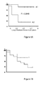
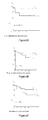
- Statistical relevancy of the biological markers primarily selected was fully corroborated by Kaplan-Meier analysis, after having assayed for the expression of the said tissue-specific markers in tumour tissue samples of breast cancer patients, as it is shown in the examples herein.
- a “biological marker” encompasses any detectable product that is synthesized upon the expression of a specific gene, and thus includes gene-specific mRNA, cDNA and protein.
- a "biological marker indicative of an occurrence of metastasis”, or a “metastasis-specific marker” encompasses any biological marker which is differentially expressed in breast tumors that generate metastasis, or will generate metastasis, in a specific given tissue or in a specific given organ, as compared to the expression of the same biological marker in breast tumors that do not.generate metastasis, or will not generate metastasis, in the said specific given tissue or in the said specific given organ.
- tissues and organs are selected from the group consisting of bone, lung, liver and brain.
- tissue-specific marker and tissue metastasis-specific marker are used interchangeably herein.
- organ-specific marker and organ metastasis-specific marker are used interchangeably herein.
- a "prediction of the occurrence of metastasis” does not consist of an absolute value, but in contrast consists of a relative value allowing to quantify the probability of occurrence of a metastasis to one or more specific tissue(s) or organ(s), in a breast cancer patient.
- the prediction of the occurrence of metastasis is expressed as a statistical value, including a P value, as calculated from the expression values obtained for each of the one or more biological markers that have been tested.
- a "tumour tissue sample” encompasses (i) a global primary tumour (as a whole), (ii) a tissue sample from the center of the tumour, (iii) a tissue sample from a location in the tumour, other than the center of the tumour and (iv) any tumor cell located outside the tumor tissue per se .
- the said tumour tissue sample originates from a surgical act of tumour resection performed on the breast cancer patient.
- the said tissue sample originates from a biopsy surgical act wherein a piece of tumour tissue is collected from the breast cancer patient for further analysis.
- the said tumor sample consists of a blood sample, including a whole blood sample, a serum sample and a plasma sample, containing tumour cells originating from the primary tumor tissue, or alternatively from metastasis that have already occurred.
- the said tumor sample consists of a blood sample, including a whole blood sample, a serum sample and a plasma sample, containing tumor proteins produced by tumor cells originating from the primary tumor tissue, or alternatively from metastasis that have already occurred.
- the various biological markers names specified herein correspond to their internationally recognised acronyms that are usable to get access to their complete amino acid and nucleic acid sequences, including their complementary DNA (cDNA) and genomic DNA (gDNA) sequences.
- cDNA complementary DNA
- gDNA genomic DNA
- the corresponding amino acid and nucleic acid sequences of each of the biological markers specified herein may be retrieved, on the basis of their acronym names, that are also termed herein "gene symbols", in the GenBank or EMBL sequence databases. All gene symbols listed in the present specification correspond to the GenBank nomenclature.
- one or more of the specified biological markers is (are) quantified.
- Quantification of a biological marker includes detection of the expression level of the said biological marker. Detection of the expression level of a specific biological marker encompasses the assessment of the amount of the corresponding specific mRNA or cDNA that is expressed in the tumour tissue sample tested, as well as the assessment of the amount of the corresponding protein that is produced in the said tumour tissue sample.
- a quantification value is obtained for each of the one or more biological markers that are used.
- step b) includes :
- step b) consists of quantifying, in a tumor tissue sample, the expression level of one or more marker genes among those specified above. Generally, the assessment of the expression level for a combination of at least two marker genes is performed. In these embodiments of step b) of the method, what is obtained at the end of step b) consists of the expression level values found for each marker gene (nucleic acid or protein expression level) specifically found in cells contained in the tumour tissue sample.
- the expression level of a metastasis-specific biological marker according to the present invention may be expressed as any arbitrary unit that reflects the amount of the corresponding mRNA of interest that has been detected in the tissue sample, such as intensity of a radioactive or of a fluorescence signal emitted by the cDNA material generated by PCR analysis of the mRNA content of the tissue sample, including (i) by Real-time PCR analysis of the mRNA content of the tissue sample and (ii) hybridization of the amplified nucleic acids to DNA microarrays.
- the said expression level value consists of a normalised relative value which is obtained after comparison of the absolute expression level value with a control value, the said control value consisting of the expression level value of a gene having the same expression level value in any breast tissue sample, regardless of whether it consists of normal or tumour breast tissue, and/or regardless whether it consists of a non-metastatic or a metastatic breast tissue.
- the said control value may consist of the amount of mRNA encoding
- the said expression level may be expressed as any arbitrary unit that reflects the amount of the protein of interest that has been detected in the tissue sample, such as intensity of a radioactive or of a fluorescence signal emitted by a labelled antibody specifically bound to the protein of interest.
- the value obtained at the end of step b) may consist of a concentration of protein(s) of interest that could be measured by various protein detection methods well known in the art, such as. ELISA, SELDI-TOF, FACS or Western blotting.
- every one of the biological markers that are specified herein consists of a biological marker (i) that is specifically expressed, including at a given expression level, exclusively in a given breast cancer metastatic tissue or organ and (ii) that is expressed at another expression level, in a distinct breast cancer metastatic tissue or organ, then the sole quantification of its expression in a tumour tissue sample originating from a primary tumour specimen allows to predict whether the breast cancer-bearing patient is likely to undergo generation of metastasis in the said tissue or organ.
- quantifying the said one or more biological markers that are specified herein brings further prediction data relating to the probability of occurrence of metastasis in one or more specific tissue or organ in a breast cancer-bearing patient, since the probability of occurrence of metastasis increases with an increased deregulation of the expression level of the said one or more biological markers tested, as compared to their control expression level that is previously measured in a breast tumor sample from a patient that has not undergone metastasis, at least in the tissue or organ of interest that is considered.
- a biological marker of interest having a "deregulated expression level” consists of a metastasis-specific biological marker for which is found, when performing step b) of the in vitro prediction method according to the invention, an expression level value that is distinct from the expression level value (that may also be termed the "control" expression value) for the said biological marker that has been previously determined (i) in tumor tissue samples originating from breast cancer patients that have never undergone metastasis, or alternatively (ii) in tumor tissue samples originating from breast cancer patients that have never undergone metastasis in the tissue or organ from which the said biological marker is metastasis-specific.
- every one of the biological markers listed herein is highly relevant for predicting the occurrence of metastasis in a specific tissue, since the markers having the lowest statistical relevance possess a P value lower than 1.10 -4 . Further, accuracy of the metastasis prediction increases, at step c) of the method, when more than one tissue-specific biological marker for a given tissue or organ is detected and/or quantified at step b).
- more than one biological marker for a given tissue or organ is detected and/or quantified, at step b) of the method.
- Quantifying a plurality of biological markers specific for a given tissue or organ at step b) of the method allows to generate an experimental expression profile of the said plurality of markers, which expreimental expression profile is then compared with at least one reference expression profile that has been previously determined from tissue-specific metastasis patients.
- the experimental expression profile that is generated from the results of quantification of the bone metastasis-specific marker genes used at step b) of the method is compared with the pre-existing reference expression profile corresponding to bone metastasis, for prediction.
- the experimental expression profile of the bone-specific metastasis markers is compared with reference expression profiles of the same genes that have been previously determined in patients having bone metastasis, as well as with reference expression profiles obtained from patients having other tissue- or organ-specific metastasis, so as to ensure that the experimental expression profile is the most close from the reference expression profile predetermined from patients having bone metastasis.
- step b) comprises quantifying at least one tissue-specific biological marker for each tissue or organ.
- step b) comprises quantifying (i) at least one bone-specific biological marker, (ii) at least one lung-specific biological marker, (iii) at least one liver-specific biological marker and (iv) at least one brain-specific biological marker.
- step b) comprises quantifying at least two tissue-specific biological markers for a given tissue or organ. The higher the number of tissue-specific markers for a given tissue or organ are quantified at step b), the more accurate is the prediction of occurrence of metastasis in the said given tissue in the breast cancer-bearing patient tested.
- the number of biological markers tested for a given tissue or organ is of at least 2, 3, 4, 5, 6, 7, 8, 9, 10, 11, 12, 13, 14, 15, 16, 17, 18, 19, 20, 21, 22, 23, 24, 25, 26, 27, 28, 29, 30, 31, 32, 33, 34, 35, 36, 37, 38, 39, 40, 41, 42, 43, 44, 45, 46, 47, 48, 49, 50, 51, 52, 53, 54, 55, 56, 57, 58, 59, 60, 61, 62, 63, 64, 65, 66, 67, 68, 69, 70, 71, 72, 73, 74, 75, 76, 77, 78, 79, 80, 81, 82, 83, 84, 85, 86, 87, 88, 89, 90, 91, 92, 93, 94, 95, 96, 97, 98, 99, 100, 101, 102, 103, 104, 105, 106, 107, 108,
- expermimental expression profiles of the patient tested may be generated, that are then compared to the reference expression profiles of the same biological markers, so as to determine to which reference expression profile the experimental expression profile is the most similar, and thus which tisue- or organ-specific metastasis may eventually be predicted.
- the one or more bone-specific biological markers that are quantified at step b) are selected from the group consisting of CLEC4A, MFNG, NXF1, FAM78A, KCTD1, BAIAP2L1, PTPN22, MEGF10, PERP, PSTPIP1, FL11, COL6A2, CD4, CFD, ZFHX1B, CD33, MMRN2, LST1, SH2D3C and RAMP3.
- Those bone-specific markers possess a high statistical relevance for predicting the occurrence of metastasis in bone tissue, with P values obtained after DNA microarray analysis that are always lower than 10 -6 , as shown in Table 2
- the one or more lung-specific biological markers that are quantified at step b) are selected from the group consisting of DSC2, HORMAD1, PLEKHG4, ODF2L, c21orf91, TFCP2L1, CHODL, CALB2, UGT8, c1orf210, SIKE, ITGB8, PAQR3, ANP32E, KIND1, ELAC1, GYLTL1B, SPSB1, PTEN and CHRM3.
- Those lung-specific markers possess a high statistical relevance for predicting the occurrence of metastasis in lung tissue or organ, with P values obtained after DNA microarray analysis that are always lower than 10 -4 , as shown in Table 2
- the one or more liver-specific biological markers that are quantified at step b) are selected from the group consisting of TBX3, c5orf30, AGXT2L1, LETM2, ZNF44, IL20RA, ZMAT1, MYRIP, WHSC1L1, SELT, GATA2, DHFRL1, ARPC2, SLC16A3, GNPTG, PRRT3, RPS6KA5, K1AA1505, ZFYVE16 and K1AA0701.
- Those liver-specific markers possess a high statistical relevance for predicting the occurrence of metastasis in liver tissue or organ, with P values obtained after DNA microarray analysis that are always lower than 10 -5 , as shown in Table 2
- the one or more brain-specific biological markers that are quantified at step b) are selected from the group consisting of PPWD1, INHA, PDGFRA, MLL5, RPS23, ANTXR1, ARRDC3, METTL7A, NPHP3, PKP2, DDX27, TRA16, HOXB13, CSPP1, RSHL1, DCBLD2, UBXD8, SURF2, ZNF655 and RAC3.
- Those brain-specific markers possess a high statistical relevance for predicting the occurrence of metastasis in brain tissue or organ, with P values obtained after DNA microarray analysis that are always equal to, or lower than, 10 -5 , as shown in Table 2.
- the one or more tissue-specific biological markers are selected from the following groups of markers :
- the one or more tissue-specific biological markers are selected from the following groups of markers :
- step b) comprises quantifying every biological marker contained in each group of markers disclosed herein. More precisely, according to those embodiments of the method, step b) comprises quantifying every biological marker contained in (i) one group of bone-specific markers, (ii) one group of lung-specific markers, (iii) one group of liver-specific markers and (iv) one group of brain-specific markers, selected among the various groups of (i) bone-specific markers, (ii) lung-specific markers, (iii) liver-specific markers and (iv) brain-specific markers disclosed in the present specification.
- every possible combination of four or more metastasis-specific biological markers comprises (i) one or more bone metastasis-specific biological marker, (ii) one or more lung metastasis-specific biological marker, (iii) one or more liver metastasis-specific biological marker and (iv) one or more liver metastasis-specific biological marker, is encompassed herein, for the purpose of performing the in vitro prediction method according to the invention.
- At least one biological marker selected from each of the groups (i), (ii), (iii) and (iv) is quantified at step b).
- all biological markers from specific groups (i), (ii), (iii) and (iv) are quantified at step b).
- the said one or more biological markers may be quantified by submitting the said breast tumour tissue sample to a gene expression analysis method.
- the said one or more biological markers may alternatively be quantified by submitting the said breast tumour tissue sample to an immunohistochemical analysis method.
- any one of the methods known by the one skilled in the art for quantifying a protein biological marker or a nucleic acid biological marker encompassed herein may be used for performing the metastasis prediction method of the invention.
- any one of the standard and non-standard (emerging) techniques well known in the art for detecting and quantifying a protein or a nucleic acid in a sample can readily be applied.
- Such techniques include detection and quantification of nucleic acid biological markers with nucleic probes or primers.
- Such techniques also include detection and quantification of protein biological markers with any type of ligand molecule that specifically binds thereto, including nucleic acids (e.g. nucleic acids selected for binding through the well known Selex method), and antibodies including antibody fragments.
- nucleic acids e.g. nucleic acids selected for binding through the well known Selex method
- antibodies including antibody fragments.
- these detection and quantification methods may also include detection and quantification of the corresponding enzyme activity.
- tissue-specific biological marker described herein may be assessed by any one of a wide variety of well known methods for detecting expression of a transcribed nucleic acid or protein.
- Non-limiting examples of such methods include immunological methods for detection of secreted, cell-surface, cytoplasmic, or nuclear proteins, protein purification methods, protein function or activity assays, nucleic acid hybridisation methods, nucleic acid reverse transcription methods, and nucleic acid amplification methods.
- expression of a marker is assessed using an antibody (e.g. a radio-labelled, chromophore-labelled, fluorophore-labeled, polymer-backbone-antibody, or enzyme-labelled antibody), an antibody derivative (e.g. an antibody conjugated with a substrate or with the protein or ligand of a protein-ligand pair ⁇ e.g. biotin-streptavidin ⁇ ), or an antibody fragment (e.g. a single-chain antibody, an isolated antibody hypervariable domain, etc.) which binds specifically with a marker protein or fragment thereof, including a marker protein which has undergone all or a portion of its normal post-translational modification.
- an antibody e.g. a radio-labelled, chromophore-labelled, fluorophore-labeled, polymer-backbone-antibody, or enzyme-labelled antibody
- an antibody derivative e.g. an antibody conjugated with a substrate or with the protein or ligand of a protein-ligand pair ⁇
- expression of a marker is assessed by preparing mRNA/cDNA (i.e. a transcribed polynucleotide) from cells in a patient tumour tissue sample, and by hybridising the mRNA/cDNA with a reference polynucleotide which is a complement of a marker nucleic acid, or a fragment thereof.
- cDNA can, optionally, be amplified using any of a variety of polymerase chain reaction methods prior to hybridisation with the reference polynucleotide.
- step b) of detection and/or quantification of the one or more biological markers is performed using DNA microarrays.
- a mixture of transcribed polynucleotides obtained from the sample is contacted with a substrate having fixed thereto a polynucleotide complementary to or homologous with at least a portion (e.g. at least 7, 10, 15, 20, 25, 30, 40, 50, 100, 500, or more nucleotide residues) of a biological marker nucleic acid. If polynucleotides complementary to or homologous with are differentially detectable on the substrate (e.g.
- the levels of expression of a plurality of markers can be assessed simultaneously using a single substrate (e.g. a "gene chip" microarray of polynucleotides fixed at selected positions).
- a single substrate e.g. a "gene chip" microarray of polynucleotides fixed at selected positions.
- An exemplary method for detecting and/or quantifying a biological marker protein or nucleic acid in a tumour tissue sample involves obtaining a tumour tissue sample. Said method includes further steps of contacting the biological sample with a compound or an agent capable of detecting the polypeptide or nucleic acid (e.g., mRNA or cDNA).
- the detection methods of the invention can thus be used to detect mRNA, protein, or cDNA, for example, in a tumour tissue sample in vitro.
- in vitro techniques for detection of mRNA include Northern hybridisation and in situ hybridisation.
- In vitro techniques for detection of a biological marker protein include enzyme linked immunosorbent assays (ELISAs), Western blots, immunoprecipitations, immunofluorescence, and RT-PCR..
- a general principle of such detection and/or quantification assays involves preparing a sample or reaction mixture that may contain a biological marker, and a probe, under appropriate conditions and for a time sufficient to allow the marker and probe to interact and bind, thus forming a complex that can be removed and/or detected in the reaction mixture.
- probe refers to any molecule which is capable of selectively binding to a specifically intended target molecule, for example, a nucleotide transcript or protein encoded by or corresponding to a biological marker. Probes can be either synthesised by one skilled in the art, or derived from appropriate biological preparations. For purposes of detection of the target molecule, probes may be specifically designed to be labelled, as described herein. Examples of molecules that can be utilised as probes include, but are not limited to, RNA, DNA, proteins, antibodies, and organic molecules.
- one method to conduct such an assay would involve anchoring the probe onto a solid phase support, also referred to as a substrate, and detecting target marker/probe complexes anchored on the solid phase at the end of the reaction.
- a sample from a subject which is to be assayed for quantification of the biological marker, can be anchored onto a carrier or solid phase support.
- the reverse situation is possible, in which the probe can be anchored to a solid phase and a sample from a subject can be allowed to react as an unanchored component of the assay.
- biotinylated assay components can be prepared from biotin-NHS (N-hydroxysuccinimide) using techniques known in the art (e.g., biotinylation kit, Pierce Chemicals, Rockford, III.), and immobilised in the wells of streptavidin-coated 96 well plates (Pierce Chemical).
- biotin-NHS N-hydroxysuccinimide
- the surfaces with immobilised assay components can be prepared in advance and stored.
- Suitable carriers or solid phase supports for such assays include any material capable of binding the class of molecule to which the marker or probe belongs.
- Well-known supports or carriers include, but are not limited to, glass, polystyrene, nylon, polypropylene, nylon, polyethylene, dextran, amylases, natural and modified celluloses, polyacrylamides, gabbros, and magnetite.
- the non-immobilised component is added to the solid phase upon which the second component is anchored.
- uncomplexed components may be removed (e.g., by washing) under conditions such that any complexes formed will remain immobilised upon the solid phase.
- the detection of marker/probe complexes anchored to the solid phase can be accomplished in a number of methods outlined herein.
- the probe when it is the unanchored assay component, can be labelled for the purpose of detection and readout of the assay, either directly or indirectly, with detectable labels discussed herein and which are well-known to one skilled in the art.
- marker/probe complex formation without further manipulation or labelling of either component (marker or probe), for example by utilising the technique of fluorescence energy transfer (see, for example, Lakowicz et al., U.S. Pat. No. 5,631,169 ; Stavrianopoulos, et al., U.S. Pat. No. 4,868,103 ).
- a fluorophore label on the first, 'donor' molecule is selected such that, upon excitation with incident light of appropriate wavelength, its emitted fluorescent energy will be absorbed by a fluorescent label on a second 'acceptor' molecule, which in turn is able to fluoresce due to the absorbed energy.
- the 'donor' protein molecule may simply utilize the natural fluorescent energy of tryptophan residues. Labels are chosen that emit different wavelengths of light, such that the 'acceptor' molecule label may be differentiated from that of the 'donor'. Since the efficiency of energy transfer between the labels is related to the distance separating the molecules, spatial relationships between the molecules can be assessed. In a situation in which binding occurs between the molecules, the fluorescent emission of the 'acceptor' molecule label in the assay should be maximal. A FRET binding event can be conveniently measured through standard fluorometric detection means well known in the art (e.g., using a fluorimeter).
- determination of the ability of a probe to recognise a marker can be accomplished without labelling either assay component (probe or marker) by utilising a technology such as real-time Biomolecular Interaction Analysis (BIA) (see, e.g., Sjolander, S. and Urbaniczky, C., 1991, Anal. Chem. 63:2338-2345 and Szabo et al., 1995, Curr. Opin. Struct. Biol. 5:699-705 ).
- BIOA Biomolecular Interaction Analysis
- surface plasmon resonance is a technology for studying biospecific interactions in real time, without labelling any of the interactants (e.g., BlAcore).
- analogous diagnostic and prognostic assays can be conducted with marker and probe as solutes in a liquid phase.
- the complexed marker and probe are separated from uncomplexed components by any of a number of standard techniques, including but not limited to: differential centrifugation, chromatography, electrophoresis and immunoprecipitation.
- differential centrifugation marker/probe complexes may be separated from uncomplexed assay components through a series of centrifugal steps, due to the different sedimentation equilibria of complexes based on their different sizes and densities (see, for example, Rivas, G., and Minton, A. P., 1993, Trends Biochem Sci.
- Standard chromatographic techniques may also be utilized to separate complexed molecules from uncomplexed ones.
- gel filtration chromatography separates molecules based on size, and through the utilization of an appropriate gel filtration resin in a column format, for example, the relatively larger complex may be separated from the relatively smaller uncomplexed components.
- the relatively different charge properties of the marker/probe complex as compared to the uncomplexed components may be exploited to differentiate the complex from uncomplexed components, for example through the utilization of ion-exchange chromatography resins.
- Such resins and chromatographic techniques are well known to one skilled in the art (see, e.g., Heegaard, N. H., 1998, J. Mol. Recognit.
- Gel electrophoresis may also be employed to separate complexed assay components from unbound components (see, e.g., Ausubel et al., ed., Current Protocols in Molecular Biology, John Wiley & Sons, New York, 1987-1999 ).
- protein or nucleic acid complexes are separated based on size or charge, for example.
- non-denaturing gel matrix materials and conditions in the absence of reducing agent are typically preferred.
- SELDI-TOF technique may also be employed on matrix or beads coupled with active surface, or not, or antibody coated surface, or beads.
- the level of marker mRNA can be determined both by in situ and by in vitro formats in a biological sample using methods known in the art.
- biological sample is intended to include tissues, cells, biological fluids and isolates thereof, isolated from a subject, as well as tissues, cells and fluids present within a subject, provided that the said biological sample is susceptible to contain (i) cells originating from the breast cancer or (ii) nucleic acids or proteins that are produced by the breast cancer cells from the patient.
- Many expression detection methods use isolated RNA.
- RNA isolation technique that does not select against the isolation of mRNA can be utilised for the purification of RNA from breast cancer (see, e.g., Ausubel et al., ed., Current Protocols in Molecular Biology, John Wiley & Sons, New York 1987-1999 ). Additionally, large numbers of tissue samples can readily be processed using techniques well known to those of skill in the art, such as, for example, the single-step RNA isolation process of Chomczynski (1989, U.S. Pat. No. 4,843,155 ).
- the isolated mRNA can be used in hybridisation or amplification assays that include, but are not limited to, Southern or Northern analyses, polymerase chain reaction analyses and probe arrays.
- One preferred diagnostic method for the detection of mRNA levels involves contacting the isolated mRNA with a nucleic acid molecule (probe) that can hybridise to the mRNA encoded by the gene being detected.
- the nucleic acid probe can be, for example, a full-length cDNA, or a portion thereof, such as an oligonucleotide of at least 7, 15, 30, 50, 100, 250 or 500 nucleotides in length and sufficient to specifically hybridise under stringent conditions to a mRNA encoding a marker of the present invention.
- step b) of the in vitro prediction method according to the invention detection and/or quantification of the metastasis-specific biological markers is performed by using suitable DNA microarrays.
- the mRNA is immobilised on a solid surface and contacted with a probe, for example by running the isolated mRNA on an agarose gel and transferring the mRNA from the gel to a membrane, such as nitrocellulose.
- the probe(s) are immobilized on a solid surface and the mRNA is contacted with the probe(s), for example, in an Affymetrix gene chip array.
- a skilled artisan can readily adapt known mRNA detection methods for use in detecting the level of mRNA encoded by the markers of the present invention.
- Specific hybridization technology which may be practiced to generate the expression profiles employed in the subject methods includes the technology described in U.S. Pat. Nos.
- an array of "probe" nucleic acids that includes a probe for each of the phenotype determinative genes whose expression is being assayed is contacted with target nucleic acids as described above.
- Contact is carried out under hybridization conditions, e.g., stringent hybridization conditions as described above, and unbound nucleic acid is then removed.
- the resultant pattern of hybridized nucleic acid provides information regarding expression for each of the genes that have been probed, where the expression information is in terms of whether or not the gene is expressed and, typically, at what level, where the expression data, i.e., expression profile, may be both qualitative and quantitative.
- An alternative method for determining the level of mRNA marker in a sample involves the process of nucleic acid amplification, e.g., by rtPCR (the experimental embodiment set forth in Mullis, 1987, U.S. Pat. No. 4,683,202 ), ligase chain reaction ( Barany, 1991, Proc. Natl. Acad. Sci. USA, 88:189-193 ), self sustained sequence replication ( Guatelli et al., 1990, Proc. Natl. Acad. Sci. USA 87:1874-1878 ), transcriptional amplification system ( Kwoh et al., 1989, Proc. Natl. Acad. Sci.
- rtPCR the experimental embodiment set forth in Mullis, 1987, U.S. Pat. No. 4,683,202
- ligase chain reaction Barany, 1991, Proc. Natl. Acad. Sci. USA, 88:189-193
- self sustained sequence replication Guatelli et al., 1990, Proc. Natl.
- amplification primers are defined as being a pair of nucleic acid molecules that can anneal to 5' or 3' regions of a gene (plus and minus strands, respectively, or vice-versa) and contain a short region in between.
- amplification primers are from about 10 to 30 nucleotides in length and flank a region from about 50 to 200 nucleotides in length. Under appropriate conditions and with appropriate reagents, such primers permit the amplification of a nucleic acid molecule comprising the nucleotide sequence flanked by the primers.
- mRNA does not need to be isolated from the breast cancer prior to detection.
- a cell or tissue sample is prepared/processed using known histological methods.
- the sample is then immobilised on a support, typically a glass slide, and then contacted with a probe that can hybridise to mRNA that encodes the marker.
- determinations may be based on the normalised expression level of the marker.
- Expression levels are normalised by correcting the absolute expression level of a marker by comparing its expression to the expression of a gene that is not a marker, e.g., a housekeeping gene that is constitutively expressed. Suitable genes for normalisation include housekeeping genes such as the actin gene, ribosomal 18S gene, GAPDH gene and TATA-box-binding protein (TBP). This normalisation allows the comparison of the expression level of one or more tissue-specific biological marker of interest in one sample.
- the expression level can be provided as a relative expression level.
- the level of expression of the marker is determined for 10 or more samples of normal versus cancer cell isolates, preferably 50 or more samples, prior to the determination of the expression level for the sample in question.
- the median expression level of each of the genes assayed in the larger number of samples is determined and this is used as a baseline expression level for the marker.
- the expression level of the marker determined for the test sample (absolute level of expression) is then divided by the mean expression value obtained for that marker. This provides a relative expression level.
- the detection/quantificattion reagent for detecting and/or quantifying a biological marker protein when performing the metastasis prediction method of the invention may consist of an antibody that specifically bind to such a biological marker protein or a fragment thereof, preferably an antibody with a detectable label.
- Antibodies can be polyclonal, or more preferably, monoclonal. An intact antibody, or a fragment or derivative thereof (e.g., Fab or F(ab').sub.2) can be used.
- labelled is intended to encompass direct labelling of the probe or antibody by coupling (i.e., physically linking) a detectable substance to the probe or antibody, as well as indirect labelling of the probe or antibody by reactivity with another reagent that is directly labelled.
- indirect labelling include detection of a primary antibody using a fluorescently labelled secondary antibody and end-labelling of a DNA probe with biotin such that it can be detected with fluorescently labelled streptavidin.
- protein isolated from breast cancer can be run on a polyacrylamide gel electrophoresis and immobilised onto a solid phase support such as nitrocellulose.
- the support can then be washed with suitable buffers followed by treatment with the detectably labeled antibody.
- the solid phase support can then be washed with the buffer a second time to remove unbound antibody.
- the amount of bound label on the solid support can then be detected by conventional means.
- a microarray may be constructed based on the metastasis-specific markers that are disclosed throughout the present specification. Metastasis-specific detection reagents including these markers may be placed on the microarray. These cancer metastasis-specific detection reagents may be different than those used in PCR methods. However, they should be designed and used in conditions such that only nucleic acids having the metastasis-specific marker may hybridize and give a positive result.
- microarrays such as those provided by Affymetrix (California), may be used with the present invention.
- the high density array will typically include a number of probes that specifically hybridize to the sequences of interest. See WO 99/32660 for methods of producing probes for a given gene or genes.
- the array will include one or more control probes.
- High density array chips include « test probes » that specifically hybridize with mRNAs or cDNAs consisting of the products of expression of the meatstasis-specific biological markers that are described herein.
- Test probes may be oligonucleotides that range from about 5 to about 500 or about 5 to about 200 nucleotides, more preferably from about 10 to about 100 nucleotides and most preferably from about 15 to about 70 nucleotides in length. In other particularly preferred embodiments, the probes are about 20 or 25 nucleotides in length. In another preferred embodiment, test probes are double or single strand DNA sequences. DNA sequences may be isolated or cloned from natural sources or amplified from natural sources using natural nucleic acid as templates. These probes have sequences complementary to particular subsequences of the metastasis-specific markers whose expression they are designed to detect.
- the high density array can contain a number of control probes.
- the control probes fall into three categories referred to herein as normalization controls; expression level controls; and mismatch controls.
- Normalization controls are oligonucleotide or other nucleic acid probes that are complementary to labeled reference oligonucleotides or other nucleic acid sequences that are added to the nucleic acid sample.
- the signals obtained from the normalization controls after hybridization provide a control for variations in hybridization conditions, label intensity, "reading" efficiency and other factors that may cause the signal of a perfect hybridization to vary between arrays.
- signals e.g.
- any probe may serve as a normalization control.
- Preferred normalization probes are selected to reflect the average length of the other probes present in the array; however, they can be selected to cover a range of lengths.
- the normalization control(s) can also be selected to reflect the (average) base composition of the other probes in the array, however in a preferred embodiment, only one or a few probes are used and they are selected such that they hybridize well (i.e., no secondary structure) and do not match any target-specific probes.
- Expression level controls are probes that hybridize specifically with constitutively expressed genes in the biological sample. Virtually any constitutively expressed gene provides a suitable target for expression level controls. Typical expression level control probes have sequences complementary to subsequences of constitutively expressed "housekeeping genes" including the .beta.-actin gene, the transferrin receptor gene, and the GAPDH gene. Mismatch controls may also be provided for the probes to the target genes, for expression level controls or for normalization controls. Mismatch controls are oligonucleotide probes or other nucleic acid probes identical to their corresponding test or control probes except for the presence of one or more mismatched bases.
- a mismatched base is a base selected so that it is not complementary to the corresponding base in the target sequence to which the probe would otherwise specifically hybridize.
- One or more mismatches are selected such that under appropriate hybridization conditions (e.g., stringent conditions) the test or control probe would be expected to hybridize with its target sequence, but the mismatch probe would not hybridize (or would hybridize to a significantly lesser extent).
- Preferred mismatch probes contain a central mismatch. Thus, for example, where a probe is a twenty-mer, a corresponding mismatch probe may have the identical sequence except for a single base mismatch (e.g., substituting a G, a C or a T for an A) at any of positions 6 through 14 (the central mismatch).
- Mismatch probes thus provide a control for non-specific binding or cross hybridization to a nucleic acid in the sample other than the target to which the probe is directed. Mismatch probes also indicate whether hybridization is specific or not.
- Solid supports containing oligonucleotide probes for differentially expressed genes can be any solid or semisolid support material known to those skilled in the art. Suitable examples include, but are not limited to, membranes, filters, tissue culture dishes, polyvinyl chloride dishes, beads, test strips, silicon or glass based chips and the like. Suitable glass wafers and hybridization methods are widely available. Any solid surface to which oligonucleotides can be bound, either directly or indirectly, either covalently or non-covalently, can be used. In some embodiments, it may be desirable to attach some oligonucleotides covalently and others non-covalently to the same solid support.
- a preferred solid support is a high density array or DNA chip.
- oligonucleotide probes contain a particular oligonucleotide probe in a predetermined location on the array.
- Each predetermined location may contain more than one molecule of the probe, but each molecule within the predetermined location has an identical sequence.
- Such predetermined locations are termed features. There may be, for example, from 2, 10, 100, 1000 to 10,000, 100,000 or 400,000 of such features on a single solid support. The solid support or the area within which the probes are attached may be on the order of a square centimeter. Methods of forming high density arrays of oligonucleotides with a minimal number of synthetic steps are known.
- the oligonucleotide analogue array can be synthesized on a solid substrate by a variety of methods, including, but not limited to, light-directed chemical coupling, and mechanically directed coupling (see U.S. Pat. No. 5,143,854 to Pirrung et al .; U.S. Pat. No. 5,800,992 to Fodor et al .; U.S. Pat. No. 5,837,832 to Chee et al .
- a glass surface is derivatized with a silane reagent containing a functional group, e.g., a hydroxyl or amine group blocked by a photolabile protecting group.
- a functional group e.g., a hydroxyl or amine group blocked by a photolabile protecting group.
- Photolysis through a photolithographic mask is used selectively to expose functional groups which are then ready to react with incoming 5' photoprotected nucleoside phosphoramidites.
- the phosphoramidites react only with those sites which are illuminated (and thus exposed by removal of the photolabile blocking group).
- the phosphoramidites only add to those areas selectively exposed from the preceding step. These steps are repeated until the desired array of sequences has been synthesized on the solid surface. Combinatorial synthesis of different oligonucleotide analogues at different locations on the array is determined by the pattern of illumination during synthesis and the order of addition of coupling reagents.
- High density nucleic acid arrays can also be fabricated by depositing premade or natural nucleic acids in predetermined positions. Synthesized or natural nucleic acids are deposited on specific locations of a substrate by light directed targeting and oligonucleotide directed targeting. Another embodiment uses a dispenser that moves from region to region to deposit nucleic acids in specific spots.
- Oligonucleotide probe arrays for expression monitoring can be made and used according to any techniques known in the art (see for example, Lockhart et al., Nat. Biotechnol. 14, 1675-1680 (1996 ); McGall et al., Proc. Nat. Acad. Sci. USA 93, 13555-13460 (1996 ).
- Such probe arrays may contain at least two or more oligonucleotides that are complementary to or hybridize to two or more of the genes described herein.
- Such arrays may also contain oligonucleotides that are complementary to or hybridize to at least 3, 4, 5, 6, 7, 8, 9, 10, 20, 30, 50, 70 or more of the genes described therein.
- Gene Signature Differential analysis is a method designed to detect nucleic acids, like mRNAs or cDNAs differentially expressed in different samples.
- Nucleic acid hybridization simply involves contacting a probe and target nucleic acid under conditions where the probe and its complementary target can form stable hybrid duplexes through complementary base pairing (see WO 99/32660 to Lockhart ). The nucleic acids that do not form hybrid duplexes are then washed away leaving the hybridized nucleic acids to be detected, typically through detection of an attached detectable label. It is generally recognized that nucleic acids are denatured by increasing the temperature or decreasing the salt concentration of the buffer containing the nucleic acids. Under low stringency conditions (e.g., low temperature and/or high salt) hybrid duplexes (e.g., DNA-DNA, RNA-RNA or RNA-DNA) will form even where the annealed sequences are not perfectly complementary.
- low stringency conditions e.g., low temperature and/or high salt
- hybridization conditions may be selected to provide any degree of stringency.
- hybridization is performed at low stringency, in this case in 6.times.SSPE-T at 37.degree. C. (0.005% Triton x-100) to ensure hybridization and then subsequent washes are performed at higher stringency (e.g., 1.times.SSPE-T at 37.degree. C.) to eliminate mismatched hybrid duplexes. Successive washes may be performed at increasingly higher stringency (e.g.
- Hybridization specificity may be evaluated by comparison of hybridization to the test probes with hybridization to the various controls that can be present (e.g., expression level controls, normalization controls, mismatch controls, etc.).
- the wash is performed at the highest stringency that produces consistent results and that provides a signal intensity greater than approximately 10% of the background intensity.
- the hybridized array may be washed at successively higher stringency solutions and read between each wash. Analysis of the data sets thus produced will reveal a wash stringency above which the hybridization pattern is not appreciably altered and which provides adequate signal for the particular oligonucleotide probes of interest.
- the hybridized nucleic acids are typically detected by detecting one or more labels attached to the sample nucleic acids.
- the labels may be incorporated by any of a number of means well known to those of skill in the art (see WO 99/32660 to Lockhart ). Any suitable methods can be used to detect one or more of the markers described herein.
- gas phase ion spectrometry can be used. This technique includes, e.g., laser desorption/ionization mass spectrometry.
- the sample can be prepared prior to gas phase ion spectrometry, e.g., pre-fractionation, two-dimensional gel chromatography, high performance liquid chromatography, etc. to assist detection of markers.
- a biological marker may be quantified with any one of the immunohistochemistry methods known in the art.
- one thin section of the tumour is firstly incubated with labelled antibodies directed against one biological marker of interest. After washing, the labelled antibodies that are bound to said biological marker of interest are revealed by the appropriate technique, depending of the kind of label, e.g. radioactive, fluorescent or enzyme label. Multiple labelling can be performed simultaneously.
- label e.g. radioactive, fluorescent or enzyme label.
- a biological marker may be quantified with any one of the nucleic acid amplification methods known in the art.
- PCR polymerase chain reaction
- a pair of primers that specifically hybridise with the target mRNA or with the target cDNA is required.
- a pair of primers that specifically hybridise with the target nucleic acid biological marker of interest may be designed by any one of the numerous methods known in the art.
- At least one pair of specific primers, as well as the corresponding detection nucleic acid probe is already referenced and entirely described in the public "Quantitative PCR primer database", notably at the following Internet address : http://Ipgws.nci.nih.gov/cgi-bin/PrimerViewer.
- a specific pair of primers may be designed using the method disclosed in the US Patent n° US 6,892,141 to Nakae et al ., the entire disclosure of which is herein incorporated by reference.
- QC-PCR quantitative competitive PCR
- an internal control competitor in each reaction ( Becker-Andre, M., Meth. Mol. Cell Biol. 2:189-201 (1991 ); Piatak, M. J., et al., BioTechniques 14:70-81 (1993 ); and Piatak, M. J., et al., Science 259:1749-1754 (1993 )).
- the efficiency of each reaction is normalised to the internal competitor.
- a known amount of internal competitor is typically added to each sample.
- the unknown target PCR product is compared with the known competitor PCR product to obtain relative quantitation.
- a difficulty with this general approach lies in developing an internal control that amplifies with the same efficiency of the target molecule.
- the nucleic acid amplification method that is used may consist of Real-Time quantitative PCR analysis.
- QPCR Real-time or quantitative PCR
- Fluorescent reporter molecules include dyes that bind double-stranded DNA (i.e. SYBR Green I) or sequence-specific probes (i.e. Molecular Beacons or TaqMan® Probes).
- Preferred nucleic acid amplification methods are quantitative PCR amplification methods, including multiplex quantitative PCR method such as the technique disclosed in the published US patent Application n° US 2005/0089862, to Therianos et al ., the entire disclosure of which is herein incorporated by reference.
- tumor tissue samples are snap-frozen shortly after biopsy collection. Then, total RNA from a "tumour tissue sample” is isolated and quantified. Then, each sample of the extracted and quantified RNA is reverse-transcribed and the resulting cDNA is amplified by PCR, using a pair of specific primers for each biological marker that is quantified. Control pair of primers are simultaneously used as controls, such as pair of primers that specifically hybridise with TBP cDNA, 18S cDNA and GADPH cDNA, or any other well known "housekeeping" gene.
- sequences of specific pairs of nucleic acid primers for detecting and/or quantifying various tissue-specific biological markers specified herein are expressly listed.
- pairs of primers for the other tissue-specific markers disclosed herein are obtainable by the one skilled in the art, starting from the nucleic acid sequences of the said markers that are publicly available in the sequence databases.
- a pair of primers that may be used for quantifying the TATA-box Binding Protein (TBP) as a control consists of the nucleic acids of SEQ ID N°113-114 disclosed herein.
- the primers having the nucleic acid sequences SEQ ID N°1 to SEQ ID N°114 are also described in Table 3 herein.
- the invention also relates to a kit for the in vitro prediction of the occurrence of metastasis in one or more tissue or organ in a patient (e.g. in a tumour tissue sample previously collected from a breast cancer patient).
- the kit comprises a plurality of reagents, each of which is capable of binding specifically with a biological marker nucleic acid or protein.
- Suitable reagents for binding with a marker protein include antibodies, antibody derivatives, antibody fragments, and the like.
- Suitable reagents for binding with a marker nucleic acid include complementary nucleic acids.
- the nucleic acid reagents may include oligonucleotides (labelled or non-labelled) fixed to a substrate, labelled oligonucleotides not bound with a substrate, pairs of PCR primers, molecular beacon probes, and the like.
- kits for the in vitro prediction of the occurrence of metastasis in a patient which kit comprises means for detecting and/or quantifying one or more biological markers that are indicative of an occurrence of metastasis in one or more tissue or organ, wherein the said one or more biological markers are selected from the group consisting of :
- the present invention also encompasses various alternative embodiments of the said prediction kit, wherein the said prediction kit comprises combination of marker detection and/or marker quantification means, for detecting and/or quantifying various combinations of the markers described in the present specification.
- a prediction kit according to the invention consists of a DNA microarray comprising probes hybridizing to the nucleic acid expression products (mRNAs or cDNAs) of the metastasis-specific biological markers described herein.
- kits for monitoring the anti-metastasis effectiveness of a therapeutic treatment of a patient affected with a breast cancer with a pharmaceutical agent which kit comprises means for quantifying one or more biological markers that are indicative of an occurrence of metastasis in one or more tissue or organ, wherein the said one or more biological markers are selected from the group consisting of :
- the present invention also encompasses various alternative embodiments of the said monitoring kit, wherein the said monitoring kit comprises combination of marker detection and/or marker quantification means, for detecting and/or quantifying various combinations of the markers described in the present specification.
- kits may optionally comprise additional components useful for performing the methods of the invention.
- the kits may comprise fluids (e.g. SSC buffer) suitable for annealing complementary nucleic acids or for binding an antibody with a protein with which it specifically binds, one or more sample compartments, an instructional material which describes performance of the prediction method or of the monitoring method of the invention, and the like.
- Kits comprising antibodies
- kits according to the invention comprises one or a combination or a set of antibodies, each kind of antibodies being directed specifically against one biological marker of the invention.
- said kit comprises a combination or a set of antibodies comprising at least two kind of antibodies, each kind of antibodies being selected from the group consisting of antibodies directed against one of the biological markers disclosed herein.
- An antibody kit according to the invention may comprise 2 to 20 kinds of antibodies, each kind of antibodies being directed specifically against one biological marker of the invention.
- an antibody kit according to the invention may comprise 2, 3, 4, 5, 6, 7, 8, 9, 10, 11, 12, 13, 14, 15, 16, 17, 18, 19 or 20 kinds of antibodies, each kind of antibodies being directed specifically against one biological marker as defined herein.
- antibodies directed against biological markers encompass antibodies directed against biological markers selected from the group consisting of those metastasis-specific markers that are listed in Table 4. Specific embodiments of these antibodies are listed in Table 4 herein.
- Fluorescently distinguishable microspheres may be improved by rendering these microspheres magnetically responsive.
- Examples of methods for forming fluorescent magnetic microspheres are described in U.S. Pat. No. 5,283,079 to Wang et al ., which is incorporated by reference as if fully set forth herein.
- the methods described by Wang et al. include coating a fluorescent core microsphere with magnetite and additional polymer or mixing a core microsphere with magnetite, dye, and polymerizable monomers and initiating polymerization to produce a coated microsphere. These methods are relatively simple approaches to the synthesis of fluorescent magnetic microspheres.
- Fluorescent magnetic microspheres are also described in U.S. Pat. No. 6,268,222 to Chandler et al ., which is incorporated by reference as if fully set forth herein.
- nanospheres are coupled to a polymeric core microsphere, and the fluorescent and magnetic materials are associated with either the core microsphere or the nanospheres. This method produces microspheres with desirable characteristics.
- a further embodiment of fluorescent magnetic microspheres is a magnetically responsive microspheres that can be dyed using established techniques, such as those described in U.S. Pat. No. 6,514,295 to Chandler et al . In general, this method uses solvents that swell the microsphere thereby allowing migration of the fluorescent material into the microsphere. These dyeing solvents include one or more organic solvents.
- kits according to the invention comprises one or a combination or a set of pair of ligands or specfic soluble molecules binding with one or more of the biological marker(s), of the invention.
- Kits comprising nucleic acid primers
- a kit according to the invention comprises one or a combination or a set of pair of primers, each kind of pair of primers hybridising specifically with one biological marker of the invention.
- said kit comprises a combination or a set of pair of primers comprising at least two kind of pair of primers, each kind of pair of primers being selected from the group consisting of pair of primers hybridising with one of the biological markers disclosed in the present specification, including the pairs of primers of SEQ ID N°1-112 that are detailed earlier in the specification.
- a primer kit according to the invention may comprise 2 to 20 kinds of pair or primers, each kind of pair of primers hybridising specifically with one biological marker of the invention.
- a primer kit according to the invention may comprise 2, 3, 4, 5, 6, 7, 8, 9, 10, 11, 12, 13, 14, 15, 16, 17, 18, 19 or 20 kinds of pairs of primers, each kind of pair of primers hybridising specifically against one biological marker as defined herein.
- At least one pair of specific primers, as well as the corresponding detection nucleic acid probe, that hybridise specifically with one biological marker of interest, is already referenced and entirely described in the public "Quantitative PCR primer database", notably at the following Internet address : htt://lpgws.nci.nih.gov/cgi-bin/PrimerViewer .
- a prediction kit or a monitoring kit according to the invention may comprise one or more specific pairs of primers for detecting and/or amplifying various tissue-specific biological markers, among the primers specified below :
- kits may also comprise one or more pairs of primers for detecting and/or quantifying a control marker.
- these kits may comprise a pair of primer for detecting and/or quantifying the TATA-box Binding Protein (TBP), such as the nucleic acids of SEQ ID N°113-114 disclosed herein.
- TBP TATA-box Binding Protein
- Monitoring the influence of agents (e.g., drug compounds) on the level of expression of one or more tissue-specific biological markers of the invention can be applied for monitoring the metastatic potency of the treated breast cancer of the patient with time.
- agents e.g., drug compounds
- the effectiveness of an agent to affect biological marker expression can be monitored during treatments of subjects receiving anti-cancer, and especially anti-metastasis, treatments.
- the present invention provides a method for monitoring the effectiveness of treatment of a subject with an agent (e.g., an agonist, antagonist, peptidomimetic, protein, peptide, nucleic acid, small molecule, or other drug candidate) comprising the steps of (i) obtaining a pre-administration sample from a subject prior to administration of the agent; (ii) detecting the level of expression of one or more selected biological markers of the invention in the pre-administration sample; (iii) obtaining one or more post-administration samples from the subject; (iv) detecting the level of expression of the biological marker(s) in the post-administration samples; (v) comparing the level of expression of the biological marker(s) in the pre-administration sample with the level of expression of the marker(s) in the post-administration sample or samples; and (vi) altering the administration of the agent to the subject accordingly.
- an agent e.g., an agonist, antagonist, peptidomimetic, protein, peptide, nucleic acid, small molecule, or other drug
- preferred biological samples consist of blood samples susceptible to contain (i) cells originating from the patient's breast cancer tissue, or (ii) metastasis-specific marker expression products synthesized by cells originating from the patients breast cancer tissue, including nucleic acids and proteins.
- the said "breast cancer patient's tissue includes the primary tumor tissue as well as a organ-specific or tissue-specific metastasis tissue.
- performing the metastasis prediction method of the invention may indicate, with more precision than the prior art methods, those patients at high-risk of tumor recurrence who may benefit from adjuvant therapy, including immunotherapy.
- the subsequent anti-cancer treatment will not comprise any adjuvant chemotherapy.
- the patient is administered with the appropriate composition of adjuvant chemotherapy.
- the present invention also relates to a method for adapting a cancer treatment in a breast cancer patient, wherein said method comprises the steps of :
- Another object of the invention consists of a kit for monitoring the effectiveness of treatment (adjuvant or neo-adjuvant) of a subject with an agent, which kit comprises means for quantifying at least one biological marker of the invention that is indicative of the probability of occurrence of metastasis in a breast cancer patient.
- This invention also pertains to methods for selecting one or more biological markers that are indicative of the probability of occurrence of metastasis in one or more tissue or organ in a breast cancer-bearing patient.
- the said tissue-specific marker selection method according to the invention preferably comprises the steps of :
- biological marker is used in its conventionally acknowledged meaning by the one skilled in the art, i.e. herein a product of expression of any human gene, including nucleic acids and proteins.
- the marker detection and/or quantification means encompass means for detecting or quantifying marker proteins, such as antibodies, or marker gene-specific nucleic acids, such as oligonucleotide primers or probes.
- marker proteins such as antibodies
- marker gene-specific nucleic acids such as oligonucleotide primers or probes.
- DNA or antibodies microarrays may be used at step a) of the selection method above.
- Means for specifically detecting and/or quantifying any one of the known biological marker e.g. any known protein or any gene-specific nucleic acid, may be provided at step a) of the selection method.
- Each collection of tissue samples that is provided at step b) of the selection method above comprises a number of metastasis tissue samples originating from a unique metastatic tissue (e.g. bone, lung, liver, brain and skin) that are collected from the same number of breast cancer-bearing individuals.
- each collection of metastasis tissue samples comprises samples originating from at least 10 distinct breast cancer individuals, and most preferably at least 20, 25 or 30 distinct breast cancer individuals.
- the statistical relevance of the tissue-specific markers that are finally selected at the end of the selection method generally increases with the number of distinct breast cancer individuals tested, and thus with the number of metastasis tissue samples comprised in each collection that is provided at step b).
- detection and/or quantification of the biological markers on the tissue samples provided at step b), using the detection and/or quantification means provided at step a), may be performed according to any one of the detection and/or quantification methods that are described elsewhere in the present specification.
- each marker detected at step c) in a first specific collection of tissue samples is compared to the detection and/or quantification results found for the same marker in all of the other collections of tissue samples (e.g. lung, liver and brain). Then, only those markers that are differentially expressed (i.e. (i) expressed, (ii) not expressed, (iii) over-expressed and (iv) under-expressed) in the said first collection of tissue samples, as compared to the other collections of tissue samples are positively selected as markers indicative of a probability of occurrence of breast cancer metastasis in the said specific tissue (e.g. bone metastasis tissue).
- tissue samples e.g. bone metastasis tissue
- step d) the selection of statistically relevant metastasis tissue-specific markers, by comparing marker expression in one breast cancer metastatic tissue with the expression of the said marker in the group of all other distinct breast metastatic breast cancer tissue(s), is termed a "One Versus All” ("OVA”) pairwise comparison, as it is fully described in the examples herein.
- OVA One Versus All
- the statistical relevance of each marker tested, at step d), may be performed by calculating the p value for the said marker, for example using a univariate t-test, as disclosed in the examples herein.
- a marker is selected at step d) of the selection method above, when its p value is lower than 10 -3 .
- the statistical relevance of the marker selection, at step d) of the method may be further increased by performing a multivariate permutation test, to provide 90% confidence that a false marker selection rate is less than 10%, as disclosed in the examples herein.
- further selection filters may be included, such as removing every marker consisting of a tissue-specific gene, i.e. every marker that is selectively differentially expressed in a first specific normal non-cancerous tissue (e.g. bone), as compared to the other normal non-cancerous tissues (e.g. lung, liver, brain, etc.).
- those markers that were initially selected as described above may be submitted to a further cycle of selection, for example by assaying the initially selected markers on further collections of breast cancer metastasis tissue samples.
- This further cycle of selection may consist of, for example, performing a further expression analysis of the initially selected markers, for example by technique of quantitative RT-PCR expression analysis, as shown in the examples herein.
- DNA microarrays may also be used.
- the quantification measure of expression of each initially selected marker is normalised against a control value, e.g. the quantification measure of expression of a control gene such as TBP.
- the results may be expressed as N-fold difference of each marker relative to the value in normal breast tissues.
- Statistical relevance of each initially selected marker is then confirmed, for example at confidence levels of more than 95% ( P of less than 0.05) using the Mann-Whitney U Test (SEM), as described in the examples herein.
- a total of 33 metastases from human breast cancer were used for microarray and quantitative RT-PCR analyses. These samples were snap-frozen in liquid nitrogen and stored at -196°C. Seven bone metastases (osteolytic type) and 6 normal bone samples were obtained from University of L'Aquila (L'Aquila, Italy), 4 brain metastases and 4 normal brain samples from IDIBELL (Barcelona, Spain). All other metastatic samples (8 lung, 8 liver and 6 skin metastases) and the 35 breast primary tumors samples were obtained from Centre René Huguenin (Saint-Cloud, France). All these primary tumor samples were resected from female patients who did not receive chemotherapy. Normal breast, lung, brain and liver RNA samples were purchased from Clontech, Biochain and Invitrogen.
- RNA pools were prepared with at least 5 samples each.
- Publicly available microarray data of a cohort of breast primary tumors with clinical follow up (with known site of relapse: bone, lung and other) were downloaded as on the NCBI website (GSE2603 record).
- Additional breast cancer metastatic paraffin-embedded samples were obtained from the Department of Pathology (Pr. J. Boniver) at the University of Liège, Belgium (12 bone, 6 liver, 4 lung), from IDIBELL, Spain (6 brain), from the Anatomopathology Department of the Centre René Huguenin, France (2 lung and 1 liver) and used for the immunohistochemical analysis.
- the purified cDNA was amplified using GeneChip Expression 3'-Amplification Reagents For IVT Labelling (Affymetrix) to produce 20-70 ⁇ g of biotin labeled cRNA (complement RNA). 13 ⁇ g of labeled cRNA (per array) was fragmented at 94°C for 35 min. The fragmented cRNA was hybridized to the Human Genome U133 Plus 2.0 Array for 16 h at 45°C.
- the hybridized arrays were washed and stained using Streptavidin-Phycoerythrin (Molecular Probes) and amplified with biotinylated anti-streptavidin (Vector Laboratories) using a GeneChip Fluidics Station 450 (Affymetrix).
- the arrays were scanned in an Affymetrix GeneChip scanner 3000 at 570 nm.
- the 5'/3' GAPDH ratios averaged 3.5 and the average background, noise average, % Present calls and average signal of the Present calls were comparable for all the arrays utilized in this experiment.
- U133 Plus 2.0 expression arrays contain 56000 probe sets corresponding to 47000 transcripts.
- the raw bone metastasis signature was defined as follow: we selected probes differentially expressed between "Bone metastases” vs "All other metastases", i.e. "Bone metastases” vs "Lung + Liver + Brain + Skin metastases”.
- cDNA of human samples used for real-time RT-PCR analysis were synthesized as previously described (Bieche et al., 2001). All of the PCR reactions were performed using a ABI Prism 7700 Sequence Detection System (Perkin-Elmer Applied Biosystems) and the SYBR Green PCR Core Reagents kit (Perkin-Elmer Applied Biosystems). 10 ⁇ l of diluted sample cDNA (produced from 2 ⁇ g of total RNA) was added to 15 ⁇ l of the PCR master-mix. The thermal cycling conditions were composed of an initial denaturation step at 95°C for 10 min, and 50 cycles at 95°C for 15 s and 65°C for 1 min.
- RNAs from primary tumors and relapses were analyzed for expression of 56 genes selected among the organ-specific metastasis associated genes by using quantitative real-time reverse transcription-PCR (RT-PCR) assay, as previously described (Bieche et al., 2001). Expression of these genes were measured in 29 metastatic samples from human breast cancer including 19 already tested by microarray and10 new samples (1 bone, 3 lung, 2 liver, and 4 skin relapses). TATA-box-binding protein (TBP) transcripts were used as an endogenous RNA control, and each sample was normalized on the basis of its TBP content.
- RT-PCR quantitative real-time reverse transcription-PCR
- N X 2 ⁇ Ctsample , where the ⁇ Ct ( ⁇ Cycle Threshold) value of the sample was determined by subtracting the average Ct value of the X gene from the average Ct value of the TBP gene.
- the N X values of the samples were subsequently normalized such that the median of the N X values of the four normal breast samples would have a value of 1.
- the nucleotide sequences of the primers used for real-time RT-PCR amplification are described in Table 3. Total RNA extraction, cDNA synthesis and the PCR reaction conditions have been previously described in detail ( Bieche et al., 2001, Cancer Res, Vol 61 (4) : 1652-1658 ).
- Metastatic biopsies from breast cancer patients were fixed in 4% formaldehyde in 0.1 M phosphate buffer, pH 7.2, and embedded in paraffin. Bone specimens were decalcified either with a solution of ethylenediaminetetraacetic acid (EDTA) and hydrochloric acid (Decalcifier II, Labonord) or with a solution of formalin (20%) and nitric acid (5%). Sections were cut using a Reichert-Jung 1150/Autocut microtome. Slide-mounted tissue sections (4 ⁇ m thick) were deparaffinized in xylene and hydrated serially in 100%, 95%, and 80% ethanol.
- EDTA ethylenediaminetetraacetic acid
- Decalcifier II Hydrochloric acid
- Endogenous peroxidases were quenched in 3% H 2 O 2 in PBS for 1 hour, then slides were incubated with the indicated primary antibodies overnight at 4°C. Sections were washed three times in PBS, and antibody binding was revealed using the Ultra-Vision Detection System anti-Polyvalent HRP/DAB kit according to the manufacturer's instructions (Lab Vision). Finally, the slides were counterstained with Mayer's hematoxylin and washed in distilled water.
- the anti-OMD and the anti-IBSP antibodies were kindly provided respectively by Dick Heinegard (Department of Experimental Medical Science, Lund University), Sweden and L.W. Fisher (National Institute of Dental Craniofacial Research, NIH, Bethesda, MD, USA).
- the anti-KIND1 and anti-TOP1 antibodies were purchased from Abcam, the anti-MMP-9 from Chemicon, and anti-DSC2 antibody from Progen respectively.
- OVA one-versus-all
- the genes related to "cell signaling" were the most frequently found (61/190, i.e. 32,1%).
- the bone metastasis associated genes contains high proportion of genes involved in immune response (Smid et al., 2006, ). Highly represented annotations in each of the organ-specific metastatic gene list might point to particular biological processes, which are potentially linked to the site of relapse.
- MFNG Manic Fringe protein which modulates the Notch signaling pathway. This pathway has been reported to be involved in bone metastasis of prostate cancer (Zayzafoon et al, 2003), while TNFAIP8L2 and CSF-1 could be implicated in the enhancement of osteoclast formation and activity typically observed in breast cancer bone metastases.
- the genes differentially expressed in the 4 target organs were then analyzed to define an organ-specific metastatic signature.
- 56 were tested by quantitative RT-PCR on a series of 29 breast cancer metastases consisting of 19 distant relapses already used for microarray analysis (4 bone metastases, 5 lung metastases, 6 liver metastases, 4 brain metastases) and 10 additional samples (1 bone metastasis, 3 lung metastases, 2 liver metastases and 4 skin metastases).
- genes upregulated or downregulated in bone metastases presented respectively higher or lower expression in breast tumor relapsing to bone than in those relapsing elsewhere.
- the organ-specific metastatic signature (represented by only 25 probes present on U133A chips) allowed a classification of those tumors within 3 main clusters corresponding to tumors giving rise mainly to lung metastases, bone metastases and no metastasis ( figure 2 ).
Landscapes
- Chemical & Material Sciences (AREA)
- Life Sciences & Earth Sciences (AREA)
- Health & Medical Sciences (AREA)
- Organic Chemistry (AREA)
- Proteomics, Peptides & Aminoacids (AREA)
- Engineering & Computer Science (AREA)
- Immunology (AREA)
- Pathology (AREA)
- Analytical Chemistry (AREA)
- Zoology (AREA)
- Genetics & Genomics (AREA)
- Wood Science & Technology (AREA)
- Physics & Mathematics (AREA)
- Biotechnology (AREA)
- Microbiology (AREA)
- Molecular Biology (AREA)
- Hospice & Palliative Care (AREA)
- Biophysics (AREA)
- Oncology (AREA)
- Biochemistry (AREA)
- Bioinformatics & Cheminformatics (AREA)
- General Engineering & Computer Science (AREA)
- General Health & Medical Sciences (AREA)
- Measuring Or Testing Involving Enzymes Or Micro-Organisms (AREA)
- Investigating Or Analysing Biological Materials (AREA)
Priority Applications (10)
| Application Number | Priority Date | Filing Date | Title |
|---|---|---|---|
| EP07300823A EP1961825A1 (fr) | 2007-02-26 | 2007-02-26 | Procédé pour prévoir l'apparition de métastase dans les patients souffrant d'un cancer du sein |
| US12/528,747 US20100113297A1 (en) | 2007-02-26 | 2008-02-26 | Method for predicting the occurrence of metastasis in breast cancer patients |
| PCT/EP2008/052318 WO2008104543A2 (fr) | 2007-02-26 | 2008-02-26 | Procédé de prévision de l'occurrence de métastases chez des patients souffrant de cancer du sein |
| JP2009551932A JP2010518878A (ja) | 2007-02-26 | 2008-02-26 | 乳癌患者における転移発生の予測方法 |
| CA002679208A CA2679208A1 (fr) | 2007-02-26 | 2008-02-26 | Procede de prevision de l'occurrence de metastases chez des patients souffrant de cancer du sein |
| EP10175619A EP2253721A1 (fr) | 2007-02-26 | 2008-02-26 | Procédé pour prévoir l'apparition de métastase du poumon dans les patients souffrant d'un cancer du sein |
| EP08709219A EP2115170A2 (fr) | 2007-02-26 | 2008-02-26 | Procédé pour prévoir l'apparition de métastase dans les patients souffrant d'un cancer du sein |
| AU2008220841A AU2008220841A1 (en) | 2007-02-26 | 2008-02-26 | Method for predicting the occurrence of metastasis in breast cancer patients |
| IL200181A IL200181A0 (en) | 2007-02-26 | 2009-07-30 | Method for predicting the occurrence of metastasis in breast cancer patients |
| ZA200905897A ZA200905897B (en) | 2007-02-26 | 2009-08-25 | Method for predicting the occurrence of metastasis in breast cancer patients |
Applications Claiming Priority (1)
| Application Number | Priority Date | Filing Date | Title |
|---|---|---|---|
| EP07300823A EP1961825A1 (fr) | 2007-02-26 | 2007-02-26 | Procédé pour prévoir l'apparition de métastase dans les patients souffrant d'un cancer du sein |
Publications (1)
| Publication Number | Publication Date |
|---|---|
| EP1961825A1 true EP1961825A1 (fr) | 2008-08-27 |
Family
ID=38561766
Family Applications (3)
| Application Number | Title | Priority Date | Filing Date |
|---|---|---|---|
| EP07300823A Ceased EP1961825A1 (fr) | 2007-02-26 | 2007-02-26 | Procédé pour prévoir l'apparition de métastase dans les patients souffrant d'un cancer du sein |
| EP10175619A Withdrawn EP2253721A1 (fr) | 2007-02-26 | 2008-02-26 | Procédé pour prévoir l'apparition de métastase du poumon dans les patients souffrant d'un cancer du sein |
| EP08709219A Withdrawn EP2115170A2 (fr) | 2007-02-26 | 2008-02-26 | Procédé pour prévoir l'apparition de métastase dans les patients souffrant d'un cancer du sein |
Family Applications After (2)
| Application Number | Title | Priority Date | Filing Date |
|---|---|---|---|
| EP10175619A Withdrawn EP2253721A1 (fr) | 2007-02-26 | 2008-02-26 | Procédé pour prévoir l'apparition de métastase du poumon dans les patients souffrant d'un cancer du sein |
| EP08709219A Withdrawn EP2115170A2 (fr) | 2007-02-26 | 2008-02-26 | Procédé pour prévoir l'apparition de métastase dans les patients souffrant d'un cancer du sein |
Country Status (8)
| Country | Link |
|---|---|
| US (1) | US20100113297A1 (fr) |
| EP (3) | EP1961825A1 (fr) |
| JP (1) | JP2010518878A (fr) |
| AU (1) | AU2008220841A1 (fr) |
| CA (1) | CA2679208A1 (fr) |
| IL (1) | IL200181A0 (fr) |
| WO (1) | WO2008104543A2 (fr) |
| ZA (1) | ZA200905897B (fr) |
Cited By (42)
| Publication number | Priority date | Publication date | Assignee | Title |
|---|---|---|---|---|
| WO2009106578A1 (fr) * | 2008-02-27 | 2009-09-03 | Vib Vzw | Utilisation de sip1 comme déterminant de cellules souches tumorales dans le cancer du sein |
| WO2010063454A1 (fr) * | 2008-12-01 | 2010-06-10 | University Of Ulster | Procédé de stratification de patientes atteintes de cancer du sein basé sur l’expression génique |
| WO2010142651A3 (fr) * | 2009-06-10 | 2011-03-17 | Fundació Privada Institut D'investigació Biomèdica De Bellvitge (Idibell) | Procédé pour déterminer le risque de développement d'une métastase cérébrale, et trousse pour mettre en oeuvre ledit procédé |
| WO2011073629A2 (fr) | 2009-12-17 | 2011-06-23 | Ucl Business Plc | Diagnostic et traitement du cancer |
| US8034910B2 (en) | 2008-05-06 | 2011-10-11 | Academia Sinica, Taiwan | SUMO fusion protein expression system for producing native proteins |
| EP2383578A1 (fr) * | 2010-04-30 | 2011-11-02 | Externautics S.p.A. | Marqueur de tumeur et procédés d'utilisation correspondants |
| WO2012001178A1 (fr) * | 2010-07-02 | 2012-01-05 | Vib Vzw | Rôle du gène responsable du syndrome du x fragile et de la protéine associée dans les métastases cancéreuses |
| WO2012045905A2 (fr) | 2010-10-06 | 2012-04-12 | Fundació Privada Institut De Recerca Biomèdica | Méthode de diagnostic, pronostic et traitement de la métastase du cancer du sein |
| ES2379918A1 (es) * | 2010-10-06 | 2012-05-07 | Fundació Privada Institut De Recerca Biomèdica | Método para el diagnóstico, pronóstico y tratamiento de la met�?stasis de c�?ncer de mama. |
| WO2012060760A1 (fr) * | 2010-11-05 | 2012-05-10 | Fujirebio Diagnostics Ab | Marqueur moléculaire pour le cancer |
| WO2013011153A2 (fr) | 2011-07-21 | 2013-01-24 | Fundació Institut D'investigació Biomèdica De Bellvitge (Idibell) | Méthode pour le pronostic et le traitement d'une métastase dans le cancer du sein |
| CN102998459A (zh) * | 2011-09-12 | 2013-03-27 | 香港中文大学 | 胃癌的生物标志物 |
| WO2013063035A1 (fr) * | 2011-10-24 | 2013-05-02 | The General Hospital Corporation | Biomarqueurs du cancer |
| EP2615181A1 (fr) * | 2012-01-10 | 2013-07-17 | Universite Joseph Fourier - Grenoble 1 | Expression de cyclone pour l'identification et le contrôle des cellules cancéreuses |
| US20130224192A1 (en) * | 2010-09-02 | 2013-08-29 | Institut National De La Sante Et De La Recherche Medicale (Inserm) | Method for the prognosis of the progression of cancer |
| EP2650682A1 (fr) * | 2012-04-09 | 2013-10-16 | Fundació Privada Institut de Recerca Biomèdica | Esters asymétriques d'acides gras utiles en tant que lubrifiants |
| EP2670862A1 (fr) * | 2011-01-31 | 2013-12-11 | Institut National de la Santé et de la Recherche Médicale | Procédé pour déterminer le pronostic du cancer |
| WO2013182912A3 (fr) * | 2012-06-06 | 2014-02-20 | Fundacio Privada Institut De Recerca Biomedica | Méthode pour le diagnostic, le pronostic et le traitement d'une métastase du cancer du poumon |
| WO2014059028A1 (fr) * | 2012-10-09 | 2014-04-17 | Igenica, Inc. | Anticorps anti-c16orf54 et leurs méthodes d'utilisation |
| WO2014140896A2 (fr) | 2013-03-15 | 2014-09-18 | Fundacio Privada Institut De Recerca Biomedica | Méthode de diagnostic, de pronostic et de traitement des métastases d'un cancer |
| WO2015052583A2 (fr) | 2013-10-09 | 2015-04-16 | Fundacio Institut De Recerca Biomedica (Irb Barcelona) | Méthode de pronostic et de traitement d'une métastase cancéreuse |
| EP2880443A4 (fr) * | 2012-08-01 | 2016-04-06 | Pacylex Pharmaceuticals Inc | Méthodes et compositions pour le diagnostic et le pronostic du cancer du sein |
| CN106244684A (zh) * | 2016-07-29 | 2016-12-21 | 北京泱深生物信息技术有限公司 | 一种食管鳞癌的分子靶标 |
| CN106536749A (zh) * | 2014-01-22 | 2017-03-22 | 蔚山大学校产学协力团 | 用于预测硫代嘌呤诱导的白细胞减少症风险的组合物,所述组合物含有nudt15基因中的单核苷酸多态性标记 |
| KR20170097950A (ko) * | 2016-02-19 | 2017-08-29 | 이화여자대학교 산학협력단 | 유방암 전이 진단 지표로서의 혈중 페리오스틴 |
| CN107367619A (zh) * | 2017-08-30 | 2017-11-21 | 福建师范大学 | Tcp1蛋白在制备乳腺癌术后预后评估试剂盒中的应用、乳腺癌预后评估试剂盒及方法 |
| WO2017203468A1 (fr) | 2016-05-25 | 2017-11-30 | Inbiomotion S.L. | Traitement thérapeutique du cancer du sein sur la base de l'état c-maf |
| CN107904309A (zh) * | 2017-11-20 | 2018-04-13 | 薛守海 | 甲基化基因组合物及制备诊断预示三阴性型乳腺癌骨转移试剂盒的用途 |
| CN108753834A (zh) * | 2018-05-28 | 2018-11-06 | 上海海洋大学 | ddx27基因缺失斑马鱼突变体的制备方法 |
| CN109758576A (zh) * | 2019-02-26 | 2019-05-17 | 南方医科大学南方医院 | Dlc3用于制备靶向治疗胃癌的药物的用途 |
| WO2019102380A1 (fr) | 2017-11-22 | 2019-05-31 | Inbiomotion S.L. | Traitement thérapeutique du cancer du sein sur la base de c-maf |
| WO2019113744A1 (fr) * | 2017-12-11 | 2019-06-20 | 中国科学院深圳先进技术研究院 | Application de mdsc tumoraux ciblant le gène tipe2 dans la préparation de médicaments pour la prévention ou le traitement de tumeurs |
| CN110372779A (zh) * | 2019-04-11 | 2019-10-25 | 南京医科大学 | 一种能保护及延长卵巢功能的多肽bpp及其应用 |
| CN110476843A (zh) * | 2019-08-23 | 2019-11-22 | 上海海洋大学 | Elovl6基因及其应用 |
| CN111526894A (zh) * | 2017-12-28 | 2020-08-11 | 汉阳大学校产学协力团 | 将chi3l1抑制剂作为有效成分的用于预防或治疗癌症的肺转移的药物组合物 |
| US11142800B2 (en) | 2010-10-07 | 2021-10-12 | The General Hospital Corporation | Biomarkers of cancer |
| KR20220010868A (ko) * | 2020-07-20 | 2022-01-27 | 한양대학교 산학협력단 | 전이성 유방암 진단 또는 예후 예측용 마커 및 이의 용도 |
| CN114107507A (zh) * | 2021-09-29 | 2022-03-01 | 暨南大学 | Zswim1蛋白在调控肺腺癌细胞增殖和转移中的应用 |
| CN115407064A (zh) * | 2021-05-26 | 2022-11-29 | 复旦大学附属肿瘤医院 | 肿瘤干细胞血管拟态标志物tem8在制备诊断试剂和试剂盒中的用途 |
| CN117625790A (zh) * | 2024-01-23 | 2024-03-01 | 杭州华得森生物技术有限公司 | 用于前列腺癌诊断和预后判断的生物标志物及其应用 |
| WO2024125274A1 (fr) * | 2022-12-13 | 2024-06-20 | 清华大学 | Utilisation de la protéine feimin dans la régulation de l'homéostasie glucidique |
| US12152241B2 (en) | 2014-06-25 | 2024-11-26 | The General Hospital Corporation | Targeting human satellite II (HSATII) |
Families Citing this family (60)
| Publication number | Priority date | Publication date | Assignee | Title |
|---|---|---|---|---|
| SG157299A1 (en) * | 2008-05-09 | 2009-12-29 | Agency Science Tech & Res | Diagnosis and treatment of kawasaki disease |
| JP5461959B2 (ja) * | 2008-10-31 | 2014-04-02 | 株式会社Dnaチップ研究所 | 神経膠腫予後予測方法、およびそれに用いるキット |
| EP2352847B1 (fr) * | 2008-11-10 | 2014-01-08 | The United States of America, as represented by The Secretary, Department of Health and Human Services | Signature génétique utilisée pour évaluer le pronostic chez des patients atteints de tumeurs solides |
| WO2010120554A1 (fr) | 2009-04-01 | 2010-10-21 | The Regents Of The University Of California | Détection et traitement de la résistance du cancer du sein aux inhibiteurs d'egfr |
| WO2010118782A1 (fr) * | 2009-04-17 | 2010-10-21 | Universite Libre De Bruxelles | Procédés et outils pour prédire l'efficacité d'anthracyclines dans le traitement du cancer |
| US7615353B1 (en) * | 2009-07-06 | 2009-11-10 | Aveo Pharmaceuticals, Inc. | Tivozanib response prediction |
| US20130029339A1 (en) | 2009-09-09 | 2013-01-31 | The General Hospital Corporation | Use of microvesicles in analyzing kras mutations |
| EP2475988B1 (fr) * | 2009-09-09 | 2018-11-14 | The General Hospital Corporation | Utilisation de microvésicules dans l'analyse de profils d'acide nucléique |
| JP5676892B2 (ja) * | 2010-03-09 | 2015-02-25 | 河田 則文 | サイトグロビン遺伝子ノックアウト非ヒト癌モデル動物 |
| WO2012031008A2 (fr) | 2010-08-31 | 2012-03-08 | The General Hospital Corporation | Matières biologiques liées au cancer dans des microvésicules |
| EP2975399B1 (fr) | 2010-09-15 | 2022-05-11 | Almac Diagnostic Services Limited | Test de diagnostic moléculaire pour le cancer |
| CA2817111C (fr) | 2010-11-10 | 2019-03-05 | Exosome Diagnostics, Inc. | Procedes d'isolement de particules contenant des acides nucleiques et extraction d'acides nucleiques a partir de celles-ci |
| CA2823339A1 (fr) * | 2010-12-29 | 2012-07-05 | Expression Pathology, Inc. | Biomarqueurs proteiques du cancer du sein de stade tardif |
| WO2012157239A1 (fr) * | 2011-05-16 | 2012-11-22 | Oncotherapy Science, Inc. | Gène chodl comme marqueur tumoral et cible thérapeutique pour le cancer |
| JP2015511121A (ja) * | 2012-01-20 | 2015-04-16 | ジ・オハイオ・ステート・ユニバーシティ | 浸潤性および予後に関する乳がんバイオマーカーシグネチャー |
| CN103656673B (zh) * | 2012-08-29 | 2017-10-13 | 上海吉凯基因化学技术有限公司 | 人ywhaq基因的用途及其相关药物 |
| CN110317811A (zh) * | 2012-09-17 | 2019-10-11 | 霍夫曼-拉罗奇有限公司 | Usp30抑制剂及其使用方法 |
| US10119171B2 (en) | 2012-10-12 | 2018-11-06 | Inbiomotion S.L. | Method for the diagnosis, prognosis and treatment of prostate cancer metastasis |
| CA2888122A1 (fr) * | 2012-10-12 | 2014-04-17 | Inbiomotion S.L. | Procede pour le diagnostic, le pronostic et le traitement d'une metastase du cancer de la prostate au moyen de c-maf |
| US20160032399A1 (en) * | 2013-03-15 | 2016-02-04 | Inbiomotion S.L. | Method for the Prognosis and Treatment of Renal Cell Carcinoma Metastasis |
| US10655187B2 (en) * | 2013-04-18 | 2020-05-19 | Gencurix Inc. | Genetic marker for early breast cancer prognosis prediction and diagnosis, and use thereof |
| US20150005183A1 (en) * | 2013-07-01 | 2015-01-01 | Expression Pathology, Inc. | Protein biomarkers of late stage breast cancer |
| WO2015013455A2 (fr) * | 2013-07-23 | 2015-01-29 | Oncocyte Corp | Méthodes et compositions pour le traitement et le diagnostic du cancer |
| US9550828B2 (en) * | 2013-09-05 | 2017-01-24 | Boise State University | Oncostatin M (OSM) antagonists for preventing cancer metastasis and IL-6 related disorders |
| CN104436162A (zh) * | 2013-09-24 | 2015-03-25 | 中国科学院上海生命科学研究院 | 抑制乳腺癌骨转移的方法和制剂 |
| WO2015111852A1 (fr) * | 2014-01-22 | 2015-07-30 | 울산대학교 산학협력단 | Composition permettant de prédire le risque de leucopénie induite par une thiopurine, contenant un marqueur de polymorphisme mononucléotidique au sein du gène nudt15 |
| WO2015119362A1 (fr) * | 2014-02-07 | 2015-08-13 | 아주대학교산학협력단 | Composition d'adjuvant anticancéreux contenant un promoteur d'expression de rip3 en tant que substance active, procédé de criblage d'un adjuvant anticancéreux augmentant la sensibilité à un médicament anticancéreux par stimulation de l'expression de rip3 et procédé de surveillance de l'efficacité d'un médicament anticancéreux |
| JP6464436B2 (ja) | 2014-02-07 | 2019-02-06 | アジュ ユニバーシティー インダストリー−アカデミック コーオペレイション ファウンデーションAjou University Industry−Academic Cooperation Foundation | 受容体共役タンパク質キナーゼ−3(rip3)発現促進剤を有効成分として含む抗癌補助用組成物、受容体共役タンパク質キナーゼ−3(rip3)の発現を促して抗癌剤感受性を増進させる抗癌補助剤のスクリーニング方法及び抗癌剤感受性のモニターリング方法 |
| WO2015168484A1 (fr) * | 2014-04-30 | 2015-11-05 | Expression Pathlogy, Inc. | Dosage srm/mrm de la protéine (axl) ufo du récepteur de la protéine tyrosine kinase |
| WO2016022514A2 (fr) * | 2014-08-07 | 2016-02-11 | Merck Sharp & Dohme Corp. | Arn inhibiteurs permettant la production améliorée de protéines dans des cellules recombinantes de mammifères |
| KR101834378B1 (ko) * | 2015-08-05 | 2018-03-06 | 아주대학교산학협력단 | Rip3 발현촉진제를 유효성분으로 포함하는 항암보조용 조성물, rip3 발현을 촉진하여 항암제 감수성을 증진시키는 항암 보조제 스크리닝 방법 및 항암제 감수성 모니터링 방법 |
| KR20170049024A (ko) * | 2015-10-28 | 2017-05-10 | 숙명여자대학교산학협력단 | ANKs1a 단백질 발현 또는 활성 억제제를 유효성분으로 포함하는 암 예방 또는 치료용 조성물 및 이의 스크리닝 방법 |
| CR20200608A (es) * | 2016-04-21 | 2021-01-20 | Immatics Biotechnologies Gmbh | INMUNOTERAPIA CONTRA EL MELANOMA Y OTROS TIPOS DE CÁNCER (Divisional 2018-0551) |
| CN106636444B (zh) * | 2017-02-28 | 2020-03-31 | 青岛泱深生物医药有限公司 | Fam78a基因的用途 |
| WO2019033041A1 (fr) | 2017-08-11 | 2019-02-14 | Board Of Regents, The University Of Texas System | Ciblage de kinases pour le traitement de métastases cancéreuses |
| US12480162B2 (en) * | 2017-10-06 | 2025-11-25 | The Regents Of The University Of Michigan | Detection of metastatic disease and related methods |
| CN109799347B (zh) * | 2017-11-17 | 2022-06-28 | 瑞博奥(广州)生物科技股份有限公司 | 白血病生物标志物igfbp3及其应用 |
| WO2019235729A1 (fr) * | 2018-06-04 | 2019-12-12 | 울산대학교 산학협력단 | Composition de biomarqueur destinée à prédire un pronostic de cancer solide comprenant du tfcp2l1 phosphorylé et son utilisation |
| KR102171774B1 (ko) * | 2018-06-04 | 2020-10-29 | 울산대학교 산학협력단 | 인산화된 tfcp2l1을 포함하는 고형암 예후 예측용 바이오마커 조성물 및 이의 용도 |
| CN111487408B (zh) * | 2019-01-28 | 2023-04-07 | 中国科学院大连化学物理研究所 | Calb2及定量检测calb2的试剂的应用及工具 |
| CN110004228B (zh) * | 2019-04-03 | 2023-05-23 | 清华大学深圳研究生院 | 一种与乳腺癌分子分型相关的诊断标志物及其用途 |
| US11633457B2 (en) | 2019-04-11 | 2023-04-25 | Boise State University | Pharmaceutical compositions comprising oncostatin m (OSM) antagonist derivatives and methods of use |
| CN111041091B (zh) * | 2019-07-05 | 2021-11-19 | 江苏医药职业学院 | 检测thump结构域蛋白3表达水平的试剂的应用和试剂盒 |
| CN110358829A (zh) * | 2019-07-09 | 2019-10-22 | 江苏医药职业学院 | 检测重组人肽基脯氨酰顺反异构酶-h表达水平的试剂的应用和试剂盒 |
| WO2021137506A1 (fr) * | 2019-12-31 | 2021-07-08 | 한국원자력연구원 | Composition pour inhiber la croissance de cellules souches cancéreuses, contenant un inhibiteur de wdr34 et utilisation correspondante |
| KR20210121505A (ko) * | 2020-03-30 | 2021-10-08 | 한국원자력의학원 | 미세플라스틱 노출에 의해 유도되는 암 악성화 예후 예측용 바이오마커 조성물 |
| EP4168548A1 (fr) | 2020-06-22 | 2023-04-26 | Regeneron Pharmaceuticals, Inc. | Traitement de l'obésité au moyen d'inhibiteurs du récepteur 75 couplé à la protéine g (gpr75) |
| KR102672646B1 (ko) * | 2020-07-02 | 2024-06-07 | 연세대학교 산학협력단 | 암세포의 부착 의존성 변환을 통한 암 전이 억제 방법 |
| KR102434325B1 (ko) * | 2020-07-20 | 2022-08-18 | 한양대학교 산학협력단 | 전이성 유방암 진단 또는 예후 예측용 마커 및 이의 용도 |
| KR102293110B1 (ko) * | 2020-12-07 | 2021-08-25 | 주식회사 온코크로스 | Ai를 이용한 유방암 예후 예측 방법 |
| CN114317756B (zh) * | 2022-01-07 | 2024-05-07 | 深圳市龙华区人民医院 | 标志物的应用 |
| CN114561469B (zh) * | 2022-03-19 | 2023-12-19 | 新乡医学院 | Er阳性乳腺癌分子标志物及其应用 |
| CN114561470B (zh) * | 2022-03-19 | 2024-03-01 | 新乡医学院 | 三阴性乳腺癌分子标志物及其应用 |
| WO2023191543A1 (fr) * | 2022-03-31 | 2023-10-05 | ㈜베르티스 | Composition pour le diagnostic du cancer |
| CN115028718A (zh) * | 2022-05-10 | 2022-09-09 | 上海大学 | 一种usf3兔多克隆抗体的制备方法及其应用 |
| CN117269494A (zh) * | 2023-09-18 | 2023-12-22 | 厦门大学 | 一组用于乳腺癌肺转移的代谢标志物群 |
| TWI858946B (zh) | 2023-10-03 | 2024-10-11 | 臺北醫學大學 | 辨識量化系統及其運作方法 |
| CN118006779B (zh) * | 2024-02-19 | 2024-11-15 | 广东药科大学 | 一种靶向znf468/prmt1/vegf-c信号轴在食管癌淋巴结转移早期诊断及治疗中的应用 |
| CN118675613B (zh) * | 2024-08-20 | 2024-11-22 | 阿里巴巴达摩院(杭州)科技有限公司 | 遗传变异位点的处理方法、表型预测方法及系统 |
| CN119555940B (zh) * | 2024-12-13 | 2025-09-02 | 中山大学肿瘤防治中心(中山大学附属肿瘤医院、中山大学肿瘤研究所) | 一种与肿瘤骨转移及疼痛相关的血浆多肽标志物col1a2及应用 |
Citations (2)
| Publication number | Priority date | Publication date | Assignee | Title |
|---|---|---|---|---|
| WO2000022130A2 (fr) * | 1998-10-15 | 2000-04-20 | Chiron Corporation | Genes regules dans les cellules du cancer du sein metastatique et du cancer du colon |
| EP1754795A1 (fr) * | 2005-08-02 | 2007-02-21 | Veridex, LLC | Prédiction des métastases osseuses après cancer du sein |
Family Cites Families (35)
| Publication number | Priority date | Publication date | Assignee | Title |
|---|---|---|---|---|
| US4683202A (en) | 1985-03-28 | 1987-07-28 | Cetus Corporation | Process for amplifying nucleic acid sequences |
| US4868103A (en) | 1986-02-19 | 1989-09-19 | Enzo Biochem, Inc. | Analyte detection by means of energy transfer |
| US5525464A (en) | 1987-04-01 | 1996-06-11 | Hyseq, Inc. | Method of sequencing by hybridization of oligonucleotide probes |
| US5202231A (en) | 1987-04-01 | 1993-04-13 | Drmanac Radoje T | Method of sequencing of genomes by hybridization of oligonucleotide probes |
| US6013531A (en) | 1987-10-26 | 2000-01-11 | Dade International Inc. | Method to use fluorescent magnetic polymer particles as markers in an immunoassay |
| US5091206A (en) | 1987-10-26 | 1992-02-25 | Baxter Diagnostics Inc. | Process for producing magnetically responsive polymer particles and application thereof |
| US4843155A (en) | 1987-11-19 | 1989-06-27 | Piotr Chomczynski | Product and process for isolating RNA |
| GB8810400D0 (en) | 1988-05-03 | 1988-06-08 | Southern E | Analysing polynucleotide sequences |
| US5143854A (en) | 1989-06-07 | 1992-09-01 | Affymax Technologies N.V. | Large scale photolithographic solid phase synthesis of polypeptides and receptor binding screening thereof |
| US5800992A (en) | 1989-06-07 | 1998-09-01 | Fodor; Stephen P.A. | Method of detecting nucleic acids |
| US6040138A (en) | 1995-09-15 | 2000-03-21 | Affymetrix, Inc. | Expression monitoring by hybridization to high density oligonucleotide arrays |
| US5547839A (en) | 1989-06-07 | 1996-08-20 | Affymax Technologies N.V. | Sequencing of surface immobilized polymers utilizing microflourescence detection |
| EP0430881A3 (en) | 1989-11-29 | 1991-10-23 | Ciba-Geigy Ag | Photochromic compounds, process for their preparation and their use |
| US5288644A (en) | 1990-04-04 | 1994-02-22 | The Rockefeller University | Instrument and method for the sequencing of genome |
| US5324633A (en) | 1991-11-22 | 1994-06-28 | Affymax Technologies N.V. | Method and apparatus for measuring binding affinity |
| DE69233087T2 (de) | 1991-11-22 | 2003-12-24 | Affymetrix, Inc. (N.D.Ges.D.Staates Delaware) | Verfahren zur Herstellung von Polymerarrays |
| EP0552108B1 (fr) | 1992-01-17 | 1999-11-10 | Lakowicz, Joseph R. | Essai fluoro-immunologique impliquant un transfert d'énergie par phase-modulation |
| JP2775346B2 (ja) | 1992-04-03 | 1998-07-16 | アプライド バイオシステムズ,インコーポレイテッド | プローブ構成物および方法 |
| US5503980A (en) | 1992-11-06 | 1996-04-02 | Trustees Of Boston University | Positional sequencing by hybridization |
| US5837832A (en) | 1993-06-25 | 1998-11-17 | Affymetrix, Inc. | Arrays of nucleic acid probes on biological chips |
| US5858659A (en) | 1995-11-29 | 1999-01-12 | Affymetrix, Inc. | Polymorphism detection |
| US5648124A (en) | 1993-07-09 | 1997-07-15 | Seradyn, Inc. | Process for preparing magnetically responsive microparticles |
| US5470710A (en) | 1993-10-22 | 1995-11-28 | University Of Utah | Automated hybridization/imaging device for fluorescent multiplex DNA sequencing |
| GB9401833D0 (en) | 1994-02-01 | 1994-03-30 | Isis Innovation | Method for discovering ligands |
| WO1997027317A1 (fr) | 1996-01-23 | 1997-07-31 | Affymetrix, Inc. | Evaluation rapide de difference d'abondance d'acides nucleiques, avec un systeme d'oligonucleotides haute densite |
| GB9507238D0 (en) | 1995-04-07 | 1995-05-31 | Isis Innovation | Detecting dna sequence variations |
| US5661028A (en) | 1995-09-29 | 1997-08-26 | Lockheed Martin Energy Systems, Inc. | Large scale DNA microsequencing device |
| US5981180A (en) | 1995-10-11 | 1999-11-09 | Luminex Corporation | Multiplexed analysis of clinical specimens apparatus and methods |
| US5854033A (en) | 1995-11-21 | 1998-12-29 | Yale University | Rolling circle replication reporter systems |
| US5948627A (en) | 1997-05-30 | 1999-09-07 | One Lambda | Immunobead flow cytometric detection of anti-HLA panel-reactive antibody |
| US6632526B1 (en) | 1997-10-14 | 2003-10-14 | Luminex Corporation | Precision fluorescently dyed particles and methods of making and using same |
| DE69822206T2 (de) | 1997-12-19 | 2005-02-17 | Affymetrix, Inc., Santa Clara | Erkenntnisse der genomforschung für die suche nach neuartigen wirkstoffen |
| CA2318779C (fr) | 1998-01-22 | 2009-08-25 | Luminex Corporation | Microparticules emettant des signaux fluorescents multiples |
| US6892141B1 (en) | 2000-03-17 | 2005-05-10 | Hitachi, Ltd. | Primer design system |
| CA2468713A1 (fr) | 2001-11-30 | 2003-06-12 | University Of Rochester | Pcr quantitative multiplex en temps reel |
-
2007
- 2007-02-26 EP EP07300823A patent/EP1961825A1/fr not_active Ceased
-
2008
- 2008-02-26 US US12/528,747 patent/US20100113297A1/en not_active Abandoned
- 2008-02-26 WO PCT/EP2008/052318 patent/WO2008104543A2/fr not_active Ceased
- 2008-02-26 AU AU2008220841A patent/AU2008220841A1/en not_active Abandoned
- 2008-02-26 JP JP2009551932A patent/JP2010518878A/ja active Pending
- 2008-02-26 EP EP10175619A patent/EP2253721A1/fr not_active Withdrawn
- 2008-02-26 CA CA002679208A patent/CA2679208A1/fr not_active Abandoned
- 2008-02-26 EP EP08709219A patent/EP2115170A2/fr not_active Withdrawn
-
2009
- 2009-07-30 IL IL200181A patent/IL200181A0/en unknown
- 2009-08-25 ZA ZA200905897A patent/ZA200905897B/xx unknown
Patent Citations (2)
| Publication number | Priority date | Publication date | Assignee | Title |
|---|---|---|---|---|
| WO2000022130A2 (fr) * | 1998-10-15 | 2000-04-20 | Chiron Corporation | Genes regules dans les cellules du cancer du sein metastatique et du cancer du colon |
| EP1754795A1 (fr) * | 2005-08-02 | 2007-02-21 | Veridex, LLC | Prédiction des métastases osseuses après cancer du sein |
Non-Patent Citations (8)
| Title |
|---|
| "Affymetrix GeneChip Human Genome U133 Plus 2.0 Array", GEO EXPRESSION, 7 November 2003 (2003-11-07), XP002361326 * |
| KAKIUCHI S ET AL: "Genome-wide analysis of organ-preferential metastasis of human small cell lung cancer in mice", MOLECULAR CANCER RESEARCH, XX, XX, vol. 1, May 2003 (2003-05-01), pages 485 - 499, XP002336377, ISSN: 1541-7786 * |
| MINN ANDY J ET AL: "Distinct organ-specific metastatic potential of individual breast cancer cells and primary tumors.", THE JOURNAL OF CLINICAL INVESTIGATION JAN 2005, vol. 115, no. 1, January 2005 (2005-01-01), pages 44 - 55, XP002454669, ISSN: 0021-9738 * |
| MINN ANDY J ET AL: "Genes that mediate breast cancer metastasis to lung.", NATURE 28 JUL 2005, vol. 436, no. 7050, 28 July 2005 (2005-07-28), pages 518 - 524, XP002454668, ISSN: 1476-4687 * |
| SMID MARCEL ET AL: "Genes associated with breast cancer metastatic to bone.", JOURNAL OF CLINICAL ONCOLOGY : OFFICIAL JOURNAL OF THE AMERICAN SOCIETY OF CLINICAL ONCOLOGY 20 MAY 2006, vol. 24, no. 15, 20 May 2006 (2006-05-20), pages 2261 - 2267, XP002454667, ISSN: 1527-7755 * |
| VEER VAN 'T L J ET AL: "Gene expression profiling predicts clinical outcome of breast cancer", NATURE, NATURE PUBLISHING GROUP, LONDON, GB, vol. 415, no. 6871, 31 January 2002 (2002-01-31), pages 530 - 536, XP002259781, ISSN: 0028-0836 * |
| WALTREGNY D ET AL: "INCREASED EXPRESSION OF BONE SIALOPROTEIN IN BONE METASTATES COMPARED WITH VISCERAL METASTATES IN HUMAN BREAST AND PROSTATE CANCERS", JOURNAL OF BONE AND MINERAL RESEARCH, NEW YORK, NY, US, vol. 15, no. 5, May 2000 (2000-05-01), pages 834 - 843, XP009004698, ISSN: 0884-0431 * |
| WOELFLE U ET AL: "MOLECULAR SIGNATURE ASSOCIATED WITH BONE MARROW MICROMETASTASIS IN HUMAN BREAST CANCER", CANCER RESEARCH, AMERICAN ASSOCIATION FOR CANCER RESEARCH, BALTIMORE, MD, US, vol. 63, no. 18, 15 September 2003 (2003-09-15), pages 5679 - 5684, XP008062521, ISSN: 0008-5472 * |
Cited By (95)
| Publication number | Priority date | Publication date | Assignee | Title |
|---|---|---|---|---|
| WO2009106578A1 (fr) * | 2008-02-27 | 2009-09-03 | Vib Vzw | Utilisation de sip1 comme déterminant de cellules souches tumorales dans le cancer du sein |
| US8034910B2 (en) | 2008-05-06 | 2011-10-11 | Academia Sinica, Taiwan | SUMO fusion protein expression system for producing native proteins |
| WO2010063454A1 (fr) * | 2008-12-01 | 2010-06-10 | University Of Ulster | Procédé de stratification de patientes atteintes de cancer du sein basé sur l’expression génique |
| WO2010142651A3 (fr) * | 2009-06-10 | 2011-03-17 | Fundació Privada Institut D'investigació Biomèdica De Bellvitge (Idibell) | Procédé pour déterminer le risque de développement d'une métastase cérébrale, et trousse pour mettre en oeuvre ledit procédé |
| WO2011073629A2 (fr) | 2009-12-17 | 2011-06-23 | Ucl Business Plc | Diagnostic et traitement du cancer |
| WO2011073629A3 (fr) * | 2009-12-17 | 2011-08-11 | Ucl Business Plc | Diagnostic et traitement du cancer |
| EP2383578A1 (fr) * | 2010-04-30 | 2011-11-02 | Externautics S.p.A. | Marqueur de tumeur et procédés d'utilisation correspondants |
| WO2012001178A1 (fr) * | 2010-07-02 | 2012-01-05 | Vib Vzw | Rôle du gène responsable du syndrome du x fragile et de la protéine associée dans les métastases cancéreuses |
| US20130224192A1 (en) * | 2010-09-02 | 2013-08-29 | Institut National De La Sante Et De La Recherche Medicale (Inserm) | Method for the prognosis of the progression of cancer |
| EP3517630A1 (fr) | 2010-10-06 | 2019-07-31 | Institució Catalana de Recerca i Estudis Avançats | Procédé pour le diagnostic, le pronostic et le traitement des métastases du cancer du sein |
| CN108192972A (zh) * | 2010-10-06 | 2018-06-22 | 生物医学研究机构基金会 | 用于乳腺癌转移的诊断、预后和治疗的方法 |
| WO2012045905A3 (fr) * | 2010-10-06 | 2012-06-21 | Fundació Privada Institut De Recerca Biomèdica | Méthode de diagnostic, pronostic et traitement de la métastase du cancer du sein |
| AU2011311452B2 (en) * | 2010-10-06 | 2016-09-01 | Fundacio Institut De Recerca Biomedica (Irb Barcelona) | Method for the diagnosis, prognosis and treatment of breast cancer metastasis |
| CN108192972B (zh) * | 2010-10-06 | 2022-09-09 | 生物医学研究机构基金会 | 用于乳腺癌转移的诊断、预后和治疗的方法 |
| KR20190009835A (ko) * | 2010-10-06 | 2019-01-29 | 펀다시오 인스티튜트 드 르세르카 바이오메디카(아이알비 바르셀로나) | 유방암 전이의 진단, 예후 및 치료 방법 |
| AU2016266009B2 (en) * | 2010-10-06 | 2018-12-06 | Fundacio Institut De Recerca Biomedica (Irb Barcelona) | Method for the diagnosis, prognosis and treatment of breast cancer metastasis |
| EP3091085A1 (fr) * | 2010-10-06 | 2016-11-09 | Institució Catalana de Recerca I Estudis Avançats | Procédé pour le diagnostic, le pronostic et le traitement de la métastase du cancer du sein |
| ES2379918A1 (es) * | 2010-10-06 | 2012-05-07 | Fundació Privada Institut De Recerca Biomèdica | Método para el diagnóstico, pronóstico y tratamiento de la met�?stasis de c�?ncer de mama. |
| CN103339265A (zh) * | 2010-10-06 | 2013-10-02 | 生物医学研究机构私人基金会 | 用于乳腺癌转移的诊断、预后和治疗的方法 |
| US10047398B2 (en) | 2010-10-06 | 2018-08-14 | Fundacio Institut De Recerca Biomedica (Irb Barcelona) | Method for the diagnosis, prognosis and treatment of breast cancer metastasis |
| US12312642B2 (en) | 2010-10-06 | 2025-05-27 | Fundacio Institut De Recerca Biomedica (Irb Barcelona) | Method for the diagnosis, prognosis and treatment of breast cancer metastasis |
| US11072831B2 (en) | 2010-10-06 | 2021-07-27 | Fundació Institut De Recerca Biomèdica (Irb Barcelona) | Method for the diagnosis, prognosis and treatment of breast cancer metastasis |
| CN103339265B (zh) * | 2010-10-06 | 2018-03-30 | 生物医学研究机构私人基金会 | 用于乳腺癌转移的诊断、预后和治疗的方法 |
| WO2012045905A2 (fr) | 2010-10-06 | 2012-04-12 | Fundació Privada Institut De Recerca Biomèdica | Méthode de diagnostic, pronostic et traitement de la métastase du cancer du sein |
| AU2019201493B2 (en) * | 2010-10-06 | 2021-03-11 | Fundacio Institut De Recerca Biomedica (Irb Barcelona) | Method for the diagnosis, prognosis and treatment of breast cancer metastasis |
| KR20140071946A (ko) * | 2010-10-06 | 2014-06-12 | 펀다시오 인스티튜트 드 르세르카 바이오메디카(아이알비 바르셀로나) | 유방암 전이의 진단, 예후 및 치료 방법 |
| KR102240323B1 (ko) * | 2010-10-06 | 2021-04-14 | 펀다시오 인스티튜트 드 르세르카 바이오메디카(아이알비 바르셀로나) | 유방암 전이의 진단, 예후 및 치료 방법 |
| US11142800B2 (en) | 2010-10-07 | 2021-10-12 | The General Hospital Corporation | Biomarkers of cancer |
| WO2012060760A1 (fr) * | 2010-11-05 | 2012-05-10 | Fujirebio Diagnostics Ab | Marqueur moléculaire pour le cancer |
| EP2670862A1 (fr) * | 2011-01-31 | 2013-12-11 | Institut National de la Santé et de la Recherche Médicale | Procédé pour déterminer le pronostic du cancer |
| WO2013011153A2 (fr) | 2011-07-21 | 2013-01-24 | Fundació Institut D'investigació Biomèdica De Bellvitge (Idibell) | Méthode pour le pronostic et le traitement d'une métastase dans le cancer du sein |
| CN102998459B (zh) * | 2011-09-12 | 2015-09-02 | 香港中文大学 | 胃癌的生物标志物 |
| CN102998459A (zh) * | 2011-09-12 | 2013-03-27 | 香港中文大学 | 胃癌的生物标志物 |
| CN104011543A (zh) * | 2011-10-24 | 2014-08-27 | 通用医疗公司 | 癌症的生物标记 |
| GB2510539A (en) * | 2011-10-24 | 2014-08-06 | Gen Hospital Corp | Biomarkers of cancer |
| GB2510539B (en) * | 2011-10-24 | 2016-04-20 | Gen Hospital Corp | Biomarkers of cancer |
| CN104011543B (zh) * | 2011-10-24 | 2016-06-15 | 通用医疗公司 | 癌症的生物标记 |
| WO2013063035A1 (fr) * | 2011-10-24 | 2013-05-02 | The General Hospital Corporation | Biomarqueurs du cancer |
| EP2615181A1 (fr) * | 2012-01-10 | 2013-07-17 | Universite Joseph Fourier - Grenoble 1 | Expression de cyclone pour l'identification et le contrôle des cellules cancéreuses |
| WO2013104688A1 (fr) * | 2012-01-10 | 2013-07-18 | Universite Joseph Fourier - Grenoble 1 | Expression du cyclon pour identifier et réguler des cellules cancéreuses |
| WO2013153458A3 (fr) * | 2012-04-09 | 2014-02-27 | Inbiomotion S.L. | Méthode de pronostic et de traitement de métastases cancéreuses |
| CN104797935A (zh) * | 2012-04-09 | 2015-07-22 | 生物医学研究机构基金会 | 用于癌症转移的预后和治疗的方法 |
| US9702878B2 (en) | 2012-04-09 | 2017-07-11 | Fundació Institut de Recera Biomèdica (IRB Barcelona) | Method for the prognosis and treatment of cancer metastasis |
| EP3825692A1 (fr) | 2012-04-09 | 2021-05-26 | Fundació Institut de Recerca Biomèdica (IRB Barcelona) | Procédé pour le pronostic et le traitement de métastases cancéreuses |
| US12196758B2 (en) | 2012-04-09 | 2025-01-14 | FUNDACIO INSTITUT DE RECERCA BIOMEDICA (IRB BARCELONA) and INSTITUCIO CATALANA DE RECERCA I ESTUDIS AVANCATS | Method for the prognosis and treatment of cancer metastasis |
| AU2013246618B2 (en) * | 2012-04-09 | 2019-04-11 | Fundacio Institut De Recerca Biomedica (Irb Barcelona) | Method for the prognosis and treatment of cancer metastasis |
| AU2013246618B9 (en) * | 2012-04-09 | 2019-04-18 | Fundacio Institut De Recerca Biomedica (Irb Barcelona) | Method for the prognosis and treatment of cancer metastasis |
| EP2650682A1 (fr) * | 2012-04-09 | 2013-10-16 | Fundació Privada Institut de Recerca Biomèdica | Esters asymétriques d'acides gras utiles en tant que lubrifiants |
| US10866241B2 (en) | 2012-04-09 | 2020-12-15 | Institucio Catalana De Recerca I Estudis Avancats | Method for the prognosis and treatment of cancer metastasis |
| CN104797935B (zh) * | 2012-04-09 | 2018-04-27 | 生物医学研究机构基金会 | 用于癌症转移的预后和治疗的方法 |
| WO2013153458A2 (fr) | 2012-04-09 | 2013-10-17 | Inbiomotion S.L. | Méthode de pronostic et de traitement de métastases cancéreuses |
| US10006091B2 (en) | 2012-06-06 | 2018-06-26 | Fundació Institut De Recerca Biomèdica (Irb Barcelona) | Method for the diagnosis, prognosis and treatment of lung cancer metastasis |
| WO2013182912A3 (fr) * | 2012-06-06 | 2014-02-20 | Fundacio Privada Institut De Recerca Biomedica | Méthode pour le diagnostic, le pronostic et le traitement d'une métastase du cancer du poumon |
| US11352673B2 (en) | 2012-06-06 | 2022-06-07 | Fundacio Institut De Recerca Biomedica (Irb Barcelona) | Method for the diagnosis, prognosis and treatment of lung cancer metastasis |
| EP3467124A1 (fr) * | 2012-06-06 | 2019-04-10 | Fundació Institut de Recerca Biomèdica IRB (Barcelona) | Procédé pour le diagnostic, le pronostic et le traitement des métastases du cancer du poumon |
| AU2013273242B2 (en) * | 2012-06-06 | 2019-04-11 | Fundacio Institut De Recerca Biomedica (Irb Barcelona) | Method for the diagnosis, prognosis and treatment of lung cancer metastasis |
| EP2880443A4 (fr) * | 2012-08-01 | 2016-04-06 | Pacylex Pharmaceuticals Inc | Méthodes et compositions pour le diagnostic et le pronostic du cancer du sein |
| US20150231240A1 (en) * | 2012-10-09 | 2015-08-20 | Igenica Biotherapeutics, Inc. | Anti-c16orf54 antibodies and methods of use thereof |
| WO2014059028A1 (fr) * | 2012-10-09 | 2014-04-17 | Igenica, Inc. | Anticorps anti-c16orf54 et leurs méthodes d'utilisation |
| EP3272880A2 (fr) | 2013-03-15 | 2018-01-24 | Fundació Institut de Recerca Biomèdica IRB (Barcelona) | Procédé pour le diagnostic, le pronostic et le traitement de cancer métastatique |
| WO2014140896A2 (fr) | 2013-03-15 | 2014-09-18 | Fundacio Privada Institut De Recerca Biomedica | Méthode de diagnostic, de pronostic et de traitement des métastases d'un cancer |
| US11591599B2 (en) | 2013-03-15 | 2023-02-28 | Fundació Institut De Recerca Biomèdica (Irb Barcelona) | Method for the diagnosis, prognosis and treatment of cancer metastasis |
| WO2015052583A2 (fr) | 2013-10-09 | 2015-04-16 | Fundacio Institut De Recerca Biomedica (Irb Barcelona) | Méthode de pronostic et de traitement d'une métastase cancéreuse |
| EP3524698A1 (fr) | 2013-10-09 | 2019-08-14 | Fundació Institut de Recerca Biomèdica IRB (Barcelona) | Procédé pour le pronostic et le traitement de métastases cancéreuses |
| WO2015052583A3 (fr) * | 2013-10-09 | 2015-07-23 | Fundacio Institut De Recerca Biomedica (Irb Barcelona) | Méthode de pronostic et de traitement d'une métastase cancéreuse |
| CN105980576A (zh) * | 2013-10-09 | 2016-09-28 | 生物医学研究机构基金会 | 用于源自乳腺癌的骨转移癌的预后和治疗的方法 |
| CN106536749B (zh) * | 2014-01-22 | 2020-02-14 | 蔚山大学校产学协力团 | 用于预测硫代嘌呤诱导的白细胞减少症风险的组合物,所述组合物含有nudt15基因中的单核苷酸多态性标记 |
| CN106536749A (zh) * | 2014-01-22 | 2017-03-22 | 蔚山大学校产学协力团 | 用于预测硫代嘌呤诱导的白细胞减少症风险的组合物,所述组合物含有nudt15基因中的单核苷酸多态性标记 |
| US12152241B2 (en) | 2014-06-25 | 2024-11-26 | The General Hospital Corporation | Targeting human satellite II (HSATII) |
| KR20170097950A (ko) * | 2016-02-19 | 2017-08-29 | 이화여자대학교 산학협력단 | 유방암 전이 진단 지표로서의 혈중 페리오스틴 |
| EP3901283A1 (fr) | 2016-05-25 | 2021-10-27 | Inbiomotion S.L. | Traitement thérapeutique du cancer du sein sur la base de l'état c-maf |
| WO2017203468A1 (fr) | 2016-05-25 | 2017-11-30 | Inbiomotion S.L. | Traitement thérapeutique du cancer du sein sur la base de l'état c-maf |
| US11596642B2 (en) | 2016-05-25 | 2023-03-07 | Inbiomotion S.L. | Therapeutic treatment of breast cancer based on c-MAF status |
| CN106244684A (zh) * | 2016-07-29 | 2016-12-21 | 北京泱深生物信息技术有限公司 | 一种食管鳞癌的分子靶标 |
| CN106244684B (zh) * | 2016-07-29 | 2019-07-12 | 北京泱深生物信息技术有限公司 | 一种食管鳞癌的分子靶标 |
| CN107367619A (zh) * | 2017-08-30 | 2017-11-21 | 福建师范大学 | Tcp1蛋白在制备乳腺癌术后预后评估试剂盒中的应用、乳腺癌预后评估试剂盒及方法 |
| CN107904309A (zh) * | 2017-11-20 | 2018-04-13 | 薛守海 | 甲基化基因组合物及制备诊断预示三阴性型乳腺癌骨转移试剂盒的用途 |
| WO2019102380A1 (fr) | 2017-11-22 | 2019-05-31 | Inbiomotion S.L. | Traitement thérapeutique du cancer du sein sur la base de c-maf |
| US11654153B2 (en) | 2017-11-22 | 2023-05-23 | Inbiomotion S.L. | Therapeutic treatment of breast cancer based on c-MAF status |
| US12233077B2 (en) | 2017-11-22 | 2025-02-25 | Inbiomotion S.L. | Therapeutic treatment of breast cancer based on c-MAF status |
| WO2019113744A1 (fr) * | 2017-12-11 | 2019-06-20 | 中国科学院深圳先进技术研究院 | Application de mdsc tumoraux ciblant le gène tipe2 dans la préparation de médicaments pour la prévention ou le traitement de tumeurs |
| CN111526894B (zh) * | 2017-12-28 | 2024-03-01 | 汉阳大学校产学协力团 | 将chi3l1抑制剂作为有效成分的用于预防或治疗癌症的肺转移的药物组合物 |
| CN111526894A (zh) * | 2017-12-28 | 2020-08-11 | 汉阳大学校产学协力团 | 将chi3l1抑制剂作为有效成分的用于预防或治疗癌症的肺转移的药物组合物 |
| CN108753834A (zh) * | 2018-05-28 | 2018-11-06 | 上海海洋大学 | ddx27基因缺失斑马鱼突变体的制备方法 |
| CN109758576A (zh) * | 2019-02-26 | 2019-05-17 | 南方医科大学南方医院 | Dlc3用于制备靶向治疗胃癌的药物的用途 |
| CN110372779A (zh) * | 2019-04-11 | 2019-10-25 | 南京医科大学 | 一种能保护及延长卵巢功能的多肽bpp及其应用 |
| CN110476843A (zh) * | 2019-08-23 | 2019-11-22 | 上海海洋大学 | Elovl6基因及其应用 |
| CN110476843B (zh) * | 2019-08-23 | 2022-03-01 | 上海海洋大学 | Elovl6基因及其应用 |
| KR20220010868A (ko) * | 2020-07-20 | 2022-01-27 | 한양대학교 산학협력단 | 전이성 유방암 진단 또는 예후 예측용 마커 및 이의 용도 |
| CN115407064A (zh) * | 2021-05-26 | 2022-11-29 | 复旦大学附属肿瘤医院 | 肿瘤干细胞血管拟态标志物tem8在制备诊断试剂和试剂盒中的用途 |
| CN114107507B (zh) * | 2021-09-29 | 2023-04-25 | 暨南大学 | Zswim1蛋白在调控肺腺癌细胞增殖和转移中的应用 |
| CN114107507A (zh) * | 2021-09-29 | 2022-03-01 | 暨南大学 | Zswim1蛋白在调控肺腺癌细胞增殖和转移中的应用 |
| WO2024125274A1 (fr) * | 2022-12-13 | 2024-06-20 | 清华大学 | Utilisation de la protéine feimin dans la régulation de l'homéostasie glucidique |
| CN117625790B (zh) * | 2024-01-23 | 2024-04-19 | 杭州华得森生物技术有限公司 | 用于前列腺癌诊断和预后判断的生物标志物及其应用 |
| CN117625790A (zh) * | 2024-01-23 | 2024-03-01 | 杭州华得森生物技术有限公司 | 用于前列腺癌诊断和预后判断的生物标志物及其应用 |
Also Published As
| Publication number | Publication date |
|---|---|
| WO2008104543A3 (fr) | 2008-11-06 |
| EP2115170A2 (fr) | 2009-11-11 |
| AU2008220841A1 (en) | 2008-09-04 |
| JP2010518878A (ja) | 2010-06-03 |
| CA2679208A1 (fr) | 2008-09-04 |
| EP2253721A1 (fr) | 2010-11-24 |
| US20100113297A1 (en) | 2010-05-06 |
| IL200181A0 (en) | 2010-04-15 |
| WO2008104543A2 (fr) | 2008-09-04 |
| ZA200905897B (en) | 2010-07-28 |
Similar Documents
| Publication | Publication Date | Title |
|---|---|---|
| US20100113297A1 (en) | Method for predicting the occurrence of metastasis in breast cancer patients | |
| CA2480045C (fr) | Procede et compositions pour le diagnostic et le traitement du cancer bronchopulmonaire "non a petites cellules" au moyen de profiles d'expression genique | |
| US8349555B2 (en) | Methods and compositions for predicting death from cancer and prostate cancer survival using gene expression signatures | |
| US8580496B2 (en) | Method and kit for the detection of genes associated with PIK3CA mutation and involved in PI3K/AKT pathway activation in the ER-postitive and HER2-positive subtypes with clinical implications | |
| KR101443214B1 (ko) | 폐암 환자 또는 폐암 치료를 받은 폐암 환자의 폐암 재발 위험을 진단하기 위한 조성물, 키트 및 마이크로어레이 | |
| US8017321B2 (en) | Gefitinib sensitivity-related gene expression and products and methods related thereto | |
| US11591655B2 (en) | Diagnostic transcriptomic biomarkers in inflammatory cardiomyopathies | |
| WO2008089577A1 (fr) | Puce génétique du cancer du sein | |
| AU2010251792A1 (en) | Molecular signature of the PTEN tumor suppressor | |
| US20110143946A1 (en) | Method for predicting the response of a tumor in a patient suffering from or at risk of developing recurrent gynecologic cancer towards a chemotherapeutic agent | |
| CN101180407A (zh) | 白血病疾病基因和其用途 | |
| WO2010036924A2 (fr) | Gènes inflammatoires et micro-arn 21 utilisables en tant que biomarqueurs dans le pronostic en matière de cancer du colon | |
| US20110039723A1 (en) | Malignancy-risk signature from histologically normal breast tissue | |
| KR20070084488A (ko) | 고형 종양의 예후 및 치료를 위한 방법 및 시스템 | |
| WO2007037611A1 (fr) | Marqueurs pour predire la reponse d'un patient souffrant de leucemie aigue myeloide a des medicaments anticancereux | |
| EP2041313B1 (fr) | Procédés de détermination du pronostic d'un adénocarcinome | |
| EP2138589A1 (fr) | Signature moléculaire de degré de tumeur du foie et utilisation pour évaluer le pronostic et le régime thérapeutique | |
| US20110224089A1 (en) | Gene expression signature of genomic instability in breast cancer | |
| WO2010076887A1 (fr) | Biomarqueurs prédictifs utiles pour une thérapie anticancéreuse à médiation par un inhibiteur de wee1 | |
| KR20090025898A (ko) | 폐암 환자의 폐암 재발 위험을 예측하기 위한 마커, 키트,마이크로어레이 및 방법 | |
| US20100015620A1 (en) | Cancer-linked genes as biomarkers to monitor response to impdh inhibitors | |
| EP1889922A1 (fr) | Méthode de diagnostic de la leucémie lymphoblastique aiguë commune pédiatrique (cALL) par détermination du niveau de l'expression de gènes | |
| WO2011038763A1 (fr) | Procédé de détection biomoléculaire de maladies hépatiques humaines, compositions et trousses utilisés dans ledit procédé | |
| US20260078449A1 (en) | Methods and Reagents for Determining Distance Recurrence and Overall Survival in Subjects Afflicted with Breast Cancer | |
| WO2024147809A9 (fr) | Diagnostic d'affections cutanées chez des patients vétérinaires et humains |
Legal Events
| Date | Code | Title | Description |
|---|---|---|---|
| PUAI | Public reference made under article 153(3) epc to a published international application that has entered the european phase |
Free format text: ORIGINAL CODE: 0009012 |
|
| AK | Designated contracting states |
Kind code of ref document: A1 Designated state(s): AT BE BG CH CY CZ DE DK EE ES FI FR GB GR HU IE IS IT LI LT LU LV MC NL PL PT RO SE SI SK TR |
|
| AX | Request for extension of the european patent |
Extension state: AL BA HR MK RS |
|
| STAA | Information on the status of an ep patent application or granted ep patent |
Free format text: STATUS: THE APPLICATION HAS BEEN REFUSED |
|
| 18R | Application refused |
Effective date: 20080914 |