EP1970034B1 - Apparat zur Ankopplung eines Elements an das Auge - Google Patents
Apparat zur Ankopplung eines Elements an das Auge Download PDFInfo
- Publication number
- EP1970034B1 EP1970034B1 EP07005280A EP07005280A EP1970034B1 EP 1970034 B1 EP1970034 B1 EP 1970034B1 EP 07005280 A EP07005280 A EP 07005280A EP 07005280 A EP07005280 A EP 07005280A EP 1970034 B1 EP1970034 B1 EP 1970034B1
- Authority
- EP
- European Patent Office
- Prior art keywords
- suction
- eye
- cornea
- suction ring
- ring
- Prior art date
- Legal status (The legal status is an assumption and is not a legal conclusion. Google has not performed a legal analysis and makes no representation as to the accuracy of the status listed.)
- Active
Links
- 210000004087 cornea Anatomy 0.000 claims description 55
- 230000003287 optical effect Effects 0.000 claims description 27
- 230000005855 radiation Effects 0.000 claims description 13
- 230000008878 coupling Effects 0.000 claims description 12
- 238000010168 coupling process Methods 0.000 claims description 12
- 238000005859 coupling reaction Methods 0.000 claims description 12
- 238000005259 measurement Methods 0.000 claims description 8
- 238000001356 surgical procedure Methods 0.000 claims description 8
- 238000004891 communication Methods 0.000 claims description 7
- 238000011282 treatment Methods 0.000 claims description 4
- 238000004659 sterilization and disinfection Methods 0.000 claims 2
- 230000001954 sterilising effect Effects 0.000 claims 1
- 230000004410 intraocular pressure Effects 0.000 description 8
- 239000011521 glass Substances 0.000 description 7
- 238000000034 method Methods 0.000 description 7
- 238000000149 argon plasma sintering Methods 0.000 description 4
- 230000008901 benefit Effects 0.000 description 4
- ORQBXQOJMQIAOY-UHFFFAOYSA-N nobelium Chemical compound [No] ORQBXQOJMQIAOY-UHFFFAOYSA-N 0.000 description 4
- XLYOFNOQVPJJNP-UHFFFAOYSA-N water Substances O XLYOFNOQVPJJNP-UHFFFAOYSA-N 0.000 description 4
- 208000027418 Wounds and injury Diseases 0.000 description 3
- 230000000052 comparative effect Effects 0.000 description 3
- 238000001816 cooling Methods 0.000 description 3
- 239000012530 fluid Substances 0.000 description 3
- 230000008569 process Effects 0.000 description 3
- 238000004611 spectroscopical analysis Methods 0.000 description 3
- 238000002679 ablation Methods 0.000 description 2
- 230000006378 damage Effects 0.000 description 2
- 238000011156 evaluation Methods 0.000 description 2
- 239000000835 fiber Substances 0.000 description 2
- 208000014674 injury Diseases 0.000 description 2
- 239000000523 sample Substances 0.000 description 2
- 230000006978 adaptation Effects 0.000 description 1
- 238000000418 atomic force spectrum Methods 0.000 description 1
- 239000011324 bead Substances 0.000 description 1
- 230000008859 change Effects 0.000 description 1
- 230000003750 conditioning effect Effects 0.000 description 1
- 239000012809 cooling fluid Substances 0.000 description 1
- 238000012937 correction Methods 0.000 description 1
- 230000002950 deficient Effects 0.000 description 1
- 230000001419 dependent effect Effects 0.000 description 1
- 238000001514 detection method Methods 0.000 description 1
- 238000011161 development Methods 0.000 description 1
- 230000018109 developmental process Effects 0.000 description 1
- 230000005611 electricity Effects 0.000 description 1
- 230000008030 elimination Effects 0.000 description 1
- 238000003379 elimination reaction Methods 0.000 description 1
- 230000005284 excitation Effects 0.000 description 1
- 239000007789 gas Substances 0.000 description 1
- 239000003365 glass fiber Substances 0.000 description 1
- 238000011065 in-situ storage Methods 0.000 description 1
- 238000007373 indentation Methods 0.000 description 1
- 230000001939 inductive effect Effects 0.000 description 1
- 239000000463 material Substances 0.000 description 1
- 230000007246 mechanism Effects 0.000 description 1
- 239000002245 particle Substances 0.000 description 1
- 230000002093 peripheral effect Effects 0.000 description 1
- 230000009467 reduction Effects 0.000 description 1
- 208000014733 refractive error Diseases 0.000 description 1
- 238000009774 resonance method Methods 0.000 description 1
- 238000007789 sealing Methods 0.000 description 1
- 238000000926 separation method Methods 0.000 description 1
- 239000000126 substance Substances 0.000 description 1
- 230000007704 transition Effects 0.000 description 1
Images


Classifications
-
- A—HUMAN NECESSITIES
- A61—MEDICAL OR VETERINARY SCIENCE; HYGIENE
- A61F—FILTERS IMPLANTABLE INTO BLOOD VESSELS; PROSTHESES; DEVICES PROVIDING PATENCY TO, OR PREVENTING COLLAPSING OF, TUBULAR STRUCTURES OF THE BODY, e.g. STENTS; ORTHOPAEDIC, NURSING OR CONTRACEPTIVE DEVICES; FOMENTATION; TREATMENT OR PROTECTION OF EYES OR EARS; BANDAGES, DRESSINGS OR ABSORBENT PADS; FIRST-AID KITS
- A61F9/00—Methods or devices for treatment of the eyes; Devices for putting in contact-lenses; Devices to correct squinting; Apparatus to guide the blind; Protective devices for the eyes, carried on the body or in the hand
- A61F9/007—Methods or devices for eye surgery
- A61F9/008—Methods or devices for eye surgery using laser
- A61F9/009—Auxiliary devices making contact with the eyeball and coupling in laser light, e.g. goniolenses
-
- A—HUMAN NECESSITIES
- A61—MEDICAL OR VETERINARY SCIENCE; HYGIENE
- A61F—FILTERS IMPLANTABLE INTO BLOOD VESSELS; PROSTHESES; DEVICES PROVIDING PATENCY TO, OR PREVENTING COLLAPSING OF, TUBULAR STRUCTURES OF THE BODY, e.g. STENTS; ORTHOPAEDIC, NURSING OR CONTRACEPTIVE DEVICES; FOMENTATION; TREATMENT OR PROTECTION OF EYES OR EARS; BANDAGES, DRESSINGS OR ABSORBENT PADS; FIRST-AID KITS
- A61F9/00—Methods or devices for treatment of the eyes; Devices for putting in contact-lenses; Devices to correct squinting; Apparatus to guide the blind; Protective devices for the eyes, carried on the body or in the hand
- A61F9/007—Methods or devices for eye surgery
-
- A—HUMAN NECESSITIES
- A61—MEDICAL OR VETERINARY SCIENCE; HYGIENE
- A61F—FILTERS IMPLANTABLE INTO BLOOD VESSELS; PROSTHESES; DEVICES PROVIDING PATENCY TO, OR PREVENTING COLLAPSING OF, TUBULAR STRUCTURES OF THE BODY, e.g. STENTS; ORTHOPAEDIC, NURSING OR CONTRACEPTIVE DEVICES; FOMENTATION; TREATMENT OR PROTECTION OF EYES OR EARS; BANDAGES, DRESSINGS OR ABSORBENT PADS; FIRST-AID KITS
- A61F9/00—Methods or devices for treatment of the eyes; Devices for putting in contact-lenses; Devices to correct squinting; Apparatus to guide the blind; Protective devices for the eyes, carried on the body or in the hand
- A61F9/007—Methods or devices for eye surgery
- A61F9/008—Methods or devices for eye surgery using laser
-
- A—HUMAN NECESSITIES
- A61—MEDICAL OR VETERINARY SCIENCE; HYGIENE
- A61F—FILTERS IMPLANTABLE INTO BLOOD VESSELS; PROSTHESES; DEVICES PROVIDING PATENCY TO, OR PREVENTING COLLAPSING OF, TUBULAR STRUCTURES OF THE BODY, e.g. STENTS; ORTHOPAEDIC, NURSING OR CONTRACEPTIVE DEVICES; FOMENTATION; TREATMENT OR PROTECTION OF EYES OR EARS; BANDAGES, DRESSINGS OR ABSORBENT PADS; FIRST-AID KITS
- A61F9/00—Methods or devices for treatment of the eyes; Devices for putting in contact-lenses; Devices to correct squinting; Apparatus to guide the blind; Protective devices for the eyes, carried on the body or in the hand
- A61F9/007—Methods or devices for eye surgery
- A61F9/013—Instruments for compensation of ocular refraction ; Instruments for use in cornea removal, for reshaping or performing incisions in the cornea
-
- A—HUMAN NECESSITIES
- A61—MEDICAL OR VETERINARY SCIENCE; HYGIENE
- A61F—FILTERS IMPLANTABLE INTO BLOOD VESSELS; PROSTHESES; DEVICES PROVIDING PATENCY TO, OR PREVENTING COLLAPSING OF, TUBULAR STRUCTURES OF THE BODY, e.g. STENTS; ORTHOPAEDIC, NURSING OR CONTRACEPTIVE DEVICES; FOMENTATION; TREATMENT OR PROTECTION OF EYES OR EARS; BANDAGES, DRESSINGS OR ABSORBENT PADS; FIRST-AID KITS
- A61F9/00—Methods or devices for treatment of the eyes; Devices for putting in contact-lenses; Devices to correct squinting; Apparatus to guide the blind; Protective devices for the eyes, carried on the body or in the hand
- A61F9/007—Methods or devices for eye surgery
- A61F9/008—Methods or devices for eye surgery using laser
- A61F2009/00855—Calibration of the laser system
- A61F2009/00857—Calibration of the laser system considering biodynamics
Definitions
- the invention relates to a device for ophthalmic surgery.
- Pulsed laser radiation is used in eye surgery, for example, for applying corneal sections (cornea) or for ablation of tissue from the cornea.
- the irradiated laser radiation causes a photodisruptive or photoablative process in the corneal tissue, which leads to tissue separation or removal of tissue material.
- Such treatments of the cornea take place, for example, in the context of refractive methods for the reduction or complete elimination of refractive errors of the eye, in which the cornea is reshaped and thereby its refractive properties are changed.
- LASIK laser in situ keratomileusis
- a small lid is cut out of the cornea either mechanically (by means of an oscillating cutting blade in a so-called microkeratome) or optically (by means of laser radiation, for example by femtosecond laser systems), which still hangs on the cornea with part of its edge.
- this lid which is usually also referred to as a flap, is folded aside so that the underlying stroma becomes accessible.
- laser radiation stromal tissue is then removed in accordance with an ablation profile determined for the respective patient. The lid is then folded back, allowing the wound to heal relatively quickly.
- fixation device For a precise coupling of the laser radiation into the eye, it is known to fix the eye by means of a fixation device, which is sucked by negative pressure on the eye.
- the fixation device may have a serving as a coupling element for the laser radiation glass.
- fixation devices are also referred to as suction rings.
- the suction ring is arranged on the eye of a patient and fixed by means of a negative pressure on the eye, energy is introduced into the interior of the cornea by means of pulses of a femtosecond laser.
- a cut is made in the cornea, the flap can be unfolded and the correction of defective vision can be achieved by a defined removal of exposed corneal tissue.
- Suction rings per se are known in the art, for example, the disclose U.S. 5,336,215 and US 5,549,632 Suction rings, which in their peripheral region as suction areas formed openings for sucking to an eye.
- the EP 0 993 814 A1 as well as the US Pat. No. 6,342,053 B1 disclose suction rings in which a negative pressure is generated in the region of an applanation surface, so that the cornea of the eye rests against it.
- 6,344,040 B1 shows a suction ring, in which a negative pressure in the region of an applanation surface is generated, wherein the suction ring further comprises a probe which pierces the cornea during use and sucks by means of a suction, the resulting during the photodisruptive process gases and particles.
- the WO 03/002008 A1 discloses a suction ring with a suction area formed on the circumference of the suction ring, wherein on the suction ring by means of a pincer-like gripper, a cone-shaped lens holder is arranged with a lens.
- the WO 00/41660 A1 describes a device for performing an operation on the eye with a first annular, fixed vacuum region and a central, movable vacuum region.
- the movable vacuum area is in use over the cornea to be operated and can give this a desired shape for the operation.
- the second vacuum region may comprise a plurality of elements so that its shape and consequently the contour of the cornea may be altered during surgery.
- the WO 03/001991 A1 discloses a contact lens having a plurality of strain gauges for measuring intraocular pressure.
- the power supply of the strain gauges and the communication are contactless.
- WO 2004/096106 A1 deals with a mechanical microkeratome for applying cuts in the cornea of an eye.
- the microkeratome has a suction ring that can be fixed on the eye, with which a receptacle for an applanation punch is screw-coupled.
- the stamp is held by means of negative pressure in the recording.
- EP 1 199 055 A1 shows in his FIG. 4 also a mechanical microkeratome with a cutting blade for cutting a flap in the cornea.
- the microkeratome has a suction ring, referred to as a positioning ring, as well as a carrier, on which a contact glass which can be used for viewing is held, which with its underside provides for a leveling of the cornea.
- Both the suction ring and the carrier are each firmly sucked by a suction chamber on the eye.
- the suction ring and the carrier are firmly connected.
- the contact glass presses against the cornea and levels it. Subsequently, the two suction chambers are evacuated.
- US 4,546,773 relates to a measuring probe with an ultrasonic transducer for measuring the bead thickness.
- WO 2006/090217 A1 deals with a device for coupling an eye to be treated to a laser system, wherein according to the embodiment of the FIG. 4 the device has a funnel-shaped expanding suction ring which can be brought into engagement with a conical adapter coupled to the laser system.
- the suction ring carries a lens, on the underside of which the cornea of the eye conforms.
- WO 2006/121066 A1 is concerned with a cooling attachment for a treated by laser radiation eye.
- the cooling attachment is placed on the eye without being sucked into the eye. It contains a directly to the eye surface open and separated by a contact element from the eye cooling chamber, which is filled with a cooling fluid.
- EP 0 327 693 A1 describes a device for examining the ocular fundus and determining intraocular pressure.
- the device comprises a pressure body with a concavely curved pressure surface, with which it is placed on the eye.
- the pressure body is equipped with various sensors, which deliver their measurement data to an evaluation circuit.
- US 6,126,668 in turn relates to a mechanical microkeratome with a suction ring and an applanation plate to be pressed onto the eye.
- WO 2005/048895 A1 shows in its figure 7 an adapter for coupling an eye to a laser system.
- the adapter has a contact glass glued into a holder for engagement with the cornea of the eye.
- the holder has a protruding annular process which first comes into contact with the ocular surface when the contact lens is placed on the eye.
- a limited between the ring extension and the bottom of the contact glass chamber is evacuated via a socket.
- the holder is festsaugbar with the contact glass on the eye.
- US 2005/0192562 A1 shows an eye positioning system that WO 2006/090217 A1 equivalent.
- a device for eye surgery comprising a suction ring to be treated on a suction ring with a first suction region, which is adapted to suck the suction ring to the eye by generating negative pressure in the first suction region, and a to the Suction ring coupling functional element with an optical element for conditioning the cornea of the eye.
- the innovation here is that the suction ring has a further suction region, which is auge tangible to suck by vacuum generation in the further suction region, the functional element to the suction ring and suck the cornea to the optical element.
- the suction region can be formed, for example, by an opening or a recess in which there is a negative pressure in use.
- the negative pressure can be generated for example by a suction pump connected to the suction.
- a suction pump connected to the suction.
- the negative pressure during coupling of the functional element to the suction ring or when coupling the suction ring to the eye automatically arises, for example, by a sealing lip is moved, so that an evacuated suction arises.
- the functional element comprises an optical element, for example a glass or a lens, through which the laser radiation is introduced into the cornea.
- the optical element may be a lens or an applanation plate.
- the functional element may be a support element which is designed so that further elements can be arranged thereon.
- the optical element can be arranged on the support element. This creates a particularly versatile device, since the optical element can be easily replaced.
- the functional element can be designed to be coupled to an optical device.
- the optical device may be a laser system, such as a femtosecond laser system, with associated optics.
- the functional element can also be an initially mentioned mechanical microkeratome.
- the functional element can also be an adaptation cone with which the suction ring is coupled to an ophthalmological device.
- the functional element can be provided both for holding an applanation element and for coupling the eye-suction device with the ophthalmological device. In the following, such a functional element is referred to as a holding element.
- the apparatus may include a first vacuum supply connected to the first suction region and a third vacuum supply connected to a third suction region.
- the third suction region is arranged on the suction ring or the functional element and is designed to suck in use the functional element to the suction ring.
- the vacuum feeds can be connected to one or more suction pumps. In use, a different negative pressure prevail in the first vacuum feed than in the third vacuum feed, resulting in different negative pressures in the first and in the third suction region. This makes it possible to securely couple the functional element to the eye-suction device, without causing such a high negative pressure in the first suction region that the eye could be injured.
- the device can be designed such that in use at least a portion of the cornea of an eye rests against the functional element and / or on an element arranged thereon. This results in an accurate fixation of the cornea, which ensures a safe surgical procedure.
- the further suction region is in fluid communication with the surface of the functional element and / or with the surface of the element attached thereto, on which the cornea of the eye rests in use.
- the cornea is set particularly well in its position, since the region between the cornea and the functional element or an element arranged on the retaining element is evacuated or is under negative pressure.
- the vacuum feed of the further suction region may be in fluid communication with the first or the third vacuum feed.
- the device may comprise at least one measuring means.
- the term measuring means in this context also includes the qualitative and / or quantitative determination or determination of a geometric, physical and / or chemical quantity.
- At least one of the measuring means of the device can be designed to perform a measurement with respect to a property of the eye.
- the measuring means can be designed to determine the intraocular pressure, wherein the intraocular pressure can be determined, for example, by means of tactile, mechanical, acoustic and optical methods, in particular also resonance methods.
- At least one measuring device of the device can be designed to measure properties of the cornea of the eye.
- the measuring means for measuring the properties of the cornea of the eye can measure the water content of the cornea, a biomechanical property of the cornea and / or the transparency of the cornea.
- the water content can be determined, for example, by means of an optical spectrometer, the biomechanical properties of the cornea can be determined, for example, by mechanical spectroscopy methods and the transparency of the cornea can be determined by light scattering.
- the device may comprise a measuring means for measuring a quantity acting on the eye.
- the measuring means for measuring a quantity acting on the eye can measure a force acting on the eye.
- pressure sensors for example piezoelectric pressure sensors, or force sensors can be integrated into the suction ring.
- MEM systems microelectromechanical systems
- the device may comprise at least one measuring device for measuring a property of the eye and / or the eye-suction device with respect to the environment.
- the measuring means for measuring a property of the eye and / or the eye-suction device with respect to the environment may, for example, measure the position of the eye and / or the eye-suction device in space.
- the means for measuring the property of the eye and / or the suction ring relative to the environment may be adapted to cooperate with positioning means for a treatment couch and / or positioning means for laser radiation. This will ensure that the eye is always in the correct position and the laser radiation hits the cornea of the eye at the correct position and at the correct angle.
- the measurement of the position of the eye and / or the suction ring can be based on a mechanical, high-frequency-based, acoustic or (three-dimensional) optical position detection.
- the measuring means may be arranged on a functional element, which is sucked to the suction ring.
- the measuring means can be designed to transmit determined measured data.
- the measured data can be transmitted inductively, via a cable, via an optical interface or via an electromagnetic interface.
- the measuring means may comprise a battery, be supplied with power inductively or be supplied via a supply line with electricity.
- the measuring means may further comprise a communication device.
- the measuring means may be formed as a transponder, so that an inductive or electromagnetic excitation takes place from outside and the communication device of the measuring means transmits the determined measured values inductively or electromagnetically.
- FIG. 1 shows a suction ring 2 with a suction region 4, which is adapted to suck in use the suction ring 2 to an eye 18, and a suction region 10 which is adapted to suck in use a functional element 12.
- the functional element 12 comprises an optical element 11, which is formed as a lens or plate 11. Furthermore, the functional element 12 comprises a connection region 13 with which the functional element can be coupled to an optical device (not shown) of a laser system, for example a femtosecond laser system.
- the suction region 4 forms a first suction region in the sense of the invention.
- the suction ring 2 comprises a suction region 6, which is a further suction region in the sense of the invention and in fluid communication with the space between the functional element 12 and the cornea 19 of the eye 18 and at least partially evacuated, so that at least a part of the cornea the functional element 12 abuts.
- the region on which the cornea 19 rests can be designed as an applanation lens or applanation plate 11.
- the device comprises a plurality of vacuum feeds 20, 22, 24 (in FIG. 1 is only one recognizable), which are connected to one or each of a suction pump or to a suction pump with three separate control devices or three separate control valves. As a result, a different negative pressure can be generated in the suction region 4, the suction region 10 and the suction region 6.
- a high negative pressure can be set so that the functional element 12 is fixedly arranged on the suction ring 2.
- a lower negative pressure can be set so that the eye 18 is not injured.
- the surgeon can also apply before the operation only a negative pressure in the suction region 10 in order to arrange the functional element 12 on the suction ring 2.
- a negative pressure can be generated in the suction area 4 in order to safely arrange the suction ring 2 on the eye 18.
- a negative pressure in the third suction region 6 can be generated, so that the cornea 19 of the eye 18 is securely arranged on the functional element 12.
- the device according to the invention avoids injury to the eye, since a different negative pressure can be set in the suction region 4 than in the second suction region 10 and / or in the suction region 6. Further, the present invention provides redundancy, since the suction ring 2 both through the suction region 4 and is held by the suction area 6 on the eye.
- the suction region 4 is formed as a circumferential groove in the suction ring 2.
- other embodiments are possible, for example, a plurality of circumferential grooves, one or more circumferential or non-circumferential indentations or a plurality of openings.
- the suction region 10 is formed by two circular circumferential grooves on the front or top of the suction ring 2. Again, the above regarding the Saug Hochses 4 described embodiments possible.
- the suction region 6 acts in the transition region from the suction ring 2 to the functional element 12. This makes it possible to ensure that part of the cornea 19 is sucked onto the surface of the functional element 12.
- the functional element 12 can be changed easily, without the need for special tools and / or complex operating steps. In use, the functional element 12 can simply be replaced by another which, for example, comprises another optical element 11, applanation element 11 and / or another connection region 13.
- FIG. 2a shows a second embodiment of the invention, wherein like reference numerals as in FIG. 1 denote the same elements.
- suction regions 4, 6, 10 and the vacuum feeds is based on the description of the embodiment according to FIG. 1 directed.
- a support member 14 by a negative pressure in the suction region 10 on the suction ring 2 is arranged.
- the support member 14 may be any element on which further elements can be arranged or fastened.
- the support member 14 comprises at its the suction ring 2 facing region a circular circumferential recess in which an optical element 16 is arranged.
- the optical element 16 can be designed as an applanation element or as an applanation lens.
- the support member 14 also includes a terminal portion (not shown) for coupling the support member to the optics of a laser system.
- the cornea 19 is sucked onto the optical element 16 arranged on the holding element 14.
- FIG. 2b shows a third embodiment of the invention, which is similar to the second embodiment.
- suction region 10 ' is arranged in the functional element 14' instead of the suction ring 2 '.
- Via the vacuum supply 22 ' a negative pressure in the suction region 10' is generated.
- the remaining embodiments of the eye-suction device 2 'and the functional element 14 ' correspond to the embodiments of the eye-suction device 2 and the functional element 14 of the second embodiment.
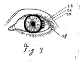
- FIG. 3 shows a front view of the suction ring without attached functional element in use.
- the suction ring includes a vacuum supply 20 connected to the suction portion 4, a vacuum supply 22 connected to the suction portion 10, and a vacuum supply 24 connected to the suction portion 6.
- the suction ring does not have to be substantially circular. It can also be designed, for example, as an elliptical ring or polygon ring.
- FIGS. 4 and 5 show a comparative example with a suction ring 116, a support member 102, an applanation element 104, a feeder 110 with at least one vacuum line and a power supply and a signal connection for at least one measuring means.
- a suction ring 116 At the bottom of the suction ring 116 is a first suction region 128, with which the suction ring 116 is fixed relative to the eye 122.
- the suction ring 116 can fix the holding element 102 via a second suction region 124 in which a negative pressure prevails.
- the holding element 102 fixes the applanation element 104 via a third, optional suction region 126.
- the radiation of a laser system (not shown), for example a femtosecond laser system, is coupled in by the applanation element 104.
- the applanation element can also be designed as a lens.
- the applanation element 104 abuts the cornea of the eye 122 at its lower end, thereby determining the position of the cornea.
- the mechanical guides 106 may include a force sensor (not shown). Furthermore, the force sensor can be arranged on the mechanical guides, in which case the optics of the laser are coupled to the force sensors.
- the force sensor may for example be a piezoelectric sensor or it may be constructed by means of strain gauges.
- the measuring means can be provided by microelectromechanical systems (MEM systems).
- MEM systems microelectromechanical systems
- At the edge of the applanation element 104 is a plurality of fiber sensors 108 for performing a spectroscopic method, for example in the near infrared range, and / or for carrying out a method for determining the light scattering.
- Properties of the cornea for example its water content, can be determined with these fiber sensors.
- the transparency of the cornea can also be determined.
- a separate transparency sensor 114 is provided, which determines its transparency, for example by determining the light scattering of the cornea.
- a mechanical spectroscopy device 112 determines, for example due to mechanical resonance, the biomechanical properties of the cornea.
- the device is supplied with negative pressure. Furthermore, a power supply is ensured with the feed device 110, and the measurement signals of the measuring means are transmitted to an evaluation device (not shown) via an electrical cable, a glass fiber and / or radio.
- contact elements may be provided to transmit electrical signals and to provide a power supply.
- position measuring means 120 are further provided to determine the position of the suction ring 116 and the support member 102 in space.
- the position measuring means 120 may be, for example, acoustic or optical sensors that determine the position relative to a reference geometry.
- the position sensors 120 may be purely passive sensors which receive a beam emitted by a reference location, the position of the suction ring 116 or of the support element 102 being determined on the basis of the received signal.
- the position-measuring means 120 may be purely active elements that have an optical or acoustic Send out beam, which is received by a corresponding receiving device at a reference location, wherein on the basis of the received signal, the position of the position measuring means 120 is determined in space.
- the support member 102 and reference marks can be arranged, which are detected by a camera arranged in space.
- optical sensors can be arranged in the interior of the eye-suction device, which detect the position of the eye. With the aid of the detected position of the eyes relative to the suction ring 116 and the position of the suction ring in the space detected via the position measuring means 120, the position of the eye 122 in space can be determined.
- the means for determining the position of the suction ring in the space may be coupled to a position control (not shown) of a treatment couch or a position control (not shown) of a laser system.
- the suction ring 116 touches the eye in use.
- a force sensor 118 is integrated to measure the force acting on the eye, from which the intraocular pressure can be determined.
- a plurality of force sensors 118 may be arranged. Thereby, a force profile along the circumference of the suction ring 116 can be created, whereby the corneal profile can be determined and accordingly the intraocular pressure can be determined more accurately.
- the suction ring 116 or the support member 102 may further include a fourth suction area (not shown) that evacuates the area between the applanation element and the cornea so that the cornea of the eye 122 securely abuts the applanation element 104.
- the suction ring 116 and the support member 102 may be integrally formed or positively or positively connected.
- the present invention has the advantage that it provides a redundancy, since on the one hand the suction ring is sucked to the eye and on the other hand a part of the cornea is sucked to a functional element or an element arranged thereon. Furthermore, the invention has the advantage that before, during and / or after the operation, the surgeon can easily change a functional element, for example a lens, an applanation lens or a connection area for the optics of a laser system, without the use of a complicated tool.
- the invention also has the advantage that for arranging the functional element No mechanical fasteners are required, which helps miniaturize the eye-suction device and reduce its weight. Since no mechanical fasteners are required, the eye-suction can be made of fewer components and simpler, whereby the surgeon receives a better view of the surgical field and the risk of failure is reduced.
- each of the suction areas can be divided into a plurality of self-sufficient suction areas, whereby a further redundancy with increased security arises.
- the present invention has the advantage that it provides measurement data of the eye before, during and / or after an operation.
- the invention provides measurement data relating to the intraocular pressure, the position of the suction ring in space, the mechanical force acting on an eye, measurement data of the cornea, for example its water content, its biomechanical properties and its transparency. Furthermore, no additional gauges need to be provided to monitor the eye during surgery.
Landscapes
- Health & Medical Sciences (AREA)
- Ophthalmology & Optometry (AREA)
- Life Sciences & Earth Sciences (AREA)
- Animal Behavior & Ethology (AREA)
- Veterinary Medicine (AREA)
- Surgery (AREA)
- Engineering & Computer Science (AREA)
- Biomedical Technology (AREA)
- Heart & Thoracic Surgery (AREA)
- Vascular Medicine (AREA)
- Public Health (AREA)
- Nuclear Medicine, Radiotherapy & Molecular Imaging (AREA)
- General Health & Medical Sciences (AREA)
- Physics & Mathematics (AREA)
- Optics & Photonics (AREA)
- Eye Examination Apparatus (AREA)
- Laser Surgery Devices (AREA)
- Prostheses (AREA)
- Instructional Devices (AREA)
- Surgical Instruments (AREA)
Description
- Die Erfindung betrifft eine Vorrichtung für die ophthalmologische Chirurgie.
- Gepulste Laserstrahlung wird in der Augenchirurgie beispielsweise zum Anbringen von Schnitten in der Hornhaut (Kornea) oder zum Abtrag (Ablation) von Gewebe aus der Hornhaut verwendet. Die eingestrahlte Laserstrahlung bewirkt im Hornhautgewebe einen photodisruptiven bzw. photoablativen Prozess, der zur Gewebetrennung bzw. zur Entfernung von Gewebematerial führt. Derartige Bearbeitungen der Kornea finden beispielsweise im Rahmen von refraktiven Verfahren zur Minderung oder vollständigen Behebung von Fehlsichtigkeiten des Auges statt, bei denen die Kornea neu geformt wird und hierdurch ihre Brechungseigenschaften verändert werden.
- Das dominierende refraktive Verfahren der Hornhautchirurgie ist die sogenannte LASIK (Laser in situ Keratomileusis). Hierbei wird aus der Hornhaut entweder mechanisch (mittels einer oszillierenden Schneidklinge in einem sogenannten Mikrokeratom) oder optisch (mittels Laserstrahlung, z.B. mittels Femtosekunden-Lasersysteme) ein kleiner Deckel herausgeschnitten, der mit einem Teil seines Randes noch an der Hornhaut hängt. Anschließend wird dieser üblicherweise auch als Flap bezeichnete Deckel zur Seite geklappt, wodurch das darunter liegende Stroma zugänglich wird. Mit Laserstrahlung wird dann nach Maßgabe eines für den jeweiligen Patienten ermittelten Ablationsprofils Stromagewebe abgetragen. Der Deckel wird danach wieder zurückgeklappt, wodurch die Wunde relativ schnell verheilen kann.
- Für eine präzise Einkopplung der Laserstrahlung in das Auge ist hierbei bekannt, das Auge mittels einer Fixationsvorrichtung zu fixieren, welche durch Unterdruck am Auge angesaugt wird. Die Fixationsvorrichtung kann ein als Einkoppelelement für die Laserstrahlung dienendes Glas aufweisen. Derartige Fixationsvorrichtungen werden auch als Saugringe bezeichnet.
- Sobald der Saugring am Auge eines Patienten angeordnet und mittels eines Unterdruckes an dem Auge fixiert ist, wird mittels Impulsen eines Femtosekunden-Lasers Energie in das Hornhautinnere eingebracht. Dadurch wird in der Hornhaut ein Schnitt erzeugt, der Flap kann aufgeklappt werden und die Korrektur der Fehlsichtigkeit kann durch einen definierten Abtrag von freigelegtem Hornhautgewebe erfolgen.
- Saugringe per se sind dem Fachmann bekannt, beispielsweise offenbaren die
US 5 336 215 undUS 5 549 632 Saugringe, die in ihrem Umfangsbereich als Saugbereiche ausgebildete Öffnungen zum Ansaugen an ein Auge umfassen. DieEP 0 993 814 A1 sowie dieUS 6 342 053 B1 offenbaren Saugringe, bei denen ein Unterdruck im Bereich einer Applanationsfläche erzeugt wird, so dass die Hornhaut des Auges daran anliegt. DieUS 6 344 040 B1 zeigt einen Saugring, bei dem ein Unterdruck im Bereich einer Applanationsfläche erzeugt wird, wobei der Saugring ferner eine Sonde umfasst, die im Einsatz die Hornhaut durchsticht und mittels einer Absaugung die während des photodisruptiven Verfahrens entstehenden Gase und Partikel absaugt. DieWO 03/002008 A1 - Die
WO 00/41660 A1 - Die
WO 03/001991 A1 -
WO 2004/096106 A1 befasst sich mit einem mechanischen Mikrokeratom zur Anbringung von Schnitten in der Kornea eines Auges. Das Mikrokeratom weist einen am Auge fixierbaren Saugring auf, mit dem eine Aufnahme für einen Applanationsstempel schraubgekoppelt ist. Der Stempel ist mittels Unterdruck in der Aufnahme gehalten. -
EP 1 199 055 A1 zeigt in seinerFigur 4 ebenfalls ein mechanisches Mikrokeratom mit einer Schneidklinge zum Schneiden eines Flaps in der Kornea. Das Mikrokeratom besitzt einen als Positionierring bezeichneten Saugring sowie einen Träger, an dem ein zum Durchschauen nutzbares Kontaktglas gehalten ist, das mit seiner Unterseite für eine Einebnung der Kornea sorgt. Sowohl der Saugring als auch der Träger sind jeweils durch eine Saugkammer am Auge festgesaugt. Der Saugring und der Träger sind fest miteinander verbunde. Beim Aufsetzen des Mikrokeratoms auf das Auge drückt das Kontaktglas gegen die Kornea und ebnet diese ein. Anschließend werden die beiden Saugkammern evakuiert. -
US 4,546,773 betrifft eine Messsonde mit einem Ultraschallwandler zur Messung der Korneadicke. -
WO 2006/090217 A1 befasst sich mit einer Vorrichtung zur Ankopplung eines zu behandelnden Auges an ein Lasersystem, wobei gemäß dem Ausführungsbeispiel derFigur 4 die Vorrichtung einen sich trichterförmig erweiternden Saugring aufweist, der mit einem mit dem Lasersystem gekoppelten konischen Adapter in Eingriff bringbar ist. Der Saugring trägt eine Linse, an dessen Unterseite sich die Hornhaut des Auges anschmiegt. -
WO 2006/121066 A1 ist mit einem Kühlaufsatz für ein mittels Laserstrahlung behandeltes Auge befasst. Der Kühlaufsatz wird auf das Auge aufgesetzt, ohne dabei an das Auge angesaugt zu werden. Er enthält eine unmittelbar zur Augenoberfläche hin offene und durch ein Kontaktelement vom Auge getrennte Kühlkammer, welche mit einem Kühlfluid befüllt wird. -
EP 0 327 693 A1 beschreibt ein Gerät zur Untersuchung des Augenhintergrunds und Bestimmung des Augeninnendrucks. Das Gerät umfasst einen Andruckkörper mit einer konkav gewölbten Andruckfläche, mit welcher er auf das Auge aufgesetzt wird. Der Andruckkörper ist mit verschiedenen Sensoren bestückt, welche ihre Messdaten an eine Auswerteschaltung liefern. -
US 6,126,668 betrifft wiederum ein mechanisches Mikrokeratom mit einem Saugring und einer auf das Auge aufzudrückenden Applanationsplatte. -
WO 2005/048895 A1 zeigt in ihrer Figur 7 einen Adapter zur Ankopplung eines Auges an ein Lasersystem. Der Adapter besitzt ein in einen Halter eingeklebtes Kontaktglas zur Anlage an der Hornhaut des Auges. Der Halter weist einen abstehende Ringfortsatz auf, der beim Aufsetzen des Kontaktglases auf das Auge zuerst in Kontakt mit der Augenoberfläche kommt. Eine zwischen dem Ringfortsatz und der Unterseite des Kontaktglases begrenzte Kammer ist über einen Stutzen evakuierbar. Hierdurch ist der Halter mit dem Kontaktglas am Auge festsaugbar. -
US 2005/0192562 A1 zeigt ein Augenpositioniersystem, das dem ausWO 2006/090217 A1 entspricht. - Erfindungsgemäß ist in Übereinstimmung mit Anspruch 1 eine Vorrichtung für die Augenchirurgie vorgesehen, umfassend einen auf ein zu behandelndes Auge aufsetzbaren Saugring mit einem ersten Saugbereich, der dazu ausgebildet ist, durch Unterdruckerzeugung in dem ersten Saugbereich den Saugring an das Auge anzusaugen, und ein an den Saugring ankoppelbares Funktionselement mit einem optischen Element zur Anlage der Hornhaut des Auges. Die Neuerung besteht hierbei darin, dass der Saugring einen weiteren Saugbereich aufweist, welcher dazu augebildet ist, durch Unterdruckerzeugung in dem weiteren Saugbereich das Funktionselement an den Saugring anzusaugen und die Hornhaut an das optische Element anzusaugen.
- Bevorzugte Weiterbildungen dieses Erfindungsgedankens finden sich in den abhängigen Ansprüchen 2 bis 13.
- Der Saugbereich kann beispielsweise durch eine Öffnung oder eine Ausnehmung gebildet sein, in der im Einsatz ein Unterdruck herrscht. Der Unterdruck kann beispielsweise durch eine mit dem Saugbereich verbundene Saugpumpe erzeugt werden. Es ist jedoch auch möglich, dass der Unterdruck beim Ankoppeln des Funktionselementes an den Saugring oder beim Ankoppeln des Saugrings an das Auge automatisch entsteht, indem beispielsweise eine Dichtlippe verschoben wird, so dass ein evakuierter Saugbereich entsteht.
- Das Funktionselement umfasst ein optisches Element, beispielsweise ein Glas oder eine Linse, durch welche die Laserstrahlung in die Hornhaut eingebracht wird. Das optische Element kann eine Linse oder eine Applanationsplatte sein. Das Funktionselement kann ein Halterungselement sein, das dazu ausgebildet ist, dass weitere Elemente daran angeordnet werden können. Das optische Element kann an dem Halterungselement angeordnet sein. Dadurch entsteht eine besonders vielseitig einsetzbare Vorrichtung, da das optische Element sehr einfach ausgewechselt werden kann. Das Funktionselement kann dazu ausgebildet sein, mit einer optischen Einrichtung gekoppelt zu werden. Die optische Einrichtung kann ein Lasersystem, beispielsweise ein Femtosekunden-Lasersystem, mit einer dazugehörigen Optik sein. Das Funktionselement kann auch ein eingangs genanntes mechanisches Mikrokeratom sein. Das Funktionselement kann auch ein Adaptionskegel sein, mit dem der Saugring an eine ophthalmologische Einrichtung gekoppelt wird. Das Funktionselement kann sowohl zur Halterung eines Applanationselementes als auch zum Koppeln der Augensaugeinrichtung mit der ophthalmologischen Einrichtung vorgesehen sein. Im Folgenden wird ein derartiges Funktionselement als Halterungselement bezeichnet.
- Die Vorrichtung kann eine erste Unterdruckzuführung, die mit dem ersten Saugbereich verbunden ist, und eine dritte Unterdruckzuführung, die mit einem dritten Saugbereich verbunden ist, umfassen. Der dritte Saugbereich ist an dem Saugring oder dem Funktionselement angeordnet und ist dazu ausgebildet, im Einsatz das Funktionselement an den Saugring zu saugen. Die Unterdruckzuführungen können an eine oder mehrere Saugpumpen angeschlossen sein. Im Einsatz kann in der ersten Unterdruckzuführung ein anderer Unterdruck herrschen als in der dritten Unterdruckzuführung, wodurch sich unterschiedliche Unterdrücke im ersten und im dritten Saugbereich ergeben. Dadurch ist es möglich, das Funktionselement sicher mit der Augensaugeinrichtung zu koppeln, ohne dass dabei im ersten Saugbereich ein so hoher Unterdruck erzeugt wird, dass das Auge verletzt werden könnte.
- Die Vorrichtung kann derart ausgebildet sein, dass im Einsatz zumindest ein Bereich der Hornhaut eines Auges an dem Funktionselement und/oder an einem daran angeordneten Element anliegt. Dadurch ergibt sich eine genaue Fixierung der Hornhaut, was einen sicheren Operationsvorgang sicherstellt. Der weitere Saugbereich steht im Einsatz mit der Oberfläche des Funktionselementes und/oder mit der Oberfläche des daran befestigen Elementes, an dem/denen im Einsatz die Hornhaut des Auges anliegt, in Fluidverbindung . Dadurch wird die Hornhaut besonders gut in ihrer Position festgelegt, da der Bereich zwischen der Hornhaut und dem Funktionselement bzw. einem an dem Halterungselement angeordneten Element evakuiert wird bzw. unter einem Unterdruck steht. Ferner ist es möglich, den in dem Bereich zwischen der Hornhaut und dem Funktionselement und/oder einem daran angeordneten Element herrschenden Unterdruck unabhängig vom Unterdruck im ersten und dritten Saugbereich einzustellen. Dadurch wird die Verletzungsgefahr minimiert und/oder der Behandlungskomfort für einen Patienten erhöht, da sich die Kraft, mit der die Augensaugeinrichtung an das Auge gesaugt wird, von der Kraft unterscheiden kann, mit der die Hornhaut an das Funktionselement und/oder an ein daran angeordnetes Element gesaugt wird. Zudem entsteht auch eine besonders sichere Vorrichtung, die eine Redundanz aufweist, da zwei Unterdrucksysteme verwendet werden, um einerseits den Saugring am Auge zu fixieren und andererseits die Hornhaut an einem Funktionselement und/oder an einem daran angeordneten Element zu fixieren.
- Bei einer vereinfachten Ausführungsform kann die Unterdruckzuführung des weiteren Saugbereichs mit der ersten oder der dritten Unterdruckzuführung in Fluidverbindung stehen.
- Die Vorrichtung kann zumindest ein Messmittel umfassen. Der Ausdruck Messmittel umfasst in diesem Kontext auch das qualitative und/oder quantitative Bestimmen oder Ermitteln einer geometrischen, physikalischen und/oder chemischen Größe.
- Zumindest eines der Messmittel der Vorrichtung kann dazu ausgebildet sein, eine Messung bezüglich einer Eigenschaft des Auges durchzuführen. Das Messmittel kann dazu ausgebildet sein, den Augeninnendruck zu bestimmen, wobei der Augeninnendruck beispielsweise mittels taktiler, mechanischer, akustischer und optischer Verfahren, insbesondere auch Resonanzverfahren, ermittelt werden kann.
- Zumindest ein Messmittel der Vorrichtung kann dazu ausgebildet sein, Eigenschaften der Hornhaut des Auges zu messen. Das Messmittel zum Messen der Eigenschaften der Hornhaut des Auges kann den Wassergehalt der Hornhaut, eine biomechanische Eigenschaft der Hornhaut und/oder die Transparenz der Hornhaut messen. Der Wassergehalt kann beispielsweise mittels eines optischen Spektrometers bestimmt werden, die biomechanischen Eigenschaften der Hornhaut können beispielsweise durch mechanische Spektroskopieverfahren bestimmt werden und die Transparenz der Hornhaut kann durch Lichtstreuung bestimmt werden.
- Die Vorrichtung kann ein Messmittel zum Messen einer auf das Auge wirkenden Größe umfassen. Das Messmittel zum Messen einer auf das Auge wirkenden Größe kann eine auf das Auge wirkende Kraft messen. Dazu können Drucksensoren, beispielsweise piezoelektrische Drucksensoren, oder Kraftsensoren in den Saugring integriert werden. Ferner ist es möglich, mikroelektromechanische Systeme (MEM-Systeme) zu verwenden, die beispielsweise an dem optischen Element angeordnet sind, über das die Laserstrahlung eingebracht wird.
- Die Vorrichtung kann zumindest ein Messmittel zur Messung einer Eigenschaft des Auges und/oder der Augensaugeinrichtung bezüglich der Umgebung umfassen. Das Messmittel zum Messen einer Eigenschaft des Auges und/oder der Augensaugeinrichtung bezüglich der Umgebung kann beispielsweise die Position des Auges und/oder der Augensaugeinrichtung im Raum messen. Das Mittel zum Messen der Eigenschaft des Auges und/oder des Saugringes gegenüber der Umgebung kann dazu ausgelegt sein, mit Positionierungsmitteln für eine Behandlungsliege und/oder Positionierungsmitteln für Laserstrahlung zusammenzuwirken. Dadurch kann sichergestellt werden, dass sich das Auge immer in der korrekten Position befindet und die Laserstrahlung an der korrekten Position und unter dem korrekten Winkel auf die Hornhaut des Auges auftrifft. Die Messung der Position des Auges und/oder des Saugringes kann auf einer mechanischen, hochfrequenzbasierten, akustischen oder (dreidimensionalen) optischen Positionserkennung beruhen. Die Messmittel können an einem Funktionselement angeordnet sein, das an den Saugring gesaugt wird.
- Die Messmittel können dazu ausgelegt sein, ermittelte Messdaten zu übertragen. Die Messdaten können induktiv, über ein Kabel, über eine optische Schnittstelle oder über eine elektromagnetische Schnittstelle übertragen werden. Das Messmittel kann eine Batterie umfassen, induktiv mit Strom versorgt werden oder über eine Zuleitung mit Strom versorgt werden. Das Messmittel kann ferner eine Kommunikationseinrichtung umfassen. Beispielsweise kann das Messmittel als Transponder ausgebildet sein, so dass von außen eine induktive oder elektromagnetische Anregung erfolgt und die Kommunikationseinrichtung des Messmittels die ermittelten Messwerte induktiv oder elektromagnetisch überträgt.
- Die Erfindung wird nachfolgend anhand der beigefügten Zeichnungen näher erläutert. Es stellen dar:
-
Figur 1 einen schematischen, nicht maßstabsgetreuen Schnitt durch eine erste Ausführungsform der Erfindung; -
Figur 2a einen schematischen, nicht maßstabsgetreuen Schnitt durch eine zweite Ausführungsform der Erfindung; -
Figur 2b einen schematischen, nicht maßstabsgetreuen Schnitt durch eine dritte Ausführungsform der Erfindung; -
Figur 3 eine schematische Frontansicht der Erfindung; -
Figur 4 eine perspektivische, nicht maßstabsgetreue Ansicht eines Vergleichsbeispiels, das einem Saugring mit einer Messeinrichtung umfasst; und -
Figur 5 einen schematischen, nicht maßstabsgetreuen Schnitt durch das Vergleichsbeispiel. -
Figur 1 zeigt einen Saugring 2 mit einem Saugbereich 4, der dazu ausgebildet ist, im Einsatz den Saugring 2 an ein Auge 18 zu saugen, und einem Saugbereich 10, der dazu ausgebildet ist, im Einsatz ein Funktionselement 12 anzusaugen. Das Funktionselement 12 umfasst ein optisches Element 11, das als Linse oder Platte 11 ausgebildet ist. Ferner umfasst das Funktionselement 12 einen Anschlussbereich 13, mit dem das Funktionselement an eine optische Einrichtung (nicht gezeigt) eines Lasersystems, beispielsweise eines Femtosekunden-Lasersystems, gekoppelt werden kann. Der Saugbereich 4 bildet einen ersten Saugbereich im Sinne der Erfindung. - Ferner umfasst der Saugring 2 einen Saugbereich 6, der ein weiterer Saugbereich im Sinne der Erfindung ist und mit dem Raum zwischen dem Funktionselement 12 und der Hornhaut 19 des Auges 18 in Fluidverbindung steht und diesen zumindest teilweise evakuiert, so dass zumindest ein Teil der Hornhaut an dem Funktionselement 12 anliegt. Der Bereich, an dem die Hornhaut 19 anliegt, kann als Applanationslinse bzw. Applanationsplatte 11 ausgebildet sein. Die Vorrichtung umfasst eine Mehrzahl von Unterdruckzuführungen 20, 22, 24 (in
Figur 1 ist nur eine erkennbar), die an einer oder je an einer Saugpumpe oder an einer Saugpumpe mit drei separaten Regelungseinrichtungen bzw. drei separaten Regelungsventilen angeschlossen sind. Dadurch kann in dem Saugbereich 4, dem Saugbereich 10 und dem Saugbereich 6 ein unterschiedlicher Unterdruck erzeugt werden. Im Saugbereich 10 kann ein hoher Unterdruck eingestellt werden, damit das Funktionselement 12 fest an dem Saugring 2 angeordnet ist. Im Saugbereich 4 und/oder im Saugbereich 6 kann ein niedrigerer Unterdruck eingestellt werden, damit das Auge 18 nicht verletzt wird. Der Operateur kann ferner vor der Operation lediglich einen Unterdruck im Saugbereich 10 anlegen, um das Funktionselement 12 am Saugring 2 anzuordnen. Beim Positionieren des Saugrings 2 an dem Auge 18 des Patienten kann im Saugbereich 4 ein Unterdruck erzeugt werden, um den Saugring 2 am Auge 18 sicher anzuordnen. Schließlich kann ein Unterdruck im dritten Saugbereich 6 erzeugt werden, damit die Hornhaut 19 des Auges 18 sicher am Funktionselement 12 angeordnet ist. Es ist jedoch auch denkbar, zuerst dem Saugring 2 am Auge 18 anzuordnen und anschließend ein Vakuum im zweiten Saugbereich 10 zu erzeugen, um das Funktionselement 12 anzuordnen. - Die erfindungsgemäße Vorrichtung vermeidet eine Verletzung des Auges, da im Saugbereich 4 ein anderer Unterdruck eingestellt werden kann als im zweiten Saugbereich 10 und/oder im Saugbereich 6. Ferner schafft die vorliegende Erfindung eine Redundanz, da der Saugring 2 sowohl durch den Saugbereich 4 als auch durch den Saugbereich 6 am Auge gehalten wird.
- Der Saugbereich 4 ist als umlaufende Nut im Saugring 2 ausgebildet. Es sind jedoch auch andere Ausgestaltungen möglich, beispielsweise eine Mehrzahl von umlaufenden Nuten, eine oder mehrere umlaufende oder nicht umlaufende Einbuchtungen oder eine Mehrzahl von Öffnungen. Der Saugbereich 10 ist durch zwei kreisförmig umlaufende Nuten an der Vorderseite oder Oberseite des Saugring 2 ausgebildet. Auch hier sind die zuvor bezüglich des Saugbereiches 4 beschriebenen Ausgestaltungen möglich. Der Saugbereich 6 wirkt bei der ersten Ausführungsform im Übergangsbereich vom Saugring 2 zum Funktionselement 12. Dadurch kann sichergestellt werden, dass ein Teil der Hornhaut 19 an die Oberfläche des Funktionselementes 12 gesaugt wird.
- Das Funktionselement 12 lässt sich einfach wechseln, ohne dass spezielle Werkzeuge und/oder aufwändige Bedienschritte dafür erforderlich sind. Das Funktionselement 12 kann im Einsatz einfach durch ein anderes ausgetauscht werden, das beispielsweise ein anderes optisches Element 11, Applanationselement 11 und/oder einen anderen Anschlussbereich 13 umfasst.
-
Figur 2a zeigt eine zweite Ausführungsform der Erfindung, wobei gleiche Bezugszeichen wie inFigur 1 gleiche Elemente bezeichnen. Bezüglich der Ausgestaltung der Saugbereiche 4, 6, 10 sowie der Unterdruckzuführungen wird auf die Beschreibung der Ausführungsform gemäßFigur 1 verwiesen. - Im Gegensatz zur Ausführungsform nach
Figur 1 wird bei der Ausführungsform nachFigur 2a ein Halterungselement 14 durch einen Unterdruck im Saugbereich 10 am Saugring 2 angeordnet. Das Halterungselement 14 kann ein beliebiges Element sein, an dem weitere Elemente angeordnet oder befestigt werden können. Das Halterungselement 14 umfasst an seinem dem Saugring 2 zugewandten Bereich eine kreisförmig umlaufende Ausnehmung, in der ein optisches Element 16 angeordnet ist. Das optische Element 16 kann als Applanationselement oder als Applanationslinse ausgebildet sein. Das Halterungselement 14 umfasst auch einen (nicht gezeigten) Anschlussbereich, mit dem das Halterungselement mit der Optik eines Lasersystems gekoppelt werden kann. - Durch den Unterdruck im Saugbereich 6 wird die Hornhaut 19 an das am Halterungselement 14 angeordnete optische Element 16 gesaugt.
-
Figur 2b zeigt eine dritte Ausführungsform der Erfindung, die der zweiten Ausführungsform ähnelt. Im Gegensatz zur zweiten Ausführungsform ist bei der dritten Ausführungsform Saugbereich 10' in dem Funktionselement 14' statt am Saugring 2' angeordnet. Über die Unterdruckzuführung 22' wird ein Unterdruck in dem Saugbereich 10' erzeugt. Die übrigen Ausgestaltungen der Augensaugeinrichtung 2' und des Funktionselementes 14' entsprechen den Ausgestaltungen der Augensaugeinrichtung 2 und des Funktionselementes 14 der zweiten Ausführungsform. -
Figur 3 zeigt eine Frontansicht des Saugrings ohne aufgesetztes Funktionselement im Einsatz. - Der Saugring umfasst eine Unterdruckzuführung 20, die mit dem Saugbereich 4 verbunden ist, eine Unterdruckzuführung 22, die mit dem Saugbereich 10 verbunden ist, und eine Unterdruckzuführung 24, die mit dem Saugbereich 6 verbunden ist. Der Saugring muss nicht im Wesentlichen kreisrund sein. Er kann beispielsweise auch als elliptischer Ring oder Polygonring ausgebildet sein.
-
Figuren 4 und 5 zeigen ein Vergleichsbeispiel mit einem Saugring 116, einem Halterungselement 102, einem Applanationselement 104, einer Zuführeinrichtung 110 mit zumindest einer Unterdruckleitung sowie einer Stromversorgung und einer Signalverbindung für zumindest ein Messmittel. An der Unterseite des Saugrings 116 befindet sich ein erster Saugbereich 128, mit dem der Saugring 116 gegenüber dem Auge 122 fixiert wird. Der Saugring 116 kann über einen zweiten Saugbereich 124, in dem ein Unterdruck herrscht, das Halterungselement 102 fixieren. Das Halterungselement 102 fixiert über einen dritten, optionalen Saugbereich 126 das Applanationselement 104. Durch das Applanationselement 104 wird die Strahlung eines (nicht gezeigten) Lasersystems, beispielsweise eines Femtosekunden-Lasersystems, eingekoppelt. Das Applanationselement kann auch als Linse ausgebildet sein. Das Applanationselement 104 liegt an seinem unteren Ende an der Hornhaut des Auges 122 an, wodurch die Position der Hornhaut festgelegt wird. - An der Oberseite des Halterungselementes 102 ist eine Mehrzahl von mechanischen Führungen 106 zum Ankoppeln des Strahlenganges des Lasers und/oder eines Verschlussmechanismus ausgebildet. Die mechanischen Führungen 106 können einen Kraftsensor (nicht dargestellt) umfassen. Ferner kann der Kraftsensor an den mechanischen Führungen angeordnet werden, wobei die Optik des Lasers in diesem Fall an die Kraftsensoren gekoppelt wird. Der Kraftsensor kann beispielsweise ein piezoelektrischer Sensor sein oder er kann mittels Dehnungsmessstreifen aufgebaut sein.
- An dem Applanationselement 104 können Messmittel zum Messen des Augeninnendruckes angeordnet sein. Die Messmittel können durch mikroelektromechanische Systeme (MEM-Systeme) bereitgestellt werden. Am Rand des Applanationselementes 104 befindet sich eine Mehrzahl von Fasersensoren 108 zum Durchführen eines Spektroskopieverfahrens, beispielsweise im nahen Infrarotbereich, und/oder zum Durchführen eines Verfahrens zur Bestimmung der Lichtstreuung. Mit diesen Fasersensoren können Eigenschaften der Hornhaut, beispielsweise deren Wassergehalt, bestimmt werden. Durch die Bestimmung der Lichtstreuung kann auch die Transparenz der Hornhaut bestimmt werden. Hierzu ist ein separater Transparenzsensor 114 vorgesehen, der beispielsweise durch Bestimmung der Lichtstreuung der Hornhaut deren Transparenz ermittelt. Eine mechanische Spektroskopieeinrichtung 112 ermittelt, beispielsweise aufgrund mechanischer Resonanz, die biomechanischen Eigenschaften der Hornhaut. Über die Zuführeinrichtung 110 wird der Vorrichtung Unterdruck zugeführt. Ferner wird mit der Zuführeinrichtung 110 eine Stromversorgung gewährleistet, und die Messsignale der Messmittel werden an eine (nicht gezeigte) Auswerteeinrichtung über ein elektrisches Kabel, eine Glasfaser und/oder Funk übertragen. An dem Applanationselement 104 und dem Halterungselement 102 können Kontaktelemente vorgesehen werden, um elektrische Signale zu übertagen und eine Stromversorgung bereitzustellen.
- An dem Halterungselement 102 sind ferner Positionsmessmittel 120 vorgesehen, um die Position des Saugrings 116 bzw. des Halterungselementes 102 im Raum zu bestimmen. Die Positionsmessmittel 120 können beispielsweise akustische oder optische Sensoren sein, die die Position gegenüber einer Referenzgeometrie bestimmen. Ferner kann es sich bei den Positionssensoren 120 um rein passive Sensoren handeln, die einen von einem Referenzort ausgestrahlten Strahl empfangen, wobei auf Basis des empfangenen Signals die Position des Saugrings 116 bzw. des Halterungselementes 102 bestimmt wird. Ebenso können die Positionsmessmittel 120 rein aktive Elemente sein, die einen optischen oder akustischen Strahl aussenden, der von einer entsprechenden Empfangseinrichtung an einem Referenzort empfangen wird, wobei auf Basis des empfangenen Signals die Position der Positionsmessmittel 120 im Raum bestimmt wird. An dem Halterungselement 102 können auch Referenzmarken angeordnet sein, die von einer im Raum angeordneten Kamera erfasst werden. Ebenso können im Inneren der Augensaugeinrichtung optische Sensoren angeordnet sein, die die Position des Auges erfassen. Mit Hilfe der erfassten Position der Augen gegenüber dem Saugring 116 und der über die Positionsmessmittel 120 erfassten Position des Saugringes im Raum kann die Position des Auges 122 im Raum bestimmt werden.
- Die Mittel zum Bestimmen der Position des Saugringes im Raum können mit einer (nicht gezeigten) Positionssteuerung einer Behandlungsliege oder einer (nicht gezeigten) Positionssteuerung eines Lasersystems gekoppelt werden.
- Der Saugring 116 berührt im Einsatz das Auge. In den Saugring 116 ist ein Kraftsensor 118 integriert, um die auf das Auge wirkende Kraft zu messen, woraus der Augeninnendruck bestimmt werden kann. Im Saugring116 kann eine Mehrzahl von Kraftsensoren 118 angeordnet sein. Dadurch kann ein Kraftprofil entlang dem Umfang des Saugrings 116 erstellt werden, wodurch das Hornhautprofil ermittelt werden kann und demgemäss der Augeninnendruck genauer ermittelt werden kann.
- Der Saugring 116 oder das Halterungselement 102 kann ferner einen vierten Saugbereich (nicht gezeigt) umfassen, der den Bereich zwischen dem Applanationselement und der Hornhaut evakuiert, damit die Hornhaut des Auges 122 sicher an dem Applanationselement 104 anliegt.
- Der Saugring 116 und das Halterungselement 102 können integral ausgebildet sein oder kraft- oder formschlüssig verbunden sein.
- Die vorliegende Erfindung hat den Vorteil, dass sie eine Redundanz schafft, da einerseits der Saugring an das Auge gesaugt wird und andererseits ein Teil der Hornhaut an ein Funktionselement oder ein daran angeordnetes Element gesaugt wird. Ferner hat die Erfindung den Vorteil, dass der Operateur vor, während und/oder nach der Operation ohne Verwendung eines aufwändig zu bedienenden Werkzeuges ein Funktionselement, beispielsweise eine Linse, eine Applanationslinse oder einen Anschlussbereich für die Optik eines Lasersystems, einfach wechseln kann. Die Erfindung hat ferner den Vorteil, dass zum Anordnen des Funktionselementes keine mechanischen Befestigungselemente erforderlich sind, was dazu beiträgt, die Augensaugeinrichtung zu miniaturisieren und deren Gewicht zu reduzieren. Da keine mechanischen Befestigungselemente erforderlich sind, kann die Augensaugeinrichtung aus weniger Bauteilen und einfacher aufgebaut werden, wodurch der Operateur eine bessere Sicht auf das Operationsfeld erhält und das Ausfallsrisiko reduziert wird.
- Es versteht sich, dass jeder der Saugbereiche in eine Mehrzahl von autarken Saugbereichen aufgeteilt werden kann, wodurch eine weitere Redundanz mit erhöhter Sicherheit entsteht.
- Die vorliegende Erfindung hat den Vorteil, dass sie vor, während und/oder nach einer Operation Messdaten des Auges liefert. Die Erfindung liefert Messdaten bezüglich des Augeninnendrucks, der Position des Saugringes im Raum, der mechanischen Kraft, die auf ein Auge wirkt, Messdaten der Hornhaut, beispielsweise deren Wassergehalt, deren biomechanische Eigenschaften und deren Transparenz. Ferner müssen keine zusätzlichen Messgeräte bereitgestellt werden, um das Auge während einer Operation zu überwachen.
- Die Erfindung wurde mittels mehrerer Ausführungsformen beschrieben. Der Fachmann versteht, dass die Merkmale und Merkmalskombinationen der verschiedenen Ausführungsformen kombiniert werden können.
Claims (13)
- Vorrichtung für die Augenchirurgie, umfassend- einen auf ein zu behandelndes Auge aufsetzbaren Saugring (2) mit einem ersten Saugbereich (4), der dazu ausgebildet ist, durch Unterdruckerzeugung in dem ersten Saugbereich den Saugring an das Auge (18) anzusaugen, und- ein an den Saugring ankoppelbares Funktionselement (14) mit einem optischen Element (16) zur Anlage der Hornhaut des Auges, wobei der Saugring einen weiteren Saugbereich (6) aufweist, welcher dazu ausgebildet ist, durch Unterdruckerzeugung in dem weiteren Saugbereich das Funktionselement an den Saugring anzusaugen, dadurch gekennzeichnet, dass der weitere Saugbereich dazu ausgebildet ist, durch Unterdruckerzeugung in dem weiteren Saugbereich die Hornhaut an das optische Element anzusaugen.
- Vorrichtung nach Anspruch 1, dadurch gekennzeichnet, dass das Funktionselement (14) einen Anschlussbereich zur Ankopplung an ein Lasersystem aufweist.
- Vorrichtung nach Anspruch 1 oder 2, dadurch gekennzeichnet, dass das Funktionselement (14) ein Halterungselement mit einer Ausnehmung ist, in Welcher das optische Element (16) angeordnet ist.
- Vorrichtung nach einem der Ansprüche 1 bis 3, dadurch gekennzeichnet, dass das optische Element eine Linse ist.
- Vorrichtung nach einem der Ansprüche 1 bis 3, dadurch gekennzeichnet, dass das optische Element eine Platte ist.
- Vorrichtung nach einem der vorhergehenden Ansprüche, dadurch gekennzeichnet, dass der Saugring (2) eine mit dem ersten Saugbereich (4) verbundene erste Unterdruckzuführung (20) sowie eine mit dem weiteren Saugbereich (6) verbundene zweite Unterdruekzuführung (24) aufweist.
- Vorrichtung nach einem der vorhergehenden Ansprüche, dadurch gekennzeichnet, dass der Unterdruck in dem weiteren Saugbereich (6) unabhängig von dem Unterdruck in dem ersten Saugbereich (4) einstellbar ist.
- Vorrichtung nach einem der vorhergehenden Ansprüche, gekennzeichnet durch mindestens ein an dem Saugring (116) oder/und an dem Funktionselement (102) angeordnetes Messmittel (120) zur Messung einer Eigenschaft des Auges und/oder des Saugrings bezüglich der Umgebung.
- Vorrichtung nach Anspruch 8, dadurch gekennzeichnet, dass zumindest eines der Messmittel (120) als Positionsmessmittel zur Messung der Position des Saugrings (116) oder/und des Funktionsetements (102) im Raum ausgebildet ist
- Vorrichtung nach Anspruch 9, dadurch gekennzeichnet, dass das Positionsmessmittel (120) dazu ausgelegt ist, mit Positionierungsmitteln für eine Behandlungsliege und/oder Positionierungsmitteln für eine Laserstrahlung zusammenzuwirken.
- Vorrichtung nach einem der Ansprüche 8 bis 10, dadurch gekennzeichnet, dass das Messmittel (120) zum leichteren Desinfizieren und/oder Sterilisieren hermetisch abgeschlossen ist.
- Vorrichtung nach einem der Ansprüche 8 bis 11, dadurch gekennzeichnet, dass das Messmittel (120) dazu ausgelegt ist, ermittelte Messdaten zu übertragen.
- Vorrichtung nach Anspruch 12, dadurch gekennzeichnet, dass das Messmittel (120) ferner eine Kommunikationseinrichtung umfasst.
Priority Applications (20)
| Application Number | Priority Date | Filing Date | Title |
|---|---|---|---|
| ES09012579T ES2720365T3 (es) | 2007-03-14 | 2007-03-14 | Aparato para el acoplamiento de un elemento al ojo |
| EP07005280A EP1970034B1 (de) | 2007-03-14 | 2007-03-14 | Apparat zur Ankopplung eines Elements an das Auge |
| DE502007002400T DE502007002400D1 (de) | 2007-03-14 | 2007-03-14 | Apparat zur Ankopplung eines Elements an das Auge |
| EP09012579.0A EP2133048B1 (de) | 2007-03-14 | 2007-03-14 | Apparat zur Ankopplung eines Elementes an das Auge |
| ES07005280T ES2336611T3 (es) | 2007-03-14 | 2007-03-14 | Aparato para el acoplamiento de un elemento al ojo. |
| MX2009009744A MX2009009744A (es) | 2007-03-14 | 2008-03-13 | Aparato para acoplar un elemento al ojo. |
| CN201210101390.2A CN102697598B (zh) | 2007-03-14 | 2008-03-13 | 用于将元件连接到眼睛上的器械 |
| KR1020127018219A KR101189819B1 (ko) | 2007-03-14 | 2008-03-13 | 안과 수술용 장치 |
| CA2819455A CA2819455C (en) | 2007-03-14 | 2008-03-13 | Apparatus for coupling an element to the eye |
| BR122014017011A BR122014017011B8 (pt) | 2007-03-14 | 2008-03-13 | dispositivo para cirurgia ocular |
| PCT/EP2008/002014 WO2008110368A1 (de) | 2007-03-14 | 2008-03-13 | Apparat zur ankopplung eines elementes an das auge |
| US12/531,217 US8475433B2 (en) | 2007-03-14 | 2008-03-13 | Apparatus for coupling an element to the eye |
| BRPI0808324A BRPI0808324B8 (pt) | 2007-03-14 | 2008-03-13 | dispositivo para cirurgia ocular |
| RU2009133106/14A RU2463028C2 (ru) | 2007-03-14 | 2008-03-13 | Устройство для фиксации элемента к глазу |
| KR1020097021481A KR101274264B1 (ko) | 2007-03-14 | 2008-03-13 | 눈에 요소를 결합시키는 장치 |
| CA2680072A CA2680072C (en) | 2007-03-14 | 2008-03-13 | Apparatus for coupling an element to the eye |
| JP2009553076A JP5295134B2 (ja) | 2007-03-14 | 2008-03-13 | 素子を眼球に結合する装置 |
| CN2008800080831A CN101646406B (zh) | 2007-03-14 | 2008-03-13 | 用于将元件连接到眼睛上的器械 |
| US13/435,693 US8753321B2 (en) | 2007-03-14 | 2012-03-30 | Apparatus for coupling an element to the eye |
| JP2012288583A JP5525594B2 (ja) | 2007-03-14 | 2012-12-28 | 素子を眼球に結合する装置 |
Applications Claiming Priority (1)
| Application Number | Priority Date | Filing Date | Title |
|---|---|---|---|
| EP07005280A EP1970034B1 (de) | 2007-03-14 | 2007-03-14 | Apparat zur Ankopplung eines Elements an das Auge |
Related Child Applications (1)
| Application Number | Title | Priority Date | Filing Date |
|---|---|---|---|
| EP09012579.0A Division EP2133048B1 (de) | 2007-03-14 | 2007-03-14 | Apparat zur Ankopplung eines Elementes an das Auge |
Publications (2)
| Publication Number | Publication Date |
|---|---|
| EP1970034A1 EP1970034A1 (de) | 2008-09-17 |
| EP1970034B1 true EP1970034B1 (de) | 2009-12-23 |
Family
ID=38110731
Family Applications (2)
| Application Number | Title | Priority Date | Filing Date |
|---|---|---|---|
| EP07005280A Active EP1970034B1 (de) | 2007-03-14 | 2007-03-14 | Apparat zur Ankopplung eines Elements an das Auge |
| EP09012579.0A Active EP2133048B1 (de) | 2007-03-14 | 2007-03-14 | Apparat zur Ankopplung eines Elementes an das Auge |
Family Applications After (1)
| Application Number | Title | Priority Date | Filing Date |
|---|---|---|---|
| EP09012579.0A Active EP2133048B1 (de) | 2007-03-14 | 2007-03-14 | Apparat zur Ankopplung eines Elementes an das Auge |
Country Status (12)
| Country | Link |
|---|---|
| US (2) | US8475433B2 (de) |
| EP (2) | EP1970034B1 (de) |
| JP (2) | JP5295134B2 (de) |
| KR (2) | KR101274264B1 (de) |
| CN (2) | CN102697598B (de) |
| BR (2) | BRPI0808324B8 (de) |
| CA (2) | CA2680072C (de) |
| DE (1) | DE502007002400D1 (de) |
| ES (2) | ES2720365T3 (de) |
| MX (1) | MX2009009744A (de) |
| RU (1) | RU2463028C2 (de) |
| WO (1) | WO2008110368A1 (de) |
Cited By (2)
| Publication number | Priority date | Publication date | Assignee | Title |
|---|---|---|---|---|
| DE102022114573A1 (de) | 2022-06-09 | 2023-12-14 | Schwind Eye-Tech-Solutions Gmbh | Behandlungsvorrichtung und Verfahren zum Steuern einer Fixiervorrichtung zum Fixieren eines menschlichen oder tierischen Auges |
| DE102022005095B4 (de) | 2022-06-09 | 2026-06-25 | Schwind Eye-Tech-Solutions Gmbh | Behandlungsvorrichtung und Verfahren zum Steuern einer Fixiervorrichtung zum Fixieren eines menschlichen oder tierischen Auges |
Families Citing this family (99)
| Publication number | Priority date | Publication date | Assignee | Title |
|---|---|---|---|---|
| US20120016349A1 (en) * | 2001-01-29 | 2012-01-19 | Amo Development, Llc. | Hybrid ophthalmic interface apparatus and method of interfacing a surgical laser with an eye |
| US8262646B2 (en) | 2006-01-20 | 2012-09-11 | Lensar, Inc. | System and method for providing the shaped structural weakening of the human lens with a laser |
| US9545338B2 (en) | 2006-01-20 | 2017-01-17 | Lensar, Llc. | System and method for improving the accommodative amplitude and increasing the refractive power of the human lens with a laser |
| US10842675B2 (en) | 2006-01-20 | 2020-11-24 | Lensar, Inc. | System and method for treating the structure of the human lens with a laser |
| US9889043B2 (en) | 2006-01-20 | 2018-02-13 | Lensar, Inc. | System and apparatus for delivering a laser beam to the lens of an eye |
| DE502007002400D1 (de) * | 2007-03-14 | 2010-02-04 | Wavelight Ag | Apparat zur Ankopplung eines Elements an das Auge |
| WO2009036098A2 (en) * | 2007-09-10 | 2009-03-19 | Lensx Lasers, Inc. | Apparatus, systems and techniques for interfacing with an eye in laser surgery |
| US8632526B2 (en) * | 2007-11-07 | 2014-01-21 | Amo Development, Llc | System and method of interfacing a surgical laser with an eye |
| ES2442002T3 (es) * | 2008-06-20 | 2014-02-07 | Wavelight Gmbh | Dispositivo para cortar una parte de tejido con una radiación láser focalizada |
| US8500723B2 (en) | 2008-07-25 | 2013-08-06 | Lensar, Inc. | Liquid filled index matching device for ophthalmic laser procedures |
| US8480659B2 (en) | 2008-07-25 | 2013-07-09 | Lensar, Inc. | Method and system for removal and replacement of lens material from the lens of an eye |
| RU2485923C2 (ru) * | 2008-08-25 | 2013-06-27 | Уэйвлайт Гмбх | Способ обеспечения контакта глаза с лазерным устройством |
| US10624787B2 (en) | 2009-07-10 | 2020-04-21 | Alcon Inc. | Apparatus for cutting a tissue section of an eye by laser radiation |
| WO2011003431A1 (de) * | 2009-07-10 | 2011-01-13 | Wavelight Gmbh | Vorrichtung zum schneiden eines gewebeteils eines auges mittels laserstrahlung |
| JP2013500078A (ja) | 2009-07-24 | 2013-01-07 | レンサー, インク. | 眼の水晶体にレーザ照射パターンを照射するシステムおよび方法 |
| AU2010276360A1 (en) * | 2009-07-24 | 2012-02-16 | Lensar, Inc. | Liquid holding interface device for ophthalmic laser procedures |
| US8758332B2 (en) | 2009-07-24 | 2014-06-24 | Lensar, Inc. | Laser system and method for performing and sealing corneal incisions in the eye |
| US8617146B2 (en) | 2009-07-24 | 2013-12-31 | Lensar, Inc. | Laser system and method for correction of induced astigmatism |
| CA2769097A1 (en) | 2009-07-24 | 2011-01-27 | Lensar, Inc. | System and method for performing ladar assisted procedures on the lens of an eye |
| US8382745B2 (en) | 2009-07-24 | 2013-02-26 | Lensar, Inc. | Laser system and method for astigmatic corrections in association with cataract treatment |
| CN102811685A (zh) * | 2010-01-29 | 2012-12-05 | 雷萨公司 | 用于眼科应用的伺服控制的对接力设备 |
| CN102843955A (zh) | 2010-02-01 | 2012-12-26 | 雷萨公司 | 眼科应用中吸环基于浦肯野图像的对准 |
| US9408746B2 (en) | 2010-03-31 | 2016-08-09 | Ocuject, Llc | Device and method for intraocular drug delivery |
| AU2011235002B2 (en) * | 2010-03-31 | 2016-08-11 | Ocuject, Llc | Device and method for intraocular drug delivery |
| WO2011151812A1 (en) | 2010-05-10 | 2011-12-08 | Ramot At Tel-Aviv University Ltd. | System for treating glaucoma by directing electromagnetic energy to the limbal area of an eye |
| US8845624B2 (en) * | 2010-06-25 | 2014-09-30 | Alcon LexSx, Inc. | Adaptive patient interface |
| CN103228239B (zh) * | 2010-09-02 | 2015-04-01 | 眼科医疗公司 | 用于眼科诊断和介入操作的患者接口 |
| USD695408S1 (en) | 2010-10-15 | 2013-12-10 | Lensar, Inc. | Laser system for treatment of the eye |
| USD694890S1 (en) | 2010-10-15 | 2013-12-03 | Lensar, Inc. | Laser system for treatment of the eye |
| WO2012051490A1 (en) | 2010-10-15 | 2012-04-19 | Lensar, Inc. | System and method of scan controlled illumination of structures within an eye |
| US10463541B2 (en) | 2011-03-25 | 2019-11-05 | Lensar, Inc. | System and method for correcting astigmatism using multiple paired arcuate laser generated corneal incisions |
| DE102011006085A1 (de) | 2011-03-25 | 2012-09-27 | Carl Zeiss Meditec Ag | Ophthalmologisches Gerät |
| US20130102895A1 (en) * | 2011-10-21 | 2013-04-25 | Optimedica Corporation | Patient interface for ophthalmologic diagnostic and interventional procedures |
| US8863749B2 (en) | 2011-10-21 | 2014-10-21 | Optimedica Corporation | Patient interface for ophthalmologic diagnostic and interventional procedures |
| CA3050497C (en) * | 2011-10-21 | 2022-08-09 | Optimedica Corporation | Patient interface for ophthalmologic diagnostic and interventional procedures |
| US9237967B2 (en) | 2011-10-21 | 2016-01-19 | Optimedica Corporation | Patient interface for ophthalmologic diagnostic and interventional procedures |
| US9044302B2 (en) | 2011-10-21 | 2015-06-02 | Optimedica Corp. | Patient interface for ophthalmologic diagnostic and interventional procedures |
| WO2013126653A1 (en) * | 2012-02-22 | 2013-08-29 | Amo Development, Llc | Preformed lens systems and methods |
| EP2633841B1 (de) | 2012-02-28 | 2017-08-16 | Ziemer Ophthalmic Systems AG | Vorrichtung zur Behandlung von Augengewebe mittels gepulster Laserstrahlen |
| US9504603B2 (en) | 2012-04-02 | 2016-11-29 | Ocuject, Llc | Intraocular delivery devices and methods therefor |
| US9421129B2 (en) | 2012-04-02 | 2016-08-23 | Ocuject, Llc | Intraocular delivery devices and methods therefor |
| JP6202252B2 (ja) * | 2012-06-02 | 2017-09-27 | 株式会社ニデック | 眼科用レーザ手術装置 |
| US10314746B2 (en) | 2012-11-02 | 2019-06-11 | Optimedica Corporation | Laser eye surgery system calibration |
| US9445946B2 (en) | 2012-11-02 | 2016-09-20 | Optimedica Corporation | Laser eye surgery system |
| US9987165B2 (en) * | 2012-11-02 | 2018-06-05 | Optimedica Corporation | Liquid optical interface for laser eye surgery system |
| US20170056243A1 (en) * | 2012-11-02 | 2017-03-02 | Optimedica Corporation | Free floating patient interface for laser surgery system |
| US10285860B2 (en) | 2012-11-02 | 2019-05-14 | Optimedica Corporation | Vacuum loss detection during laser eye surgery |
| US10292863B2 (en) | 2012-11-02 | 2019-05-21 | Optimedica Corporation | Interface force feedback in a laser eye surgery system |
| US9603744B2 (en) | 2012-11-09 | 2017-03-28 | Technolas Perfect Vision Gmbh | Adaptable patient interface |
| US9724238B2 (en) * | 2012-11-30 | 2017-08-08 | Amo Development, Llc | Ophthalmic interface apparatus, method of interfacing a surgical laser with an eye, and support ring for use with a suction ring |
| US9861275B2 (en) | 2012-12-20 | 2018-01-09 | Wavelight Gmbh | Apparatus, interface unit, suction ring and method to monitor corneal tissue |
| US10335315B2 (en) * | 2013-02-01 | 2019-07-02 | Alcon Lensx, Inc. | Bi-radial patient interface |
| ES2799154T5 (en) | 2013-02-26 | 2026-06-25 | Belkin Vision Ltd | System for glaucoma treatment |
| US9398979B2 (en) | 2013-03-11 | 2016-07-26 | Technolas Perfect Vision Gmbh | Dimensional compensator for use with a patient interface |
| US10092393B2 (en) | 2013-03-14 | 2018-10-09 | Allotex, Inc. | Corneal implant systems and methods |
| CA3114952A1 (en) * | 2013-03-15 | 2014-09-25 | Amo Development, Llc | Hybrid ophthalmic interface apparatus |
| US20140276674A1 (en) * | 2013-03-15 | 2014-09-18 | Amo Development Llc. | System and method for ophthalmic laser surgery employing eye tracking without eye docking |
| JP6549099B2 (ja) | 2013-04-17 | 2019-07-24 | オプティメディカ コーポレイション | 白内障手術における軸アラインメントのためのレーザ基準 |
| US9610197B2 (en) | 2013-06-14 | 2017-04-04 | Wavelight Gmbh | Automatic machine settings for customized refractive surgery |
| CA2854507C (en) * | 2013-06-21 | 2017-01-03 | Alcon Inc. | An apparatus for performing ophthalmic surgery using a contact element |
| CN108378984B (zh) * | 2013-06-25 | 2020-06-26 | Tec晶体有限责任公司 | 用于眼睛的光线疗法的装置 |
| KR101435435B1 (ko) * | 2013-07-25 | 2014-09-01 | 주식회사 루트로닉 | 컨택트 렌즈 및 이를 갖는 안과용 치료장치 |
| JP6580679B2 (ja) | 2014-09-25 | 2019-09-25 | オプティメディカ・コーポレイションOptimedica Corporation | 角膜トポグラフィー、瞬き検出、及び、レーザ眼手術システムのための方法及びシステム |
| DE102014225636A1 (de) * | 2014-12-11 | 2016-06-16 | Carl Zeiss Meditec Ag | Vorrichtung und Verfahren zur Festlegung einer relativen geometrischen Lage eines Auges |
| DE102014225635A1 (de) * | 2014-12-11 | 2016-06-16 | Carl Zeiss Meditec Ag | Vorrichtung und Verfahren zur Festlegung einer relativen geometrischen Lage eines Auges |
| WO2016106217A1 (en) | 2014-12-22 | 2016-06-30 | TECLens, LLC | Corneal crosslinking with oxygenation |
| CN107920919B (zh) | 2015-03-18 | 2020-10-27 | 光学医疗公司 | 激光眼科手术期间的真空损失检测 |
| EP3092985B1 (de) * | 2015-05-15 | 2019-12-18 | WaveLight GmbH | Patientenadapter für eine augenlaservorrichtung |
| US10449090B2 (en) | 2015-07-31 | 2019-10-22 | Allotex, Inc. | Corneal implant systems and methods |
| ES2670672T3 (es) * | 2015-11-10 | 2018-05-31 | Novartis Ag | Adaptador modular de paciente para un dispositivo láser ocular |
| US10219948B2 (en) * | 2016-02-24 | 2019-03-05 | Perfect Ip, Llc | Ophthalmic laser treatment system and method |
| EP4091589A1 (de) | 2016-04-05 | 2022-11-23 | AMO Development, LLC | Verbindungsstück für die augen-laser-chirurgie |
| CN109843205B (zh) * | 2016-04-07 | 2022-04-05 | 雷萨公司 | 用于激光方法和系统的患者接合装置 |
| EP3260093B1 (de) * | 2016-06-22 | 2021-12-22 | Ziemer Ophthalmic Systems AG | Ophthalmologische patientenschnittstelle |
| JP7201441B2 (ja) | 2016-06-30 | 2023-01-10 | イリデックス・コーポレーション | 取り替え可能接触先端部材及び処置案内部材を有する手持ち式眼科レーザー・システム |
| WO2018052418A1 (en) * | 2016-09-14 | 2018-03-22 | Optimedica Corporation | Free floating patient interface for laser surgery system |
| RU2631108C1 (ru) * | 2016-10-06 | 2017-09-18 | Федеральное государственное автономное учреждение "Межотраслевой научно-технический комплекс "Микрохирургия глаза" имени академика С.Н. Федорова" Министерства здравоохранения Российской Федерации | Инструмент для защиты роговичного лоскута от лазерного воздействия при эксимерлазерной хирургии |
| EP3528758B1 (de) * | 2016-10-19 | 2021-10-13 | Alcon Inc. | Systeme für das andocken in der femtosekunden-laser-augenchirurgie |
| FR3063217B1 (fr) * | 2017-02-24 | 2019-04-05 | Keranova | Interface de couplage entre une source laser et un tissu a traiter |
| WO2018195139A1 (en) | 2017-04-19 | 2018-10-25 | Amo Development, Llc | Patient interface device for ophthalmic surgical laser system |
| US10966865B2 (en) * | 2017-07-27 | 2021-04-06 | Alcon Inc. | Femtosecond laser docking apparatus |
| DE102017215589A1 (de) | 2017-09-05 | 2019-03-07 | Carl Zeiss Meditec Ag | Flüssigkeits-Patienteninterface |
| DE102017123300B4 (de) | 2017-10-06 | 2026-07-02 | Schwind Eye-Tech-Solutions Gmbh | Patienteninterfacesystem, Verfahren zum Koppeln eines Patienteninterfaces mit einem Patienteninterfacehalter, Patienteninterface und Patienteninterfacehalter |
| US11135093B2 (en) * | 2017-12-12 | 2021-10-05 | Alcon Inc. | Patient interface for ophthalmic surgery |
| US10864075B2 (en) * | 2017-12-31 | 2020-12-15 | Rxsight, Inc. | Intraocular lens visualization and tracking system |
| IL279749B2 (en) | 2018-07-02 | 2024-04-01 | Belkin Vision Ltd | Direct selective laser trabeculoplasty |
| KR102120643B1 (ko) * | 2018-10-23 | 2020-06-10 | 한국표준과학연구원 | 눈 운동 저항과 힘 측정기 |
| WO2020089737A1 (en) | 2018-10-28 | 2020-05-07 | Belkin Laser Ltd. | Protection for direct selective laser trabeculoplasty |
| EP3945994B1 (de) * | 2019-03-27 | 2024-10-23 | Alcon Inc. | System und verfahren zur verwendung eines oder mehrerer bilder eines auges in medizinischen verfahren |
| JP2023519074A (ja) * | 2020-01-03 | 2023-05-10 | レンサー インク | 眼に対して音波とレーザーを複合適用するための方法及びシステム |
| CN115666464B (zh) | 2020-07-19 | 2026-04-14 | 贝尔金视觉有限公司 | 自动囊切开术 |
| FR3112681B1 (fr) * | 2020-07-22 | 2022-07-15 | Univ Bordeaux | Dispositif d’aplanation destiné à être couplé à un système de chirurgie ophtalmologique laser |
| US12564517B2 (en) | 2020-10-26 | 2026-03-03 | Belvin Vision Ltd. | Avoiding blood vessels during direct selective laser trabeculoplasty |
| US12465522B2 (en) | 2020-12-07 | 2025-11-11 | Iridex Corporation | Methods and probes for intrascleral laser surgery |
| CN114848292B (zh) * | 2022-04-19 | 2023-12-05 | 南京朔视科技开发有限公司 | 一种用于固定眼球的接口单元 |
| US12611101B2 (en) | 2022-10-11 | 2026-04-28 | Belkin Vision Ltd. | Gonioscopic laser surgery |
| CN117414249B (zh) * | 2023-09-26 | 2026-06-26 | 上海新眼光医疗器械股份有限公司 | 可精确调控吸附力的近视手术用负压吸引环及其使用方法 |
| CN117281684B (zh) * | 2023-11-24 | 2024-03-29 | 超目科技(北京)有限公司 | 眼球吸附装置和眼球吸附系统 |
| CN118453258B (zh) * | 2024-07-10 | 2024-10-11 | 广东麦特维逊医学研究发展有限公司 | 人眼对接接口装置 |
Family Cites Families (49)
| Publication number | Priority date | Publication date | Assignee | Title |
|---|---|---|---|---|
| US4546773A (en) * | 1981-01-23 | 1985-10-15 | Accutome, Inc. | Apparatus to measure conical thickness |
| US4798599A (en) * | 1984-01-03 | 1989-01-17 | George Thomas | Eye washing method and apparatus |
| DE3433581C2 (de) * | 1984-09-13 | 1986-08-07 | Fa. Carl Zeiss, 7920 Heidenheim | Vorrichtung zur lamellierenden, refraktiven Hornhautchirurgie |
| US4669466A (en) * | 1985-01-16 | 1987-06-02 | Lri L.P. | Method and apparatus for analysis and correction of abnormal refractive errors of the eye |
| US5133708A (en) * | 1988-01-14 | 1992-07-28 | Smith Robert F | Method for controlled corneal ablation |
| US5112328A (en) * | 1988-01-25 | 1992-05-12 | Refractive Laser Research & Development Program, Ltd. | Method and apparatus for laser surgery |
| CH673760A5 (de) * | 1988-02-09 | 1990-04-12 | Yves Dr Med Robert | |
| US5215104A (en) * | 1988-08-16 | 1993-06-01 | Steinert Roger F | Method for corneal modification |
| US6099522A (en) * | 1989-02-06 | 2000-08-08 | Visx Inc. | Automated laser workstation for high precision surgical and industrial interventions |
| US5133726A (en) * | 1990-02-14 | 1992-07-28 | Ruiz Luis A | Automatic corneal shaper |
| US6342053B1 (en) | 1990-07-23 | 2002-01-29 | Laser Biotech, Inc. | Apparatus for cornea reshaping |
| EP0629137A4 (de) * | 1992-01-15 | 1997-01-22 | Premier Laser Systems Inc | Hornhaut umformung durch laserenergie. |
| US5549632A (en) | 1992-10-26 | 1996-08-27 | Novatec Laser Systems, Inc. | Method and apparatus for ophthalmic surgery |
| EP0627207B1 (de) * | 1992-11-20 | 1998-09-16 | OKAMOTO, Shinseiro | Gerät für chirurgische eingriffe an der kornea |
| US5336215A (en) | 1993-01-22 | 1994-08-09 | Intelligent Surgical Lasers | Eye stabilizing mechanism for use in ophthalmic laser surgery |
| US5496339A (en) * | 1994-05-17 | 1996-03-05 | Koepnick; Russell G. | Universal automated keratectomy apparatus and method |
| US5586980A (en) * | 1994-10-14 | 1996-12-24 | Kremer; Frederic B. | Microkeratome |
| US5643299A (en) * | 1996-01-16 | 1997-07-01 | Sentinel Medical, Inc. | Hydrojet apparatus for refractive surgery |
| US5624456A (en) * | 1996-02-07 | 1997-04-29 | Hellenkamp; Johann F. | Automatic surgical device for cutting a cornea |
| US5828454A (en) * | 1996-07-29 | 1998-10-27 | Gust; Gary R. | Optical heterodyne interferometer for measurement of ocular displacement |
| US6126668A (en) | 1997-04-25 | 2000-10-03 | Innovative Optics, Inc. | Microkeratome |
| RU2170563C2 (ru) * | 1997-05-27 | 2001-07-20 | Валерий Григорьевич Боровик | Устройство для офтальмологических операций |
| RU2130762C1 (ru) * | 1997-12-10 | 1999-05-27 | Федоров Святослав Николаевич | Устройство для офтальмохирургических операций |
| US6143011A (en) * | 1998-04-13 | 2000-11-07 | Surgijet, Inc. | Hydrokeratome for refractive surgery |
| US6254595B1 (en) * | 1998-10-15 | 2001-07-03 | Intralase Corporation | Corneal aplanation device |
| WO2000041660A1 (en) | 1999-01-15 | 2000-07-20 | Medjet, Inc. | Corneal microjet cutter with adjustable applanation template |
| US6165189A (en) * | 1999-02-10 | 2000-12-26 | Sis, Ltd. | Microkeratome for performing lasik surgery |
| US6344040B1 (en) | 1999-03-11 | 2002-02-05 | Intralase Corporation | Device and method for removing gas and debris during the photodisruption of stromal tissue |
| RU2160575C1 (ru) * | 2000-02-02 | 2000-12-20 | ООО "Медицинский научно-исследовательский офтальмологический центр "НОВЫЙ ВЗГЛЯД" | Способ проведения операции рефракционно-корригирующей эксимерлазерной интрастромальной кератэктомии (рэик) |
| US7090669B2 (en) * | 2000-02-14 | 2006-08-15 | Paul Phillip Van Saarloos | Apparatus and procedure for ultraviolet laser ablation |
| EP1199055A1 (de) | 2000-10-16 | 2002-04-24 | Gebauer GmbH | Klinge mit amorpher Schneide |
| US6730073B2 (en) * | 2000-10-20 | 2004-05-04 | Medtronic, Inc. | Method of performing a lasik procedure and tonometer system for use therewith |
| US6899707B2 (en) * | 2001-01-29 | 2005-05-31 | Intralase Corp. | Applanation lens and method for ophthalmic surgical applications |
| US6569153B1 (en) * | 2001-06-28 | 2003-05-27 | Lahaye Leon C. | Multi-function surgical instrument for facilitating ophthalmic laser surgery |
| US6497700B1 (en) * | 2001-06-28 | 2002-12-24 | Lahaye Leon C. | Multi-function surgical instrument for facilitating ophthalmic laser surgery |
| DE60117792T2 (de) | 2001-06-29 | 2006-11-23 | Ecole polytechnique fédérale de Lausanne (EPFL) | Vorrichtung zur messung des intraokularen druckes |
| JP4436903B2 (ja) * | 2001-12-28 | 2010-03-24 | 株式会社ニデック | 角膜手術装置 |
| US20030153904A1 (en) * | 2002-02-13 | 2003-08-14 | Patel Anilbhai S. | Environmental chamber for laser refractive surgery |
| US7141047B2 (en) * | 2002-12-18 | 2006-11-28 | Thomas John | Eye aspirating device and method of use |
| US7922735B2 (en) * | 2003-05-02 | 2011-04-12 | Albert Daxer | Device for cutting the cornea of an eye |
| DE10354025B4 (de) * | 2003-11-19 | 2022-03-24 | Carl Zeiss Meditec Ag | Adapter zum mechanischen Koppeln einer Laserbearbeitungsvorrichtung mit einem Objekt |
| US7402159B2 (en) * | 2004-03-01 | 2008-07-22 | 20/10 Perfect Vision Optische Geraete Gmbh | System and method for positioning a patient for laser surgery |
| US7390089B2 (en) | 2005-02-25 | 2008-06-24 | 20/10 Perfect Vision Optische Geraete Gmbh | Device and method for aligning an eye with a surgical laser |
| JP4879897B2 (ja) * | 2005-05-10 | 2012-02-22 | 卓也 片岡 | 眼科レーザー治療用器具 |
| US7955324B2 (en) * | 2005-10-21 | 2011-06-07 | Technolas Perfect Vision Gmbh | Cornea contact system |
| US10729585B2 (en) * | 2006-07-19 | 2020-08-04 | Alcon Inc. | Computer program for ophthalmological surgery |
| DE502007002400D1 (de) * | 2007-03-14 | 2010-02-04 | Wavelight Ag | Apparat zur Ankopplung eines Elements an das Auge |
| WO2010007819A1 (ja) | 2008-07-15 | 2010-01-21 | シャープ株式会社 | カラーフィルタ基板及び液晶表示装置 |
| US10624787B2 (en) * | 2009-07-10 | 2020-04-21 | Alcon Inc. | Apparatus for cutting a tissue section of an eye by laser radiation |
-
2007
- 2007-03-14 DE DE502007002400T patent/DE502007002400D1/de active Active
- 2007-03-14 EP EP07005280A patent/EP1970034B1/de active Active
- 2007-03-14 ES ES09012579T patent/ES2720365T3/es active Active
- 2007-03-14 ES ES07005280T patent/ES2336611T3/es active Active
- 2007-03-14 EP EP09012579.0A patent/EP2133048B1/de active Active
-
2008
- 2008-03-13 BR BRPI0808324A patent/BRPI0808324B8/pt not_active IP Right Cessation
- 2008-03-13 BR BR122014017011A patent/BR122014017011B8/pt not_active IP Right Cessation
- 2008-03-13 CA CA2680072A patent/CA2680072C/en not_active Expired - Fee Related
- 2008-03-13 MX MX2009009744A patent/MX2009009744A/es active IP Right Grant
- 2008-03-13 KR KR1020097021481A patent/KR101274264B1/ko not_active Expired - Fee Related
- 2008-03-13 CN CN201210101390.2A patent/CN102697598B/zh not_active Expired - Fee Related
- 2008-03-13 WO PCT/EP2008/002014 patent/WO2008110368A1/de not_active Ceased
- 2008-03-13 CN CN2008800080831A patent/CN101646406B/zh not_active Expired - Fee Related
- 2008-03-13 KR KR1020127018219A patent/KR101189819B1/ko not_active Expired - Fee Related
- 2008-03-13 JP JP2009553076A patent/JP5295134B2/ja active Active
- 2008-03-13 RU RU2009133106/14A patent/RU2463028C2/ru not_active IP Right Cessation
- 2008-03-13 US US12/531,217 patent/US8475433B2/en active Active
- 2008-03-13 CA CA2819455A patent/CA2819455C/en not_active Expired - Fee Related
-
2012
- 2012-03-30 US US13/435,693 patent/US8753321B2/en active Active
- 2012-12-28 JP JP2012288583A patent/JP5525594B2/ja active Active
Cited By (3)
| Publication number | Priority date | Publication date | Assignee | Title |
|---|---|---|---|---|
| DE102022114573A1 (de) | 2022-06-09 | 2023-12-14 | Schwind Eye-Tech-Solutions Gmbh | Behandlungsvorrichtung und Verfahren zum Steuern einer Fixiervorrichtung zum Fixieren eines menschlichen oder tierischen Auges |
| DE102022114573B4 (de) | 2022-06-09 | 2024-05-02 | Schwind Eye-Tech-Solutions Gmbh | Behandlungsvorrichtung und Verfahren zum Steuern einer Fixiervorrichtung zum Fixieren eines menschlichen oder tierischen Auges |
| DE102022005095B4 (de) | 2022-06-09 | 2026-06-25 | Schwind Eye-Tech-Solutions Gmbh | Behandlungsvorrichtung und Verfahren zum Steuern einer Fixiervorrichtung zum Fixieren eines menschlichen oder tierischen Auges |
Also Published As
Similar Documents
| Publication | Publication Date | Title |
|---|---|---|
| EP2133048B1 (de) | Apparat zur Ankopplung eines Elementes an das Auge | |
| EP2349149B1 (de) | Ophthalmologisches lasersystem | |
| EP2654636B1 (de) | Vorrichtung zur materialbearbeitung eines werkstücks und verfahren zum kalibrieren einer solchen vorrichtung | |
| EP2729099B1 (de) | Vorrichtung und verfahren für ein lasergestütztes augenchirurgisches behandlungssystem | |
| DE102019007148A1 (de) | Anordnung zur OCT-gestützten Laser-Vitreolyse | |
| EP2337534B1 (de) | Vorrichtung, verfahren und steuerprogramm für ophthalmologische, insbesondere refraktive laserchirurgie | |
| EP2306948B1 (de) | Vorrichtung zur ophthalmologischen, insbesondere refraktiven laserchirurgie | |
| EP2364105B1 (de) | Ophthalmologisches messsystem und verfahren zu dessen kalibrierung und/oder justierung | |
| EP4041151A1 (de) | Anordnung zur laser-vitreolyse | |
| DE112008002511T5 (de) | Verfahren und Vorrichtungen für integrierte Kataraktchirurgie | |
| EP3454802B1 (de) | Planungseinrichtung und verfahren zum erzeugen von steuerdaten für eine behandlungsvorrichtung zur augenchirurgie | |
| WO2013017513A2 (de) | Ophthalmologische laservorrichtung und verfahren zur prävention und zur behandlung von nachstar | |
| DE112008002446T5 (de) | Vorrichtungen, Systeme und Techniken zur Kopplung mit einem Auge in der Laserchirurgie | |
| EP3618787B1 (de) | Nachbehandlung bei augenchirurgischer refraktionskorrektur | |
| EP1916977A2 (de) | Kontaktglas für die augenchirurgie | |
| WO2010070020A2 (de) | Ophthalmologisches lasersystem und betriebsverfahren | |
| EP1758498B1 (de) | Vorrichtung und verfahren zum erfassen der räumlichen lage der optischen achse eines auges | |
| EP2482770B1 (de) | Vorrichtung für die ophthalmologische laserchirurgie | |
| EP2621421B1 (de) | Einrichtung für die augenchirurgie | |
| EP4216889A1 (de) | Anordnung zur laserbearbeitung von augentrübungen | |
| DE10349296B4 (de) | Adapter für Laserbearbeitung, Laserbearbeitungsvorrichtung und Verwendung eines Adapters | |
| DE102019135609B4 (de) | Verfahren zur Steuerung eines augenchirurgischen Lasers, sowie Behandlungsvorrichtung | |
| WO2013057176A1 (de) | Ophthalmologisches lasersystem und verfahren zur laserchirurgischen behandlung der cornea | |
| EP1250884B1 (de) | Vorrichtung zur Messung des Augeninnendrucks, insbesondere Tonometer | |
| DE102019213734A1 (de) | Augenchirurgische Behandlungsvorrichtung |
Legal Events
| Date | Code | Title | Description |
|---|---|---|---|
| PUAI | Public reference made under article 153(3) epc to a published international application that has entered the european phase |
Free format text: ORIGINAL CODE: 0009012 |
|
| AK | Designated contracting states |
Kind code of ref document: A1 Designated state(s): AT BE BG CH CY CZ DE DK EE ES FI FR GB GR HU IE IS IT LI LT LU LV MC MT NL PL PT RO SE SI SK TR |
|
| AX | Request for extension of the european patent |
Extension state: AL BA HR MK RS |
|
| 17P | Request for examination filed |
Effective date: 20081023 |
|
| 17Q | First examination report despatched |
Effective date: 20090327 |
|
| AKX | Designation fees paid |
Designated state(s): DE ES FR GB IT |
|
| GRAP | Despatch of communication of intention to grant a patent |
Free format text: ORIGINAL CODE: EPIDOSNIGR1 |
|
| GRAS | Grant fee paid |
Free format text: ORIGINAL CODE: EPIDOSNIGR3 |
|
| GRAA | (expected) grant |
Free format text: ORIGINAL CODE: 0009210 |
|
| AK | Designated contracting states |
Kind code of ref document: B1 Designated state(s): DE ES FR GB IT |
|
| REG | Reference to a national code |
Ref country code: GB Ref legal event code: FG4D Free format text: NOT ENGLISH |
|
| REF | Corresponds to: |
Ref document number: 502007002400 Country of ref document: DE Date of ref document: 20100204 Kind code of ref document: P |
|
| REG | Reference to a national code |
Ref country code: ES Ref legal event code: FG2A Ref document number: 2336611 Country of ref document: ES Kind code of ref document: T3 |
|
| PLBE | No opposition filed within time limit |
Free format text: ORIGINAL CODE: 0009261 |
|
| STAA | Information on the status of an ep patent application or granted ep patent |
Free format text: STATUS: NO OPPOSITION FILED WITHIN TIME LIMIT |
|
| 26N | No opposition filed |
Effective date: 20100924 |
|
| PG25 | Lapsed in a contracting state [announced via postgrant information from national office to epo] |
Ref country code: IT Free format text: LAPSE BECAUSE OF NON-PAYMENT OF DUE FEES Effective date: 20100314 |
|
| REG | Reference to a national code |
Ref country code: FR Ref legal event code: PLFP Year of fee payment: 10 |
|
| REG | Reference to a national code |
Ref country code: FR Ref legal event code: PLFP Year of fee payment: 11 |
|
| REG | Reference to a national code |
Ref country code: FR Ref legal event code: PLFP Year of fee payment: 12 |
|
| PGFP | Annual fee paid to national office [announced via postgrant information from national office to epo] |
Ref country code: CZ Payment date: 20190116 Year of fee payment: 13 |
|
| PGFP | Annual fee paid to national office [announced via postgrant information from national office to epo] |
Ref country code: ES Payment date: 20190401 Year of fee payment: 13 |
|
| REG | Reference to a national code |
Ref country code: GB Ref legal event code: 732E Free format text: REGISTERED BETWEEN 20200116 AND 20200122 Ref country code: DE Ref legal event code: R082 Ref document number: 502007002400 Country of ref document: DE Representative=s name: WUESTHOFF & WUESTHOFF, PATENTANWAELTE PARTG MB, DE Ref country code: DE Ref legal event code: R081 Ref document number: 502007002400 Country of ref document: DE Owner name: ALCON INC., CH Free format text: FORMER OWNER: WAVELIGHT AG, 91058 ERLANGEN, DE |
|
| REG | Reference to a national code |
Ref country code: ES Ref legal event code: PC2A Owner name: ALCON INC. Effective date: 20200402 |
|
| REG | Reference to a national code |
Ref country code: ES Ref legal event code: FD2A Effective date: 20210729 |
|
| PG25 | Lapsed in a contracting state [announced via postgrant information from national office to epo] |
Ref country code: ES Free format text: LAPSE BECAUSE OF NON-PAYMENT OF DUE FEES Effective date: 20200315 |
|
| PG25 | Lapsed in a contracting state [announced via postgrant information from national office to epo] |
Ref country code: IT Free format text: LAPSE BECAUSE OF NON-PAYMENT OF DUE FEES Effective date: 20200314 |
|
| P01 | Opt-out of the competence of the unified patent court (upc) registered |
Effective date: 20230510 |
|
| PGFP | Annual fee paid to national office [announced via postgrant information from national office to epo] |
Ref country code: DE Payment date: 20250218 Year of fee payment: 19 |
|
| PGFP | Annual fee paid to national office [announced via postgrant information from national office to epo] |
Ref country code: FR Payment date: 20250224 Year of fee payment: 19 |
|
| PGFP | Annual fee paid to national office [announced via postgrant information from national office to epo] |
Ref country code: GB Payment date: 20250220 Year of fee payment: 19 |