EP1987354B1 - Procédés pour évaluer la coagulation sanguine - Google Patents
Procédés pour évaluer la coagulation sanguine Download PDFInfo
- Publication number
- EP1987354B1 EP1987354B1 EP07706053A EP07706053A EP1987354B1 EP 1987354 B1 EP1987354 B1 EP 1987354B1 EP 07706053 A EP07706053 A EP 07706053A EP 07706053 A EP07706053 A EP 07706053A EP 1987354 B1 EP1987354 B1 EP 1987354B1
- Authority
- EP
- European Patent Office
- Prior art keywords
- tfpi
- microparticles
- ratio
- expression
- coagulation
- Prior art date
- Legal status (The legal status is an assumption and is not a legal conclusion. Google has not performed a legal analysis and makes no representation as to the accuracy of the status listed.)
- Not-in-force
Links
- 238000000034 method Methods 0.000 title claims abstract description 53
- 230000023555 blood coagulation Effects 0.000 title description 14
- 239000011859 microparticle Substances 0.000 claims abstract description 161
- 102000002262 Thromboplastin Human genes 0.000 claims abstract description 154
- 108010000499 Thromboplastin Proteins 0.000 claims abstract description 153
- 108010013555 lipoprotein-associated coagulation inhibitor Proteins 0.000 claims abstract description 138
- 102100030951 Tissue factor pathway inhibitor Human genes 0.000 claims abstract description 137
- 230000000694 effects Effects 0.000 claims abstract description 62
- 210000004369 blood Anatomy 0.000 claims abstract description 55
- 239000008280 blood Substances 0.000 claims abstract description 55
- 230000015271 coagulation Effects 0.000 claims abstract description 45
- 238000005345 coagulation Methods 0.000 claims abstract description 45
- 230000001413 cellular effect Effects 0.000 claims abstract description 21
- 229940127215 low-molecular weight heparin Drugs 0.000 claims description 28
- 208000007536 Thrombosis Diseases 0.000 claims description 18
- 238000003556 assay Methods 0.000 claims description 16
- 230000035602 clotting Effects 0.000 claims description 15
- 206010053567 Coagulopathies Diseases 0.000 claims description 12
- 201000005665 thrombophilia Diseases 0.000 claims description 10
- 238000011282 treatment Methods 0.000 claims description 9
- 238000011269 treatment regimen Methods 0.000 claims description 8
- 210000002889 endothelial cell Anatomy 0.000 claims description 6
- 210000000265 leukocyte Anatomy 0.000 claims description 4
- HTTJABKRGRZYRN-UHFFFAOYSA-N Heparin Chemical compound OC1C(NC(=O)C)C(O)OC(COS(O)(=O)=O)C1OC1C(OS(O)(=O)=O)C(O)C(OC2C(C(OS(O)(=O)=O)C(OC3C(C(O)C(O)C(O3)C(O)=O)OS(O)(=O)=O)C(CO)O2)NS(O)(=O)=O)C(C(O)=O)O1 HTTJABKRGRZYRN-UHFFFAOYSA-N 0.000 claims description 3
- 239000005557 antagonist Substances 0.000 claims description 3
- BSYNRYMUTXBXSQ-UHFFFAOYSA-N Aspirin Chemical compound CC(=O)OC1=CC=CC=C1C(O)=O BSYNRYMUTXBXSQ-UHFFFAOYSA-N 0.000 claims description 2
- 239000005552 B01AC04 - Clopidogrel Substances 0.000 claims description 2
- 102000003886 Glycoproteins Human genes 0.000 claims description 2
- 108090000288 Glycoproteins Proteins 0.000 claims description 2
- 229960001138 acetylsalicylic acid Drugs 0.000 claims description 2
- 229960003009 clopidogrel Drugs 0.000 claims description 2
- GKTWGGQPFAXNFI-HNNXBMFYSA-N clopidogrel Chemical compound C1([C@H](N2CC=3C=CSC=3CC2)C(=O)OC)=CC=CC=C1Cl GKTWGGQPFAXNFI-HNNXBMFYSA-N 0.000 claims description 2
- 229960002768 dipyridamole Drugs 0.000 claims description 2
- IZEKFCXSFNUWAM-UHFFFAOYSA-N dipyridamole Chemical compound C=12N=C(N(CCO)CCO)N=C(N3CCCCC3)C2=NC(N(CCO)CCO)=NC=1N1CCCCC1 IZEKFCXSFNUWAM-UHFFFAOYSA-N 0.000 claims description 2
- 210000003743 erythrocyte Anatomy 0.000 claims description 2
- 229960002897 heparin Drugs 0.000 claims description 2
- 229920000669 heparin Polymers 0.000 claims description 2
- 239000000041 non-steroidal anti-inflammatory agent Substances 0.000 claims description 2
- 229940021182 non-steroidal anti-inflammatory drug Drugs 0.000 claims description 2
- 229960005080 warfarin Drugs 0.000 claims description 2
- PJVWKTKQMONHTI-UHFFFAOYSA-N warfarin Chemical compound OC=1C2=CC=CC=C2OC(=O)C=1C(CC(=O)C)C1=CC=CC=C1 PJVWKTKQMONHTI-UHFFFAOYSA-N 0.000 claims description 2
- 101100540159 Candida albicans (strain SC5314 / ATCC MYA-2876) TFP1 gene Proteins 0.000 claims 1
- 101100316793 Saccharomyces cerevisiae (strain ATCC 204508 / S288c) VMA1 gene Proteins 0.000 claims 1
- 239000000427 antigen Substances 0.000 description 36
- 102000036639 antigens Human genes 0.000 description 36
- 108091007433 antigens Proteins 0.000 description 36
- 206010012601 diabetes mellitus Diseases 0.000 description 17
- 210000002381 plasma Anatomy 0.000 description 17
- 230000002792 vascular Effects 0.000 description 17
- 239000003146 anticoagulant agent Substances 0.000 description 16
- 229940127219 anticoagulant drug Drugs 0.000 description 15
- 210000001772 blood platelet Anatomy 0.000 description 14
- 239000003805 procoagulant Substances 0.000 description 13
- 206010020608 Hypercoagulation Diseases 0.000 description 12
- 230000003027 hypercoagulation Effects 0.000 description 11
- MHMNJMPURVTYEJ-UHFFFAOYSA-N fluorescein-5-isothiocyanate Chemical compound O1C(=O)C2=CC(N=C=S)=CC=C2C21C1=CC=C(O)C=C1OC1=CC(O)=CC=C21 MHMNJMPURVTYEJ-UHFFFAOYSA-N 0.000 description 10
- 210000004027 cell Anatomy 0.000 description 8
- 239000003055 low molecular weight heparin Substances 0.000 description 8
- 208000037265 diseases, disorders, signs and symptoms Diseases 0.000 description 7
- 238000001943 fluorescence-activated cell sorting Methods 0.000 description 7
- 230000035935 pregnancy Effects 0.000 description 7
- 102000004169 proteins and genes Human genes 0.000 description 7
- 108090000623 proteins and genes Proteins 0.000 description 7
- 208000008960 Diabetic foot Diseases 0.000 description 5
- 238000002965 ELISA Methods 0.000 description 5
- 206010028980 Neoplasm Diseases 0.000 description 5
- 108010094028 Prothrombin Proteins 0.000 description 5
- 108090000190 Thrombin Proteins 0.000 description 5
- 201000011510 cancer Diseases 0.000 description 5
- 238000006243 chemical reaction Methods 0.000 description 5
- 208000035475 disorder Diseases 0.000 description 5
- 230000002526 effect on cardiovascular system Effects 0.000 description 5
- 230000007170 pathology Effects 0.000 description 5
- 239000012071 phase Substances 0.000 description 5
- 239000007787 solid Substances 0.000 description 5
- 229960004072 thrombin Drugs 0.000 description 5
- PGOHTUIFYSHAQG-LJSDBVFPSA-N (2S)-6-amino-2-[[(2S)-5-amino-2-[[(2S)-2-[[(2S)-2-[[(2S)-2-[[(2S)-4-amino-2-[[(2S)-2-[[(2S)-2-[[(2S)-2-[[(2S)-2-[[(2S)-5-amino-2-[[(2S)-5-amino-2-[[(2S)-2-[[(2S)-2-[[(2S)-2-[[(2S,3R)-2-[[(2S)-5-amino-2-[[(2S)-2-[[(2S)-2-[[(2S,3R)-2-[[(2S)-2-[[(2S)-2-[[(2S)-2-[[(2S)-2-[[(2S)-5-amino-2-[[(2S)-1-[(2S,3R)-2-[[(2S)-2-[[(2S)-2-[[(2R)-2-[[(2S)-2-[[(2S)-2-[[2-[[(2S)-2-[[(2S)-2-[[(2S)-2-[[(2S)-1-[(2S)-2-[[(2S)-2-[[(2S)-2-[[(2S)-2-amino-4-methylsulfanylbutanoyl]amino]-3-(1H-indol-3-yl)propanoyl]amino]-5-carbamimidamidopentanoyl]amino]propanoyl]pyrrolidine-2-carbonyl]amino]-3-methylbutanoyl]amino]-4-methylpentanoyl]amino]-4-methylpentanoyl]amino]acetyl]amino]-3-hydroxypropanoyl]amino]-4-methylpentanoyl]amino]-3-sulfanylpropanoyl]amino]-4-methylsulfanylbutanoyl]amino]-5-carbamimidamidopentanoyl]amino]-3-hydroxybutanoyl]pyrrolidine-2-carbonyl]amino]-5-oxopentanoyl]amino]-3-hydroxypropanoyl]amino]-3-hydroxypropanoyl]amino]-3-(1H-imidazol-5-yl)propanoyl]amino]-4-methylpentanoyl]amino]-3-hydroxybutanoyl]amino]-3-(1H-indol-3-yl)propanoyl]amino]-5-carbamimidamidopentanoyl]amino]-5-oxopentanoyl]amino]-3-hydroxybutanoyl]amino]-3-hydroxypropanoyl]amino]-3-carboxypropanoyl]amino]-3-hydroxypropanoyl]amino]-5-oxopentanoyl]amino]-5-oxopentanoyl]amino]-3-phenylpropanoyl]amino]-5-carbamimidamidopentanoyl]amino]-3-methylbutanoyl]amino]-4-methylpentanoyl]amino]-4-oxobutanoyl]amino]-5-carbamimidamidopentanoyl]amino]-3-(1H-indol-3-yl)propanoyl]amino]-4-carboxybutanoyl]amino]-5-oxopentanoyl]amino]hexanoic acid Chemical compound CSCC[C@H](N)C(=O)N[C@@H](Cc1c[nH]c2ccccc12)C(=O)N[C@@H](CCCNC(N)=N)C(=O)N[C@@H](C)C(=O)N1CCC[C@H]1C(=O)N[C@@H](C(C)C)C(=O)N[C@@H](CC(C)C)C(=O)N[C@@H](CC(C)C)C(=O)NCC(=O)N[C@@H](CO)C(=O)N[C@@H](CC(C)C)C(=O)N[C@@H](CS)C(=O)N[C@@H](CCSC)C(=O)N[C@@H](CCCNC(N)=N)C(=O)N[C@@H]([C@@H](C)O)C(=O)N1CCC[C@H]1C(=O)N[C@@H](CCC(N)=O)C(=O)N[C@@H](CO)C(=O)N[C@@H](CO)C(=O)N[C@@H](Cc1cnc[nH]1)C(=O)N[C@@H](CC(C)C)C(=O)N[C@@H]([C@@H](C)O)C(=O)N[C@@H](Cc1c[nH]c2ccccc12)C(=O)N[C@@H](CCCNC(N)=N)C(=O)N[C@@H](CCC(N)=O)C(=O)N[C@@H]([C@@H](C)O)C(=O)N[C@@H](CO)C(=O)N[C@@H](CC(O)=O)C(=O)N[C@@H](CO)C(=O)N[C@@H](CCC(N)=O)C(=O)N[C@@H](CCC(N)=O)C(=O)N[C@@H](Cc1ccccc1)C(=O)N[C@@H](CCCNC(N)=N)C(=O)N[C@@H](C(C)C)C(=O)N[C@@H](CC(C)C)C(=O)N[C@@H](CC(N)=O)C(=O)N[C@@H](CCCNC(N)=N)C(=O)N[C@@H](Cc1c[nH]c2ccccc12)C(=O)N[C@@H](CCC(O)=O)C(=O)N[C@@H](CCC(N)=O)C(=O)N[C@@H](CCCCN)C(O)=O PGOHTUIFYSHAQG-LJSDBVFPSA-N 0.000 description 4
- 208000004476 Acute Coronary Syndrome Diseases 0.000 description 4
- FAPWRFPIFSIZLT-UHFFFAOYSA-M Sodium chloride Chemical compound [Na+].[Cl-] FAPWRFPIFSIZLT-UHFFFAOYSA-M 0.000 description 4
- 238000004458 analytical method Methods 0.000 description 4
- 239000011324 bead Substances 0.000 description 4
- 239000003153 chemical reaction reagent Substances 0.000 description 4
- 238000003018 immunoassay Methods 0.000 description 4
- 239000012528 membrane Substances 0.000 description 4
- 239000000203 mixture Substances 0.000 description 4
- 230000003331 prothrombotic effect Effects 0.000 description 4
- 230000002829 reductive effect Effects 0.000 description 4
- QTBSBXVTEAMEQO-UHFFFAOYSA-N Acetic acid Chemical compound CC(O)=O QTBSBXVTEAMEQO-UHFFFAOYSA-N 0.000 description 3
- 102000015081 Blood Coagulation Factors Human genes 0.000 description 3
- 108010039209 Blood Coagulation Factors Proteins 0.000 description 3
- OYPRJOBELJOOCE-UHFFFAOYSA-N Calcium Chemical compound [Ca] OYPRJOBELJOOCE-UHFFFAOYSA-N 0.000 description 3
- 108090000790 Enzymes Proteins 0.000 description 3
- 102000004190 Enzymes Human genes 0.000 description 3
- 108010074860 Factor Xa Proteins 0.000 description 3
- 238000012413 Fluorescence activated cell sorting analysis Methods 0.000 description 3
- WSFSSNUMVMOOMR-UHFFFAOYSA-N Formaldehyde Chemical compound O=C WSFSSNUMVMOOMR-UHFFFAOYSA-N 0.000 description 3
- 206010062506 Heparin-induced thrombocytopenia Diseases 0.000 description 3
- 102100027378 Prothrombin Human genes 0.000 description 3
- 208000001435 Thromboembolism Diseases 0.000 description 3
- 108010072035 antithrombin III-protease complex Proteins 0.000 description 3
- 239000011575 calcium Substances 0.000 description 3
- 229910052791 calcium Inorganic materials 0.000 description 3
- 239000003593 chromogenic compound Substances 0.000 description 3
- 238000007820 coagulation assay Methods 0.000 description 3
- 230000006378 damage Effects 0.000 description 3
- 238000001514 detection method Methods 0.000 description 3
- 230000003511 endothelial effect Effects 0.000 description 3
- 229940088598 enzyme Drugs 0.000 description 3
- 238000011156 evaluation Methods 0.000 description 3
- 239000000284 extract Substances 0.000 description 3
- 208000009429 hemophilia B Diseases 0.000 description 3
- 230000000977 initiatory effect Effects 0.000 description 3
- 238000002372 labelling Methods 0.000 description 3
- 239000000463 material Substances 0.000 description 3
- 238000010369 molecular cloning Methods 0.000 description 3
- 239000013642 negative control Substances 0.000 description 3
- 229940039716 prothrombin Drugs 0.000 description 3
- 239000011780 sodium chloride Substances 0.000 description 3
- 238000012360 testing method Methods 0.000 description 3
- 230000002885 thrombogenetic effect Effects 0.000 description 3
- 230000001732 thrombotic effect Effects 0.000 description 3
- 210000001519 tissue Anatomy 0.000 description 3
- UXVMQQNJUSDDNG-UHFFFAOYSA-L Calcium chloride Chemical compound [Cl-].[Cl-].[Ca+2] UXVMQQNJUSDDNG-UHFFFAOYSA-L 0.000 description 2
- 239000003154 D dimer Substances 0.000 description 2
- 238000009007 Diagnostic Kit Methods 0.000 description 2
- 208000005189 Embolism Diseases 0.000 description 2
- 108010054218 Factor VIII Proteins 0.000 description 2
- 201000003542 Factor VIII deficiency Diseases 0.000 description 2
- 206010062713 Haemorrhagic diathesis Diseases 0.000 description 2
- 208000032759 Hemolytic-Uremic Syndrome Diseases 0.000 description 2
- 208000009292 Hemophilia A Diseases 0.000 description 2
- 208000028622 Immune thrombocytopenia Diseases 0.000 description 2
- 102000000589 Interleukin-1 Human genes 0.000 description 2
- 108010002352 Interleukin-1 Proteins 0.000 description 2
- 102100034925 P-selectin glycoprotein ligand 1 Human genes 0.000 description 2
- 108010054395 P-selectin ligand protein Proteins 0.000 description 2
- 108020004511 Recombinant DNA Proteins 0.000 description 2
- 239000007983 Tris buffer Substances 0.000 description 2
- 108060008682 Tumor Necrosis Factor Proteins 0.000 description 2
- 102000000852 Tumor Necrosis Factor-alpha Human genes 0.000 description 2
- 208000027418 Wounds and injury Diseases 0.000 description 2
- 230000002159 abnormal effect Effects 0.000 description 2
- 230000005856 abnormality Effects 0.000 description 2
- 230000006907 apoptotic process Effects 0.000 description 2
- 230000015572 biosynthetic process Effects 0.000 description 2
- 210000000601 blood cell Anatomy 0.000 description 2
- 239000003114 blood coagulation factor Substances 0.000 description 2
- 238000004364 calculation method Methods 0.000 description 2
- 230000020411 cell activation Effects 0.000 description 2
- 238000012512 characterization method Methods 0.000 description 2
- 239000003795 chemical substances by application Substances 0.000 description 2
- 239000000701 coagulant Substances 0.000 description 2
- 230000007812 deficiency Effects 0.000 description 2
- 230000002950 deficient Effects 0.000 description 2
- 230000001419 dependent effect Effects 0.000 description 2
- 201000010099 disease Diseases 0.000 description 2
- 208000009190 disseminated intravascular coagulation Diseases 0.000 description 2
- 229940079593 drug Drugs 0.000 description 2
- 239000003814 drug Substances 0.000 description 2
- 201000007219 factor XI deficiency Diseases 0.000 description 2
- 108010052295 fibrin fragment D Proteins 0.000 description 2
- 238000000684 flow cytometry Methods 0.000 description 2
- 239000008187 granular material Substances 0.000 description 2
- 208000031169 hemorrhagic disease Diseases 0.000 description 2
- 230000023597 hemostasis Effects 0.000 description 2
- 230000010112 hemostatic balance Effects 0.000 description 2
- 230000000023 hypocoagulative effect Effects 0.000 description 2
- 239000003999 initiator Substances 0.000 description 2
- 208000014674 injury Diseases 0.000 description 2
- 206010025135 lupus erythematosus Diseases 0.000 description 2
- 239000003550 marker Substances 0.000 description 2
- 238000005259 measurement Methods 0.000 description 2
- 230000007246 mechanism Effects 0.000 description 2
- 230000001404 mediated effect Effects 0.000 description 2
- 239000002245 particle Substances 0.000 description 2
- 150000003904 phospholipids Chemical class 0.000 description 2
- 230000010118 platelet activation Effects 0.000 description 2
- 230000008569 process Effects 0.000 description 2
- 239000001509 sodium citrate Substances 0.000 description 2
- NLJMYIDDQXHKNR-UHFFFAOYSA-K sodium citrate Chemical compound O.O.[Na+].[Na+].[Na+].[O-]C(=O)CC(O)(CC([O-])=O)C([O-])=O NLJMYIDDQXHKNR-UHFFFAOYSA-K 0.000 description 2
- 239000000243 solution Substances 0.000 description 2
- 230000009885 systemic effect Effects 0.000 description 2
- 210000002993 trophoblast Anatomy 0.000 description 2
- 206010056867 Activated protein C resistance Diseases 0.000 description 1
- 208000030090 Acute Disease Diseases 0.000 description 1
- 108090000672 Annexin A5 Proteins 0.000 description 1
- 102000004121 Annexin A5 Human genes 0.000 description 1
- 208000003343 Antiphospholipid Syndrome Diseases 0.000 description 1
- 201000001320 Atherosclerosis Diseases 0.000 description 1
- IVRMZWNICZWHMI-UHFFFAOYSA-N Azide Chemical compound [N-]=[N+]=[N-] IVRMZWNICZWHMI-UHFFFAOYSA-N 0.000 description 1
- 208000001593 Bernard-Soulier syndrome Diseases 0.000 description 1
- KRKNYBCHXYNGOX-UHFFFAOYSA-K Citrate Chemical compound [O-]C(=O)CC(O)(CC([O-])=O)C([O-])=O KRKNYBCHXYNGOX-UHFFFAOYSA-K 0.000 description 1
- 102100022641 Coagulation factor IX Human genes 0.000 description 1
- 102100026735 Coagulation factor VIII Human genes 0.000 description 1
- 206010010356 Congenital anomaly Diseases 0.000 description 1
- 208000028702 Congenital thrombocyte disease Diseases 0.000 description 1
- 102000004127 Cytokines Human genes 0.000 description 1
- 108090000695 Cytokines Proteins 0.000 description 1
- 206010073306 Exposure to radiation Diseases 0.000 description 1
- 206010016077 Factor IX deficiency Diseases 0.000 description 1
- 108010014172 Factor V Proteins 0.000 description 1
- 102000001690 Factor VIII Human genes 0.000 description 1
- 108010054265 Factor VIIa Proteins 0.000 description 1
- 108010014173 Factor X Proteins 0.000 description 1
- 108010074864 Factor XI Proteins 0.000 description 1
- 208000034826 Genetic Predisposition to Disease Diseases 0.000 description 1
- 208000013607 Glanzmann thrombasthenia Diseases 0.000 description 1
- 201000000584 Gray platelet syndrome Diseases 0.000 description 1
- 208000032843 Hemorrhage Diseases 0.000 description 1
- 101000911390 Homo sapiens Coagulation factor VIII Proteins 0.000 description 1
- 101000635804 Homo sapiens Tissue factor Proteins 0.000 description 1
- 206010020772 Hypertension Diseases 0.000 description 1
- 108010090444 Innovin Proteins 0.000 description 1
- 206010058467 Lung neoplasm malignant Diseases 0.000 description 1
- 241000283973 Oryctolagus cuniculus Species 0.000 description 1
- 208000000733 Paroxysmal Hemoglobinuria Diseases 0.000 description 1
- 208000005764 Peripheral Arterial Disease Diseases 0.000 description 1
- 102100036050 Phosphatidylinositol N-acetylglucosaminyltransferase subunit A Human genes 0.000 description 1
- 206010073391 Platelet dysfunction Diseases 0.000 description 1
- 239000004743 Polypropylene Substances 0.000 description 1
- 201000005660 Protein C Deficiency Diseases 0.000 description 1
- 206010051292 Protein S Deficiency Diseases 0.000 description 1
- 208000006930 Pseudomyxoma Peritonei Diseases 0.000 description 1
- 208000010378 Pulmonary Embolism Diseases 0.000 description 1
- 241000220317 Rosa Species 0.000 description 1
- 208000005718 Stomach Neoplasms Diseases 0.000 description 1
- 208000031981 Thrombocytopenic Idiopathic Purpura Diseases 0.000 description 1
- 206010043561 Thrombocytopenic purpura Diseases 0.000 description 1
- 201000007023 Thrombotic Thrombocytopenic Purpura Diseases 0.000 description 1
- 102100030859 Tissue factor Human genes 0.000 description 1
- 206010067584 Type 1 diabetes mellitus Diseases 0.000 description 1
- 208000027276 Von Willebrand disease Diseases 0.000 description 1
- 238000002835 absorbance Methods 0.000 description 1
- 230000002411 adverse Effects 0.000 description 1
- 238000011256 aggressive treatment Methods 0.000 description 1
- 230000003321 amplification Effects 0.000 description 1
- 210000004102 animal cell Anatomy 0.000 description 1
- 230000003429 anti-cardiolipin effect Effects 0.000 description 1
- 230000000702 anti-platelet effect Effects 0.000 description 1
- 230000010100 anticoagulation Effects 0.000 description 1
- 239000004019 antithrombin Substances 0.000 description 1
- 238000003149 assay kit Methods 0.000 description 1
- 230000000740 bleeding effect Effects 0.000 description 1
- 238000009534 blood test Methods 0.000 description 1
- 210000001124 body fluid Anatomy 0.000 description 1
- 239000010839 body fluid Substances 0.000 description 1
- 238000002725 brachytherapy Methods 0.000 description 1
- 239000000872 buffer Substances 0.000 description 1
- 239000001110 calcium chloride Substances 0.000 description 1
- 229910001628 calcium chloride Inorganic materials 0.000 description 1
- 230000003197 catalytic effect Effects 0.000 description 1
- 238000004113 cell culture Methods 0.000 description 1
- 230000006037 cell lysis Effects 0.000 description 1
- 239000012829 chemotherapy agent Substances 0.000 description 1
- 239000007979 citrate buffer Substances 0.000 description 1
- 208000014763 coagulation protein disease Diseases 0.000 description 1
- AGVAZMGAQJOSFJ-WZHZPDAFSA-M cobalt(2+);[(2r,3s,4r,5s)-5-(5,6-dimethylbenzimidazol-1-yl)-4-hydroxy-2-(hydroxymethyl)oxolan-3-yl] [(2r)-1-[3-[(1r,2r,3r,4z,7s,9z,12s,13s,14z,17s,18s,19r)-2,13,18-tris(2-amino-2-oxoethyl)-7,12,17-tris(3-amino-3-oxopropyl)-3,5,8,8,13,15,18,19-octamethyl-2 Chemical compound [Co+2].N#[C-].[N-]([C@@H]1[C@H](CC(N)=O)[C@@]2(C)CCC(=O)NC[C@@H](C)OP(O)(=O)O[C@H]3[C@H]([C@H](O[C@@H]3CO)N3C4=CC(C)=C(C)C=C4N=C3)O)\C2=C(C)/C([C@H](C\2(C)C)CCC(N)=O)=N/C/2=C\C([C@H]([C@@]/2(CC(N)=O)C)CCC(N)=O)=N\C\2=C(C)/C2=N[C@]1(C)[C@@](C)(CC(N)=O)[C@@H]2CCC(N)=O AGVAZMGAQJOSFJ-WZHZPDAFSA-M 0.000 description 1
- 230000024203 complement activation Effects 0.000 description 1
- 150000001875 compounds Chemical class 0.000 description 1
- 208000011664 congenital factor XI deficiency Diseases 0.000 description 1
- 229960000610 enoxaparin Drugs 0.000 description 1
- 230000007613 environmental effect Effects 0.000 description 1
- 108010091897 factor V Leiden Proteins 0.000 description 1
- 229940012414 factor viia Drugs 0.000 description 1
- 229960000301 factor viii Drugs 0.000 description 1
- 230000020764 fibrinolysis Effects 0.000 description 1
- 239000012530 fluid Substances 0.000 description 1
- 206010017758 gastric cancer Diseases 0.000 description 1
- 239000011521 glass Substances 0.000 description 1
- 150000004676 glycans Chemical class 0.000 description 1
- 230000036541 health Effects 0.000 description 1
- 208000013746 hereditary thrombophilia due to congenital protein C deficiency Diseases 0.000 description 1
- 230000013632 homeostatic process Effects 0.000 description 1
- 239000005556 hormone Substances 0.000 description 1
- 229940088597 hormone Drugs 0.000 description 1
- 210000001822 immobilized cell Anatomy 0.000 description 1
- 230000003100 immobilizing effect Effects 0.000 description 1
- 238000011534 incubation Methods 0.000 description 1
- 208000027866 inflammatory disease Diseases 0.000 description 1
- 230000002757 inflammatory effect Effects 0.000 description 1
- 239000003112 inhibitor Substances 0.000 description 1
- 230000002401 inhibitory effect Effects 0.000 description 1
- 230000005764 inhibitory process Effects 0.000 description 1
- 230000003902 lesion Effects 0.000 description 1
- 230000000670 limiting effect Effects 0.000 description 1
- 201000005202 lung cancer Diseases 0.000 description 1
- 208000020816 lung neoplasm Diseases 0.000 description 1
- 238000004519 manufacturing process Methods 0.000 description 1
- 102000006240 membrane receptors Human genes 0.000 description 1
- 108020004084 membrane receptors Proteins 0.000 description 1
- 230000002906 microbiologic effect Effects 0.000 description 1
- 230000003278 mimic effect Effects 0.000 description 1
- 230000035772 mutation Effects 0.000 description 1
- 230000002071 myeloproliferative effect Effects 0.000 description 1
- 210000000885 nephron Anatomy 0.000 description 1
- 238000003199 nucleic acid amplification method Methods 0.000 description 1
- 238000007899 nucleic acid hybridization Methods 0.000 description 1
- 238000002515 oligonucleotide synthesis Methods 0.000 description 1
- 210000000056 organ Anatomy 0.000 description 1
- 238000004806 packaging method and process Methods 0.000 description 1
- 201000003045 paroxysmal nocturnal hemoglobinuria Diseases 0.000 description 1
- 230000036961 partial effect Effects 0.000 description 1
- 230000037361 pathway Effects 0.000 description 1
- 239000008363 phosphate buffer Substances 0.000 description 1
- 230000004962 physiological condition Effects 0.000 description 1
- 230000035790 physiological processes and functions Effects 0.000 description 1
- 210000002826 placenta Anatomy 0.000 description 1
- 230000003169 placental effect Effects 0.000 description 1
- 239000004033 plastic Substances 0.000 description 1
- 229920003023 plastic Polymers 0.000 description 1
- 208000004521 platelet storage pool deficiency Diseases 0.000 description 1
- 229920000515 polycarbonate Polymers 0.000 description 1
- 239000004417 polycarbonate Substances 0.000 description 1
- 229920000306 polymethylpentene Polymers 0.000 description 1
- 229920001184 polypeptide Polymers 0.000 description 1
- -1 polypropylene Polymers 0.000 description 1
- 229920001155 polypropylene Polymers 0.000 description 1
- 229920001282 polysaccharide Polymers 0.000 description 1
- 239000005017 polysaccharide Substances 0.000 description 1
- 229920001296 polysiloxane Polymers 0.000 description 1
- 229920002223 polystyrene Polymers 0.000 description 1
- 238000011533 pre-incubation Methods 0.000 description 1
- 238000002360 preparation method Methods 0.000 description 1
- 102000004196 processed proteins & peptides Human genes 0.000 description 1
- 108090000765 processed proteins & peptides Proteins 0.000 description 1
- 230000002947 procoagulating effect Effects 0.000 description 1
- 230000008742 procoagulation Effects 0.000 description 1
- 238000012514 protein characterization Methods 0.000 description 1
- 238000001742 protein purification Methods 0.000 description 1
- 108010024559 prothrombin fragment 1.2 Proteins 0.000 description 1
- 238000001959 radiotherapy Methods 0.000 description 1
- 239000000018 receptor agonist Substances 0.000 description 1
- 229940044601 receptor agonist Drugs 0.000 description 1
- 102000005962 receptors Human genes 0.000 description 1
- 108020003175 receptors Proteins 0.000 description 1
- 238000009256 replacement therapy Methods 0.000 description 1
- 238000012216 screening Methods 0.000 description 1
- 238000000926 separation method Methods 0.000 description 1
- 239000011734 sodium Substances 0.000 description 1
- 239000007790 solid phase Substances 0.000 description 1
- 201000011549 stomach cancer Diseases 0.000 description 1
- 238000003860 storage Methods 0.000 description 1
- 238000011477 surgical intervention Methods 0.000 description 1
- 208000024891 symptom Diseases 0.000 description 1
- 208000011580 syndromic disease Diseases 0.000 description 1
- 229940126585 therapeutic drug Drugs 0.000 description 1
- 230000009424 thromboembolic effect Effects 0.000 description 1
- 238000013518 transcription Methods 0.000 description 1
- 230000035897 transcription Effects 0.000 description 1
- 238000013519 translation Methods 0.000 description 1
- 208000001072 type 2 diabetes mellitus Diseases 0.000 description 1
- 210000003462 vein Anatomy 0.000 description 1
- 229920002554 vinyl polymer Polymers 0.000 description 1
- 229940088594 vitamin Drugs 0.000 description 1
- 239000011782 vitamin Substances 0.000 description 1
- 229930003231 vitamin Natural products 0.000 description 1
- 235000013343 vitamin Nutrition 0.000 description 1
- 208000012137 von Willebrand disease (hereditary or acquired) Diseases 0.000 description 1
- 238000001262 western blot Methods 0.000 description 1
Images


Classifications
-
- G—PHYSICS
- G01—MEASURING; TESTING
- G01N—INVESTIGATING OR ANALYSING MATERIALS BY DETERMINING THEIR CHEMICAL OR PHYSICAL PROPERTIES
- G01N33/00—Investigating or analysing materials by specific methods not covered by groups G01N1/00 - G01N31/00
- G01N33/48—Biological material, e.g. blood, urine; Haemocytometers
- G01N33/483—Physical analysis of biological material
- G01N33/487—Physical analysis of biological material of liquid biological material
- G01N33/49—Blood
- G01N33/4905—Determining clotting time of blood
-
- G—PHYSICS
- G01—MEASURING; TESTING
- G01N—INVESTIGATING OR ANALYSING MATERIALS BY DETERMINING THEIR CHEMICAL OR PHYSICAL PROPERTIES
- G01N33/00—Investigating or analysing materials by specific methods not covered by groups G01N1/00 - G01N31/00
- G01N33/48—Biological material, e.g. blood, urine; Haemocytometers
- G01N33/50—Chemical analysis of biological material, e.g. blood, urine; Testing involving biospecific ligand binding methods; Immunological testing
- G01N33/86—Chemical analysis of biological material, e.g. blood, urine; Testing involving biospecific ligand binding methods; Immunological testing involving blood coagulating time or factors, or their receptors
-
- G—PHYSICS
- G01—MEASURING; TESTING
- G01N—INVESTIGATING OR ANALYSING MATERIALS BY DETERMINING THEIR CHEMICAL OR PHYSICAL PROPERTIES
- G01N2333/00—Assays involving biological materials from specific organisms or of a specific nature
- G01N2333/435—Assays involving biological materials from specific organisms or of a specific nature from animals; from humans
- G01N2333/745—Assays involving non-enzymic blood coagulation factors
- G01N2333/7454—Tissue factor (tissue thromboplastin, Factor III)
Definitions
- the present invention relates to a method of determining blood coagulation.
- the procoagulant microparticles are small membrane vesicles that shed from various cellular surfaces.
- Cellular microparticles expose membrane antigens that are specific for the cells from which they are derived and they vary in size, phospholipid and protein composition [ Diamant et al., Eur J Clin Invest (2004) 34:392-401 ].
- cytokines such as interleukin-1 (IL-1) and tumor necrosis factor (TNF). Endothelial microparticles are detectable in normal human blood and are increased in patients with coagulation abnormalities.
- Concentration of circulating platelet microparticles may serve as a marker of platelet activation.
- Procoagulant PMP are known to be elevated in severe thrombotic states [ Preston et al., Hypertension (2003) 41:211-7 ].
- Leukocyte-derived microparticles, bearing both tissue factor and P-Selectin glycoprotein ligand 1 (PSGL-1) circulate in blood and are accumulated in the developing platelet-rich thrombus following a vessel wall injury [ Furie et al., Trends Mol Med (2004) 10:171-8 ].
- Endothelial microparticles are increased in patients with a coagulation abnormality associated with the lupus anticoagulant [ Combes et al., J Clin Invest (1999) 104:93-102 ], and in acute coronary syndromes [ Bernal-Mizrachi et al., Int J Cardiol (2004) 97:439-46 ].
- Microparticles that shed from cancer cells constitute the main source TF activity and contribute to the prothrombotic effects associated cancer [ Yu JL and Rak JW, J Thromb Haemost (2004) 2:2065-7 ].
- PMPs are elevated and provide a catalytic surface for thrombin generation that is associated with the increased risk for arterial or venous thrombotic event [ Villmow et al., Thromb Res (2002) 108:139-45 ].
- PMP levels were significantly higher compared to healthy controls [ Kanazawa et al., Lung Cancer (2003) 39:145-9 ].
- Type 1 and Type 2 diabetes are also associated with increased levels of circulating microparticles. However, the procoagulant activity and the cellular origin of the microparticles differ in diabetic patients [ Omoto et al., Nephron (1999) 81:271-7 ].
- Normal pregnancies are characterized by high levels of platelet and endothelial microparticles compared to the levels of microparticles found in non-pregnant healthy women [ Bretelle et al., Thromb Haemost (2003) 89:486-92 ].
- Tissue Factor the initiator of coagulation
- the microparticle associated forum is capable of initiating thrombin production (FVII-mediated), while the fluid phase form does not cause thrombin generation [ Sturk-Maquelin et al., J Thromb Haemost (2003) 1(9):1920-6 ].
- Tissue Factor Pathway Inhibitor TFPI
- TFPI Tissue Factor Pathway Inhibitor
- TF and TFPI antigens may also appear on intact cells, including endothelial cells and trophoblasts of the placenta, where they interact to maintain hemostatic balance. Endothelial cells possess an "anticoagulant character” and have been shown to express high TFPI protein levels and activity, while placental trophoblast cells possess a "procoagulant character” and have been shown to express high TF protein levels and activity [ Aharon et al., Thromb Haemost (2004) 92:776-86 ].
- PT prothrombin time
- aPTT activated partial thromboplastin time
- the PT is the more convenient assay, and is performed by adding a large quantity of thromboplastin (TF) to the citrated plasma, with subsequent initiation of the reaction by calcium addition.
- the aPTT test involves a 3-5 minute preincubation of the citrated plasma with a mixture of phospholipids and negatively charged solid surfaces. The reaction is initiated by adding calcium. For both assays, the time to clot formation is evaluated.
- Assays associated with the assessment of a hypercoagulable state include the Thrombin Anti-Thrombin Complex (TAT), Prothrombin fragment 1.2 (F1.2), and D-dimer. These blood tests are designed to measure a specific marker or product of the coagulation process.
- TAT and F1.2 assays both measure late stages in the coagulation process while the D-dimer assay reflects both late stage of clot formation and fibrinolysis.
- These assays can exclude a thromboembolic disease but cannot foresee vascular complications or a risk of thrombotic event [ Tejidor et al., U.S. Pat. No. 6,645,768 ].
- U.S. Pat. No. 5,552,290 describes a method for detecting procoagulant platelet-derived microparticles (PDMP) in whole blood by flow cytometry.
- Total platelets are first identified using specific anti-platelet labeled agent (antibody against platelet-specific antigen, such as GPIb or GPIIb-IIIa).
- procoagulant PDMP are identified with a second agent specific for procoagulant PDMP (antibodies directed against coagulant factors, such as coagulant factor II, V, VIII, or X, or a protein, such as Annexin V).
- This method enables assessment of only PDMPs and not of procoagulant microparticles from other cellular origins. Also, this method does not allow evaluation of tissue factor or TFPI expression on microparticles.
- U.S. Pat. No. 7,005,271 discloses a method for determining a thrombotic or prethrombotic state of an individual.
- the method utilizes immunoassays for detection and characterization of circulating blood microparticles and stimulated procoagulant cells.
- blood derived microparticles are immobilized on a solid phase and prothrombinase activity in the immobilized complex is determined.
- Elevated level of prothrombinase activity determined for the immobilized complex compared with a level determined for normal body fluid samples (e.g., blood) indicates a thrombotic or prethrombotic state.
- U.S. Pat. No. 20020076833 discloses the identification of blood coagulation and related medical conditions by analyzing an expression level of various markers in whole blood, platelets and microparticles. Although measurement of TF and TFPI is indicated, their specific expression on microparticles is not contemplated nor is determination of their ratio on such particles.
- Shimura et al., (1996) discloses methods and kits for determing blood coagulation status of a blood sample comprising determining the TF to TFPI ratio in plasma.
- the present invention is of a coagulation method which can be used for diagnosing the coagulation status of blood samples.
- Procoagulant microparticles are associated with changes in the coagulation balance and their quantity is highly elevated in a wide range of diseases including diabetes, cancer and acute coronary syndromes.
- Tissue Factor TF
- TFPI Tissue Factor Pathway Inhibitor
- TFPI Tissue Factor Pathway Inhibitor
- U.S. Pat. No. 5,552,290 teaches a method of detecting procoagulant platelet-derived microparticles (PDMP) in whole blood by flow cytometry. This method is not appropriate for assessment of microparticles that are of other cellular origins and does not allow evaluation of TF or TFPI expression on microparticles.
- PDMP procoagulant platelet-derived microparticles
- U.S. Pat. No. 20020076833 teaches the identification of blood coagulation by analysis of expression levels of various markers in whole blood, platelets and microparticles. Although this method allows evaluation of TF and TFPI, it does not hint to their specific expression on microparticles nor their expression ratio.
- U.S. Pat. No. 7,005,271 teaches characterization of prothrombinase activity in microparticles similarly to the aforementioned application. This method does not mention TFPI expression in microparticles nor determines TP to TFPI expression ratio in microparticles.
- TF to TFPI expression and/or activity ratio in cellular microparticles can be predictive of the blood coagulation status.
- TF to TFPI ratios that can be predictive of blood coagulation status.
- TF to TFPI expression ratio lower than 1 represents normal healthy human plasma (Example 1);
- TF to TFPI expression ratio above 1 demonstrates hypercoagulation;
- TF to TFPI expression ratio higher than 3 may predict risk of vascular complications or thrombotic events (Examples 2 and 3).
- a method of determining a coagulation status of a blood sample comprising isolating cellular microparticles from the blood sample; and determining an expression and/or activity ratio of Tissue Factor (TF) to Tissue Factor Pathway Inhibitor (TFPI) in said cellular microparticles, wherein the expression ratio is indicative of the coagulation status of the blood sample.
- TF Tissue Factor
- TFPI Tissue Factor Pathway Inhibitor
- coagulation status refers to the coagulability of the blood which either provides hemostasis (also termed normal coagulation which refers to clot formation only at the site of vessel wall injury); displays an increased tendency for blood clotting and thromboembolism (hypercoagulation); or displays a bleeding tendency (hypocoagulation).
- hemostasis also termed normal coagulation which refers to clot formation only at the site of vessel wall injury
- hypocoagulation displays an increased tendency for blood clotting and thromboembolism
- hypocoagulation displays a bleeding tendency
- blood sample refers to a blood sample that contains cellular microparticles.
- the blood sample may be fresh whole blood, fractionated whole blood, blood plasma and/ or microparticles.
- cellular microparticles refers to all blood cell derived microparticles. These microparticles are usually formed as a result of shedding (such as following cell activation, complement activity) and/or cell lysis (such as resulting from apoptosis). Examples of cellular microparticles which can be used in accordance with the present invention include, but are not limited to, platelet derived microparticles, endothelial cell derived microparticles, leukocyte derived microparticles and erythrocyte derived microparticles.
- Blood withdrawal is effected using any procedure which is known in the art and provides enough material (cellular microparticles) for analysis.
- normal venous blood collection procedures are used. Care is taken to not force blood from the subjects' veins. It is possible to use whole blood or plasma. It is especially preferred to use platelet poor plasma (PPP, see general materials and methods of the Examples section which follows).
- PPP platelet poor plasma
- the sample is supplemented with an anticoagulant solution containing, for example, sodium citrate.
- the composition of the anticoagulant solution for collection of blood samples should keep platelet activation at a level as low as possible.
- microparticle enrichment may be effected by immobilizing this fraction to a solid support such as by immunoisolation using a microparticle specific antibody or antibodies. Accordingly, a concentration and separation of these microparticles from other blood cells and other blood or vascular compounds is possible.
- a solid support may be used in such a configuration, preferably used are those which are compatible with automatic machinery such as multiwell plates, which can be used for high throughput analysis.
- solid supports include, but are not limited to tubes, beads, microtiterplates or microcarriers made of plastics for example polystyrol, polyvinyl, polypropylene, polycarbonate, polysaccharide, silicone or glass [ Maggio, Enzyme Immunoassays, CAP. Press, Florida (1980), 175-180 ; EP-A-0 063064 , Bioengineering 16 (1974), 997-1003 ; Sonderson and Wilson, Immunology (1971) 20:1061-1065 ].
- the microcarriers could be used as small columns.
- the expression and/or activity ratio of TF to TFPI in cellular microparticles of the blood sample is determined.
- tissue factor also termed thromboplastin, factor III or CD142 refers to the protein present as membrane receptor in subendothelial tissue, platelets, leukocytes and microparticles derived therefrom. TF is required for thrombin formation from the zymogen prothrombin, a stage which initiates the blood coagulation cascade.
- tissue factor pathway inhibitor refers to the single-chain polypeptide protein receptor which is present in platelets and endothelial cells.
- Tissue Factor Pathway Inhibitor TFPI
- TFPI Tissue Factor Pathway Inhibitor
- expression ratio refers to the protein expression ratio of TF to TFPI in the above-described microparticles.
- Determining TF to TFPI expression ratio in the microparticles can be effected using a homogeneous or heterogeneous assay.
- Homogeneous assays may be effected as described in U.S. Pat. No. 5,552,290 .
- Heterogeneous assays refer to two phase assays, usually involving the immobilization of the microparticles on a solid support.
- TF to TFPI expression ratio at the protein level can be effected using methods which are well known in the art. Examples include, but are not limited to immunoassays, such as ELISA, FACS, Western blot and the like. As shown in Examples 1 and 5 of the Examples section which follows, TF to TFPI expression ratio was determined by FACS and TFPI antigen level was determined by ELISA.
- the phrase “activity ratio” refers to the ratio of TF activity to TFPI activity in the above-described microparticles.
- TF activity refers to pro-coagulation activity which is TF dependent TF activity can be determined by assaying the clotting time of the microparticles using methods which are know in the art. For example, adding calcium and clotting factors (such as VIIa) to the microparticles and measuring clotting time (e.g., seconds) that may be converted to TF arbitrary units or the subsequent rate of factor Xa generation by chromogenic substrate.
- calcium and clotting factors such as VIIa
- TFPI activity refers to an anticoagulation activity which is TFPI dependent. TFPI is typically examined by a chromogenic activity assay. Basically, MPs are incubated with reagent mixture containing 0.8 nM activated factor X (Chromogenix-IL, Milan, Italy), 25 pM FVII (Sigma), 10 mM CaCl 2 , 1 % TF (Innovin) in tris saline citrate buffer (0.05 M tris, 0.1 M NaCl, 0.01 M Na 3 Citrate, 0.2 % BSA (Sigma, pH8.0) for 20 minutes at 37 °C, followed by the addition of 0.4 U/ml FX (20 ⁇ l) and further incubation for 10 minutes.
- reagent mixture containing 0.8 nM activated factor X (Chromogenix-IL, Milan, Italy), 25 pM FVII (Sigma), 10 mM CaCl 2 , 1 % TF (Innovin) in tris saline cit
- a chromogenic substrate for FXa - 0.72 mM S2765 (Chromogenix) is then added and incubated for 1 hour. The reaction is then terminated with 50 % acetic acid (50 ⁇ l). Absorbance is read at 405 nm and may be compared to a standard curve. TFPI activity may be expressed as percent inhibition of control.
- TF activity also referred to herein as functional TF
- TFPI expression also referred to herein as TFPI antigen level
- TF to TFPI expression ratio of microparticles may be predictive of coagulation status.
- a TF to TFPI expression ratio lower than about 1 is indicative of normal coagulation;
- a TF to TFPI expression ratio higher than about 1 indicates hypercoagulation, while
- TF to TFPI expression ratio higher than about 3 may be indicative of a risk of vascular complications or thrombotic events.
- Similar results were obtained with the determination of functional TF to TFPI antigen level (i.e., expression) ratio as shown in Example 6.
- Reagents (at least one) for determining expression and/or activity ratio of TF and TFPI in microparticles of blood samples may be included in a packaging kit.
- Such reagents may include antibodies directed at TF and TFPI (such markers are commercially available, see Examples section which follows).
- the antibody comprises a label.
- the label can be an enzyme, chemiluminescent, fluorescent or any other label which will allow detection of the antibody.
- indirect labeling e.g., secondary antibodies may be used.
- reagents which may be used for determining TFPI and TF activity include but are not limited to chromogenic substrate and coagulation factors such as described hereinabove.
- TF to TFPI expression ratio in microparticles may be predictive of a risk of hypercoagulation or prothrombosis.
- the present methodology may be valuable in determining treatment regimen for subjects in need thereof.
- a method of designing a treatment regimen for a subject in need thereof comprising, isolating cellular microparticles of a blood sample of the subject; determining on microparticles of a blood sample of the subject an expression and/or activity ratio of TF to TFPI as described above.
- the expression and/or activity ratio is indicative of the coagulation status of the subject; and designing the treatment regimen based on the coagulation status;
- the term "subject” refers to a mammalian subject, preferably a human subject.
- the subject may be at risk of hypercoagulation (e.g., predisposed) either because of a physiological state (e.g., pregnancy, a medical condition which affects, or results from abnormal blood coagulation), drug use or environmental or genetic predisposition.
- Examples of medical conditions which affect or result from abnormal blood coagulation include, but are not limited to, disorders of the platelet and vessel wall [Immune thrombocytopenic purpura (ITP), Thrombotic thrombocytopenic purpura (TTP), Hemolytic-uremic syndrome (HUS), Glanzmann's thrombasthenia, Bernard-Soulier syndrome, Storage pool disorders, Paroxysmal nocturnal hemoglobinuria, Gray platelet syndrome: deficient alpha granules, Delta storage pool deficiency: deficient dense granules], disorders of coagulation and thrombosis [Disseminated intravascular coagulation, Factor deficiencies: Hemophilia A (Factor VIII deficiency), Hemophilia B (Factor IX deficiency, "Christmas disease”), Hemophilia C (Factor XI deficiency), Von Willebrand disease, Factor inhibitors, Platelet Dysfunction], disorders predisposing to
- treatment regimen refers to a treatment plan that specifies the type of treatment, dosage, schedule and/or duration of a treatment provided to a subject in need thereof (e.g., a subject diagnosed with a pathology).
- the selected treatment regimen can be an aggressive one which is expected to result in the best clinical outcome (e.g., complete cure of the pathology) or a more moderate one which may relieve symptoms of the pathology yet results in incomplete cure of the pathology. It will be appreciated that in certain cases the more aggressive treatment regimen may be associated with some discomfort to the subject or adverse side effects (e.g., damage to healthy cells or tissue).
- the type of treatment can include a surgical intervention (e.g., removal of lesion, diseased cells, tissue, or organ), a cell replacement therapy, an administration of a therapeutic drug (e.g., receptor agonists, antagonists, hormones, chemotherapy agents) in a local or a systemic mode, an exposure to radiation therapy using an external source (e.g., external beam) and/or an internal source (e.g., brachytherapy) and/or any combination thereof.
- a surgical intervention e.g., removal of lesion, diseased cells, tissue, or organ
- a cell replacement therapy e.g., an administration of a therapeutic drug (e.g., receptor agonists, antagonists, hormones, chemotherapy agents) in a local or a systemic mode
- an exposure to radiation therapy using an external source e.g., external beam
- an internal source e.g., brachytherapy
- the dosage, schedule and duration of treatment can vary, depending on the severity of pathology and the selected type of treatment, and those
- the subject may be treated with, for example, blood thinners such as Low molecular weight heparins (LMWH), warfarin, aspirin, heparin, NSAIDs, Dipyridamole, Clopidogrel and Plateles glycoprotein IIb/IIIa antagonists.
- LMWH Low molecular weight heparins
- warfarin aspirin
- aspirin heparin
- NSAIDs NSAIDs
- Dipyridamole Clopidogrel
- Clopidogrel Clopidogrel
- Plateles glycoprotein IIb/IIIa antagonists glycoprotein IIb/IIIa antagonists
- the present invention provides novel methods and kits for determining blood coagulation that can be employed for designing treatment regimen for numerous subjects in need thereof.
- Blood collection and preparation The study was approved by the ethics comity of the Rambam Health Care Campus. 3 ml blood samples were collected into blood collection tubes containing 300 ⁇ l Sodium Citrate (1:10). Tubes were centrifuged twice at 1,500 x g for 15 minutes in order to reach Poor-Platelet Plasma (PPP) state. The samples were then used in the assay kit or were stored for up to one week in a -80 °C freezer.
- PPP Poor-Platelet Plasma
- Antibodies The following antibodies were used: mouse anti-human TF (American Diagnostica, Greenwich, CT, USA); FITC conjugated mouse anti-human TF (American Diagnostica, Greenwich, CT, USA); mouse anti-human TFPI (American Diagnostica, Greenwich, CT, USA); rabbit anti mouse IgG FITC (DakoCytomation, Denmark); FITC conjugated mouse IgG (BD biosciences, Fremingham, MA, USA) and Beads 0.75 micron (BD biosciences, Fremingham, MA, USA).
- Tubes containing 50 ⁇ l sample PPP were incubated for 30 minutes at room temperature (RT) with either FITC conjugated mouse anti-human TF antibody or FITC conjugated mouse anti-human TFPI antibody as primary antibodies.
- FITC conjugated mouse IgG was used as a secondary antibody and samples which were stained only with FITC conjugated mouse IgG were used as a negative control.
- FACS buffer Phosphate buffer saline (PBS), Formaldehyde 1 %, Azid 0.02 %) was added to each tube.
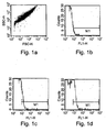
- TF labeled microparticles and TFPI labeled microparticles were identified at the M1 area (in Figures 1B, D ) and expressed as percent from the total microparticles in the gate.
- TF / TFPI ratio The ratio between TF and TFPI expression on microparticles was calculated as a measure of coagulation status.
- TF/TFPI ratio less than 1 represented normal healthy human plasma.
- TF/TFPI ratio higher than 1 demonstrated hyper-coagulability state.
- TF/TFPI ratio higher than 3 may predict a risk of vascular complication or a thrombotic event.
- TF activity Plasmatic MPs were isolated using ultracentrifuge. MPs ⁇ g/ ⁇ l TF activity was measured by a one-step clotting assay as was previously described ( Aharon et al, Thromb Haemost (2004) 92(4):776-86 ). MPs extract. (100 ⁇ l) were added to pooled normal human plasma (100 ⁇ l) and incubated for one minute at 37°C. Then, a 25 mM calcium chloride solution (100 ⁇ l) was added.
- the clotting times were measured and converted to standard curve of 10-1000 arbitrary units of TF (AU/ml) - where 180 seconds of clotting time stand for 1 TF AU, 130 seconds stand for 1.5 TF AU and 100 sec stand for 2.5 TF AU.
- Plasmatic MPs were isolated using ultracentrifuge. TFPI protein levels were measured in human MPs extracts by ELISA (American Diagnostica, Greenwich, CT, USA) according to manufacturer's instructions and expressed as TFPI ng/100 ⁇ g of MPs total proteins.
- TF activity / TFPI antigen level ratio The ratio between TF activity and TFPI antigen level on microparticles was calculated as a measure of coagulation status.
- TF activity/TFPI. antigen ratio less than 1 represented normal healthy human plasma.
- TF activity/TFPI antigen ratio higher than 1 demonstrated hyper-coagulability state.
- TF activity/TFPI antigen ratio higher than 3 may predict a risk of vascular complication or a thrombotic event.
- TF/TFPI ratio of blood microparticles (MPs)
- TF and TFPI expression were analyzed for MPs TF and TFPI expression.
- FACS analysis was used to identify positively stained MPs.
- FACS mediated detection of MPs was calibrated with 0.75 micron beads as indicated in Figure 1A .
- Sample labeling with anti mouse FITC IgG served as the negative control ( Figure 1B ).
- the ratio between TF expression (6.4%, Figure 1C ) and TFPI expression (18.5%, Figure 1D ) on microparticles was calculated (0.35).
- the TF/TFPI ratio was lower than 1 which represented normal healthy human plasma.
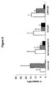
- microparticle TF/TFPI ratio was lower than 1 in non-pregnant healthy woman as expected in normal healthy human plasma.
- the microparticle TF/TFPI ratio was higher than 1 which implies a hyper-coagulability state.
- the microparticle TF/TFPI ratio was higher than 3 which may predict a thrombotic event.
- the TF/TFPI ratio was significantly reduced in the LMWH treated group.
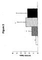
- the TF/TFPI ratio on microparticles of the healthy control group was 0.43 ( ⁇ 0.268) and was significantly increased in diabetic patients without known complications 1.41 ( ⁇ 0.734, p>0.0001).
- microparticle TF/TFPI ratio was lower than 1 in the healthy control group as expected in normal healthy human plasma.
- the microparticle TF/TFPI ratio increased to higher than 1 in all diabetic patients, with even higher ratio values in diabetic patients with diabetic foot, which implies a hyper-coagulability state.
- the microparticle TF/TFPI ratio may predict a thrombotic event.
- GVC pregnant women with gestational vascular complications
- LMWH anticoagulant low molecular weight heparin
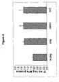
- TF activity on microparticles was significantly lower in non-pregnant women compared to pregnant women (p ⁇ 0.0001). There was no marked difference between TF activity in the three different pregnant women groups (healthy, with GVC treated with LMWH or untreated with GVC).
- GVC gestational vascular complications
- LMWH anticoagulant low molecular weight heparin
- TFPI antigen level on microparticles of non-pregnant healthy women was significantly higher compared to pregnant healthy women 1.322 ⁇ 0.0258; 0.504 ⁇ 0.0161 respectively (p ⁇ 0.0001).
- TFPI antigen level was even lower in pregnant women with GVC 0.236 ⁇ 0.013 (p ⁇ 0.0001), a value which rose in pregnant women with GVC treated with LMWH 0.445 ⁇ 0.019 (p ⁇ 0.0001).
- TF activity to TFPI antigen level on blood microparticles was assessed.
- TF activity/TFPI antigen ratio on microparticles of non-pregnant women was 0.657 ⁇ 0.035.
- the ratio increased significantly to 2.82 ⁇ 0.135 (p ⁇ 0.0001) in healthy pregnant women and increased drastically to 6.135 ⁇ 0.768 (p ⁇ 0.0001) in pregnant women with GVC.
- the microparticle TF activity/TFPI antigen ratio was significantly reduced in the LMWH treated group to 3.198 ⁇ 0.568 (p ⁇ 0.0001) compared to non-treated GVC pregnant women.
- microparticle TF activity/TFPI antigen ratio was lower than 1 in non-pregnant healthy woman as expected in normal healthy human plasma.
- the microparticle TF activity/TFPI antigen ratio was higher than 1 which implies a hyper-coagulability state.
- the microparticle TF activity/TFPI antigen ratio was higher than 3 which may predict a risk of thrombotic event.
- the TF activity/TFPI antigen ratio was significantly reduced in the LMWH treated group.
Landscapes
- Health & Medical Sciences (AREA)
- Life Sciences & Earth Sciences (AREA)
- Engineering & Computer Science (AREA)
- Biomedical Technology (AREA)
- Hematology (AREA)
- Chemical & Material Sciences (AREA)
- Immunology (AREA)
- Physics & Mathematics (AREA)
- Molecular Biology (AREA)
- Urology & Nephrology (AREA)
- Pathology (AREA)
- Food Science & Technology (AREA)
- Medicinal Chemistry (AREA)
- Analytical Chemistry (AREA)
- Biochemistry (AREA)
- General Health & Medical Sciences (AREA)
- General Physics & Mathematics (AREA)
- Ecology (AREA)
- Biophysics (AREA)
- Biotechnology (AREA)
- Cell Biology (AREA)
- Microbiology (AREA)
- Investigating Or Analysing Biological Materials (AREA)
Claims (8)
- Procédé de détermination d'un état de coagulation dans un échantillon de sang, le procédé comprenant les étapes consistant à :(a) isoler des microparticules cellulaires de l'échantillon de sang ; et(b) déterminer un rapport d'expression et/ou d'activité entre le facteur de tissu (TF) et l'inhibiteur de la voie du facteur de tissu (TFPI) dans lesdites microparticules cellulaires, où ledit rapport est indicatif de l'état de coagulation de l'échantillon de sang.
- Procédé de conception d'un régime de traitement pour un sujet qui en a besoin, le procédé comprenant les étapes consistant à :a) isoler des microparticules cellulaires d'un échantillon de sang du sujet ;(b) déterminer sur les microparticules cellulaires d'un échantillon de sang du sujet un rapport d'expression et/ou d'activité entre TF et TFPI, où ledit rapport d'expression et/ou d'activité est indicatif de l'état de coagulation du sujet ;(c) concevoir le régime de traitement en se basant sur ledit état de coagulation.
- Procédé selon la revendication 2, dans lequel le traitement est choisi dans le groupe consistant en les héparines de faible masse moléculaire (LMWH), la warfarine, l'aspirine, l'héparine, les AINS, le dipyridamole, le clopidogrel et les antagonistes de glycoprotéine IIb/IIIa des plaquettes.
- Procédé selon la revendication 1 ou 2, dans lequel lesdites microparticules cellulaires sont choisies dans le groupe consistant en les microparticules dérivées des plaquettes, les microparticules dérivées de cellules endothéliales, les microparticules dérivées de leucocytes et les microparticles dérivées d'érythrocytes.
- Procédé selon la revendication ou 2, dans lequel ledit rapport d'activité est déterminé par un dosage de coagulation.
- Procédé selon la revendication ou 2, dans lequel lorsque ledit rapport d'expression entre TF et TFPI est inférieur à environ 1, l'état de coagulation de l'échantillon de sang est normal.
- Procédé selon la revendication 1 ou 2, dans lequel lorsque ledit rapport d'expression entre TF et TFPI est au-dessus d'environ 1, l'état de coagulation de l'échantillon de sang manifeste une hypercoagulabilité.
- Procédé selon la revendication 1 ou 2, dans lequel lorsque ledit rapport d'expression entre TF et TFPI est au-dessus d'environ 3, l'état de coagulation de l'échantillon de sang est prédictif du risque de survenue d'événements thrombotiques.
Applications Claiming Priority (2)
| Application Number | Priority Date | Filing Date | Title |
|---|---|---|---|
| US76251906P | 2006-01-27 | 2006-01-27 | |
| PCT/IL2007/000110 WO2007086069A1 (fr) | 2006-01-27 | 2007-01-28 | Procédés et nécessaires pour évaluer la coagulation sanguine |
Publications (2)
| Publication Number | Publication Date |
|---|---|
| EP1987354A1 EP1987354A1 (fr) | 2008-11-05 |
| EP1987354B1 true EP1987354B1 (fr) | 2010-11-03 |
Family
ID=37950920
Family Applications (1)
| Application Number | Title | Priority Date | Filing Date |
|---|---|---|---|
| EP07706053A Not-in-force EP1987354B1 (fr) | 2006-01-27 | 2007-01-28 | Procédés pour évaluer la coagulation sanguine |
Country Status (6)
| Country | Link |
|---|---|
| US (1) | US20090042217A1 (fr) |
| EP (1) | EP1987354B1 (fr) |
| AT (1) | ATE487133T1 (fr) |
| CA (1) | CA2640167A1 (fr) |
| DE (1) | DE602007010246D1 (fr) |
| WO (1) | WO2007086069A1 (fr) |
Families Citing this family (11)
| Publication number | Priority date | Publication date | Assignee | Title |
|---|---|---|---|---|
| US8840893B2 (en) | 2004-06-10 | 2014-09-23 | Omeros Corporation | Methods for treating conditions associated with MASP-2 dependent complement activation |
| WO2009046267A1 (fr) * | 2007-10-05 | 2009-04-09 | University Of Virginia Patent Foundation | Biomarqueurs protéiques révélateurs d'un anévrisme de l'aorte abdominale |
| EP3150635A3 (fr) * | 2009-10-16 | 2017-06-21 | Omeros Corporation | Procédés de traitement de la coagulation intravasculaire disséminée par inhibition de l'activation du complément masp-2 dépendant |
| WO2012120131A1 (fr) * | 2011-03-09 | 2012-09-13 | INSERM (Institut National de la Santé et de la Recherche Médicale) | Méthodes pour générer des microparticules cellulaires |
| DK2694108T3 (en) | 2011-04-08 | 2018-08-20 | Univ Leicester | METHODS OF TREATING DISEASES ASSOCIATED WITH MASP-2 DEPENDENT COMPLEMENT ACTIVATION |
| US9644035B2 (en) | 2011-04-08 | 2017-05-09 | Omeros Corporation | Methods for treating conditions associated with MASP-2 dependent complement activation |
| FR3043780B1 (fr) * | 2015-11-16 | 2017-12-15 | Hospices Civils Lyon | Methode de diagnostic d'anomalies de la coagulation sanguine |
| US20210263052A1 (en) * | 2018-07-25 | 2021-08-26 | Sony Corporation | Blood coagulation system analysis device |
| EP4083630A4 (fr) * | 2019-12-26 | 2024-01-10 | Sekisui Medical Co., Ltd. | Procédé de mesure du temps de coagulation sanguine |
| EP4270010A4 (fr) * | 2020-12-28 | 2024-11-20 | Fujimori Kogyo Co., Ltd. | Procédé d'évaluation de performance de coagulation sanguine, et procédé d'inspection du risque de thrombose |
| IT202100018803A1 (it) * | 2021-07-15 | 2023-01-15 | Centro Cardiologico Monzino S P A | Metodo per la valutazione del potenziale trombotico residuo piastrinico in pazienti in terapia antiaggregante |
Family Cites Families (1)
| Publication number | Priority date | Publication date | Assignee | Title |
|---|---|---|---|---|
| ES2139957T3 (es) * | 1994-07-23 | 2000-02-16 | Roche Diagnostics Gmbh | Metodo para la determinacion del estado pretrombotico. |
-
2007
- 2007-01-28 EP EP07706053A patent/EP1987354B1/fr not_active Not-in-force
- 2007-01-28 DE DE602007010246T patent/DE602007010246D1/de active Active
- 2007-01-28 AT AT07706053T patent/ATE487133T1/de not_active IP Right Cessation
- 2007-01-28 CA CA002640167A patent/CA2640167A1/fr not_active Abandoned
- 2007-01-28 WO PCT/IL2007/000110 patent/WO2007086069A1/fr not_active Ceased
-
2008
- 2008-06-23 US US12/223,290 patent/US20090042217A1/en not_active Abandoned
Also Published As
| Publication number | Publication date |
|---|---|
| EP1987354A1 (fr) | 2008-11-05 |
| US20090042217A1 (en) | 2009-02-12 |
| CA2640167A1 (fr) | 2007-08-02 |
| WO2007086069A1 (fr) | 2007-08-02 |
| ATE487133T1 (de) | 2010-11-15 |
| DE602007010246D1 (de) | 2010-12-16 |
Similar Documents
| Publication | Publication Date | Title |
|---|---|---|
| US20090042217A1 (en) | Methods and Kits for Determining Blood Coagulation | |
| Favaloro | Clinical utility of the PFA-100 | |
| JP2023072032A (ja) | 凝固分析を使用した抗凝固剤の検出および分類 | |
| Wan et al. | Whole blood thrombin generation profiles of patients with cirrhosis explored with a near patient assay | |
| Favaloro et al. | Clinical and laboratory diagnosis of heparin induced thrombocytopenia: an update | |
| Tripodi et al. | Hypercoagulability in splenectomized thalassemic patients detected by whole-blood thromboelastometry, but not by thrombin generation in platelet-poor plasma | |
| JP5662802B2 (ja) | フォン−ビルブランド病ならびにフォン−ビルブランド病および血小板機能の後天性または先天性障害に関連する出血リスクの増加を評価するためのインビトロ診断法 | |
| Dashkevich et al. | Effect of pre-analytical conditions on the thrombodynamics assay | |
| Odegard et al. | Evaluation of the coagulation system in children with two-ventricle congenital heart disease | |
| Tsuchida et al. | Characterization and usefulness of clot-fibrinolysis waveform analysis in critical care patients with enhanced or suppressed fibrinolysis | |
| Laffan et al. | 17 Investigation of a thrombotic tendency | |
| Ristovska et al. | Von-Willebrand factor as a predictor of three-month mortality in patients with liver cirrhosis compared to MELD score | |
| Shavazi | The nature of changes markers of dysfunction of the endothelium in blood of women with premature bursting of amniotic waters | |
| Campbell | Hemostasis | |
| Van Dreden et al. | Procoagulant phospholipids and tissue factor activity in cerebrospinal fluid from patients with intracerebral haemorrhage | |
| Remkova | Diagnostic approach to hypercoagulable states | |
| Düz | Coagulation analyses in diagnosis and treatment | |
| Bonhomme et al. | Laboratory testing of hemostasis | |
| Rogers et al. | Laboratory Analysis of Coagulation | |
| Chadha | Review of coagulation testing in liver disease | |
| KR101608134B1 (ko) | 단백질 z의 혈액 항응고기능 분석방법 | |
| Abdelwahab | Laboratory Diagnosis of Less Common Acquired and Congenital Thrombophilic | |
| Grafeneder | The Disseminated Intravascular Coagulation (DIC) score is associated with mortality in critically ill patients and patients with liver disease. | |
| Abdullah et al. | Practical Haemostasisfor Medical Personnel | |
| Egberg et al. | Coagulation testing: basic and advanced clinical laboratory tests |
Legal Events
| Date | Code | Title | Description |
|---|---|---|---|
| PUAI | Public reference made under article 153(3) epc to a published international application that has entered the european phase |
Free format text: ORIGINAL CODE: 0009012 |
|
| 17P | Request for examination filed |
Effective date: 20080827 |
|
| AK | Designated contracting states |
Kind code of ref document: A1 Designated state(s): AT BE BG CH CY CZ DE DK EE ES FI FR GB GR HU IE IS IT LI LT LU LV MC NL PL PT RO SE SI SK TR |
|
| 17Q | First examination report despatched |
Effective date: 20090326 |
|
| GRAP | Despatch of communication of intention to grant a patent |
Free format text: ORIGINAL CODE: EPIDOSNIGR1 |
|
| RTI1 | Title (correction) |
Free format text: METHODS FOR DETERMINING BLOOD COAGULATION |
|
| GRAS | Grant fee paid |
Free format text: ORIGINAL CODE: EPIDOSNIGR3 |
|
| GRAA | (expected) grant |
Free format text: ORIGINAL CODE: 0009210 |
|
| AK | Designated contracting states |
Kind code of ref document: B1 Designated state(s): AT BE BG CH CY CZ DE DK EE ES FI FR GB GR HU IE IS IT LI LT LU LV MC NL PL PT RO SE SI SK TR |
|
| REG | Reference to a national code |
Ref country code: GB Ref legal event code: FG4D |
|
| REG | Reference to a national code |
Ref country code: CH Ref legal event code: EP |
|
| REG | Reference to a national code |
Ref country code: IE Ref legal event code: FG4D |
|
| REF | Corresponds to: |
Ref document number: 602007010246 Country of ref document: DE Date of ref document: 20101216 Kind code of ref document: P |
|
| REG | Reference to a national code |
Ref country code: NL Ref legal event code: VDEP Effective date: 20101103 |
|
| LTIE | Lt: invalidation of european patent or patent extension |
Effective date: 20101103 |
|
| PG25 | Lapsed in a contracting state [announced via postgrant information from national office to epo] |
Ref country code: LT Free format text: LAPSE BECAUSE OF FAILURE TO SUBMIT A TRANSLATION OF THE DESCRIPTION OR TO PAY THE FEE WITHIN THE PRESCRIBED TIME-LIMIT Effective date: 20101103 |
|
| PG25 | Lapsed in a contracting state [announced via postgrant information from national office to epo] |
Ref country code: SE Free format text: LAPSE BECAUSE OF FAILURE TO SUBMIT A TRANSLATION OF THE DESCRIPTION OR TO PAY THE FEE WITHIN THE PRESCRIBED TIME-LIMIT Effective date: 20101103 Ref country code: SI Free format text: LAPSE BECAUSE OF FAILURE TO SUBMIT A TRANSLATION OF THE DESCRIPTION OR TO PAY THE FEE WITHIN THE PRESCRIBED TIME-LIMIT Effective date: 20101103 Ref country code: NL Free format text: LAPSE BECAUSE OF FAILURE TO SUBMIT A TRANSLATION OF THE DESCRIPTION OR TO PAY THE FEE WITHIN THE PRESCRIBED TIME-LIMIT Effective date: 20101103 Ref country code: IS Free format text: LAPSE BECAUSE OF FAILURE TO SUBMIT A TRANSLATION OF THE DESCRIPTION OR TO PAY THE FEE WITHIN THE PRESCRIBED TIME-LIMIT Effective date: 20110303 Ref country code: AT Free format text: LAPSE BECAUSE OF FAILURE TO SUBMIT A TRANSLATION OF THE DESCRIPTION OR TO PAY THE FEE WITHIN THE PRESCRIBED TIME-LIMIT Effective date: 20101103 Ref country code: BG Free format text: LAPSE BECAUSE OF FAILURE TO SUBMIT A TRANSLATION OF THE DESCRIPTION OR TO PAY THE FEE WITHIN THE PRESCRIBED TIME-LIMIT Effective date: 20110203 Ref country code: PT Free format text: LAPSE BECAUSE OF FAILURE TO SUBMIT A TRANSLATION OF THE DESCRIPTION OR TO PAY THE FEE WITHIN THE PRESCRIBED TIME-LIMIT Effective date: 20110303 Ref country code: FI Free format text: LAPSE BECAUSE OF FAILURE TO SUBMIT A TRANSLATION OF THE DESCRIPTION OR TO PAY THE FEE WITHIN THE PRESCRIBED TIME-LIMIT Effective date: 20101103 Ref country code: LV Free format text: LAPSE BECAUSE OF FAILURE TO SUBMIT A TRANSLATION OF THE DESCRIPTION OR TO PAY THE FEE WITHIN THE PRESCRIBED TIME-LIMIT Effective date: 20101103 |
|
| PG25 | Lapsed in a contracting state [announced via postgrant information from national office to epo] |
Ref country code: GR Free format text: LAPSE BECAUSE OF FAILURE TO SUBMIT A TRANSLATION OF THE DESCRIPTION OR TO PAY THE FEE WITHIN THE PRESCRIBED TIME-LIMIT Effective date: 20110204 |
|
| PG25 | Lapsed in a contracting state [announced via postgrant information from national office to epo] |
Ref country code: BE Free format text: LAPSE BECAUSE OF FAILURE TO SUBMIT A TRANSLATION OF THE DESCRIPTION OR TO PAY THE FEE WITHIN THE PRESCRIBED TIME-LIMIT Effective date: 20101103 Ref country code: ES Free format text: LAPSE BECAUSE OF FAILURE TO SUBMIT A TRANSLATION OF THE DESCRIPTION OR TO PAY THE FEE WITHIN THE PRESCRIBED TIME-LIMIT Effective date: 20110214 Ref country code: CZ Free format text: LAPSE BECAUSE OF FAILURE TO SUBMIT A TRANSLATION OF THE DESCRIPTION OR TO PAY THE FEE WITHIN THE PRESCRIBED TIME-LIMIT Effective date: 20101103 Ref country code: EE Free format text: LAPSE BECAUSE OF FAILURE TO SUBMIT A TRANSLATION OF THE DESCRIPTION OR TO PAY THE FEE WITHIN THE PRESCRIBED TIME-LIMIT Effective date: 20101103 |
|
| PG25 | Lapsed in a contracting state [announced via postgrant information from national office to epo] |
Ref country code: RO Free format text: LAPSE BECAUSE OF FAILURE TO SUBMIT A TRANSLATION OF THE DESCRIPTION OR TO PAY THE FEE WITHIN THE PRESCRIBED TIME-LIMIT Effective date: 20101103 Ref country code: SK Free format text: LAPSE BECAUSE OF FAILURE TO SUBMIT A TRANSLATION OF THE DESCRIPTION OR TO PAY THE FEE WITHIN THE PRESCRIBED TIME-LIMIT Effective date: 20101103 Ref country code: MC Free format text: LAPSE BECAUSE OF NON-PAYMENT OF DUE FEES Effective date: 20110131 Ref country code: DK Free format text: LAPSE BECAUSE OF FAILURE TO SUBMIT A TRANSLATION OF THE DESCRIPTION OR TO PAY THE FEE WITHIN THE PRESCRIBED TIME-LIMIT Effective date: 20101103 Ref country code: PL Free format text: LAPSE BECAUSE OF FAILURE TO SUBMIT A TRANSLATION OF THE DESCRIPTION OR TO PAY THE FEE WITHIN THE PRESCRIBED TIME-LIMIT Effective date: 20101103 |
|
| PLBE | No opposition filed within time limit |
Free format text: ORIGINAL CODE: 0009261 |
|
| STAA | Information on the status of an ep patent application or granted ep patent |
Free format text: STATUS: NO OPPOSITION FILED WITHIN TIME LIMIT |
|
| 26N | No opposition filed |
Effective date: 20110804 |
|
| REG | Reference to a national code |
Ref country code: IE Ref legal event code: MM4A |
|
| REG | Reference to a national code |
Ref country code: DE Ref legal event code: R097 Ref document number: 602007010246 Country of ref document: DE Effective date: 20110804 |
|
| PG25 | Lapsed in a contracting state [announced via postgrant information from national office to epo] |
Ref country code: IT Free format text: LAPSE BECAUSE OF FAILURE TO SUBMIT A TRANSLATION OF THE DESCRIPTION OR TO PAY THE FEE WITHIN THE PRESCRIBED TIME-LIMIT Effective date: 20101103 |
|
| PG25 | Lapsed in a contracting state [announced via postgrant information from national office to epo] |
Ref country code: IE Free format text: LAPSE BECAUSE OF NON-PAYMENT OF DUE FEES Effective date: 20110128 |
|
| PGFP | Annual fee paid to national office [announced via postgrant information from national office to epo] |
Ref country code: CH Payment date: 20120123 Year of fee payment: 6 |
|
| PG25 | Lapsed in a contracting state [announced via postgrant information from national office to epo] |
Ref country code: CY Free format text: LAPSE BECAUSE OF FAILURE TO SUBMIT A TRANSLATION OF THE DESCRIPTION OR TO PAY THE FEE WITHIN THE PRESCRIBED TIME-LIMIT Effective date: 20101103 Ref country code: LU Free format text: LAPSE BECAUSE OF NON-PAYMENT OF DUE FEES Effective date: 20110128 |
|
| REG | Reference to a national code |
Ref country code: CH Ref legal event code: PL |
|
| PG25 | Lapsed in a contracting state [announced via postgrant information from national office to epo] |
Ref country code: TR Free format text: LAPSE BECAUSE OF FAILURE TO SUBMIT A TRANSLATION OF THE DESCRIPTION OR TO PAY THE FEE WITHIN THE PRESCRIBED TIME-LIMIT Effective date: 20101103 |
|
| PG25 | Lapsed in a contracting state [announced via postgrant information from national office to epo] |
Ref country code: CH Free format text: LAPSE BECAUSE OF NON-PAYMENT OF DUE FEES Effective date: 20130131 Ref country code: LI Free format text: LAPSE BECAUSE OF NON-PAYMENT OF DUE FEES Effective date: 20130131 Ref country code: HU Free format text: LAPSE BECAUSE OF FAILURE TO SUBMIT A TRANSLATION OF THE DESCRIPTION OR TO PAY THE FEE WITHIN THE PRESCRIBED TIME-LIMIT Effective date: 20101103 |
|
| REG | Reference to a national code |
Ref country code: FR Ref legal event code: PLFP Year of fee payment: 10 |
|
| REG | Reference to a national code |
Ref country code: FR Ref legal event code: PLFP Year of fee payment: 11 |
|
| REG | Reference to a national code |
Ref country code: FR Ref legal event code: PLFP Year of fee payment: 12 |
|
| PGFP | Annual fee paid to national office [announced via postgrant information from national office to epo] |
Ref country code: DE Payment date: 20200121 Year of fee payment: 14 Ref country code: GB Payment date: 20200123 Year of fee payment: 14 |
|
| PGFP | Annual fee paid to national office [announced via postgrant information from national office to epo] |
Ref country code: FR Payment date: 20200121 Year of fee payment: 14 |
|
| REG | Reference to a national code |
Ref country code: DE Ref legal event code: R119 Ref document number: 602007010246 Country of ref document: DE |
|
| GBPC | Gb: european patent ceased through non-payment of renewal fee |
Effective date: 20210128 |
|
| PG25 | Lapsed in a contracting state [announced via postgrant information from national office to epo] |
Ref country code: FR Free format text: LAPSE BECAUSE OF NON-PAYMENT OF DUE FEES Effective date: 20210131 |
|
| PG25 | Lapsed in a contracting state [announced via postgrant information from national office to epo] |
Ref country code: DE Free format text: LAPSE BECAUSE OF NON-PAYMENT OF DUE FEES Effective date: 20210803 Ref country code: GB Free format text: LAPSE BECAUSE OF NON-PAYMENT OF DUE FEES Effective date: 20210128 |