EP1992965A2 - Procédé de calibrage d'un tomographe à émission de positrons d'un dispositif de radiothérapie et dispositif de radiothérapie - Google Patents
Procédé de calibrage d'un tomographe à émission de positrons d'un dispositif de radiothérapie et dispositif de radiothérapie Download PDFInfo
- Publication number
- EP1992965A2 EP1992965A2 EP08103379A EP08103379A EP1992965A2 EP 1992965 A2 EP1992965 A2 EP 1992965A2 EP 08103379 A EP08103379 A EP 08103379A EP 08103379 A EP08103379 A EP 08103379A EP 1992965 A2 EP1992965 A2 EP 1992965A2
- Authority
- EP
- European Patent Office
- Prior art keywords
- positron emission
- emission tomograph
- measured
- dose
- sample body
- Prior art date
- Legal status (The legal status is an assumption and is not a legal conclusion. Google has not performed a legal analysis and makes no representation as to the accuracy of the status listed.)
- Granted
Links
Images

Classifications
-
- G—PHYSICS
- G01—MEASURING; TESTING
- G01T—MEASUREMENT OF NUCLEAR OR X-RADIATION
- G01T1/00—Measuring X-radiation, gamma radiation, corpuscular radiation, or cosmic radiation
- G01T1/16—Measuring radiation intensity
- G01T1/169—Exploration, location of contaminated surface areas
-
- A—HUMAN NECESSITIES
- A61—MEDICAL OR VETERINARY SCIENCE; HYGIENE
- A61B—DIAGNOSIS; SURGERY; IDENTIFICATION
- A61B6/00—Apparatus or devices for radiation diagnosis; Apparatus or devices for radiation diagnosis combined with radiation therapy equipment
- A61B6/58—Testing, adjusting or calibrating thereof
- A61B6/582—Calibration
- A61B6/583—Calibration using calibration phantoms
-
- G—PHYSICS
- G01—MEASURING; TESTING
- G01T—MEASUREMENT OF NUCLEAR OR X-RADIATION
- G01T1/00—Measuring X-radiation, gamma radiation, corpuscular radiation, or cosmic radiation
- G01T1/29—Measurement performed on radiation beams, e.g. position or section of the beam; Measurement of spatial distribution of radiation
-
- G—PHYSICS
- G01—MEASURING; TESTING
- G01T—MEASUREMENT OF NUCLEAR OR X-RADIATION
- G01T1/00—Measuring X-radiation, gamma radiation, corpuscular radiation, or cosmic radiation
- G01T1/29—Measurement performed on radiation beams, e.g. position or section of the beam; Measurement of spatial distribution of radiation
- G01T1/2914—Measurement of spatial distribution of radiation
- G01T1/2985—In depth localisation, e.g. using positron emitters; Tomographic imaging (longitudinal and transverse section imaging; apparatus for radiation diagnosis sequentially in different planes, steroscopic radiation diagnosis)
-
- A—HUMAN NECESSITIES
- A61—MEDICAL OR VETERINARY SCIENCE; HYGIENE
- A61N—ELECTROTHERAPY; MAGNETOTHERAPY; RADIATION THERAPY; ULTRASOUND THERAPY
- A61N5/00—Radiation therapy
- A61N5/10—X-ray therapy; Gamma-ray therapy; Particle-irradiation therapy
- A61N5/1048—Monitoring, verifying, controlling systems and methods
- A61N5/1049—Monitoring, verifying, controlling systems and methods for verifying the position of the patient with respect to the radiation beam
- A61N2005/1052—Monitoring, verifying, controlling systems and methods for verifying the position of the patient with respect to the radiation beam using positron emission tomography [PET] single photon emission computer tomography [SPECT] imaging
-
- A—HUMAN NECESSITIES
- A61—MEDICAL OR VETERINARY SCIENCE; HYGIENE
- A61N—ELECTROTHERAPY; MAGNETOTHERAPY; RADIATION THERAPY; ULTRASOUND THERAPY
- A61N5/00—Radiation therapy
- A61N5/10—X-ray therapy; Gamma-ray therapy; Particle-irradiation therapy
- A61N5/1048—Monitoring, verifying, controlling systems and methods
- A61N5/1075—Monitoring, verifying, controlling systems and methods for testing, calibrating, or quality assurance of the radiation treatment apparatus
- A61N2005/1076—Monitoring, verifying, controlling systems and methods for testing, calibrating, or quality assurance of the radiation treatment apparatus using a dummy object placed in the radiation field, e.g. phantom
Definitions
- the invention relates to a method for calibrating a positron emission tomograph of a radiotherapy apparatus and to a radiotherapy apparatus for performing such a method, wherein the radiotherapy apparatus is in particular a particle therapy system.
- Positron emission tomography is an imaging technique used primarily in nuclear medicine and radiotherapy.
- a radioactive decay in which a positron is emitted, detected in a body to be examined. After a short distance the positron interacts with an electron, whereby both particles are destroyed. In their place, a pair of gamma quanta is created, which move away from each other at an angle of 180 °. These gamma quanta penetrate the body to be examined and are registered by two opposing detectors.
- a positron emission tomograph for imaging usually comprises a plurality of gamma-ray detectors, which usually surround the patient to be examined in the manner of a ring. Such an arrangement is for example in the DE 32 08 178 A1 described.
- the relevant radioactive decay can be induced, for example, by injection or inhalation of a radioactively labeled radiopharmaceutical, also called a tracer. From the spatial distribution of the tracer, for example, can draw conclusions about underlying diseases.
- Radioactive decay which involves the formation of positrons, also occurs during radiotherapy the radiation of a body, depending on the applied radiation dose.
- PET systems have proven to be a suitable means to carry out such a dose validation or monitoring of radiotherapy and particle therapy. Especially in the case of particle therapy, measurements are therefore often taken to check whether the planned radiation dose coincides with the dose actually applied and / or whether the spatial distribution of an applied dose agrees with a desired spatial distribution.
- PET systems can be adapted to the requirements of a particle therapy system and deviate from the conventional ring shape.
- so-called in-beam PET systems are known which consist only of two opposing detectors.
- the additional opening between the two detectors of an in-beam PET system allows e.g. a freer positioning of the patient or a radiation of the patient with a beam through this opening, without the beam hits the detectors.
- PET systems must be calibrated at certain intervals, such as daily. So far, a calibration using radioactive sources.
- the radioactive sources are defined in a treatment room set up by the PET system is arranged.
- the radioactive sources produce a defined activity, which is measured with the PET system. These measurements are used to calibrate the PET system. This may include, for example, a review of an existing calibration of the PET system.
- the method is based on the idea that, in the case of a radiation therapy device, a defined radiation dose can be applied to the treatment beam in a sample body. This is possible because in the case of a radiation therapy device the properties of the treatment beam are checked with regard to the accuracy of the applied dose and the accuracy of the application site in separate methods and may therefore be assumed to be quality assured.
- the applied radiation dose generates in the sample body a defined radioactivity, which is transmitted via the positron emission tomograph measured and used for calibration.
- the radiation dose is in particular at a defined location in the treatment room, i. at a defined location relative to the positron emission tomograph, and / or at a defined location in the sample body. This is possible for example by controlling the deflection of a particle beam or controlling the energy of the particle beam in a known manner. But it is also possible, for example, to apply the radiation dose in the phantom and then to position the phantom then at the desired location in the same or in another room.
- the method thus makes it possible to carry out a calibration of the PET system without additional radioactive sources, thereby increasing the automation of the method. Overall, this reduces costs and reduces possible sources of error.
- the method can be made more flexible, since the intensity of the radiation dose, the application site and / or the application time can be flexibly adapted and changed in a simple manner, for example via a control of the treatment beam.
- the plurality of different radiation doses differ with regard to their dose, their application site and / or their application time. In this way, a calibration can be performed more accurately.
- a response of detector elements of the positron emission tomograph and / or a spatial resolution and / or a positional coding of the positron emission tomograph are calibrated with the aid of the measured activity or the measured activities.
- radiation doses which differ in their intensity and are applied successively are particularly suitable for calibrating the response behavior of the detector elements.
- Radiation doses applied at different locations are suitable for calibrating the spatial resolution or the spatial coding of the PET system.
- the defined radiation doses can be applied divided in accordance with a three-dimensional, predefined pattern. This allows a more accurate calibration of the spatial resolution and / or location coding of the PET system by tuning the measured activity to the three-dimensional predefined pattern. Consequently, additional information about the spatial distribution of the induced radioactivity can be obtained from the three-dimensional pattern. This information can also be used to calibrate the PET system.
- the specimen used by the radiation dose is applied, for example, a solid state phantom, in particular a so-called PMMA phantom (for polymethyl methacrylate phantom) or another known phantom.
- a solid state phantom in particular a so-called PMMA phantom (for polymethyl methacrylate phantom) or another known phantom.
- the radiotherapy device is designed as a particle therapy system
- the method can be used.
- a defined dose of particulate applied by the particle beam applies as a defined radiation dose.
- the radiotherapy device according to the invention which is designed in particular as a particle therapy system, has a positron emission tomograph in a treatment space, wherein the radiotherapy device or the positron emission tomograph is designed to carry out a calibration method according to one of claims 1 to 7.
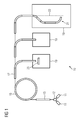
- FIG. 1 shows a schematic overview of the structure of a particle therapy system 10.
- a particle therapy system 10 takes place irradiation of a body, in particular a tumor-affected tissue, with a particle beam.
- particles mainly ions such as protons, pions, helium ions, carbon ions or other types of ions are used.
- ions are generated in a particle source 11.
- two particle sources 11 are present which produce two different types of ions can be switched between these two ion types within a short time interval.
- a solenoid 12 is used, which is arranged between the ion sources 11 on the one hand and a pre-accelerator 13 on the other.
- this allows the particle therapy system 10 to be operated simultaneously with protons and with carbon ions.
- the ions generated by the or one of the ion sources 11 and optionally selected by the switching magnet 12 are accelerated in the pre-accelerator 13 to a first energy level.
- the pre-accelerator 13 is, for example, a linear accelerator (LINAC for English: "LINear ACcelerator”).
- the particles are fed into an accelerator 15, for example a synchrotron or cyclotron.
- an accelerator 15 for example a synchrotron or cyclotron.
- they are accelerated to high energies necessary for irradiation.
- a high-energy beam transport system 17 guides the particle beam to one or more irradiation spaces 19.
- an irradiation space 19 the accelerated particles are directed onto a body to be irradiated. Depending on the configuration, this takes place from a fixed direction (in so-called "fixed beam” spaces) or else via a rotatable gantry 21 which can be moved about an axis 22 from different directions.
- FIG. 1 The basis of the FIG. 1
- the illustrated basic structure of a particle therapy system 10 is typical for many particle therapy systems, but may also deviate from this.
- the embodiments described below are both in connection with the basis of the FIG. 1 used particle therapy system as well as with other particle therapy or radiation therapy equipment used.
- a PET system 23 is indicated, with which a dose validation or a monitoring during an irradiation session in the context of particle therapy can be performed.
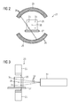
- FIG. 2 shows a frontal view of a ring-shaped PET system 23.
- the detector ring consists for example of two opposite ring segments 25 on each of which a plurality of detector elements 26 are arranged.
- Such a configuration of the detector ring allows a freer positionability of a horizontal patient couch, for example, both perpendicular to the detector ring and along this plane.
- a sample body 29 is arranged on a table top 27 of a patient bed, for example a cuboid-shaped PMMA phantom.
- FIG. 3 shows a side view of the ring-shaped PET system 23 with the sample body 29.
- a jet outlet 31 is also visible from which the particle beam 33 exits in a treatment or irradiation space and can be directed to one or more different regions in the sample body 29.
- a plurality of defined particle doses 35 arranged in a spatial pattern can now be applied in the sample body 29.
- the strength of the applied particulate cans 35 is in FIG. 2 and FIG. 3 represented by different sized circles.
- the individual particle doses 35 are successively applied one after the other in the sample body.
- the control of the particle beam 33 - and thus the control of the applied particulate cans 35 - is performed by a control device of the particle therapy system.
- deflection magnets 33 are controlled in accordance with the deflection of the particle beam.
- the applied particle doses 35 each induce in the sample body 29 a defined radioactivity, which is detected by means of the PET system 23. Since the induced activity is known on account of the defined particle dose or particle doses 35, a calibration of the PET system 23 can take place via this.
- the use of a spatial pattern in the application of the particulate cans 35 has the advantage that the calibration of the PET system 23 can be performed more accurately than with a single particulate dose, since the spatial information about the distribution pattern can be correlated with the measured activities. As a result, a more accurate, for example, redundant, calibration of the PET system 23 can be performed.
- particulate cans 35 that have been applied according to a spatial pattern can also be used to calibrate the spatial resolution of the PET system 23. For this purpose, a matching of the spatial pattern with the measured activity of the PET system 23 can take place.
- Successively applied, in their intensity different particle doses 35 can be used in particular to calibrate the response of individual detector elements 26.
- the calibration can also be done with a passive beam application. ie in a beam application, in which the treatment beam is formed by passive elements in the beam path, are used.
- FIG. 4 shows a schematic representation of the method steps that are used to calibrate a positron emission tomograph.
- a first step 41 one or more defined particle doses are applied by means of the particle beam of the particle therapy system in a sample body placed relative to the PET system.
- the applied or applied particle doses induce radioactive decays in the sample body whose activity is measured in a second step 43 using the PET system.
- a calibration of the PET system using the measured radioactivity follows.
- the calibration can be done, for example, based on the directly measured raw data, but for the calibration can also be a reconstruction of a PET image with the raw data done, which is then used for calibration.
- a spatial resolution or a spatial coding of the PET system can be calibrated, or - depending on the configuration of the method - in a fifth step 49, the response of individual detector elements.
- the method has been described in the figures in the context of a particle therapy, the method can also be used in a conventional radiotherapy, e.g. be used with X-rays.
Landscapes
- Health & Medical Sciences (AREA)
- Life Sciences & Earth Sciences (AREA)
- High Energy & Nuclear Physics (AREA)
- Molecular Biology (AREA)
- Physics & Mathematics (AREA)
- General Physics & Mathematics (AREA)
- Spectroscopy & Molecular Physics (AREA)
- Medical Informatics (AREA)
- Engineering & Computer Science (AREA)
- Nuclear Medicine, Radiotherapy & Molecular Imaging (AREA)
- Surgery (AREA)
- Optics & Photonics (AREA)
- Pathology (AREA)
- Radiology & Medical Imaging (AREA)
- Biomedical Technology (AREA)
- Heart & Thoracic Surgery (AREA)
- Biophysics (AREA)
- Animal Behavior & Ethology (AREA)
- General Health & Medical Sciences (AREA)
- Public Health (AREA)
- Veterinary Medicine (AREA)
- Radiation-Therapy Devices (AREA)
- Nuclear Medicine (AREA)
Applications Claiming Priority (1)
| Application Number | Priority Date | Filing Date | Title |
|---|---|---|---|
| DE102007020600A DE102007020600A1 (de) | 2007-05-02 | 2007-05-02 | Verfahren zur Kalibrierung eines Positronen-Emissions-Tomographen einer Strahlentherapievorrichtung sowie Strahlentherapievorrichtung |
Publications (3)
| Publication Number | Publication Date |
|---|---|
| EP1992965A2 true EP1992965A2 (fr) | 2008-11-19 |
| EP1992965A3 EP1992965A3 (fr) | 2014-03-19 |
| EP1992965B1 EP1992965B1 (fr) | 2017-06-14 |
Family
ID=39808744
Family Applications (1)
| Application Number | Title | Priority Date | Filing Date |
|---|---|---|---|
| EP08103379.7A Not-in-force EP1992965B1 (fr) | 2007-05-02 | 2008-04-04 | Procédé de calibrage d'un tomographe à émission de positrons d'un dispositif de radiothérapie et dispositif de radiothérapie |
Country Status (3)
| Country | Link |
|---|---|
| US (1) | US7679049B2 (fr) |
| EP (1) | EP1992965B1 (fr) |
| DE (1) | DE102007020600A1 (fr) |
Cited By (2)
| Publication number | Priority date | Publication date | Assignee | Title |
|---|---|---|---|---|
| EP3805805A1 (fr) * | 2019-10-09 | 2021-04-14 | Siemens Medical Solutions USA, Inc. | Étalonnage d'estimation de dose de rayonnement en imagerie nucléaire médicale |
| WO2022055539A1 (fr) * | 2020-09-10 | 2022-03-17 | Siemens Medical Solutions Usa, Inc. | Mini-gammatographe comme dosimètre |
Families Citing this family (23)
| Publication number | Priority date | Publication date | Assignee | Title |
|---|---|---|---|---|
| US7858925B2 (en) * | 2006-04-11 | 2010-12-28 | University Of Washington | Calibration method and system for PET scanners |
| US8017915B2 (en) | 2008-03-14 | 2011-09-13 | Reflexion Medical, Inc. | Method and apparatus for emission guided radiation therapy |
| WO2012030297A1 (fr) * | 2010-09-01 | 2012-03-08 | Empire Technology Development Llc | Procédé et système pour une thérapie gamma par faisceau ionique de radio-isotopes |
| CA2908092A1 (fr) * | 2013-03-28 | 2014-10-02 | Atomic Energy Of Canada Limited / Energie Atomique Du Canada Limitee | Systeme et procede pour la dosimetrie tridimensionnelle en temps reel |
| ES2768659T3 (es) | 2013-09-27 | 2020-06-23 | Mevion Medical Systems Inc | Exploración de haces de partículas |
| JP6256974B2 (ja) * | 2013-10-29 | 2018-01-10 | 株式会社日立製作所 | 荷電粒子ビームシステム |
| US9483850B2 (en) * | 2013-11-19 | 2016-11-01 | Toshiba Medical Systems Corporation | Cylinder source software-positioning method for PET calibration and image quality assurance |
| US9962560B2 (en) | 2013-12-20 | 2018-05-08 | Mevion Medical Systems, Inc. | Collimator and energy degrader |
| US10675487B2 (en) | 2013-12-20 | 2020-06-09 | Mevion Medical Systems, Inc. | Energy degrader enabling high-speed energy switching |
| US9661736B2 (en) | 2014-02-20 | 2017-05-23 | Mevion Medical Systems, Inc. | Scanning system for a particle therapy system |
| US10786689B2 (en) | 2015-11-10 | 2020-09-29 | Mevion Medical Systems, Inc. | Adaptive aperture |
| WO2018009779A1 (fr) | 2016-07-08 | 2018-01-11 | Mevion Medical Systems, Inc. | Planification de traitement |
| CN110248604B (zh) | 2016-11-15 | 2023-07-21 | 反射医疗公司 | 放射治疗患者平台 |
| WO2018093933A1 (fr) | 2016-11-15 | 2018-05-24 | Reflexion Medical, Inc. | Système d'administration de photons à haute énergie guidé par émission |
| US11103730B2 (en) | 2017-02-23 | 2021-08-31 | Mevion Medical Systems, Inc. | Automated treatment in particle therapy |
| WO2018183748A1 (fr) | 2017-03-30 | 2018-10-04 | Reflexion Medical, Inc. | Systèmes et procédés de radiothérapie avec suivi de tumeur |
| JP6938757B2 (ja) * | 2017-05-12 | 2021-09-22 | 南京中硼▲聯▼康医▲療▼科技有限公司Neuboron Medtech Ltd. | 光子発射検出装置およびそれを有するホウ素中性子捕捉治療システム |
| WO2019006253A1 (fr) | 2017-06-30 | 2019-01-03 | Mevion Medical Systems, Inc. | Collimateur configurable commandé au moyen de moteurs linéaires |
| JP7672126B2 (ja) | 2017-07-11 | 2025-05-07 | リフレクション メディカル, インコーポレイテッド | Pet検出器残光管理のための方法 |
| CN111148471B (zh) | 2017-08-09 | 2023-08-22 | 反射医疗公司 | 用于发射引导放射治疗中的故障检测的系统和方法 |
| US11369806B2 (en) | 2017-11-14 | 2022-06-28 | Reflexion Medical, Inc. | Systems and methods for patient monitoring for radiotherapy |
| EP3934752A1 (fr) | 2019-03-08 | 2022-01-12 | Mevion Medical Systems, Inc. | Administration de radiothérapie par colonne et génération d'un plan de traitement associé |
| US11445995B2 (en) | 2020-06-26 | 2022-09-20 | Raytheon Company | Gradient index scintillator for improved resolution |
Citations (1)
| Publication number | Priority date | Publication date | Assignee | Title |
|---|---|---|---|---|
| DE3208178A1 (de) | 1982-03-06 | 1983-09-08 | Max Planck Gesellschaft zur Förderung der Wissenschaften e.V., 3400 Göttingen | Positronen-emissions-tomograph |
Family Cites Families (9)
| Publication number | Priority date | Publication date | Assignee | Title |
|---|---|---|---|---|
| US4638502A (en) * | 1985-07-08 | 1987-01-20 | The Ontario Cancer Institute | Anthropomorphic phantoms |
| US6362471B1 (en) * | 1998-05-14 | 2002-03-26 | University Of Cincinnati | Design of a calibration phantom for in vivo measurement of stable lead or radioactivity in bone |
| WO2001010301A1 (fr) | 1999-08-06 | 2001-02-15 | Karolinska Innovations Ab | Fantome en medecine nucleaire sur la base de papier et procede d'utilisation de ce fantome |
| DE10035751C1 (de) * | 2000-07-22 | 2001-11-15 | Forschungszentrum Juelich Gmbh | Prüfkörper und Prüfkörpersysteme, deren Herstellung sowie Verwendung |
| US6914959B2 (en) * | 2001-08-09 | 2005-07-05 | Analogic Corporation | Combined radiation therapy and imaging system and method |
| US6963065B2 (en) * | 2003-04-18 | 2005-11-08 | Cti Pet Systems, Inc. | Normalization apparatus for panel detector PET scanners |
| US20070003010A1 (en) * | 2005-04-29 | 2007-01-04 | Varian Medical Systems Technologies, Inc. | Radiation systems with imaging capability |
| US7718954B2 (en) * | 2006-01-31 | 2010-05-18 | Koninklijke Philips Electronics N.V. | Component method and system for PET detector efficiency normalization |
| US7594753B2 (en) * | 2007-03-29 | 2009-09-29 | Accuray Incorporated | Phantom insert for quality assurance |
-
2007
- 2007-05-02 DE DE102007020600A patent/DE102007020600A1/de not_active Ceased
-
2008
- 2008-04-04 EP EP08103379.7A patent/EP1992965B1/fr not_active Not-in-force
- 2008-04-28 US US12/110,978 patent/US7679049B2/en active Active
Patent Citations (1)
| Publication number | Priority date | Publication date | Assignee | Title |
|---|---|---|---|---|
| DE3208178A1 (de) | 1982-03-06 | 1983-09-08 | Max Planck Gesellschaft zur Förderung der Wissenschaften e.V., 3400 Göttingen | Positronen-emissions-tomograph |
Cited By (5)
| Publication number | Priority date | Publication date | Assignee | Title |
|---|---|---|---|---|
| EP3805805A1 (fr) * | 2019-10-09 | 2021-04-14 | Siemens Medical Solutions USA, Inc. | Étalonnage d'estimation de dose de rayonnement en imagerie nucléaire médicale |
| US11642093B2 (en) | 2019-10-09 | 2023-05-09 | Siemens Medical Solutions Usa, Inc. | Calibration of radiation dose estimation in medical nuclear imaging |
| WO2022055539A1 (fr) * | 2020-09-10 | 2022-03-17 | Siemens Medical Solutions Usa, Inc. | Mini-gammatographe comme dosimètre |
| AU2020467836B2 (en) * | 2020-09-10 | 2024-02-01 | Siemens Medical Solutions Usa, Inc. | Mini-spect as dosimeter |
| AU2023285890B2 (en) * | 2020-09-10 | 2025-01-02 | Siemens Medical Solutions Usa, Inc. | Mini-spect as dosimeter |
Also Published As
| Publication number | Publication date |
|---|---|
| US20080272284A1 (en) | 2008-11-06 |
| US7679049B2 (en) | 2010-03-16 |
| DE102007020600A1 (de) | 2008-11-13 |
| EP1992965B1 (fr) | 2017-06-14 |
| EP1992965A3 (fr) | 2014-03-19 |
Similar Documents
| Publication | Publication Date | Title |
|---|---|---|
| EP1992965B1 (fr) | Procédé de calibrage d'un tomographe à émission de positrons d'un dispositif de radiothérapie et dispositif de radiothérapie | |
| EP2482926B1 (fr) | Procédé et dispositif pour vérifier un programme de traitement par rayonnement et installation de traitement par rayonnement | |
| EP2108402B1 (fr) | Procédé et dispositif destinés à l'établissement d'un plan de rayonnement | |
| EP1967229B1 (fr) | Fantôme et procédé de surveillance de la qualité d'une installation médicale | |
| DE102007014715B4 (de) | Bestimmung von Steuerparametern für eine Bestrahlung eines bewegten Zielvolumens in einem Körper | |
| EP2183026B1 (fr) | Équipement de thérapie particulaire | |
| EP2506929B1 (fr) | Dispositif d'irradiation | |
| EP2268359B1 (fr) | Dispositif thérapeutique à faisceau de particules et procédé de guidage d'un faisceau de particules chargées dans un dispositif thérapeutique à faisceau de particules | |
| EP1967231B1 (fr) | Dispositif et procédé de vérification de la qualité d'une installation de thérapie à particules | |
| DE10140099B4 (de) | Verfahren und System zum Liefern von Strahlung an einen Behandlungsbereich | |
| EP1987859A2 (fr) | Installation de thérapie à particules | |
| DE10141068A1 (de) | Verfahren und System zum Steuern einer Strahlungslieferung an einen Behandlungsbereich | |
| DE102007014723A1 (de) | Bestimmung eines Planungsvolumens für eine Bestrahlung eines Körpers | |
| EP2177244A1 (fr) | Installation de rayonnement de patients dotée de particules chargées et procédé de surveillance de l'installation | |
| EP2790784B1 (fr) | Procédé et appareil permettant d'établir un plan d'exposition à un rayonnement pour un volume cible se déplaçant, sans compensation du déplacement | |
| EP2292298B1 (fr) | Procédé de détermination d'un plan de rayonnement, dispositif de planification du rayonnement et installation de rayonnement | |
| EP2189184A1 (fr) | Dispositif et procédé de réduction de l'élargissement de rayon d'un rayonnement | |
| EP1479411B2 (fr) | Dispostif d'irradiation de tumeur controlée | |
| EP2366428B1 (fr) | Procédé d'enregistrement d'un premier ensemble de données représentatives vers un second ensemble de données représentatives | |
| DE102009040389A1 (de) | Strahlentherapievorrichtung und Verfahren zur Überwachung einer Bestrahlung | |
| DE102011083414A1 (de) | Vorrichtung und Verfahren zur Dosierung einer Bestrahlung | |
| DE102004009784B4 (de) | Verfahren zur Korrektur der beim Monitoring der strahlentherapeutischen Behandlung mittels in-beam Positronen-Emissions-Tomographie erhaltenen Messwerte | |
| EP2453986B1 (fr) | Procédé d'irradiation d'un volume cible en mouvement et installation d'irradiation | |
| DE102011083195B4 (de) | Verfahren und Vorrichtung zur Optimierung eines Partikelbeschleunigers | |
| DE102011083196B4 (de) | Bestimmung der Bestrahlungsdauer bei einer Partikelbestrahlungsplanung |
Legal Events
| Date | Code | Title | Description |
|---|---|---|---|
| PUAI | Public reference made under article 153(3) epc to a published international application that has entered the european phase |
Free format text: ORIGINAL CODE: 0009012 |
|
| AK | Designated contracting states |
Kind code of ref document: A2 Designated state(s): AT BE BG CH CY CZ DE DK EE ES FI FR GB GR HR HU IE IS IT LI LT LU LV MC MT NL NO PL PT RO SE SI SK TR |
|
| AX | Request for extension of the european patent |
Extension state: AL BA MK RS |
|
| RAP1 | Party data changed (applicant data changed or rights of an application transferred) |
Owner name: SIEMENS AKTIENGESELLSCHAFT |
|
| REG | Reference to a national code |
Ref country code: DE Ref legal event code: R079 Ref document number: 502008015384 Country of ref document: DE Free format text: PREVIOUS MAIN CLASS: G01T0007000000 Ipc: G01T0001169000 |
|
| PUAL | Search report despatched |
Free format text: ORIGINAL CODE: 0009013 |
|
| AK | Designated contracting states |
Kind code of ref document: A3 Designated state(s): AT BE BG CH CY CZ DE DK EE ES FI FR GB GR HR HU IE IS IT LI LT LU LV MC MT NL NO PL PT RO SE SI SK TR |
|
| AX | Request for extension of the european patent |
Extension state: AL BA MK RS |
|
| RIC1 | Information provided on ipc code assigned before grant |
Ipc: G01T 1/29 20060101ALI20140211BHEP Ipc: A61N 5/10 20060101ALI20140211BHEP Ipc: G01T 1/169 20060101AFI20140211BHEP |
|
| 17P | Request for examination filed |
Effective date: 20140905 |
|
| RBV | Designated contracting states (corrected) |
Designated state(s): AT BE BG CH CY CZ DE DK EE ES FI FR GB GR HR HU IE IS IT LI LT LU LV MC MT NL NO PL PT RO SE SI SK TR |
|
| AKX | Designation fees paid |
Designated state(s): AT BE BG CH CY CZ DE DK EE ES FI FR GB GR HR HU IE IS IT LI LT LU LV MC MT NL NO PL PT RO SE SI SK TR |
|
| RAP1 | Party data changed (applicant data changed or rights of an application transferred) |
Owner name: SIEMENS HEALTHCARE GMBH |
|
| GRAP | Despatch of communication of intention to grant a patent |
Free format text: ORIGINAL CODE: EPIDOSNIGR1 |
|
| INTG | Intention to grant announced |
Effective date: 20170109 |
|
| GRAS | Grant fee paid |
Free format text: ORIGINAL CODE: EPIDOSNIGR3 |
|
| GRAA | (expected) grant |
Free format text: ORIGINAL CODE: 0009210 |
|
| AK | Designated contracting states |
Kind code of ref document: B1 Designated state(s): AT BE BG CH CY CZ DE DK EE ES FI FR GB GR HR HU IE IS IT LI LT LU LV MC MT NL NO PL PT RO SE SI SK TR |
|
| REG | Reference to a national code |
Ref country code: GB Ref legal event code: FG4D Free format text: NOT ENGLISH |
|
| REG | Reference to a national code |
Ref country code: CH Ref legal event code: EP Ref country code: AT Ref legal event code: REF Ref document number: 901488 Country of ref document: AT Kind code of ref document: T Effective date: 20170615 |
|
| REG | Reference to a national code |
Ref country code: CH Ref legal event code: NV Representative=s name: SIEMENS SCHWEIZ AG, CH |
|
| REG | Reference to a national code |
Ref country code: IE Ref legal event code: FG4D Free format text: LANGUAGE OF EP DOCUMENT: GERMAN |
|
| REG | Reference to a national code |
Ref country code: DE Ref legal event code: R096 Ref document number: 502008015384 Country of ref document: DE |
|
| REG | Reference to a national code |
Ref country code: NL Ref legal event code: MP Effective date: 20170614 |
|
| REG | Reference to a national code |
Ref country code: LT Ref legal event code: MG4D |
|
| PG25 | Lapsed in a contracting state [announced via postgrant information from national office to epo] |
Ref country code: LT Free format text: LAPSE BECAUSE OF FAILURE TO SUBMIT A TRANSLATION OF THE DESCRIPTION OR TO PAY THE FEE WITHIN THE PRESCRIBED TIME-LIMIT Effective date: 20170614 Ref country code: GR Free format text: LAPSE BECAUSE OF FAILURE TO SUBMIT A TRANSLATION OF THE DESCRIPTION OR TO PAY THE FEE WITHIN THE PRESCRIBED TIME-LIMIT Effective date: 20170915 Ref country code: HR Free format text: LAPSE BECAUSE OF FAILURE TO SUBMIT A TRANSLATION OF THE DESCRIPTION OR TO PAY THE FEE WITHIN THE PRESCRIBED TIME-LIMIT Effective date: 20170614 Ref country code: ES Free format text: LAPSE BECAUSE OF FAILURE TO SUBMIT A TRANSLATION OF THE DESCRIPTION OR TO PAY THE FEE WITHIN THE PRESCRIBED TIME-LIMIT Effective date: 20170614 Ref country code: NO Free format text: LAPSE BECAUSE OF FAILURE TO SUBMIT A TRANSLATION OF THE DESCRIPTION OR TO PAY THE FEE WITHIN THE PRESCRIBED TIME-LIMIT Effective date: 20170914 Ref country code: FI Free format text: LAPSE BECAUSE OF FAILURE TO SUBMIT A TRANSLATION OF THE DESCRIPTION OR TO PAY THE FEE WITHIN THE PRESCRIBED TIME-LIMIT Effective date: 20170614 |
|
| PG25 | Lapsed in a contracting state [announced via postgrant information from national office to epo] |
Ref country code: NL Free format text: LAPSE BECAUSE OF FAILURE TO SUBMIT A TRANSLATION OF THE DESCRIPTION OR TO PAY THE FEE WITHIN THE PRESCRIBED TIME-LIMIT Effective date: 20170614 Ref country code: LV Free format text: LAPSE BECAUSE OF FAILURE TO SUBMIT A TRANSLATION OF THE DESCRIPTION OR TO PAY THE FEE WITHIN THE PRESCRIBED TIME-LIMIT Effective date: 20170614 Ref country code: BG Free format text: LAPSE BECAUSE OF FAILURE TO SUBMIT A TRANSLATION OF THE DESCRIPTION OR TO PAY THE FEE WITHIN THE PRESCRIBED TIME-LIMIT Effective date: 20170914 Ref country code: SE Free format text: LAPSE BECAUSE OF FAILURE TO SUBMIT A TRANSLATION OF THE DESCRIPTION OR TO PAY THE FEE WITHIN THE PRESCRIBED TIME-LIMIT Effective date: 20170614 |
|
| REG | Reference to a national code |
Ref country code: DE Ref legal event code: R081 Ref document number: 502008015384 Country of ref document: DE Owner name: VARIAN MEDICAL SYSTEMS PARTICLE THERAPY GMBH &, DE Free format text: FORMER OWNER: SIEMENS HEALTHCARE GMBH, 91052 ERLANGEN, DE Ref country code: DE Ref legal event code: R081 Ref document number: 502008015384 Country of ref document: DE Owner name: VARIAN MEDICAL SYSTEMS PARTICLE THERAPY GMBH, DE Free format text: FORMER OWNER: SIEMENS HEALTHCARE GMBH, 91052 ERLANGEN, DE |
|
| PG25 | Lapsed in a contracting state [announced via postgrant information from national office to epo] |
Ref country code: RO Free format text: LAPSE BECAUSE OF FAILURE TO SUBMIT A TRANSLATION OF THE DESCRIPTION OR TO PAY THE FEE WITHIN THE PRESCRIBED TIME-LIMIT Effective date: 20170614 Ref country code: EE Free format text: LAPSE BECAUSE OF FAILURE TO SUBMIT A TRANSLATION OF THE DESCRIPTION OR TO PAY THE FEE WITHIN THE PRESCRIBED TIME-LIMIT Effective date: 20170614 Ref country code: SK Free format text: LAPSE BECAUSE OF FAILURE TO SUBMIT A TRANSLATION OF THE DESCRIPTION OR TO PAY THE FEE WITHIN THE PRESCRIBED TIME-LIMIT Effective date: 20170614 Ref country code: CZ Free format text: LAPSE BECAUSE OF FAILURE TO SUBMIT A TRANSLATION OF THE DESCRIPTION OR TO PAY THE FEE WITHIN THE PRESCRIBED TIME-LIMIT Effective date: 20170614 |
|
| PG25 | Lapsed in a contracting state [announced via postgrant information from national office to epo] |
Ref country code: IS Free format text: LAPSE BECAUSE OF FAILURE TO SUBMIT A TRANSLATION OF THE DESCRIPTION OR TO PAY THE FEE WITHIN THE PRESCRIBED TIME-LIMIT Effective date: 20171014 Ref country code: PL Free format text: LAPSE BECAUSE OF FAILURE TO SUBMIT A TRANSLATION OF THE DESCRIPTION OR TO PAY THE FEE WITHIN THE PRESCRIBED TIME-LIMIT Effective date: 20170614 |
|
| REG | Reference to a national code |
Ref country code: DE Ref legal event code: R097 Ref document number: 502008015384 Country of ref document: DE |
|
| REG | Reference to a national code |
Ref country code: FR Ref legal event code: PLFP Year of fee payment: 11 |
|
| REG | Reference to a national code |
Ref country code: CH Ref legal event code: NV Representative=s name: KELLER AND PARTNER PATENTANWAELTE AG, CH Ref country code: CH Ref legal event code: PUE Owner name: VARIAN MEDICAL SYSTEMS PARTICLE THERAPY GMBH, DE Free format text: FORMER OWNER: SIEMENS HEALTHCARE GMBH, DE |
|
| PLBE | No opposition filed within time limit |
Free format text: ORIGINAL CODE: 0009261 |
|
| STAA | Information on the status of an ep patent application or granted ep patent |
Free format text: STATUS: NO OPPOSITION FILED WITHIN TIME LIMIT |
|
| PG25 | Lapsed in a contracting state [announced via postgrant information from national office to epo] |
Ref country code: DK Free format text: LAPSE BECAUSE OF FAILURE TO SUBMIT A TRANSLATION OF THE DESCRIPTION OR TO PAY THE FEE WITHIN THE PRESCRIBED TIME-LIMIT Effective date: 20170614 |
|
| PGFP | Annual fee paid to national office [announced via postgrant information from national office to epo] |
Ref country code: CH Payment date: 20180326 Year of fee payment: 11 |
|
| 26N | No opposition filed |
Effective date: 20180315 |
|
| PGFP | Annual fee paid to national office [announced via postgrant information from national office to epo] |
Ref country code: IT Payment date: 20180326 Year of fee payment: 11 Ref country code: FR Payment date: 20180322 Year of fee payment: 11 |
|
| REG | Reference to a national code |
Ref country code: AT Ref legal event code: PC Ref document number: 901488 Country of ref document: AT Kind code of ref document: T Owner name: VARIAN MEDICAL SYSTEMS PARTICLE THERAPY GMBH, DE Effective date: 20180614 |
|
| REG | Reference to a national code |
Ref country code: FR Ref legal event code: TP Owner name: VARIAN MEDICAL SYSTEMS PARTICLE THERAPY GMBH, DE Effective date: 20180628 |
|
| PG25 | Lapsed in a contracting state [announced via postgrant information from national office to epo] |
Ref country code: SI Free format text: LAPSE BECAUSE OF FAILURE TO SUBMIT A TRANSLATION OF THE DESCRIPTION OR TO PAY THE FEE WITHIN THE PRESCRIBED TIME-LIMIT Effective date: 20170614 |
|
| PGFP | Annual fee paid to national office [announced via postgrant information from national office to epo] |
Ref country code: AT Payment date: 20180322 Year of fee payment: 11 |
|
| PG25 | Lapsed in a contracting state [announced via postgrant information from national office to epo] |
Ref country code: MT Free format text: LAPSE BECAUSE OF FAILURE TO SUBMIT A TRANSLATION OF THE DESCRIPTION OR TO PAY THE FEE WITHIN THE PRESCRIBED TIME-LIMIT Effective date: 20170614 |
|
| PG25 | Lapsed in a contracting state [announced via postgrant information from national office to epo] |
Ref country code: MC Free format text: LAPSE BECAUSE OF FAILURE TO SUBMIT A TRANSLATION OF THE DESCRIPTION OR TO PAY THE FEE WITHIN THE PRESCRIBED TIME-LIMIT Effective date: 20170614 |
|
| REG | Reference to a national code |
Ref country code: BE Ref legal event code: MM Effective date: 20180430 |
|
| GBPC | Gb: european patent ceased through non-payment of renewal fee |
Effective date: 20180404 |
|
| REG | Reference to a national code |
Ref country code: IE Ref legal event code: MM4A |
|
| PG25 | Lapsed in a contracting state [announced via postgrant information from national office to epo] |
Ref country code: LU Free format text: LAPSE BECAUSE OF NON-PAYMENT OF DUE FEES Effective date: 20180404 |
|
| PG25 | Lapsed in a contracting state [announced via postgrant information from national office to epo] |
Ref country code: BE Free format text: LAPSE BECAUSE OF NON-PAYMENT OF DUE FEES Effective date: 20180430 Ref country code: GB Free format text: LAPSE BECAUSE OF NON-PAYMENT OF DUE FEES Effective date: 20180404 |
|
| PG25 | Lapsed in a contracting state [announced via postgrant information from national office to epo] |
Ref country code: IE Free format text: LAPSE BECAUSE OF NON-PAYMENT OF DUE FEES Effective date: 20180404 |
|
| REG | Reference to a national code |
Ref country code: CH Ref legal event code: PL |
|
| REG | Reference to a national code |
Ref country code: AT Ref legal event code: MM01 Ref document number: 901488 Country of ref document: AT Kind code of ref document: T Effective date: 20190404 |
|
| PG25 | Lapsed in a contracting state [announced via postgrant information from national office to epo] |
Ref country code: CH Free format text: LAPSE BECAUSE OF NON-PAYMENT OF DUE FEES Effective date: 20190430 Ref country code: LI Free format text: LAPSE BECAUSE OF NON-PAYMENT OF DUE FEES Effective date: 20190430 Ref country code: AT Free format text: LAPSE BECAUSE OF NON-PAYMENT OF DUE FEES Effective date: 20190404 |
|
| PG25 | Lapsed in a contracting state [announced via postgrant information from national office to epo] |
Ref country code: FR Free format text: LAPSE BECAUSE OF NON-PAYMENT OF DUE FEES Effective date: 20190430 |
|
| PG25 | Lapsed in a contracting state [announced via postgrant information from national office to epo] |
Ref country code: TR Free format text: LAPSE BECAUSE OF FAILURE TO SUBMIT A TRANSLATION OF THE DESCRIPTION OR TO PAY THE FEE WITHIN THE PRESCRIBED TIME-LIMIT Effective date: 20170614 |
|
| PG25 | Lapsed in a contracting state [announced via postgrant information from national office to epo] |
Ref country code: IT Free format text: LAPSE BECAUSE OF NON-PAYMENT OF DUE FEES Effective date: 20190404 |
|
| PG25 | Lapsed in a contracting state [announced via postgrant information from national office to epo] |
Ref country code: HU Free format text: LAPSE BECAUSE OF FAILURE TO SUBMIT A TRANSLATION OF THE DESCRIPTION OR TO PAY THE FEE WITHIN THE PRESCRIBED TIME-LIMIT; INVALID AB INITIO Effective date: 20080404 Ref country code: PT Free format text: LAPSE BECAUSE OF FAILURE TO SUBMIT A TRANSLATION OF THE DESCRIPTION OR TO PAY THE FEE WITHIN THE PRESCRIBED TIME-LIMIT Effective date: 20170614 |
|
| PG25 | Lapsed in a contracting state [announced via postgrant information from national office to epo] |
Ref country code: CY Free format text: LAPSE BECAUSE OF FAILURE TO SUBMIT A TRANSLATION OF THE DESCRIPTION OR TO PAY THE FEE WITHIN THE PRESCRIBED TIME-LIMIT Effective date: 20170614 |
|
| REG | Reference to a national code |
Ref country code: DE Ref legal event code: R082 Ref document number: 502008015384 Country of ref document: DE Representative=s name: LORENZ SEIDLER GOSSEL RECHTSANWAELTE PATENTANW, DE Ref country code: DE Ref legal event code: R082 Ref document number: 502008015384 Country of ref document: DE Ref country code: DE Ref legal event code: R081 Ref document number: 502008015384 Country of ref document: DE Owner name: VARIAN MEDICAL SYSTEMS PARTICLE THERAPY GMBH &, DE Free format text: FORMER OWNER: VARIAN MEDICAL SYSTEMS PARTICLE THERAPY GMBH, 53842 TROISDORF, DE |
|
| REG | Reference to a national code |
Ref country code: DE Ref legal event code: R082 Ref document number: 502008015384 Country of ref document: DE Representative=s name: LORENZ SEIDLER GOSSEL RECHTSANWAELTE PATENTANW, DE |
|
| PGFP | Annual fee paid to national office [announced via postgrant information from national office to epo] |
Ref country code: DE Payment date: 20240619 Year of fee payment: 17 |
|
| REG | Reference to a national code |
Ref country code: DE Ref legal event code: R119 Ref document number: 502008015384 Country of ref document: DE |
|
| PG25 | Lapsed in a contracting state [announced via postgrant information from national office to epo] |
Ref country code: DE Free format text: LAPSE BECAUSE OF NON-PAYMENT OF DUE FEES Effective date: 20251104 |