EP1994885A1 - Agencement d'une aiguille fine - Google Patents
Agencement d'une aiguille fine Download PDFInfo
- Publication number
- EP1994885A1 EP1994885A1 EP07108566A EP07108566A EP1994885A1 EP 1994885 A1 EP1994885 A1 EP 1994885A1 EP 07108566 A EP07108566 A EP 07108566A EP 07108566 A EP07108566 A EP 07108566A EP 1994885 A1 EP1994885 A1 EP 1994885A1
- Authority
- EP
- European Patent Office
- Prior art keywords
- needle
- fine needle
- arrangement according
- chamber
- sample
- Prior art date
- Legal status (The legal status is an assumption and is not a legal conclusion. Google has not performed a legal analysis and makes no representation as to the accuracy of the status listed.)
- Withdrawn
Links
- 238000000034 method Methods 0.000 claims abstract description 40
- 230000003902 lesion Effects 0.000 claims abstract description 13
- 238000010168 coupling process Methods 0.000 claims abstract description 11
- 238000005859 coupling reaction Methods 0.000 claims abstract description 11
- 230000008878 coupling Effects 0.000 claims abstract description 10
- 239000000463 material Substances 0.000 claims description 17
- 230000001965 increasing effect Effects 0.000 claims description 14
- 238000002604 ultrasonography Methods 0.000 claims description 12
- 230000007704 transition Effects 0.000 claims description 6
- 239000011248 coating agent Substances 0.000 claims description 5
- 238000000576 coating method Methods 0.000 claims description 5
- 238000009736 wetting Methods 0.000 claims description 3
- 230000001133 acceleration Effects 0.000 claims description 2
- 230000003628 erosive effect Effects 0.000 claims description 2
- 229920001343 polytetrafluoroethylene Polymers 0.000 claims description 2
- 239000004810 polytetrafluoroethylene Substances 0.000 claims description 2
- 238000005488 sandblasting Methods 0.000 claims description 2
- 239000000523 sample Substances 0.000 description 39
- 210000004027 cell Anatomy 0.000 description 31
- 238000005070 sampling Methods 0.000 description 14
- 206010028980 Neoplasm Diseases 0.000 description 11
- 239000011521 glass Substances 0.000 description 11
- 238000010899 nucleation Methods 0.000 description 8
- 238000003745 diagnosis Methods 0.000 description 5
- 239000012530 fluid Substances 0.000 description 5
- 238000001574 biopsy Methods 0.000 description 4
- 210000000481 breast Anatomy 0.000 description 4
- 238000009413 insulation Methods 0.000 description 4
- 230000035515 penetration Effects 0.000 description 4
- 210000004881 tumor cell Anatomy 0.000 description 4
- 230000002380 cytological effect Effects 0.000 description 3
- 230000000740 bleeding effect Effects 0.000 description 2
- 208000031513 cyst Diseases 0.000 description 2
- 230000000120 cytopathologic effect Effects 0.000 description 2
- 238000002405 diagnostic procedure Methods 0.000 description 2
- 230000000694 effects Effects 0.000 description 2
- 230000035945 sensitivity Effects 0.000 description 2
- 238000002560 therapeutic procedure Methods 0.000 description 2
- 208000019901 Anxiety disease Diseases 0.000 description 1
- 206010006187 Breast cancer Diseases 0.000 description 1
- 208000026310 Breast neoplasm Diseases 0.000 description 1
- 239000004809 Teflon Substances 0.000 description 1
- 229920006362 Teflon® Polymers 0.000 description 1
- 230000036506 anxiety Effects 0.000 description 1
- 230000009286 beneficial effect Effects 0.000 description 1
- 210000004369 blood Anatomy 0.000 description 1
- 239000008280 blood Substances 0.000 description 1
- 210000000601 blood cell Anatomy 0.000 description 1
- 238000010241 blood sampling Methods 0.000 description 1
- 238000010888 cage effect Methods 0.000 description 1
- 230000003047 cage effect Effects 0.000 description 1
- 201000011510 cancer Diseases 0.000 description 1
- 238000005119 centrifugation Methods 0.000 description 1
- 230000008859 change Effects 0.000 description 1
- 230000002596 correlated effect Effects 0.000 description 1
- 230000003247 decreasing effect Effects 0.000 description 1
- 230000001419 dependent effect Effects 0.000 description 1
- 230000002708 enhancing effect Effects 0.000 description 1
- 210000002919 epithelial cell Anatomy 0.000 description 1
- 210000003743 erythrocyte Anatomy 0.000 description 1
- 239000000284 extract Substances 0.000 description 1
- 238000002847 impedance measurement Methods 0.000 description 1
- 208000015181 infectious disease Diseases 0.000 description 1
- 238000001802 infusion Methods 0.000 description 1
- 208000014674 injury Diseases 0.000 description 1
- 238000003780 insertion Methods 0.000 description 1
- 230000037431 insertion Effects 0.000 description 1
- 239000007788 liquid Substances 0.000 description 1
- 238000012986 modification Methods 0.000 description 1
- 230000004048 modification Effects 0.000 description 1
- 230000005404 monopole Effects 0.000 description 1
- 230000000877 morphologic effect Effects 0.000 description 1
- 230000000149 penetrating effect Effects 0.000 description 1
- 239000004033 plastic Substances 0.000 description 1
- 239000011148 porous material Substances 0.000 description 1
- 230000008569 process Effects 0.000 description 1
- 238000010079 rubber tapping Methods 0.000 description 1
- 239000007787 solid Substances 0.000 description 1
- 238000010186 staining Methods 0.000 description 1
- 229910001220 stainless steel Inorganic materials 0.000 description 1
- 239000010935 stainless steel Substances 0.000 description 1
- 230000008733 trauma Effects 0.000 description 1
- 230000001960 triggered effect Effects 0.000 description 1
Images


Classifications
-
- A—HUMAN NECESSITIES
- A61—MEDICAL OR VETERINARY SCIENCE; HYGIENE
- A61B—DIAGNOSIS; SURGERY; IDENTIFICATION
- A61B10/00—Instruments for taking body samples for diagnostic purposes; Other methods or instruments for diagnosis, e.g. for vaccination diagnosis, sex determination or ovulation-period determination; Throat striking implements
- A61B10/02—Instruments for taking cell samples or for biopsy
- A61B10/0233—Pointed or sharp biopsy instruments
- A61B10/0283—Pointed or sharp biopsy instruments with vacuum aspiration, e.g. caused by retractable plunger or by connected syringe
-
- A—HUMAN NECESSITIES
- A61—MEDICAL OR VETERINARY SCIENCE; HYGIENE
- A61B—DIAGNOSIS; SURGERY; IDENTIFICATION
- A61B90/00—Instruments, implements or accessories specially adapted for surgery or diagnosis and not covered by any of the groups A61B1/00 - A61B50/00, e.g. for luxation treatment or for protecting wound edges
- A61B90/39—Markers, e.g. radio-opaque or breast lesions markers
- A61B2090/3925—Markers, e.g. radio-opaque or breast lesions markers ultrasonic
Definitions
- the present invention relates to a needle arrangement for taking a sample of cells from suspicious lesions with a so called fine needle aspiration (FNA) technique, which provides an increased amount of cells in the sample and increased ultrasound visibility in comparison with prior art arrangements. Furthermore, the arrangement provides for an effective killing of all lesion cells dislocated in the needle tracts after sampling to prevent local spread (seeding) and also for a way to increases the cell concentration when taking a cell sample from cysts. Moreover, the arrangement provides for methods to distribute the sample droplets on the microscope glass, without significant loss of material.
- FNA fine needle aspiration
- the inventive arrangement also relates to a method for taking samples of cells from tumours which brings about the above mentioned advantages.
- cytological diagnosis single cells and small cell complexes are aspirated from the lesion with the aid of a fine needle in which a lowered air pressure is created during the sampling process. Due to the lower adhesion between tumour cells than between healthy cells, the tumour cell concentration in the sample might be enriched. Subsequent to taking the cell sample, the cell material is examined by e.g. ejecting them onto a glass slide, where they are smeared, fixed, stained and examined cytomorphologically. A final diagnosis can be assessed within 10 min reducing the waiting time and the anxiety for the patient. Ongoing advances in genetics and functional genomics suggest that a completely objective molecular diagnostic procedure in single cells from fine needle aspirates will be available in a near future.
- the diameter of the core biopsy instruments is several times greater than for the FNA needles, with diameters of up to 3.2 mm.
- FNA and core biopsy sampling it is of utmost importance to be aware that increasing diameter of the sampling needle is correlated to increasing risk for complications, especially needle tract seeding, local bleeding and infection. In order to minimise the frequency and extension of complications there is a need to establish routines to achieve final diagnosis with needles of minimal diameters.
- FNA can be performed with virtually no or minimal side effects.
- the time consumption for the histopathological procedures is considerably longer compared to cytopathological examination making it hard or impossible to incorporate it in "a one-day diagnostic procedure", which naturally is both economically beneficial and highly demanded from the patients.
- standard needles are used for FNA, which are designed for blood sampling or infusion therapy i.e. for quantities of gram when the aspirated sample averages only a few milligrams.
- standard needles have large amounts of residual spaces between the Luer coupling and the cannula stainless steel tube, where the sample can stick to the surface and coagulate.
- the air flow-profile during sample ejection is not well defined and portions of the sample might be sheltered from the air stream during the ejection phase.
- the needles have residual spaces and registered volumes of more than 70 milligram (of H 2 O), even when attached to the Record-cone integrated to the syringe.
- the compartments are both ill matched in size and configured wrongly to yield optimum amount of sample material.
- Many experienced FNA-operators are in fact routinely tapping the needle hub against the glass slide to increase the yield. Some even use a small brush to empty the hub from sample. In our studies we have shown that only 25 % of the extracted material is obtained on the microscopic glass with a standard 0,6*25 mm needle. The rest is still trapped in the equipment and thrown away after the examination.
- This ultrasound guided procedure usually requires a longer needle (up to length 120 mm) and the needle to transducer axis angle becomes quite flat (in the order of 20 - 30 degrees).
- the ultrasound frequency commonly used goes from some 3 MHz up to approx. 20 MHz, equivalent to a wavelength-span from 0.5 mm to 0.0745 mm.
- the ⁇ /4 figure is of course a critical limit for reflection which here becomes approx. 125 ⁇ m to 18,6 ⁇ m Due to the flat angle of reflection at the actual wavelength, the reflection intensity can be quite low which means that it can be difficult to accurately follow the needle-tip approaching the tumour structure during the full procedure. Often the motion of the tissue surrounding the needle tip is used to locate the tip.
- cysts Another issue to attend is the cell sampling of cysts. If a lesion is cystic, generally several millilitres of fluid are obtained. However, the cell material in the fluid will vary significantly. Presently, the sample may for instance undergo centrifugation subsequent to the sampling procedure in order to retrieve the cells to be examined cytomorphologically. This is a rather labour intensive and time consuming procedure and often the retrieved cell concentration is low. Another method is to put a few droplets of the cystic fluid on the microscopic glass and smear it in the same way as solid cell samples. However, the big drawback with this method is that the number of cells examined is usually too few for a conclusive diagnosis.
- WO-96/32146 relates to an aspiration needle and method for use in collecting large cell samples with a source of vacuum for fine needle aspiration cytology without increasing the size of the needle having a rigid elongate tubular member having a distal and proximal extremities.
- a chamber is formed within the tubular member by a sloping uninterrupted wall leading distally to the opening of the member.
- US-5,330,443 relates to an aspiration needle for use with a syringe for fine needle aspiration cytology.
- WO-2006/036108 relates to an arrangement for cell sampling using fine needle aspiration technique.
- a longitudinal movement is applied to the needle when the needle is position inside the tumour, and in addition the arrangement is provided with heat generating means in order to apply a short pulse of heat to the needle in order to lower the risk for the tumour to spread.
- WO-2006/036112 relates to an arrangement for therapy of tumours where a needle is intended to be inserted into a tumour and radio frequency energy is intended to be applied between the needle and a ground electrode such that heat is generated in tissue surrounding the needle.
- the object of the present invention is to provide an improved needle arrangement for taking a sample of cells from a lesion with the cytological aspiration technique, which provides increased amount of cells in the sample given a certain needle diameter and increased ultrasound visibility in comparison with prior art arrangements.
- Another object of the inventive arrangement is to lower the risk of cancer spread when taking the sample.
- the present inventions also provide for an arrangement that in an effective way increases the cell concentration when taking a cell sample from cystic tumours, which provides for a faster, less labour intensive and representative procedure.
- the arrangement is provided with means to increase the visibility of the needle during insertion when using ultrasound technique.
- the arrangement provides for methods to smear the sample droplets on the microscope glass, without significant loss of material.
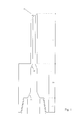
- a fine needle arrangement for taking a sample of cells from suspicious lesions by using fine needle aspiration (FNA) technique.
- the arrangement comprises a tubular needle member (1), a storage compartment (2) enclosing a chamber, and a coupling (3) to which a connector may be attached.
- At least a part (8) of the surface of the needle member has a rougher surface compared to the rest of the needle member surface in order to increase the acoustic reflection of the needle.
- This embodiment of the present invention is to adapt the FNA arrangement to ultrasonically guided procedures.
- the surface becomes randomly facetted (8) or roughened, preferably with a precision sandblasting technique or ultrasound cavitation erosion technique, which increases the acoustic reflection of the needle, thus enhancing the visibility of the needle.
- a second phenomena that has been identified by the inventors is that this short section of the needle generates an increased local friction over the treated needle-section, without adding much to the overall penetration needle force, which generates a local "tissue movement wave" during penetration - or retraction - that significantly enhances the secondary visibility from the diminutive target propagation movement volume.
- the diameter of the tubular member, the needle, in this embodiment is preferably 0.6 mm, 0.7 mm or 0.8 with lengths 50 mm and 80 mm. Even longer tubular members can be used in certain difficult cases.
- the tubular member has a proximal end connected to the storage compartment 2 and a distal end (4).
- the configuration and sharpness of the distal end (tip) has major impact on the penetration force and the amount of obtained sample.
- An increased length of the open section of the tubular member "the cutting length" (5) increases the amount of obtained sample.
- the configuration of the bevel shape and grinding has great effect on the penetration force.
- transition between the different parts 1,2 and 3 (6,7) is configured without any residual spaces and with efficient air streaming.
- transitions in inner diameter can be of any shape, for example linear or exponential.
- the material thickness of the tubular member is decreased, from the distal end and in the proximal direction, which increases the amount of sample yield.
- the inner surface of the tubular member is coated with appropriate material, e.g. PTFE (Teflon), to prevent wetting, i.e. to prevent sample material sticking to the surface.
- PTFE Teflon
- the tubular member during direct puncturing is preferably 0.5 mm, 0.6 mm or 0.7 mm in diameter with lengths 25 mm and 50 mm and with material thickness in the order of 50 to 200 ⁇ m, depending on the length of the needle. Certain stiffness is required in the needle to be able to penetrate the tumour in different directions. The material thickness should be considerable less than present standard needles. For example the inner diameter of a standard 0,6 mm needle are only 0,3 mm.
- the length and diameter of the storage compartment (2) must be carefully selected to be able to keep the sample in the needle, and not in the hub or syringe, and finally eject it. If the storage diameter is too large the air-stream speed-profile is too low to successfully eject the sample. Moreover if the length of the compartment is too short sample droplets will be thrust into the hub or syringe, due to the high energy, and be lost. Therefore it is important to arrange the shape so the energy and velocity parameters will be sufficiently distributed along the storage channel.
- the preferable outline of the compartment for a 0,6*25 mm needle is an inner diameter of 0,7 mm and length 16 mm (6,16 mm 3 ). Depending on the length and the inner diameter of the tubular member this can vary.
- the general outline of the chamber is preferably about 5-40 mm long and has an inner diameter of approximately 0,5-3 mm. This results in chamber volumes of approximately 1 - 300 mm 3 .
- the storage compartment might also be partly placed within the Luer-coupling and into the Record cone near the plunge lower turning level, in order to prevent adding extra length to the complete needle/syringe system. Moreover it might be fully integrated part of the coupling.
- the Coupling (3) is connecting the needle and the storage compartment to a male Luer coupling or the like, for example a syringe.
- the function of the coupling is to connect under pressure to the distal part of the tubular member and to provide a mechanical coupling for successful manoeuvring of the needle during sampling.
- the present invention increases the available sample material more than three times compared to the standard needles of the same size.
- a standard 0.7 mm diameter needle extracts more total sample than the present invention of size 0,6 mm.
- the available sample on the microscopic glass was similar for both needle types.
- the present invention yielded more total sample for cases with sparse material, which in combination with the better needle efficiency resulted in twice the amount of available material.
- a high frequency of inadequate specimens only occurs in the cases with the smallest sample volume.
- increasing the sampling yield often in the form of a high content of blood, in the cases with already enough volume adds little to the diagnostic properties of the yield.
- our proposed needle keeps the trauma and bleeding to a minimum but still yields more material in the critical cases.
- the change in (tip angle) "cutting length" and material thickness increase the number procedures with sufficient extracted total material, whereas the added storage compartment increases the available sample from lesions with sparse total material, such as fibrous tumours, by increasing the efficiency.
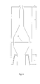
- Yet another preferred embodiment of the present invention is to provide any combinations of the above embodiments with anti-seeding properties, which is illustrated in figure 3 .
- This feature is further described in the above-mentioned WO-2006/036108 assigned to the applicant of the present application.
- the anti-seeding is established using a high peak low average radio frequency power burst pulse preferably in a monopole regime, e.g. between the tubular member and the patient body as a bulk.
- An electric insulation (9) over the tubular member's proximal section is used to prevent the patient skin from the secondary heat pulse.
- the sample inside the needle is effectively prevented from the generated heat in the tissue by the Faraday's cage effect.
- the non-insulated part of the tubular member needs to be connected to an external device preferably via the tubular member and the coupling (10).
- the anti-seeding pulse will preferably be triggered by changes in circuit impedance.
- the insulation must either be:
- a filter (11), preferably a micro-filter, is incorporated in the needle compartment or provided separately ( figure 4 ), which effectively collects the cells of the cystic fluid and thus increases the possibility to enrich the diagnostic cells, e.g. tumour cells.
- the fluid After the fluid has passed the filter, it is removed and the cells are fixed, stained and examined cytomorphologically directly on the filter.
- filters with pore diameters of 10 ⁇ m can efficiently accumulate epithelial cells whereas blood cells, e.g. erythrocytes with an average diameter of 7 ⁇ m, past the filter.
- staining and microscopic examination could be performed on cells located on the filter surface.
- the second "smearing glass could be exchanged with a low adhesion sterile spatula, which should be provided with the sampling kit.
- Other alternative embodiments might be automatic non-contact smearing devices using pressurised air streaming or acceleration methods or the like.
- the present invention may easily be incorporated in the "sampling kit" used in our other inventions described in the above-mentioned WO-2006/036108 and WO-2006/036112 .
Landscapes
- Health & Medical Sciences (AREA)
- Life Sciences & Earth Sciences (AREA)
- Medical Informatics (AREA)
- Engineering & Computer Science (AREA)
- Biomedical Technology (AREA)
- Heart & Thoracic Surgery (AREA)
- Pathology (AREA)
- Molecular Biology (AREA)
- Surgery (AREA)
- Animal Behavior & Ethology (AREA)
- General Health & Medical Sciences (AREA)
- Public Health (AREA)
- Veterinary Medicine (AREA)
- Apparatus Associated With Microorganisms And Enzymes (AREA)
- Sampling And Sample Adjustment (AREA)
Priority Applications (7)
| Application Number | Priority Date | Filing Date | Title |
|---|---|---|---|
| EP07108566A EP1994885A1 (fr) | 2007-05-21 | 2007-05-21 | Agencement d'une aiguille fine |
| CN200880025390A CN101754719A (zh) | 2007-05-21 | 2008-05-20 | 用于细胞取样的细针组件 |
| US12/601,074 US8323210B2 (en) | 2007-05-21 | 2008-05-20 | Fine needle arrangement for cell sampling |
| ES08750346T ES2400620T3 (es) | 2007-05-21 | 2008-05-20 | Disposición de aguja fina para muestreo de células |
| JP2010508820A JP2010527670A (ja) | 2007-05-21 | 2008-05-20 | 細胞採取用の細針装置 |
| PCT/EP2008/056141 WO2008142060A2 (fr) | 2007-05-21 | 2008-05-20 | Agencement d'aiguille fine pour un échantillonnage de cellules |
| EP08750346A EP2162069B1 (fr) | 2007-05-21 | 2008-05-20 | Agencement d'une aiguille fine |
Applications Claiming Priority (1)
| Application Number | Priority Date | Filing Date | Title |
|---|---|---|---|
| EP07108566A EP1994885A1 (fr) | 2007-05-21 | 2007-05-21 | Agencement d'une aiguille fine |
Publications (1)
| Publication Number | Publication Date |
|---|---|
| EP1994885A1 true EP1994885A1 (fr) | 2008-11-26 |
Family
ID=38603433
Family Applications (2)
| Application Number | Title | Priority Date | Filing Date |
|---|---|---|---|
| EP07108566A Withdrawn EP1994885A1 (fr) | 2007-05-21 | 2007-05-21 | Agencement d'une aiguille fine |
| EP08750346A Not-in-force EP2162069B1 (fr) | 2007-05-21 | 2008-05-20 | Agencement d'une aiguille fine |
Family Applications After (1)
| Application Number | Title | Priority Date | Filing Date |
|---|---|---|---|
| EP08750346A Not-in-force EP2162069B1 (fr) | 2007-05-21 | 2008-05-20 | Agencement d'une aiguille fine |
Country Status (6)
| Country | Link |
|---|---|
| US (1) | US8323210B2 (fr) |
| EP (2) | EP1994885A1 (fr) |
| JP (1) | JP2010527670A (fr) |
| CN (1) | CN101754719A (fr) |
| ES (1) | ES2400620T3 (fr) |
| WO (1) | WO2008142060A2 (fr) |
Cited By (2)
| Publication number | Priority date | Publication date | Assignee | Title |
|---|---|---|---|---|
| WO2019006303A1 (fr) * | 2017-06-30 | 2019-01-03 | Boston Scientific Scimed, Inc. | Dispositif de filtration aspiration |
| CN112869785A (zh) * | 2021-01-29 | 2021-06-01 | 重庆市人民医院 | 一种神经内科临床用取样送检设备 |
Families Citing this family (3)
| Publication number | Priority date | Publication date | Assignee | Title |
|---|---|---|---|---|
| CN102719353B (zh) * | 2012-06-13 | 2014-07-30 | 湖南大学 | 一种用于外周血中循环癌细胞的特异性捕获的装置及方法 |
| CN104605899B (zh) * | 2015-02-11 | 2017-03-01 | 昆明医科大学第一附属医院 | 一种专用于甲状腺的针吸细胞学活检穿刺装置 |
| DE102019126965A1 (de) * | 2019-10-08 | 2021-04-08 | Morpheus Ag | Chirurgisches Instrument, Verfahren zur Herstellung eines chirurgischen Instruments und Verwendung eines Drehgelenks zur Ausbildung eines Schneidewerkzeugs eines chirurgischen Instruments |
Citations (3)
| Publication number | Priority date | Publication date | Assignee | Title |
|---|---|---|---|---|
| US4869259A (en) * | 1988-05-17 | 1989-09-26 | Vance Products Incorporated | Echogenically enhanced surgical instrument and method for production thereof |
| US6086543A (en) * | 1998-06-24 | 2000-07-11 | Rubicor Medical, Inc. | Fine needle and core biopsy devices and methods |
| US20030195500A1 (en) * | 1999-06-17 | 2003-10-16 | Moorman Jack W. | Needle kit and method for microwave ablation, track coagulation, and biopsy |
Family Cites Families (9)
| Publication number | Priority date | Publication date | Assignee | Title |
|---|---|---|---|---|
| US4713061A (en) * | 1986-07-14 | 1987-12-15 | Survival Technology, Inc. | Cartridge with universal plastic hub |
| US5902279A (en) | 1993-04-20 | 1999-05-11 | Advanced Cytometrix, Inc. | Aspiration needle and method |
| US5330443A (en) * | 1993-04-20 | 1994-07-19 | Powles Trevor J | Aspiration needle, syringe for use therewith, apparatus incorporating the same and kit for use in fine needle aspiration cytology |
| US5573008A (en) * | 1993-10-29 | 1996-11-12 | Boston Scientific Corporation | Multiple biopsy sampling coring device |
| US5902280A (en) * | 1995-04-13 | 1999-05-11 | Advanced Cytometrix, Inc. | Aspiration needle apparatus incorporating its own vacuum and method and adapter for use therewith |
| US7815649B2 (en) | 2000-04-07 | 2010-10-19 | Kyphon SÀRL | Insertion devices and method of use |
| US6656161B2 (en) * | 2000-11-08 | 2003-12-02 | Ispg, Inc. | Magnifying hub |
| ATE355016T1 (de) | 2004-09-27 | 2006-03-15 | Vibratech Ab | Zellensammelvorrichtung |
| DE602004007661T2 (de) | 2004-09-27 | 2008-06-05 | Vibratech Ab | Anordnung zur therapeutischen Behandlung von Tumoren |
-
2007
- 2007-05-21 EP EP07108566A patent/EP1994885A1/fr not_active Withdrawn
-
2008
- 2008-05-20 ES ES08750346T patent/ES2400620T3/es active Active
- 2008-05-20 US US12/601,074 patent/US8323210B2/en not_active Expired - Fee Related
- 2008-05-20 WO PCT/EP2008/056141 patent/WO2008142060A2/fr not_active Ceased
- 2008-05-20 CN CN200880025390A patent/CN101754719A/zh active Pending
- 2008-05-20 EP EP08750346A patent/EP2162069B1/fr not_active Not-in-force
- 2008-05-20 JP JP2010508820A patent/JP2010527670A/ja not_active Withdrawn
Patent Citations (3)
| Publication number | Priority date | Publication date | Assignee | Title |
|---|---|---|---|---|
| US4869259A (en) * | 1988-05-17 | 1989-09-26 | Vance Products Incorporated | Echogenically enhanced surgical instrument and method for production thereof |
| US6086543A (en) * | 1998-06-24 | 2000-07-11 | Rubicor Medical, Inc. | Fine needle and core biopsy devices and methods |
| US20030195500A1 (en) * | 1999-06-17 | 2003-10-16 | Moorman Jack W. | Needle kit and method for microwave ablation, track coagulation, and biopsy |
Cited By (3)
| Publication number | Priority date | Publication date | Assignee | Title |
|---|---|---|---|---|
| WO2019006303A1 (fr) * | 2017-06-30 | 2019-01-03 | Boston Scientific Scimed, Inc. | Dispositif de filtration aspiration |
| CN112869785A (zh) * | 2021-01-29 | 2021-06-01 | 重庆市人民医院 | 一种神经内科临床用取样送检设备 |
| CN112869785B (zh) * | 2021-01-29 | 2022-09-06 | 重庆市人民医院 | 一种神经内科临床用取样送检设备 |
Also Published As
| Publication number | Publication date |
|---|---|
| EP2162069B1 (fr) | 2012-12-19 |
| WO2008142060A2 (fr) | 2008-11-27 |
| JP2010527670A (ja) | 2010-08-19 |
| US8323210B2 (en) | 2012-12-04 |
| US20100160828A1 (en) | 2010-06-24 |
| CN101754719A (zh) | 2010-06-23 |
| ES2400620T3 (es) | 2013-04-11 |
| EP2162069A2 (fr) | 2010-03-17 |
| WO2008142060A3 (fr) | 2009-02-05 |
Similar Documents
| Publication | Publication Date | Title |
|---|---|---|
| EP2982309B1 (fr) | Échangeable aiguille de biopsie | |
| EP1676533B1 (fr) | Procédé de fabrication d'un ensemble à aiguille à utiliser avec un dispositif de biopsie | |
| CN205729413U (zh) | 用于针吸活检的装置 | |
| US6673023B2 (en) | Micro-invasive breast biopsy device | |
| US5449001A (en) | Biopsy needle | |
| US10758213B2 (en) | Exchangeable core biopsy needle | |
| WO1997034531A1 (fr) | Perfectionnement concernant un ensemble aiguille a ponction-biopsie | |
| EP2162069B1 (fr) | Agencement d'une aiguille fine | |
| EP3103398B1 (fr) | Filtre pour biopsie à aiguille fine | |
| WO2013110079A1 (fr) | Système et procédé pour l'aspiration à l'aiguille fine | |
| US10182798B2 (en) | Exchangeable core biopsy needle |
Legal Events
| Date | Code | Title | Description |
|---|---|---|---|
| PUAI | Public reference made under article 153(3) epc to a published international application that has entered the european phase |
Free format text: ORIGINAL CODE: 0009012 |
|
| AK | Designated contracting states |
Kind code of ref document: A1 Designated state(s): AT BE BG CH CY CZ DE DK EE ES FI FR GB GR HU IE IS IT LI LT LU LV MC MT NL PL PT RO SE SI SK TR |
|
| AX | Request for extension of the european patent |
Extension state: AL BA HR MK RS |
|
| STAA | Information on the status of an ep patent application or granted ep patent |
Free format text: STATUS: THE APPLICATION HAS BEEN WITHDRAWN |
|
| 18W | Application withdrawn |
Effective date: 20090320 |