EP2004037B1 - Dispositif pour la determination d'une valeur comparative de données bio ainsi que pour la determination de donnees bio - Google Patents
Dispositif pour la determination d'une valeur comparative de données bio ainsi que pour la determination de donnees bio Download PDFInfo
- Publication number
- EP2004037B1 EP2004037B1 EP07722172.9A EP07722172A EP2004037B1 EP 2004037 B1 EP2004037 B1 EP 2004037B1 EP 07722172 A EP07722172 A EP 07722172A EP 2004037 B1 EP2004037 B1 EP 2004037B1
- Authority
- EP
- European Patent Office
- Prior art keywords
- signal
- sensor
- cwf
- signals
- data
- Prior art date
- Legal status (The legal status is an assumption and is not a legal conclusion. Google has not performed a legal analysis and makes no representation as to the accuracy of the status listed.)
- Active
Links
Images


Classifications
-
- A—HUMAN NECESSITIES
- A61—MEDICAL OR VETERINARY SCIENCE; HYGIENE
- A61B—DIAGNOSIS; SURGERY; IDENTIFICATION
- A61B5/00—Measuring for diagnostic purposes; Identification of persons
- A61B5/72—Signal processing specially adapted for physiological signals or for diagnostic purposes
- A61B5/7271—Specific aspects of physiological measurement analysis
- A61B5/7275—Determining trends in physiological measurement data; Predicting development of a medical condition based on physiological measurements, e.g. determining a risk factor
-
- A—HUMAN NECESSITIES
- A61—MEDICAL OR VETERINARY SCIENCE; HYGIENE
- A61B—DIAGNOSIS; SURGERY; IDENTIFICATION
- A61B5/00—Measuring for diagnostic purposes; Identification of persons
-
- A—HUMAN NECESSITIES
- A61—MEDICAL OR VETERINARY SCIENCE; HYGIENE
- A61B—DIAGNOSIS; SURGERY; IDENTIFICATION
- A61B5/00—Measuring for diagnostic purposes; Identification of persons
- A61B5/0002—Remote monitoring of patients using telemetry, e.g. transmission of vital signals via a communication network
- A61B5/0004—Remote monitoring of patients using telemetry, e.g. transmission of vital signals via a communication network characterised by the type of physiological signal transmitted
- A61B5/0006—ECG or EEG signals
-
- A—HUMAN NECESSITIES
- A61—MEDICAL OR VETERINARY SCIENCE; HYGIENE
- A61B—DIAGNOSIS; SURGERY; IDENTIFICATION
- A61B5/00—Measuring for diagnostic purposes; Identification of persons
- A61B5/02—Detecting, measuring or recording for evaluating the cardiovascular system, e.g. pulse, heart rate, blood pressure or blood flow
- A61B5/024—Measuring pulse rate or heart rate
- A61B5/02405—Determining heart rate variability
-
- A—HUMAN NECESSITIES
- A61—MEDICAL OR VETERINARY SCIENCE; HYGIENE
- A61B—DIAGNOSIS; SURGERY; IDENTIFICATION
- A61B5/00—Measuring for diagnostic purposes; Identification of persons
- A61B5/02—Detecting, measuring or recording for evaluating the cardiovascular system, e.g. pulse, heart rate, blood pressure or blood flow
- A61B5/024—Measuring pulse rate or heart rate
- A61B5/02416—Measuring pulse rate or heart rate using photoplethysmograph signals, e.g. generated by infrared radiation
-
- A—HUMAN NECESSITIES
- A61—MEDICAL OR VETERINARY SCIENCE; HYGIENE
- A61B—DIAGNOSIS; SURGERY; IDENTIFICATION
- A61B5/00—Measuring for diagnostic purposes; Identification of persons
- A61B5/02—Detecting, measuring or recording for evaluating the cardiovascular system, e.g. pulse, heart rate, blood pressure or blood flow
- A61B5/024—Measuring pulse rate or heart rate
- A61B5/02438—Measuring pulse rate or heart rate with portable devices, e.g. worn by the patient
-
- G—PHYSICS
- G16—INFORMATION AND COMMUNICATION TECHNOLOGY [ICT] SPECIALLY ADAPTED FOR SPECIFIC APPLICATION FIELDS
- G16H—HEALTHCARE INFORMATICS, i.e. INFORMATION AND COMMUNICATION TECHNOLOGY [ICT] SPECIALLY ADAPTED FOR THE HANDLING OR PROCESSING OF MEDICAL OR HEALTHCARE DATA
- G16H50/00—ICT specially adapted for medical diagnosis, medical simulation or medical data mining; ICT specially adapted for detecting, monitoring or modelling epidemics or pandemics
- G16H50/30—ICT specially adapted for medical diagnosis, medical simulation or medical data mining; ICT specially adapted for detecting, monitoring or modelling epidemics or pandemics for calculating health indices; for individual health risk assessment
-
- G—PHYSICS
- G16—INFORMATION AND COMMUNICATION TECHNOLOGY [ICT] SPECIALLY ADAPTED FOR SPECIFIC APPLICATION FIELDS
- G16H—HEALTHCARE INFORMATICS, i.e. INFORMATION AND COMMUNICATION TECHNOLOGY [ICT] SPECIALLY ADAPTED FOR THE HANDLING OR PROCESSING OF MEDICAL OR HEALTHCARE DATA
- G16H50/00—ICT specially adapted for medical diagnosis, medical simulation or medical data mining; ICT specially adapted for detecting, monitoring or modelling epidemics or pandemics
- G16H50/70—ICT specially adapted for medical diagnosis, medical simulation or medical data mining; ICT specially adapted for detecting, monitoring or modelling epidemics or pandemics for mining of medical data, e.g. analysing previous cases of other patients
Definitions
- the invention relates to a device for determining a comparison value of biodata (body parameters) of a living being for determining an individual risk, comprising at least one sensor for the noninvasive measurement of at least two signals selected from the group: CWF (continuous wave fluctuation), Sp02, pulse rate Heart rate, PTT (pulse transit time) and an evaluation device connected to the sensor, wherein the evaluation device has at least one analyzer, the signal ranges defined by signal analysis, a comparison means the signal ranges taking into account other parameters such as living organisms and existing biodata Data sets and the result can be output as an index value, wherein the sensor records the raw plethysmogram as a signal from which the evaluation device extracts a CWP (continous wave parameter) which, over time, is considered to be C WF signal is calculated.
- CWF continuous wave fluctuation
- Sp02 pulse rate Heart rate
- PTT pulse transit time
- an evaluation device connected to the sensor
- the evaluation device has at least one analyzer, the signal ranges defined by signal analysis
- a plethysmogram is used, which is recorded, for example, with a pulse oximeter.
- various CWP (continous wave parameters) are extracted.
- CWP are various fluctuating parameters, each describing a property of the plethysmogram.
- the following quantities can be calculated as CWP: the pulse wave amplitude, the integral over certain sections of the plethysmogram, the ratio of different integrals over different sections of the plethysmogram, a respiratory correlating portion of the plethysmogram, the rise angle of each pulse wave, the fall angle of each pulse wave, the Ratio of rise to fall angle, the duration of a pulse wave rise, the duration of a pulse wave drop, the rise to fall duration ratio, the pulse wave maximum, the pulse wave minimum, a magnitude related to PTT (Pulse Transit Time).
- PTT Pulse Transit Time
- CWP continuous wave fluctuation
- CWF signal the pulse wave amplitudes, parameters relating to the integral over a section of the plethysmogram, ratio of different integrals over different sections of the plethysmogram, parts of the plethysmogram correlating with the respiration, angle of rise of the pulse waves, Falling angles of the pulse waves, rising to falling angle ratios, duration of pulse wave rises, pulse wave waste durations, rising to falling duration ratios, the envelope of the pulse wave maxima or pulse wave minima, PTT (Pulse Transit Time) related quantities, the centerline of the plethysmogram, the pulse rate.
- PTT Pulse Transit Time
- any variation of the plethysmogram, each CWP and any combination of different CWP should be representable as a CWF signal.
- cardiovascular diseases or interactions that can affect the patient's quality of life. So far, such phenomena can only be statistically determined for a patient collective, but not recorded for a specific individual patient.
- Pulse wave signals are recorded and analyzed using photoplethysmography during anesthesia. The measurement result is used to control a drug pump.
- the device according to the invention and the method likewise relate to a modular system for determining biodata of a living being, comprising at least one sensor device for the non-invasive measurement of at least two signals.
- sleep-related diseases and / or cardiovascular diseases and / or metabolic disorders are to be determined and a diagnosis supported.
- the determination of sleep-related disorders or illnesses is currently always carried out by a specialist and usually during the night in a sleep laboratory.
- a polysomnography device is used for this purpose. Due to the small number of trained medical specialists and the small number of sleep laboratories, many patients often have to wait a long time for a diagnosis or a diagnosis is not even made.
- Object of the present invention is therefore to provide a device that allows a simple, fast and inexpensive diagnosis.
- an extended statement compared to the previous polysomnography, be made available. This extended statement should particularly concern the activity and assessment of the autonomic nervous system.
- At least one sensor for non-invasive measurement of at least two signals selected from the group: CWF (continous wave fluctuation), Sp02, heart rate, PTT (pulse transit time) is connected to an evaluation device, the evaluation device has at least one analyzer , the analyzer determines signal ranges that can be defined by signal analysis, a comparison device analyzes the signal ranges taking into account further parameters and the result of the analysis is transmitted as an index value to a connected output device.
- CWF continous wave fluctuation
- Sp02 heart rate
- PTT pulse transit time
- the aim of the invention is therefore not the diagnosis of a disease and not the mere determination of the frequency and severity of the occurrence of certain pathophysiological events. On the contrary, it is determined on the basis of the recorded signals how high the individual risk of the examined person is to suffer secondary diseases which impair the quality of life or life expectancy. Likewise, based on the determined risk, it can be deduced which therapy form and therapy dosage is optimal for the person. In addition, the success of existing therapy can be measured.
- CWF continous wave fluctuation
- SpO2 heart rate
- PTT pulse transit time
- Another object of the invention is to provide a modular device which makes it possible to quickly and easily adapt complementary modules as needed.
- a particular advantage of the device according to the invention is that even medical laypersons, but at least medical staff or non-specialist physicians are readily able to quickly and correctly apply the device according to the invention to the patient.
- a basic module has interfaces for adapting supplementary individual modules for ascertaining further biodata, such as ECG, heart rate, respiratory flow, PTT (pulse transit time).
- a plethysmogram is preferred, for example with a pulse oximeter and / or a multi-wavelength pulse spectrometer (according to FIG DE 102005020022 A1 . DE 10213692 A1 . DE 10321338 A1 ) recorded.
- the terms pulse oximeter and / or a pulse spectrometer are used here synonymously.
- the pulse oximeter and / or the pulse spectrometer using at least two wavelengths selected from the range 400 to 2500 nm, at least determine the following parameters: pulse rate, plethysmogram and oxygen saturation (SpO2 and / or SaO2). In the following, the terms SpO2 and SaO2 are therefore used synonymously.
- At least one CWP (continuous wave parameter) is extracted from the plethysmogram, preferably at least two or more CWPs are extracted.
- different signals can be combined in order to detect relevant fluctuations of the pulse wave and to use them for the evaluation.
- obstructive and central respiratory disorders from patterns detected in a plethysmogram-derived CWF signal.
- the characteristic patterns have frequency components which are associated with the respiratory frequency.
- a signal evaluation over a predefinable period of time is supported by the fact that the sensor is connected to a first memory unit for storing a detected measurement signal.
- the evaluation device has a course analyzer for analyzing the time profile of the signal.
- the comparison device is provided with a second memory unit for storing the calculated index value.
- a typical evaluation process takes place in that the amplitude characteristic of the measurement signal is evaluated.
- a frequency of the measurement signal is evaluated.
- a comprehensive signal evaluation can be done by performing a pattern recognition.
- a further signal evaluation can take place in that a periodic and / or transient signal analysis is performed.
- a supplementary signal evaluation can be carried out by performing a frequency analysis.
- An alternative signal evaluation can take place in that an analysis of the slope is performed.
- Signal evaluation can be carried out by forming histograms and / or distributions and / or derivatives.
- An alternative signal evaluation can take place in that threshold values are compared and / or correlated.
- a creation of a hierarchy of the signals and / or a decision tree can further support the signal evaluation.
- One way to capture signals is to evaluate a continuous wave fluctuation (CWF) signal.
- CWF continuous wave fluctuation
- the amplitudes (51) of the plethysmogram (49) are used to determine a CWF signal.
- the CWF (50) in this case represents the amplitude levels of the individual pulse waves of the plethysmogram (49).
- different signals can be combined in order to detect relevant fluctuations of the pulse wave and to use them for the evaluation.
- Another possibility for signal detection is that an oxygen saturation signal is evaluated.
- Another possibility for signal detection is that a pulse rate and / or heart rate signal is evaluated.
- PTT pulse transit time
- An additional option for signal acquisition is that an EEG signal is evaluated.
- Another measurement variant is that an oxygen saturation of the blood is evaluated.
- Another measurement variant is that a hemoglobin concentration of the blood is evaluated.
- snoring In order to record additional parameters, snoring, arousals, blood pressure, CO2, sleep stages, skin conductance, sleep depth, sleep fragmentation, parasympathetic activity can be evaluated in addition or in relation to the sympathetic, vascular compliance.
- An easy to implement measuring principle is that an optical density of at least one body region is evaluated.
- a signal evaluation is carried out with regard to present periodic signal components.
- the index of a cumulative autonomous resting intensity is associated with the regulation of the cardiovascular system.
- the detection of particularly meaningful events is achieved by performing a signal analysis regarding a period of activation of the autonomous system.
- a further increase in the quality of the statement can be achieved by evaluating at least one further parameter when evaluating periods of activation of the autonomous system.
- the increase in the quality of the statement can be achieved by changing the number of parameters acquired or derived from the acquired parameters Time of activation of the autonomic system in strength, type and timing is compared.
- the cumulative number and intensity of the activation periods of the autonomous system be taken into account when determining the index.
- a simple measurement setup is supported by the fact that the heart rate is detected by the sensor.
- the sensor detects the variability of the heart rate.
- the PTT is detected by the sensor.
- the senor detects a CWF.
- the pulse amplitude is detected by the sensor.
- a recognition of short-term signal changes is supported by the fact that a maximum value of the measured signal detected by the sensor is evaluated within a predefinable time interval.
- An easily applicable measurement method is that the signal acquisition is performed using photoplethysmography.
- An increase in system sensitivity can be achieved by carrying out the signal evaluation within at least one predeterminable frequency band.
- At least one further body parameter is evaluated by the evaluation unit.
- the age of the living being is evaluated.
- the weight of the living being may be evaluated as a further body parameter.
- the weight of the living being may be evaluated as a further body parameter.
- one or more already known risk factors for example for a cardiovascular disease, to be evaluated as a further body parameter.
- a consideration of events past in time can take place in that the patient's health history is evaluated as a further body parameter.
- a further increase in the quality of the statement can be achieved by evaluating the medication of the living being as a further body parameter.
- a further increase in the statement quality can be achieved by evaluating comparison values of other persons as further parameters.
- At least the following signals are determined, for example with a pulse oximeter sensor: continuous wave fluctuation: CWF, SpO2, pulse frequency.
- respiratory signals such as flow, pressure or snoring, heart rate, PTT (pulse transit time) are registered and analyzed by adding suitable sensors. It is also thought to use ECG signals, EEG signals, EMG signals for the evaluation. Furthermore, blood pressure and CO2 concentration can be recorded and used for the evaluation.
- the device according to the invention determines a risk index specific for the patient. This can be specified as a percentage, for example.
- the medical history of the patient is taken into account together with the current measurement data for determining the risk index.
- At least one readable memory is used as the database in the area of the device according to the invention.
- a differentiation of the risk classes is also provided. Due to the output of the device according to the invention, for example, a physician can specifically initiate a treatment.
- the CWP is first calculated from at least one measurement signal. Subsequently, the CWF is optionally determined from the original measurement signal, the CWP or both from the measurement signal and from the CWP.
- the determined CWF can be used, for example, in the context of an automated diagnostic system. In addition, however, it is also intended to evaluate the CWF directly for device control.
- the CWF can be compared with stored values or curves or with current measured values to carry out a further analysis.
- the device allows, in a preferred embodiment, a rapid determination of the risk index in that using a pulse oximetry sensor at least three signals, namely CWF, oxygen saturation of the blood and pulse rate are determined substantially simultaneously.
- Pattern recognition analyzes the signals and compares them with stored values. In particular, the change in the amplitude of the pulse wave is analyzed. The result of the comparison yields a patient-specific index value that is suitable for assessing the risk of cardiovascular disease.
- the device according to the invention is small and portable. The energy supply takes place alternatively via accumulators / batteries and / or via a mains cable.
- the measured data are stored in the device on a CompactFlash® card, as well as online via cable or optional wireless transmission to the PC.
- the data stored in the device can either be transferred to the PC via a USB interface or read into the software by reading the CompactFlash® Card via a reading device.
- the device according to the invention for determining and analyzing biodata of a living being consists of a basic module with a power supply and a memory unit and at least one sensor device for the non-invasive measurement of at least one measurement signal representing the heart activity and / or the breathing activity.
- the sensor device may for example be selected from the group: ECG, blood pressure cuff, pulse oximeter, impedance sensor, Doppler sensor.
- the measurement signal which represents the heart activity and / or the respiratory activity is selected from the group: pulse rate, plethysmogram, oxygen saturation, respiratory signal, heart signal.
- an analysis device for extracting at least one CWP (continous wave parameter) from the measurement signal, and / or a device for determining at least one CWF (continuous wave fluctuation) wherein the CWF (conti-nous wave fluctuation) is preferred the CWP and / or the measurement signal is determined.
- a classifier compares at least one CWF information and / or derived variables with stored data in order to identify physiological / pathophysiological events. Such events are, for example, an apnea, it being possible according to the invention to distinguish between central and obstructive apneas.
- a classifier compares at least one CWF information and / or quantities derived therefrom with other measurement signals and / or CWP in order to identify physiological / pathophysiological events therefrom.
- At least part of the results of the analysis device and / or at least a part of the results of the classifier are output substantially immediately acoustically and / or graphically, preferably via a display.
- At least two signals that are temporally related are evaluated for the purpose of identifying physiological events.
- the device according to the invention can be expanded to include additional modules for measuring further signals.
- the modules are preferably adapted in the sense of a "plug and play".
- complementary sensor devices are adapted for this purpose.
- At least one interface is arranged in the region of the basic module and / or in the region of supplementary modules.
- the interface also makes it possible to read data and / or control other devices, in particular therapy devices.
- ECG ECG
- EMG EMG
- EOG EEG
- EEG pulse oximetry
- blood pressure impedance measurement
- UV Ultrasonic
- Doppler CO2
- Respiratory Snore
- Oral Thoracic
- Abdomen Location Sensor
- the sensor devices can be supplemented in the form of a module which is designed, for example, to determine respiratory parameters, such as respiratory flow, PTT, motion signals, respiratory effort.
- the sensor devices can also be supplemented in the form of a module, which is designed to determine cardiac parameters, such as ECG, cardiac frequency.
- a device for data entry is provided.
- a display is provided as a display means for analysis results.
- acoustic alarms are provided.
- At least the basic module is releasably fixed to the body of a patient with fastening means, such as straps, for example.
- the device for detecting and analyzing biodata of a living organism at least one sensor device for non-invasive measurement of at least two signals, such as plethysmogram and derived therefrom CWF signals (conti-nous wave fluctuation), SpO2, pulse rate and a to the analysis device connected to the sensor device for analyzing transient and / or periodically recurring patterns of the measured signals.
- CWF signals conti-nous wave fluctuation
- SpO2 pulse rate
- a module for the evaluation of such information which are associated with the frequency or amplitude of the signals or the parameters derived therefrom.
- the device for detecting and analyzing biodata of a living being consists of a basic module with a power supply and a storage unit and at least one sensor device for non-invasive measurement of at least one measurement signal representing the respiratory activity, for example oxygen saturation, respiration signal, pressure signal, flow signal and at least one further sensor device for non-invasive measurement of at least one measurement signal representing cardiac activity, for example oxygen saturation, blood pressure, pulse rate, ECG, plethysmogram and an analysis device connected to the sensor device for extracting at least one CWP (continous wave parameter) from at least one the measuring signals.
- CWP continous wave parameter
- conclusions about apneas, hypopneas and other respiratory disorders can be obtained by analysis of the measurement signals.
- at least one sensor device for the non-invasive measurement of at least one measurement signal representing respiratory activity and at least one further sensor device for non-invasive measurement of at least one measurement signal representing cardiac activity it is possible, for example by pattern recognition, to draw conclusions about diseases of the cardiovascular system and / or the respiratory system.
- the basic module consists of a pulse oximeter, a possible interface for the control of other devices, eg therapy devices, possible further interfaces for further, optional sensors, eg connection of a respiratory flow / snore sensor, a communication interface such as USB, or a wireless protocol, a readable memory , a power supply via battery / accumulator and a data bus controller, as a connection for other modules and a display to display calculated indices, current readings, current battery charge states.
- the basic module allows bidirectional data exchange with other devices, for example with APAP therapy devices.
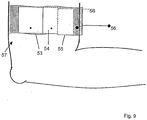
- FIG. 9 shows how the basic module (53) with adapted pneumology module (54) and adapted cardiology module (55) are positioned on a common base support (58) on the arm (57) of a user. Bidirectional data transmission is possible via the connected cable (56).
- Alternative application sites for the device according to the invention are: finger, toe, nose, ear, forehead.
- the device according to the invention has fewer input channels than conventional polysomnography devices and is thus much cheaper, smaller, lighter and more energy-efficient.
- ⁇ Pulsoximetry sensor to detect plethysmogram, oxygen saturation, pulse rate.
- this module can be adapted to the data bus of the basic module. According to the invention, it is intended to carry out the connection between basic and pneumology module as a plug-in connection, which is easy and quick to use.
- the pneumology module can also be used alone, without basic module.
- the pneumology module has at least one effort sensor system and a sensor device for determining a respiratory flow signal.
- the cardiology module allows ECG recording.
- a position sensor can be adapted.
- this module can be adapted to the data bus of the basic module and / or the pneumology module.
- the connection between the basic and / or the pneumology module and the cardiology module is designed as a plug connection which can be operated simply and quickly.
- the cardiology module can also be used alone, without basic module and / or pneumology module.
- the converter box is used for the wired data transmission of the data stored in the device.
- the data transmission is galvanically separated via a USB interface.
- the device according to the invention is charged via the power supply unit, or permanently supplied with power.
- the PC software is used for recording, storage, processing, visualization, evaluation, documentation and archiving of patient-related biosignals. This serves to support the diagnosis, therapy adjustment and therapy control of sleep disorders.
- the device software is used for acquisition, storage, processing and evaluation of biosignals. This serves to support the diagnosis, therapy adjustment and therapy control of sleep disorders.
- the device software communicates with the PC software via a secure data transfer protocol.
- the apparatus of the invention generates information signals (e.g., battery state of charge) that are graphically visualized and stored by the display and / or the personal computer system. These information signals serve to check the presence of the signals to be recorded, as well as the function check of the device. This avoids faulty recordings and eliminates the need for repetition of the night measurement.
- information signals e.g., battery state of charge
- the automated analyzes can be done online in the device and / or offline from the signals stored in the PC and assist the evaluator in the diagnosis of Sleep disorders, as well as the therapy initiation and therapy control.
- the PC software is used for visualization, evaluation, documentation and patient-related archiving of long-term examinations for the diagnosis of, for example, sleep disorders, cardiovascular diseases, diabetes.
- the system is configured and the transferred data is automatically analyzed offline.
- the software allows the user to enter comments. A manual reclassification of the analysis results by the evaluator is possible.
- the patient is able to set up the sensors and the device himself after being instructed by qualified personnel and following the patient's instructions for use.
- the device according to the invention in principle, it is possible to use the device according to the invention both in the prevention of various diseases (risk determination for secondary diseases), as well as in real-time scenarios in which it reacts directly to currently detected patterns.
- the PC software is used for visualization, evaluation, documentation and patient-related archiving of long-term examinations for the diagnosis of, for example, sleep disorders, cardiovascular diseases, diabetes.
- the system is configured and the transferred data is automatically analyzed offline.
- the software allows the user to enter comments. A manual reclassification of the analysis results by the evaluator is possible.
- the patient is able to set up the sensors and the device himself after being instructed by qualified personnel and following the patient's instructions for use.
- the device according to the invention processes and stores all measured signals on the integrated memory unit, e.g. CompactFlash® Card.
- the data is read either via a USB cable or by reading the CompactFlash® Card with a reader.
- the device according to the invention can transmit the recorded data online either wirelessly or by cable to the software, where the data is additionally stored.
- the device according to the invention has an optional position sensor.
- the sensor registers if and when the patient lies on his stomach, back or side.
- the device also has an optional effort sensor integrated in the housing. The integration reduces cleaning and increases the life of the sensor.
- LEDs or a display via an integrated display can be determined during a sensor test / impedance check, whether or which electrode is well or poorly applied.
- the device according to the invention by a yellow LED in the battery pack next to the battery icon or by a symbol on the integrated display on whether the battery is currently being charged.
- the state of charge can also be requested via the software, since a capacity monitoring is integrated in the battery.
- the stored data can be transferred to the PC.
- the converter box can also be used to charge the battery with the included power supply unit.
- a battery module can also be charged if it is not inserted in the device.
- the present results can be evaluated according to definable criteria.
- the respiratory flow snore nasal cannula in conjunction with the pressure sensor integrated in the device according to the invention, detects the respiratory flow and the snoring.
- the inspiration is registered by the generated negative pressure, the expiration by the generated overpressure.
- Snoring causes pressure fluctuations in the nostrils, which are also registered.
- the pressure measurement reacts more sensitively to small flow limitations than the thermal measurement. It is independent of the ambient temperature and additionally enables the visual assessment of the temporal flow contour. In mouth breathing, the signals may be attenuated. Alternatively, therefore, the simultaneous use of the respiratory flow mouth sensor.
- the measured pulse rate changes correspond to the heart rate changes that were triggered by a sleep-related apnea syndrome, sufficiently accurate.
- the change in the pulse wave in particular the amplitude of the pulse wave, is determined with the aid of photoplethysmography.
- the device according to the invention calculates a quality index for each detected oxygen saturation value, which characterizes the quality or the accuracy of the measured SpO2 value.
- the quality signal assumes values between 0 and 100%. When assessing long-term SpO2 measurements, the quality signal can be helpful because it suggests artifacts that occurred during the measurement.
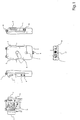
- Fig. 1 shows a handset with a pressure port (1) for connection to a Druckmeßschlauch, electrode terminals (2), a RIB (3) and a terminal (4) for an abdomen sensor (35), not shown.
- LEDs (5), a button (6) and a battery lock (7) to recognize.
- a connection (8) for a charge / data transfer cable and a battery (9) can be seen.
- a second pressure port (10) is provided.
- the functionality is further enhanced by a thorax sensor (11) and a connection (12) for a pulse oximetry sensor.
- an insert card (13) with application locations is provided in the area of a back of the handset.
- a Z-electrode (14) and a terminal (15) can be seen.
- the terminal (15) is used for connection to a in Fig. 2 illustrated respiratory flow snore sensor (16) or a respiratory mouth sensor (27).
- the port (1) is provided for connection to a respiratory snoring nasal cannula (22) or a pressure gauge tube (28).
- the connections (1, 10) together serve for connection to a pneumo-T adapter (33).
- Fig. 2 shows in addition to the sensors already mentioned above for connection to the mobile part sensor beads (17), a sleeve (18), a microphone (19), a support plate (20) and a sensor plug (21). Also shown are a sleeve (23), cannulas (24), a tab (25) and a port (26) of the respiratory nasal cannula (22).
- the pressure measuring hose (28) comprises a connecting piece (29), a connecting hose (30), a plastic hose (32) and a thread (31) for a CPAP connection.
- Further sensors include a pulse oximetry sensor (34) and an abdomen sensor (35).
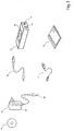
- Fig. 3 shows components for the data transfer:
- the handset is typically connected to an evaluation device, which may be designed as a personal computer.
- the evaluation device in this case comprises a CD-ROM drive with CD (36), a charger (37) with power supply (38) and plug (39), a charge-data transfer cable (40) and a USB cable (41).
- a converter box (42) is equipped with a socket (43) for the load data transfer cable (40), a USB socket (44) and a charger socket (45).
- a data transfer from the handset to the evaluation device can also be done directly by plugging a memory card (46).
- Fig. 4 shows a device strap (47) and an abdominal belt (48) to aid a mobile application.
- the device according to the invention is attached to the user.
- the strap is closed with the buckle.
- the Velcro straps By adjusting the Velcro straps, the belt can be adjusted to the body circumference.
- the strap consists of a skin-friendly elastic loop tape.
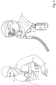
- Fig. 5 shows in the left drawing part an application with a respiratory snoring sensor (16) and in the right drawing part an application for pressure measurement in the area of a respiratory mask.
- the device is attached to the user with a pulse oximetry sensor and a respiratory flow snore sensor or connected to a respiratory tube with a Pneumo-T adapter.
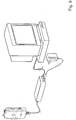
- Fig. 6 shows a connected device to an evaluation device, which is designed here as a personal computer.
- Data are transmitted to a PC via a medically approved power supply unit with a combination cable for charging the battery and for data transfer of the stored data via a galvanically isolated USB interface of the converter box.
- the converter box is used for the wired data transmission of the data stored in the device.
- the data transmission is galvanically separated via a USB interface.
- the device according to the invention is charged via the power supply unit, or permanently supplied with power.
- the PC software is running on the PC.
- the PC software is used for recording, storage, processing, visualization, evaluation, documentation and archiving of patient-related biosignals. This serves to support the diagnosis, therapy adjustment and therapy control of sleep disorders.
- the pulse oximetry sensor (34) measures the pulse oximetry signals, the oxygen saturation of the blood and the pulse rate of your patient.
- the main components of the sensor are at least two light emitting diodes and a receiver diode. For example, several SpO2 values are determined for each pulse wave (split-pulse-wave algorithm).
- the measured pulse rate changes correspond to the heart rate changes that were triggered by a sleep-related apnea syndrome, sufficiently accurate.
- sensors are used which alternatively and / or additionally determine the concentration of hemoglobin (cHb), oxyhemoglobin (HbO2), deoxygenated Hemoglobin (HbDe), carboxyhemoglobin (HbCO), methemoglobin (MetHb), sulfhemoglobin (HbSulf), bilirubin, glucose.
- cHb hemoglobin
- HbO2 oxyhemoglobin
- HbDe deoxyhemoglobin
- HbCO carboxyhemoglobin
- MetHb methemoglobin
- HbSulf sulfhemoglobin
- bilirubin glucose
- the sensors emit at least one light source to which alternatively and / or additionally the following wavelengths are selected, selected from the group: 150nm ⁇ 15%, 400nm ⁇ 15%, 460nm ⁇ 15%, 480nm ⁇ 15%, 520nm ⁇ 15%, 550nm ⁇ 15%, 560nm ⁇ 15%, 606nm ⁇ 15%, 617nm ⁇ 15%, 620 nm ⁇ 15%, 630 nm ⁇ 15%, 650 nm ⁇ 15%, 660 nm ⁇ , 705 nm ⁇ 15%, 710 nm ⁇ 15%, 720 nm ⁇ 10%, 805 nm ⁇ 15%, 810 nm ⁇ 15%, 880 nm ⁇ 15%, 890 nm, 905 nm ⁇ 15%, 910 nm ⁇ 15%, 950 nm ⁇ 15%, 980 nm ⁇ 15%, 980 nm ⁇ 15%, 1000 nm ⁇ 15% , 1030
- the device calculates a quality index for each detected oxygen saturation value, which characterizes the quality or the accuracy of the measured SpO2 value. If the signal is disturbed by movements, the number of values is low. Noise-free signals have a high number of values. Accordingly, a disturbed measurement signal produces a low quality value, an undisturbed measurement signal results in a high quality value.
- the quality signal assumes values between 0 and 100%. When assessing long-term SpO2 measurements, the quality signal can be helpful because it suggests artifacts that occurred during the measurement.
- Fig. 7 shows above the measured signal (49) as RohPlethysmogramm.
- a CWF signal (50) is extracted from the plethysmogram (49) .
- the fluctuation may represent the pulse wave amplitude.
- CWF integral of the plethysmogram
- any fluctuation of the plethysmogram should be representable as CWF.
- Fig. 8 shows a possible embodiment of the CWF signal.
- the amplitudes (51) of the plethysmogram (49) were used to determine the CWF signal.
- the CWF (50) in this case represents the amplitude height of the plethysmogram (49).
- different signals can be combined in order to detect relevant fluctuations of the pulse wave and to use them for the evaluation.
- Thoracic and abdominal sensors are used to record the thoracic and abdominal respiratory movements.
- Breathing movements cause alternating tensile stresses on the sensors in the fastening straps.
- the sensors convert the movements into electrical signals due to the piezoelectric effect.
- the abdominal sensor along with the abdominal straps, detects the abdominal respiratory movements.
- the sensor consists of a skin-friendly plastic.
- the respiratory flow snore sensor detects nasal and oral respiratory flow and snoring sounds.
- the sensor beads are made of thermistors. They record the respiratory flow via the temperature of the exhaled and inhaled air.
- the microphone registers the snoring sounds of the patient.
- the respiratory flow mouth sensor oral respiratory flow is detected during diagnosis with the respiratory flow snoring nasal cannula, therapy control or therapy setting.
- the sensor beads are made of thermistors. They record the respiratory flow via the temperature of the exhaled and inhaled air.
- the Breath Flow Oral Sensor (27) is used in conjunction with the Pneumo-T Adapter (28) for diagnosis along with the Respiratory Snore Nasopharyngeal or for Therapy Control or Therapy Adjustment to detect Oral Breathing.
- the Pneumo-T adapter is used for therapy control together with a nasal mask. Through it the respiratory flow and the snoring of the patient are registered during the therapy and the applied therapy pressure in the mask is measured. Via the pressure measuring tubes, in- and expiratory pressure fluctuations are conducted from the mask to the device. The exhalation of air generates a slight overpressure, the inhalation corresponding to a negative pressure. From the pressure differences, the breaths can be derived. Snoring sounds are measured by rapid pressure changes. The therapy pressure is derived from the static component of the pressure signal.
- the Pneumo-T Adapter (28) is used with therapy setting and therapy control with xPAP devices. The Pneumo-T Adapter can be used with the Breath Flow Oral Sensor (27) to detect mouth breathing and mouth leakage.
- the Pneumo-T adapter has a standard cone (ISO 22) for connection to therapy masks.
- the quantity detected with the electrodes is the voltage.
- a voltage difference between two points of the body is measured. Since the measurement on the skin surface is noninvasive, the measurable voltage differences are very small. They are in the range ⁇ V for the EEG, EOG and EMG, and in the range mV for the ECG.
- the Bluetooth-USB adapter can be used to receive or transmit data wirelessly from the device according to the invention, to configure the device, and to carry out an application check.
- the device according to the invention can be operated via a network.
- the device according to the invention can receive data wirelessly via the USB server in connection with the Bluetooth USB adapter, furthermore the device can be configured and an application control can be carried out.
- the device according to the invention can be connected via the converter box wired.
- the data stored on the CompactFlash® Card can be read out by the device according to the invention.
- the device according to the invention can be configured via the CompactFlash® card reader and / or several CompactFlash® cards with different configurations can be created.
- a supplementary PC software enables the reading and presentation of the therapy control data and the remote adjustment of all mentioned therapy devices via the software, as well as the PC-based evaluation of titration data from a ventilation titration device.
- the device according to the invention can be combined with conventional CPAP, BiLevel and APAP titration home ventilation therapy systems.
- the coupling of the systems is quick and easy via the Pneumo-T adapter, which is inserted between the hose and the mask.
- the software is used for analysis and offers alternative evaluation suggestions.
- the evaluation of automatically generated analysis results is the responsibility of the physician.
- the time in the basic unit is compared with the system time of the PC. If the data transfer to the PC is interrupted, the measurement data is still stored in the device.
- the software displays the signals as a zero line. All data is readable.
- the ECG is derived bipolar.
- the polysomnographic derivative of the device according to the invention is based on the Einthoven derivative.
- the reference electrode in the device according to the invention is the earth electrode at an arbitrary body location.
- the device according to the invention can be applied to a patient.
- Velcro straps the straps can be adjusted to the body circumference of the patient.
- the strap consists of a skin-friendly elastic loop tape.
- a test can be performed after the sensors and devices have been installed. To do this, press the button (6). process device Sensor test is running During the sensor test, the LED of the currently tested sensor flashes quickly (4x per second). Sensor test ok The LED of the corresponding signal stops flashing after the end of the impedance test: Impedance of the electrode ⁇ 5 k ⁇ OK or sensor signal present. Sensor test medium The LED of the corresponding signal flashes slowly after the end of the impedance test: electrode impedance ⁇ 10 k ⁇ not optimal, but acceptable quality. Green LEDs flash slowly at 0.5Hz. Sensor test not ok The LED of the corresponding signal flashes quickly after the impedance test has ended: Impedance of the electrode> 10 k ⁇ or no sensor signal (check electrode or sensor, unacceptable signal quality). Green LEDs flash rapidly at 1Hz.
- the sensor test checks all channels, including the effort and pulsoximetry sensors, as well as the thermistor and nasal cannula for the presence of a signal.
- the LED When the LED is off, it means: "Sensor is connected and transmits a (physiological) signal”.
- Position sensor Position sensor integrated sensor in the device value range re. Side, left Side, stomach, back, standing accuracy about 45 ° ⁇ 15 ° location CPAP / BiPAP / SmartPAP printing measuring range 0 to 40 hPa accuracy ⁇ 0.6 hPa SpO2 measuring range 50 to 100% SpO2 accuracy 70% ⁇ SpO2 ⁇ 100% better than 2% accuracy SpO2 measuring range 30 to 250 bpm Pulse accuracy ⁇ 1 bpm to 2% of the displayed value signal quality > 90% Respiratory flow snore sensor 3 thermistors as sum signal, no measuring function at ambient temperatures between 33 - 38 ° C Respiratory flow nasal cannula inspiratory / expiratory pressure fluctuations Respiratory flow oral sensor A thermistor, no measuring function at ambient temperatures between 33 - 38 ° C channel ECG EEG EMG EOG Dynamic range (physical value range) ⁇ 5m
Landscapes
- Health & Medical Sciences (AREA)
- Life Sciences & Earth Sciences (AREA)
- Engineering & Computer Science (AREA)
- Public Health (AREA)
- Medical Informatics (AREA)
- Pathology (AREA)
- General Health & Medical Sciences (AREA)
- Biomedical Technology (AREA)
- Cardiology (AREA)
- Molecular Biology (AREA)
- Physics & Mathematics (AREA)
- Heart & Thoracic Surgery (AREA)
- Biophysics (AREA)
- Surgery (AREA)
- Animal Behavior & Ethology (AREA)
- Veterinary Medicine (AREA)
- Physiology (AREA)
- Data Mining & Analysis (AREA)
- Primary Health Care (AREA)
- Epidemiology (AREA)
- Databases & Information Systems (AREA)
- Psychiatry (AREA)
- Computer Networks & Wireless Communication (AREA)
- Artificial Intelligence (AREA)
- Computer Vision & Pattern Recognition (AREA)
- Signal Processing (AREA)
- Measurement Of The Respiration, Hearing Ability, Form, And Blood Characteristics Of Living Organisms (AREA)
- Measuring Pulse, Heart Rate, Blood Pressure Or Blood Flow (AREA)
Claims (8)
- Dispositif destiné à déterminer une valeur comparative de données biologiques ou de paramètres corporels d'un organisme vivant pour déterminer un risque individuel, constitué d'un modèle de base portable muni d'au moins un capteur (11, 14, 16, 17, 19, 22, 27, 28, 34, 35) pour la mesure non invasive d'au moins deux signaux choisis dans le groupe : CWF (50), Sp02, fréquence du pouls, fréquence cardiaque, PTT et
muni d'un dispositif d'analyse connecté au capteur (11, 14, 16, 17, 19, 22, 27, 28, 34, 35), dans lequel le dispositif d'analyse comporte au moins un analyseur qui détermine des plages de signaux définissables par analyse de signaux, et
muni d'une interface pour la commande d'un appareil de thérapie et
d'une interface USB et
d'une unité de stockage intégrée et
d'un dispositif de comparaison qui harmonise les plages de signaux en tenant compte d'autres paramètres tels que des données biologiques existantes ou des paramètres corporels de l'organisme vivant et/ou des ensembles de données statiques et qui délivre le résultat sous la forme d'une valeur d'indice,
dans lequel le capteur enregistre le pléthysmogramme brut sous la forme d'un signal à partir duquel le dispositif d'analyse extrait un CWP, qui est calculé au cours du temps sous la forme d'un signal CWF, et en ce que le signal CWF est analysé pour la commande de l'appareil, et
dans lequel tous les signaux mesurés sont traités et stockés sur l'unité de mémoire intégrée, les données sont lues par l'intermédiaire d'un câble USB et en ce que les données sont transmises par l'intermédiaire de l'interface USB. - Dispositif selon la revendication 1, caractérisé en ce que les paramètres des données biologiques de l'organisme vivant comprennent tels que les traitements médicaux, les antécédents pathologiques, l'âge, le sexe, des données diagnostiques.
- Dispositif selon la revendication 1 ou 2, caractérisé en ce que les ensembles de données statistiques peuvent être établis à partir de groupes d'êtres vivants, et comprennent des paramètres tels que les traitements médicaux, l'âge, le sexe, des données diagnostiques et autres.
- Dispositif selon l'une des revendications 1 à 3, caractérisé en ce que le dispositif de comparaison peut être couplé à des dispositifs périphériques tels que des ordinateurs, des imprimantes ou des appareils d'affichage.
- Dispositif selon l'une des revendications 1 à 4, caractérisé en ce que le dispositif d'analyse est conçu pour déterminer un CWP à partir d'au moins un signal de mesure acquis.
- Dispositif selon la revendication 5, caractérisé en ce que le dispositif d'analyse est conçu pour déterminer une CWF (50) à partir d'au moins l'un des signaux de mesure acquis.
- Dispositif selon l'une des revendications 1 à 6, caractérisé en ce que le dispositif d'analyse est conçu pour déterminer une CWF (50) à partir du CWP.
- Dispositif selon l'une des revendications 1 à 7, caractérisé en ce que le dispositif d'analyse destiné à déterminer la CWF (50) est réalisé à la fois à partir d'au moins un signal de mesure acquis et du CWP.
Applications Claiming Priority (3)
| Application Number | Priority Date | Filing Date | Title |
|---|---|---|---|
| DE102006018041 | 2006-04-07 | ||
| DE102006018040 | 2006-04-07 | ||
| PCT/DE2007/000614 WO2007115553A1 (fr) | 2006-04-07 | 2007-04-04 | Dispositif et procede pour la determination d'une valeur comparative de données bio ainsi que pour la determination de donnees bio |
Publications (2)
| Publication Number | Publication Date |
|---|---|
| EP2004037A1 EP2004037A1 (fr) | 2008-12-24 |
| EP2004037B1 true EP2004037B1 (fr) | 2018-09-12 |
Family
ID=38148507
Family Applications (1)
| Application Number | Title | Priority Date | Filing Date |
|---|---|---|---|
| EP07722172.9A Active EP2004037B1 (fr) | 2006-04-07 | 2007-04-04 | Dispositif pour la determination d'une valeur comparative de données bio ainsi que pour la determination de donnees bio |
Country Status (3)
| Country | Link |
|---|---|
| US (1) | US9801589B2 (fr) |
| EP (1) | EP2004037B1 (fr) |
| WO (1) | WO2007115553A1 (fr) |
Cited By (1)
| Publication number | Priority date | Publication date | Assignee | Title |
|---|---|---|---|---|
| EP4596012A1 (fr) | 2024-01-30 | 2025-08-06 | Georg-August-Universität Göttingen Stiftung Öffentlichen Rechts, Universitätsmedizin | Tube respiratoire avec capteur acoustique |
Families Citing this family (47)
| Publication number | Priority date | Publication date | Assignee | Title |
|---|---|---|---|---|
| US10512429B2 (en) | 2004-12-23 | 2019-12-24 | ResMed Pty Ltd | Discrimination of cheyne-stokes breathing patterns by use of oximetry signals |
| NZ593988A (en) | 2004-12-23 | 2012-12-21 | Resmed Ltd | Apparatus and method of diagnosis of Cheyne-Stokes breathing in a person |
| US8437843B1 (en) | 2006-06-16 | 2013-05-07 | Cleveland Medical Devices Inc. | EEG data acquisition system with novel features |
| JP5028143B2 (ja) | 2007-05-23 | 2012-09-19 | ローレル精機株式会社 | 安全管理システム |
| JP4974761B2 (ja) * | 2007-05-25 | 2012-07-11 | ローレル精機株式会社 | 安全管理システム |
| US10426399B1 (en) | 2007-06-08 | 2019-10-01 | Cleveland Medial Devices Inc. | Method and device for in-home sleep and signal analysis |
| US9202008B1 (en) | 2007-06-08 | 2015-12-01 | Cleveland Medical Devices Inc. | Method and device for sleep analysis |
| TWM326235U (en) * | 2007-07-23 | 2008-01-21 | Inpaq Technology Co Ltd | Structure of linear polarized flat antenna |
| US20170188940A9 (en) | 2007-11-26 | 2017-07-06 | Whispersom Corporation | Device to detect and treat Apneas and Hypopnea |
| EP2116181B1 (fr) | 2007-12-28 | 2017-09-13 | Löwenstein Medical Technology S.A. | Dispositif destinés à la détermination de données biologiques |
| US9492105B1 (en) * | 2009-02-13 | 2016-11-15 | Cleveland Medical Devices Inc. | Device for sleep diagnosis |
| US8355769B2 (en) * | 2009-03-17 | 2013-01-15 | Advanced Brain Monitoring, Inc. | System for the assessment of sleep quality in adults and children |
| NZ614025A (en) * | 2009-04-20 | 2014-10-31 | Resmed Ltd | Discrimination of cheyne-stokes breathing patterns by use of oximetry signals |
| CN101933798A (zh) * | 2009-06-29 | 2011-01-05 | 周常安 | 无线多重睡眠生理检测系统 |
| US8430817B1 (en) | 2009-10-15 | 2013-04-30 | Masimo Corporation | System for determining confidence in respiratory rate measurements |
| US9724016B1 (en) | 2009-10-16 | 2017-08-08 | Masimo Corp. | Respiration processor |
| US9307928B1 (en) | 2010-03-30 | 2016-04-12 | Masimo Corporation | Plethysmographic respiration processor |
| WO2011150916A2 (fr) * | 2010-05-31 | 2011-12-08 | Seca Ag | Équipement d'analyse modulaire |
| US9239890B2 (en) | 2011-05-31 | 2016-01-19 | Fanhattan, Inc. | System and method for carousel context switching |
| US9778818B2 (en) | 2011-05-31 | 2017-10-03 | Fanhattan, Inc. | System and method for pyramidal navigation |
| DE102011110486A1 (de) * | 2011-08-17 | 2013-02-21 | Daimler Ag | Verfahren und Vorrichtung zur Überwachung zumindest eines Fahrzeuginsassen und Verfahren zum Betrieb zumindest einer Assistenzvorrichtung |
| US8870764B2 (en) | 2011-09-06 | 2014-10-28 | Resmed Sensor Technologies Limited | Multi-modal sleep system |
| US9146616B2 (en) * | 2012-01-10 | 2015-09-29 | Fanhattan Inc. | Touch-enabled remote control |
| US9060745B2 (en) | 2012-08-22 | 2015-06-23 | Covidien Lp | System and method for detecting fluid responsiveness of a patient |
| US8731649B2 (en) | 2012-08-30 | 2014-05-20 | Covidien Lp | Systems and methods for analyzing changes in cardiac output |
| US9357937B2 (en) | 2012-09-06 | 2016-06-07 | Covidien Lp | System and method for determining stroke volume of an individual |
| US9241646B2 (en) | 2012-09-11 | 2016-01-26 | Covidien Lp | System and method for determining stroke volume of a patient |
| US20140081152A1 (en) | 2012-09-14 | 2014-03-20 | Nellcor Puritan Bennett Llc | System and method for determining stability of cardiac output |
| TW201421417A (zh) * | 2012-11-29 | 2014-06-01 | 周源祥 | 透過藍牙/Wi-Fi與行動電話裝置以傳輸生理偵測訊號的方法 |
| US8977348B2 (en) | 2012-12-21 | 2015-03-10 | Covidien Lp | Systems and methods for determining cardiac output |
| US10441181B1 (en) | 2013-03-13 | 2019-10-15 | Masimo Corporation | Acoustic pulse and respiration monitoring system |
| WO2014183003A1 (fr) | 2013-05-10 | 2014-11-13 | University Of Utah Research Foundation | Dispositifs, systèmes et procédés permettant de mesurer une perte de sang |
| US10690684B2 (en) | 2013-05-10 | 2020-06-23 | Majelco Medical, Inc. | Apparatus and system for measuring volume of blood loss |
| WO2014208289A1 (fr) * | 2013-06-28 | 2014-12-31 | 株式会社村田製作所 | Dispositif d'estimation d'état biologique |
| WO2017180656A1 (fr) | 2016-04-11 | 2017-10-19 | Alfred Akerman | Appareil et système de mesurer du volume de perte de sang |
| US20180206762A1 (en) * | 2017-01-25 | 2018-07-26 | Intel Corporation | Sleep apnea therapy enhancement method and apparatus |
| US10953192B2 (en) | 2017-05-18 | 2021-03-23 | Advanced Brain Monitoring, Inc. | Systems and methods for detecting and managing physiological patterns |
| EP3684463B1 (fr) | 2017-09-19 | 2025-05-14 | Neuroenhancement Lab, LLC | Procédé et appareil de neuro-activation |
| US11717686B2 (en) | 2017-12-04 | 2023-08-08 | Neuroenhancement Lab, LLC | Method and apparatus for neuroenhancement to facilitate learning and performance |
| US12280219B2 (en) | 2017-12-31 | 2025-04-22 | NeuroLight, Inc. | Method and apparatus for neuroenhancement to enhance emotional response |
| US11478603B2 (en) | 2017-12-31 | 2022-10-25 | Neuroenhancement Lab, LLC | Method and apparatus for neuroenhancement to enhance emotional response |
| US11364361B2 (en) | 2018-04-20 | 2022-06-21 | Neuroenhancement Lab, LLC | System and method for inducing sleep by transplanting mental states |
| CN113382683A (zh) | 2018-09-14 | 2021-09-10 | 纽罗因恒思蒙特实验有限责任公司 | 改善睡眠的系统和方法 |
| US11786694B2 (en) | 2019-05-24 | 2023-10-17 | NeuroLight, Inc. | Device, method, and app for facilitating sleep |
| US12533552B2 (en) * | 2020-08-28 | 2026-01-27 | Robert Bosch Gmbh | Controller and a method to determine a swim stroke |
| EP4282323B1 (fr) * | 2021-07-29 | 2025-07-02 | Beijing Honor Device Co., Ltd. | Méthode d'évaluation de la qualité du signal de détection physiologique, dispositif électronique |
| US20250302377A1 (en) * | 2022-05-06 | 2025-10-02 | Oxama Medical Corp | Biomedical Parameters Monitoring System for the Diagnosis of Sleep Disorders |
Family Cites Families (10)
| Publication number | Priority date | Publication date | Assignee | Title |
|---|---|---|---|---|
| SE465551B (sv) | 1990-02-16 | 1991-09-30 | Aake Oeberg | Anordning foer bestaemning av en maenniskas hjaert- och andningsfrekvens genom fotopletysmografisk maetning |
| IL116020A (en) | 1995-11-16 | 2000-06-01 | Optelmed Ltd | Apparatus and method for measuring the variability of cardiovascular parameters |
| US6804551B2 (en) | 1998-03-17 | 2004-10-12 | University Of Virginia Patent Foundation | Method and apparatus for the early diagnosis of subacute, potentially catastrophic illness |
| EP1063917B1 (fr) | 1998-03-17 | 2008-12-24 | The University Of Virginia Patent Foundation | Methode et appareil de diagnostic precoce de maladies subaigus potentiellement catastrophiques |
| US6319587B1 (en) | 1998-09-24 | 2001-11-20 | Toray Industries, Inc. | Biaxially-oriented polyester film |
| US6340346B1 (en) * | 1999-11-26 | 2002-01-22 | T.A.O. Medical Technologies Ltd. | Method and system for system identification of physiological systems |
| AUPQ966600A0 (en) * | 2000-08-25 | 2000-09-21 | Jankov, Vladimir | System for physiological monitoring during sleep |
| US6856829B2 (en) | 2000-09-07 | 2005-02-15 | Denso Corporation | Method for detecting physiological condition of sleeping patient based on analysis of pulse waves |
| KR100519758B1 (ko) | 2003-01-22 | 2005-10-07 | 삼성전자주식회사 | 용적맥파를 이용한 인체 안정도 평가방법 및 장치 |
| US7635337B2 (en) * | 2005-03-24 | 2009-12-22 | Ge Healthcare Finland Oy | Determination of clinical stress of a subject in pulse oximetry |
-
2007
- 2007-04-04 EP EP07722172.9A patent/EP2004037B1/fr active Active
- 2007-04-04 US US12/225,980 patent/US9801589B2/en active Active
- 2007-04-04 WO PCT/DE2007/000614 patent/WO2007115553A1/fr not_active Ceased
Non-Patent Citations (1)
| Title |
|---|
| None * |
Cited By (1)
| Publication number | Priority date | Publication date | Assignee | Title |
|---|---|---|---|---|
| EP4596012A1 (fr) | 2024-01-30 | 2025-08-06 | Georg-August-Universität Göttingen Stiftung Öffentlichen Rechts, Universitätsmedizin | Tube respiratoire avec capteur acoustique |
Also Published As
| Publication number | Publication date |
|---|---|
| WO2007115553A1 (fr) | 2007-10-18 |
| EP2004037A1 (fr) | 2008-12-24 |
| US20090240119A1 (en) | 2009-09-24 |
| US9801589B2 (en) | 2017-10-31 |
Similar Documents
| Publication | Publication Date | Title |
|---|---|---|
| EP2004037B1 (fr) | Dispositif pour la determination d'une valeur comparative de données bio ainsi que pour la determination de donnees bio | |
| JP7692015B2 (ja) | 睡眠呼吸障害のスクリーニング、診断および監視のためのシステムおよび方法 | |
| DE102004042797B4 (de) | Erfassungsgerät sowie Verfahren zur Observation schlafbezogener Atmungsstörungen | |
| DE69930720T2 (de) | Integrierte anordnung zur anzeige von apnoe während des schlafes | |
| JP4753881B2 (ja) | 心電図から導出された睡眠呼吸障害の監視、検出及び分類のための装置及び信号処理方法 | |
| EP1353594B1 (fr) | Evaluation des risques d'apnee du sommeil | |
| JP2013533021A (ja) | 睡眠障害分析のための方法およびシステム | |
| DE102019129288A1 (de) | Vorrichtung und Verfahren zur Bestimmung des Schweregrads einer Schlafapnoe | |
| EP2116181B1 (fr) | Dispositif destinés à la détermination de données biologiques | |
| CN115607123A (zh) | 一种心肺功能监测和呼吸机闭环控制一体化装置 | |
| EP1859733B1 (fr) | Procédé et dispositif destinés à mettre en corrélation les signaux de respiration du système cardiocirculatoire | |
| DE102007016447A1 (de) | Vorrichtung zur Ermittlung von Biosignalen | |
| EP3803896B1 (fr) | Dispositif d'analyse médicale permettant d'évaluer l'aptitude à narcose d'un patient | |
| DE102007009984B4 (de) | Verfahren und Vorrichtung zur Aufzeichnung von Biosignalen | |
| WO2013182173A1 (fr) | Appareil d'analyse et de diagnostic du stress et de l'épuisement professionnel | |
| US20200345275A1 (en) | A method for measuring a sedation state of a patient | |
| DE102007063934B3 (de) | Vorrichtung zur Ermittlung von Biosignalen | |
| DE10021784B4 (de) | Anordnung zur Diagnose und/oder Therapie schlafbezogener Atmungsstörungen | |
| EP4248858A1 (fr) | Détection de sommeil/éveil | |
| DE102007016446A1 (de) | Vorrichtung und Verfahren zur Bestimmung eines Vergleichswertes von Biodaten | |
| Riha | Methods of different sleep tests | |
| KR20190032577A (ko) | 개선된 호흡 모니터 및 시스템 | |
| Coomar | Clinical and electronic monitoring during procedural sedation and analgesia for dentistry. | |
| DE102025126833A1 (de) | Tragbare Ausrüstung mit künstlicher Intelligenz zur Überwachung der Atmung und des Herzzustands und System dafür | |
| GGGGLGGGLS | American Sleep Disorders Association Role And Oualification Of Technologists Performing Polysomnography |
Legal Events
| Date | Code | Title | Description |
|---|---|---|---|
| PUAI | Public reference made under article 153(3) epc to a published international application that has entered the european phase |
Free format text: ORIGINAL CODE: 0009012 |
|
| 17P | Request for examination filed |
Effective date: 20080930 |
|
| AK | Designated contracting states |
Kind code of ref document: A1 Designated state(s): AT BE BG CH CY CZ DE DK EE ES FI FR GB GR HU IE IS IT LI LT LU LV MC MT NL PL PT RO SE SI SK TR |
|
| 17Q | First examination report despatched |
Effective date: 20100422 |
|
| DAX | Request for extension of the european patent (deleted) | ||
| RAP1 | Party data changed (applicant data changed or rights of an application transferred) |
Owner name: LOEWENSTEIN MEDICAL TECHNOLOGY GMBH + CO. KG |
|
| RAP1 | Party data changed (applicant data changed or rights of an application transferred) |
Owner name: LOEWENSTEIN MEDICAL TECHNOLOGY S.A. |
|
| GRAP | Despatch of communication of intention to grant a patent |
Free format text: ORIGINAL CODE: EPIDOSNIGR1 |
|
| STAA | Information on the status of an ep patent application or granted ep patent |
Free format text: STATUS: GRANT OF PATENT IS INTENDED |
|
| INTG | Intention to grant announced |
Effective date: 20170302 |
|
| GRAJ | Information related to disapproval of communication of intention to grant by the applicant or resumption of examination proceedings by the epo deleted |
Free format text: ORIGINAL CODE: EPIDOSDIGR1 |
|
| STAA | Information on the status of an ep patent application or granted ep patent |
Free format text: STATUS: EXAMINATION IS IN PROGRESS |
|
| INTC | Intention to grant announced (deleted) | ||
| GRAP | Despatch of communication of intention to grant a patent |
Free format text: ORIGINAL CODE: EPIDOSNIGR1 |
|
| STAA | Information on the status of an ep patent application or granted ep patent |
Free format text: STATUS: GRANT OF PATENT IS INTENDED |
|
| INTG | Intention to grant announced |
Effective date: 20180524 |
|
| GRAS | Grant fee paid |
Free format text: ORIGINAL CODE: EPIDOSNIGR3 |
|
| GRAA | (expected) grant |
Free format text: ORIGINAL CODE: 0009210 |
|
| STAA | Information on the status of an ep patent application or granted ep patent |
Free format text: STATUS: THE PATENT HAS BEEN GRANTED |
|
| AK | Designated contracting states |
Kind code of ref document: B1 Designated state(s): AT BE BG CH CY CZ DE DK EE ES FI FR GB GR HU IE IS IT LI LT LU LV MC MT NL PL PT RO SE SI SK TR |
|
| REG | Reference to a national code |
Ref country code: GB Ref legal event code: FG4D Free format text: NOT ENGLISH |
|
| REG | Reference to a national code |
Ref country code: CH Ref legal event code: EP |
|
| REG | Reference to a national code |
Ref country code: IE Ref legal event code: FG4D Free format text: LANGUAGE OF EP DOCUMENT: GERMAN |
|
| REG | Reference to a national code |
Ref country code: DE Ref legal event code: R096 Ref document number: 502007016384 Country of ref document: DE |
|
| REG | Reference to a national code |
Ref country code: AT Ref legal event code: REF Ref document number: 1039612 Country of ref document: AT Kind code of ref document: T Effective date: 20181015 |
|
| REG | Reference to a national code |
Ref country code: NL Ref legal event code: MP Effective date: 20180912 |
|
| REG | Reference to a national code |
Ref country code: LT Ref legal event code: MG4D |
|
| PG25 | Lapsed in a contracting state [announced via postgrant information from national office to epo] |
Ref country code: FI Free format text: LAPSE BECAUSE OF FAILURE TO SUBMIT A TRANSLATION OF THE DESCRIPTION OR TO PAY THE FEE WITHIN THE PRESCRIBED TIME-LIMIT Effective date: 20180912 Ref country code: LT Free format text: LAPSE BECAUSE OF FAILURE TO SUBMIT A TRANSLATION OF THE DESCRIPTION OR TO PAY THE FEE WITHIN THE PRESCRIBED TIME-LIMIT Effective date: 20180912 Ref country code: BG Free format text: LAPSE BECAUSE OF FAILURE TO SUBMIT A TRANSLATION OF THE DESCRIPTION OR TO PAY THE FEE WITHIN THE PRESCRIBED TIME-LIMIT Effective date: 20181212 Ref country code: GR Free format text: LAPSE BECAUSE OF FAILURE TO SUBMIT A TRANSLATION OF THE DESCRIPTION OR TO PAY THE FEE WITHIN THE PRESCRIBED TIME-LIMIT Effective date: 20181213 Ref country code: SE Free format text: LAPSE BECAUSE OF FAILURE TO SUBMIT A TRANSLATION OF THE DESCRIPTION OR TO PAY THE FEE WITHIN THE PRESCRIBED TIME-LIMIT Effective date: 20180912 |
|
| PG25 | Lapsed in a contracting state [announced via postgrant information from national office to epo] |
Ref country code: LV Free format text: LAPSE BECAUSE OF FAILURE TO SUBMIT A TRANSLATION OF THE DESCRIPTION OR TO PAY THE FEE WITHIN THE PRESCRIBED TIME-LIMIT Effective date: 20180912 Ref country code: ES Free format text: LAPSE BECAUSE OF FAILURE TO SUBMIT A TRANSLATION OF THE DESCRIPTION OR TO PAY THE FEE WITHIN THE PRESCRIBED TIME-LIMIT Effective date: 20180912 |
|
| PG25 | Lapsed in a contracting state [announced via postgrant information from national office to epo] |
Ref country code: IS Free format text: LAPSE BECAUSE OF FAILURE TO SUBMIT A TRANSLATION OF THE DESCRIPTION OR TO PAY THE FEE WITHIN THE PRESCRIBED TIME-LIMIT Effective date: 20190112 Ref country code: EE Free format text: LAPSE BECAUSE OF FAILURE TO SUBMIT A TRANSLATION OF THE DESCRIPTION OR TO PAY THE FEE WITHIN THE PRESCRIBED TIME-LIMIT Effective date: 20180912 Ref country code: PL Free format text: LAPSE BECAUSE OF FAILURE TO SUBMIT A TRANSLATION OF THE DESCRIPTION OR TO PAY THE FEE WITHIN THE PRESCRIBED TIME-LIMIT Effective date: 20180912 Ref country code: IT Free format text: LAPSE BECAUSE OF FAILURE TO SUBMIT A TRANSLATION OF THE DESCRIPTION OR TO PAY THE FEE WITHIN THE PRESCRIBED TIME-LIMIT Effective date: 20180912 Ref country code: CZ Free format text: LAPSE BECAUSE OF FAILURE TO SUBMIT A TRANSLATION OF THE DESCRIPTION OR TO PAY THE FEE WITHIN THE PRESCRIBED TIME-LIMIT Effective date: 20180912 Ref country code: NL Free format text: LAPSE BECAUSE OF FAILURE TO SUBMIT A TRANSLATION OF THE DESCRIPTION OR TO PAY THE FEE WITHIN THE PRESCRIBED TIME-LIMIT Effective date: 20180912 Ref country code: RO Free format text: LAPSE BECAUSE OF FAILURE TO SUBMIT A TRANSLATION OF THE DESCRIPTION OR TO PAY THE FEE WITHIN THE PRESCRIBED TIME-LIMIT Effective date: 20180912 |
|
| PG25 | Lapsed in a contracting state [announced via postgrant information from national office to epo] |
Ref country code: PT Free format text: LAPSE BECAUSE OF FAILURE TO SUBMIT A TRANSLATION OF THE DESCRIPTION OR TO PAY THE FEE WITHIN THE PRESCRIBED TIME-LIMIT Effective date: 20190112 Ref country code: SK Free format text: LAPSE BECAUSE OF FAILURE TO SUBMIT A TRANSLATION OF THE DESCRIPTION OR TO PAY THE FEE WITHIN THE PRESCRIBED TIME-LIMIT Effective date: 20180912 |
|
| REG | Reference to a national code |
Ref country code: DE Ref legal event code: R097 Ref document number: 502007016384 Country of ref document: DE |
|
| PLBE | No opposition filed within time limit |
Free format text: ORIGINAL CODE: 0009261 |
|
| STAA | Information on the status of an ep patent application or granted ep patent |
Free format text: STATUS: NO OPPOSITION FILED WITHIN TIME LIMIT |
|
| PG25 | Lapsed in a contracting state [announced via postgrant information from national office to epo] |
Ref country code: DK Free format text: LAPSE BECAUSE OF FAILURE TO SUBMIT A TRANSLATION OF THE DESCRIPTION OR TO PAY THE FEE WITHIN THE PRESCRIBED TIME-LIMIT Effective date: 20180912 |
|
| 26N | No opposition filed |
Effective date: 20190613 |
|
| PG25 | Lapsed in a contracting state [announced via postgrant information from national office to epo] |
Ref country code: SI Free format text: LAPSE BECAUSE OF FAILURE TO SUBMIT A TRANSLATION OF THE DESCRIPTION OR TO PAY THE FEE WITHIN THE PRESCRIBED TIME-LIMIT Effective date: 20180912 |
|
| REG | Reference to a national code |
Ref country code: BE Ref legal event code: MM Effective date: 20190430 |
|
| GBPC | Gb: european patent ceased through non-payment of renewal fee |
Effective date: 20190404 |
|
| PG25 | Lapsed in a contracting state [announced via postgrant information from national office to epo] |
Ref country code: MC Free format text: LAPSE BECAUSE OF FAILURE TO SUBMIT A TRANSLATION OF THE DESCRIPTION OR TO PAY THE FEE WITHIN THE PRESCRIBED TIME-LIMIT Effective date: 20180912 Ref country code: LU Free format text: LAPSE BECAUSE OF NON-PAYMENT OF DUE FEES Effective date: 20190404 |
|
| PG25 | Lapsed in a contracting state [announced via postgrant information from national office to epo] |
Ref country code: GB Free format text: LAPSE BECAUSE OF NON-PAYMENT OF DUE FEES Effective date: 20190404 |
|
| PG25 | Lapsed in a contracting state [announced via postgrant information from national office to epo] |
Ref country code: BE Free format text: LAPSE BECAUSE OF NON-PAYMENT OF DUE FEES Effective date: 20190430 |
|
| PG25 | Lapsed in a contracting state [announced via postgrant information from national office to epo] |
Ref country code: TR Free format text: LAPSE BECAUSE OF FAILURE TO SUBMIT A TRANSLATION OF THE DESCRIPTION OR TO PAY THE FEE WITHIN THE PRESCRIBED TIME-LIMIT Effective date: 20180912 |
|
| PG25 | Lapsed in a contracting state [announced via postgrant information from national office to epo] |
Ref country code: IE Free format text: LAPSE BECAUSE OF NON-PAYMENT OF DUE FEES Effective date: 20190404 |
|
| REG | Reference to a national code |
Ref country code: AT Ref legal event code: MM01 Ref document number: 1039612 Country of ref document: AT Kind code of ref document: T Effective date: 20190404 |
|
| PG25 | Lapsed in a contracting state [announced via postgrant information from national office to epo] |
Ref country code: AT Free format text: LAPSE BECAUSE OF NON-PAYMENT OF DUE FEES Effective date: 20190404 |
|
| PG25 | Lapsed in a contracting state [announced via postgrant information from national office to epo] |
Ref country code: CY Free format text: LAPSE BECAUSE OF FAILURE TO SUBMIT A TRANSLATION OF THE DESCRIPTION OR TO PAY THE FEE WITHIN THE PRESCRIBED TIME-LIMIT Effective date: 20180912 |
|
| PG25 | Lapsed in a contracting state [announced via postgrant information from national office to epo] |
Ref country code: MT Free format text: LAPSE BECAUSE OF FAILURE TO SUBMIT A TRANSLATION OF THE DESCRIPTION OR TO PAY THE FEE WITHIN THE PRESCRIBED TIME-LIMIT Effective date: 20180912 Ref country code: HU Free format text: LAPSE BECAUSE OF FAILURE TO SUBMIT A TRANSLATION OF THE DESCRIPTION OR TO PAY THE FEE WITHIN THE PRESCRIBED TIME-LIMIT; INVALID AB INITIO Effective date: 20070404 |
|
| P01 | Opt-out of the competence of the unified patent court (upc) registered |
Effective date: 20230524 |
|
| PGFP | Annual fee paid to national office [announced via postgrant information from national office to epo] |
Ref country code: DE Payment date: 20250417 Year of fee payment: 19 |
|
| PGFP | Annual fee paid to national office [announced via postgrant information from national office to epo] |
Ref country code: FR Payment date: 20250422 Year of fee payment: 19 |
|
| PGFP | Annual fee paid to national office [announced via postgrant information from national office to epo] |
Ref country code: CH Payment date: 20250501 Year of fee payment: 19 |