EP2009441B1 - Procédé pour déterminer la quantité d'une protéine liant l'ADN - Google Patents
Procédé pour déterminer la quantité d'une protéine liant l'ADN Download PDFInfo
- Publication number
- EP2009441B1 EP2009441B1 EP08159220A EP08159220A EP2009441B1 EP 2009441 B1 EP2009441 B1 EP 2009441B1 EP 08159220 A EP08159220 A EP 08159220A EP 08159220 A EP08159220 A EP 08159220A EP 2009441 B1 EP2009441 B1 EP 2009441B1
- Authority
- EP
- European Patent Office
- Prior art keywords
- nucleic acid
- acid probe
- fluorescent
- diffusion time
- dna binding
- Prior art date
- Legal status (The legal status is an assumption and is not a legal conclusion. Google has not performed a legal analysis and makes no representation as to the accuracy of the status listed.)
- Not-in-force
Links
Images

Classifications
-
- G—PHYSICS
- G01—MEASURING; TESTING
- G01N—INVESTIGATING OR ANALYSING MATERIALS BY DETERMINING THEIR CHEMICAL OR PHYSICAL PROPERTIES
- G01N33/00—Investigating or analysing materials by specific methods not covered by groups G01N1/00 - G01N31/00
- G01N33/48—Biological material, e.g. blood, urine; Haemocytometers
- G01N33/50—Chemical analysis of biological material, e.g. blood, urine; Testing involving biospecific ligand binding methods; Immunological testing
- G01N33/53—Immunoassay; Biospecific binding assay; Materials therefor
- G01N33/5308—Immunoassay; Biospecific binding assay; Materials therefor for analytes not provided for elsewhere, e.g. nucleic acids, uric acid, worms, mites
-
- G—PHYSICS
- G01—MEASURING; TESTING
- G01N—INVESTIGATING OR ANALYSING MATERIALS BY DETERMINING THEIR CHEMICAL OR PHYSICAL PROPERTIES
- G01N33/00—Investigating or analysing materials by specific methods not covered by groups G01N1/00 - G01N31/00
- G01N33/48—Biological material, e.g. blood, urine; Haemocytometers
- G01N33/50—Chemical analysis of biological material, e.g. blood, urine; Testing involving biospecific ligand binding methods; Immunological testing
- G01N33/68—Chemical analysis of biological material, e.g. blood, urine; Testing involving biospecific ligand binding methods; Immunological testing involving proteins, peptides or amino acids
- G01N33/6872—Intracellular protein regulatory factors and their receptors, e.g. including ion channels
Definitions
- the present invention relates to a method for determining the amount of a DNA binding protein.
- EP1138781 As a method of determining the amount of an objective DNA binding protein from a contaminant-containing sample such as a biological sample, there is a method disclosed in EP1138781 . In this method, a complex having an obj ective DNA binding protein bound specifically to a probe is separated, and then the amount of the resulting complex is determined by an immunological quantification method or PCR.
- An operation for separating the objective DNA binding protein/probe complex from a sample is troublesome.
- An operation for determining the amount of the objective DNAbinding protein and DNA contained in the complex by an immunological method or PCR is also troublesome. Accordingly, the method disclosed in EP1138781 takes some time until quantification results are obtained.
- US2006166237 has proposed a method of detecting an objective DNA binding protein only in a short time by utilizing fluorescence correlation spectroscopy without an operation of separating and washing the DNA binding protein from a sample containing a plurality of proteins.
- US2006166237 shows in the Examples that transcription factors such as AP-1 and NF- ⁇ B in a nuclear extract from a biological sample can be detected with fluorescent-labeled DNA probes. It is also shown therein that in the reaction of binding between a DNA binding probe used and an objective transcription factor, there is correlation between the concentration of the transcription factor and translational diffusion time to be measured. Further, correlation between addition of a stimulating factor (TNF- ⁇ ) for activating a transcription factor (AP-1) and a labeled DNA probe-transcription factor complex is shown.
- Fluorescence correlation spectroscopy is a method of measuring the translational diffusion time by the Brownian motion of molecules contained in a sample in a solution state.
- a protein that specifically binds to a probe is detected by the prolongation of translational diffusion time by formation of a probe-protein complex.
- the Brownian motion of molecules in a sample in a solution state is liable to the influence of the viscosity of a solution.
- a biological sample contains a large amount of high-molecular materials such as contaminating proteins. It follows that even if the amount of an object sample-probe complex is the same in a biological sample, the diffusion time measured in the biological sample may change due to the influence of high-molecular substances contained in the solution sample.
- EP1835283 discloses a method of supporting diagnosis of systemic inflammatory response syndrome (SIRS) including sepsis by measuring a transcription factor of inflammatory cytokine.
- SIRS systemic inflammatory response syndrome
- pathological conditions change every hour. Accordingly, therapy to be used needs to depend on pathological conditions. Therefore, the amount of an object protein as a diagnostic material is determined desirably within 1 hour.
- Quantification methods requiring separation and washing of an objective protein can be applied to nonemergency protein quantification such as in medical checkup. However, such methods cannot be applied to diagnosis of a change in pathological conditions wherein the amount of an object protein contained in a sample collected from a patient should be determined in a short time. Accordingly, transcription factors should be measured in a short time by fluorescence correlation spectroscopy and the like.
- the object of the present invention is to provide a method for accurately determining the amount of an objective DNA binding protein contained in a contaminant-containing sample such as a biological sample by fluorescence correlation spectroscopy.
- the content of an objective DNA binding protein can be accurately quantified in a short time without troublesome operations such as separation and purification of the objective protein. Accordingly, the quantification method of the present invention can be applied to diagnosis of a change in pathological conditions.
- the kit for determining the amount of a DNAbinding protein is applied to measurement by fluorescence correlation spectroscopy.
- the quantification kit has a first measuring reagent and a second measuring reagent.
- the first measuring reagent contains a fluorescent-labeled nucleic acid probe (Pfw) and a first unlabeled nucleic acid probe (Pw).
- An objective DNA binding protein can bind to a nucleic acid sequence of each of the fluorescent-labeled nucleic acid probe (Pfw) and the first unlabeled nucleic acid probe (Pw).
- the second measuring reagent contains a fluorescent-labeled nucleic acid probe (Pfw) and a second unlabeled nucleic acid probe (Pn).
- An objective DNA binding protein does not bind to a nucleic acid sequence of the second unlabeled nucleic acid probe (Pn). That is, the second measuring reagent contains the fluorescent-labeled nucleic acid probe (Pfw) having a nucleic acid sequence that binds to the objective DNA binding protein and the unlabeled nucleic acid probe (Pn) having a nucleic acid sequence that does not bind to the objective DNA binding protein.
- DNAbinding protein is a general name of proteins having affinity for a DNA and binding specifically or nonspecifically to the DNA.
- the DNA binding protein include a protein binding to a double-stranded DNA, a protein binding to a single-stranded DNA, a protein involved in maintaining a high-order structure of a chromosome, a DNA-dependent ATPase, and a DNA topoisomerase.
- the objective DNA binding protein to be measured in the present invention is preferably a DNA binding protein that binds specifically to a double-stranded DNA, more preferably a DNA binding protein that regulates gene expression.
- a more specific example of the objective DNA binding protein is a transcription factor.
- the transcription factor is preferably a transcription factor that regulates transcription of cytokine.
- the transcription factor of cytokine includes NF- ⁇ , AP-1, NFAT, STAT family, IRF family, C/EBP, Sp1, and CREB. Among them, NF- ⁇ B is particularly preferable.
- the nucleic acid sequence to which the objective DNA binding protein binds is not particularly limited as long as it contains a consensus sequence (Wseq) to which the objective DNAbinding protein binds.
- the nucleic acid sequence is suitably selected depending on the type of the objective DNA binding protein.
- the objective DNA binding protein is a transcription factor
- a transcriptional regulatory domain involved in expression of the transcription factor, particularly an enhancer domain can be selected as the nucleic acid sequence.
- the fluorescent-labeled nucleic acid probe (Pfw) used in the present invention is a nucleic acid probe which is labeled with a fluorescent substance described later and which has a consensus sequence (Wseq) to which the objective DNA binding protein binds.
- the nucleic acid probe can be composed of DNA, RNA and PNA.
- bases constituting the nucleic acid adenine, guanine, cytosine, thymine, uracil and modified bases thereof can be used.
- the fluorescent-labeled nucleic acid probe (Pfw) is preferably a nucleic acid probe that forms a stem-loop structure.
- This nucleic acid probe can be formed by allowing a sense chain and antisense chain of a double-stranded DNA containing a consensus sequence (Wseq) to be bridged via adenine or thymine forming a loop structure.
- the total number of bases in the fluorescent-labeled nucleic acid probe (Pfw) is not particularly limited, but is preferably 46 to 74.
- the fluorescent substance used as a label is not particularly limited as long as it is a fluorescent substance capable of labeling the nucleic acid probe.
- fluorescent substances include TAMRA, Rhodamine Green, Alexa 546, TMR, Alexa 488, and Alexa 647. Particularly, TAMRA and Rhodamine Green are preferable. These fluorescent substances are known in the art and are generally available.
- the first unlabeled nucleic acid probe (Pw) used in the present invention is a nucleic acid probe which is not labeled with a fluorescent substance and which has a consensus sequence (Wseq) to which the objective DNA binding protein binds. It is desirable that the first unlabeled nucleic acid probe (Pw) and the fluorescent-labeled nucleic acid probe (Pfw) be the same nucleic acid sequence. However, their sequences other than the consensus sequence (Wseq) may be different in several bases from each other as long as their binding to the DNA binding protein is not influenced.
- the first unlabeled nucleic acid probe (Pw) is also preferably a nucleic acid probe that forms a stem-loop structure similarly to the fluorescent-labeled nucleic acid probe (Pfw) described above.
- the second unlabeled nucleic acid probe (Pn) used in the present invention is a nucleic acid probe which is not labeled with a florescent substance and which does not have a consensus sequence (Wseq) to which the objective DNA binding protein binds.
- the total number of bases in the second unlabeled nucleic acid probe (Pn) is preferably the same as in the first unlabeled nucleic acid probe (Pw).
- the first unlabeled nucleic acid probe (Pw) forms a stem-loop structure
- it is preferable that the second unlabeled nucleic acid probe (Pn) also forms a stem-loop structure.
- the first measuring reagent in the quantification kit contains the fluorescent-labeled probe (Pfw) and the first unlabeled nucleic acid probe (Pw).
- the fluorescent-labeled probe (Pfw) is contained in such an amount that the objective DNA binding protein contained in a measurement sample can bind sufficiently to the fluorescent-labeled probe.
- the first unlabeled nucleic acid probe (Pw) is contained in a larger amount than the fluorescent-labeled probe (Pfw).
- the content of the first unlabeled nucleic acid probe (Pw) in the first measuring reagent is preferably 10-fold or more, more preferably 50-fold or more, based on the content of the fluorescent-labeled probe (Pfw).
- the first measuring reagent further contains poly (dIdC) to suppress nonspecific binding to the nucleic acid probe.
- poly (dIdC) to suppress nonspecific binding to the nucleic acid probe.
- a solvent used for the first measuring reagent is a solvent wherein the nucleic acid probe and the objective DNA binding protein are stably present and can bind to each other.
- the solvent can be prepared appropriately depending on the obj ective DNA binding protein. Examples of such solvents include solutions having sodium chloride, EDTA and DTT added as necessary to buffers such as Tris buffer, HEPES buffer and phosphate buffer.
- the second measuring reagent in the quantification kit contains the fluorescent-labeled probe (Pfw) and the second unlabeled nucleic acid probe (Pn).
- the second measuring reagent is the same as the first measuring reagent except that the second unlabeled nucleic acid probe (Pn) is contained in place of the first unlabeled nucleic acid probe (Pw). That is, the solvent for the second measuring reagent is the same as for the first measuring reagent.
- the second measuring reagent contains the fluorescent-labeled probe (Pfw) at the same concentration as in the first measuring reagent.
- the second measuring reagent contains the second unlabeled nucleic acid probe (Pn) in excess of Pfw.
- the concentration of the first unlabeled nucleic acid probe (Pw) in the first measuring reagent and the concentration of the second unlabeled nucleic acid probe (Pn) in the second measuring reagent are preferably the same.
- the measurement kit contains the first measuring reagent and the second measuring reagent.
- This measurement kit preferably contains instructions containing data showing the relationship between the objective DNA binding protein and translational diffusion time.
- the data are preferably in the form of a calibration curve showing the relationship between the amount of the objective DNA binding protein and translational diffusion time.
- the measurement kit may contain a calculation program by which the amount of the objective DNA binding protein in a sample is calculated, on basis of the data described above, from the measured translational diffusion time.
- the calculation program can be provided via a recording medium recording it or via a phone line to the user of the measurement kit.
- the recording medium includes a flexible disk (FD), a compact disk (CD) and a magnetic recording disk (for example, DVD).
- the calibration curve can be obtained by using a reaction solution for calibration containing substantially the fluorescent-labeled nucleic acid probe (Pfw) only and samples for calibration to which known amounts of the objective DNA binding protein were added. That is, two or more samples for calibration to which different amounts of the DNAbinding protein were added are prepared. Then, each sample for calibration is mixed with the reaction solution for calibration. Then, the translational diffusion time of each mixture is measured. From the relationship between the amount of the DNA binding protein in each of the samples for calibration and the measured translational diffusion time, a calibration curve is obtained.
- a plurality of samples for calibration different in concentration are prepared by a serial dilution method.
- a line that shows correlation between the amount of the DNAbinding protein and the translational diffusion time in the respective samples for calibration is obtained by a least-squares method, whereby a calibration curve can be obtained.
- the solvent used for the sample for calibration has the same basic composition as in the solvent for the first measuring reagent, but is preferably free of poly(dIdC).
- the method for determining the amount of a DNA binding protein according to the present invention is a method for determining the amount of an objective DNA binding protein in a liquid sample, comprising steps of:
- the liquid sample may be any liquid sample wherein the objective DNA binding protein is present in a free state without binding to a nucleic acid.
- the quantification method of the present invention can be adapted to a contaminant-containing liquid sample. Accordingly, the quantification method of the present invention can be preferably adapted to a biological sample collected from a patient.
- the biological sample is not particularly limited as long as it is a sample containing cell components collected from a patient.
- the biological sample includes, for example, blood, urine, bronchioloalveolar lavage, saliva, sputum, brain bone marrow aspirate, synovial fluid, infiltrating fluid, and solubilized tissues.
- transcription factors for example, NF- ⁇ B and NFAT
- NF- ⁇ B and NFAT transcription factors that occur in an inactive state in cytoplasm, but upon activation, are localized in nuclei. Accordingly, when the amount of such a transcription factor in an activated state is tobedetermined, a nuclear extract is preferably used as a sample.
- Biological samples including an intranuclear extract can be prepared by methods known in the art. For example, cells are swollen by treatment with a hypotonic solution, and cell membranes are destroyed with a homogenizer or a syringe. Then, a nuclear pellet is isolated by centrifugation. Then, the isolated nuclear pellet is treated with a hypertonic solution or a surfactant to extract nuclear proteins. Then, the resulting nuclear protein extract is centrifuged to recover a supernatant. This supernatant contains proteins extracted from nuclei.
- the first measuring reagent in the quantification kit is mixed with the above liquid sample to prepare a first mixture.
- the mixing ratio is a ratio at which the objective DNA binding protein that can be contained in the liquid sample can sufficiently bind to the fluorescent-labeled probe (Pfw) contained in the first measuring reagent.
- the prepared first mixture is left for 5 to 15 minutes, and then the first translational diffusion time (Ta) in the first mixture is measured.
- Ta first translational diffusion time
- the objective DNA binding protein contained in the liquid sample binds to the fluorescent-labeled probe (Pfw) and the first unlabeled nucleic acid probe (Pw) contained in the first measuring reagent.
- the unlabeled nucleic acid probe Pw is contained in excess of the fluorescent-labeled nucleic acid probe (Pfw). Accordingly, the objective DNA binding protein binds predominantly to Pw. Therefore, it is estimated that the first mixture contains the fluorescent-labeled nucleic acid probe (Pfw), an unlabeled nucleic acid probe Pw-objective DNA binding protein complex (Pw-protein complex), and the unlabeled nucleic acid probe (Pw). That is, it is estimated that the fluorescent-labeled substance in the first mixture is the fluorescent-labeled nucleic acid probe (Pfw).
- the second measuring reagent in the measuring reagent kit is mixed with the above liquid sample, to prepare a second mixture.
- the mixing ratio of the second measuring reagent to the liquid sample is the same as the above-mentioned mixing ratio of the first mixture to the liquid sample.
- the second translational diffusion time (Tb) in the prepared second mixture is measured.
- the time for which the second mixture is left is the same as the time for which the first mixture is left.
- the objective DNA binding protein contained in the liquid sample only binds to the fluorescent-labeled probe (Pfw) contained in the second measuring reagent.
- the fluorescent-labeled substance contained in the second mixture is assumed to be a fluorescent-labeled nucleic acid probe (Pfw) -target DNAbinding protein complex (Pfw-objective protein complex) and the unreacted fluorescent-labeled nucleic acid probe Pfw.
- the first translational diffusion time (Ta) and the second translational diffusion time (Tb) can be determined by fluorescence correlation spectroscopy (FCS) by irradiating the first mixture and second mixtures respectively with a confocal laser light.
- FCS fluorescence correlation spectroscopy
- the amount of the objective DNA binding protein contained in the liquid sample is determined.
- the fluorescent-labeled substance in the first mixture can be regarded as the nucleic acid probe (Pfw) not forming a complex. Accordingly, the first translational diffusion time (Ta) in the first mixture can be regarded as the translational diffusion time of Pfw.
- the fluorescent-labeled substance in the second mixture can be regarded as the Pfw-objective protein complex and the unreacted fluorescent-labeled nucleic acid probe Pfw.
- the second translational diffusion time (Tb) in the second mixture can be regarded as the translational diffusion time of the Pfw-objective protein complex and the unreacted fluorescent-labeled nucleic acid probe Pfw.
- ) between the first translational diffusion time (Ta) and the second translational diffusion time (Tb) is considered to correspond to the difference in translational diffusion time based on the presence or absence of the Pfw-objective protein complex.
- the amount of the objective DNA binding protein contained in the liquid sample can be determined from the data showing the relationship between the amount of the objective DNA binding protein and the translational diffusion time and from the difference (
- the data are in the form of a calibration curve showing the correlation between the translational diffusion time and target protein, the slope of the curve is determined and the difference in translational diffusion time is divided by the slope, whereby the amount of the protein can be calculated.
- FCS fluorescence correlation spectroscopy
- FCS is a method of measuring the fluorescence emitted by molecules in solution when passing through a confocal region. Molecules in solution move freely by Brownian motion. Small molecules move rapidly and thus pass rapidly through the confocal region. Accordingly, the signal fluorescence intensity of small molecules rapidly changes. On the other hand, large molecules move slowly and thus pass slowly through the confocal region. Accordingly, the signal fluorescence intensity of large molecules slowly changes.
- FCS is a method wherein the movement velocity (translational diffusion time) of molecules is determined from the fluctuation rate of this signal fluorescence intensity by an autocorrelation method.
- the assay system utilizing FCS is influenced by the viscosity of a solution. This is because the viscosity of a solution influences the Brownian motion of molecules. That is, the translational diffusion time may be prolonged by the factor other than molecular weight. More specifically, the translational diffusion time in a liquid sample with less contaminants and the translational diffusion time in abiological liquid sample are subtly different from each other even if the liquid sample and the biological sample contain the same amount of the Pfw-objective protein complex.
- the viscosity of a biological sample is generally high because it contains a large amount of high-molecular compounds such as proteins. Accordingly, the motion of the complex in the biological sample is inhibited, so the translational diffusion time tends to be prolonged.
- the inhibitory condition, in the first mixture, of the Brownian motion of the Pfw-objective protein complex is the same as that in the second mixture. Accordingly, the prolongation of translational diffusion time attributable to the influence of contaminants contained in the liquid sample has been canceled in the difference (
- the quantification method of the present invention can cancel the prolongation of translational diffusion time attributable to the influence of contaminants contained in a liquid sample. Accordingly, the amount of the objective DNA binding protein even in a biological sample can be accurately determined.
- the first translational diffusion time (Ta) and the second translational diffusion time (Tb) can be measured within 30 minutes. Accordingly, the amount of the objective DNA binding protein can be determined within 30 minutes by using the measuring reagent kit provided with the calculation program described above.
- This nucleic acid probe is prepared by binding a fluorescent dye TAMRA (Sigma Genosys) to the 5'-terminus of a nucleic acidhaving the following nucleotide sequence that forms a stem-loop structure with 4 adenine bases as a loop.
- Wseq is a region of from the 8- to 17-positions counted from the 5'-terminal.
- an oligonucleotide (lyophilized product) synthesized by Sigma Genosys was dissolved at a Pfw concentration of 100 ⁇ M in 10 mM Tris-HCl buffer, then kept at 95°C for 10 minutes and at 65°C for 30 minutes to make it single-stranded, then returned again to room temperature to form a stem-loop structure and subjected to use.
- This nucleic acid probe (Pw) has a nucleotide sequence (sequence shown below) which is the same as that of Pfw except that the fluorescent dye is not bound thereto.
- Pw was made once single-stranded by heat denaturation, then returned again to room temperature to form a stem-loop structure and subjected to use.
- This nucleic acid probe (Pn) has the following nucleotide sequence forming a stem loop structure with 4 adenine bases as a loop.
- Pn was also made once single-stranded by heat denaturation, then returned again to room temperature to form a stem-loop structure and subjected to use.
- FCS buffer is a solvent used for preparation of FCS measurement solutions and has the following composition.
- a commercial product of Promega Corporation was used after dilution with a predetermined diluent.
- the diluent used was a solution having the following composition: 50 mM CaCl, 5 mM DTT, 20 mM HEPES (pH 7.9), 0.1% NP-40, and 10% glycol.
- Blood collected in a vacuum blood collection tube was diluted twice by adding an equal amount of physiological saline, and then layered on Ficoll (Amersham Bioscience) as a solution of specific gravity for lymphocyte isolation and centrifuged thereby separating it into erythrocytes, peripheral blood mononuclear cells and plasma components.
- the peripheral blood mononuclear cell fraction was recovered, then washed with physiological saline and centrifuged thereby removing the supernatant, to give a cell pellet.
- NucBuster (Trademark) that is a protein extraction kit of Novagen was used. That is, the cell pellet was dispersed in 75 ⁇ l of Reagent 1, then homogenized with Vortex for 15 seconds, and left on ice for 5 minutes. Thereafter, the sample was homogenized again with Vortex for 15 seconds and centrifuged at 15000 rpm at 4°C for 5 minutes to recover a supernatant as a cytoplasmic fraction.
- a culture of HeLa cells was centrifuged at 190 G for 5 minutes, and the recovered HeLa cells were washed with 1 ml ice-cold phosphate-buffered saline (PBS) to give a cell pellet.
- NucBuster Trademark
- the cell pellet was dispersed in 150 ⁇ l of Reagent 1, then homogenized with Vortex for 15 seconds, and left on ice for 5 minutes. Thereafter, the sample was homogenized again with Vortex for 15 seconds and centrifuged at 15000 rpm at 4°C for 5 minutes to recover a supernatant as a cytoplasmic fraction.
- recombinant NF- ⁇ B 1 ⁇ l of recombinant NF- ⁇ B (Promega) was mixed with 39 ⁇ l of a diluent (50 mM NaCl, 5 mM DTT, 20 mM HEPES (pH 7.9), 0.1% NP-40, 10% glycerol) to prepare a calibrator stock solution containing recombinant NF- ⁇ B at a concentration of 6 ng/ ⁇ l.
- a diluent 50 mM NaCl, 5 mM DTT, 20 mM HEPES (pH 7.9), 0.1% NP-40, 10% glycerol
- Dilutions of the calibrator stock solution were mixed with the reaction solution for calibration, to prepare calibrators containing recombinant NF- ⁇ B at concentrations of 1.5 ng/ ⁇ l, 0.8 ng/ ⁇ l and 0.2 ng/ ⁇ l, respectively.
- the respective samples were reacted at room temperature for 10 minutes, and then the translational diffusion time of each calibrator was measured with MF20 (Olympus).
- a mixture of a dilution containing recombinant NF- ⁇ B at a concentration of 0 ng/ ⁇ l and the reaction solution for calibration was also measured for its translational diffusion time in the same manner as for the above calibrators.
- the concentration of recombinant NF- ⁇ B was plotted on the X-axis and the translational diffusion time on the Y-axis, to prepare a calibration curve by the least-squares method in the concentration range where linearity can be obtained.
- the obtained calibration curve is shown in FIG. 1 .
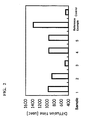
- Sample Nos. 1 to 5 the control, and the reference sample (Reference Example) prepared above were measured for their translational diffusion time with MF20 manufactured by Olympus. The results are shown in FIG. 2 .
- the translational diffusion time (969.3 ⁇ sec) of Sample No. 1 was longer due to formation of a fluorescent-labeled probe Pfw-NF- ⁇ B complex (abbreviated as Pfw-NF complex).
- Pfw-NF complex a fluorescent-labeled probe Pfw-NF- ⁇ B complex
- the translational diffusion time (868 ⁇ sec) of Sample 2 is shorter than that of Sample No. 1, but is longer than that of the control.
- Samples 4 and 5 contain the nucleic acid probe to which NF- ⁇ B does not bind, and both of their translational diffusion times were the same degree as that of Sample 1. As can be seen from this result, the fluorescent substance in Samples 4 and 5 was the Pfw-NF complex.
- a nuclear extract of HeLa cells in a cell strain of human cervical cancer was used to determine the amount of activated NF- ⁇ B (NF- ⁇ B transferred into nuclei) as a DNA binding protein.
- TNF- ⁇ was added to a medium of HeLa cells, and then a nuclear extract was prepared, and as shown in Table 3, the nuclear extract, a solvent for FCS measurement, poly(dIdC), and a nucleic acid probe were mixed to prepare Samples 10 to 13. The reaction time was 5 minutes.
- Sample 10 was a nuclear extract prepared without adding TNF- ⁇ . In the control, the nuclear extract was not added. Table 3 Sample No. 10 11 12 13 Control TNF- ⁇ - + + + - Nuclear extract (6 mg/ml) 5 5 5 5 5 - Reagent 2 - - - - 5 FCS buffer 4 4 4 4 4 Water 10 10 10 10 10 10 Poly (dIdC) (1.3 ⁇ g/ ⁇ l) 0.8 0.8 0.8 0.8 0.8 0.8 0.8 0.8 0.8 0.8 Pfw (100 nM) 0.2 0 0.2 0.2 0.2 (1 nM) (1 nM) (1 nM) Pw (1 ⁇ M) 0 0 1 (50 nM) 0 0 Pn (1 ⁇ M) 0 0 0 1 (50 nM) 0 0 Pn (1 ⁇ M) 0 0 0 1 (50 nM) 0 In the table, the content of each sample is expressed in ⁇ l, and a numerical value in round brackets shows a final
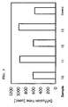
- Samples 10 to 13 and the control were measured for their translational diffusion time with MF20 manufactured by Olympus. The results are shown in FIG. 3 .
- the difference between Sample 10 and Sample 11 is the presence or absence of simulation for converting NF- ⁇ B into activated NF- ⁇ B.
- the translational diffusion time of Sample 11 to which TNF- ⁇ was added is longer than that of Sample 10 to which TNF- ⁇ was not added, and it can be confirmed that the fluorescent-labeled probe Pfw has bound to NF- ⁇ B to form a complex (Pfw-NF complex). It can be seen that addition of TNF- ⁇ is necessary for transporting NF- ⁇ B into nuclei.
- the translational diffusion time of Sample 13 was almost equal to the translational diffusion time of Sample 11. Because the coexisting unlabeled probe Pn in Sample 13 is a probe with which NF- ⁇ B does not form a complex, the fluorescent substance present in Sample 13 is considered to be the Pfw-NF complex. Accordingly, the prolongation in translational diffusion time by formation of the Pfw-NF complex can be known not only by the difference between the translational diffusion time of Sample 10 and the translational diffusion time of Sample 11 by the presence or absence of NF- ⁇ B, but also by the difference between the translational diffusion time of Sample 12 and the translational diffusion time of Sample 13 by the presence or absence of formation of the Pfw-NF complex due to the coexistence of the competitive probe. The difference between the translational diffusion time of the control and the translational diffusion time of Sample 10 is considered attributable to the slight influence of an increase in solution viscosity on the degree of freedom of the Brownian motion of the fluorescent-labeled probe Pfw.
- Recombinant NF- ⁇ B was added at a concentration of 0.4 ng/ ⁇ l to 10 ⁇ g of a nuclear extract prepared from a human monocyte lineage cell line U937 and then mixed with nucleic acid probes (Pfw, Pw, Pn), FCS buffer, Reagent 2, poly(dIdC) and water, as shown in Table 4, to prepare Measurement Samples 21, 22 and 23.
- Measurement Samples 21, 22 and 23 thus prepared were measured 3 times for the translational diffusion time of the fluorescent substance by FCS, to determine average translational diffusion time. The composition of each measurement sample and average translational diffusion time are shown in Table 4. Table 4 Sample No.
- the amount of NF- ⁇ B was calculated from translational diffusion time.
- the amount of NF- ⁇ B calculated from the difference between the translational diffusion time (650.7 ⁇ sec) of Sample 21 and the translational diffusion time (533 ⁇ sec) of the control wherein the concentration of NF- ⁇ B used in preparing the calibration curve is 0 ng/ ⁇ l was 0.50 ng/ ⁇ l. This corresponds to 124% recovery based on the amount of the NF- ⁇ B added to the sample.
- the amount of NF- ⁇ B calculated from the difference between the translational diffusion time of Sample 22 and the translational diffusion time of Sample 23 was 0.42 ng/ ⁇ l. This corresponds to 105% recovery based on the amount of the NF- ⁇ B added to the sample.
- the quantification method of the present invention utilizing a competitive assay system of unlabeled nucleic acid probes (Pw, Pn) could be confirmed to be a highly accurate quantification method wherein the content of a DNA binding protein calculated from a calibration curve is made highly reliable by accurately knowing the prolongation of translational diffusion time based on formation of a complex with a fluorescent-labeled nucleic acid probe, by cancelling the influence of contaminants contained in a sample.
Landscapes
- Life Sciences & Earth Sciences (AREA)
- Health & Medical Sciences (AREA)
- Engineering & Computer Science (AREA)
- Molecular Biology (AREA)
- Immunology (AREA)
- Chemical & Material Sciences (AREA)
- Urology & Nephrology (AREA)
- Biomedical Technology (AREA)
- Hematology (AREA)
- Cell Biology (AREA)
- Medicinal Chemistry (AREA)
- Biochemistry (AREA)
- Pathology (AREA)
- Food Science & Technology (AREA)
- Biotechnology (AREA)
- Physics & Mathematics (AREA)
- Analytical Chemistry (AREA)
- Microbiology (AREA)
- General Health & Medical Sciences (AREA)
- General Physics & Mathematics (AREA)
- Proteomics, Peptides & Aminoacids (AREA)
- Tropical Medicine & Parasitology (AREA)
- Measuring Or Testing Involving Enzymes Or Micro-Organisms (AREA)
- Investigating, Analyzing Materials By Fluorescence Or Luminescence (AREA)
- Investigating Or Analysing Materials By The Use Of Chemical Reactions (AREA)
Claims (10)
- Procédé de détermination de la quantité d'une protéine liant l'ADN cible dans un échantillon liquide, qui comprend les étapes consistant à :mesurer un premier temps de diffusion translationnelle de la substance marquée par fluorescence présente dans un premier mélange préparé par mélange d'un premier réactif de mesure et de l'échantillon liquide, le premier réactif de mesure comprenant une sonde d'acide nucléique marquée par fluorescence et une première sonde d'acide nucléique non marquée, et la protéine liant l'ADN cible étant capable de se lier à la sonde d'acide nucléique marquée par fluorescence et à la première sonde d'acide nucléique non marquée ;mesurer un second temps de diffusion translationnelle de la substance marquée par fluorescence présente dans un second mélange préparé par mélange d'un second réactif de mesure et de l'échantillon liquide, le second réactif de mesure comprenant la sonde d'acide nucléique marquée par fluorescence et une seconde sonde d'acide nucléique non marquée, et la protéine liant l'ADN cible n'étant pas capable de se lier à la seconde sonde d'acide nucléique non marquée ; etdéterminer la quantité de la protéine liant l'ADN cible dans l'échantillon liquide en se basant sur une différence entre le premier temps de diffusion translationnelle et le second temps de diffusion translationnelle.
- Procédé selon la revendication 1, dans lequel l'étape de détermination est réalisée en vue de déterminer la quantité de la protéine liant l'ADN cible dans un échantillon liquide en se basant sur la différence entre le premier temps de diffusion translationnelle et le second temps de diffusion translationnelle, et les données représentant une relation entre la quantité de la protéine liant l'ADN cible et une durée de diffusion translationnelle.
- Procédé selon la revendication 2, dans lequel les données représentant la relation entre la quantité de la protéine liant l'ADN cible et le temps de diffusion translationnelle sont une courbe d'étalonnage obtenue par mesure d'un temps de diffusion translationnelle dans un mélange préparé par mélange d'une solution liquide comprenant la sonde d'acide nucléique marquée par fluorescence et une solution liquide comprenant une quantité prédéterminée de la protéine liant l'ADN cible.
- Procédé selon l'une quelconque des revendications 1 à 3, dans lequel
la quantité de la première sonde d'acide nucléique non marquée comprise dans le premier réactif de mesure est supérieure à la quantité de la sonde d'acide nucléique marquée par fluorescence comprise dans le premier réactif de mesure, et
la quantité de la seconde sonde d'acide nucléique non marquée comprise dans le second réactif de mesure est supérieure à la quantité de la sonde d'acide nucléique marquée par fluorescence comprise dans le second réactif de mesure. - Procédé selon l'une quelconque des revendications 1 à 4, dans lequel la concentration de la première sonde d'acide nucléique non marquée comprise dans le premier réactif de mesure et la concentration de la seconde sonde d'acide nucléique non marquée comprise dans le second réactif de mesure sont identiques.
- Procédé selon l'une quelconque des revendications 1 à 5, dans lequel l'échantillon liquide comprend un extrait nucléaire.
- Procédé selon l'une quelconque des revendications 1 à 6, dans lequel la protéine liant l'ADN cible est un facteur de transcription.
- Procédé selon la revendication 7, dans lequel le facteur de transcription est un facteur de transcription des cytokines.
- Procédé selon l'une quelconque des revendications 1 à 8, dans lequel la séquence d'acide nucléique de la sonde d'acide nucléique marquée par fluorescence et la séquence d'acide nucléique de la première sonde d'acide nucléique non marquée sont identiques.
- Procédé selon l'une quelconque des revendications 1 à 9, dans lequel au moins l'une de la sonde d'acide nucléique marquée par fluorescence, de la première sonde d'acide nucléique non marquée et de la seconde sonde d'acide nucléique non marquée forment une structure tige-boucle.
Applications Claiming Priority (1)
| Application Number | Priority Date | Filing Date | Title |
|---|---|---|---|
| JP2007172216A JP2009008608A (ja) | 2007-06-29 | 2007-06-29 | Dna結合タンパクの定量方法及び定量用キット |
Publications (2)
| Publication Number | Publication Date |
|---|---|
| EP2009441A1 EP2009441A1 (fr) | 2008-12-31 |
| EP2009441B1 true EP2009441B1 (fr) | 2010-08-11 |
Family
ID=39789298
Family Applications (1)
| Application Number | Title | Priority Date | Filing Date |
|---|---|---|---|
| EP08159220A Not-in-force EP2009441B1 (fr) | 2007-06-29 | 2008-06-27 | Procédé pour déterminer la quantité d'une protéine liant l'ADN |
Country Status (6)
| Country | Link |
|---|---|
| US (1) | US20090004749A1 (fr) |
| EP (1) | EP2009441B1 (fr) |
| JP (1) | JP2009008608A (fr) |
| CN (1) | CN101334412A (fr) |
| AT (1) | ATE477488T1 (fr) |
| DE (1) | DE602008002106D1 (fr) |
Families Citing this family (3)
| Publication number | Priority date | Publication date | Assignee | Title |
|---|---|---|---|---|
| JP2012524907A (ja) * | 2009-04-24 | 2012-10-18 | ディスカヴァーエックス コーポレイション | 検出可能なタンパク質を用いた細胞分析 |
| CN118345145A (zh) | 2016-12-09 | 2024-07-16 | 乌尔蒂维尤股份有限公司 | 用于使用标记的核酸成像剂进行多路复用成像的改进方法 |
| EP3601339A4 (fr) | 2017-03-31 | 2020-11-04 | Ultivue, Inc. | Échange adn-antigène et amplification |
Family Cites Families (6)
| Publication number | Priority date | Publication date | Assignee | Title |
|---|---|---|---|---|
| US5985575A (en) * | 1998-05-20 | 1999-11-16 | Wisconsin Alumni Research Foundation | Tethered function assay for protein function |
| EP1138781A3 (fr) * | 2000-03-31 | 2004-07-14 | Health Research, Inc. | Méthode pour la quantification de l'activité de liaison des protéines de liaison d'ADN |
| JP4274856B2 (ja) | 2003-06-19 | 2009-06-10 | オリンパス株式会社 | Dnaとdna結合タンパク質との反応を検出する方法 |
| JP2005269957A (ja) * | 2004-03-24 | 2005-10-06 | National Institute Of Advanced Industrial & Technology | 核酸結合タンパク質の検出方法 |
| JP4338590B2 (ja) * | 2004-06-03 | 2009-10-07 | 独立行政法人科学技術振興機構 | 蛍光相関分光法による抗原の迅速検出及び/又は測定法。 |
| WO2006062127A1 (fr) | 2004-12-10 | 2006-06-15 | Sysmex Corporation | Procédé support de diagnostic de maladie liée à un dysfonctionnement du système immunitaire et dispositif de sortie d’informations support de diagnostic |
-
2007
- 2007-06-29 JP JP2007172216A patent/JP2009008608A/ja active Pending
-
2008
- 2008-06-24 CN CNA2008101265184A patent/CN101334412A/zh active Pending
- 2008-06-27 DE DE602008002106T patent/DE602008002106D1/de active Active
- 2008-06-27 AT AT08159220T patent/ATE477488T1/de not_active IP Right Cessation
- 2008-06-27 US US12/163,431 patent/US20090004749A1/en not_active Abandoned
- 2008-06-27 EP EP08159220A patent/EP2009441B1/fr not_active Not-in-force
Also Published As
| Publication number | Publication date |
|---|---|
| DE602008002106D1 (de) | 2010-09-23 |
| JP2009008608A (ja) | 2009-01-15 |
| US20090004749A1 (en) | 2009-01-01 |
| EP2009441A1 (fr) | 2008-12-31 |
| ATE477488T1 (de) | 2010-08-15 |
| CN101334412A (zh) | 2008-12-31 |
Similar Documents
| Publication | Publication Date | Title |
|---|---|---|
| Li et al. | An optimized procedure for exosome isolation and analysis using serum samples: application to cancer biomarker discovery | |
| Geeurickx et al. | The generation and use of recombinant extracellular vesicles as biological reference material | |
| Lee et al. | Multiplexed profiling of single extracellular vesicles | |
| Lee et al. | ssDNA aptamer-based surface plasmon resonance biosensor for the detection of retinol binding protein 4 for the early diagnosis of type 2 diabetes | |
| Gildea et al. | Urinary exosome miRNome analysis and its applications to salt sensitivity of blood pressure | |
| Kontopoulou et al. | Evaluation of dsDNA from extracellular vesicles (EVs) in pediatric AML diagnostics | |
| EP2904118B1 (fr) | Arnm d'exosome d'urine et procédés d'utilisation de ceux-ci pour détecter une néphropathie diabétique | |
| Tamkovich et al. | Blood circulating exosomes contain distinguishable fractions of free and cell-surface-associated vesicles | |
| JP7344631B2 (ja) | サンプル収集関連応用、分析および診断のためのデバイス、溶液および方法 | |
| MX2013010594A (es) | Un metodo para analizar una muestra de sangre de un sujeto por la presencia de un marcador de enfermedad. | |
| Klebes et al. | Multianalyte lateral flow immunoassay for simultaneous detection of protein-based inflammation biomarkers and pathogen DNA | |
| JP2023113877A (ja) | 膵がんの検出を補助する方法 | |
| La Salvia et al. | Extracellular vesicles in essential hypertension: hidden messengers | |
| KR101913163B1 (ko) | 이중가닥 핵산 신호 프로브 및 이를 이용한 표적분자의 검출방법 | |
| Fernández‐Rhodes et al. | Defining the influence of size‐exclusion chromatography fraction window and ultrafiltration column choice on extracellular vesicle recovery in a skeletal muscle model | |
| EP2009441B1 (fr) | Procédé pour déterminer la quantité d'une protéine liant l'ADN | |
| JP2025510610A (ja) | 可溶性st2タンパク質を特異的に認識する核酸アプタマー及びその使用 | |
| Saadi et al. | Evaluation of microRNA-192 in patients with diabetic nephropathy | |
| KR102681006B1 (ko) | 뇌의 레닌-안지오텐신 시스템 인자를 이용한 알츠하이머병의 진단 방법 | |
| Janovičová et al. | Isolation and quantification of extracellular dna from biofluids | |
| KR101704828B1 (ko) | 체액 내 세포밖 소포체의 단백질 또는 유전자 분석을 통한 염증성 질환 진단 방법 | |
| Meutelet et al. | Extraction and purification of short DNA fragments from plasma using aqueous two-phase systems for liquid biopsy | |
| Ababneh et al. | The utility of whole-exome sequencing in accurate diagnosis of neuromuscular disorders in consanguineous families in Jordan | |
| Song et al. | Upregulated Tie2 expression in plasma: a potential non-invasive biomarker for the diagnosis of breast cancer | |
| KR101911859B1 (ko) | 이중가닥 핵산 신호 프로브 및 이를 이용한 표적분자의 검출방법 |
Legal Events
| Date | Code | Title | Description |
|---|---|---|---|
| PUAI | Public reference made under article 153(3) epc to a published international application that has entered the european phase |
Free format text: ORIGINAL CODE: 0009012 |
|
| AK | Designated contracting states |
Kind code of ref document: A1 Designated state(s): AT BE BG CH CY CZ DE DK EE ES FI FR GB GR HR HU IE IS IT LI LT LU LV MC MT NL NO PL PT RO SE SI SK TR |
|
| AX | Request for extension of the european patent |
Extension state: AL BA MK RS |
|
| 17P | Request for examination filed |
Effective date: 20090619 |
|
| AKX | Designation fees paid |
Designated state(s): AT BE BG CH CY CZ DE DK EE ES FI FR GB GR HR HU IE IS IT LI LT LU LV MC MT NL NO PL PT RO SE SI SK TR |
|
| GRAP | Despatch of communication of intention to grant a patent |
Free format text: ORIGINAL CODE: EPIDOSNIGR1 |
|
| RTI1 | Title (correction) |
Free format text: METHOD FOR DETERMINING THE AMOUNT OF DNA BINDING PROTEIN |
|
| GRAS | Grant fee paid |
Free format text: ORIGINAL CODE: EPIDOSNIGR3 |
|
| GRAA | (expected) grant |
Free format text: ORIGINAL CODE: 0009210 |
|
| AK | Designated contracting states |
Kind code of ref document: B1 Designated state(s): AT BE BG CH CY CZ DE DK EE ES FI FR GB GR HR HU IE IS IT LI LT LU LV MC MT NL NO PL PT RO SE SI SK TR |
|
| REG | Reference to a national code |
Ref country code: GB Ref legal event code: FG4D |
|
| REG | Reference to a national code |
Ref country code: CH Ref legal event code: EP |
|
| REG | Reference to a national code |
Ref country code: IE Ref legal event code: FG4D |
|
| REF | Corresponds to: |
Ref document number: 602008002106 Country of ref document: DE Date of ref document: 20100923 Kind code of ref document: P |
|
| REG | Reference to a national code |
Ref country code: NL Ref legal event code: VDEP Effective date: 20100811 |
|
| LTIE | Lt: invalidation of european patent or patent extension |
Effective date: 20100811 |
|
| PG25 | Lapsed in a contracting state [announced via postgrant information from national office to epo] |
Ref country code: NL Free format text: LAPSE BECAUSE OF FAILURE TO SUBMIT A TRANSLATION OF THE DESCRIPTION OR TO PAY THE FEE WITHIN THE PRESCRIBED TIME-LIMIT Effective date: 20100811 Ref country code: AT Free format text: LAPSE BECAUSE OF FAILURE TO SUBMIT A TRANSLATION OF THE DESCRIPTION OR TO PAY THE FEE WITHIN THE PRESCRIBED TIME-LIMIT Effective date: 20100811 Ref country code: FI Free format text: LAPSE BECAUSE OF FAILURE TO SUBMIT A TRANSLATION OF THE DESCRIPTION OR TO PAY THE FEE WITHIN THE PRESCRIBED TIME-LIMIT Effective date: 20100811 Ref country code: LT Free format text: LAPSE BECAUSE OF FAILURE TO SUBMIT A TRANSLATION OF THE DESCRIPTION OR TO PAY THE FEE WITHIN THE PRESCRIBED TIME-LIMIT Effective date: 20100811 Ref country code: NO Free format text: LAPSE BECAUSE OF FAILURE TO SUBMIT A TRANSLATION OF THE DESCRIPTION OR TO PAY THE FEE WITHIN THE PRESCRIBED TIME-LIMIT Effective date: 20101111 |
|
| PG25 | Lapsed in a contracting state [announced via postgrant information from national office to epo] |
Ref country code: CY Free format text: LAPSE BECAUSE OF FAILURE TO SUBMIT A TRANSLATION OF THE DESCRIPTION OR TO PAY THE FEE WITHIN THE PRESCRIBED TIME-LIMIT Effective date: 20100811 Ref country code: SI Free format text: LAPSE BECAUSE OF FAILURE TO SUBMIT A TRANSLATION OF THE DESCRIPTION OR TO PAY THE FEE WITHIN THE PRESCRIBED TIME-LIMIT Effective date: 20100811 Ref country code: BG Free format text: LAPSE BECAUSE OF FAILURE TO SUBMIT A TRANSLATION OF THE DESCRIPTION OR TO PAY THE FEE WITHIN THE PRESCRIBED TIME-LIMIT Effective date: 20101111 Ref country code: PL Free format text: LAPSE BECAUSE OF FAILURE TO SUBMIT A TRANSLATION OF THE DESCRIPTION OR TO PAY THE FEE WITHIN THE PRESCRIBED TIME-LIMIT Effective date: 20100811 Ref country code: IS Free format text: LAPSE BECAUSE OF FAILURE TO SUBMIT A TRANSLATION OF THE DESCRIPTION OR TO PAY THE FEE WITHIN THE PRESCRIBED TIME-LIMIT Effective date: 20101211 Ref country code: HR Free format text: LAPSE BECAUSE OF FAILURE TO SUBMIT A TRANSLATION OF THE DESCRIPTION OR TO PAY THE FEE WITHIN THE PRESCRIBED TIME-LIMIT Effective date: 20100811 |
|
| PG25 | Lapsed in a contracting state [announced via postgrant information from national office to epo] |
Ref country code: GR Free format text: LAPSE BECAUSE OF FAILURE TO SUBMIT A TRANSLATION OF THE DESCRIPTION OR TO PAY THE FEE WITHIN THE PRESCRIBED TIME-LIMIT Effective date: 20101112 Ref country code: LV Free format text: LAPSE BECAUSE OF FAILURE TO SUBMIT A TRANSLATION OF THE DESCRIPTION OR TO PAY THE FEE WITHIN THE PRESCRIBED TIME-LIMIT Effective date: 20100811 Ref country code: SE Free format text: LAPSE BECAUSE OF FAILURE TO SUBMIT A TRANSLATION OF THE DESCRIPTION OR TO PAY THE FEE WITHIN THE PRESCRIBED TIME-LIMIT Effective date: 20100811 Ref country code: BE Free format text: LAPSE BECAUSE OF FAILURE TO SUBMIT A TRANSLATION OF THE DESCRIPTION OR TO PAY THE FEE WITHIN THE PRESCRIBED TIME-LIMIT Effective date: 20100811 |
|
| PG25 | Lapsed in a contracting state [announced via postgrant information from national office to epo] |
Ref country code: DK Free format text: LAPSE BECAUSE OF FAILURE TO SUBMIT A TRANSLATION OF THE DESCRIPTION OR TO PAY THE FEE WITHIN THE PRESCRIBED TIME-LIMIT Effective date: 20100811 |
|
| PG25 | Lapsed in a contracting state [announced via postgrant information from national office to epo] |
Ref country code: RO Free format text: LAPSE BECAUSE OF FAILURE TO SUBMIT A TRANSLATION OF THE DESCRIPTION OR TO PAY THE FEE WITHIN THE PRESCRIBED TIME-LIMIT Effective date: 20100811 Ref country code: EE Free format text: LAPSE BECAUSE OF FAILURE TO SUBMIT A TRANSLATION OF THE DESCRIPTION OR TO PAY THE FEE WITHIN THE PRESCRIBED TIME-LIMIT Effective date: 20100811 Ref country code: CZ Free format text: LAPSE BECAUSE OF FAILURE TO SUBMIT A TRANSLATION OF THE DESCRIPTION OR TO PAY THE FEE WITHIN THE PRESCRIBED TIME-LIMIT Effective date: 20100811 Ref country code: IT Free format text: LAPSE BECAUSE OF FAILURE TO SUBMIT A TRANSLATION OF THE DESCRIPTION OR TO PAY THE FEE WITHIN THE PRESCRIBED TIME-LIMIT Effective date: 20100811 Ref country code: SK Free format text: LAPSE BECAUSE OF FAILURE TO SUBMIT A TRANSLATION OF THE DESCRIPTION OR TO PAY THE FEE WITHIN THE PRESCRIBED TIME-LIMIT Effective date: 20100811 |
|
| PLBE | No opposition filed within time limit |
Free format text: ORIGINAL CODE: 0009261 |
|
| STAA | Information on the status of an ep patent application or granted ep patent |
Free format text: STATUS: NO OPPOSITION FILED WITHIN TIME LIMIT |
|
| PG25 | Lapsed in a contracting state [announced via postgrant information from national office to epo] |
Ref country code: ES Free format text: LAPSE BECAUSE OF FAILURE TO SUBMIT A TRANSLATION OF THE DESCRIPTION OR TO PAY THE FEE WITHIN THE PRESCRIBED TIME-LIMIT Effective date: 20101122 |
|
| 26N | No opposition filed |
Effective date: 20110512 |
|
| PGFP | Annual fee paid to national office [announced via postgrant information from national office to epo] |
Ref country code: FR Payment date: 20110621 Year of fee payment: 4 |
|
| REG | Reference to a national code |
Ref country code: DE Ref legal event code: R097 Ref document number: 602008002106 Country of ref document: DE Effective date: 20110512 |
|
| PGFP | Annual fee paid to national office [announced via postgrant information from national office to epo] |
Ref country code: DE Payment date: 20110622 Year of fee payment: 4 |
|
| PG25 | Lapsed in a contracting state [announced via postgrant information from national office to epo] |
Ref country code: MT Free format text: LAPSE BECAUSE OF FAILURE TO SUBMIT A TRANSLATION OF THE DESCRIPTION OR TO PAY THE FEE WITHIN THE PRESCRIBED TIME-LIMIT Effective date: 20100811 |
|
| REG | Reference to a national code |
Ref country code: IE Ref legal event code: MM4A |
|
| PG25 | Lapsed in a contracting state [announced via postgrant information from national office to epo] |
Ref country code: IE Free format text: LAPSE BECAUSE OF NON-PAYMENT OF DUE FEES Effective date: 20110627 |
|
| REG | Reference to a national code |
Ref country code: CH Ref legal event code: PL |
|
| REG | Reference to a national code |
Ref country code: CH Ref legal event code: PL |
|
| GBPC | Gb: european patent ceased through non-payment of renewal fee |
Effective date: 20120627 |
|
| REG | Reference to a national code |
Ref country code: FR Ref legal event code: ST Effective date: 20130228 |
|
| REG | Reference to a national code |
Ref country code: DE Ref legal event code: R119 Ref document number: 602008002106 Country of ref document: DE Effective date: 20130101 |
|
| PG25 | Lapsed in a contracting state [announced via postgrant information from national office to epo] |
Ref country code: MC Free format text: LAPSE BECAUSE OF NON-PAYMENT OF DUE FEES Effective date: 20110630 Ref country code: GB Free format text: LAPSE BECAUSE OF NON-PAYMENT OF DUE FEES Effective date: 20120627 Ref country code: LI Free format text: LAPSE BECAUSE OF NON-PAYMENT OF DUE FEES Effective date: 20120630 Ref country code: DE Free format text: LAPSE BECAUSE OF NON-PAYMENT OF DUE FEES Effective date: 20130101 Ref country code: FR Free format text: LAPSE BECAUSE OF NON-PAYMENT OF DUE FEES Effective date: 20120702 Ref country code: CH Free format text: LAPSE BECAUSE OF NON-PAYMENT OF DUE FEES Effective date: 20120630 |
|
| PG25 | Lapsed in a contracting state [announced via postgrant information from national office to epo] |
Ref country code: LU Free format text: LAPSE BECAUSE OF NON-PAYMENT OF DUE FEES Effective date: 20110627 |
|
| PG25 | Lapsed in a contracting state [announced via postgrant information from national office to epo] |
Ref country code: TR Free format text: LAPSE BECAUSE OF FAILURE TO SUBMIT A TRANSLATION OF THE DESCRIPTION OR TO PAY THE FEE WITHIN THE PRESCRIBED TIME-LIMIT Effective date: 20100811 |
|
| PG25 | Lapsed in a contracting state [announced via postgrant information from national office to epo] |
Ref country code: HU Free format text: LAPSE BECAUSE OF FAILURE TO SUBMIT A TRANSLATION OF THE DESCRIPTION OR TO PAY THE FEE WITHIN THE PRESCRIBED TIME-LIMIT Effective date: 20100811 |
|
| PG25 | Lapsed in a contracting state [announced via postgrant information from national office to epo] |
Ref country code: PT Free format text: LAPSE BECAUSE OF FAILURE TO SUBMIT A TRANSLATION OF THE DESCRIPTION OR TO PAY THE FEE WITHIN THE PRESCRIBED TIME-LIMIT Effective date: 20100811 |