EP2020440A1 - Compositions et procédés pour inhiber l'expression du gène car par interférence d'ARN - Google Patents
Compositions et procédés pour inhiber l'expression du gène car par interférence d'ARN Download PDFInfo
- Publication number
- EP2020440A1 EP2020440A1 EP08252595A EP08252595A EP2020440A1 EP 2020440 A1 EP2020440 A1 EP 2020440A1 EP 08252595 A EP08252595 A EP 08252595A EP 08252595 A EP08252595 A EP 08252595A EP 2020440 A1 EP2020440 A1 EP 2020440A1
- Authority
- EP
- European Patent Office
- Prior art keywords
- car
- sirna
- gene
- cell
- expression
- Prior art date
- Legal status (The legal status is an assumption and is not a legal conclusion. Google has not performed a legal analysis and makes no representation as to the accuracy of the status listed.)
- Withdrawn
Links
Images
Classifications
-
- C—CHEMISTRY; METALLURGY
- C12—BIOCHEMISTRY; BEER; SPIRITS; WINE; VINEGAR; MICROBIOLOGY; ENZYMOLOGY; MUTATION OR GENETIC ENGINEERING
- C12N—MICROORGANISMS OR ENZYMES; COMPOSITIONS THEREOF; PROPAGATING, PRESERVING, OR MAINTAINING MICROORGANISMS; MUTATION OR GENETIC ENGINEERING; CULTURE MEDIA
- C12N15/00—Mutation or genetic engineering; DNA or RNA concerning genetic engineering, vectors, e.g. plasmids, or their isolation, preparation or purification; Use of hosts therefor
- C12N15/09—Recombinant DNA-technology
- C12N15/11—DNA or RNA fragments; Modified forms thereof; Non-coding nucleic acids having a biological activity
- C12N15/113—Non-coding nucleic acids modulating the expression of genes, e.g. antisense oligonucleotides; Antisense DNA or RNA; Triplex- forming oligonucleotides; Catalytic nucleic acids, e.g. ribozymes; Nucleic acids used in co-suppression or gene silencing
- C12N15/1138—Non-coding nucleic acids modulating the expression of genes, e.g. antisense oligonucleotides; Antisense DNA or RNA; Triplex- forming oligonucleotides; Catalytic nucleic acids, e.g. ribozymes; Nucleic acids used in co-suppression or gene silencing against receptors or cell surface proteins
-
- A—HUMAN NECESSITIES
- A61—MEDICAL OR VETERINARY SCIENCE; HYGIENE
- A61P—SPECIFIC THERAPEUTIC ACTIVITY OF CHEMICAL COMPOUNDS OR MEDICINAL PREPARATIONS
- A61P43/00—Drugs for specific purposes, not provided for in groups A61P1/00-A61P41/00
-
- G—PHYSICS
- G01—MEASURING; TESTING
- G01N—INVESTIGATING OR ANALYSING MATERIALS BY DETERMINING THEIR CHEMICAL OR PHYSICAL PROPERTIES
- G01N33/00—Investigating or analysing materials by specific methods not covered by groups G01N1/00 - G01N31/00
- G01N33/48—Biological material, e.g. blood, urine; Haemocytometers
- G01N33/50—Chemical analysis of biological material, e.g. blood, urine; Testing involving biospecific ligand binding methods; Immunological testing
- G01N33/68—Chemical analysis of biological material, e.g. blood, urine; Testing involving biospecific ligand binding methods; Immunological testing involving proteins, peptides or amino acids
- G01N33/6875—Nucleoproteins
-
- C—CHEMISTRY; METALLURGY
- C12—BIOCHEMISTRY; BEER; SPIRITS; WINE; VINEGAR; MICROBIOLOGY; ENZYMOLOGY; MUTATION OR GENETIC ENGINEERING
- C12N—MICROORGANISMS OR ENZYMES; COMPOSITIONS THEREOF; PROPAGATING, PRESERVING, OR MAINTAINING MICROORGANISMS; MUTATION OR GENETIC ENGINEERING; CULTURE MEDIA
- C12N2310/00—Structure or type of the nucleic acid
- C12N2310/10—Type of nucleic acid
- C12N2310/14—Type of nucleic acid interfering nucleic acids [NA]
-
- C—CHEMISTRY; METALLURGY
- C12—BIOCHEMISTRY; BEER; SPIRITS; WINE; VINEGAR; MICROBIOLOGY; ENZYMOLOGY; MUTATION OR GENETIC ENGINEERING
- C12N—MICROORGANISMS OR ENZYMES; COMPOSITIONS THEREOF; PROPAGATING, PRESERVING, OR MAINTAINING MICROORGANISMS; MUTATION OR GENETIC ENGINEERING; CULTURE MEDIA
- C12N2320/00—Applications; Uses
- C12N2320/10—Applications; Uses in screening processes
-
- G—PHYSICS
- G01—MEASURING; TESTING
- G01N—INVESTIGATING OR ANALYSING MATERIALS BY DETERMINING THEIR CHEMICAL OR PHYSICAL PROPERTIES
- G01N2500/00—Screening for compounds of potential therapeutic value
- G01N2500/10—Screening for compounds of potential therapeutic value involving cells
Definitions
- the present invention relates to inhibition by short interference RNA (hereinafter, referred to as siRNA in some cases) of expression of the nuclear receptor CAR that acts on transcriptional control of drug-metabolizing enzymes.
- siRNA short interference RNA
- CAR Constitutive Active/Androstane Receptor
- Knockout animals such as CAR knockout mice have been used to examine the relationship between tumorigenic transformation by increased expression of CAR-related drug-metabolizing enzymes and toxicity of a compound like phenobarbital in animals such as mice ( Yamamoto et al 2004 Cancer Res 64 7197-7200 ).
- the present invention is directed to inhibition of CAR gene expression by RNA interference.
- the present invention provides:
- siRNAs compositions and methods for inhibiting the CAR gene expression simply and rapidly, which can be used for evaluating toxicity of a chemical substance.
- RNA interference is performed by allowing to act on the target gene a short double-stranded oligoribonucleotide (hereinafter referred to as ds oligoribonucleotide in some cases) which comprises an overhang of about two nucleotides at least at the terminal of the first strand of the duplex.
- the ds oligoribonucleotide is known as the short interference RNA (siRNA).
- siRNA has a nucleotide sequence matched to a partial sequence of the nucleotide sequence of the CAR gene when the CAR gene is used as the target gene.
- the siRNA is modified with a material such as cholesterol. Further, in order to improve preservation stability, it is more preferable that either the sense or antisense strand of the siRNA is chemically modified with 2'-O-methyl or 2'-deoxy.
- siRNA against CAR mRNA is delivered into cultured cells (the term "cultured cells” used herein includes established cell lines or primary cultured cells obtained from, for example, mammals), preferable examples of the delivery method include forward and reverse transfection methods using the calcium chloride method, lipofection method, and electroporation method and the like. Although the lipofection method is more preferable, it is preferable to select an optimal delivery method depending on the type of cultured cell to be introduced.
- Examples of the method for delivering siRNA against CAR mRNA to cells in a living body include various methods of administration to target animals.
- Preferable examples of the method of administration include oral administration, dermal administration, subcutaneous administration, airway administration and transocular administration, intramuscular administration, and intravascular administration.
- the blood vessels include all arteries and veins in living organisms. Preferable examples thereof include hepatic portal, hepatic artery, tail vein, and jugular vein.
- siRNA may be administered directly without an external capsule.
- siRNA may be included in or bound to carriers such as liposomes. More preferably, siRNA may be included in or bound to a mixture of liposome and cholesterol, further preferably DMRIE-C Reagent (Invitrogen).
- siRNA may be mixed with foods for feeding organisms.
- the effective amount of siRNA to be administered varies depending on the cell types, animal species, administration methods, and the like.
- the appropriate dosage can vary depending on each condition. For example, as for a single daily administration to living animals, the amount is preferably 0.01 to 1000 mg/kg body weight/day, more preferably 10 mg/kg body weight/day in the case of tail vein administration to mice. In the case of tail vein administration to rats, the amount is preferably 0.01 to 1000 mg/kg body weight/day, more preferably 10 mg/kg body weight/day.
- the dose per day divided into multiple doses can be given.
- hepatic portal administration or hepatic arterial administration may include placement of catheter in the blood vessel, implantation of an osmotic pump containing siRNA, and the like.
- osmotic pump containing siRNA and the like.
- tail vein administration, jugular vein administration, and the like are preferred.
- the dosage interval and the number of repeated-doses can be changed for each condition of the embodiments.
- siRNA is administered at a dose of preferably 0.01 to 1000 mg/kg body weight/day, more preferably 10 mg/kg body weight/day for multiple days.
- the administration can be performed at least one day apart.
- siRNA is administered at a dose of preferably 0.01 to 1000 mg/kg body weight/day, more preferably 10 mg/kg body weight/day for multiple days.
- the administration can be performed at least one day apart.
- the expression level of the CAR gene which is the target gene, can be measured by a method for measuring the amount of transcripts of the gene per unit cell mass or a method for measuring the amount of the translated products per unit cell mass.
- the amount of mRNA i.e., transcripts of the gene is measured.
- the amount of mRNA can be measured by methods such as the quantitative real-time polymerase chain reaction (hereinafter, referred to as quantitative RT-PCR.), the northern hybridization method [ J. Sambrook, E. F. Frisch, T. Maniatis work; Molecular Cloning 2nd edition, issued by Cold Spring Harbor Laboratory, 1989 ], the DNA array method, and the in situ hybridization method.
- the amount of the protein of the amino acid sequence encoded by the nucleotide sequence of the gene is measured.
- the amount of protein can be measured by methods including immunological measurement using a specific antibody against the protein such as ELISA, Western blot, RIA, and immunohistochemistry, two-dimensional electrophoresis, and a method using high performance liquid chromatography.
- the specific antibody against the protein can be prepared using the protein as an immunogen in accordance with a conventional method.
- the level of inhibition of expression of the CAR gene i.e., the target gene is determined by comparing the expression level, which is measured by the above-described methods, of the CAR gene in animals receiving the siRNA with the expression level of the CAR gene in the liver of animals such as mammals of the same species, line, sex, and age in week receiving a control siRNA having a nucleotide sequence that is not matched to the nucleotide sequence of the CAR gene of the animal.
- the mammals are not limited, humans, monkeys, marmosets, dogs, rabbits, guinea pigs, rats mice, or the like can be used.
- the method for evaluating the toxicity of a chemical substance includes the following three steps:
- Examples of the cells to be used in the first step may include cultured cells or cells in a.body of animals. Specific examples thereof may include hypatocytes of mammals. These cells may be used directly, alternatively, cells prepared by separation, fractionation, or immobilization of such cells may be used. Examples of the mammal to obtain cells include rodents such as rats and mice.
- the inhibition can be performed by allowing siRNA to act on the CAR gene as described above.
- a solution containing a chemical substance is added to the culture medium of the cultured cells 24 hours after the treatment of siRNA in the first step.
- the concentration of the chemical substance of the solution to be added at the time can be changed depending on the combination of cultured cells to be used and the chemical substance. Desirably, the concentration is set to the level that the toxicity of the chemical substance can be evaluated in the third step. Further, it is preferable to newly add the solution of the chemical substance to the culture medium for each replacement of the culture medium.
- the chemical substance is administered to the cells after the first step, preferably after multiple days.
- the method for administering the chemical substance include oral (gavage or mixed in drinking water or diet), intramuscular, intravenous, subcutaneous, intraperitoneal, and airway routes of administration.
- the dose, number of doses, and dosing period may be within the range that does not have a serious impact on general condition of the mammals and organ tissues throughout the body, e.g. below the maximal tolerated dose.
- the cells thus obtained from mammals for example, tissue, cell separated from the tissue, cultured cells of the cells, can be brought into contact with the chemical substance by direct or indirect means.
- the chemical substance when administered to the cells, for example, of mammals which are obtained in the first step, they are administered orally, subcutaneously, or inhalationally for three days or more in the case where the mammals are adult mammals. In the case of juvenile animals, they are administered orally, subcutaneously, or inhalationally for three days or at least once a day.
- the oral administration in the case is carried out using the following procedures.
- the required amount of the chemical substance is weighed and an appropriate solvent such as corn oil, about 0.25 to 0.5% methylcellulose solution, or the like, is added to the chemical substance, if necessary. Then, a solution or a uniform suspension is prepared, which is used as a dosing solution.
- an appropriate solvent such as corn oil, about 0.25 to 0.5% methylcellulose solution, or the like.
- a solution or a uniform suspension is prepared, which is used as a dosing solution.
- the dosing solution is administered to the mammals, it is preferable to administer at a dose of 5 mL/kg/day or less at least once a day using a syringe, an elastic catheter, and the like.
- the subcutaneous administration in the case is carried out using the following procedures.
- the required amount of the chemical substance is weighed and an appropriate solvent such as corn oil, about 0.25 to 0.5% methylcellulose solution, or the like, is added to the chemical substance, if necessary. Then, a solution or a uniform suspension is prepared, which is used as a dosing solution.
- an appropriate solvent such as corn oil, about 0.25 to 0.5% methylcellulose solution, or the like.
- a solution or a uniform suspension is prepared, which is used as a dosing solution.
- the dosing solution is administered to the mammals, it is preferable to administer at a dose of 4 mL/kg/day or less at least once a day using a syringe, a needle, and.the like.
- the required amount of the chemical substance is weighed and an appropriate solvent such as corn oil, acetone, etc. , is added to the chemical substance, if necessary. Then, a solution or a uniform suspension is prepared, which is used as a dosing solution. Then, the dosing solution is placed in a nebulizer and a suitable exposure chamber is used to allow the mammals to inhale the solution during spontaneous breathing. The inhalation is performed at least once a day (continuously for 4 hours or more per inhalation).
- the toxicity of the chemical substance to the cells obtained in the second step is evaluated.
- the toxicity evaluation can be performed by:
- the level of inhibition of expression of the CAR gene can be detected by measuring the expression level of the CAR gene.
- methods generally known in the art may be used in order to detect increase or decrease in the expression level of the CAR gene-related drug-metabolizing enzyme, that is, the level of the increase in the expression of the CAR gene-related drug-metabolizing enzyme, in cultured cells or in cells in the body of mammals which have been brought into contact with the chemical substance.
- Specific examples thereof include a method for measuring the amount of transcripts or translated products of the gene of the CAR gene-related drug-metabolizing enzyme per unit cell mass.
- the term "gene of drug-metabolizing enzyme" which is expressed in the liver means an enzyme gene expressed in liver cells which is involved in metabolic reaction of hormone or drug, which are chemical substances.
- enzymes involved in the first phase reaction including oxidation, reduction, and hydrolysis in the liver include CYP (Cytochrome P450), ADH (alcohol dehydrogenase), ALDH (aldehyde dehydrogenase), or the like.
- Enzymes involved in the second phase reaction including conjugation in the liver include UGT (UDP-glucuronosyltransferase), SULT (sulfotransferase), or the like.
- the CYP includes subtypes of CYP1, CYP2, and CYP3 in rats.
- CYP2 includes a plurality of subtypes such as CYP2B1.
- UGT includes subtypes of UGT1A and UGT2B in rats.
- UGT1A includes a plurality of subtypes such as UGT1A1, UGT1A2, UGT1A3p, UGT1A4, UGT1A5p, UGT1A6, UGT1A7, UGT1A8, UGT1A9, UGT1A10, and UGT1A11. Since respective subtypes have different substrates, subtypes can be selected depending on the intended use.
- phenobarbital which is a tumor promoter in rat liver and thyroid
- it can be determined by comparing the results of animals receiving the test substance with the results of animals receiving phenobarbital using the expression level of CYP2B1, UGT1A1, UGT1A6 induced by phenobarbital or UGT2B1 as indicators.
- selection of the subtypes can be readily understand by those skilled in the art who can develop drugs or evaluate the safety of test substances.
- Examples of the detection (iii) described above include methods for detecting the increase or decrease in the activity of the CAR gene-related drug-metabolizing enzyme, the increase or decrease in the amount of cell proliferation, the increase or decrease in the amount of apoptosis in cells, the increase or decrease in the amount of production of active oxygen, the increase or decrease in wet weight of the liver, or the increase or decrease in the cell number causing centrilobular hypertrophy of hepatocytes.
- methods generally known in the art may be used in order to detect the increase or decrease in the activity of the CAR gene-related drug-metabolizing enzyme in cultured cells or cells in the body of mammals which are contacted with chemical substances.
- the activity can be determined by bringing a substrate of CAR gene-related drug-metabolizing enzyme into contact with cells and measuring changes in the substrate or its metabolite per unit cell mass.
- the method for measuring the amount of substrate or metabolite include a measuring method using reporter gene assay, a measuring method using absorptiometer, a method for measuring the amount of fluorescence, a method for using a radiation marker to measure the radiation dose, a measuring method using immunological measurement using a specific antibody against the substrate or metabolite such as ELISA, Western blot, RIA, and immunohistochemistry, a measuring method using two dimensional electrophoresis, and a measuring method using high performance liquid chromatography.
- methods generally known in the art may be used in order to detect the increase or decrease in the amount of cell proliferation in cultured cells or cells in the body of mammals which are contacted with chemical substances.
- Specific examples thereof include a method for measuring the increase or decrease in the cell number per unit cell mass and a method for measuring indicators correlated with the increase or decrease in the amount of cell proliferation such as uptake of thymidine or BrdU.
- methods generally known in the art may be used in order to detect the increase or decrease in the amount of apoptosis in cultured cells which are contacted with chemical substances or cells in the body of mammals.
- examples of the method for measuring the apoptosis cell number per unit cell mass include morphological detection at the cell level, histochemical test using an apoptosis-related protein such as Fas as an antigen, TUNEL assay, and cytochrome C release assay (for further information, see “ New Experimental Method of Apoptosis, revised 2nd Ed., Yodosha Co., Ltd., issued in 1999 ").
- methods known in the art may be used in order to detect the increase or decrease in the amount of production of active oxygen in cultured cells which are contacted with chemical substances or cells in the body of mammals.
- the increase or decrease in the amount of production of active oxygen is detected by measurement of O 2 - , H 2 O 2 per unit cell mass, measurement of NO, peroxynitrite, measurement of Lipid peroxide, analysis by Chemiluminescence, ESR analysis using a spin trapping agent, analysis of peroxides by FACS, bioassay for antioxidant activity, or the like (for further information, see " Active Oxygen Experimental Protocol (under the editorial supervision of Naoyuki Taniguchi, Shujunsha Co., Ltd., issued in 1994 )").
- methods known in the art may be used in order to detect the increase or decrease in wet weight of the liver in cells derived from mammals which are contacted with chemical substances.
- an anesthetic for example, ether is administered to the mammals. Thereafter, they are sacrificed by collecting the blood from the abdominal aorta. After the sacrifice, the liver is removed and the wet weight is immediately measured using a balance.
- methods generally known in the art may be used in order to detect the increase or decrease in the cell number causing centrilobular hypertrophy of hepatocytes in cells in the body of mammals which are contacted with chemical substances.
- mammals are sacrificed by the above-described method, followed by removing the liver.
- a portion of the liver is fixed in 10% neutral buffered formalin solution in order to use as cells for histopathological examination and histopathological examination based on optical microscope observations is performed in accordance with an ordinary method. Then, the cell number causing centrilobular hypertrophy of hepatocytes is calculated.
- siRNA comprising a sequence complementary to mRNA of CAR was produced (sense strand: SEQ ID NO: 1; 5' GCU CAC ACA CUU UGC AGA UAU CAA U 3' and antisense strand: SEQ ID NO: 2; 5' AUU GAU AUC UGC ACA GUG UGU GAG C 3'), and was introduced into cultured cells using Lipofectamine RNAiMax reagent (Invitrogen).
- control siRNA (Stealth RNAi Negative Control with Midium GC, manufactured by Invitrogen, Catalog No.: 12935-300) comprising a sequence which is not complementary to any gene sequence was introduced into cultured cells to evaluate as a control group.
- siRNA into CAR was performed on the day following the inoculation of the cells (on day 1) and treatment was carried out at a final concentration of 100 nM. After 4 hours, the culture medium was replaced with a new culture medium. The cell density was about 50% confluent at the time of introduction.
- RNA was prepared 72 hours after the introduction of siRNA (on day 4).
- the cells were washed with PBS and 1 ml of ISOGEN (manufactured by NIPPON GENE CO., LTD.) was added thereto.
- ISOGEN manufactured by NIPPON GENE CO., LTD.
- the resulting product was pipetted and homogenized by vortexing while it was cooled with ice, which was left at room temperature for 5 minutes.
- 0.2 ml of chloroform manufactured by Kanto Chemical Co., Inc. was added to the resulting product, which was vigorously stirred up and down for 15 seconds and then left at room temperature for 5 minutes.
- RNA solution was further DNase-treated and purified as directed in the instruction manual using the RNeasy Kit (manufactured by Qiagen).
- the reagent contained in TaqMan Reverse Transcription Regents (manufactured by ABI) (10x Taq Man RT buffer 1 ⁇ L, 25 mM MgCl 2 2.2 pL, DeoxyNTPs Mixure 2 pL, Oligo dT 0.5 ⁇ L, RNase Inhibitor 0.2 ⁇ L, MultiScribe RT 0.25 ⁇ L) was mixed with 2.85 ⁇ l of sterile distilled water treated with DEPC.
- RNA prepared as described in (3) was added thereto and the obtained mixture was subjected to reverse transcription reaction by incubating at 25° C for 10 minutes, then at 48° C for 30 minutes, and then heating at 95° C for 5 minutes. After the reaction, the reaction mixture was cooled at 4°C, which was used as cDNA solution.
- cDNA prepared as described in (4) was used as a template and PCR was performed under the following conditions, then amplified DNAs were quantitated. That is, 25 ⁇ l of the reaction solution containing the cDNA 2 ⁇ l, Forward primer (SEQ ID NO: 3:5' CCA TCA CCG GCC TTT CC 3') 22.5 pmol, Reverse primer (SEQ ID NO: 4:5' GCT GCA CCA TGA AAG TAT TGA TAT CT 3') 22.5 pmol, probe (SEQ ID NO: 5:5' CCT GGC CCC CGT GTT GCC T 3') 6.25 pmol, and TaqMan Universal Master Mix (manufactured by ABI) 12.5 ⁇ l was prepared, which was subjected to incubation at 50°C for 5 minutes, then at 95° C for 10 minutes, followed by 40 PCR cycles, 1 cycle being incubation at 95°C for 15 seconds, and then at 60°C for 1 minute, using GeneAmp5700 Sequence detection
- the amount of mRNA of Glyceraldehyde-3-phosphate dehydrogenase (GAPDH) gene was also quantified in the same manner as described above (Forward primer: SEQ ID NO: 6:5' GCT GCC TTC TCT TGT GAC AAA GT 3', Reverse primer: SEQ ID NO: 7:5' CTC AGC CTT GAC TGT GCC ATT 3', and Probe: SEQ ID NO: 8:5' TGT TCC AGT ATG ATT CTA CCC ACG GCA AG 3').
- the ratio of the amount of mRNA of CAR gene to the amount of mRNA of GAPDH gene was calculated, which was used as the CAR gene expression level.
- the CAR gene expression level in cells was respectively determined.
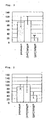
- the CAR gene expression level in the group receiving siRNA(CAR) was reduced to about 30% as compared to the CAR gene expression level in the group treated with siRNA(Cont) (see Fig. 1 ).
- slRNA(CAR) using CAR as a target gene (sense strand: SEQ ID NO: 1; 5' GCU CAC ACA CUU UGC AGA UAU CAA U 3' and antisense strand: SEQ ID NO: 2; 5' AUU GAU AUC UGC AAA GUG UGU GAG C 3') (manufactured by Invitrogen) was dissolved in Ringer's solution at a concentration of 0.8 mg/mL, which was mixed with an equivalent volume of DMRIE-C reagent (Invitrogen).
- control siRNA(Cont) (Stealth RNAi Negative Control with Midium GC, manufactured by Invitrogen, Catalog No.: 12935-300) was dissolved in Ringer's solution at a concentration of 0.8 mg/mL, which was mixed with an equivalent volume of DMRIE-C reagent.
- the weight of the mammalian subjects prepared in (1) was measured for each individual using a balance, and then the dosing solution containing siRNA(CAR) prepared in (2) was administered via the tail vein by injection at a dose of 10 mg/kg/day in a volume of 12 mL/mg/day once daily (hereinafter referred to as mammals receiving siRNA(CAR)).
- mammals receiving siRNA(CAR) a dosing solution containing control siRNA(Cont) was administered in place of the dosing solution containing siRNA(CAR) in the same manner as the case of the mammals receiving siRNA(CAR) (hereinafter referred to as mammals receiving control siRNA(Cont)).
- untreated animals animals which received nothing were determined (hereinafter referred to as untreated animals). In each case, three to five mammalian subjects were used.
- viability test With reference to all of the animals administered, viability test, measurement of body weight (just before the administration and at the time of dissection), necropsy at the time of dissection, blood biochemical test (for example, ALT, AST, LDH, gamma-GTP, etc.), measurement of liver weight, and histopathological examination of the liver were performed in accordance with an ordinary method in order to grasp the toxicity by administration. Consequently, no significant toxicity was observed in mammals receiving control siRNA(Cont).
- an appropriate anesthetic for example, ether was administered to mammals being dissected. Thereafter, they were sacrificed by collecting the blood from the abdominal aorta. After the sacrifice, organs throughout the body were necropsied (gross pathological observation). Then, the liver was removed and the wet weight was immediately measured using a balance. After measuring the weight, the liver was isolated and a portion of the liver was placed into the RNA later (manufactured by Ambion) to use as the liver tissue for RNA expression analysis, which was stored at 4°C until the analysis. Alternatively, another portion of the liver was freezed with liquid nitrogen to use as the liver tissue for protein analysis, which was then stored at -80°C until the analysis. Further, histopathological examination based on optical microscope observations was performed in accordance with an ordinary method. Further, the obtained blood was heparinized and the plasma was preparatively isolated, followed by performing the blood biochemical test of the plasma.
- an appropriate anesthetic for example, ether was administered to mammals being dissected. Thereafter
- RNA solution was further DNase-treated and purified as directed in the instruction manual using the RNeasy Kit (manufactured by Qiagen).
- the reagent contained in TaqMan Reverse Transcription Regents (manufactured by ABI) (10x Taq Man RT buffer 1 ⁇ L, 25 mM MgCl 2 2.2 ⁇ L, DeoxyNTPs Mixure 2 ⁇ L, Oligo dT 0.5 ⁇ L, RNase Inhibitor 0.2 ⁇ L, MultiScribe RT 0.25 ⁇ L) was mixed with 2.85 ⁇ l of sterile distilled water treated with DEPC.
- RNA prepared as described in (6) was added thereto and the obtained mixture was subjected to reverse transcription reaction by incubating at 25° C for 10 minutes, then at 48°C for 30 minutes, and then heating at 95°C for 5 minutes. After the reaction, the reaction mixture was cooled at 4°C, which was used as cDNA solution.
- cDNA prepared as described in (7) was used as a template and PCR was performed under the following conditions, then amplified DNAs were quantitated. That is, 25 ⁇ l of the reaction solution containing the cDNA 2 ⁇ l, Forward primer (SEQ ID NO: 3: 5' CCA TCA CCG GCC TTT CC 3') 22.5 pmol, Reverse primer (SEQ ID NO: 4: 5' GCT GCA CCA TGA AA GTA TTG ATA TCT 3') 22.5 pmol, probe (SEQ ID NO: 5: 5' CCT GGC CCC CGT GTT GCC T 3') 6.25 pmol, and TaqMan Universal Master Mix (manufactured by ABI) 12.5 ⁇ l was prepared, which was subjected to incubation at 50° C for 5 minutes, then at 95° C for 10 minutes, followed by 40 PCR cycles, 1 cycle being incubation at 95°C for 15 seconds, and then at 60° C for 1 minute, using GeneAmp5700 Sequence
- the amount of mRNA of Glyceraldehyde-3-phosphate dehydrogenase (GAPDH) gene was quantified in the same manner as described above (Forward primer: SEQ ID NO: 6: 5' GCT GCC TTC TCT TGT GAC AAA GT 3' , Reverse primer: SEQ ID NO: 7: 5' CTC AGC CTT GAC TGT GCC ATT 3', and Probe: SEQ ID NO: 8: 5' TGT TCC AGT ATG ATT CTA CCC ACG GCA AG 3').
- the ratio of the amount of mRNA of CAR gene to the amount of mRNA of GAPDH gene was calculated, which was used as the CAR gene expression level.
- the CAR gene expression level in the liver tissue was respectively determined for each individual.
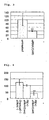
- the CAR gene expression level in the liver tissue of the mammal group receiving siRNA(Cont) was reduced up to 40% ( Fig. 2 ) one day following the administration (the day after administration).
- the liver tissue for protein analysis stored by the method described in (5) of Example 2 was homogenized in 20 mL of buffer for grinding (PBS solution containing 10 mg/mL of leupeptin, 1 mg/mL of pepstatin, and 200 ⁇ M of PMSF as protease inhibitors) using a homogenizer (manufactured by Polytron). A portion of the sample was aliquoted into a 1.5 mL tube (manufactured by Eppendorf Co., Ltd.), which was centrifuged at 15,000 rpm at 4° C for 30 minutes and the resulting supernatant was used for protein analysis.
- buffer for grinding PBS solution containing 10 mg/mL of leupeptin, 1 mg/mL of pepstatin, and 200 ⁇ M of PMSF as protease inhibitors
- the concentration of proteins contained was measured at an absorbance of 595 nm with an absorptiometer using the Protein assay reagent (manufactured by Bio-Rad Laboratories, Inc.).
- Protein assay reagent manufactured by Bio-Rad Laboratories, Inc.
- 50 ⁇ g of protein sample was added to the precast gel (manufactured by Bio-Rad Laboratories, Inc.), which was subjected to electrophoresis under electrophoresis conditions (120V, 30 mA, 80 min). Transfer was performed under electrophoresis conditions (270 V, 350 mA, 60 min) using Hybond membrane (manufactured by GE Healthcare).
- In the treatment of primary antibody blocking was carried out with TBST solution containing 5% skim milk for 1 hour.
- the membrane was washed with TBST solution several times, which was treated with TBST solution containing anti-CAR polyclonal antibody at a 1:1000 dilution (manufactured by Santa Cruz Biotechnology, Inc.) at 4° C for about 12 hours.
- the membrane was washed with TBST solution several times, which was treated with TBST solution containing anti-rabbit IgG antibody at a 1:1000 dilution (manufactured by Santa Cruz Biotechnology, Inc.) at room temperature for about 2 hours.
- bands were visualized using Chemi-Lumi.
- the CAR protein expression level in the liver tissue was respectively determined for each individual.
- the CAR protein expression level in the liver tissue of the mammal group receiving siRNA(Cont) was reduced up to 50% three days following the administration.
- Example 2 Five(5)-week-old male Crj:CD-1 mice (purchased from CHARLES RIVER LABORATORIES JAPAN, INC.) were quarantined for one week and then the 5-week-old mice were subjected to experiments.
- siRNA(CAR) using CAR as a target gene was produced (sense strand: SEQ ID NO: 1; 5' GCU CAC ACA CUU UGC AGA UAU CAA U 3', and antisense strand: SEQ ID NO: 2; 5' AUU GAU AUC UGC AAA GUG UGU GAG C 3').
- the dosing solution containing the siRNA(CAR) was subjected to processes described in (3) to (7) of Example 2 and cDNA was prepared.
- PCR of CAR gene was performed and as a control gene, PCR of Glyceraldehyde-3-phosphate dehydrogenase (GAPDH) gene was performed by the process described in (8) of Example 2 using the prepared cDNA as a template. The amplified DNAs were quantitated.
- Glyceraldehyde-3-phosphate dehydrogenase Glyceraldehyde-3-phosphate dehydrogenase
- primers and probes for mouse CAR gene (Forward primer: SEQ ID NO: 9: 5' GCA TCA CCG GCC TTT CC 3', Reverse primer: SEQ ID NO: 10 : 5' CCA TAA ACG TGT TGA TAT CTG CAA A 3', and probe: SEQ ID NO: 11: 5' CCC CGT GTT GCC TCT GCT.CAC AC 3') and primers and probes for mouse GAPDH gene (Forward primer: SEQ ID NO: 12: 5' TGT GTC CGT CGT GGA TCT GA 3', Reverse primer: SEQ ID NO: 13: 5' CCT GCT TCA CCA CCT TCT TGA 3', and probe: SEQ ID NO: 14: 5' CCG CCT GGA GAA ACC TGC CAA GTA TG 3') were used.
- the ratio of the amount of mRNA of CAR gene to the amount of mRNA of GAPDH gene was calculated, which was used as the CAR gene expression level.
- the CAR gene expression level in the liver tissue was respectively determined for each individual.
- the CAR gene expression level in the liver tissue of the mammal group receiving siRNA (CAR) was reduced up to 40% ( Fig. 3 ) one day following the administration (the day after administration).
- PB solution was added to the culture medium at a final concentration of 50 nM (on day 2). The compound was newly added when the culture medium was replaced every day.
- a group (untreated group) to which PB was not added was prepred as a control.
- cDNA prepared by the methods described in (1) to (4) of Example 1 from the cells obtained in (1) of Example 5 was used as a template, PCR was performed in the same manner as described in (5) of Example 1 under the following conditions, and then the amount of DNA for each amplified gene was quantitated.
- CYP2B1 gene expression 25 ⁇ l of the reaction solution containing Forward primer against CYP2B1 (SEQ ID NO: 15: 5' GCT CAA GTA CCC CCA TGT CG 3') 45 pmol, Reverse primer (SEQ ID NO: 16:5' ATC AGT GTA TGG CAT TTT ACT GCG G 3') 45 pmol, and Power SYBR Green PCR Master Mix (manufactured by ABI) 12.5 ⁇ l was prepared, which was subjected to incubation at 95° C for 10 minutes followed by 40 PCR cycles, 1 cycle being incubation at 95° C for 30 seconds, and then at 60° C for 30 seconds, and further at 72° C for 45 seconds, using GeneAmp5700 Sequence detection System (manufactured by ABI). The amount of mRNA of CYP2B1 gene was quantified from the amount of amplified DNAs.
- the amount of mRNA of Glyceraldehyde-3-phosphate dehydrogenase(GAPDH) gene was quantified in the same manner as described above (Forward primer: SEQ ID NO: 6: 5' GCT GCC TTC TCT TGT GAC AAA GT 3', Reverse primer: SEQ ID NO: 7: 5' CTC AGC CTT GAC TGT GCC ATT 3', and Probe: SEQ ID NO: 8: 5' TGT TCC AGT ATG ATT CTA CCC ACG GCA AG 3').
- the ratio of the amount of mRNA of CAR gene to the amount of mRNA of GAPDH gene was calculated, which was used as expression levels of CAR genes.
- the CAR gene expression level in untreated cells, cells treated with control siRNA(Cont), and cells treated with siRNA(CAR) was respectively determined.
- the ratio of the amount of mRNA of CYP2B1 gene to the amount of mRNA of GAPDH gene was calculated, which was used as expression levels of CYP2B1 genes.
- the CYP2B1 gene expression level in untreated cells, cells treated with control siRNA(Cont), and cells treated withsiRNA(CAR) was respectively determined.
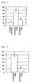
- the CAR gene expression level in the cells treated with PB and siRNA(CAR) was reduced to about 40% as compared to the CAR gene expression level in the cells treated with PB and control siRNA(Cont) (see Fig. 4 ).
- CYP2B1 mRNA expression was increased about fivefold compared to cells untreated with PB ( Fig. 5 ).
- CYP2H1 expression by PB treatment was increased about twofold compared to cells untreated with compound, and thus a significant reduction was observed ( Fig. 5 ).
- the amount of change in the induced part obtained by subtracting the CYP2B1 expression in the cells untreated with compound was reduced to 80% or more by inhibition of CAR ( Fig. 6 ). This shows that induction of CYP2B1 expression by PB treatment is achieved via CAR.
- the culture medium is washed with William's culture medium twice 72 hours after the introduction of siRNA (on day 4). Then, the culture medium is replaced with 2 mL of William's culture medium containing 7 ⁇ M of 7-pentoxyresorufin and 7 ⁇ M of dicumarol. The culture medium is incubated at 37° C for 4 hours, and then the culture medium is recovered. The cells are mixed with 1000 U of ⁇ -glucronidase (bovine liver-derived) and 50 mM of sodium acetate solution (pH 5), which is left at 37°C for 3 hours.
- Measurement of pentoxyresorufin O-depentylation activity is performed by measuring the amount of fluorescence per unit solution. Further, the measurement is carried out at an excitation wavelength of 530 nm and a fluorescence wavelength of 585 nm using a fluorophotometer (F-4010, manufactured by Hitachi). In evaluation of the results, the total amount of protein in the cells in which the substrate was treated is measured by the Protein assay kit (Bio-Rad) and the production amount of resorufin per unit protein (amount of pentoxyresorufin O-depentylation) is calculated on the basis of the calibration curve of resorufin.
- a BrdU-labeled solution is added to the culture medium using the cell proliferation ELISA, BrdU chemiluminescence kit (manufactured by Roche Diagnostics) 48 hours after the introduction of siRNA (on day 3) and then cultured at 37° C for 24 hours.
- the solution is sucked 72 hours after the introduction of siRNA (on day 4) and 2 mL of FixDenat is added thereto, which is left at room temperature for 30 minutes. Thereafter, FixDenat is removed and anti-BrdU-POD was added thereto, which is left at room temperature for 90 minutes.
- the solution is removed and the remained cells are washed with PBS 3 times.
- the cells are peeled from the plate by trypsin treatment.
- 2 mL of substrate solution is added to the cells and then 200 ⁇ L of the cell culture solution is aliquoted into a 96-well plate.
- the bottom of the transparent plate is sealed with a black adhesive foil, which is left on a shaker for 3 minutes, followed by measuring with a microplate luminometer (MicroLumat Plus LB96V, manufactured by Berthold).
- a microplate luminometer MicroLumat Plus LB96V, manufactured by Berthold.
- the amounts of fluorescence as to untreated cells, cells treated with PB and control siRNA (Cont), and cells treated with PB and siRNA (CAR) are respectively measured.
- the uptake of BrdU is enhanced by PB and control siRNA (Cont) treatment as compared to the case of the untreated cells.
- the uptake of BrdU into the cells treated with PB and siRNA (CAR) is the same as that of the untreated cells. This shows that the enhancement of cell proliferation by PB treatment is achieved via CAR.
- apoptotic cells are stained by the TUNEL assay 72 hours after the introduction of siRNA (on day 4). Then, the total mumber per well is counted. When apoptosis proceeds, DNA degradation is caused.
- FITC-labeled dUTP is bound to the DNA fragment by using TdT (terminal deoxynucleotidyl transferase). When the labeled fragment is observed with a fluorescence microscope, apoptotic cells generate fluorescence.
- TdT terminal deoxynucleotidyl transferase
- fluorescence values as to untreated cells, cells treated with PB and control siRNA (Cont), and cells treated with PB and siRNA (CAR) are respectively determined and the resulting values are used as the amount of apoptosis.
- the amount of cell apoptosis is decreased by PB and control siRNA (Cont) treatment as compared to the case of the untreated cells.
- the uptake of BrdU into the cells treated with PB and siRNA (CAR) is the same as that of the untreated cells. This shows that the decrease of apoptosis by PB treatment is achieved via CAR.
- the bottom of the transparent plate is sealed with a black adhesive foil, which is left on a shaker for 3 minutes, followed by measuring with a microplate luminometer (SpectraMaxL, manufactured by Molecular Devices Corporation).
- the amounts of fluorescence as to untreated cells, cells treated with PB and control siRNA (Cont), and cells treated with PB and siRNA (CAR) are respectively measured.
- the amount of active oxygen is increased by PB and control siRNA (Cont) treatment as compared to the case of the untreated cells.
- the amount of active oxygen in the cells treated with PB and siRNA (CAR) is the same as that in the untreated cells. This shows that the increase in the amount of active oxygen by PB treatment is achieved via CAR.
- the wet weight of the liver in the rats to which siRNA (CAR) or control siRNA (Cont) was administered by the methods described in (1) to (3) of Example 2 is measured by the method described in (5) of Example 2.
- the liver weight is increased in the group treated with PB and control siRNA (Cont) as compared to the case of the untreated animal group.
- the liver weight in the group receiving PB and siRNA (CAR) is the same as that in the untreated group. This shows that the increase in the liver weight by PB treatment is achieved via CAR.
Landscapes
- Life Sciences & Earth Sciences (AREA)
- Health & Medical Sciences (AREA)
- Engineering & Computer Science (AREA)
- Biomedical Technology (AREA)
- Chemical & Material Sciences (AREA)
- Molecular Biology (AREA)
- Genetics & Genomics (AREA)
- Biotechnology (AREA)
- General Health & Medical Sciences (AREA)
- Organic Chemistry (AREA)
- Bioinformatics & Cheminformatics (AREA)
- Wood Science & Technology (AREA)
- Biochemistry (AREA)
- Physics & Mathematics (AREA)
- Urology & Nephrology (AREA)
- Microbiology (AREA)
- Zoology (AREA)
- General Engineering & Computer Science (AREA)
- Hematology (AREA)
- Immunology (AREA)
- Medicinal Chemistry (AREA)
- Cell Biology (AREA)
- Pathology (AREA)
- Plant Pathology (AREA)
- Food Science & Technology (AREA)
- Biophysics (AREA)
- Analytical Chemistry (AREA)
- Proteomics, Peptides & Aminoacids (AREA)
- General Physics & Mathematics (AREA)
- Pharmacology & Pharmacy (AREA)
- Animal Behavior & Ethology (AREA)
- Public Health (AREA)
- Nuclear Medicine, Radiotherapy & Molecular Imaging (AREA)
- Veterinary Medicine (AREA)
- General Chemical & Material Sciences (AREA)
- Chemical Kinetics & Catalysis (AREA)
- Measuring Or Testing Involving Enzymes Or Micro-Organisms (AREA)
- Pharmaceuticals Containing Other Organic And Inorganic Compounds (AREA)
- Investigating Or Analysing Biological Materials (AREA)
Applications Claiming Priority (1)
| Application Number | Priority Date | Filing Date | Title |
|---|---|---|---|
| JP2007198725A JP2009033986A (ja) | 2007-07-31 | 2007-07-31 | RNA干渉による遺伝子発現抑制のためのターゲット遺伝子としてのcar遺伝子の使用 |
Publications (1)
| Publication Number | Publication Date |
|---|---|
| EP2020440A1 true EP2020440A1 (fr) | 2009-02-04 |
Family
ID=39884529
Family Applications (1)
| Application Number | Title | Priority Date | Filing Date |
|---|---|---|---|
| EP08252595A Withdrawn EP2020440A1 (fr) | 2007-07-31 | 2008-07-31 | Compositions et procédés pour inhiber l'expression du gène car par interférence d'ARN |
Country Status (3)
| Country | Link |
|---|---|
| US (1) | US7816338B2 (fr) |
| EP (1) | EP2020440A1 (fr) |
| JP (1) | JP2009033986A (fr) |
Citations (2)
| Publication number | Priority date | Publication date | Assignee | Title |
|---|---|---|---|---|
| WO2002025270A1 (fr) * | 2000-09-21 | 2002-03-28 | Baylor College Of Medicine | Systemes de criblage et methodes d'identification de modulateurs d'un metabolisme xenobiotique |
| WO2006064197A2 (fr) * | 2004-12-13 | 2006-06-22 | Iti Scotland Limited | Animaux transgeniques pour evaluer le metabolisme et la toxicite d'un medicament |
Family Cites Families (2)
| Publication number | Priority date | Publication date | Assignee | Title |
|---|---|---|---|---|
| US20040006775A1 (en) * | 2001-09-21 | 2004-01-08 | Moore David D. | Screening systems and methods for identifying modulators of xenobiotic matabolism |
| CA2429814C (fr) * | 2000-12-01 | 2014-02-18 | Thomas Tuschl | Petites molecules d'arn mediant l'interference arn |
-
2007
- 2007-07-31 JP JP2007198725A patent/JP2009033986A/ja active Pending
-
2008
- 2008-07-31 EP EP08252595A patent/EP2020440A1/fr not_active Withdrawn
- 2008-07-31 US US12/183,354 patent/US7816338B2/en not_active Expired - Fee Related
Patent Citations (2)
| Publication number | Priority date | Publication date | Assignee | Title |
|---|---|---|---|---|
| WO2002025270A1 (fr) * | 2000-09-21 | 2002-03-28 | Baylor College Of Medicine | Systemes de criblage et methodes d'identification de modulateurs d'un metabolisme xenobiotique |
| WO2006064197A2 (fr) * | 2004-12-13 | 2006-06-22 | Iti Scotland Limited | Animaux transgeniques pour evaluer le metabolisme et la toxicite d'un medicament |
Non-Patent Citations (10)
| Title |
|---|
| "Active Oxygen Experimental Protocol", 1994, SHUJUNSHA CO., LTD. |
| CHEN ET AL: "Human constitutive androstane receptor mediated methotrexate induction of human dehydroepiandrosterone sulfotransferase (hSULT2A1)", TOXICOLOGY, vol. 231, no. 2-3, 24 February 2007 (2007-02-24), pages 224 - 233, XP005904702, ISSN: 0300-483X * |
| J. SAMBROOK; E. F. FRISCH; T. MANIATIS: "Molecular Cloning", 1989, COLD SPRING HARBOR LABORATORY |
| SWALES KAREN ET AL: "CAR, driving into the future", MOLECULAR ENDOCRINOLOGY, vol. 18, no. 7, July 2004 (2004-07-01), pages 1589 - 1598, XP002503410, ISSN: 0888-8809 * |
| SWALES; NEGISHI, MOL. ENDO., vol. 18, no. 7, 2004, pages 1589 - 1598 |
| YAMADA HIDEYUKI ET AL: "Induction of the hepatic cytochrome P4502B subfamily by xenobiotics: Research history, evolutionary aspect, relation to tumorigenesis, and mechanism", CURRENT DRUG METABOLISM, vol. 7, no. 4, May 2006 (2006-05-01), pages 397 - 409, XP008098330, ISSN: 1389-2002 * |
| YAMAMOTO ET AL., CANCER RES, vol. 64, 2004, pages 7197 - 7200 |
| YAMAMOTO ET AL., CANCER RES., vol. 64, 2004, pages 7197 - 7200 |
| YAMAMOTO YUKIO ET AL: "The orphan nuclear receptor constitutive active/androstane receptor is essential for liver tumor promotion by phenobarbital in mice", CANCER RESEARCH, vol. 64, no. 20, 15 October 2004 (2004-10-15), pages 7197 - 7200, XP002503409, ISSN: 0008-5472 * |
| YANG ET AL: "A novel RNAi library based on partially randomized consensus sequences of nuclear receptors: Identifying the receptors involved in amyloid beta degradation", GENOMICS, vol. 88, no. 3, 23 August 2006 (2006-08-23), pages 282 - 292, XP005742398, ISSN: 0888-7543 * |
Also Published As
| Publication number | Publication date |
|---|---|
| JP2009033986A (ja) | 2009-02-19 |
| US7816338B2 (en) | 2010-10-19 |
| US20090053298A1 (en) | 2009-02-26 |
Similar Documents
| Publication | Publication Date | Title |
|---|---|---|
| Feng et al. | miR-200b mediates endothelial-to-mesenchymal transition in diabetic cardiomyopathy | |
| Fang et al. | Adipocyte-specific loss of PPARγ attenuates cardiac hypertrophy | |
| US20140323551A1 (en) | Targeting micrornas mir-409-5p, mir-379 and mir-154* to treat prostate cancer bone metastasis and drug resistant lung cancer | |
| AU2020322415B2 (en) | KLF induced cardiomyogenesis | |
| Chen et al. | Inhibition of myocyte-specific enhancer factor 2A improved diabetic cardiac fibrosis partially by regulating endothelial-to-mesenchymal transition | |
| US12397011B2 (en) | Method for preventing or treating cancer using SYT11 inhibitor | |
| Kuwagata et al. | MicroRNA148b-3p inhibits mTORC1-dependent apoptosis in diabetes by repressing TNFR2 in proximal tubular cells | |
| Gao et al. | High glucose-upregulated PD-L1 expression through RAS signaling-driven downregulation of PTRH1 leads to suppression of T cell cytotoxic function in tumor environment | |
| EP2351585B1 (fr) | Une composition d' antivirus de l' hépatite c comprenant une acide nucléique contre pa28-gamma | |
| Cui et al. | Enhancing Acsl4 in absence of mTORC2/Rictor drove β-cell dedifferentiation via inhibiting FoxO1 and promoting ROS production | |
| Tran et al. | Phosphatidylinositol 4-kinase III alpha governs cytoskeletal organization for invasiveness of liver cancer cells | |
| Zhang et al. | Hsa_circ_0001495 contributes to cervical cancer progression by targeting miR-526b-3p/TMBIM6/mTOR axis | |
| Lee et al. | miR-10a regulates cell death and inflammation in adipose tissue of male mice with diet-induced obesity | |
| JPWO2008041665A1 (ja) | C型肝炎に伴う疾患の予防及び/又は治療薬のスクリーニング方法 | |
| CN113679735A (zh) | Slc7a11基因在肝细胞癌介入栓塞术后中的应用 | |
| US7816338B2 (en) | Compositions and methods for inhibiting car gene expression by RNA interference | |
| AU2006204051A1 (en) | Development and use of a new orthotopic genetically tractable non-human animal model for liver cancer | |
| JP6475550B2 (ja) | リン脂質ヒドロペルオキシド依存性細胞死の検出方法及びキット、リン脂質ヒドロペルオキシド依存性細胞死の抑制剤、並びにリン脂質ヒドロペルオキシド依存性細胞死が関連する疾患の予防又は治療剤 | |
| CN113498440A (zh) | 新型胰腺癌上皮间质转化标记物 | |
| US20100239566A1 (en) | Enigma-mdm2 interaction and uses thereof | |
| CN116949174A (zh) | 一种lncRNA及其下游级联物质在芳香化酶抑制剂耐药ER阳性乳腺癌中的用途 | |
| JP2010265190A (ja) | Rna干渉による遺伝子発現抑制のためのターゲット遺伝子としてのugt遺伝子の使用 | |
| EP4355432A1 (fr) | Compositions de copolymère d'arnsi et procédés d'utilisation pour le traitement du cancer du foie | |
| JP5688229B2 (ja) | 肝保護作用を有するタンパク質、肝障害予防・保護用化合物のスクリーニング方法 | |
| CN101914541A (zh) | 抑制mbd2表达的抑制剂及其应用 |
Legal Events
| Date | Code | Title | Description |
|---|---|---|---|
| PUAI | Public reference made under article 153(3) epc to a published international application that has entered the european phase |
Free format text: ORIGINAL CODE: 0009012 |
|
| AK | Designated contracting states |
Kind code of ref document: A1 Designated state(s): AT BE BG CH CY CZ DE DK EE ES FI FR GB GR HR HU IE IS IT LI LT LU LV MC MT NL NO PL PT RO SE SI SK TR |
|
| AX | Request for extension of the european patent |
Extension state: AL BA MK RS |
|
| 17P | Request for examination filed |
Effective date: 20090804 |
|
| 17Q | First examination report despatched |
Effective date: 20090907 |
|
| AKX | Designation fees paid |
Designated state(s): AT BE BG CH CY CZ DE DK EE ES FI FR GB GR HR HU IE IS IT LI LT LU LV MC MT NL NO PL PT RO SE SI SK TR |
|
| STAA | Information on the status of an ep patent application or granted ep patent |
Free format text: STATUS: THE APPLICATION IS DEEMED TO BE WITHDRAWN |
|
| 18D | Application deemed to be withdrawn |
Effective date: 20100318 |